
Evolutionary Conservation of Divergent Pro-Inflammatory and Homeostatic Responses in LampreyPhagocytesJeffrey J. Havixbeck1, Aja M. Rieger1, Michael E. Wong1, Michael P. Wilkie3, Daniel R. Barreda1,2*
1Department of Biological Sciences, University of Alberta, Edmonton, Alberta, Canada, 2Department of Agricultural, Food and Nutritional Science, University of Alberta,
Edmonton, Alberta, Canada, 3Department of Biology, Wilfrid Laurier University, Waterloo, Ontario, Canada
Abstract
In higher vertebrates, phagocytosis plays a critical role in development and immunity, based on the internalization andremoval of apoptotic cells and invading pathogens, respectively. Previous studies describe the effective uptake of theseparticles by lower vertebrate and invertebrate phagocytes, and identify important molecular players that contribute to thisinternalization. However, it remains unclear if individual phagocytes mediate internalization processes in these ancientorganisms, and how this impacts the balance of pro-inflammatory and homeostatic events within their infection sites.Herein we show that individual phagocytes of the jawless vertebrate Petromyzon marinus (sea lamprey), like those of teleostfish and mice, display the capacity for divergent pro-inflammatory and homeostatic responses following internalization ofzymosan and apoptotic cells, respectively. Professional phagocytes (macrophages, monocytes, neutrophils) were theprimary contributors to the internalization of pro-inflammatory particles among goldfish (C. auratus) and lamprey (P.marinus) hematopoietic leukocytes. However, goldfish showed a greater ability for zymosan phagocytosis when comparedto their jawless counterparts. Coupled to this increase was a significantly lower sensitivity of goldfish phagocytes tohomeostatic signals derived from apoptotic cell internalization. Together, this translated into a significantly greater capacityfor induction of antimicrobial respiratory burst responses compared to lamprey phagocytes, but also a decreased efficacy inapoptotic cell-driven leukocyte homeostatic mechanisms that attenuate this pro-inflammatory process. Overall, our resultsshow the long-standing evolutionary contribution of intrinsic phagocyte mechanisms for the control of inflammation, andillustrate one effective evolutionary strategy for increased responsiveness against invading pathogens. In addition, theyhighlight the need for development of complementary regulatory mechanisms of inflammation to ensure continuedmaintenance of host integrity amidst increasing challenges from invading pathogens.
Citation: Havixbeck JJ, Rieger AM, Wong ME, Wilkie MP, Barreda DR (2014) Evolutionary Conservation of Divergent Pro-Inflammatory and Homeostatic Responsesin Lamprey Phagocytes. PLoS ONE 9(1): e86255. doi:10.1371/journal.pone.0086255
Editor: Sebastian D. Fugmann, Chang Gung University, Taiwan
Received October 16, 2013; Accepted December 9, 2013; Published January 20, 2014
Copyright: � 2014 Havixbeck et al. This is an open-access article distributed under the terms of the Creative Commons Attribution License, which permitsunrestricted use, distribution, and reproduction in any medium, provided the original author and source are credited.
Funding: This work was supported by Natural Sciences and Engineering Council of Canada (NSERC) Discovery Grants to DRB and MPW. JJH was supported by anNSERC CGS-M scholarship and AMR was supported by NSERC Vanier doctoral scholarship. The funders had no role in study design, data collection and analysis,decision to publish, or preparation of the manuscript.
Competing Interests: The authors have declared that no competing interests exist.
* E-mail: [email protected]
Introduction
The immune system has evolved to confer effective protection
against infection, driven by continuous interactions between hosts
and microbes. The resulting multi-layered system increasingly
requires complex cross-regulatory systems [1,2]. At the core of
these responses, phagocytosis continues to fill increasing roles as an
inducer and regulator of host immunity. Internalization of
pathogens by phagocytes leads to stimulation of potent killing
mechanisms such as the production of reactive oxygen species
(ROS) that have evolved to degrade and kill foreign invaders and
contribute to downstream adaptive mechanisms [3]. Importantly,
the capacity for effective internalization and clearance of apoptotic
cells is already well established in developmental pathways of early
multi-cellular organisms [4–6]. Phagocytic receptors in Caenorhab-
ditis elegans (CED-1) and Drosophila melanogaster (Draper), for
example, drive recognition and internalization of apoptotic corpses
and activate downstream processing pathways that are central to
morphogenesis and the maintenance of tissue integrity and
function [7,8].
At the site of infection, mammalian phagocytes effectively shape
the environmental milieu for destruction of invading pathogens or
resolution of tissue inflammation. Pathogen engagement leads to
rapid production of pro-inflammatory mediators including the
production of reactive oxygen and nitrogen species [9], release of
antimicrobial peptides [10], and the secretion of tumor necrosis
factor alpha (TNF-a), interferon gamma (IFN-c) and IL-1 beta
(IL-1b) [11]. In contrast, internalization of apoptotic cells initiates
the shift towards resolution mechanisms that promote tissue repair
and a return to homeostasis once the pathogen has been effectively
cleared. This is marked by increases in interleukin 10 (IL-10),
transforming growth factor beta 1 (TGF-b1), prostaglandin E2 and
platelet activating factor [12–14], combined with decreases in pro-
inflammatory mediators, including tumor necrosis factor alpha
(TNF-a), IL-6, IL-8, IL-12, IL-17, IL-23, leukotriene C4 and
thromboxane B2 [13,15,16].
PLOS ONE | www.plosone.org 1 January 2014 | Volume 9 | Issue 1 | e86255

We previously showed that phagocytes of teleost fish contrib-
uted to both pro-inflammatory and anti-inflammatory (resolution)
responses at infectious foci [17]. Like murine phagocytes, they
possessed the capacity to balance between these two seemingly
contradictory processes. However, teleost phagocytes displayed
significant differences in vivo with regards to the level of
responsiveness to zymosan and apoptotic bodies, the identity of
leukocytes infiltrating the infectious site, their rate of infiltration,
and the kinetics and strength of resulting antimicrobial responses
[17]. The striking evolutionary differences observed in inflamma-
tory control between mice and goldfish provided a platform to
investigate these differences down the evolutionary scale in a
primordial vertebrate.
In this study, we compared the effects of homeostatic
phagocytosis on the regulation of pro-inflammatory responses in
goldfish (Carassius auratus) and sea lamprey (Petromyzon marinus). As
one of the earliest vertebrates, along with hagfish (Myxinidae), sea
lampreys were an appropriate model to investigate the conserva-
tion of this dichotomy. Through the use of zymosan, a pro-
inflammatory stimulus known to induce production of reactive
intermediates [18–24], the impact of apoptotic cells on the
production of reactive oxygen species (ROS) was examined.
Interestingly, we found notable differences between goldfish and
lamprey ROS responses in phagocytes that had internalized both
zymosan and apoptotic bodies, though responses in cells that had
internalized only zymosan or only apoptotic bodies were largely
conserved. This work underscores the importance of phagocytosis
as a phylogenetically ancient process essential to the innate
immune response.
Materials and Methods
Ethics StatementAll animals were maintained according to the guidelines of the
Canadian Council on Animal Care, and protocols were approved
by the University of Alberta Animal Care and Use Committee
(ACUC-Biosciences; protocol numbers 595807 and 706). Both
goldfish and lampreys were terminated via cervical dislocations
using approved procedures following anaesthetization with
tricaine methanesulfonate (TMS-222). All efforts were made to
minimize animal stress and to ensure that termination procedures
were performed efficiently.
AnimalsGoldfish (Carassius auratus L.) 10–15 cm in length were
purchased from Mount Parnell (Mercersburg, PA). The fish were
held at 18uC in a flow-through water system with constant
aeration. Ammocoete larvae (8–11 cm in length) of the sea
lamprey (Petromyzon marinus) were received from Wilfred Laurier
University, previously captured by electrofishing from freshwater
streams in New Brunswick, Canada. Lamprey were maintained in
sand-lined aquaria with constant aeration at 18uC and fed
brewer’s yeast. All animals were housed in the Aquatic Facility
of the Department of Biological Sciences, University of Alberta, on
a simulated natural photosystem.
Isolation of Hematopoietic LeukocytesGoldfish primary kidney leukocytes (PKL) were isolated by
maceration of goldfish kidney tissue between two glass slides
suspended in incomplete MGFL-15 medium. Lamprey primary
typhlosole leukocytes were isolated as previously described [25].
Briefly, larval lampreys were dissected along the ventral side to
extract the intestine and the associated typhlosole. Cells were
harvested by maceration between two glass slides suspended in one
part water and two parts incomplete MGFL-15 medium.
Light Microscopy Phagocytosis AssayLatex beads (3 mm; Polysciences) were added at ratios of 1:1, 5:1
and 10:1 (bead: cell) to 16105 cells in 24 well plates and incubated
at 18uC for the indicated times. Cells were stained by Hema3 stain
set (Fisher Scientific) and counted by light microscopy. Phagocytic
index was calculated based on the number of beads internalized
per phagocyte. A minimum of 200 cells were evaluated per
sample. As per previous reports, internalization of particles larger
than 0.5 mm leads to activation of downstream phagocytic events
[26].
Preparation of Zymosan and Apoptotic CellsUnlabeled zymosan particles (Molecular Probes) were labeled
overnight with 250 ng/mL FITC (Sigma) with continuous shaking
at 4uC in carbonate buffer (0.1 M sodium carbonate, 0.1 M
sodium bicarbonate; pH 9.6). After staining, zymosan-FITC was
washed twice with 16PBS2/2. Unlabeled zymosan particles
(Molecular Probes) were labeled overnight with 75 mg/mL
allophycocyanin (APC; Sigma) with continuous shaking at 4uCin 16PBS2/2. Apoptotic cells were generated by incubating cells
for 24 h in the presence of 10 mg/mL cycloheximide. Treated cells
were harvested, washed twice in 16 PBS2/2 and stained
overnight with 1.5 mg/mL wheat germ agglutinin Alexa Fluor
555 (Molecular Probes). Apoptotic cells were then washed twice in
16 PBS2/2. For goldfish experiments, apoptotic cells were
generated from catfish 3B11 B cells. For lamprey, apoptotic cells
were generated from primary typhlosole leukocytes. We have
previously shown that apoptotic cells derived from primary or cell
line leukocytes induce equivalent phagocyte responses [17].
ImageStream Phagocytosis AssayLatex beads (3 mm; Polysciences), E. coli DH5a-GFP or
zymosan-FITC were added to 16105 cells at ratios of 5:1 and
10:1 (particles: cells) and incubated for the indicated times.
Following incubation the cells were washed twice in 16 PBS2/2
and fixed in 1% formaldehyde at 4uC overnight. Phagocytic index
was calculated based on the number of particles internalized per
phagocyte. Data was acquired on an ImageStream multi-spectral
imaging flow cytometer (Amnis) and analyzed using INSPIRE
software. At least 10000 cells were acquired. Preliminary
experiments indicated that cells remained within their correspond-
ing leukocyte gate following internalization of E. coli or zymosan,
but not following internalization of 3 mm beads, where internal
complexity increased significantly. Development of specific mark-
ers for each of these leukocyte subsets should further enhance the
characterization of phagocytic events within each of these
phagocyte subsets.
Respiratory Burst AssayThis assay was performed as previously described with minor
modifications [27,28]. Cells were harvested and collected into
5 mL polystyrene round bottom tubes (BD Falcon). Dihydrorho-
damine (DHR, Molecular Probes) was added to the cells at a final
concentration of 1 mM and incubated for 5 min to allow the cells
to take up the DHR. Phorbol 12-myristate 13-acetate (PMA;
Sigma) was then added at a final concentration of 10 ng/mL. Cells
were further incubated for 30 minutes to allow oxidation of the
DHR. All samples were appropriately staggered with respect to
timing to accommodate for the transient state of oxidized DHR
fluorescence. DHR fluorescence was not quenched by the
Control of Lamprey Phagocyte Inflammatory Response
PLOS ONE | www.plosone.org 2 January 2014 | Volume 9 | Issue 1 | e86255

presence of other fluorochromes, including the wheat germ
agglutinin or APC labels on phagocytosed particles (data not
shown).
AnalysisImageStream data was analyzed using IDEAS software (Amnis),
as previously described [27]. Statistics were performed by
Students’ T-test using Prism 6 software (GraphPad Prism).
Results
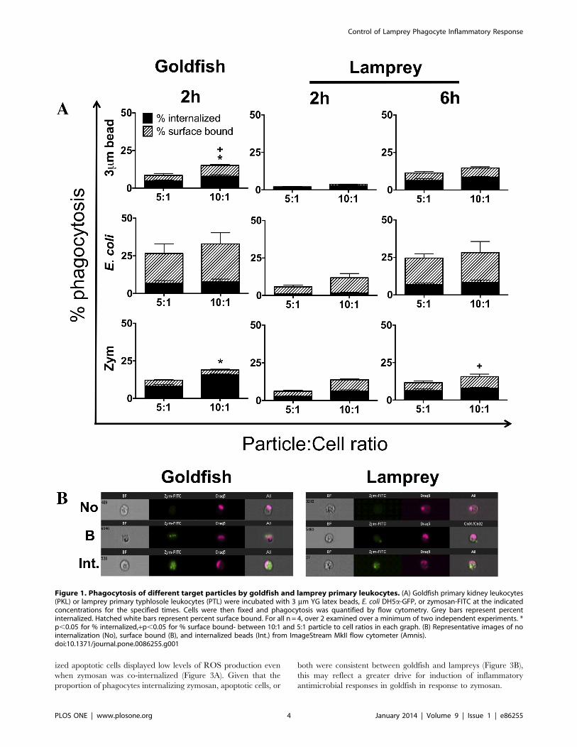
Goldfish Phagocytes Display a Greater Capacity for theInternalization of Pro-inflammatory Particles thanLamprey PhagocytesAs a first step in the characterization of differences between the
contributions of agnathan (jawless fish) and teleost (bony fish)
phagocytes to the control of inflammation, we compared the
phagocytic capacity of primary leukocytes from sea lamprey
typhlosole and goldfish kidney. These corresponded to the primary
hematopoietic tissues of the animals examined and provided
sufficient numbers of their primary phagocyte populations for ex
vivo examination [29–32]. Phagocytosis of three commonly used
model particles was examined: i) 3 mm latex beads, ii) E. coli, and
iii) zymosan (Figure 1A). Phagocytosis was assessed by imaging
flow cytometry, which allowed discrimination of bound and
internalized particles (Figure 1B; [27]). From the outset, our
experiments indicated differences in the efficiency of phagocytosis
for goldfish and lamprey leukocytes ex vivo. Two hours was
sufficient to examine basal levels of phagocytosis in goldfish
(Figure 1A). In contrast, six hours were required to achieve
equivalent levels of phagocytosis among sea lamprey leukocytes.
This was further corroborated by a conventional light microscopy
phagocytosis assay, and determination of optimal respiratory burst
levels (Figure S1 and S2, respectively). Results were not consistent
across the three pro-inflammatory particles examined. Lamprey
leukocytes continued to display lower levels of zymosan phagocy-
tosis that goldfish even after six hours of incubation (Figure 1A,
bottom row).
A greater capacity for phagocytosis of zymosan suggested an
increased ability of goldfish phagocytes to effectively mount
antimicrobial defenses against fungal pathogens. We wondered
whether this difference was associated with differences in the
relative abundance of distinct phagocyte groups within the
hematopoietic leukocyte pool and/or differences in their phago-
cytic capacity compared to those in lamprey (Figure S3). We
focused on the granulocyte, monocyte, and macrophage popula-
tions as the classical professional phagocytes of fish, and the
lymphocyte population as the newest members of the phagocyte
group [33–35]. Although leukocyte populations in lamprey
displayed similar cellular characteristics to those of goldfish based
on size, morphology and internal complexity the range of reagents
available to help define various subsets is still limited. As such, they
remain as lymphocyte-like, granulocyte-like, monocyte-like and
macrophage-like cells. For goldfish, granulocytes/monocytes
represented the majority of the leukocytes isolated from kidney
hematopoietic tissues (73%), with smaller contributions from
macrophage and lymphocyte populations (19% and 9% respec-
tively; Figure 2A). For the lamprey, lymphocyte-like cells
comprised the majority of the total typhlosole leukocyte pool
(50%; Figure 2A, 6 h). Granulocyte/monocyte and macrophage-
like cells followed and contributed approximately 32% and 18%,
respectively. Evaluation of phagocytosis indicated that macro-
phage and macrophage-like cells were the primary mediators of
zymosan internalization in goldfish and lamprey, respectively
(Figure 2B). However, these cells represented different proportions
of the hematopoietic phagocyte pool that internalized zymosan in
goldfish and lamprey (11% and 35%, respectively; Figure 2C).
Further, they also displayed a differential ability to internalize
zymosan (28% and 17% phagocytosis, respectively; Figure 2B).
Despite their lower abundance within the hematopoietic tissues
examined, goldfish macrophages showed a marked greater
capacity to internalize zymosan, which contributed to overall
greater levels of zymosan phagocytosis for goldfish kidney
leukocytes (16% in goldfish versus 6% in lampreys; Figure 1A).
The increased relative efficacy for zymosan internalization in
goldfish was not limited to the macrophage population. Despite
their lower contribution to the internalization of zymosan,
monocytes/granulocytes also displayed a four-fold greater capacity
for zymosan phagocytosis in goldfish compared to the monocyte/
granulocyte-like pool in lamprey (8% versus 2%, respectively;
Figure 2B). Thus, professional phagocytes (macrophages, mono-
cytes, neutrophils) are the primary contributors to the internali-
zation of zymosan in both goldfish and lamprey hematopoietic
leukocytes; however, our teleost phagocytes displayed a signifi-
cantly greater ability for zymosan phagocytosis when compared to
their agnathan counterparts. Notably, the low levels of phagocy-
tosis observed among goldfish lymphocytes and lamprey lympho-
cyte-like cells in these experiments (0.27% and 0.8%, respectively)
may stem from their limitation for internalization of larger
particles (2.5–3 mm for zymosan) and not an overall inability for
phagocytosis. Alternatively, this may be associated with a reduced
capacity to interact with zymosan. Indeed, evaluation of the
capacity for E. coli internalization among the hematopoietic
leukocyte subsets examined showed increased levels of phagocy-
tosis among lamprey lymphocyte-like cells when compared to
zymosan. Whereas 0.8% of lamprey lymphocyte-like cells showed
zymosan internalization under the experimental conditions tested,
2.3% showed E. coli uptake. Given the levels of bacterial uptake
observed, this attributed 13% of the E. coli phagocytosis to the
lymphocyte-like population. As lymphocyte-like cells corresponded
to approximately 50% of the leukocyte population examined in
lamprey (Figure 2A), this points to a potentially significant
contribution by these cells to bacterial phagocytosis.
Goldfish Phagocytes Display Prominent Respiratory BurstResponses, even when Apoptotic Cells are InternalizedWe previously determined that teleost phagocytes, like those of
mice, displayed the capacity for divergent pro-inflammatory and
homeostatic responses, following internalization of zymosan and
apoptotic cells, respectively [17]. Further, internalization of
zymosan, apoptotic cells, or both by individual phagocytes
contributed differentially to the modulation of antimicrobial
inflammatory responses [17]. Accordingly, we investigated wheth-
er or not a similar mechanism of inflammatory control at the level
of the individual phagocyte was already displayed in the lamprey.
As expected, zymosan internalization resulted in an increase in the
level of the respiratory burst response in both goldfish and lamprey
(Figure 3A). Conversely, internalization of apoptotic cells led to a
significant reduction in ROS production in both species. Thus, our
results indicated that sea lamprey phagocytes were already capable
of mediating divergent pro- and anti-inflammatory responses.
Interestingly, when we analyzed the level of ROS production in
cells that had internalized both zymosan and apoptotic cells, we
saw a striking difference between goldfish and lamprey phagocytes.
Goldfish phagocytes that had internalized both zymosan and
apoptotic cells showed equivalent levels of ROS production
compared to those that internalized zymosan alone (p = 0.88;
Figure 3A). In sharp contrast, lamprey phagocytes that internal-
Control of Lamprey Phagocyte Inflammatory Response
PLOS ONE | www.plosone.org 3 January 2014 | Volume 9 | Issue 1 | e86255
ized apoptotic cells displayed low levels of ROS production even
when zymosan was co-internalized (Figure 3A). Given that the
proportion of phagocytes internalizing zymosan, apoptotic cells, or
both were consistent between goldfish and lampreys (Figure 3B),
this may reflect a greater drive for induction of inflammatory
antimicrobial responses in goldfish in response to zymosan.
Figure 1. Phagocytosis of different target particles by goldfish and lamprey primary leukocytes. (A) Goldfish primary kidney leukocytes(PKL) or lamprey primary typhlosole leukocytes (PTL) were incubated with 3 mm YG latex beads, E. coli DH5a-GFP, or zymosan-FITC at the indicatedconcentrations for the specified times. Cells were then fixed and phagocytosis was quantified by flow cytometry. Grey bars represent percentinternalized. Hatched white bars represent percent surface bound. For all n = 4, over 2 examined over a minimum of two independent experiments. *p,0.05 for % internalized,+p,0.05 for % surface bound- between 10:1 and 5:1 particle to cell ratios in each graph. (B) Representative images of nointernalization (No), surface bound (B), and internalized beads (Int.) from ImageStream MkII flow cytometer (Amnis).doi:10.1371/journal.pone.0086255.g001
Control of Lamprey Phagocyte Inflammatory Response
PLOS ONE | www.plosone.org 4 January 2014 | Volume 9 | Issue 1 | e86255

Examination of the Zym+AC group presented an opportunity
to examine the mechanism(s) by which individual phagocytes
regulate inflammatory processes following internalization of pro-
inflammatory or homeostatic particles. One possibility for the
greater capacity of goldfish phagocytes to display robust respira-
tory burst responses was that individual goldfish phagocytes
internalized a greater number of zymosan particles. Alternatively,
they may have internalized fewer apoptotic cells. However, our
results showed that goldfish and lamprey primary hematopoietic
tissue phagocytes did not display a differential capacity to
internalize zymosan or apoptotic cells (Figure 3C). Instead,
subsequent analysis revealed that goldfish phagocytes were more
responsive to zymosan internalization than those in lamprey, as
evidenced by the relative strength of respiratory burst responses
(Figure 3D). When the number of internalized apoptotic cells was
kept constant, relative increases in the internalization of zymosan
(1 apoptotic cell with 1–2, 3–4, or .5 zymosan particles)
translated to a dramatic increase in ROS production in goldfish
phagocytes (Figure 3D). In contrast, lamprey phagocytes displayed
much less prominent respiratory burst responses in all groups
examined when compared to that of zymosan alone. Thus,
internalization of even a single apoptotic cell impaired the ability
of lamprey, but not goldfish phagocytes, to mount robust pro-
inflammatory respiratory burst antimicrobial responses when a
phagocyte encountered both pro-inflammatory and homeostatic
particles, as would commonly occur within an infection site.
Importantly, the increased strength in goldfish phagocyte
respiratory burst responses described above did not preclude their
effective inhibition by homeostatic stimuli. Pre-incubation of
primary leukocytes with apoptotic cells (22 h) was sufficient to
impair the respiratory burst response of goldfish leukocytes
(Zym+AC group; Figure 4A). The relative proportion of phago-
cytes internalizing zymosan, apoptotic cells, or both remained
consistent between goldfish and lamprey (Figure 4B), as was
observed when both particles were added at the same time
(Figure 3B). Further, the phagocytic index of the Zym+AC
Figure 2. Macrophages are the dominant phagocyte of goldfish and lamprey primary hematopoietic tissues. (A) Goldfish PKL andlamprey PTL were incubated with zymosan (5:1 ratio) for the specified times. The total leukocyte pool was broken down into cellular subpopulationsbased on morphology and flow cytometry forward and side scatter parameters. (B) The percent of phagocytic cells from each subpopulationpreviously determined in (A). * p,0.05 and ** p,0.01 compared to lymphocytes;+p,0.05 and++p,0.01 compared to monocytes/granulocytes. (C)The percent of monocytes/granulocytes and macrophages that make up the professional population of phagocytes. For all n = 4, examined over aminimum of two independent experiments.doi:10.1371/journal.pone.0086255.g002
Control of Lamprey Phagocyte Inflammatory Response
PLOS ONE | www.plosone.org 5 January 2014 | Volume 9 | Issue 1 | e86255

Control of Lamprey Phagocyte Inflammatory Response
PLOS ONE | www.plosone.org 6 January 2014 | Volume 9 | Issue 1 | e86255

population remained constant in both organisms even when
apoptotic cells were added two hours prior to zymosan (Figure 4C).
Thus, despite the greater ability of goldfish phagocytes to mount
robust respiratory burst responses to zymosan, a model develops
whereby goldfish phagocytes can remain as important contributors
to the resolution phase of inflammation following early induction
of potent antimicrobial pro-inflammatory mechanisms.
The experiments presented also showcase the responses of
individual phagocytes amidst a microenvironment that contains
mixed populations of phagocytes that have internalized zymosan,
apoptotic cells, both particle types or none. Both goldfish and
lamprey displayed marked compartmentalization in the activation
of respiratory burst responses among phagocyte subgroups
following the internalization of pro-inflammatory and/or homeo-
static particles. As such, these results highlight the importance of
intrinsic mechanisms of inflammation control at the level of the
individual phagocyte in both of these animal groups.
Decreased Goldfish Phagocyte Sensitivity to ApoptoticCells Contributes to Pronounced Antimicrobial ROSProduction but Decreased Efficacy in LeukocyteHomeostatic ResponsesExamination of ROS production amidst mixed cellular
populations allowed us to assess the broader impact of phagocyte
responses on the total leukocyte pool. Specifically, we sought to
determine how the differential sensitivity to pro-inflammatory and
homeostatic particles for goldfish and lamprey phagocytes
identified above contributed to the control of antimicrobial
respiratory burst responses. Varying ratios of zymosan to apoptotic
cells (pro-inflammatory and homeostatic particles, respectively)
were co-incubated with goldfish and lamprey total hematopoietic
leukocyte isolates prior to evaluation of respiratory burst responses
(Figure 5). The goal was to mimic the natural shift that occurs at
an infection site, where phagocytes initially encounter greater
proportions of pro-inflammatory particles (pathogens) followed by
increasing proportions of homeostatic particles (apoptotic cells),
which ultimately contribute to the activation of tissue repair
mechanisms and a return to homeostasis [13,36,37]. Consistent
with a higher capacity for pro-inflammatory ROS production,
goldfish leukocytes displayed greater efficacy in the induction of
respiratory burst responses than those of lamprey. However,
examination of decreasing Zym to AC ratios showed that this
further translated into a lower sensitivity to apoptotic cell
homeostatic signals in goldfish compared to lamprey. Goldfish
leukocytes required a three-fold greater amount of apoptotic cells
than zymosan (1:3 Zym to AC group, Figure 5) to reach basal
levels of ROS production. In contrast, lamprey leukocytes reached
these levels even when zymosan out-numbered apoptotic cells
three to one (i.e. not statistically significant to basal levels of ROS
production). For our experiments, basal levels of ROS production
(grey dashed line, Figure 5) were derived from those phagocytes
that exclusively internalized apoptotic cells (Figure 3A). As such,
our results suggest that the reduced sensitivity of goldfish
phagocytes to apoptotic cells translates to overall greater capacity
for induction of antimicrobial respiratory burst responses but also
a decreased efficacy in leukocyte homeostatic mechanisms that
attenuate this pro-inflammatory process.
Discussion
Phagocytosis is a phylogenetically ancient innate defense strategy
that has served as an important platform for the evolution of
mechanismsof inflammationcontrol [13,17,37–39].Previous studies
fromour lab focusedon thedivergent responses of teleost andmurine
phagocytes following internalization of pathogen-derived and
homeostatic particles [17]. In the present study, we show that
individual phagocytes of the jawless vertebratePetromyzonmarinus (sea
lamprey), like those of teleost fish and mice, display the capacity for
divergent pro-inflammatory and homeostatic responses. Phagocytes
isolated from sea lamprey typhlosole and goldfish kidney hemato-
poietic tissues were able to internalize a range of particles including
latex beads, E. coli, and zymosan. However, goldfish leukocytes
displayed greater efficiency in the internalization of these pro-
inflammatory particles. Examination of phagocytic subsets at the
single cell level indicated that, for zymosan, macrophages displayed
the greatest capacity of internalization despite representing a
significantly lower proportion of the phagocytes within the hemato-
poietic leukocyte pool. Thus, although professional phagocytes
(macrophages,monocytes, neutrophils) were important contributors
to the internalization of zymosan in both goldfish and lamprey
hematopoietic leukocytes, teleostphagocytesdisplayedasignificantly
greater ability for zymosan phagocytosis when compared to their
agnathan counterparts.
Although we found some conservation of phagocyte functional
responses between lamprey and goldfish, we also saw significant
differences in the level and control of these responses to pro-
inflammatory and homeostatic stimuli. Goldfish phagocytes that
had internalized both zymosan and apoptotic cells showed ROS
levels similar to those induced in cells that internalized only
zymosan. In sharp contrast, lamprey phagocytes that internalized
both stimuli displayed basal levels of ROS production. Goldfish
and lamprey primary hematopoietic tissue phagocytes did not
display a differential capacity to internalize zymosan or apoptotic
cells - the phagocytic index of each particle was similar in
phagocytes derived from each animal. Instead, our results suggest
that goldfish phagocytes are more responsive to internalized
zymosan than those in lamprey based on the relative strength of
respiratory burst responses observed. Internalization of even a
single apoptotic cell impaired the ability of lamprey, but not
goldfish phagocytes, to mount robust pro-inflammatory respiratory
burst antimicrobial responses when a phagocyte encountered both
pro-inflammatory and homeostatic particles, as would commonly
occur within an infection site. Importantly, priming goldfish and
lamprey leukocytes in an anti-inflammatory environment (pre-
incubation with apoptotic cells) resulted in similar responses in
both animal groups. This was particularly relevant in the
Zym+AC group, where the suppression of respiratory burst
responses was now observed in both goldfish and lamprey, in
contrast to experiments where both particles were added at the
Figure 3. Divergent pro-inflammatory and homeostatic responses of lamprey and goldfish phagocytes. Goldfish PKL and lamprey PTLwere incubated with both zymosan and apoptotic cells (5:1 ratio for each) for 2 h and 6 h, respectively. (A) Respiratory burst (measured as % DHRpositive) was then analyzed based on phagocytic capacity across the four resulting sub-populations: non-phagocytic cells, phagocytes containingonly zymosan, phagocytes containing only apoptotic cells, and phagocytes that contain both. (B) The percent of total population found in each of thefour sub-populations of (A); no internalization, zymosan only, apoptotic cells only, zymosan and apoptotic cells. * p,0.05 and ** p,0.01 compared toNo;+p,0.05 and++p,0.01 compared to Zym. (C) The phagocytic index of the Zym+AC group in (A). (D) Respiratory burst analyzed according to thenumber of zymosan particles internalized in the Zym+AC group. * p,0.05 and ** p,0.01 compared to goldfish. No- no internalized particle; AC-apoptotic cells; Zym- zymosan. For all n = 4, examined over a minimum of two independent experiments.doi:10.1371/journal.pone.0086255.g003
Control of Lamprey Phagocyte Inflammatory Response
PLOS ONE | www.plosone.org 7 January 2014 | Volume 9 | Issue 1 | e86255

same time. As such, our results suggest that goldfish phagocytes
remain as central contributors to the resolution phase of
inflammation, even though they showcased an improved ability
to induce strong antimicrobial inflammatory responses.
Following an infectious challenge, pathogen load and the
number of apoptotic cells vary at each point along the
inflammatory process. By altering the density of zymosan to
apoptotic cells we were able to mimic this natural progression.
Analysis of ROS production among mixed cell populations
allowed us to assess the broader impact of phagocyte responses
on the total leukocyte pool. Goldfish leukocytes required three
times the number of apoptotic cells to zymosan to return to basal
levels of ROS, whereas phagocytes from the sea lamprey reached
basal levels when zymosan outnumbered apoptotic cells three to
one. Consistent with an increased capacity for antimicrobial pro-
inflammatory responses in goldfish, our results suggested a reduced
sensitivity of their phagocytes to apoptotic cell homeostatic signals,
and a greater potency for ROS production compared to lamprey
phagocytes following zymosan stimulation. It remains to be
determined if these features are shared across the range of
Figure 4. Effect of pre-incubation with zymosan and apoptotic cells on respiratory burst responses of individual phagocytes.Goldfish PKL and lamprey PTL were incubated with both zymosan and apoptotic cells (5:1 ratio for each) for 2 h and 6 h, respectively. (A) Toinvestigate the effects of pre-incubation with apoptotic cells, apoptotic cells were added 2 h prior to zymosan. Respiratory burst (measured as % DHRpositive) was then analyzed based on phagocytic capacity across the four resulting sub-populations: non-phagocytic cells, phagocytes containingonly zymosan, phagocytes containing only apoptotic cells, and phagocytes that contain both. (B) The percent of total population found in each of thefour sub-populations of (A); no internalization, zymosan only, apoptotic cells only, zymosan and apoptotic cells. (C) The phagocytic index of theZym+AC group in (A). For all n = 4, examined over a minimum of two independent experiments. * p,0.05 and ** p,0.01 compared to No;+p,0.05and++p,0.01 compared to Zym.doi:10.1371/journal.pone.0086255.g004
Control of Lamprey Phagocyte Inflammatory Response
PLOS ONE | www.plosone.org 8 January 2014 | Volume 9 | Issue 1 | e86255

potential pathogenic challenges (bacterial, fungal, viral, parasitic)
and whether they are consistent across all phagocyte subsets. It is
possible that each phagocyte population (e.g. macrophage,
monocyte, neutrophil) may display distinct abilities to take up
zymosan or apoptotic cells, or that the capacity of apoptotic cells
to inhibit the ROS potential for each phagocyte population is
different. As such, the overall capacity to elicit or inhibit ROS
responses may depend on the type of phagocyte that is primarily
present at that site of infection or injury, and their relative
contributions to this control. Similarly, it remains to be determined
if these features are part of a broadly used strategy for the
regulation of phagocyte-driven inflammatory processes beyond
ROS antimicrobial responses.
A reduced sensitivity of goldfish phagocytes to apoptotic cells
coupled to greater potency for ROS production would presumably
translate into increased efficacy for killing of invading pathogens by
respiratory burst responses. However, a decreased efficacy in
apoptotic cell-driven phagocyte mechanisms that attenuate this
pro-inflammatory process could come at a cost unless complemen-
tary regulatory strategies are developed to ensure continued
maintenance of host integrity. Collectively, our results suggest an
evolvingcontributionof intrinsicphagocytemechanisms tocontrolof
inflammation, and illustrate one effective strategy that allows for
increased responsiveness against invading pathogens while ensuring
continued participation in its resolution phase. Importantly, future
studies should expand on the contributions of phylogeny and
ontogeny to the relative sensitivities of phagocytes to pro-inflamma-
tory and homeostatic signals. Among others, differing life cycles pose
unique physiological challenges that drive the development of novel
strategies for inflammation control. For example, during the first 4–6
years of their life, larval sea lampreys burrow in tributary sediment
where they interactwith theirenvironmentasblind filter feeders [40].
Subsequently, these lampreys undergo an 8-month metamorphosis
period, where tissue loss and reorganization is accompanied by
extensive cell death [41]. Finally, in their 12-month adult stage, the
parasitic nature of adult lampreys is likely to set unique requirements
for induction and control of inflammatory reactions, given that a
compromise must be struck to allow for continued surveillance of
natural infectionswhile ensuring that sustained immune competence
does not impinge on the fragile relationship between the parasite and
its host.
Supporting Information
Figure S1 Comparative analysis of goldfish and lam-prey primary leukocyte phagocytosis. (A) Goldfish primary
kidney leukocytes (PKLs) or lamprey primary typhlosole leuko-
cytes (PTLs) were plated in a 6-well plate and incubated with 3 mmlatex beads at the indicated concentrations for the specified times.
Phagocytosis was quantified by light microscopy at 100x
magnification. Phagocytic index represents the average number
of beads internalized per phagocytic cell in the sample. (B)
Representative images are of positive phagocytosis (beads marked
with x) and surface bound beads at 100x magnification. For all
n = 4 animals examined over a minimum of two independent
experiments, * p,0.05 and ** p,0.01.
(TIF)
Figure S2 Kinetics of goldfish PKL and Lamprey PTLactivation as measured by ROS production. Goldfish PKL
and lamprey PTL were incubated with zymosan (5:1 ratio) for the
indicated times. Respiratory burst was measured in the total PKL
and PTL population. For all n = 4, examined over a minimum of
two independent experiments. * p,0.05 and ** p,0.01 compared
to 0 h.
(TIF)
Figure S3 Gating strategy for cell subpopulationsisolated from hematopoietic tissues. (A) Primary hemato-
poietic leukocytes isolated from goldfish kidney and representative
images of cells from within each gate. (B) Primary hematopoietic
leukocytes isolated from lamprey typhlosole and representative
images of cells from within each gate. Cell populations were
determined based on internal complexity (dark field) and area. L-
lymphocytes; M/G- monocytes and granulocytes; Mac- macro-
phage.
(TIF)
Author Contributions
Conceived and designed the experiments: JJH AMR MEW DRB.
Performed the experiments: JJH AMR MEW. Analyzed the data: JJH
AMR MEW MPW DRB. Contributed reagents/materials/analysis tools:
MPW. Wrote the paper: JJH DRB.
Figure 5. Respiratory burst responses of goldfish and lamprey hematopoietic leukocytes following zymosan and apoptotic cellstimulation. Goldfish PKL and lamprey PTL were incubated with both zymosan and apoptotic cells (5:1 ratio for each) for 2 h and 6 h, respectively.To examine the effects of a dose response, zymosan and apoptotic cells were added at varying concentrations. Respiratory burst (measured as % DHRpositive) was then analyzed for the entire leukocyte population. For all n = 6, examined over a minimum of two independent experiments. Greydashed line represents the respiratory burst of phagocytes internalizing only AC cells. * p,0.05 and ** p,0.01 compared to grey dashed line (% DHRof phagocytes internalizing only apoptotic cells).doi:10.1371/journal.pone.0086255.g005
Control of Lamprey Phagocyte Inflammatory Response
PLOS ONE | www.plosone.org 9 January 2014 | Volume 9 | Issue 1 | e86255

References
1. Cooper MD, Herrin BR (2010) How did our complex immune system evolve?Nat Rev Immunol 10: 2–3.
2. Janeway CA, Jr., Medzhitov R (2002) Innate immune recognition. Annu Rev
Immunol 20: 197–216.
3. Dupre-Crochet S, Erard M, Nubetae O (2013) ROS production in phagocytes:
why, when, and where? J Leukoc Biol 94: 657–670.
4. Franc NC, White K, Ezekowitz RA (1999) Phagocytosis and development: backto the future. Curr Opin Immunol 11: 47–52.
5. Fristrom D (1969) Cellular degeneration in the production of some mutant
phenotypes in Drosophila melanogaster. Mol Gen Genet 103: 363–379.
6. Reddien PW, Horvitz HR (2004) The engulfment process of programmed cell
death in Caenorhabditis elegans. Annu Rev Cell Dev Biol 20: 193–221.
7. Manaka J, Kuraishi T, Shiratsuchi A, Nakai Y, Higashida H, et al. (2004)
Draper-mediated and phosphatidylserine-independent phagocytosis of apoptotic
cells by Drosophila hemocytes/macrophages. J Biol Chem 279: 48466–48476.
8. Zhou Z, Hartwieg E, Horvitz HR (2001) CED-1 is a transmembrane receptor
that mediates cell corpse engulfment in C. elegans. Cell 104: 43–56.
9. Rieger AM, Barreda DR (2011) Antimicrobial mechanisms of fish leukocytes.Dev Comp Immunol 35: 1238–1245.
10. Noga EJ, Silphaduang U, Park NG, Seo JK, Stephenson J, et al. (2009) Piscidin
4, a novel member of the piscidin family of antimicrobial peptides. CompBiochem Physiol B Biochem Mol Biol 152: 299–305.
11. Jung HC, Eckmann L, Yang SK, Panja A, Fierer J, et al. (1995) A distinct array
of proinflammatory cytokines is expressed in human colon epithelial cells inresponse to bacterial invasion. J Clin Invest 95: 55–65.
12. Fadok VA, Bratton DL, Konowal A, Freed PW, Westcott JY, et al. (1998)
Macrophages that have ingested apoptotic cells in vitro inhibit proinflammatorycytokine production through autocrine/paracrine mechanisms involving TGF-
beta, PGE2, and PAF. J Clin Invest 101: 890–898.
13. Maderna P, Godson C (2003) Phagocytosis of apoptotic cells and the resolutionof inflammation. Biochim Biophys Acta 1639: 141–151.
14. Voll RE, Herrmann M, Roth EA, Stach C, Kalden JR, et al. (1997)
Immunosuppressive effects of apoptotic cells. Nature 390: 350–351.
15. Kim S, Elkon KB, Ma X (2004) Transcriptional suppression of interleukin-
12 gene expression following phagocytosis of apoptotic cells. Immunity 21: 643–
653.
16. Stark MA, Huo Y, Burcin TL, Morris MA, Olson TS, et al. (2005) Phagocytosis
of apoptotic neutrophils regulates granulopoiesis via IL-23 and IL-17. Immunity
22: 285–294.
17. Rieger AM, Konowalchuk JD, Grayfer L, Katzenback BA, Havixbeck JJ, et al.
(2012) Fish and mammalian phagocytes differentially regulate pro-inflammatory
and homeostatic responses in vivo. PLoS One 7: e47070.
18. Ariel A, Fredman G, Sun YP, Kantarci A, Van Dyke TE, et al. (2006) Apoptotic
neutrophils and T cells sequester chemokines during immune response
resolution through modulation of CCR5 expression. Nat Immunol 7: 1209–1216.
19. Bannenberg GL, Chiang N, Ariel A, Arita M, Tjonahen E, et al. (2005)
Molecular circuits of resolution: formation and actions of resolvins andprotectins. J Immunol 174: 4345–4355.
20. Brown GD, Taylor PR, Reid DM, Willment JA, Williams DL, et al. (2002)
Dectin-1 is a major beta-glucan receptor on macrophages. J Exp Med 196: 407–412.
21. Cash JL, White GE, Greaves DR (2009) Chapter 17. Zymosan-induced
peritonitis as a simple experimental system for the study of inflammation.Methods Enzymol 461: 379–396.
22. Chadzinska M, Leon-Kloosterziel KM, Plytycz B, Lidy Verburg-van KemenadeBM (2008) In vivo kinetics of cytokine expression during peritonitis in carp:
evidence for innate and alternative macrophage polarization. Dev Comp
Immunol 32: 509–518.23. Kolaczkowska E, Barteczko M, Plytycz B, Arnold B (2008) Role of lymphocytes
in the course of murine zymosan-induced peritonitis. Inflamm Res 57: 272–278.24. Schwab JM, Chiang N, Arita M, Serhan CN (2007) Resolvin E1 and protectin
D1 activate inflammation-resolution programmes. Nature 447: 869–874.
25. Mayer WE, Uinuk-Ool T, Tichy H, Gartland LA, Klein J, et al. (2002) Isolationand characterization of lymphocyte-like cells from a lamprey. Proc Natl Acad
Sci U S A 99: 14350–14355.26. Flannagan RS, Jaumouille V, Grinstein S (2012) The cell biology of
phagocytosis. Annu Rev Pathol 7: 61–98.27. Rieger AM, Hall BE, Barreda DR (2010) Macrophage activation differentially
modulates particle binding, phagocytosis and downstream antimicrobial
mechanisms. Dev Comp Immunol 34: 1144–1159.28. Stafford JL, McLauchlan PE, Secombes CJ, Ellis AE, Belosevic M (2001)
Generation of primary monocyte-like cultures from rainbow trout head kidneyleukocytes. Dev Comp Immunol 25: 447–459.
29. Zapata A (1979) Ultrastructural study of the teleost fish kidney. Dev Comp
Immunol 3: 55–65.30. Jordan H.E. Speidel CC (1930) Blood formation in the cyclostomes. Am J Anat
46: 355–379.31. Percy Lord R, Potter IC (1976) Blood cell formation in the river lamprey,
Lampetra fluviatilis. J Zool 178: 319–340.32. Piavis G.W. Hiatt JL (1971) Blood cell lineage in the sea lamprey, Petromyzon
marinus (Pisces. Petromyzontidae). Copeia 1971: 722–728.
33. Li J, Barreda DR, Zhang YA, Boshra H, Gelman AE, et al. (2006) Blymphocytes from early vertebrates have potent phagocytic and microbicidal
abilities. Nat Immunol 7: 1116–1124.34. Overland HS, Pettersen EF, Ronneseth A, Wergeland HI (2010) Phagocytosis
by B-cells and neutrophils in Atlantic salmon (Salmo salar L.) and Atlantic cod
(Gadus morhua L.). Fish Shellfish Immunol 28: 193–204.35. Zhang YA, Salinas I, Li J, Parra D, Bjork S, et al. (2010) IgT, a primitive
immunoglobulin class specialized in mucosal immunity. Nat Immunol 11: 827–835.
36. Devitt A, Marshall LJ (2011) The innate immune system and the clearance ofapoptotic cells. J Leukoc Biol 90: 447–457.
37. Erwig LP, Henson PM (2007) Immunological consequences of apoptotic cell
phagocytosis. Am J Pathol 171: 2–8.38. Bianchi SM, Prince LR, McPhillips K, Allen L, Marriott HM, et al. (2008)
Impairment of apoptotic cell engulfment by pyocyanin, a toxic metabolite ofPseudomonas aeruginosa. Am J Respir Crit Care Med 177: 35–43.
39. Fullard JF, Kale A, Baker NE (2009) Clearance of apoptotic corpses. Apoptosis
14: 1029–1037.40. Morkert SB, Swink WD, Sellye JG (1998) Evidence for early metamorphosis of
sea lampreys in the Chippewa River. N Am J Fish Manage 18: 966–971.41. Youson JH (2003) The biology of metamorphosis in sea lampreys: endocrine,
environmental, and physiological cues and events, and their potentialapplication to lamprey control. J Great Lakes Res 29: 26–49.
Control of Lamprey Phagocyte Inflammatory Response
PLOS ONE | www.plosone.org 10 January 2014 | Volume 9 | Issue 1 | e86255
Recommended


