Exercise During Pregnancy
Thesis for the degree of Philosophiae Doctor
Trondheim, May 2012
Norwegian University of Science and TechnologyFaculty of MedicineDepartment of Public Health and General Practice National Center for Fetal Medicine, Department of Laboratory Medicine, Children’s and Women’s Health
Signe Nilssen Stafne

NTNUNorwegian University of Science and Technology
Thesis for the degree of Philosophiae Doctor
Faculty of MedicineDepartment of Public Health and General Practice National Center for Fetal Medicine, Department of Laboratory Medicine, Children’s and Women’s Health
© Signe Nilssen Stafne
ISBN 978-82-471-3574-7 (printed ver.)ISBN 978-82-471-3575-4 (electronic ver.)ISSN 1503-8181
Doctoral theses at NTNU, 2012:144
Printed by NTNU-trykk
SAMMENDRAG
Friske gravide anbefales å trene regelmessig under svangerskapet for å fortsette å oppnå de
samme helsegevinstene som ved trening i ikke-gravid tilstand. Når det gjelder forebygging og
behandling av svangerskapsrelaterte plager, har det ikke tidligere vært gjennomført store
randomiserte kontrollerte studier for å studere mulig effekt av trening i svangerskapet. Dagens
kunnskap er hovedsakelig basert på observasjonsstudier.
TRIP-studien er en to-armet, to-senter, randomisert kontrollert studie og ble gjennomført i
perioden 2007-2010. Studien inkluderte 855 friske, gravide kvinner fra Trondheim og
Stavanger. Kvinnene ble tilfeldig fordelt i en treningsgruppe og i en kontrollgruppe. Kvinnene
i treningsgruppen ble tilbudt et 12-ukers standardisert treningsprogram med ukentlig
gruppetrening, ledet av fysioterapeut, og to dager egentrening. Kvinnene i kontrollgruppen
fulgte vanlig svangerskapsomsorg. Kvinnene ble testet i svangerskapsuke 18-22 og retestet i
svangerskapsuke 32-36.
Målet med denne studien var: I) å undersøke om deltagelse i treningsprogrammet kunne
redusere forekomsten av svangerskapsdiabetes og gi bedre insulinresistens, II) å undersøke
om kvinner i treningsgruppen rapporterte mer rygg- og bekkensmerter enn kvinner i
kontrollgruppen, III) å undersøke om kvinner i treningsgruppen som ble tilbudt et generelt
treningsprogram inkludert bekkenbunnstrening, rapporterte mindre inkontinens enn
kontrollgruppen og IV) å undersøke om energiforbruket, målt med en fysisk aktivitets-
monitor, SenseWearTM Pro2 Armband, er forskjellig fra energiforbruket målt med indirekte
kalorimetri.
Totalt 55 % av kvinnene i treningsgruppen trente tre ganger per uke eller mer på moderat til
høy intensitet på slutten av svangerskapet. Til sammenligning trente 10 % i kontrollgruppen
tilsvarende (p<0.001). I) Det var ingen forskjell på gruppene i forekomst av
svangerskapsdiabetes eller insulinresistens. II) Andelen kvinner med rygg- og bekkensmerter
var lik i begge gruppene, men færre kvinner i treningsgruppen var sykmeldt på grunn av rygg-
og bekkensmerter. III) Forekomsten av urininkontinens var mindre i treningsgruppen, og
treningsprogrammet viste seg å ha både forebyggende og behandlende effekt. Det var ingen
forskjell på gruppene i andel kvinner som rapporterte analinkontinens. IV) SenseWearTM Pro2

Armband viste seg å være en god fysisk aktivitetsmonitor for å registrere totalt energiforbruk
hos gravide.
Resultater fra TRIP-studien understøtter de generelle anbefalingene om at gravide kvinner bør
trene i svangerskapet. En grundig instruksjon i korrekt bekkenbunnstrening og et
bekkenbunnstreningsprogram bør inngå i treningsgrupper for gravide kvinner.
TABLE OF CONTENTS ACKNOWLEDGEMENTS ..................................................................................................... 5 LIST OF PAPERS .................................................................................................................... 9 ABBREVATIONS .................................................................................................................. 10 SUMMARY ............................................................................................................................. 11 INTRODUCTION .................................................................................................................. 13
Definitions ........................................................................................................................ 13
History .............................................................................................................................. 13
Physiology ........................................................................................................................ 14
Exercise recommendations for non-pregnant women ...................................................... 16
Exercise recommendations for pregnant women ............................................................. 17
Disease prevention ........................................................................................................... 18
Pregnancy and level of physical activity .......................................................................... 20
Beliefs and behavior ......................................................................................................... 21
Validation of physical activity ......................................................................................... 22
Gestational diabetes ........................................................................................................... 25 Definition ......................................................................................................................... 25
Prevalence of GDM .......................................................................................................... 25
Risk factors for GDM ....................................................................................................... 25
Metabolic changes during pregnancy ............................................................................... 26
GDM and associated short- and long-term risk factors .................................................... 26
Exercise and glucose homeostasis .................................................................................... 27
The role of exercise in prevention of GDM ..................................................................... 28
Lumbopelvic pain ............................................................................................................... 32 History .............................................................................................................................. 32
Definition ......................................................................................................................... 32
Prevalence of LPP ............................................................................................................ 32
Risk factors for LPP ......................................................................................................... 33
Influence of LPP on daily life .......................................................................................... 34
Etiology, anatomy and function ....................................................................................... 35
Sick leave ......................................................................................................................... 37
Exercise ............................................................................................................................ 37
The influence of exercise on LPP .................................................................................... 38
Incontinence ........................................................................................................................ 45 Definition ......................................................................................................................... 45
Prevalence of UI ............................................................................................................... 45
Risk factors for UI ............................................................................................................ 46
Prevalence of AI ............................................................................................................... 48
Risk factors of AI ............................................................................................................. 48
Incontinence and quality of life ........................................................................................ 49
Anatomy of the pelvic floor ............................................................................................. 50
Pelvic floor muscle training ............................................................................................. 52
The influence of PFMT on UI and AI .............................................................................. 54
AIMS OF THE THESIS ........................................................................................................ 61 MATERIAL AND METHODS ............................................................................................. 62
Study design ........................................................................................................................ 62 Study population ................................................................................................................ 62
Studies I-III ...................................................................................................................... 62
Study IV ........................................................................................................................... 65
Intervention ......................................................................................................................... 65 Methods ............................................................................................................................... 66
Study I – Assessment of GDM ......................................................................................... 66
Study II – Assessment of LPP .......................................................................................... 67
Study III – Assessment of incontinence ........................................................................... 67
Study IV – Assessment of energy expenditure ................................................................ 68
Ethics ................................................................................................................................... 70 Power calculations .............................................................................................................. 70
Studies I-III ...................................................................................................................... 70
Study IV ........................................................................................................................... 70
Statistical analyses .............................................................................................................. 71 Study I .............................................................................................................................. 71
Studies II-III ..................................................................................................................... 71
Study IV ........................................................................................................................... 71
RESULTS ................................................................................................................................ 73 Group characteristics ........................................................................................................ 74
Main results......................................................................................................................... 75 Study I – GDM ................................................................................................................. 75
Study II – LPP .................................................................................................................. 75
Study III – Incontinence ................................................................................................... 75
Study IV – Energy expenditure ........................................................................................ 76
DISCUSSION ......................................................................................................................... 77 Methodological aspects ...................................................................................................... 77
Design ............................................................................................................................... 77
Study population .............................................................................................................. 78
Intervention ...................................................................................................................... 79
Compliance ....................................................................................................................... 81
Withdrawals and loss to follow-up ................................................................................... 81
Statistics and power calculations ...................................................................................... 82
Outcome measurements ..................................................................................................... 83 Physical activity and exercise .......................................................................................... 83
Gestational diabetes .......................................................................................................... 84
Lumbopelvic pain ............................................................................................................. 84
Incontinence ..................................................................................................................... 85
Main results......................................................................................................................... 87 Study I – GDM ................................................................................................................. 87
Study II – LPP .................................................................................................................. 89
Study III – Incontinence ................................................................................................... 91
General discussion .............................................................................................................. 93 The current guidelines ...................................................................................................... 93
Exercise training in antenatal care ................................................................................... 95
CONCLUSIONS ..................................................................................................................... 97 IMPLICATIONS FOR FUTURE RESEARCH .................................................................. 98 REFERENCES ....................................................................................................................... 99

ACKNOWLEDGEMENTS
In January 2006 I started as a PhD candidate at the Norwegian University of Science and
Technology (NTNU) without knowing about all the work, stimulation and challenges that lay
ahead of me. Six years after, including two breaks for maternity leave, I have completed my
thesis and I am really pleased that I have been given this opportunity.
I would like to express my sincere gratitude to the numerous people who have contributed in
different ways to make this thesis possible. My special thanks are due to:
Professor Siv Mørkved at the Faculty of Medicine, NTNU who has been my main supervisor
and the principal investigator. She introduced me to the field of women’s health and gave
endless support during my research. She had already drafted the study protocol when I started,
and I really appreciate her trust in me letting me complete the project she initiated. She was an
inspiring colleague, solution-oriented, full of new research questions and ideas. In short an
inspiration with spirit, professional engagement and professional pride.
Professor Kjell Åsmund Salvesen at the Faculty of Medicine, NTNU, my co-supervisor, is to
be thanked for always being available for my questions during the study period and for his
constructive feedback and guidance in scientific writing. I feel honored and privileged
working with the two of you and that you shared your professional and scientific knowledge
with me. You both have an amazing ability to get things done, and have wonderful
personalities. Being down to earth, positive and always seeing more opportunities than
limitations. I am looking forward to further collaboration with you.
Accomplishing a large RCT such as the TRIP trial is demanding and would not be possible
without the dedicated persons involved and contributing. At NTNU/St. Olavs Hospital my
special thanks go to medical secretary Elin Ørndahl Holte and the physiotherapists Marit
Lindland Ree, Wilma van der Veen, Karen Schei and Marte Sundby. Elin Ørndahl Holte
deserves special thanks for her contribution and overview during the whole study period, and
for becoming a close friend. At Stavanger University Hospital, gynecologist Torbjørn Moe
Eggebø, medical secretary Heidi Larsen and physiotherapists Irene Hiim Torjusen and
Henriette Tokvam Larsen deserve my special thanks. You are all doing a fantastic job and
5
showed excellent abilities in working together. I am grateful for your contribution. In addition
I would like to thank staff engineer Kristin Rian for taking care of all the blood samples and
for doing some of the blood analysis.
Special thanks to all the women who participated in this trial, for showing an interest and
taking time for the examinations and answering questionnaires. Without your participation
this thesis would definitely not be possible.
Thanks to medical information specialist Ingrid Riphagen for thoroughly searching the
literature upon my completion of this thesis.
I am also grateful for the advice received from Stewart Clark, NTNU’s English language
adviser, and his help with the editing of the final version of this thesis.
In writing articles I have been honored to work with several co-authors and would like to
thank Sven Magnus Carlsen for your valuable contribution with the protocol and the GDM-
paper. Thank you for always giving me time and answering my questions regarding your
special field. Pål Richard Romundstad, thank you for spending hours with me trying to
explain the essence of statistics. Britt Stuge for sharing your expertise regarding lumbopelvic
pain and your invaluable contribution with constructive critique and encouragement when
needed. Torbjørn Moe Eggebø in particular for your knowledge both in statistics and
obstetrics. Sveinung Berntsen Stølevik for your engagement in the validity study and for
sharing your expertise with me.
Financial support for this trial is gratefully acknowledged from The Norwegian Fund for
Postgraduate Training in Physiotherapy and The Liaison Committee for Central Norway
Regional Health Authority (RHA) and NTNU.
All midwifes, nurses, secretaries and leaders at the outpatient’s clinic for pregnant women at
St. Olavs Hospital and Stavanger University hospital deserve special thanks for their
generosity in offering room for our examinations 1-3 days per week for three years. You had
limited space basically, but you showed flexibility and willingness to cooperate. I really
appreciate your assistance.
6

The Department of Clinical Services, St. Olavs Hospital, is acknowledged for lending us
rooms for the exercise groups and the Department of Physiotherapy, Stavanger University
Hospital, is also thanked for lending us rooms for the examinations.
All secretaries at the National Center for Fetal Medicine, St. Olavs Hospital, and at the
outpatient’s clinic for pregnant women, Stavanger University Hospital, for ensuring that all
women invited to the routine ultrasound scan received an invitation to the TRIP trial.
All my colleagues at the Department of Public Health and General Practice, in particular those
of you at the second floor, for all the pleasant breaks, good laughs and interesting discussions
about research but most of all life in general.
Lise Støylen, the Head Clinician and the previous departmental head Guri Tokle and the
current head Anne Sørlie at the Department of Clinical Services, St. Olavs Hospital are
acknowledged for encouraging me to complete my master’s degree in the first place, and later
my PhD. I am grateful for the opportunity to take leave of absence from the clinic. And thanks
to all my colleagues at the Department of Clinical Services, especially those at “Bikuben”, for
your interest in my research and your encouragement. After six years of absence, I really look
forward to new challenges in a clinical setting with such good colleagues.
The Nordic network of lumbopelvic pain research is thanked for their stimulating and
inspiring discussions. I appreciate being a member, and meeting such skilled researchers has
widened my perspective in this field.
Norwegian Physiotherapist Association, Interest group of Women’s health, where I have been
a board member since 2009 is acknowledged. I really appreciate our work together and the
ability to promote women’s health.
To all my friends outside work, for showing an interest in my work. For being there and for
all the “non-scientific” talks and gatherings.
7

Special thanks to my parents, my best role models, for your endless love and always believing
in me. And to my two sisters - the four of you mean a lot to me, and I appreciate your interest
and efforts to try to understand what I do for a living.
And finally, I would like to thank my dear husband Børge and our wonderful children,
Sigmund and Ingrid, for reminding me about the most important things in life.
Trondheim, 31 January, 2012
Signe Nilssen Stafne
8
LIST OF PAPERS
I. Stafne SN, Salvesen KÅ, Romundstad PR, Eggebø TM, Carlsen SM, Mørkved S.
Regular exercise during pregnancy to prevent gestational diabetes: A randomized
controlled trial. Obstet Gynecol 2012;119:29-36.
II. Stafne SN, Salvesen KÅ, Romundstad PR, Stuge B, Mørkved S. Does regular
exercise during pregnancy influence lumbopelvic pain? A randomized controlled trial.
Acta Obstet Gynecol Scand 2012;91:552-559.
III. Stafne SN, Salvesen KÅ, Romundstad PR, Torjusen IH, Mørkved S. Does regular
exercise including pelvic floor muscle training prevent urinary and anal incontinence
during pregnancy? A randomized controlled trial. Accepted for publication in BJOG.
IV. Berntsen S, Stafne SN, Mørkved S. Physical activity monitor for recording energy
expenditure in pregnancy. Acta Obstet Gynecol Scand 2011;90:903-7.
9
ABBREVATIONS
ACOG American College of Obstetricians and Gynecologists
AI Anal incontinence
ASLR Active straight leg raise
BMI Body mass index
CI Confidence interval
DRI Disability rating index
FI Fecal incontinence
GDM Gestational diabetes mellitus
HOMA-IR Homeostasis model assessment–insulin resistance
LBP Low back pain
LPP Lumbopelvic pain
mFABQ Modified fear-avoidance beliefs questionnaire
MoBa study The Norwegian Mother and Child Cohort Study
OGTT Oral glucose tolerance test
OR Odds ratio
PF Pelvic floor
PFM Pelvic floor muscle
PFMC Pelvic floor muscle contraction
PFMT Pelvic floor muscle training
P4 Posterior pelvic pain provocation test
PGP Pelvic girdle pain
RCT Randomized controlled trial
SUI Stress urinary incontinence
TRIP trial The Training In Pregnancy trial
T2DM Type 2 Diabetes Mellitus
UI Urinary incontinence
UUI Urgency urinary incontinence
VAS Visual analogue scale
VPFMC Voluntary pelvic floor muscle contraction
WHO World Health Organization
10
SUMMARY
Healthy pregnant women are encouraged to engage in regular exercise during pregnancy to
continue to derive same health benefits as in the non-pregnant state. However, as research on
exercise during pregnancy is mainly based on observational studies, there is a lack of large-
scale randomized controlled trials to evaluate the role of exercise during pregnancy in the
prevention and treatment of pregnancy-related conditions.
The TRIP trial was a two-armed, two-centered randomized controlled trial conducted in 2007-
2010. The trial included 855 healthy pregnant women from the areas of Trondheim and
Stavanger in Norway. Women were randomized to an intervention or control group. Women
in the intervention group were offered a 12-week standardized exercise program with weekly
group training led by a physiotherapist and a training program to be performed twice per week
at home. Women in the control group received standard antenatal care. Women were enrolled
at 18-22 weeks of pregnancy and follow-up was done at 32-36 weeks of pregnancy.
The aims of this trial were: I) to assess the efficacy of offering pregnant women a regular
exercise program to prevent gestational diabetes and improve insulin resistance, II) to assess
if women randomized to a regular exercise program were more likely to report lumbopelvic
pain than women receiving standard antenatal care, III) to assess if pregnant women
following a general exercise course including pelvic floor muscle training were less likely to
report incontinence in late pregnancy than a group of women receiving standard antenatal
care, and IV) to assess whether the energy expenditure recorded with the physical activity
monitor SenseWearTM Pro2 Armband differs from that recorded with indirect calorimetry.
The adherence to the protocol (exercising three days per week or more at moderate to high
intensity) in the intervention group was 55 %. By comparison, 10 % in the control group
exercised three days per week at moderate to high intensity at follow-up (p<0.001). I) There
was no difference between groups in the prevalence of gestational diabetes or level of insulin
resistance. II) The prevalence of lumbopelvic pain in both groups was similar, however, less
women in the intervention group were on sick leave due to lumbopelvic pain. III) The
intervention group reported less urinary incontinence in late gestation, and the intervention
had both a primary and secondary prevention effect. No difference was found in anal
11

incontinence between groups. IV) The physical activity monitor SenseWearTM Pro2 Armband
was found to be a valid instrument in pregnant women.
The TRIP trial supports the general recommendations that women should engage in regular
exercise during pregnancy and that thorough instructions in pelvic floor muscle training and a
pelvic floor muscle training program should be included in general exercise classes for
pregnant women.
12
INTRODUCTION
During the last 20 years there has been increasing interest in the possible benefits of exercise
in pregnancy, and pregnant women are encouraged to exercise throughout the pregnancy.
However, today’s knowledge about the importance of regular exercise in pregnancy is mainly
based on results from observational studies. This thesis examines the effect of regular exercise
during pregnancy based on a randomized controlled trial (RCT) performed in the period 2007-
2010 including 855 pregnant women.
Definitions
Physical activity is defined as any bodily movement produced by skeletal muscles that results
in energy expenditure.62 Exercise is a subset of physical activity that is planned, structured,
and repetitive and has as objective to improve or maintain physical fitness.62 Physical activity
and exercise promote health and longevity, and minimal adherence to current physical activity
guidelines is associated with a significant 20-30 % reduction in risk of all-cause mortality.185
Further reductions in risk are observed at higher volumes of energy expenditure.185 Disease
outcomes and conditions inversely related to regular physical activity include cardiovascular
disease, thromboembolic stroke, hypertension, type 2 diabetes mellitus (T2DM), osteoporosis,
some form of cancers (colon and breast cancer), anxiety, depression and obesity.163
According to the World Health Organization (WHO) physical inactivity has been identified as
the fourth leading risk factor for global mortality causing an estimated 3.2 million deaths
globally.338 Among women of reproductive age in the US, physical inactivity declined from
25 % to 23 %, however, obesity increased from 18 % to 25 % from 2001/2003 to 2009.143
History
Recognizing the beneficial effects of exercise during pregnancy is not new. In 1901, J. W.
Ballantyne, who helped pioneer antenatal care in Edinburgh, designed a card to make sure
essential advice during pregnancy was remembered and recorded by his “pre-maternity
nurses”. This included a tick box regarding exercise and rest.22 In 1985, the American College
of Obstetricians and Gynecologists (ACOG) issued a technical bulletin regarding exercise
13
during pregnancy.14 Women were advised to reduce physical activity levels and non-
exercising women were to refrain from initiating strenuous exercise programs. The maximum
heart rate during pregnancy should not exceed 140 beats per minute and women should not
take part in strenuous exercise for more than 15 minutes.14 This advice was based on concerns
that exercise would have negative effect on the mother and fetus secondary due to increased
core temperature during embryogenesis, circulating stress hormones, biomechanical stress and
increased risk of maternal musculoskeletal injury due to changes in posture and ligamentous
laxity, and by shunting the transport of oxygen and nutrients to maternal skeletal muscles
rather than to the developing fetus.83 The recommendations were primarily based on expert
opinions because of limited evidence available at that time.
Following these original ACOG guidelines, research over the next 10 years focused on the
safety, as well as the potential benefit of physical activity performed during the peripartum
period. Results from most studies showed that any effects of physical activity on the maternal-
fetal unit are likely to be beneficial. In 1994, ACOG released a new bulletin removing specific
limitations and prohibitions regarding exercise during pregnancy.13 ACOG still recommended
that women should avoid exhaustion during exercise. In 2002, ACOG published “Exercise
during pregnancy and the postpartum period: ACOG Committee Opinion 267.” In this paper,
the ACOG Committee recognized that “in the absence of contraindications, pregnant women
should be encouraged to engage in regular, moderate intensity physical activity to continue to
derive health benefits during their pregnancy as they did prior to their pregnancy”.5
Physiology
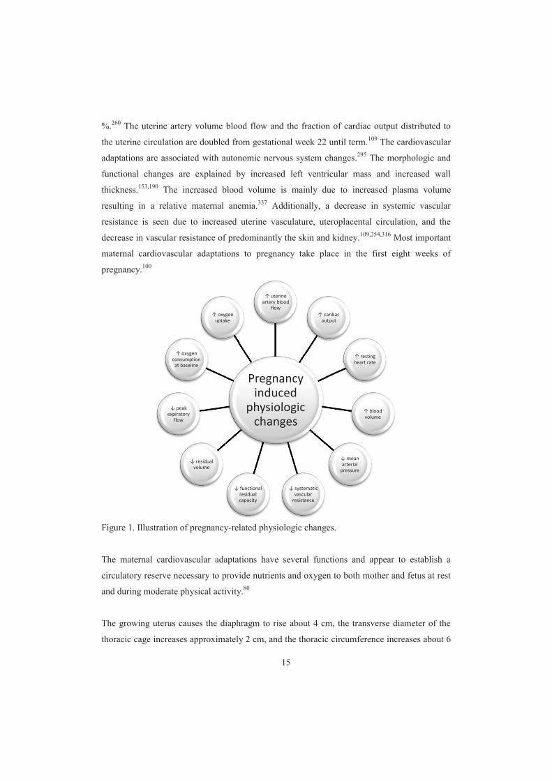
There are profound anatomical, physiological (Figure 1), and biochemical adaptations to
pregnancy. Many of these remarkable changes begin soon after fertilization and continue
throughout gestation, and most occur in response to physiological stimuli provided by the
fetus and placenta.80 These profound cardiovascular system alterations occur in pregnant
women regardless of their physical fitness status.207 Within five weeks of pregnancy cardiac
output increases and reaches 24 % above the non-pregnant level in pregnancy week 24,153 and
almost 50 % above the non-pregnant level in pregnancy week 32.270 This occurs as a result of
increased resting heart rate by 10 beats/min,295 increased blood volume,100,153,260,337 and
decreased mean arterial pressure.100,190 At term, the average increase in blood volume is 48
14
%.260 The uterine artery volume blood flow and the fraction of cardiac output distributed to
the uterine circulation are doubled from gestational week 22 until term.109 The cardiovascular
adaptations are associated with autonomic nervous system changes.295 The morphologic and
functional changes are explained by increased left ventricular mass and increased wall
thickness.153,190 The increased blood volume is mainly due to increased plasma volume
resulting in a relative maternal anemia.337 Additionally, a decrease in systemic vascular
resistance is seen due to increased uterine vasculature, uteroplacental circulation, and the
decrease in vascular resistance of predominantly the skin and kidney.109,254,316 Most important
maternal cardiovascular adaptations to pregnancy take place in the first eight weeks of
pregnancy.100
Figure 1. Illustration of pregnancy-related physiologic changes.
The maternal cardiovascular adaptations have several functions and appear to establish a
circulatory reserve necessary to provide nutrients and oxygen to both mother and fetus at rest
and during moderate physical activity.80
The growing uterus causes the diaphragm to rise about 4 cm, the transverse diameter of the
thoracic cage increases approximately 2 cm, and the thoracic circumference increases about 6
Pregnancy induced
physiologic changes
uterine artery blood
flow cardiac
output
resting heart rate
blood volume
mean arterial
pressure
systematic vascular
resistance
functional residual capacity
residual volume
peak expiratory
flow
oxygen consumption
at baseline
oxygen uptake
15
cm.80 As a consequence of the elevated diaphragm, the functional residual capacity, the
residual volume and the peak expiratory flow rates decline progressively as gestation
advances.135 There is increased oxygen uptake and an increase in baseline oxygen
consumption.172 The subjective workload and maximum exercise performance decreases due
to increased resting oxygen requirements. Because of the increased work of breathing, caused
by pressure of the enlarged uterus on the diaphragm, there is decreased oxygen availability for
the performance of aerobic exercise during pregnancy.18
The fetal body temperature is about 1.0 °C higher than maternal temperature. To prevent
hyperthermia during exercise due to maternal basal metabolic rate and heat production, a
steady state of heat production versus dissipation is accomplished by increased conductance
of heat from the core to the periphery circulation as well as through evaporative cooling
through sweat.18
Due to the enlarged uterus, the supine resting position and motionless standing will decrease
venous return and cardiac output due to vena cava compression.74,158 However, exercise in a
supine position has not been found to significantly decrease venous return and cardiac
output,158 and does not support guidelines advising against exercises in supine position after
16 weeks of gestation.5,83 However, when designing exercise programs for pregnant women
one should be aware of the possible obstruction of vena cava.
Exercise recommendations for non-pregnant women
In 1995, the Centers for Disease Control and Prevention (CDC) and the American College of
Sports Medicine (ACSM), issued a public health recommendation about physical activity with
the purpose to encourage increased participation in physical activity by a largely sedentary US
population.247 The original recommendations issued that “every US adult should accumulate
30 minutes or more of moderate-intensity physical activity on most, preferably all, days of the
week”.247 However, the recommendations were not accepted by some and others have
misinterpreted the recommendations. In 2007, the ACSM and the American Heart Association
issued an updated version of the 1995 recommendations.137 The intent was to provide a more
comprehensive and explicit public health recommendation for adults based upon available
evidence of the health benefits of physical activity.137
16
In the latest recommendations for adults, both aerobic physical activity and strength exercises
are recommended to promote and maintain good health and physical independence.137
Moderate-intensity aerobic physical activity for a minimum of 30 minutes should be
performed on five days each week or vigorous-intensity aerobic activity for a minimum of 20
minutes on three days each week. A combination of moderate- and vigorous intensity activity
can be performed to meet this recommendation.137 Activities that maintain or increase
muscular strength and endurance should be performed for a minimum of two days each week.
It is recommended that 8-10 exercises are performed on two or more nonconsecutive days
each week using the major muscle groups.137 It is further stated that participation in aerobic
and muscle-strengthening physical activities above minimum recommended amounts provides
additional health benefits and results in higher levels of physical fitness.137
Exercise recommendations for pregnant women
From the traditional view that pregnant women should reduce their habitual levels of exercise
in pregnancy, there is now agreement that healthy pregnant women should be encouraged to
include exercise as part of a healthy lifestyle. Pregnancy is recognized as a unique time for
behavior modification and is no longer considered a condition for confinement. It is currently
recognized that habits adopted during pregnancy could affect a woman’s health for the rest of
her life.18 Current ACOG guidelines for exercise during pregnancy recommend 30 minutes or
more of moderate exercise on most if not all days of the week for pregnant women in the
absence of medical or obstetric complications.5 Women with complicated pregnancies have
been discouraged from participating in exercise activities for fear of impacting the underlying
disorder or maternal or fetal outcome.83 The recommendations also promote exercise for
sedentary women and those with medical or obstetric complications, but only after medical
evaluation and clearance (Table 1). Pregnant women should be cautious about participating in
contact sports and avoid scuba diving. Due to the anatomical and physical changes during
pregnancy, such as weight gain, increased forces across hip- and knee joints and altered levels
of hormones, pregnant women are theoretically more predisposed to musculoskeletal injuries.
Nevertheless, these possibilities should be considered when designing exercise programs.
17
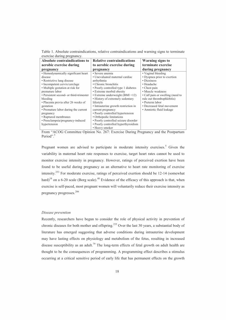
Table 1. Absolute contraindications, relative contraindications and warning signs to terminate exercise during pregnancy. Absolute contraindications to aerobic exercise during pregnancy
Relative contraindications to aerobic exercise during pregnancy
Warning signs to terminate exercise during pregnancy
• Hemodynamically significant heart disease • Restrictive lung disease • Incompetent cervix/cerclage • Multiple gestation at risk for premature labor • Persistent second- or third-trimester bleeding • Placenta previa after 26 weeks of gestation • Premature labor during the current pregnancy • Ruptured membranes • Preeclampsia/pregnancy-induced hypertension
• Severe anemia • Unevaluated maternal cardiac arrhythmia • Chronic bronchitis • Poorly controlled type 1 diabetes • Extreme morbid obesity • Extreme underweight (BMI <12) • History of extremely sedentary lifestyle • Intrauterine growth restriction in current pregnancy • Poorly controlled hypertension • Orthopedic limitations • Poorly controlled seizure disorder • Poorly controlled hyperthyroidism • Heavy smoker
• Vaginal bleeding • Dyspnea prior to exertion • Dizziness • Headache • Chest pain • Muscle weakness • Calf pain or swelling (need to rule out thrombophlebitis) • Preterm labor • Decreased fetal movement • Amniotic fluid leakage
From “ACOG Committee Opinion No. 267: Exercise During Pregnancy and the Postpartum Period”.5
Pregnant women are advised to participate in moderate intensity exercises.5 Given the
variability in maternal heart rate responses to exercise, target heart rates cannot be used to
monitor exercise intensity in pregnancy. However, ratings of perceived exertion have been
found to be useful during pregnancy as an alternative to heart rate monitoring of exercise
intensity.253 For moderate exercise, ratings of perceived exertion should be 12-14 (somewhat
hard)18 on a 6-20 scale (Borg scale).40 Evidence of the efficacy of this approach is that, when
exercise is self-paced, most pregnant women will voluntarily reduce their exercise intensity as
pregnancy progresses.206
Disease prevention
Recently, researchers have begun to consider the role of physical activity in prevention of
chronic diseases for both mother and offspring.255 Over the last 30 years, a substantial body of
literature has emerged suggesting that adverse conditions during intrauterine development
may have lasting effects on physiology and metabolism of the fetus, resulting in increased
disease susceptibility as an adult.79 The long-term effects of fetal growth on adult health are
thought to be the consequences of programming. A programming effect describes a stimulus
occurring at a critical sensitive period of early life that has permanent effects on the growth
18
and metabolism of the organism. These stimuli may lead to alterations in birth size and
metabolic changes, such as reduced insulin sensitivity and increased susceptibility to disease
later in life.26 Fetal adaption to an adverse intrauterine environment involves programming of
metabolic pathways that might predispose to cardiovascular disease in later life. This adverse
environment may change gene expression, leading to physiological phenotypes associated
with morbidity and mortality.27,160 Many of the genes epigenetically influenced by nutrition
are also affected by exercise or are involved in metabolic processes that are modulated by
exercise.204 It is therefore reasonable to hypothesize that exercise during pregnancy also could
mediate future health outcomes through epigenetic modifications.96
Body size at birth is the best available marker for fetal growth and is a rough indicator of the
in utero environment. Both high and low birth weight has been found to have adverse
associations with later disease. There is a robust relationship between low birth size and all-
cause mortality in adulthood, implicating a major impact of intrauterine conditions on health
during later life.160 In a recently published meta-analysis Risnes et al.264 found that birth
weight was inversely associated with adult mortality from all causes and cardiovascular
mortality. For cancer mortality there was a linear relationship with birth weight for men, but
not for women.
Children with a high birth weight are more likely to become obese.16,335 In the long run,
childhood overweight and obesity is strongly associated with adult obesity, giving more than
a fivefold increase in the risk for being overweight in early adulthood.122,296,311 These results
suggest that prevention of obesity should begin as early as possible.
There is now increasing evidence that the effect of the in utero environment on the
development of obesity and risk factors for adult disease is U-shaped. Here, the rates of
T2DM among persons with low birth weight (<2500 g) and persons with high birth weight
(>4500 g) are being almost twice as high compared to persons with normal birth weight.238,252
Hopkins et al.151 found that women who exercised regularly from gestational week 20
onwards gave birth to significantly lighter babies than controls (3426 ± 427 vs. 3569 ± 433).
Clapp et al.73 found that women who performed a high volume of exercise in early pregnancy
and then cut back their exercise in late pregnancy delivered offspring who were significantly
heavier at birth. In contrast, although not statistically significant, the offspring of women who
19
increased from a low volume of exercise in early pregnancy to a high volume in the second
half of pregnancy were on average 100 g lighter than the offspring of a comparative group
who maintained a moderate volume of exercise throughout pregnancy. These results suggest
that fetoplacental adaptations are dependent on the period of gestation in which exercise
training is initiated and maintained, as well as the intensity or volume of the exercise
performed.152
In utero exposure to hyperglycemia is found to have an intergenerational effect, with women
born to diabetic mothers being more predisposed to diabetes in their own pregnancy than
women born to non-diabetic mothers.205 Promoting the health of women of reproductive age
before conception and ensuring that women receive the services they need to improve
outcomes while they are pregnant is a primary goal of preconception care and the prevention
of chronic diseases in future generations.20 Helping women to maintain a healthy weight
before and during pregnancy may be the best hope for controlling the obesity epidemic seen
in some countries.
Today’s recommendations for exercise in pregnancy are proactive,5,230 suggesting that
virtually all women having a normal pregnancy can benefit from a physical activity program.
However, there is a lack of studies examining the potential epigenetic programming effects of
exercise during pregnancy.96
Pregnancy and level of physical activity
Although exercise is recommended during pregnancy, it is documented that being pregnant is
an event that leads to decreased physical activity. Women seem to reduce all levels of
physical activity such as household tasks, care giving and active living, but the largest
reduction is seen in participation in sports and exercise.105 In the Norwegian Mother and Child
Cohort Study (MoBa study), including 34,508 pregnancies in Norway, the proportion of
regular exercise defined as exercising three times per week or more, was 46 % before
pregnancy and declined to 28 % and 20 % by gestational weeks 17 and 30, respectively.244
Multiple pregnancy, pelvic girdle pain, nausea, musculo-skeletal pain, uterine contractions
and sick-leave were factors inversely associated with regular exercise in the MoBa study.244
Sociodemographic characteristics positively associated with exercising during pregnancy are
20
high education, low body mass index (BMI), primiparity and being a cohabitant.60,105,149,244
Regular exercise prior to pregnancy is strongly associated with regular exercise during
pregnancy, and women with high levels of pre-pregnancy physical activity are more likely to
exercise during pregnancy.60,105,149,244
Beliefs and behavior
In a qualitative study of 19 Australian pregnant women, motivating factors that facilitated
women’s engagement in physical activity included access to exercise classes, using a personal
trainer, receiving advice from health professionals, being with other pregnant women,
wanting to stay in shape with a controlled weight gain, and receiving reassurance that their
pregnancy was progressing satisfactorily. In addition control of physiological events such as
constipation and incontinence, and the baby’s well-being were motivating factors.72 The
same study identified barriers to be physical active such as feeling unwell or tired, physical
discomforts, such as backache and sore knees, pressure from the uterus, breathlessness and
decreased motivation and lack of time because of work and having previous children.72
Most women report that they obtain the information about exercise in pregnancy from books,
the web, parental magazines, publications issued free at antenatal clinics, and health care
professionals.72,75 In a study by Clarke et al.,75 only 18 % reported receiving advice directly
from the health professionals involved in their antenatal care. Women reported that the
information received was often unclear, confusing, inconsistent or conflicting.72,75 On the
other hand, women who are informed about the benefits and risks of physical activity and
exercise techniques, have more favorable attitudes towards physical activity.60
Following the reduction in activity during pregnancy, the activity levels are found to be 1.4
hours per week below pre-pregnancy levels at six months postpartum. This is associated with
increased body weight retention during the first six months of the postpartum period.250
Several factors related to the time available for exercise predicted the likelihood of
insufficient activity, including employment, number of children at home, and child care
barriers.250 Women who temporary halt exercise during pregnancy, tend not to resume their
exercise habits postpartum to the same extent as women who continue exercising during
pregnancy.103
21
Among pregnant women the importance of rest and relaxation are rated significantly higher
than having an active lifestyle and exercising regularly.75 Although most mothers understand
the benefits of physical activity in pregnancy, that does not seem to translate in to practice.
The pregnancy and the postnatal periods are prime opportunities for raising women’s
awareness of health strategies to enable them to nurture their optimal well-being.72 There is
considerable scope for improving the quantity and quality of advice in this area.75 Antepartum
care providers should increase women’s awareness of the benefits of activity in pregnancy,
provide reassurance about safety concerns, and present options to women that can guide their
incorporation of recommended activity levels into their daily lives.72
Validation of physical activity
Physical activity is characterized by its intensity, duration, frequency and mode of activity.218
Ideally, all these aspects should be recorded during physical activity measurements. However,
in most studies addressing effects of physical activity in pregnancy, the validity of activity
reports is open to discussion.
Data collection of physical activity most frequently involves self-reporting (subjective)
measures, such as questionnaires, diaries, logs, surveys and interviews. The benefits of self-
reporting measures are their practicality, low cost, low participant burden and general
acceptance.94 However, the self-reporting methods have limitations such as recall and
response bias and the inability to capture the absolute level of physical activity.259 Direct
measures are believed to offer more precise estimates of energy expenditure and remove
many of the issues of recall and response bias. There are several methods for assessing direct
physical activity such as calorimetry (i.e., doubly labelled water, indirect, direct),
physiological markers (i.e., cardiorespiratory fitness, biomarkers), motion sensors and
monitors (i.e., accelerometers, pedometers, heart rate monitors).259 However, there is no
single “golden standard” for measuring physical activity or assessing its validity.94
A systematic review included 148 studies on non-pregnant adults and reported the correlation
between self-reports and direct measurements of physical activity. However, there was no
clear trend in the degree of correlation between self-reported and directly measured physical
activity, regardless of the direct method used. The correlation was generally low-to-moderate
22
with a mean of 0.37, ranging from -0.71 to 0.96.259 The correlation was higher in studies
reporting results for males (r=0.47 on average) versus studies reporting results for females
(r=0.36 on average), but with very similar ranges. In general, self-reported physical activity
estimates were higher than those measured by directly methods.259 In overweight or obese
individuals, self-reported physical activity was overestimated in all cases.259 Among 153
adults aged 35-65 years, Ferrari et al.107 found that the accuracy of physical activity
questionnaire measurements was higher for men than for women, for younger individuals and
for those with lower BMI.
One challenge in assessing physical activity is that women as a group spend more time on
household and family activities than men. In surveys that did not include these activities,
women tend to underestimate their activity level.7 In a review by Prince et al.259 some results
suggest that patterns in the agreement between self-reports and direct measurements of
physical activity may exist, but they are likely to differ depending on the direct methods used
for comparison and gender. Differences between the self-report and direct measurements
increase with the higher category levels of intensity.259 The fact that intensity is the least valid
component assessed by self-reporting is supported by Sallis and Saelens who found that
young people and adults overestimate their physical activity, particularly vigorous intensity
activities.276
Despite the advantages of using direct methods, these types of measurements are often
expensive and time consuming resulting in difficulties in applying them to large populations.
The direct methods also require specialized training and the physical proximity of the
participant for data collection.
Since resting metabolic rate and absolute energy cost during physical activity are increased in
pregnant women,271,322 activity monitors must be validated in this specific population.330 To
be able to give recommendations based on evidence, valid measurement tools are important.68
One possible bias is that the activity monitors used have not been validated in the pregnant
population.
Validation studies quantifying the impact of measurement errors on physical activity
estimates are essential to evaluate the impact of physical inactivity on health.107 The validity
23

of activity monitors needs to be carefully examined in the actual research population.330 To
my knowledge, few have evaluated activity monitors for use during pregnancy.68 Valid and
reliable measurements of energy expenditure and physical activity are critical to understand
the influence of physical activity on pregnancy outcomes.68
24
In this thesis I will look into the influence of exercise during pregnancy on gestational
diabetes and the two most common pregnancy-related muscelosceletal disorders; lumbopelvic
pain and incontinence.
Gestational diabetes
Definition
Gestational diabetes mellitus (GDM) is defined as carbohydrate intolerance with onset or first
recognition during pregnancy210 and is usually diagnosed by an oral glucose tolerance test
(OGTT), however, the OGTT procedure and the diagnostic criteria used vary.70
Prevalence of GDM
The prevalence of GDM has been reported up to 14 %.159 It has increased worldwide,
however, there is variation in diagnostic criteria, ethnicity and the population studied.159,343 A
prevalence of 5.6 % was found (unpublished data) in a cohort of a Scandinavian population in
Oslo. This was in the “Stork” study conducted in 2002-2008 (N=1,032). In the “Stork
Groruddalen” research program, conducted at the same time that included an urban
Norwegian population (N=759), a prevalence of 13 % was found in gestational week 28 ±
2.224 In 2009, 1.3 % of all pregnancies in Norway were registered as having GDM in the
Medical Birth Registry of Norway.307 However, pregnant Norwegian women are not screened
for GDM on a regular basis, and only women with identified risk factors are offered OGTT.
Thus, the prevalence is probably underreported in the Medical Birth Registry of Norway, and
the true prevalence is unknown.
Risk factors for GDM
The risk of diabetes in pregnancy reflects the underlying frequency of T2DM for a given
population.168 Identified risk factors for GDM are obesity (BMI>30) diagnosed before
pregnancy,314 ethnicity,168 polycystic ovarian syndrome,315 essential hypertension,
25
hypertension in pregnancy,145 family history of diabetes in first-degree relatives and a history
of GDM in previous pregnancy.1 In a population-based cohort study of 96,801 nulliparous
pregnancies in the US, the proportion of women who developed GDM, consistently increased
with increasing BMI. Both pre-pregnancy obesity and overweight were found to increase the
risk of GDM.21
Metabolic changes during pregnancy
Pregnancy has been characterized as a diabetogenic event due to hormones with diabetogenic
effects (estrogen, prolactin, human chorionic somatomammotropin, HPL, cortisol, and
progesterone). The diabetogenic effects of these hormones lead to insulin resistance and
increased insulin requirements.17 Insulin resistance is a physiological condition in which the
natural hormone insulin becomes less effective in lowering blood sugars. The metabolic
changes are in response to the increased demands of the rapidly growing fetus and placenta.
By the third trimester, the maternal basal metabolic rate is increased by 10-20 % compared
with that of the non-pregnant state.80 Among the nutrients crossing the placenta, glucose is
quantitatively the most important, and is the primary energy source for feto-placental tissues.
The glucose supply to the fetus is via passive glucose diffusion and therefore it is
concentration dependent.142
A normal pregnancy is characterized by mild fasting hypoglycemia, postprandial
hyperglycemia, and hyperinsulinemia. This response is consistent with a pregnancy-induced
state of peripheral insulin resistance, the purpose of which is likely to ensure a sustained
postprandial supply of glucose to the fetus. Insulin sensitivity in late normal pregnancy is 50-
70 % lower than that of non-pregnant women.49 The mechanisms responsible for insulin
resistance are not completely understood.80
GDM and associated short- and long-term risk factors
GDM is a risk factor for pregnancy complications,165,283 and is associated with short- and
long-term morbidity in both the mother and the offspring.39,81,147,166,167 It is unclear if the
complications are due to elevated glucose levels per se or by unidentified factors. In the case
of maternal hyperglycemia and placental normal function, there will be increased placental
26
transfer of glucose, fetal hyperglycemia develops and hyperinsulinism secondary to this
alteration. As insulin is one of the main growth factors during fetal life, this hyperinsulinemia
leads to macrosomia and to the complications secondary to the delivery of a large baby. Even
minor degrees of hyperglycemia are associated with adverse outcomes. In the HAPO study,
including 23,316 pregnant women who underwent a 75 g OGTT at 24-32 weeks of gestation,
a strong continuous association was found between maternal glucose tolerance and a number
of different adverse pregnancy outcome events even in the sub-diabetes range. There were
positive associations between increasing levels of fasting, 1-hour, and 2-hour plasma glucose
obtained by OGTT and birth weight above the 90th percentile, preterm delivery, shoulder
dystocia, birth injury, intensive neonatal care, hyperbilirubinemia, and preeclampsia.211
In the long term GDM is associated with risk factors for cardiovascular disease,180 and a
higher risk of developing T2DM during the next 5-10 years for the mother.167 A meta-analysis
found that women with GDM have at least a seven-fold increased risk of developing T2DM
compared with those who had a normoglycaemic pregnancy (RR 7.43, 95 % CI 4.79-11.51).29
And women developing GDM have a higher risk of the recurrence of GDM in the next
pregnancy.166 Children born in GDM pregnancies have a higher risk of childhood obesity,147
metabolic syndrome39 and T2DM as adolescents.81 In addition, children born in GDM
pregnancies have a higher risk of developing GDM in their own pregnancy compared to
children born to non-diabetic mothers.205
Many of the risk factors for GDM and T2DM are the same (a family history of diabetes,
raised BMI, increased age, and Asian and black origin) suggesting that the two disorders
might have overlapping causes.29
Exercise and glucose homeostasis
In the non-pregnant state, physical activity improves glucose homeostasis through a direct or
indirect impact on insulin sensitivity.15,41,88 Both aerobic and resistance training improve
insulin sensitivity as shown by a reduction in the insulin response to a glucose load, and an
improvement in peripheral insulin sensitivity. Muscle contractions stimulate glucose uptake in
the complete absence of insulin. The effects of muscle contraction and insulin are additive,
and the contraction and insulin stimulate glucose transport by separate pathways.140 However,
27
exercise must be undertaken regularly to have beneficial effects.77,140 Both in prevention and
treatment of T2DM, exercise plays an important role.77 A meta-analysis found that all types of
exercise reduced the HbA1c levels in T2DM to a similar magnitude as those exerted by long-
term dietary, drug and insulin therapy.291 Therefore, regular sustained aerobic exercise in
pregnant women may have the same effect and counteract the normal state of insulin
resistance in late gestation. In the ACOG’s latest version from 2002 it is suggested that
exercise has a possible role in the prevention and management of GDM.5
Among non-pregnant women with T2DM, the intensity and duration of physical activity seem
to be the most important factors that influence energy expenditure. However, there are
conflicting results about whether the intensity or the volume of exercise is the most important
determinant for the improvement of insulin action.139 Physical activity causes increased
glucose uptake into active muscles balanced by hepatic glucose production, with a greater
reliance on carbohydrate to fuel muscular activity as intensity increases. An increase in
muscle mass from resistance training may contribute to blood glucose uptake from a mass
effect.63 Combined strength and aerobic training is found to be superior to either type of
exercise alone.291 In addition, the supervision of exercise sessions by qualified exercise
trainers is found to be important for the best effect on blood glucose, and there should be no
more than two consecutive days between bouts of aerobic activity.77 For those with T2DM,
strength and aerobic training are recommended as well as initial instruction and periodic
supervision by a qualified exercise trainer.77
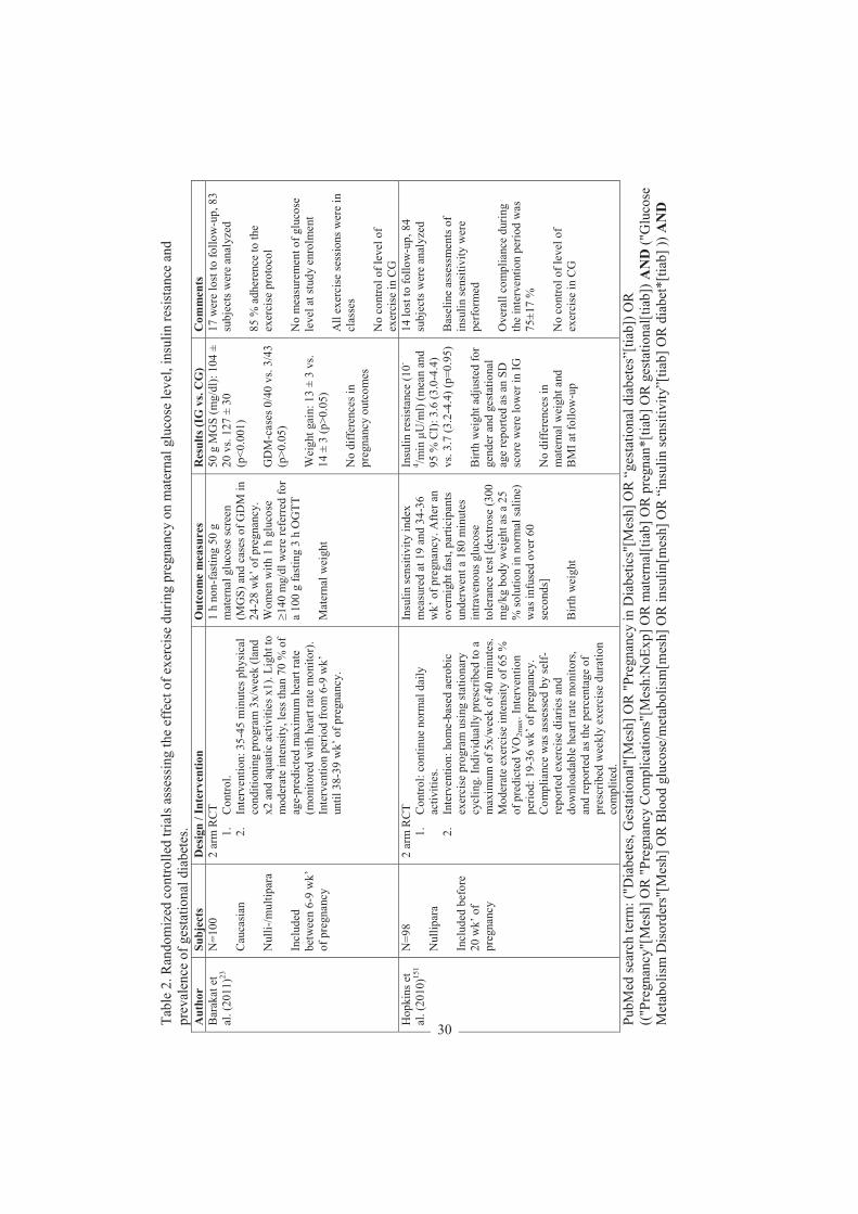
The role of exercise in prevention of GDM
The effect of exercise on the development of gestational diabetes has been sparsely studied,
and the study results are conflicting.23,59,151,313 In a systematic search on PubMed no previous
RCT’s addressing the effect of physical activity in prevention of GDM in a general population
of pregnant women were found (Table 2). Two recent trials assessed maternal glucose level
and insulin sensitivity respectively with conflicting results. However, the study populations
were small, only 84 and 83 subjects.23,151 Results from one meta-analysis demonstrate that
greater total physical activity before or during early pregnancy is associated with a lower risk
of GDM, with the magnitude of the association being stronger for pre-pregnancy physical
activity.313 There is a lack of large RCTs that have a sufficient methodological quality to
28

assess the effects of adequate regular exercise in pregnancy on glucose homeostasis.220 Two
protocols for assessing exercise in the prevention of GDM in women at risk are
published.69,243 A protocol for a Cochrane review on exercise for pregnant women in the
prevention of GDM has recently been published.127
The prevalence of GDM doubled from 1994 to 2002 in a multiethnic population in Colorado,
US.82 The trend toward older maternal age, the epidemic of obesity and diabetes, and the
decrease in physical activity and the adoption of modern lifestyles in developing countries
may all contribute to an increase in the prevalence of GDM.106 Identifying ways that might
help prevent GDM is a matter of urgent public health importance.
29
Tabl
e 2.
Ran
dom
ized
con
trolle
d tri
als a
sses
sing
the
effe
ct o
f exe
rcis
e du
ring
preg
nanc
y on
mat
erna
l glu
cose
leve
l, in
sulin
resis
tanc
e an
d pr
eval
ence
of g
esta
tiona
l dia
bete
s. A
utho
r Su
bjec
ts
Des
ign
/ Int
erve
ntio
n O
utco
me
mea
sure
s R
esul
ts (I
G v
s. C
G)
Com
men
ts
Bar
akat
et
al. (
2011
)23
N=1
00
Cau
casi
an
Nul
li -/m
ultip
ara
Incl
uded
be
twee
n 6-
9 w
k’
of p
regn
ancy
2 ar
m R
CT
1.
Con
trol.
2.
Inte
rven
tion:
35-
45 m
inut
es p
hysi
cal
cond
ition
ing
prog
ram
3x/
wee
k (la
nd
x2 a
nd a
quat
ic a
ctiv
ities
x1)
. Lig
ht to
m
oder
ate
inte
nsity
, les
s tha
n 70
% o
f ag
e-pr
edic
ted
max
imum
hea
rt ra
te
(mon
itore
d w
ith h
eart
rate
mon
itor)
. In
terv
entio
n pe
riod
from
6-9
wk’
un
til 3
8-39
wk’
of p
regn
ancy
.
1 h
non-
fast
ing
50 g
m
ater
nal g
luco
se sc
reen
(M
GS)
and
cas
es o
f GD
M in
24
-28
wk’
of p
regn
ancy
. W
omen
with
1 h
glu
cose
14
0 m
g/dl
wer
e re
ferr
ed fo
r a
100
g fa
stin
g 3
h O
GTT
M
ater
nal w
eigh
t
50 g
MG
S (m
g/dl
): 10
4 ±
20 v
s. 12
7 ±
30
(p<0
.001
) G
DM
-cas
es 0
/40
vs. 3
/43
(p>0
.05)
W
eigh
t gai
n: 1
3 ±
3 vs
. 1 4
± 3
(p>0
.05)
N
o di
ffere
nces
in
preg
nanc
y ou
tcom
es
17 w
ere
lost
to fo
llow
-up,
83
subj
ects
wer
e an
alyz
ed
85 %
adh
eren
ce to
the
exer
cise
pro
toco
l N
o m
easu
rem
ent o
f glu
cose
le
vel a
t stu
dy e
nrol
men
t A
ll ex
erci
se se
ssio
ns w
ere
in
clas
ses
No
cont
rol o
f lev
el o
f ex
erci
se in
CG
H
opki
ns e
t al
. (20
10)15
1 N
=98
N
ullip
ara
Incl
uded
bef
ore
20 w
k’ o
f pr
egna
ncy
2 ar
m R
CT
1.
Con
trol:
cont
inue
nor
mal
dai
ly
activ
ities
. 2.
In
terv
entio
n: h
ome-
base
d ae
robi
c ex
erci
se p
rogr
am u
sing
stat
iona
ry
cycl
ing.
Indi
vidu
ally
pre
scrib
ed to
a
max
imum
of 5
x/w
eek
of 4
0 m
inut
es.
Mod
erat
e ex
erci
se in
tens
ity o
f 65
%
of p
redi
cted
VO
2max
. Int
erve
ntio
n pe
riod:
19-
36 w
k’ o
f pre
gnan
cy.
Com
plia
nce
was
ass
esse
d by
self-
repo
rted
exer
cise
dia
ries a
nd
dow
nloa
dabl
e he
art r
ate
mon
itors
, an
d re
porte
d as
the
perc
enta
ge o
f pr
escr
ibed
wee
kly
exer
cise
dur
atio
n co
mpl
ited.
Insu
lin se
nsiti
vity
inde
x m
easu
red
at 1
9 an
d 34
-36
wk’
of p
regn
ancy
. Afte
r an
over
nigh
t fas
t, pa
rtici
pant
s un
derw
ent a
180
min
utes
in
trave
nous
glu
cose
to
lera
nce
test
[dex
trose
(300
m
g/kg
bod
y w
eigh
t as a
25
% so
lutio
n in
nor
mal
salin
e)
was
infu
sed
over
60
seco
nds]
B
irth
wei
ght
Insu
lin re
sist
ance
(10-
4 /min
U
/ml)
(mea
n an
d 95
% C
I): 3
.6 (3
.0-4
.4)
vs. 3
.7 (3
.2-4
.4) (
p=0.
95)
Birt
h w
eigh
t adj
uste
d fo
r ge
nder
and
ges
tatio
nal
age
repo
rted
as a
n SD
sc
ore
wer
e lo
wer
in IG
N
o di
ffere
nces
in
mat
erna
l wei
ght a
nd
BM
I at f
ollo
w-u
p
14 lo
st to
follo
w-u
p, 8
4 su
bjec
ts w
ere
anal
yzed
B
asel
ine
asse
ssm
ents
of
insu
lin se
nsiti
vity
wer
e pe
rfor
med
O
vera
ll co
mpl
ianc
e du
ring
the
inte
rven
tion
perio
d w
as
75±1
7 %
N
o co
ntro
l of l
evel
of
exer
cise
in C
G
PubM
ed se
arch
term
: ("D
iabe
tes,
Ges
tatio
nal"
[Mes
h] O
R "P
regn
ancy
in D
iabe
tics"
[Mes
h] O
R “
gest
atio
nal d
iabe
tes”
[tiab
]) O
R
(("P
regn
ancy
"[M
esh]
OR
"Pre
gnan
cy C
ompl
icat
ions
"[M
esh:
NoE
xp] O
R m
ater
nal[t
iab]
OR
pre
gnan
*[tia
b] O
R g
esta
tiona
l[tia
b]) A
ND
("G
luco
se
Met
abol
ism
Dis
orde
rs"[
Mes
h] O
R B
lood
glu
cose
/met
abol
ism
[mes
h] O
R in
sulin
[mes
h] O
R “
insu
lin se
nsiti
vity
”[tia
b] O
R d
iabe
t*[ti
ab] )
) AN
D
30

("Ex
erci
se"[
Mes
h] O
R "
Exer
cise
The
rapy
"[M
esh]
OR
"Ex
erci
se M
ovem
ent T
echn
ique
s"[M
esh]
OR
"Phy
sica
l Fitn
ess"
[Mes
h] O
R "P
hysi
cal
Exer
tion"
[Mes
h] O
R "M
otor
Act
ivity
"[M
esh]
OR
"Spo
rts"[
Mes
h] O
R e
xerc
is*[
tiab]
OR
trai
ning
[tiab
] OR
("ph
ysic
al a
ctiv
ity"[
tiab]
NO
T m
edlin
e[sb
])) A
ND
(clin
ical
tria
l[pt]
OR
rand
om*[
tiab]
OR
tria
l[tia
b] O
R g
roup
[tiab
] OR
gro
ups[
tiab]
OR
syst
emat
ic[s
b]) N
OT
((A
nim
als[
mes
h] N
OT
Hum
ans[
mes
h]) O
R C
ase
repo
rts[p
t])
BM
I den
otes
Bod
y M
ass I
ndex
, CG
con
trol g
roup
, GD
M g
esta
tiona
l dia
bete
s, IG
inte
rven
tion
grou
p, O
GTT
ora
l glu
cose
tole
ranc
e te
st.
31
Lumbopelvic pain
History
Historical evidence shows that pelvic girdle pain (PGP) in pregnancy was already known and
recognized many centuries ago. Symphysis pubis dysfunction was mentioned by Hippocrates
(ca. 400 BC) in his theory of “disjunction pelvica”.28 According to Hippocrates, the widening
of the symphysis pubis only occurred during the first parturition, and then remained
permanent and sufficient for later childbirths. The first to give a description of the clinical
aspects of pelvic insufficiency in Norway was Skajaa, who in 1929 observed symptoms of
pelvic insufficiency in 31 (16 %) of 185 patients from his private consultations.115
Definition
Pain is defined as an unpleasant sensory and emotional experience associated with actual or
potential tissue damage, or described in terms of such damage.157 Pain is an individual and
subjective experience that depends on a variety of biological, psychological and social
factors.78 Low back pain (LBP) is usually defined as pain between the 12th rib and the gluteal
fold, while PGP is defined as pain experienced between the posterior iliac crest and the
gluteal fold, particularly in the vicinity of the sacroiliac joints. The pain may radiate in the
posterior thigh and can also occur in conjunction with/or separately in the symphysis.326
Although similar and overlapping mechanisms may be involved between PGP and LBP, some
researchers argue that a distinction should be made.326 A clinical examination including pain
provocation tests and/or tests of functional disturbances must be performed to distinguish
between LBP and PGP,326 but the criteria are unclear. In this thesis the pain is referred to as
lumbopelvic pain (LPP), no specific distinction is made between LBP and PGP except when
this distinction is made in the references.
Prevalence of LPP
LPP during pregnancy is a common disorder. A review has reported an average prevalence of
45 % with variations from 4-90 %.342 The variation is probably due to differences in study
design and diagnostic procedures.326 Most of the literature use self-reports of LPP. It has been
32
shown that self-reporting generally reports 20 % higher prevalence than medical records,
indicating that women in questionnaires report mild pain that normally would not come to the
attention of the medical profession.342 In the MoBa study including 75,939 pregnancies, 58 %
reported pain in one or more pelvic locations during pregnancy week 30, with 13 % reporting
severe pain in one or more pelvic locations.34
It has been stated that LPP is more common in the Scandinavian countries than the rest of the
world.193 However, studies show that LPP is common in many
countries,35,67,76,98,216,219,221,312,329 irrespective of the socio-economic status of the countries.35
The majority of studies on LPP have been performed in the Scandinavian countries. One
explanation for the assumed higher prevalence in Scandinavia may be that outside of
Scandinavia pregnancy-related PGP is regarded as a normal pregnancy complaint rather than
a syndrome.28
Risk factors for LPP
Identified risk factors with strong evidence are reported to be strenuous work, previous low
back pain, previous LPP during or after pregnancy, and previous trauma to the pelvis.326,342
It has been suggested that the hormone relaxin, which is known to remodel pelvic connective
tissue in several mammalian species during pregnancy, plays an important role in the
development of LPP. However, study results on the levels of relaxin and LPP are
conflicting,10,178 and further research is needed to assess the possible role of relaxin and other
pregnancy-related hormones in the development of LPP. Bjelland et al.33 found an inverse
association between age at menarche and pelvic girdle syndrome (pain in all three pelvic
joints) in the MoBa study, suggesting an association between hormonal factors and PGP.
In the same cohort, Bjelland et al.34 found a positive relationship between parity and pain in
all three pelvic locations, with a stronger association for severe pain. Other factors associated
were previous LBP, emotional distress, BMI 30 kg/m2, physically demanding work, daily
smoking, low maternal age and lower educational level.34 In another cohort (N=891), high
BMI, low educational level, parity, previous LPP, a history of hypermobility and amenorrhea
were factors influencing the risk of developing LPP.216 Albert et al.9 divided pregnant women
33
with PGP into five subgroups regarding pain location. They found different risk factors for
the different subgroups, but common risk factors for all subgroups were a history of previous
LBP, trauma of the back or pelvis, multiparity, high levels of stress, and job dissatisfaction. In
a cross-sectional study including 283 pregnant women, Robinson et al.268 found that results
from the Posterior Pelvic Pain Provocation test (P4) and Active Straight Leg Raise (ASLR)
contributed independently to decreased physical ability in pregnancy week 30.
LPP is considered to be a normal condition of pregnancy expected to spontaneously disappear
after delivery.233 However, some women do not recover after birth. Albert et al.9 found a rapid
decline in the number of diseased in the first three months after delivery, but after three
months recovery was slow. Women classified with pelvic girdle syndrome had the worst
prognosis, and those with symphysolysis had the best.9 The average prevalence of LPP
postpartum is 25 %, with variations from 0.3 % to 67 %, and approximately 5 % report
serious LPP.342 High age, early onset of symptoms in the pregnancy, severe pain at early
gestation and low rate of weight retention postpartum are associated with persistent pain two
years after pregnancy.312 High pain intensity during pregnancy indicated a poor prognosis
after pregnancy.349 Women who have not recovered, have reduced quality of life compared to
the general population.298,299 It has been suggested that poor coping strategies may be
involved in the development of long-term pelvic pain postpartum.133 Olsson et al.242 found
that pain catastrophizing and physical ability mid-pregnancy are predictors of postpartum
LPP. Vøllestad and Stuge found that the ASLR test and belief in improvement were strong
and independent predictors of clinical significance in women with PGP postpartum.328
Influence of LPP on daily life
LPP interferes with ordinary daily activities249 and reduces the quality of life.241 Considerable
difficulties with activities such as housekeeping, walking, working and sexual life have been
reported.134,214 Although the reported prevalence of LPP is more than 50 % in most
studies,34,67,123,216,221,241,268,312,329 the majority of women are mild to moderately affected as
shown by the average scores in pain and disability.67,266
The median Disability Rating Index (DRI; an instrument for assessment of physical disability
with scores in the range of 0-100) in a group of healthy women aged 30-39 years was <1.275
34
Olsson et al.240 found a median DRI score for pregnant women without LPP to be 10 and 27
in women with LPP in mid-pregnancy. In late pregnancy the corresponding numbers were 24
versus 54 in women without and with LPP,241 indicating more limited physical activity with
advancing gestation. Higher DRI scores in women with LPP are also found in other
studies.123,177,266 This suggests that pregnancy and LPP has an additive effect. Higher DRI in
pregnant women without LPP than non-pregnant women underscores that even pregnancy in
itself constitutes physical disability.
Evidently, there is a wide range in the expression of the symptoms of LPP during pregnancy,
and women are affected to various degrees.214 Some women experience pain in a very early
stage of pregnancy, while others only experience pain in the final stage of pregnancy.266 In
addition, some women have more restricted activities due to pain compared with other
women. This suggests that other factors than hormonal and/or physiological changes might
influence the prevalence of LPP during pregnancy. One recent study found that women with
LPP in mid-pregnancy showed more pain catastrophizing and fear-avoidance beliefs than
women without LPP.240 Of women with LPP during pregnancy, around 45 % have mild
symptoms only; 25 % have severe pain; and 8 % are severely disabled.342 In a retrospective
cohort study from Norway in 1998-99, 7 % of all women reported the use of crutches during
pregnancy due to PGP.267 The localization and the nature of the pain vary. In pain research it
is well known that there are large individual variations in pain ratings, even in patients with
the same condition.231 The proportion of women with severe LPP in need of treatment has
been shown to be close to 25 %.342
Etiology, anatomy and function
The etiology and pathophysiology of LPP is unknown. During pregnancy there are
considerable changes in biomechanics and posture, when the center of gravity is moved
anteriorly. Progressive lordosis is a characteristic feature of normal pregnancy. Compensating
for the anterior position of the enlarging uterus, the lordosis shifts the center of gravity back
over the lower extremities. This curvature and reinforcement of the lumbar vertebrae has
evolved in humans to permit bipedal posture and locomotion despite up to a 31 % increase in
the maternal abdominal mass by term.336 The anatomy and function of the pelvis is due to a
combination of specific anatomic features (form closure) and the compression generated by
35
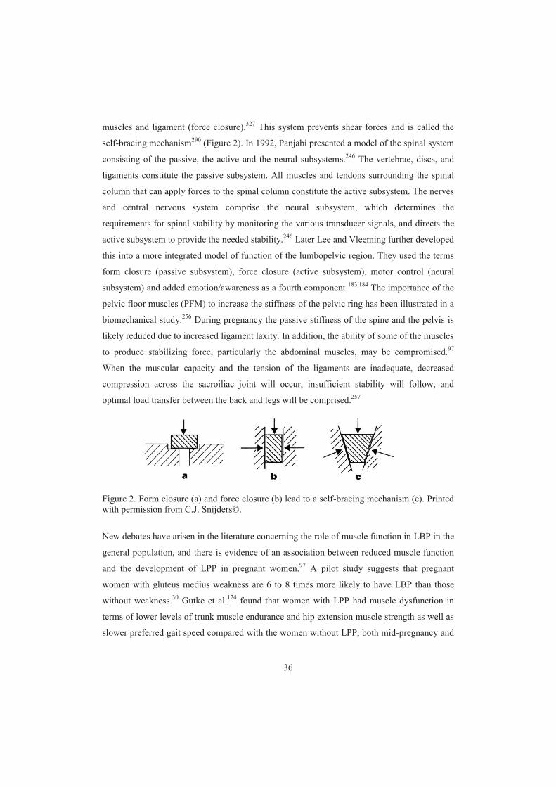
muscles and ligament (force closure).327 This system prevents shear forces and is called the
self-bracing mechanism290 (Figure 2). In 1992, Panjabi presented a model of the spinal system
consisting of the passive, the active and the neural subsystems.246 The vertebrae, discs, and
ligaments constitute the passive subsystem. All muscles and tendons surrounding the spinal
column that can apply forces to the spinal column constitute the active subsystem. The nerves
and central nervous system comprise the neural subsystem, which determines the
requirements for spinal stability by monitoring the various transducer signals, and directs the
active subsystem to provide the needed stability.246 Later Lee and Vleeming further developed
this into a more integrated model of function of the lumbopelvic region. They used the terms
form closure (passive subsystem), force closure (active subsystem), motor control (neural
subsystem) and added emotion/awareness as a fourth component.183,184 The importance of the
pelvic floor muscles (PFM) to increase the stiffness of the pelvic ring has been illustrated in a
biomechanical study.256 During pregnancy the passive stiffness of the spine and the pelvis is
likely reduced due to increased ligament laxity. In addition, the ability of some of the muscles
to produce stabilizing force, particularly the abdominal muscles, may be compromised.97
When the muscular capacity and the tension of the ligaments are inadequate, decreased
compression across the sacroiliac joint will occur, insufficient stability will follow, and
optimal load transfer between the back and legs will be comprised.257
Figure 2. Form closure (a) and force closure (b) lead to a self-bracing mechanism (c). Printed with permission from C.J. Snijders©.
New debates have arisen in the literature concerning the role of muscle function in LBP in the
general population, and there is evidence of an association between reduced muscle function
and the development of LPP in pregnant women.97 A pilot study suggests that pregnant
women with gluteus medius weakness are 6 to 8 times more likely to have LBP than those
without weakness.30 Gutke et al.124 found that women with LPP had muscle dysfunction in
terms of lower levels of trunk muscle endurance and hip extension muscle strength as well as
slower preferred gait speed compared with the women without LPP, both mid-pregnancy and
36
postpartum. Women with PGP had lower levels of back flexor endurance compared to women
without LPP.124 In addition, Gutke et al.125 found that low endurance of back flexors, older
age, combined pain in early pregnancy and work dissatisfaction were predictors of having
persistent PGP or LPP three months postpartum. Sihvonen et al.289 found a relationship
between LBP and muscle activity. They reported that the lower the back muscles activity
during the first trimester of pregnancy, the greater the chances to experience LBP and
disability throughout the pregnancy. De Groot et al.84 showed that pregnant women with LPP
used more muscle activity, produced less force and scored higher on the ASLR compared to
controls.
The combined effect of the muscle dysfunction identified in these studies and the hormone-
induced increased ligament and joint capsule laxity in pregnancy, may increase the risk of
insufficient force closure of the pelvis giving continuous strain on the pelvic ligaments and
resulting in pain.257 Results from these studies strengthen the hypothesis that there is an
association between muscle dysfunction and women who develop or have persistent LPP.
However, it is not known if identifying women at risk in early pregnancy and offering them
an intervention of strengthening adequate muscles, will prevent LPP.
Sick leave
In Norway, 59 % of pregnant women are on sick leave at least once during pregnancy with an
average of 21.5 days off work.223 Comparable numbers for other countries may differ with
type of social welfare system.304 LPP accounts for most of the sick leave among pregnant
women in Scandinavian countries,121,223,305,350 causing a high impact in terms of loss of
production and socio-economic costs. It has been demonstrated that women with postpartum
PGP may have difficulties in returning to work.133
Exercise
Despite that women are encouraged to exercise during pregnancy, being pregnant seems to be
an event that leads to reduced physical activity105,244 and women experiencing LPP are less
likely to exercise regularly.244 A recent cross-sectional study showed that sedentary coping
strategies were more frequent in women with LPP.67 This is supported by another study which
37
found that rest and relaxation were perceived as being significantly more important during
pregnancy than regular exercise.75
In the short term, resting and guarding may temporarily relieve pain. However, in the long run
inactivity leads to deconditioning and weakening of muscles, which in turn predisposes a
person to loss of function and pain. In the non-pregnant population there is general agreement
that the best strategy in the treatment of chronic LBP seems to be encourage physical activity
and exercise.8 A cross-cultural myth still suggests that pregnant women should be inactive
and rest in order to protect the safety of the fetus.72 European guidelines326 and a Cochrane
review249 clearly indicate the importance of both exercise and relaxation for pregnant women
to enhance overall health and prevent pain during pregnancy.
Besides being physically active maintain or improve fitness,208 observational studies have
demonstrated that physical activity before pregnancy may reduce the risk of developing
LPP.89,182,215 Also there is a reduced need for sick leave due to LPP.350 The explanation may
be that women in good physical condition will probably handle the hormonal and
biomechanical changes during pregnancy better. Another explanation may be that previous
physical activity clearly is a determinant of actual physical status.215
The influence of exercise on LPP
There is a lack of RCT’s assessing the effects of physical activity to prevent or treat LPP in
pregnant women. In a systematic literature search on PubMed (Table 3) only one RCT was
found that assessed the preventive effect of exercise on LPP. The trial, including 301
nulliparous women, assessed the effect of a 12-week intervention program with main focus on
pelvic floor muscle exercises. At gestational week 36 the authors found a prevalence of self-
reported LPP of 44 % in the intervention group versus 56 % in the control group (p = 0.03).
No difference was seen in sick leave due to LPP.228 Ten trials have assessed the role played
by physical exercise on treatment of LPP. In general, seven out of ten trials found a positive
effect from the intervention in the meaning of reduced pain intensity and/or improved
functional ability compared to the control group.114,120,161,164,170,287,302 However, the trials are
not comparable due to different interventions, length of intervention, intensity of intervention
38

and outcome measure. Four trials found reduced need for sick leave in the intervention
group.120,164,234,350
There is a lack of RCT’s assessing the effects of physical activity for pregnant women. Thus,
both prevention and possible adverse effects on LPP and general disability as well as the
effects on work related outcomes are sparsely studied.249
39
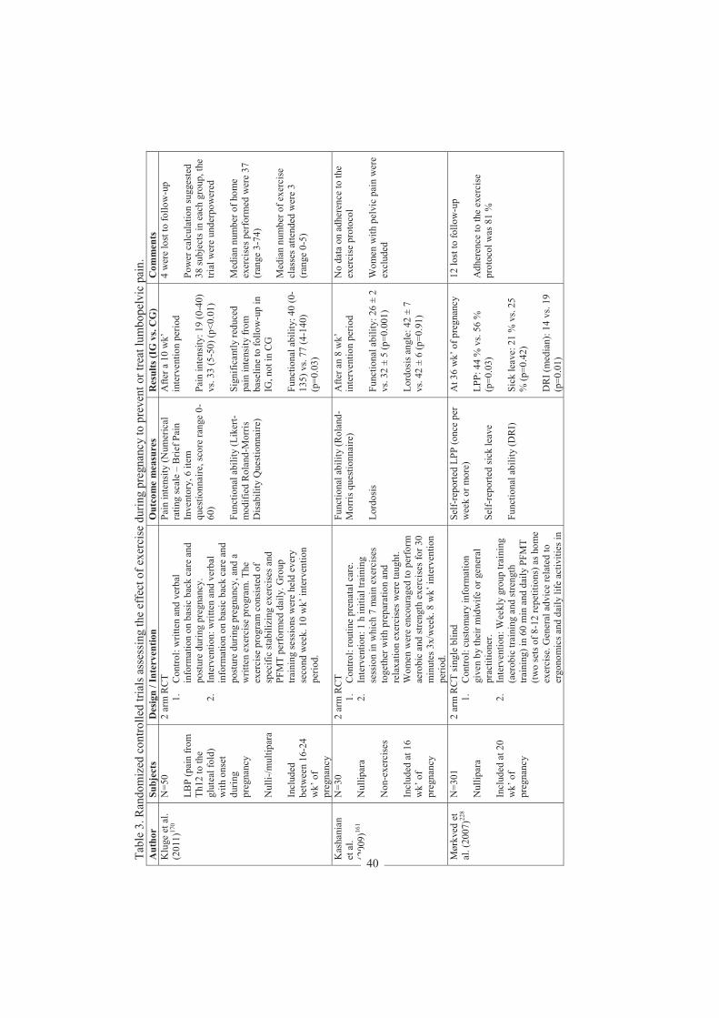
Tabl
e 3.
Ran
dom
ized
con
trolle
d tri
als a
sses
sing
the
effe
ct o
f exe
rcis
e du
ring
preg
nanc
y to
pre
vent
or t
reat
lum
bope
lvic
pai
n.
Aut
hor
Subj
ects
D
esig
n / I
nter
vent
ion
Out
com
e m
easu
res
Res
ults
(IG
vs.
CG
) C
omm
ents
K
luge
et a
l. (2
011)
170
N=5
0 L B
P (p
ain
from
Th
12 to
the
glut
eal f
old)
w
ith o
nset
du
ring
preg
nanc
y N
ulli-
/mul
tipar
a In
clud
ed
betw
een
16-2
4 w
k’ o
f pr
egna
ncy
2 ar
m R
CT
1.
Con
trol:
writ
ten
and
verb
al
info
rmat
ion
on b
asic
bac
k ca
re a
nd
post
ure
durin
g pr
egna
ncy.
2.
In
terv
entio
n: w
ritte
n an
d ve
rbal
in
form
atio
n on
bas
ic b
ack
care
and
po
stur
e du
ring
preg
nanc
y, a
nd a
w
ritte
n ex
erci
se p
rogr
am. T
he
exer
cise
pro
gram
con
sist
ed o
f sp
ecifi
c st
abili
zing
exe
rcis
es a
nd
PFM
T pe
rfor
med
dai
ly. G
roup
tra
inin
g se
ssio
ns w
ere
held
eve
ry
seco
nd w
eek.
10
wk’
inte
rven
tion
perio
d.
Pain
inte
nsity
(Num
eric
al
ratin
g sc
ale
– B
rief P
ain
Inve
ntor
y, 6
item
qu
estio
nnai
re, s
core
rang
e 0-
60)
F unc
tiona
l abi
lity
(Lik
ert-
mod
ified
Rol
and-
Mor
ris
Dis
abili
ty Q
uest
ionn
aire
)
Afte
r a 1
0 w
k’
inte
rven
tion
perio
d Pa
in in
tens
ity: 1
9 (0
-40)
vs
. 33
(5-5
0) (p
<0.0
1)
Sign
ifica
ntly
redu
ced
pain
inte
nsity
from
ba
selin
e to
follo
w-u
p in
IG
, not
in C
G
Func
tiona
l abi
lity:
40
(0-
135)
vs.
77 (4
-140
) (p
=0.0
3)
4 w
ere
lost
to fo
llow
-up
Pow
er c
alcu
latio
n su
gges
ted
38 su
bjec
ts in
eac
h gr
oup,
the
trial
wer
e un
d erp
ower
ed
Med
ian
num
ber o
f hom
e ex
erci
ses p
erfo
rmed
wer
e 37
(r
ange
3-7
4)
Med
ian
num
ber o
f exe
rcis
e cl
asse
s atte
nded
wer
e 3
(ran
ge 0
-5)
Kas
hani
an
et a
l. (2
009)
161
N=3
0 N
ullip
ara
Non
-exe
rcis
es
Incl
uded
at 1
6 w
k’ o
f pr
egna
ncy
2 ar
m R
CT
1.
Con
trol:
rout
ine
pren
atal
car
e.
2.
Inte
rven
tion:
1 h
initi
al tr
aini
ng
sess
ion
in w
hich
7 m
ain
exer
cise
s to
geth
er w
ith p
repa
ratio
n an
d re
laxa
tion
exer
cise
s wer
e ta
ught
. W
omen
wer
e en
cour
aged
to p
erfo
rm
aero
bic
and
stre
ngth
exe
rcis
es fo
r 30
min
utes
3x/
wee
k. 8
wk’
inte
rven
tion
perio
d.
Func
tiona
l abi
lity
(Rol
and-
Mor
ris q
uest
ionn
aire
) Lo
rdos
is
Afte
r an
8 w
k’
inte
rven
tion
perio
d Fu
nctio
nal a
bilit
y: 2
6 ±
2 vs
. 32
± 5
(p=0
.001
) Lo
rdos
is a
ngle
: 42
± 7
vs. 4
2 ±
6 (p
=0.9
1)
No
data
on
adhe
renc
e to
the
exer
cise
pro
toco
l W
omen
with
pel
vic
pain
wer
e ex
clud
ed
Mør
kved
et
al. (
2007
)228
N=3
01
Nul
lipar
a In
clud
ed a
t 20
wk’
of
preg
nanc
y
2 ar
m R
CT
sing
le b
lind
1.
Con
trol:
cust
omar
y in
form
atio
n gi
ven
by th
eir m
idw
ife o
r gen
eral
pr
actit
ione
r. 2.
In
terv
entio
n: W
eekl
y gr
oup
train
ing
(aer
obic
trai
ning
and
stre
ngth
tra
inin
g) in
60
min
and
dai
ly P
FMT
(two
sets
of 8
-12
repe
titio
ns) a
s hom
e ex
erci
se. G
ener
al a
dvic
e re
late
d to
er
gono
mic
s and
dai
ly li
fe a
ctiv
ities
in
Self-
repo
rted
LPP
(onc
e pe
r w
eek
or m
ore)
Se
lf-re
porte
d si
ck le
ave
Func
tiona
l abi
lity
(DR
I)
At 3
6 w
k’ o
f pre
gnan
cy
LPP:
44
% v
s. 56
%
(p=0
.03)
Si
ck le
ave:
21
% v
s. 25
%
(p=0
.42)
D
RI (
med
ian)
: 14
vs. 1
9 (p
=0.0
1)
12 lo
st to
follo
w-u
p A
dher
ence
to th
e ex
erci
se
prot
ocol
was
81
%
40

preg
nanc
y. 1
2 w
k’ in
terv
entio
n pe
riod
betw
een
20-3
6 w
k’ o
f pr
egna
ncy.
Adh
eren
ce w
as re
gist
ered
in
the
pers
onal
trai
ning
dia
ry.
At 3
mon
ths p
ostp
artu
m
LPP:
26
% v
s. 37
%
(p=0
.06)
H
augl
and
et
al. (
2006
)138
N=5
60
PGP
(ver
ified
w
ith c
linic
al
test
s)
Nul
li-/m
ultip
ara
Incl
uded
be
twee
n 18
-32
wk’
of
preg
nanc
y
2 ar
m R
CT
1.
Con
trol:
rece
ived
no
inte
rven
tion
but
wer
e fr
ee to
seek
adv
ice
for t
heir
pelv
ic p
robl
em o
r rec
eive
oth
er
treat
men
t. 2.
In
terv
entio
n: E
duca
tiona
l pro
gram
in
smal
l gro
ups.
The
grou
ps h
ad o
ne
sess
ion
for 2
h o
nce
a w
eek
in 4
co
nsec
utiv
e w
k’. T
he e
duca
tiona
l pr
ogra
m in
clud
ed g
ener
al
info
rmat
ion
on P
GP ,
erg
onom
ics,
exer
cise
s, pa
in m
anag
emen
t, ad
vice
fo
r dai
ly li
fe m
ovem
ent,
dem
onst
ratio
n of
pel
vic
belt/
crut
ches
/eas
yslid
e an
d in
form
atio
n ab
out d
eliv
ery
and
the
puer
pera
l per
iod.
Pain
(VA
S) fo
r int
ensi
ty o
f pa
in c
ause
d by
four
dai
ly
activ
ities
(wal
king
10
min
, st
andi
ng 1
0 m
in, c
limbi
ng
stai
rs 2
floo
rs, a
nd tu
rnin
g in
be
d)
P4 a
nd sy
mph
ysis
pre
ssur
e te
st
6 an
d 12
mon
ths P
P: n
o di
ff ere
nce
betw
een
grou
ps in
pai
n in
tens
ity
P 4 a
t 6 m
onth
s PP:
55
%
vs. 5
5 %
(p=0
.29)
P4
at 1
2 m
onth
s PP:
80
% v
s. 72
% (p
=0.9
2)
60 %
of C
G re
ceiv
ed
treat
men
t
Gra
nath
et
al. (
2006
)120
N=3
90
LPP
Nul
li-/m
ultip
ara
Incl
uded
at 1
1-12
wk’
of
preg
nanc
y
2 ar
m R
CT
1.
Land
-bas
ed p
hysi
cal e
xerc
ise
(LB
PE)
2.
Wat
er a
erob
ics (
WA
) B
oth
inte
rven
tions
focu
sed
on st
reng
th,
flexi
bilit
y an
d fit
ness
and
con
sist
ed o
f 45
min
utes
act
ivity
follo
wed
by
15 m
inut
es o
f re
laxa
tion.
The
inte
rven
tion
star
ted
at 1
1-12
w
k’ o
f pre
gnan
cy a
nd c
ontin
ued
thro
ugho
ut
preg
nanc
y.
Sick
leav
e LP
P
LPP:
34/
134
in L
BPE
vs.
19/1
32 in
WA
(p=0
.04)
Si
ck le
ave
due
to L
PP:
6/13
4 in
LB
PE v
s. 0/
132
in W
A (p
=0.0
3)
Sick
leav
e du
e to
any
ca
use,
num
ber o
f day
s:
9.8
± 27
.6 v
s. 6.
1 ±
22.4
(p
<0.0
01)
Non
-blin
ded
rand
omiz
atio
n,
rand
omiz
atio
n by
dat
e of
bi
rth
Ana
lysi
s wer
e do
ne p
er
prot
ocol
, 1/3
in e
ach
grou
p w
ere
excl
uded
from
ana
lysi
s be
caus
e th
ey d
id n
ot fo
llow
th
e ex
erci
se p
roto
col (
266
subj
ects
wer
e an
alyz
ed)
Ave
rage
wk’
of p
regn
ancy
at
incl
usio
n w
as 1
9 (r
ange
16-
29)
Shim
et a
l. N
=62
2 ar
m c
lust
er-R
CT
Pain
inte
nsity
(VA
S)
Afte
r 12
wk’
inte
rven
tion
6 lo
st to
follo
w-u
p
41
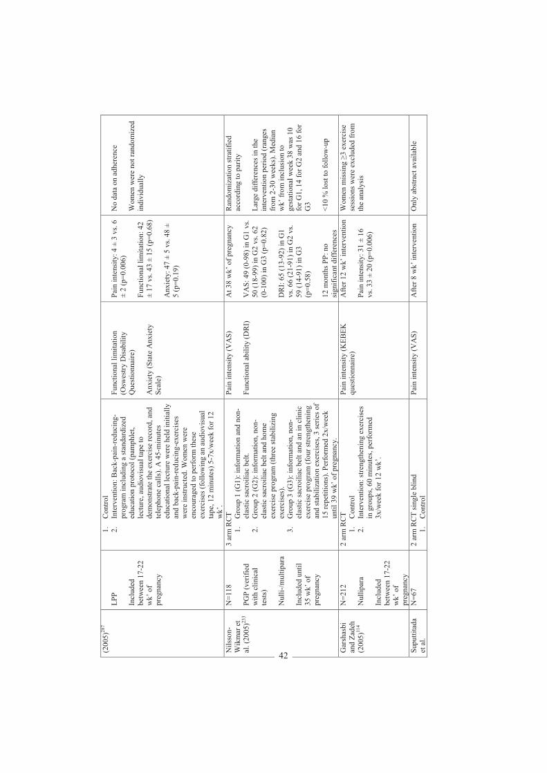
(200
5)28
7 LP
P In
clud
ed
betw
een
17-2
2 w
k’ o
f pr
egna
ncy
1.
Con
trol
2.
Inte
rven
tion:
Bac
k-pa
in-r
educ
ing-
prog
ram
incl
udin
g a
stan
dard
ized
ed
ucat
ion
prot
ocol
(pam
phle
t, le
ctur
e, a
udio
visu
al ta
pe to
de
mon
stra
te th
e ex
erci
se re
cord
, and
te
leph
one
calls
). A
45-
min
utes
ed
ucat
iona
l lec
ture
wer
e he
ld in
itial
ly
and
back
-pai
n-re
duci
ng-e
xerc
ises
w
ere
inst
ruct
ed. W
omen
wer
e en
cour
aged
to p
erfo
rm th
ese
exer
cise
s (fo
llow
ing
an a
udio
visu
al
tape
, 12
min
utes
) 5-7
x/w
eek
for 1
2 w
k’.
Func
tiona
l lim
itatio
n (O
swes
try D
isab
ility
Q
uest
ionn
aire
) A
nxie
ty (S
tat e
Anx
iety
Sc
ale)
Pain
inte
nsity
: 4 ±
3 v
s. 6
± 2
(p=0
.006
) Fu
nctio
nal l
imita
tion:
42
± 17
vs.
43 ±
15
(p=0
.68)
A
nxie
ty: 4
7 ±
5 vs
. 48
± 5
(p=0
.19)
No
data
on
adhe
renc
e W
omen
wer
e no
t ran
dom
ized
in
divi
dual
ly
Nils
son-
Wik
mar
et
al. (
2005
)233
N=1
18
PGP
(ver
ified
w
ith c
linic
al
test
s)
Nul
li-/m
ultip
ara
Incl
u ded
unt
il 35
wk’
of
preg
nanc
y
3 ar
m R
CT
1.
Gro
up 1
(G1)
: inf
orm
atio
n an
d no
n-el
astic
sacr
oilia
c be
lt.
2.
Gro
up 2
(G2)
: inf
orm
atio
n, n
on-
elas
tic sa
croi
liac
belt
and
hom
e ex
erci
se p
rogr
am (t
hree
stab
ilizi
ng
exer
cise
s).
3.
Gro
up 3
(G3)
: inf
orm
atio
n, n
on-
elas
tic sa
croi
liac
belt
and
an in
clin
ic
exer
cise
pro
gram
(fou
r stre
ngth
enin
g an
d st
abili
zatio
n ex
erci
ses,
3 se
ries o
f 15
repe
titio
ns).
Perf
orm
ed 2
x/w
eek
until
39
wk’
of p
regn
ancy
.
Pain
inte
nsity
(VA
S)
Func
tiona
l abi
lity
(DR
I)
At 3
8 w
k’ o
f pre
gnan
cy
VA
S: 4
9 (0
-98)
in G
1 vs
. 50
(18-
99) i
n G
2 vs
. 62
(0-1
00) i
n G
3 (p
=0.8
2)
DR
I: 65
(13 -
92) i
n G
1 vs
. 66
(21-
91) i
n G
2 vs
. 59
(14 -
91) i
n G
3 (p
=0.5
8)
12 m
onth
s PP:
no
sign
ifica
nt d
iffer
ence
s
Ran
dom
izat
ion
stra
tifie
d ac
cord
ing
to p
arity
La
rge
diffe
renc
es in
the
inte
rven
tion
perio
d (r
ange
s fr
om 2
-30
wee
ks).
Med
ian
wk’
from
incl
usio
n to
ge
stat
iona
l wee
k 38
was
10
for G
1, 1
4 fo
r G2
and
16 fo
r G
3 <1
0 %
lost
to fo
ll ow
-up
Gar
shas
bi
and
Zade
h (2
005)
114
N=2
12
Nul
lipar
a In
clud
ed
betw
een
17-2
2 w
k’ o
f pr
egna
ncy
2 ar
m R
CT
1.
Con
trol
2.
Inte
rven
tion:
stre
ngth
enin
g e x
erci
ses
in g
roup
s, 60
min
utes
, per
form
ed
3x/w
eek
for 1
2 w
k’.
Pain
inte
nsity
(KEB
EK
ques
tionn
aire
)
Afte
r 12
wk’
inte
rven
tion
Pain
inte
nsity
: 31
± 16
vs
. 33
± 20
(p=0
.006
)
Wom
en m
issi
ng
3 ex
erci
se
sess
ions
wer
e ex
clud
ed fr
om
the
anal
ysis
Supu
ttita
da
et a
l. N
=67
2
arm
RC
T si
ngle
blin
d 1.
C
ontro
l Pa
in in
tens
ity (V
AS)
A
fter 8
wk’
inte
rven
tion
O
nly
abst
ract
ava
ilabl
e
42
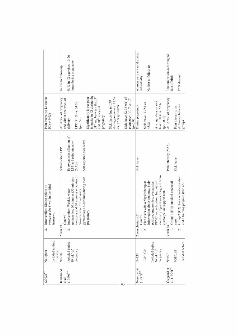
(200
2)30
2 N
ullip
ara
Incl
uded
in th
ird
trim
este
r
2.
Inte
rven
tion:
Sitt
ing
pelv
ic ti
lt ex
erci
ses f
or 8
wk’
in th
e th
ird
trim
este
r.
Pain
inte
nsity
: Low
er in
IG
(p<0
.05)
Kih
lstra
nd
et a
l. (1
999)
164
N=2
58
Incl
uded
bef
ore
19 w
k’ o
f pr
egna
ncy
2 ar
m R
CT
1.
Con
trol
2.
Inte
rven
tion:
Wee
kly
wat
er
gym
nast
ics,
60 m
inut
es (3
0 m
inut
es
exer
cise
s and
30
min
utes
rela
xatio
n).
Wom
en w
ere
offe
red
wat
er
gym
nast
ics 1
7-20
tim
es d
urin
g th
eir
preg
nanc
y.
Self-
repo
rted
LPP
Ever
yday
cla
ssifi
catio
n of
LP
P an
d p a
in in
tens
ity
(VA
S)
Self-
repo
rted
sick
leav
e
At 3
4 w
k’ o
f pre
gnan
cy
and
with
in o
ne w
eek
of
deliv
ery
LPP:
71
% v
s. 74
%
(p=0
.57)
Si
gnifi
cant
ly lo
wer
pai
n in
tens
ity in
IG d
urin
g th
e 31
st a
nd b
etw
een
the
33rd
an
d 38
th w
eeks
of
preg
nanc
y Si
ck le
ave
due
to L
PP
durin
g pr
egna
ncy:
13
%
vs. 2
2 %
(p=0
.09)
Si
ck le
ave
32-3
3 w
k’ o
f pr
egna
ncy
(n) :
7 vs
. 17
(p=0
.03)
14 lo
st to
follo
w-u
p 88
% in
IG e
xerc
ised
10-
20
times
dur
ing
preg
nanc
y
Nor
én e
t at.
(199
7)23
4 N
=135
LB
P/PG
P In
clud
ed b
efor
e 36
wk’
of
preg
nanc
y
2 ar
m c
lust
er-R
CT
1.
Con
trol
2.
Five
vis
its w
ith a
phy
siot
hera
pist
. In
form
atio
n ab
out a
nato
my,
bod
y po
stur
e, e
rgon
omic
s, gy
mna
stic
s, PF
MT
and
rela
xatio
n. In
divi
dual
ex
erci
se p
rogr
am w
as d
esig
ned.
Non
-el
astic
pel
vic
supp
ort b
elt.
Sick
leav
e D
urin
g pr
egna
ncy
Sick
leav
e: 3
3/54
vs.
45/8
1 A
vera
ge d
ays o
n si
ck
leav
e: 3
0.4
vs. 5
3.6
(p<0
.001
)
Wom
en w
ere
not r
ando
miz
ed
indi
vidu
ally
N
o lo
st to
follo
w-u
p
Öst
gaar
d et
al
. (19
94)35
0 N
=407
PG
P/LB
P In
clud
ed b
efor
e
3 ar
m R
CT
1.
Gro
up 1
(G1)
: sta
ndar
d an
tena
tal
care
. 2.
G
roup
2 (G
2): b
ack
scho
ol e
duca
tion
and
a tra
inin
g pr
ogra
m (t
wo
45-
Pain
inte
nsity
(VA
S)
Sick
leav
e
At 3
6 w
k’ o
f pre
gnan
cy
Pain
inte
nsity
: no
diff e
renc
e be
twee
n gr
oups
Ran
dom
izat
ion
acco
rdin
g to
da
te o
f birt
h 17
% d
ropo
ut
43

18 w
k’ o
f pr
egna
ncy
min
utes
cla
sses
in g
roup
s) b
efor
e 20
w
k’ o
f pre
gnan
cy. T
he p
rogr
am
incl
uded
info
rmat
ion
on a
nato
my,
po
stur
e, e
rgon
omic
s, m
uscl
e tra
inin
g an
d re
laxa
tion.
3.
G
roup
(G3)
: bac
k sc
hool
edu
catio
n,
an in
divi
dual
ized
exe
rcis
e pr
ogra
m
(fiv
e 30
-min
utes
less
ons b
etw
een
18-
32 w
k’ o
f pre
gnan
cy) a
nd a
writ
ten
hom
e ex
erci
ses p
rogr
am to
be
perf
orm
ed 3
x/w
eek.
The
pro
gram
in
clud
ed in
form
atio
n on
ana
tom
y,
post
ure,
erg
onom
ics,
mus
cle
train
ing
and
rela
xatio
n.
Sick
leav
e: 5
0 %
in G
1 vs
. 52
% in
G2
vs. 3
7 %
in
G3
(p<0
.05)
Per p
roto
col a
naly
sis
PubM
ed se
arch
term
: ("P
elvi
c Pa
in"[
Mes
h] O
R "B
ack
Pain
"[M
esh]
OR
"Pe
lvic
Pai
n"[ti
ab] O
R "
Bac
k Pa
in"[
tiab]
OR
"Pel
vic
Gird
le P
ain"
[tiab
])
AN
D ("
Preg
nanc
y"[M
esh]
OR
"Pre
gnan
cy C
ompl
icat
ions
"[M
esh:
NoE
xp] O
R p
regn
an*[
tiab]
) AN
D ("
Exer
cise
"[M
esh]
OR
"Exe
rcis
e Th
erap
y"[M
esh]
OR
"Exe
rcis
e M
ovem
ent T
echn
ique
s"[M
esh]
OR
"Phy
sica
l Fitn
ess"
[Mes
h] O
R "P
hysi
cal E
xerti
on"[
Mes
h] O
R "M
otor
A
ctiv
ity"[
Mes
h] O
R "S
ports
"[M
esh]
OR
exe
rcis
*[tia
b] O
R tr
aini
ng[ti
ab] O
R ("
phys
ical
act
ivity
"[tia
b] N
OT
med
line[
sb])
) AN
D (c
linic
al tr
ial[p
t] O
R ra
ndom
*[tia
b] O
R tr
ial[t
iab]
OR
gro
up[ti
ab] O
R g
roup
s[tia
b] O
R sy
stem
atic
[sb]
) NO
T ((
Ani
mal
s[m
esh]
NO
T H
uman
s[m
esh]
) OR
Cas
e re
ports
[pt])
C
G d
enot
es c
ontro
l gro
up, D
RI D
isab
ility
Rat
ing
Inde
x, IG
inte
rven
tion
grou
p, L
BP
low
bac
k pa
in, L
PP lu
mbo
pelv
ic p
ain,
PFM
T pe
lvic
floo
r m
uscl
e tra
inin
g, P
4 Po
ster
ior P
elvi
c Pa
in P
rovo
catio
n te
st, P
GP
pelv
ic g
irdle
pai
n, P
P po
stpa
rtum
, VA
S vi
sual
ana
logu
e sc
ale.
44
Incontinence
Definition
Urinary incontinence (UI) is defined as “complaint of involuntary loss of urine”, while the
term anal incontinence (AI) is defined as “complaint of involuntary loss of feces or flatus”.144
AI can further be divided into fecal incontinence (FI; “complaint of involuntary loss of
feces”) and flatal incontinence (“complaint of involuntary loss of flatus”).144
Prevalence of UI
UI is a common condition among women.213 In a Norwegian cohort study, EPINCONT
(N=15,307), the overall prevalence of UI in nulliparous women were 10 %, and the
prevalence increases with age.272 The prevalence of urinary leakage in pregnancy has been
reported to vary between 9 % and 74 %.64,261 This variation is probably due to different study
populations (various proportions of nulliparous/parous women), different definitions of
incontinence (self-reports or standardized pad tests), different pregnancy lengths, and
different definitions. The International Continence Society has changed the definition several
times.3,4,144 In the first standardization of terminology of lower urinary tract function, UI was
defined as “involuntary loss of urine which is objectively demonstrable and a social or
hygienic problem”.3 As the definition changes, so will the prevalence estimate. Chaliha et
al.64 (N=286) reported a prevalence of 9 % with genuine stress urinary incontinence (SUI) in
gestational week 34 measured with urodynamics. Raza-Khan et al.261 (N=113) included
women in the third trimester and registered any type of UI in a questionnaire and reported a
prevalence of 74 %.
In the MoBa study including 43,279 women from 1999-2006, 58 % of the women reported
any UI and 35 % reported UI weekly or more in pregnancy week 30.334 The prevalence of UI
increased from 26 % before pregnancy to 58 % in pregnancy week 30. The prevalence among
nulliparous and parous women was 15 % and 35 % before pregnancy and 48 % and 67 % at
30 weeks.334 A cohort of nulliparous women (N=1,128), who were continent before
pregnancy, reported an UI incidence of 39 % at some point during pregnancy.292 Another
observational study of primiparous women (N=1,501) found that 4 % reported UI before
45
pregnancy and 38 % during pregnancy.187 In both studies the incidence of UI was low in the
first trimester, rising rapidly in the second trimester and continued to rise more slowly in the
third trimester.187,292 In a third cohort of nulliparous women (N=1,507), 11 % reported UI at
least once per month before pregnancy, and the prevalence increased to 17 % in early
gestation and 56 % in the third trimester of pregnancy.44 In all three studies the severity
increased slightly with increasing gestation.44,187,292 A correlation between severity of UI and
level of interference in daily living was found.292
The increase in UI during pregnancy is mainly due to incidence of SUI.187,292 The prevalence
of pure SUI is reported to be 1.7-8 times higher than the prevalence of pure urgency urinary
incontinence (UUI) in pregnancy. Prevalence of mixed UI is reported to be 0.2-2.2 times of
the prevalence of pure SUI in pregnancy.44,118,187,261
In a systematic review including 33 studies on UI postpartum, a pooled prevalence of any UI
postpartum was found to be 33 %, with weekly UI to be 12 % and daily UI 3 %.310 There
were only small changes in the prevalence of UI over the first year postpartum.310
Risk factors for UI
Data suggest that pregnancy contributes to pelvic floor dysfunction later in life.194 The precise
etiology of UI in pregnancy is unknown, but there is a variety of changes associated with
pregnancy, e.g. hormonal and mechanical changes that could contribute.154 Prenatal
physiological changes such as increases in intra-abdominal pressure of the growing uterus and
fetal weight on the PFM throughout pregnancy together with pregnancy-related hormonal
changes may lead to reduced strength and reduced supportive and sphincter functions of the
PFM.148
Incontinence during pregnancy has been linked to age,274 BMI,131 strenuous physical
exercise,102 smoking history,131 and a family history of UI.292 In the EPINCONT study
Rørtveit et al.274 found that women having their first baby after the age of 25 had higher
prevalence of UI than women having their first baby before the age of 25 (28 % versus 23 %,
p<0.01). However, the effect attenuated with increasing actual age, and it disappeared in the
age group 50 to 64 years.274
46
Developing de novo SUI during pregnancy was associated with BMI >30.187 In the MoBa
study weight gain in pregnancy does not seem to be a clinically important risk factor for UI.
However, weight loss postpartum may be important for regaining continence 6 months
postpartum.333 Brown et al.44 found that women with subclinical urinary symptoms before
pregnancy, younger women (18-24 years), older women ( 40 years), and those with a BMI
30 were at an increased risk of developing de novo UI in pregnancy. The factor most
strongly associated with the onset of UI during pregnancy was the presence of occasional
leakage of urine before pregnancy.44 Solans-Domenech et al.292 found that the risk of UI
increased in pregnant women >35 years, with overweight or obesity at baseline and a family
history of UI. Hannestad et al.130 found that women were more likely to develop UI if their
mother or older sisters were incontinent, suggesting that a genetic predisposition plays a role
in the development of UI.
Antenatal UI is found to be a risk factor for UI postpartum, and many authors have found that
UI in pregnancy is an important predictor for UI later in life.48,91,110,155,286,319,324,332 A history
of UI prior to pregnancy significantly increases the chance that postpartum UI will
persist.104,294 In a cohort study it was found that most women experiencing postpartum UI had
symptoms of UI during the third trimester.261 Another study found that five years after the
first delivery, 33 % of women reported UI.323 In a twelve year prospective study, Viktrup et
al.324 found that in women with onset of UI in pregnancy or shortly after their first delivery
had increased risk of long-lasting symptoms. Among women who were continent during their
first pregnancy and the postpartum period, the prevalence of UI 12 years after the first
delivery was 33 % compared to 66 % in women who became incontinent during their first
pregnancy and the postpartum period.325
In the EPINCONT study including 27,936 women, the reported prevalence of any UI was 25
% in the adult population, with the prevalence of incontinence increasing with age.132 In the
Women’s Health Australia Project, including three age cohorts of women completed in 1996,
women were asked if they had experienced leaking urine last year. The estimated prevalence
of UI in young women (18-23 years, N=14,000) was 13 %, middle-aged women (45-50 years,
N=13,738) 37 % and old women (70-75 years, N=12,417) 35 %.71 Middle-aged and older
nulliparous women were almost three times more likely to report UI than nulliparous young
women. UI was significantly associated with parity, constipation, obesity, past gynecological
47
surgery and conditions which can impact on bladder control, such as coughing and sneezing
on a regular basis.71 In twin studies it has been found that women (mean age 47 years) with at
least two births are four times more likely to report UI than their nulliparous twin sisters.117
Among premenopausal women, parous women have higher prevalence of UI than nulliparous
women.273 Among postmenopausal women, a history of pregnancy and childbirth has little
impact on the prevalence of UI. Old nulliparous women are as likely to have UI as parous
women.43,273 It is assumed that other factors, such a comorbid medical conditions and age-
related changes, outweigh the effect of previous pregnancies.235 The numbers of striated
muscle fibers of the urethral sphincter decrease with age and parity, with an average loss of 2
% per year from 15 to 80 years of age.19 In an epidemiological study it was found that 50 %
of incontinence can be attributed to pregnancy and childbirth among parous women.248
However, there are many unanswered questions, and the available literature cannot
distinguish the effects of pregnancy from the effects of childbirth.
High BMI and the presence of antenatal UI are modifiable risk factors. The presence of these
risk factors may help clinicians target at-risk women for early intervention.
Prevalence of AI
In a review of 16 studies, the estimated prevalence of AI varied from 2-24 %, and the
estimated prevalence of FI varied from 0.4-18 % in community-dwelling adults.195 AI is less
studied in pregnancy, but the prevalence of AI has been found to increase from pre-pregnancy
to late pregnancy.65,66,292 Prevalence rates for AI before, during, and after the first pregnancy
of 0-1 %, 0-8 %, and 2-26 % have been reported.65,126,191,192,284,301,306,318,346 The presence of AI
is mainly characterized by loss of flatus in more than 90 % of cases.292
The prevalence of AI postpartum has been reported to be 9 % - 27 %.66,101,113,288,301
Risk factors of AI
Pregnancy and delivery are risk factors for AI in later life,47 and the first pregnancy and
delivery is associated with a high prevalence of objective and subjective bowel dysfunction.66
Age and excess weight gain during pregnancy are associated with the occurrence of AI during
48
pregnancy,292 however, risk factors for development of AI during pregnancy have been
sparsely studied.
In twin studies, it has been found that women (mean age 47 years) with at least two births are
three times more likely to report AI than their nulliparous twin sisters.2 Obstetric trauma is
one of the major causes of AI in women. During delivery a sphincter trauma and/or pudendal
neuropathy can occur. Risk factors for sphincter injury are vaginal birth, midline episiotomy,
forceps delivery and vacuum-assisted delivery.101 A review including 18 studies found that
symptoms of AI in the first year postpartum were associated with the mode of delivery.258
Women with spontaneous delivery had increased risk of AI postpartum compared to cesarean
delivery (OR 1.32, 95 % CI 1.04-1.68). In forceps deliveries, women had doubled risk of AI
compared to cesarean delivery (OR 2.01, 95 % CI 1.47-2.74).258 Solans-Domenech et al.292
identified AI during pregnancy and vaginal delivery as risk factors for AI postpartum. Women
with FI de novo postpartum are more likely to have had a third or fourth degree anal sphincter
rupture (OR 8.0, 95 % CI 4.6-13.8).318 A systematic review including 31 longitudinal studies
found that third- or fourth-degree sphincter rupture was the only etiological factor strongly
associated with postpartum AI.38 A meta-analysis included five studies with a total of 717
women who underwent an endoanal ultrasonography to diagnose anal sphincter defects
following a vaginal delivery.237 The study reports an incidence of 27 % anal sphincter defects
in primiparous women and an incidence of 9 % new sphincter defects in multiparous women.
Overall, 30 % of anal sphincter tears had symptoms, and 3 % of women experienced
postpartum FI without anal sphincter defect.237
Incontinence and quality of life
In one prospective study (N=597), 42 % and 19 % reported SUI and UUI in gestational week
36, of those only 6 % and 3 % reported it to be causing bother.320 Although UI is common, it
reduces quality of life and affects social, physical, occupational and leisure activities.156
AI is less prevalent than UI, but this condition is clearly a social and psychological burden
with a significant impact on quality of life.108,129,150,189 FI further reduces quality of life in
women with UI,108 and FI and UI together have a greater impact on quality of life than either
condition alone.129 Women affected by moderate or severe FI may suffer embarrassment,
49
social isolation, curtailed activities, and severely impaired quality of life.31 Despite the
negative effect on quality of life, few women discuss AI with their medical providers.189 In a
cohort study (N=906) women were asked about FI symptoms 10 months after birth, and 36 (4
%) reported new FI after birth. In all, 27 (75 %) of 36 women had symptoms several times a
week, however only five consulted a doctor.191 FI symptoms are known to be
underreported.186,191 The reason may be embarrassment, but could also be little knowledge
about treatment.
Anatomy of the pelvic floor
The pelvic floor (PF) is an important part of the continence mechanisms.19 The PF consists of
several components between the pelvic peritoneum and the vulvar skin. These are (from
above downwards) the peritoneum, viscera and endopelvic fascia, levator ani muscles,
perineal membrane, and external genital muscles.86 The pelvic organs (vagina, bladder and
rectum) are ensured by fascias and ligaments.
The levator ani group of muscles (Figure 3A and 3B) keeps the urogenital hiatus closed.86 The
different muscles have different fiber directions, and if each muscle could contract in
isolation, they would all have different functions. However, the only known voluntary
function of the PFM is a mass contraction best described as an inward lift and squeeze around
the urethra, vagina and rectum.57 The levator ani muscles have constant activity, like that of
other postural muscles.86 In addition, the PFM close the PF and carry the weight of the
abdominal and pelvic organs, preventing constant strain on the ligaments.86 As long as the
levator ani muscles function properly, the PF is closed and the ligaments and fascia are under
no tension; the fasciae simply act to stabilize the organs in their position above the levator ani
muscles. In continent women an automatic (unconscious) co-contraction occurs when
abdominal pressure increases.50 The amount of striated muscle declines considerable with
age251 and is associated with a decline in innervating nerves.245
50
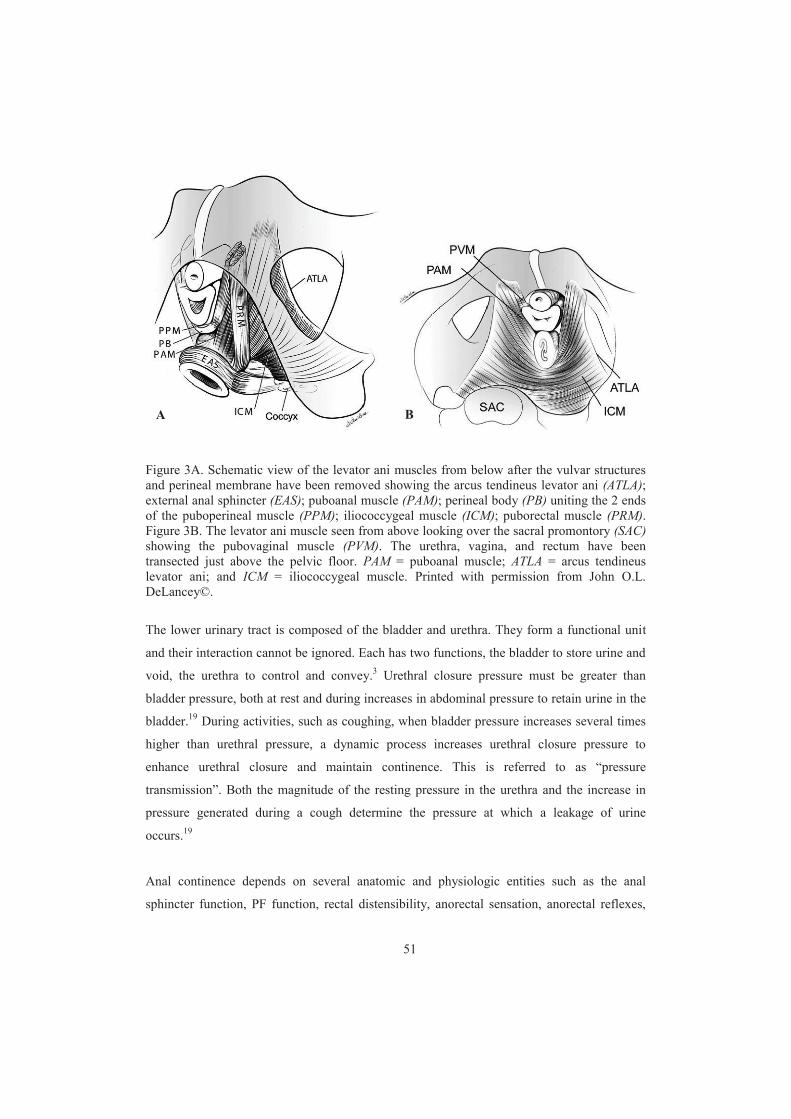
Figure 3A. Schematic view of the levator ani muscles from below after the vulvar structures and perineal membrane have been removed showing the arcus tendineus levator ani (ATLA); external anal sphincter (EAS); puboanal muscle (PAM); perineal body (PB) uniting the 2 ends of the puboperineal muscle (PPM); iliococcygeal muscle (ICM); puborectal muscle (PRM). Figure 3B. The levator ani muscle seen from above looking over the sacral promontory (SAC) showing the pubovaginal muscle (PVM). The urethra, vagina, and rectum have been transected just above the pelvic floor. PAM = puboanal muscle; ATLA = arcus tendineus levator ani; and ICM = iliococcygeal muscle. Printed with permission from John O.L. DeLancey©.
The lower urinary tract is composed of the bladder and urethra. They form a functional unit
and their interaction cannot be ignored. Each has two functions, the bladder to store urine and
void, the urethra to control and convey.3 Urethral closure pressure must be greater than
bladder pressure, both at rest and during increases in abdominal pressure to retain urine in the
bladder.19 During activities, such as coughing, when bladder pressure increases several times
higher than urethral pressure, a dynamic process increases urethral closure pressure to
enhance urethral closure and maintain continence. This is referred to as “pressure
transmission”. Both the magnitude of the resting pressure in the urethra and the increase in
pressure generated during a cough determine the pressure at which a leakage of urine
occurs.19
Anal continence depends on several anatomic and physiologic entities such as the anal
sphincter function, PF function, rectal distensibility, anorectal sensation, anorectal reflexes,
A B
51
intact nervous system, mental function, stool volume, stool consistency, and colonic transit.
Deficiency in one or more of these factors can lead to AI.196 The anal sphincter muscles are
located in the distal part of the anal canal. The anal sphincter involves the internal anal
sphincter, external anal sphincter and puborectalis muscle.196
In the non-pregnant women, the uterus is an almost solid structure weighing about 70 g and
with a cavity of 10 ml or less. During pregnancy, the uterus is transformed to accommodate
the fetus, placenta, and amnionic fluid. The total volume of the contents at term averages
about 5 l, but may be up to 20 l or more. By the end of pregnancy, the uterus has achieved a
capacity that is 500 to 1000 times greater than in the non-pregnant state.80 In late gestation,
the fetal head markedly alters bladder geometry.90
Pregnancy and delivery are associated with functional and anatomical changes in the muscles,
nerves, and connective tissue of the PFM.128 In women with de novo SUI after their first birth,
injury to the levator ani muscle is found twice as often as in women who are continent after
delivery.87 The first vaginal delivery also results in changes in EMG patterns of striated
sphincter musculature that is consistent with impaired muscular function persisting for at least
six months beyond delivery.331 Deindl et al.85 compared parous women with SUI (n=8) and
nulliparous continent women (n=10) with EMG-measures of the PFM. The muscle activation
pattern was in principle similar except for lower holding time in a voluntary pelvic floor
muscle contraction (VPFMC) and asymmetric and uncoordinated activity pattern in the
levator ani muscles among parous incontinent women.85 Mørkved et al.227 (N=103) assessed
muscle strength and thickness in nulliparous pregnant women at 20 weeks of gestation, and
found higher maximal vaginal squeeze pressure and increment in muscle thickness in
continent compared to incontinent women.
Pelvic floor muscle training
Pelvic floor muscle training (PFMT) for the treatment of UI was popularized by the American
gynecologist Arnold Kegel in 1948.162 However, PFMT has been a part of exercise programs
in Chinese Taoism for over 6000 years.99 PFMT has been found to have a high cure rate in the
adult female population and is recommended as first line treatment for women with SUI, UUI
or mixed incontinence.99 There are two hypothesis explaining the effect of PFMT; behavior
52
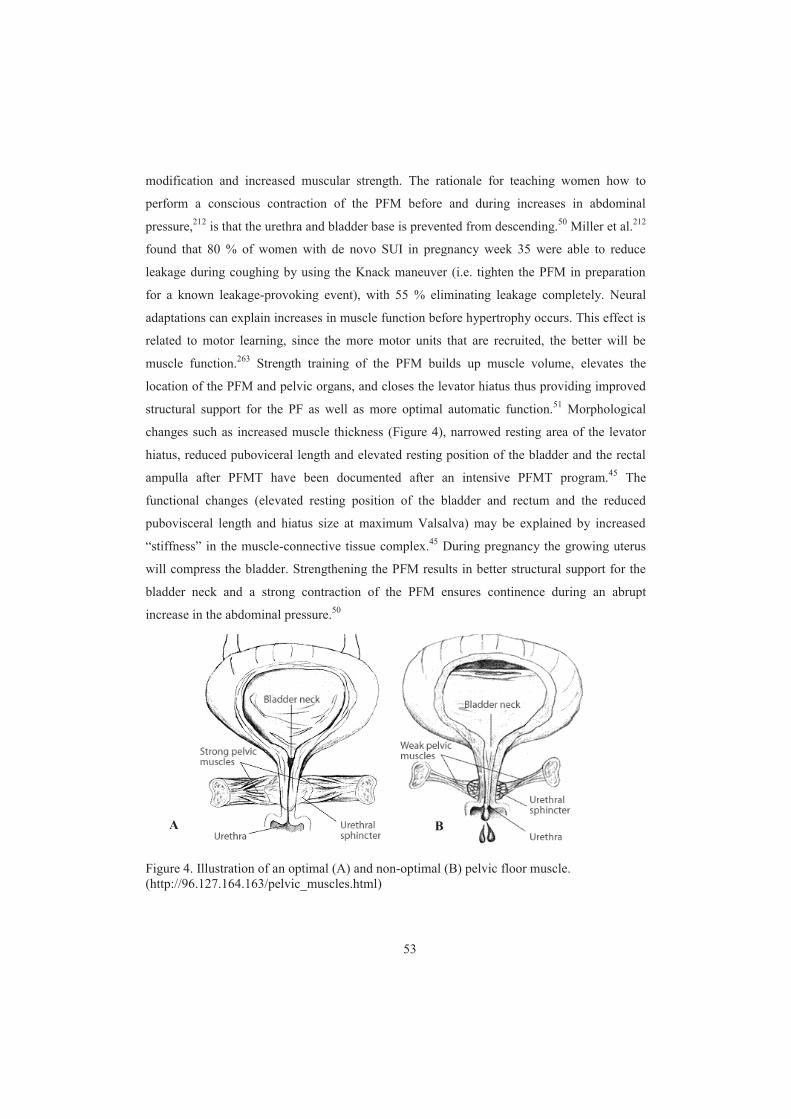
modification and increased muscular strength. The rationale for teaching women how to
perform a conscious contraction of the PFM before and during increases in abdominal
pressure,212 is that the urethra and bladder base is prevented from descending.50 Miller et al.212
found that 80 % of women with de novo SUI in pregnancy week 35 were able to reduce
leakage during coughing by using the Knack maneuver (i.e. tighten the PFM in preparation
for a known leakage-provoking event), with 55 % eliminating leakage completely. Neural
adaptations can explain increases in muscle function before hypertrophy occurs. This effect is
related to motor learning, since the more motor units that are recruited, the better will be
muscle function.263 Strength training of the PFM builds up muscle volume, elevates the
location of the PFM and pelvic organs, and closes the levator hiatus thus providing improved
structural support for the PF as well as more optimal automatic function.51 Morphological
changes such as increased muscle thickness (Figure 4), narrowed resting area of the levator
hiatus, reduced puboviceral length and elevated resting position of the bladder and the rectal
ampulla after PFMT have been documented after an intensive PFMT program.45 The
functional changes (elevated resting position of the bladder and rectum and the reduced
pubovisceral length and hiatus size at maximum Valsalva) may be explained by increased
“stiffness” in the muscle-connective tissue complex.45 During pregnancy the growing uterus
will compress the bladder. Strengthening the PFM results in better structural support for the
bladder neck and a strong contraction of the PFM ensures continence during an abrupt
increase in the abdominal pressure.50
Figure 4. Illustration of an optimal (A) and non-optimal (B) pelvic floor muscle. (http://96.127.164.163/pelvic_muscles.html)
A B
53
The influence of PFMT on UI and AI
A variety of hypotheses have been suggested about why PFMT might help prevent and treat
incontinence in pregnancy and after delivery. A trained muscle might be less prone to injury,
and previously trained muscles might be easier to retrain after damage as the appropriate
motor patterns are already learned. It may be that previously trained muscle has a greater
reserve of strength so that injury to the muscle itself, or its nerve supply, does not cause
sufficient loss of muscle function to reach the threshold where reduced urethral pressure
results in leakage.141 During pregnancy, training the PFM might help to counteract the
increased intra-abdominal pressure caused by the growing fetus, the hormonally mediated
reduction in urethral pressure, and the increased laxity of fascia and ligament in the pelvic
area. A similar rationale might be used to support the use of PFMT to improve the function of
the external anal sphincter and thus prevent AI.141
In a systematic literature search on PubMed (Table 4) three previous trials were found
addressing primary prevention of UI during pregnancy.119,200,262 Four trials included both
continent and incontinent women in pregnancy.54,171,226,279 Three trials have assessed the
effect of treatment of UI during pregnancy.92,282,340 No previously published trial addressing
primary prevention of AI was found. One previous trial included PFMT in a general fitness
program for pregnant women.54 This trial demonstrated that there was no difference in UI or
AI between intervention and control groups. However, this trial had some limitations such as
being underpowered, including no control of the women’s ability to perform a correct
VPFMC, high dropout rates and low adherence to the exercise training protocol.54
Common for all trials with a positive effect of PFMT are that they are based on thorough
clinical assessment of the participants’ ability to contract the PFM correctly, had close follow-
up of the pregnant women either in groups or individually, and had high adherence to the
exercise protocol. In contrast, trials with no effect had an inadequate training dosage, left the
patients alone to train or had low adherence to the exercise protocol. As for all other strength
training regimens, the effects of PFMT are dependent on the training dosage and adherence to
training. Type of exercise, frequency, intensity and duration of the training in addition to the
adherence to the exercise protocol will decide the effect size.55,136 The concept of intensive
PFMT was introduced by Bø et al.55 The recommendations for effective strength training to
54
increase muscle cross-sectional area and strength are 3 sets of 8-12 close to maximum
contractions 3-4 times a week.136
The success of training depends on the ability to effectively contract the PFM. It is estimated
that 30 % of women are unable to contract the PFM on the first attempt.46,56 Bump et al.46
found that only 49 % performed an ideal VPFMC after brief standardized verbal instruction,
and Dinc et al.92 found that only 68 % of pregnant women were able to perform a correct
VPMFC one week after individual thorough instruction. Vaginal palpation is most commonly
used by physiotherapist to evaluate the function and strength of the PFM, however ultrasound
is a more objective measure and is becoming an important clinical tool.58
RCT’s have documented the effects of intensive PFMT and close follow-up of
physiotherapists in the prevention and treatment of pregnancy-related UI.92,119,171,226,262,279,282
A Cochrane review addressing the prevention of urinary- and fecal incontinence included
mixed prevention and treatment trials.141 This review concluded that continent women
randomized to intensive PFMT were 56 % less likely to report UI in late pregnancy and about
30 % up to six months postpartum.141 Nevertheless, the Cochrane review and two other
systematic reviews conclude that the effect of PFMT during pregnancy to prevent UI and AI
is still open to question.42,141,225 The Cochrane review strongly recommends that all future
trials of PFMT during pregnancy should collect data on AI, and highlights the need for large,
pragmatic trials with population-based approaches, using intensive PFMT and recruiting
antenatal women regardless of continence status or parity.141 There is a lack of trials
investigating the effect of implementing PFMT in a more general training program for
pregnant women. The cost of illness for incontinence is a substantial economic and human
burden, highlighting the need for effective forms of management.213 The importance of
effective prevention is obvious.
55

Tabl
e 4.
Ran
dom
ized
con
trolle
d tri
als a
sses
sing
the
effe
ct o
f pel
vic
floor
mus
cle
train
ing
durin
g pr
egna
ncy
to p
reve
nt a
nd tr
eat i
ncon
tinen
ce
incl
udin
g bo
th w
omen
with
and
with
out i
ncon
tinen
ce a
t inc
lusi
on.
Aut
hor
Subj
ects
D
esig
n / I
nter
vent
ion
Out
com
e R
esul
ts (I
G v
s. C
G)
Com
men
ts
Sang
saw
ang
et a
l. (2
011)
282
N=7
0 N
ulli -
/mul
tipar
a SU
I (1
epis
ode
of u
rinar
y le
akag
e la
st
mon
th)
Incl
uded
be
twee
n 20
-30
wk’
of
preg
nanc
y
Qua
si-e
xper
imen
tal s
tudy
, pre
-pos
t tes
t with
co
ntro
l gro
up
1.
Con
trol:
usua
l nur
sing
car
e (in
clud
ing
writ
ten
inst
ruct
ions
in
PFM
T).
2.
Inte
rven
tion:
writ
ten
and
indi
vidu
al
oral
inst
ruct
ion
in c
orre
ct V
PFM
C
(no
vagi
nal p
alpa
tion,
but
in
stru
ctio
ns in
“st
op- te
st”
i.e. t
ryin
g to
stop
or s
low
urin
ary
flow
) and
PF
MT.
PFM
T (4
5 m
inut
es) i
n gr
oups
ev
ery
seco
nd w
eek
for 6
wk’
. Hom
e ex
erci
ses,
with
40
repe
titio
ns d
aily
, 5
x/w
eek
(slo
w c
ontra
ctio
ns w
ith 1
0 se
cond
hol
ding
tim
e an
d 10
fast
co
ntra
ctio
ns a
t the
end
of h
oldi
ng
time)
. Per
form
ing
PFM
T w
as
reco
rded
in a
trai
ning
dia
ry.
SUI s
ever
ity (f
requ
ency
and
am
ount
) SU
I sev
erity
(VA
S)
Afte
r 6 w
k’ in
terv
entio
n:
No
leak
age:
39
% v
s. 0
% (p
=0.0
01)
Freq
uenc
y SU
I: 2
± 4
vs.
1 5 ±
10
(p<0
.001
) Se
verit
y SU
I (V
AS)
: 1.3
±
1.3
vs. 6
.8 ±
2.2
(p
<0.0
01)
Wom
en in
the
IG w
ere
mat
ched
to C
G w
omen
re
gard
ing
age,
par
ity a
nd
seve
rity
of S
UI
Wom
en w
ho fa
iled
to
perf
orm
PFM
C w
ere
cate
goriz
ed a
s dro
pout
4
lost
to fo
llow
-up
in IG
N
o as
sess
men
t of c
orre
ct
VPF
MC
10
0 %
in IG
adh
ered
to th
e ex
erci
se p
roto
col (
28 d
ays
of P
FMT)
Bø
and
Haa
ksta
d (2
011)
54
N=1
05
Nul
lipar
a C
ontin
ent/
inco
ntin
ent
Sede
ntar
y In
clud
ed w
ithin
24
wk’
of
preg
nanc
y
2 ar
m R
CT
1.
Con
trol
2.
Inte
rven
tion:
atte
ndin
g ae
robi
c fit
ness
cla
sses
incl
udin
g PF
MT,
2-
3x/w
eek,
and
per
form
ing
10 d
aily
PF
MC
at h
ome.
The
PFM
T in
stru
cted
in c
lass
es c
onsi
sted
of
thre
e se
ts o
f clo
se to
max
imum
co
ntra
ctio
ns o
f 8- 1
2 re
petit
ions
with
ho
ldin
g pe
riods
of 6
-8 se
cond
s pe
rfor
med
in d
iffer
ent p
ositi
ons.
Wom
en w
ere
enco
urag
ed to
be
phys
ical
ly a
ctiv
e 30
min
utes
dai
ly.
Inte
rven
tion
perio
d w
as 1
2-16
wk’
.
UI a
nd A
I rep
orte
d in
a
pers
onal
inte
rvie
w
At 3
6-38
wk’
of
preg
nanc
y U
I: 17
/42
vs. 1
6/42
(p
=0.8
2)
Flat
us: 1
1/42
vs.
9/16
(p
=0.6
1)
AI:
1/42
vs.
1/42
A
t 6-8
wk’
PP
UI:
12/4
3 vs
. 13/
47
(p=0
.99)
Fl
atus
: 10/
43 v
s. 8/
47
(p=0
.46)
A
I: 1/
43 v
s. 3/
47
(p=0
.62)
No
asse
ssm
ent o
f cor
rect
V
PFM
C
Cla
sses
led
by a
n ae
robi
c in
stru
ctor
, ver
bal i
nstru
ctio
ns
wer
e gi
ven
40 %
of I
G w
omen
atte
nded
at
leas
t 80
% o
f the
exe
rcis
e se
ssio
ns
20 %
wer
e lo
st to
follo
w-u
p N
o da
ta o
n le
vel o
f exe
rcis
e an
d PF
MT
in th
e C
G
Ko
et a
l. N
=300
2
arm
RC
T U
I rep
orte
d in
a p
erso
nal
At 3
6 w
k’ o
f pre
gnan
cy:
Adh
eren
ce (d
efin
ed a
s
56
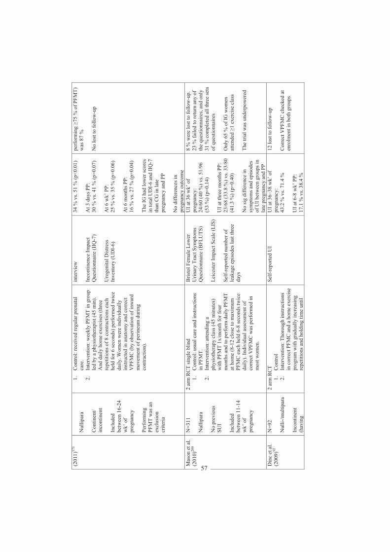
(201
1)17
1 N
ullip
ara
Con
tinen
t/ in
cont
inen
t In
clud
ed
betw
een
16-2
4 w
k’ o
f pr
egna
ncy
Perf
orm
ing
PFM
T w
as a
n ex
clus
ion
crite
ria
1.
Con
trol:
rece
ived
regu
lar p
rena
tal
care
. 2.
In
terv
entio
n: w
eekl
y PF
MT
in g
roup
le
d by
a p
hysi
othe
rapi
st (4
5 m
in).
And
dai
ly h
ome
exer
cise
s (th
ree
repe
titio
ns o
f 8 c
ontra
ctio
ns e
ach
held
for 6
seco
nds)
per
form
ed tw
ice
daily
. Wom
en w
ere
indi
vidu
ally
in
stru
cted
in a
nato
my
and
corr
ect
VPF
MC
(by
obse
rvat
ion
of in
war
d m
ovem
ent o
f per
ineu
m d
urin
g co
ntra
ctio
n).
inte
rvie
w
Inco
ntin
ence
Impa
ct
Que
stio
nnai
re (I
IQ-7
) U
roge
nita
l Dis
tress
In
vent
ory
(UD
I -6)
34 %
vs.
51 %
(p<0
.01)
A
t 3 d
ays P
P:
30 %
vs.
41 %
(p=0
.07)
A
t 6 w
k’ P
P:
25 %
vs.
35 %
(p=0
.06)
A
t 6 m
onth
s PP:
16
% v
s. 27
% (p
=0.0
4)
The
IG h
ad lo
wer
scor
es
in to
tal U
DI-
6 an
d II
Q-7
th
an C
G in
late
pr
egna
ncy
and
PP
No
diffe
renc
es in
pr
egna
ncy
outc
ome
perf
orm
ing
75 %
of P
FMT)
w
as 8
7 %
N
o lo
st to
follo
w-u
p
Mas
on e
t al.
(201
0)20
0 N
=311
N
ullip
ara
No
prev
ious
SU
I In
clud
ed
betw
een
11-1
4 w
k’ o
f pr
egna
ncy
2 ar
m R
CT
sing
le b
lind
1.
Con
trol:
usua
l car
e an
d in
stru
ctio
ns
in P
FMT.
2.
In
terv
entio
n: a
ttend
ing
a ph
ysio
ther
apy
clas
s (45
min
utes
) w
ith P
FMT
1x/m
onth
for f
our
mon
ths a
nd to
per
form
dai
ly P
FMT
at h
ome
(8-1
2 cl
ose
to m
axim
um
PFM
C e
ach
held
6-8
seco
nds t
wic
e da
ily).
Indi
vidu
al a
sses
smen
t of
corr
ect V
PFM
C w
as p
erfo
rmed
in
mos
t wom
en.
Bris
tol F
emal
e Lo
wer
U
rinar
y Tr
act S
ympt
oms
Que
stio
nnai
re (B
FLU
TS)
Leic
este
r Im
pact
Sca
le (L
IS)
Self -
repo
rted
num
ber o
f le
akag
e ep
isod
es la
st th
ree
days
UI a
t 36
wk’
of
preg
nanc
y:
24/6
0 (4
0 %
) vs.
51/9
6 (5
3 %
) (p=
0.14
) U
I at t
hree
mon
ths P
P:
23/6
8 (3
3.8
%) v
s. 33
/80
(41.
3 %
) (p=
0.40
) N
o si
g di
ffer
ence
in
sym
ptom
s and
epi
sode
s of
UI b
etw
een
grou
ps in
la
te p
regn
ancy
and
PP
8 %
wer
e lo
st to
follo
w-u
p.
23 %
faile
d to
retu
rn a
ny o
f th
e qu
estio
nnai
res,
and
only
31
% c
ompl
eted
all
thre
e se
ts
of q
uest
ionn
aire
s O
nly
65 %
of I
G w
omen
at
tend
ed
1 ex
erci
se c
lass
Th
e tri
al w
as u
nder
pow
ered
Din
c et
al.
(200
9)92
N
=92
Nul
li -/m
ultip
ara
Inco
ntin
ent
(hav
ing
2 ar
m R
CT
1.
Con
trol
2.
Inte
rven
tion:
Tho
roug
h in
stru
ctio
ns
in c
orre
ct P
FMC
and
a h
ome
exer
cise
pr
ogra
m w
ith g
radu
ally
incr
easi
ng
repe
titio
ns a
nd h
oldi
ng ti
me
until
Self-
repo
rted
UI
UI a
t 36–
38 w
k’ o
f pr
egna
ncy:
43
.2 %
vs.
71.4
%
UI a
t 6-8
wk’
PP:
17
.1 %
vs.
38.4
%
12 lo
st to
follo
w-u
p C
orre
ct V
PFM
C c
heck
ed a
t en
rolm
ent i
n bo
th g
roup
s
57

com
plai
nts o
f st
ress
/mix
ed U
I in
thei
r his
tory
) In
clud
ed
betw
een
20-3
4 w
k’ o
f pr
egna
ncy
thre
e se
ts o
f 15
repe
titio
ns w
ith 1
0 se
cond
s hol
ding
tim
e an
d fa
st
cont
ract
ions
add
ed, r
epea
ted
3x/d
ay.
Sig n
ifica
nt d
iffer
ence
in
epis
odes
of U
I, ur
genc
y,
num
ber o
f voi
ds a
nd
amou
nt o
f urin
e in
pad
te
st in
fav o
r of t
he IG
bo
th a
t 36 –
38 w
k’
preg
nanc
y an
d at
6-8
wk’
PP
W
oldr
ingh
et
al.
(200
6)34
0
N=2
64
Inco
ntin
ent (
2 ep
isod
es o
f los
s of
urin
e la
st
mon
th)
Nul
li-/m
ultip
ara
In
clud
ed
betw
een
17-2
0 w
k’ o
f pr
egna
ncy
2 ar
m R
CT
1.
Con
trol:
rout
ine
care
. 2.
In
terv
entio
n: F
our s
essi
ons o
f ind
ivid
ual
PFM
T; th
ree
sess
ions
(with
2 w
eek
inte
rval
) bet
wee
n 23
-30
wk’
of p
regn
ancy
an
d a
four
th se
ssio
n 6
wk’
PP.
Writ
ten
info
rmat
ion
incl
udin
g a
deta
iled
PFM
T pr
ogra
m.
Self-
repo
rted
seve
rity
of U
I In
cont
inen
ce Im
pact
Q
uest
ionn
aire
(IIQ
)
No
diffe
renc
e be
twee
n IG
and
CG
with
resp
ect
to th
e se
verit
y of
UI a
nd
impa
ct o
f UI o
n da
ily li
fe
No
vagi
nal p
alpa
tion
of
VPF
MC
, but
obs
erva
tion
and
palp
atio
n of
the
perin
eal b
ody
50 %
lost
to fo
llow
-up
Low
adh
eren
ce, o
nly
37 %
re
porte
d to
exe
rcis
e al
mos
t ev
ery
day
Gor
bea
Cha
vez
et
al. (
2004
)119
N=7
2 N
ullip
ara
Con
tinen
t
2 ar
m R
CT
1.
Con
trol:
no P
FMT.
2.
In
terv
entio
n: in
divi
dual
PFM
T w
ith
phys
ioth
erap
ist.
10 P
FMC
eac
h he
ld
for 8
seco
nds a
nd fo
llow
ed b
y 3
fast
co
ntra
ctio
ns. C
linic
al a
ppoi
ntm
ents
w
eekl
y fo
r 8 w
k’, t
hen
wee
kly
phon
e ca
lls u
p to
20
wk’
. Bio
feed
back
and
tra
inin
g di
ary.
Indi
vidu
al a
sses
smen
t of
cor
rect
VPF
MC
was
per
form
ed.
Self-
repo
rted
UI
UI a
t 28
wk’
of
preg
nanc
y:
0 %
vs.
17.2
% (p
<0.0
5)
UI a
t 35
wk’
of
preg
nanc
y:
0 %
vs.
47 %
(p<0
.05)
U
I at 6
wk’
PP:
15
% v
s. 47
% (p
<0.0
5)
Onl
y ab
stra
ct a
vaila
ble
Mør
kved
et
al. (
2003
)226
N=3
01
Nul
lipar
a
2 ar
m R
CT
sing
le b
lind
1.
Con
trol:
cust
omar
y in
form
atio
n gi
ven
by th
eir m
idw
ife o
r gen
eral
pr
actit
ione
r. C
orre
ct V
PFM
C
Self-
repo
rted
UI (
wom
en
repo
rting
UI
1/w
eek
wer
e ca
tego
rized
as i
ncon
tinen
t)
UI a
t 36
wk’
of
preg
nanc
y:
32 %
vs.
48 %
(p=0
.07)
Low
dro
pout
rate
, 4 %
A
dher
ence
81
%
58

Con
tinen
t/ in
cont
inen
t In
clud
ed a
t 20
wk’
of
preg
nanc
y
chec
ked
at in
clus
ion.
2.
In
terv
entio
n: 1
2 w
k’ w
eekl
y PF
MT
in g
roup
s led
by
phys
ioth
erap
ist,
with
ad
ditio
nal h
ome
exer
cise
s (10
clo
se
to m
axim
um P
FMC
eac
h he
ld fo
r 6
seco
nds,
3-4
fast
con
tract
ions
add
ed
on th
e la
st 4
exe
rcis
es. R
epea
ted
2x/d
ay).
Inte
rven
tion
perio
d be
twee
n 20
-36
wk’
of p
regn
ancy
. Cor
rect
V
PFM
C c
heck
ed a
t inc
lusi
on.
UI a
t 3 m
onth
s PP:
20
% v
s. 32
% (p
=0.0
2)
Rei
lly e
t al.
(200
2)26
2 N
=268
N
ullip
ara
Con
tinen
t with
in
crea
sed
blad
der n
eck
mob
ility
In
clud
ed a
t 20
wk’
of
preg
nanc
y
2 ar
m R
CT
1.
Con
trol:
rout
ine
ante
nata
l car
e lik
ely
to in
clud
e ve
rbal
inst
ruct
ions
in
PFM
T.
2.
Inte
rven
tion:
Indi
vidu
al P
FMT
with
ph
ysio
ther
apis
t at m
onth
ly in
terv
als
from
20
wk’
of p
regn
ancy
unt
il de
liver
y, w
ith a
dditi
onal
hom
e ex
erci
ses.
3 se
ts o
f 8 c
ontra
ctio
ns
(eac
h he
ld fo
r 6 se
cond
s), r
epea
ted
twic
e da
ily. A
t 34
wk’
of p
regn
ancy
th
e nu
mbe
r of c
ontra
ctio
ns p
er se
t w
as in
crea
sed
from
8 to
12.
Wom
en
wer
e in
stru
cted
to c
ontra
ct th
e PF
M
whe
n co
ughi
ng o
r sne
ezin
g.
Perf
orm
ing
PFM
T w
as re
cord
ed in
a
pers
onal
trai
ning
dia
ry.
Self-
repo
rted
UI
UI a
t 3 m
onth
s PP:
19
% v
s. 33
% (p
=0.0
2)
14 %
lost
to fo
llow
-up
46 %
IG w
omen
per
form
ed
PFM
T 28
day
s
Sam
psel
le e
t al
. (19
98)27
9 N
=72
Nul
lipar
a C
ontin
ent/
inco
ntin
ent
Incl
uded
at 2
0 w
k’ o
f pr
egna
ncy
2 ar
m R
CT
sing
le b
lind
1.
Con
trol:
rout
ine
care
. 2.
In
terv
entio
n: P
FMT
tailo
red
to th
e w
oman
’s in
divi
dual
abi
lity,
with
m
uscl
e id
entif
icat
ion
exer
cise
s pr
eced
ing
stre
ngth
-bui
ldin
g ef
forts
. 30
PFM
C d
aily
at m
axim
um o
r nea
r-m
axim
um in
tens
ity w
as
reco
mm
ende
d fo
r stre
ngth
bui
ldin
g.
Cor
rect
VPF
MC
wer
e ch
ecke
d.
Self-
repo
rted
UI (
resu
lts
repo
rted
as c
hang
e in
mea
n U
I sym
ptom
scor
e)
At 3
5 w
k’ o
f pre
gnan
cy:
Less
UI s
ympt
oms w
ere
seen
in th
e IG
vs.
CG
(p
=0.0
4)
At 6
wk’
PP:
Le
ss U
I sym
ptom
s wer
e se
en in
the
IG v
s. C
G
Adh
eren
ce 8
5 %
St
udy
parti
cipa
nts w
ere
finan
cial
ly c
ompe
nsat
ed,
$150
59
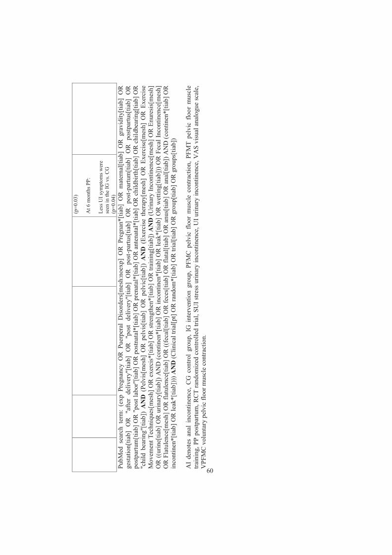
(p
=0.0
3)
At 6
mon
ths P
P:
Less
UI s
ympt
oms w
ere
seen
in th
e IG
vs.
CG
(p
=0.0
4)
PubM
ed s
earc
h te
rm:
(exp
Pre
gnan
cy O
R P
uerp
eral
Dis
orde
rs[m
esh:
noex
p] O
R P
regn
an*[
tiab]
OR
mat
erna
l[tia
b] O
R g
ravi
dity
[tiab
] O
R
gest
atio
n[tia
b] O
R "
afte
r de
liver
y"[ti
ab]
OR
"po
st d
eliv
ery"
[tiab
] O
R p
ost-p
artu
s[tia
b] O
R p
ost-p
artu
m[ti
ab]
OR
pos
tpar
tus[
tiab]
OR
po
stpa
rtum
[tiab
] OR
"po
st la
bor"
[tiab
] OR
pos
tnat
al*[
tiab]
OR
pre
nata
l*[ti
ab] O
R a
nten
atal
*[tia
b] O
R c
hild
birth
[tiab
] OR
chi
ldbe
arin
g[tia
b] O
R
"chi
ld b
earin
g"[ti
ab])
AN
D (
Pelv
is[m
esh]
OR
pel
vis[
tiab]
OR
pel
vic[
tiab]
) A
ND
(Ex
erci
se t
hera
py[m
esh]
OR
Exe
rcis
e[m
esh]
OR
Exe
rcis
e M
ovem
ent T
echn
ique
s[m
esh]
OR
exe
rcis
*[tia
b] O
R s
treng
then
*[tia
b] O
R tr
aini
ng[ti
ab])
AN
D (U
rinar
y In
cont
inen
ce[m
esh]
OR
Enu
resi
s[m
esh]
O
R ((
urin
e[tia
b] O
R u
rinar
y[tia
b]) A
ND
(con
tinen
*[tia
b] O
R in
cont
inen
*[tia
b] O
R le
ak*[
tiab]
OR
wet
ting[
tiab]
)) O
R F
ecal
Inco
ntin
ence
[mes
h]
OR
Fla
tule
nce[
mes
h] O
R fl
atul
ence
[tiab
] OR
((fe
cal[t
iab]
OR
fece
s[tia
b] O
R fl
atal
[tiab
] OR
anu
s[tia
b] O
R a
nal[t
iab]
) AN
D (c
ontin
en*[
tiab]
OR
in
cont
inen
*[tia
b] O
R le
ak*[
tiab]
))) A
ND
(Clin
ical
tria
l[pt]
OR
rand
om*[
tiab]
OR
tria
l[tia
b] O
R g
roup
[tiab
] OR
gro
ups[
tiab]
) A
I de
note
s an
al i
ncon
tinen
ce,
CG
con
trol
grou
p, I
G i
nter
vent
ion
grou
p, P
FMC
pel
vic
floor
mus
cle
cont
ract
ion,
PFM
T pe
lvic
flo
or m
uscl
e tra
inin
g, P
P po
stpa
rtum
, RC
T ra
ndom
ized
con
trolle
d tri
al, S
UI s
tress
urin
ary
inco
ntin
ence
, UI u
rinar
y in
cont
inen
ce, V
AS
visu
al a
nalo
gue
scal
e,
VPF
MC
vol
unta
ry p
elvi
c flo
or m
uscl
e co
ntra
ctio
n.
60
AIMS OF THE THESIS
A general aim of this thesis was to evaluate the efficacy of regular exercise during pregnancy
in the prevention of pregnancy-related diseases and complications during labor.
More specifically, the aims of the separate papers were:
Study I
To assess the efficacy of offering pregnant women a regular exercise program to prevent
GDM and improve insulin resistance.
Study II
To assess if women randomized to a regular exercise program were more likely to report LPP
than women receiving standard antenatal care.
Study III
To assess if pregnant women following a general exercise course including PFMT, were less
likely to report UI and AI in late pregnancy than a group of women receiving standard
antenatal care.
Study IV
To assess whether the energy expenditure recorded with the physical activity monitor
SenseWearTM Pro2 Armband differs from that recorded with indirect calorimetry in pregnant
women.
61
MATERIAL AND METHODS
Study design
A two-armed, two-center RCT (Training in Pregnancy (TRIP) trial) of a 12-week regular
exercise program was conducted and compared with standard antenatal care. Pre-tests were
done at 18-22 weeks and post-tests at 32-36 weeks of pregnancy.
Concealed randomization in blocks of 30 was performed at the Unit for Applied Clinical
Research, Norwegian University of Science and Technology, by a web-based computerized
procedure. The randomization was undertaken separately for each hospital to ensure that both
sites would have approximately 50 % of their participants in each group. The staff involved
with training or outcome assessments had no influence on the randomization procedure.
Because of the nature of the study it was not blinded.
Study population
Studies I-III
Pregnant women booking for routine ultrasound scan at St. Olavs Hospital, Trondheim
University Hospital and Stavanger University Hospital were invited to participate in the trial.
Women in Trondheim were recruited from April 2007 to June 2009, and women in Stavanger
were recruited from October 2007 to January 2009. During the study period approximately
12,000 women had a routine scan at the two hospitals. All women were invited with written
information about the study attached to the invitation letter for the routine ultrasound scan.
Women willing to participate were asked to contact the project coordinator. Inclusion criteria
were Caucasian women aged 18 years with a singleton live fetus. Exclusion criteria were
high-risk pregnancies and/or diseases that could interfere with participation (Appendix 1). For
62
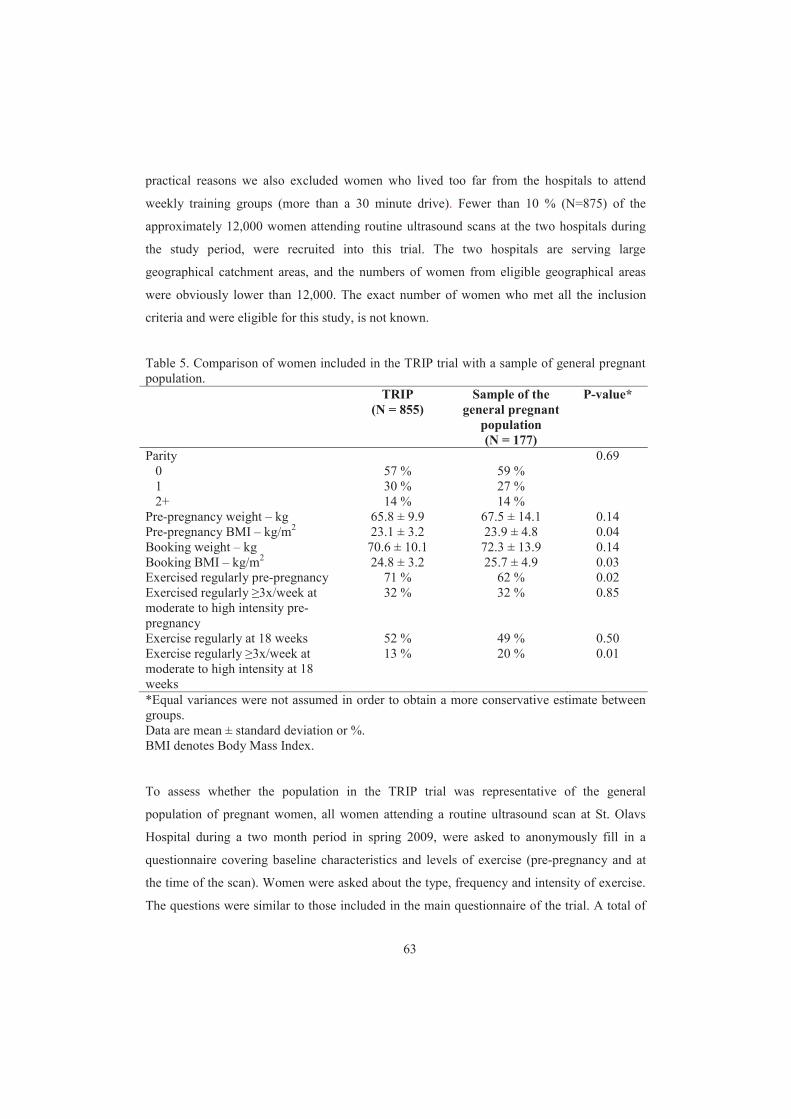
practical reasons we also excluded women who lived too far from the hospitals to attend
weekly training groups (more than a 30 minute drive). Fewer than 10 % (N=875) of the
approximately 12,000 women attending routine ultrasound scans at the two hospitals during
the study period, were recruited into this trial. The two hospitals are serving large
geographical catchment areas, and the numbers of women from eligible geographical areas
were obviously lower than 12,000. The exact number of women who met all the inclusion
criteria and were eligible for this study, is not known.
Table 5. Comparison of women included in the TRIP trial with a sample of general pregnant population. TRIP
(N = 855) Sample of the
general pregnant population (N = 177)
P-value*
Parity 0.69 0 57 % 59 % 1 30 % 27 % 2+ 14 % 14 % Pre-pregnancy weight – kg 65.8 ± 9.9 67.5 ± 14.1 0.14 Pre-pregnancy BMI – kg/m2 23.1 ± 3.2 23.9 ± 4.8 0.04 Booking weight – kg 70.6 ± 10.1 72.3 ± 13.9 0.14 Booking BMI – kg/m2 24.8 ± 3.2 25.7 ± 4.9 0.03 Exercised regularly pre-pregnancy 71 % 62 % 0.02 Exercised regularly 3x/week at moderate to high intensity pre-pregnancy
32 % 32 % 0.85
Exercise regularly at 18 weeks 52 % 49 % 0.50 Exercise regularly 3x/week at moderate to high intensity at 18 weeks
13 % 20 % 0.01
*Equal variances were not assumed in order to obtain a more conservative estimate between groups. Data are mean ± standard deviation or %. BMI denotes Body Mass Index.
To assess whether the population in the TRIP trial was representative of the general
population of pregnant women, all women attending a routine ultrasound scan at St. Olavs
Hospital during a two month period in spring 2009, were asked to anonymously fill in a
questionnaire covering baseline characteristics and levels of exercise (pre-pregnancy and at
the time of the scan). Women were asked about the type, frequency and intensity of exercise.
The questions were similar to those included in the main questionnaire of the trial. A total of
63
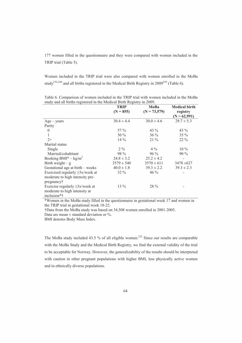
177 women filled in the questionnaire and they were compared with women included in the
TRIP trial (Table 5).
Women included in the TRIP trial were also compared with women enrolled in the MoBa
study232,244 and all births registered in the Medical Birth Registry in 2009229 (Table 6).
Table 6. Comparison of women included in the TRIP trial with women included in the MoBa study and all births registered in the Medical Birth Registry in 2009. TRIP
(N = 855) MoBa
(N = 73,579) Medical birth
registry (N = 62,991)
Age – years 30.4 ± 4.4 30.0 ± 4.6 29.7 ± 5.3 Parity 0 57 % 43 % 43 % 1 30 % 36 % 35 % 2+ 14 % 21 % 22 % Marital status Single 2 % 4 % 10 % Married/cohabitant 98 % 96 % 90 % Booking BMI* – kg/m2 24.8 ± 3.2 25.2 ± 4.2 - Birth weight – g 3579 ± 540 3570 ± 611 3478 ±627 Gestational age at birth – weeks 40.0 ± 1.8 39.3 ± 2.2 39.3 ± 2.3 Exercised regularly 3x/week at moderate to high intensity pre-pregnancy†
32 % 46 % -
Exercise regularly 3x/week at moderate to high intensity at inclusion*†
13 % 28 % -
*Women in the MoBa study filled in the questionnaire in gestational week 17 and women in the TRIP trial in gestational week 18-22. †Data from the MoBa study was based on 34,508 women enrolled in 2001-2005. Data are mean ± standard deviation or %. BMI denotes Body Mass Index.
The MoBa study included 43.5 % of all eligible women.232 Since our results are comparable
with the MoBa Study and the Medical Birth Registry, we find the external validity of the trial
to be acceptable for Norway. However, the generalizability of the results should be interpreted
with caution in other pregnant populations with higher BMI, less physically active women
and in ethnically diverse populations.
64
Study IV
During a period of two months, women included in the RCT were also asked to participate in
a study aiming to validate a physical activity monitor (SenseWearTM Pro2 Armband). Twenty-
nine pregnant women (24–43 years old) in the second (n=13) and third trimester (n=16) were
recruited. None of the participating women used medications or smoked, which could have
influenced the energy expenditure.
Intervention
Studies I-III
Women in the intervention group were encouraged to exercise three days per week during the
12-week intervention period. They received a standardized exercise program including
aerobic activity, strength training and balance exercises. The training protocol followed
recommendations from the ACOG and the Norwegian National Report on Physical Activity
and Health.5,230 Training sessions of 60 minutes in groups of 8-15 women instructed by a
physiotherapist were offered once a week over a period of 12 weeks (between 20 and 36
pregnancy weeks). Each group session consisted of three parts:
1) 30-35 minutes low impact aerobics (no running or jumping). Step length and body rotations
were reduced to a minimum, and crossing of legs and sharp and sudden changes of position
were avoided. The aerobic dance program was performed at moderate intensity, defined as 12
and 14 on Borg’s rating scale of perceived exertion40 (Appendix 2).
2) 20-25 minutes strength exercises using body weight as resistance, including exercises for
the upper and lower limbs, back extensors, deep abdominal muscles and PFM. Three sets of
ten repetitions of each exercise were performed. The PFMT was based on the one used by Bø
et al.55 and repeated in the trial by Mørkved et al.226
3) 5-10 minutes of light stretching, body awareness, breathing and relaxation exercises.
In addition, women were encouraged to follow a written 45 minute home exercise program at
least twice a week (30 minutes endurance training and 15 minutes strength and balance
exercises) (Appendix 3).
65
Women in the control group received standard antenatal care and the customary information
given by their midwife or general practitioner. They were not discouraged from exercising on
their own. Women in both groups received written recommendations on PFMT, diet and
pregnancy related lumbopelvic pain.
Methods
Pre-tests were done at 18-22 weeks and post-tests at 32-36 weeks of pregnancy. The women
met at the hospital for assessment. Height and weight were measured by an assessor.
Frequency, intensity, duration and type of physical activity including PFMT were recorded
for both groups in a questionnaire at inclusion and follow-up. In addition, women in the
intervention group registered PFMT in a training diary. Adherence to the protocol was
defined as exercising three days per week or more on moderate to high intensity. Performing
the exercise program was strongly emphasized and recorded in the women's personal training
diary and through reports from the physiotherapists leading the training groups.
Study I – Assessment of GDM
The primary outcomes of the RCT were the prevalence of GDM and insulin resistance
estimated by the homeostasis model assessment method (HOMA-IR).202 HOMA-IR is an
empirical mathematical formula based on fasting plasma glucose and fasting plasma insulin
levels to calculate insulin resistance [(fasting plasma insulin ( IU/mL) x fasting plasma
glucose (mmol/L))/22.5]. All participants underwent a 75 g OGTT at inclusion (18-22 weeks)
and at the end of the training period (32-36 weeks). Fasting and 2 hour glucose levels were
measured in serum by the routine methods used by the hospital laboratory. GDM was
diagnosed according to the WHO criteria as fasting glucose level in fasting whole blood 6.1
mmol/L or plasma glucose 7.0 mmol/L, or 2 hour value 7.8 mmol/l.11 In addition,
secondary outcomes, such as pregnancy complications and outcomes (e.g. newborn weight,
gestational age, Apgar scores) were registered from the birth journal.
66
Study II – Assessment of LPP
Possible effects of regular exercise during pregnancy on LPP were one of six other pre-
specified outcomes. LPP and sick leave due to LPP were measured on self-reports. The
prevalence estimate of LPP was based on a simple question: “Do you have pain in the pelvic
and/or lumbar area?” (Yes/No).
Secondary outcomes were disability, pain intensity and fear-avoidance beliefs. Disability was
measured by The Disability Rating Index (DRI) containing 12 questions concerning physical
function, of which each question is scored on a 100 mm visual analog scale from “no
disability” to “totally unable”.275 The DRI index is calculated as the mean of 12 questions.
Since pain intensity varies throughout the day,209 the women were asked about worst pain in
the morning and the evening on Visual Analog Scales (VAS 0-100 mm). The modified Fear-
Avoidance Beliefs Questionnaire (mFABQ) consists of four of the five items from the fear-
avoidance beliefs about physical activity.188 Ratings were made on 0 to 6 point scale ranging
from strongly disagree to strongly agree. Scores may range from 0 to 24 with high scores
showing stronger fear-avoidance beliefs.
Study III – Assessment of incontinence
Possible effects of regular exercise during pregnancy on incontinence were another of the six
pre-specified outcomes. UI was measured by self-reports using a severity index.280,281 Women
were asked to report their urinary leakage when A) coughing / sneezing / laughing, B) when
being physically active (running or jumping), C) when sudden changes in position / lifting or
D) leakage accompanied by a strong urgency to void. Answer alternatives were “Yes” or “No.
Urinary leakage was subclassified according to the definitions given in the standardized
terminology of lower urinary tract symptoms.144 Women confirming any type of urinary
leakage (alternative A, B, C or D) were referred to as having UI. Women confirming loss of
urine in association with alternatives A, B or C were defined as having SUI, while women
confirming loss of urine with alternative D were defined as having UUI, The outcome
measures of UI were further divided into two severity categories with respect to frequency,
“urinary leakage < once per week” or “urinary leakage once per week (severe UI)”.
67
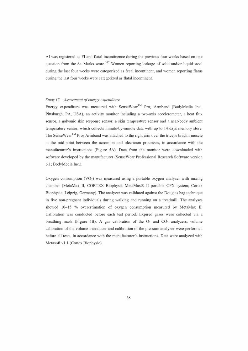
AI was registered as FI and flatal incontinence during the previous four weeks based on one
question from the St. Marks score.317 Women reporting leakage of solid and/or liquid stool
during the last four weeks were categorized as fecal incontinent, and women reporting flatus
during the last four weeks were categorized as flatal incontinent.
Study IV – Assessment of energy expenditure
Energy expenditure was measured with SenseWearTM Pro2 Armband (BodyMedia Inc.,
Pittsburgh, PA, USA), an activity monitor including a two-axis accelerometer, a heat flux
sensor, a galvanic skin response sensor, a skin temperature sensor and a near-body ambient
temperature sensor, which collects minute-by-minute data with up to 14 days memory store.
The SenseWearTM Pro2 Armband was attached to the right arm over the triceps brachii muscle
at the mid-point between the acromion and olecranon processes, in accordance with the
manufacturer’s instructions (Figure 5A). Data from the monitor were downloaded with
software developed by the manufacturer (SenseWear Professional Research Software version
6.1; BodyMedia Inc.).
Oxygen consumption (VO2) was measured using a portable oxygen analyzer with mixing
chamber (MetaMax II, CORTEX Biophysik MetaMax® II portable CPX system; Cortex
Biophysic, Leipzig, Germany). The analyzer was validated against the Douglas bag technique
in five non-pregnant individuals during walking and running on a treadmill. The analyses
showed 10–15 % overestimation of oxygen consumption measured by MetaMax II.
Calibration was conducted before each test period. Expired gases were collected via a
breathing mask (Figure 5B). A gas calibration of the O2 and CO2 analyzers, volume
calibration of the volume transducer and calibration of the pressure analyzer were performed
before all tests, in accordance with the manufacturer’s instructions. Data were analyzed with
Metasoft v1.1 (Cortex Biophysic).
68
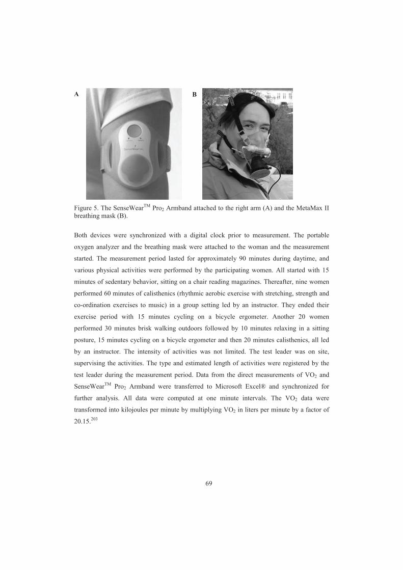
Figure 5. The SenseWearTM Pro2 Armband attached to the right arm (A) and the MetaMax II breathing mask (B).
Both devices were synchronized with a digital clock prior to measurement. The portable
oxygen analyzer and the breathing mask were attached to the woman and the measurement
started. The measurement period lasted for approximately 90 minutes during daytime, and
various physical activities were performed by the participating women. All started with 15
minutes of sedentary behavior, sitting on a chair reading magazines. Thereafter, nine women
performed 60 minutes of calisthenics (rhythmic aerobic exercise with stretching, strength and
co-ordination exercises to music) in a group setting led by an instructor. They ended their
exercise period with 15 minutes cycling on a bicycle ergometer. Another 20 women
performed 30 minutes brisk walking outdoors followed by 10 minutes relaxing in a sitting
posture, 15 minutes cycling on a bicycle ergometer and then 20 minutes calisthenics, all led
by an instructor. The intensity of activities was not limited. The test leader was on site,
supervising the activities. The type and estimated length of activities were registered by the
test leader during the measurement period. Data from the direct measurements of VO2 and
SenseWearTM Pro2 Armband were transferred to Microsoft Excel® and synchronized for
further analysis. All data were computed at one minute intervals. The VO2 data were
transformed into kilojoules per minute by multiplying VO2 in liters per minute by a factor of
20.15.203
A B
69
Ethics
The procedures followed were in accordance with ethical standards of research and the
Helsinki declaration.309 The women received written (Appendix 4) and oral information, and
they signed informed consent forms. The participants were not compensated financially. The
study was approved by the Regional Committee for Medical and Health Research Ethics
(REK 4.2007.81) and registered with Clinical trial gov (NCT 00476567).
Power calculations
Studies I-III
For the principal power calculation of the trial we assumed a GDM prevalence of 9 % in the
control group and a prevalence of 4 % in the intervention group (risk difference of 5 %).
Under these assumptions a two-sample comparison (Chi squared) with a 5 % level of
significance and power of 0.80 gave a study population of 381 patients in each group. This
sample size was able to detect a 0.2 SD difference on continuous variables (insulin resistance)
given the same power and significance level. Assuming a 10 % dropout rate we needed to
include about 880 pregnant women.
Study IV
Sample size calculation was based on a standard deviation of 5 and 7 kJ (per min) with a
significance level of 0.05 and power of 0.80. We needed 24 subjects to detect a mean
difference of 5 kJ between indirect calorimetry and SenseWearTM Pro2 Armband.
70
Statistical analyses
Study I
The primary data analysis was according to the “intention–to-treat” principle using Stata
software version 10.0 for Windows (Stata Corporation, College Station, Texas). A two sample
test of proportions for univariable analyses was used for complete case analysis of continuous
variables and a simple linear regression was used for univariable analysis and analysis of
covariance when adjusting for baseline values. The estimated risk differences were
accompanied with 95 % confidence intervals (CI) and corresponding p-values. A mixed
model with random slopes was used to assess the potential impact from missing data. This
model used the interaction between the treatment group and time to estimate the baseline
adjusted differences between the exercise and control groups. Exploratory analyses using the
same statistical models were performed to compare women in the intervention group who
adhered to the protocol with the total control group.
Studies II-III
The data were analyzed according to the “intention–to-treat” principle using binary logistic
regression for dichotomous variables and ANCOVA for continuous variables. The results
were presented as crude and baseline adjusted estimates with 95 % CI. The statistical analyses
were performed with SPSS® (Statistical Package for Social Sciences, version 19 for
Windows; SPSS Inc., Chicago, USA).
Study IV
A Bland-Altman plot was constructed to demonstrate the relation between the differences
(SenseWearTM Pro2 Armband minus indirect calorimetry) and the average values of the two
methods for recording energy expenditure during the measurement period. The mean
differences and limits of agreement were calculated according to Bland and Altman.36 A two-
way mixed, single-measure, parametric intraclass correlation [ICC (3,1)] was performed for
evaluating the extent of agreement between SenseWearTM Pro2 Armband and indirect
71

calorimetry. Analyses were conducted in SPSS® (Statistical Package for Social Sciences,
version 15 for Windows; SPSS Inc., Chicago, USA).
72
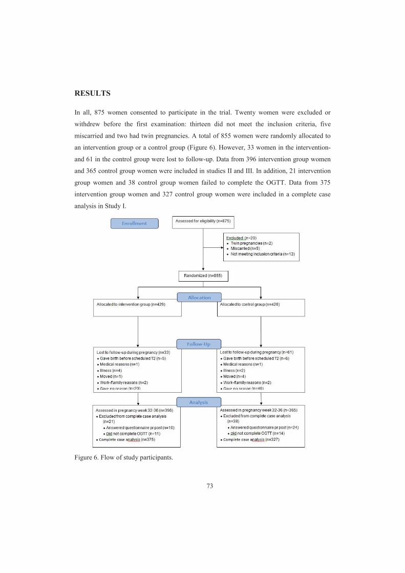
RESULTS
In all, 875 women consented to participate in the trial. Twenty women were excluded or
withdrew before the first examination: thirteen did not meet the inclusion criteria, five
miscarried and two had twin pregnancies. A total of 855 women were randomly allocated to
an intervention group or a control group (Figure 6). However, 33 women in the intervention-
and 61 in the control group were lost to follow-up. Data from 396 intervention group women
and 365 control group women were included in studies II and III. In addition, 21 intervention
group women and 38 control group women failed to complete the OGTT. Data from 375
intervention group women and 327 control group women were included in a complete case
analysis in Study I.
Figure 6. Flow of study participants.
73
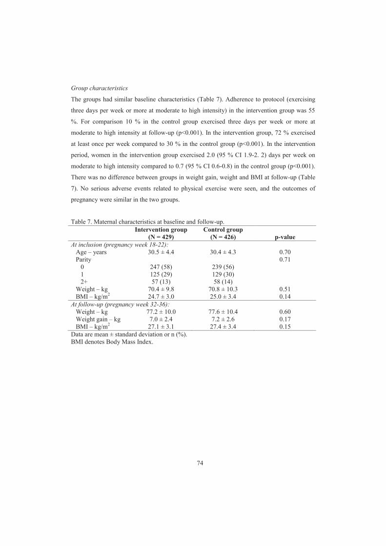
Group characteristics
The groups had similar baseline characteristics (Table 7). Adherence to protocol (exercising
three days per week or more at moderate to high intensity) in the intervention group was 55
%. For comparison 10 % in the control group exercised three days per week or more at
moderate to high intensity at follow-up (p<0.001). In the intervention group, 72 % exercised
at least once per week compared to 30 % in the control group (p<0.001). In the intervention
period, women in the intervention group exercised 2.0 (95 % CI 1.9-2. 2) days per week on
moderate to high intensity compared to 0.7 (95 % CI 0.6-0.8) in the control group (p<0.001).
There was no difference between groups in weight gain, weight and BMI at follow-up (Table
7). No serious adverse events related to physical exercise were seen, and the outcomes of
pregnancy were similar in the two groups.
Table 7. Maternal characteristics at baseline and follow-up. Intervention group
(N = 429) Control group
(N = 426)
p-value At inclusion (pregnancy week 18-22): Age – years 30.5 ± 4.4 30.4 ± 4.3 0.70 Parity 0.71 0 247 (58) 239 (56) 1 125 (29) 129 (30) 2+ 57 (13) 58 (14) Weight – kg 70.4 ± 9.8 70.8 ± 10.3 0.51 BMI – kg/m2 24.7 ± 3.0 25.0 ± 3.4 0.14 At follow-up (pregnancy week 32-36): Weight – kg 77.2 ± 10.0 77.6 ± 10.4 0.60 Weight gain – kg 7.0 ± 2.4 7.2 ± 2.6 0.17 BMI – kg/m2 27.1 ± 3.1 27.4 ± 3.4 0.15 Data are mean ± standard deviation or n (%). BMI denotes Body Mass Index.
74
Main results
The results are described in detail in each of the papers, hence only a summary of the main
results is given here.
Study I – GDM
No differences were found in the prevalence of GDM between groups; 25/375 (7 %, 95 % CI
4.3-9.7) intervention group women compared to 18/327 (6 %, 95 % CI 3.3-8.6) control group
women (p=0.52). Complete case analysis demonstrated lower fasting insulin and insulin
resistance in the intervention group compared to the control group at follow-up, but when
adjusting for baseline values the differences were reduced. To evaluate the potential effects of
attrition and missing data, a linear mixed effects model was applied for all included subjects,
N=855. The results were similar (lower fasting insulin and insulin resistance in the
intervention group), but the differences were halved when adjusting for baseline values.
Study II – LPP
There were no differences in the percentage of women reporting LPP in the two groups after
the 12 weeks intervention period. However, the proportion of women on sick leave due to
LPP and the odds ratio (OR) were lower in the intervention group (p=0.01). When adjusting
for baseline sick leave due to LPP, the estimated OR was still significantly lower (p=0.04).
No differences were found between groups regarding disability, pain intensity or fear-
avoidance beliefs both unadjusted and adjusted for baseline values of the outcome measure.
Study III – Incontinence
After the intervention period, significantly less women in the intervention group reported UI
and SUI. The findings were consistent when adjusting for baseline values. A lower proportion
of women in the intervention group (3 %) than the control group (5 %) reported FI, however,
the difference was not statistically significant.
75
Subgroup analyses were done based on groups stratified according to urinary continence
status at inclusion. Sixty-nine percent of the continent women and 40 % of the incontinent
women were nulliparous. The subgroup analyses showed that women in the intervention
group who were continent for urine at inclusion, reported significantly less severe SUI ( once
per week) and UUI (p=0.03 and p=0.006) after the intervention period. Furthermore, among
women who were incontinent for urine at inclusion, the proportion reporting UI, UI once per
week and SUI after the intervention period was lowest in the intervention group (p=0.002,
p=0.03 and p=0.007). The findings were consistent when adjusting for baseline values.
Subgroup analyses on groups stratified according to anal continence status at inclusion
showed no significant difference.
In the intervention group, 95 % reported weekly PFMT at 32-36 weeks of gestation compared
to 79 % in the control group (p<0.001). The proportion of intervention group women
performing PFMT three times per week or more was 67 % compared to 40 % in the control
group (p<0.01).
Study IV – Energy expenditure
A total of 29 women were included in the validation study. Thirteen women were in the
second trimester and 16 in the third trimester. Twenty women carried out conditioning
exercises such as brisk walking, running or bicycling in addition to strength exercises,
whereas nine participated in calisthenics and rhythmic aerobic exercise. The mean period with
energy expenditure recordings was 85 minutes (range 80–89).
Mean total energy expenditure during the period with free-living activities was 1,618 kJ (95
% CI 1,492–1,744) for indirect calorimetry and 1,482 kJ (95 % CI 1,364–1,600) for
SenseWearTM Pro2 Armband. When comparing energy expenditure during free-living
activities, the mean difference and limit of agreement from the Bland–Altman plot was 136
± 343 kJ. SenseWearTM Pro2 Armband underestimated energy expenditure by 9 %.
76
DISCUSSION
The central aspects in each of the studies have been discussed in the respective papers. Here I
will concentrate on some methodological aspects and address the main issues across the
studies.
To my knowledge this is the largest RCT assessing exercise during pregnancy in the
prevention and treatment of pregnancy-related conditions. Healthy pregnant women were
included. Women in the intervention group were offered a 12-week exercise program,
including PFMT, to be performed three times per week in the second half of pregnancy. A
total of 55 % of intervention group women exercised three times per week in late pregnancy,
compared to 10 % in the control group. At follow-up, there was no difference between groups
in the prevalence of GDM or level of insulin resistance. The prevalence of LPP in both groups
was similar, however, less women in the intervention group were on sick leave due to LPP.
The intervention group reported less UI in late gestation, and this group had both a primary
and secondary prevention effect. A physical activity monitor was validated and found to be a
valid instrument in pregnant women. The TRIP trial was a two-armed, two-centered
randomized controlled trial with concealed randomization. However, there are some
limitations.
Methodological aspects
Design
RCT is considered to be the gold standard design when assessing the effects of treatment,
providing the most reliable evidence on the efficacy of healthcare interventions.217 Proper
randomization reduces selection bias at trial entry, balancing both known and unknown
factors. The intervention group was offered a regular exercise program. The control group
women received no intervention, but were not advised against exercising on their own. The
optimal study design would be to have a control group free from exercise. However, it would
be unethical to stop control group women exercising on their own, and adding a control group
77
of sedentary pregnant women would ruin the randomized design. Including only sedentary
women with no previous habit of regular exercise in the trial would have been another option.
Blinding is considered an important methodological factor to ensure high internal validity of a
RCT. In the TRIP trial the investigator was aware of group allocation. Addition of a blinded
assessor would increase the costs and thus was difficult for practical reasons. However,
analysis of blood glucose and insulin was done blinded to group allocation, and prevalence of
LPP and incontinence were based on self-reports minimizing the effect of bias due to the non-
blinded study design.
The possibility of recall bias is minimal in the TRIP trial, since women were asked to report
LPP and incontinence at present, except for AI in which women were asked to recall
symptoms last four weeks.
It is possible that women in the intervention group were «eager to please» because they were
offered a free exercise class. Thus, they may have reported fewer symptoms than they actually
experienced. On the other hand, the exercise program exposed women attending the
intervention group to a variety of situations that trigger incontinence and challenges the local
and global spinal stabilizing muscles. This means that the effects of the training program may
be underestimated regarding incontinence and LPP.
Study population
Another limitation to be considered is selection bias. Less than 10 % of all women attending a
routine ultrasound scan in the study period assigned for the trial (875/12,000). To be able to
attend, women should be Caucasian, healthy with low risk pregnancies and live within 30
minutes drive from the hospital. The actual number of women meeting the inclusion criteria is
not known. The women volunteering for the trial contacted the project coordinator after
receiving a written invitation, and were mainly well motivated to exercise during pregnancy.
Women in the TRIP trial might therefore be a selected group of women already in good
physical condition.
78
Women included in the TRIP trial were from an urban area in Norway. When compared with
a general population of pregnant women in Trondheim, women in the MoBa study and the
Medical Birth Registry, the external validity was found to be acceptable for Norwegian
pregnant women regarding age, parity, BMI, exercising habits, gestational age at birth and
birth weight (Table 5 and 6). However, in the MoBa study the youngest women (<25 years),
those living alone, mothers with >2 previous births, previous stillbirths and smokers were
strongly under-represented compared with the total population pregnant women in Norway in
the same time period.232
The intervention was based on both group training and individual training. This may have
caused practical difficulties for some of the invited women (childcare, work, transport). We
have not been able to assess the characteristics of non-participants. However, women in the
TRIP trial were healthy Caucasian women with mean BMI within the normal range.
Therefore, the results from our trial should be interpreted with caution in groups with less
physical activity, higher BMI and other ethnicities.
Intervention
The intervention was based on the current recommendations for the general population
(exercising three times per week including both aerobic and strength training). Healthy
pregnant women are encouraged to follow the same exercise recommendations as non-
pregnant. The exercise program required no equipment, all strength exercises were performed
with own body weight as resistance. The women chose according to their own preferences
what type of endurance training to perform when doing the home exercise program. The
strength and balance exercises were similar to those performed in the group sessions. All
strength exercises were shown in three different positions reflecting various degrees of
difficulty. The weekly group sessions were led by skilled physiotherapists, who followed a
standardized exercise program. Supervision of exercises is critically important in improving
quality of exercise performance. The supervision by a qualified professional and proper
prescription of the program variables have been found to be important for effective resistance
training.175 In addition, a strong correlation between the quality of exercise performance and
decrease in pain in non-pregnant patients with neck pain and LBP has been found.112 The
exercise program also included information on related topics such as incontinence, LPP,
79
ergonomics, exercise in pregnancy and the postpartum period. Most of the exercise groups
were visited by a gynecologist who informed about exercise in pregnancy, contractions, and
answered questions from the women.
The control group received standard care and written information about LPP, PFMT and diet.
The brochure on PFMT included a detailed evidence-based PFMT program similar to the
PFMT program included in the intervention. A high percentage of women in the control group
reported performing PFMT. In spite of this, the intervention group demonstrated significantly
less UI than the control group women. However, women in the control group were not
instructed in correct VPFMC at enrolment in the trial. Since it is well known that many
women do not perform a VPFMC correctly after only verbal or written information,46,56 there
is a possibility that some women in the control group performed the contractions incorrectly.
Other important issues are thorough instructions and motivation. Women in the intervention
group had weekly instructions and practical training in groups led by skilled physiotherapists.
The two examinations during the study period were done at the hospital, and all women
diagnosed with GDM were referred to standard follow-up of diabetes nurses and
endocrinologists. If other problems were discovered, women were referred to a midwife or
gynecologist. Both examinations were in addition to standard antenatal care.
The 12-week intervention period was during the second half of pregnancy. There were several
reasons for this approach. First, the study invitation was attached to an invitation to a routine
ultrasound scan. Thus, we were able to invite all eligible pregnant women in the catchment
areas. Second, all women had ultrasound before inclusion, and we were assured that only
women with one single fetus and a low risk pregnancy were included. And third, at study start
there were some discussions between experts regarding leisure time physical activity and the
risk of early miscarriage.197 However, in the light of our findings and previous research, I
believe that promoting health of women of reproductive age before conception and helping
women to have a healthy and active lifestyle during pregnancy are the most promising
approaches in preventing pregnancy-related conditions such as GDM, LPP and incontinence.
80
Compliance
Compliance is essential for proper interpretation of the effect of an exercise intervention. In
this trial only 55 % managed to follow the exercise protocol. Reported reasons for not
following the protocol were mostly attributable to pregnancy symptoms, child care and work
commitments. Exercising three times per week can be hard to achieve for a pregnant women
taking into account family- and work commitments and a body in continuous change. Two-
thirds of the exercise protocol was exercises to be performed home. From a public health
perspective strategies to increase adherence to exercise must be of importance. This might be
through public health information campaigns, information about health benefits of regular
exercise given by a physiotherapist during antenatal care, offering free exercise courses
during pregnancy and that the employer offers time for exercise included in working hours. It
is important to emphasize that health benefits of regular exercise is not only limited to
maternal profits, but is most likely to influence the fetus and protect against overweight,
metabolic syndrome, diabetes and other cardiovascular diseases in the adolescents and
adulthood.
Withdrawals and loss to follow-up
Loss to follow-up after recruitment and attrition affects the generalizability of the results. Any
loss to follow-up decreases internal validity, but differential rates of loss to follow-up among
comparison groups cause major damage.285 In general, less than 5 % loss to follow-up in RCT
is acceptable, whereas greater than 20 % loss to follow-up will potentially imply serious
threats to validity.285 In the TRIP trial we experienced 11 % loss to follow-up on self-reported
outcomes and 18 % on OGTT. The respective percentages in intervention and control groups
were 8 % vs. 14 % on self-reports and 13 % vs. 23 % on OGTT. These differences were
considered acceptable. One reason for loss of data at the OGTT was that performing the
OGTT caused nausea in some women who therefore did not complete the test. In addition, the
tests were performed in daytime due to the need of an overnight fast, and lasted for almost
three hours including OGTT, questionnaires and physical tests. This may have been difficult
for some women due to work commitments.
81
Statistics and power calculations
All participants were included in the analysis in the groups to which they were originally
assigned (“intention-to-treat” analysis). The intention-to-treat principle underlies the primary
analysis in an RCT to avoid bias associated with non-random loss of participants.181 Since
only 55 % adhered to the exercise protocol it is difficult to conclude on the direct effect of the
exercise protocol on glucose metabolism, LPP and incontinence. Explorative analysis can be
performed on the protocol adherents. However, such analysis has some limitations since they
are based on only a subset of the subjects initially included in the trial. The results may be
explained by other confounding variables and results should therefore be interpreted with
caution.
When the present trial was planned no prevalence studies on GDM from Norway were
published. The power calculation of the trial was based on previous international studies
reporting prevalence up to 14 %. Since the TRIP trial was a population-based approach and
invited all women irrespective of risk factors, a prevalence of 9 % was chosen and a reduction
to 4 % in the intervention group was assumed. The overall GDM prevalence in the TRIP trial
was 6.1 % at 32-36 weeks of pregnancy indicating that the study was slightly underpowered.
However, even with a larger trial, there would probably be no statistically significant
difference in GDM prevalence between groups (7 % vs. 6 % in the present trial). This is
supported by our secondary outcome, 120 minutes OGTT, which was not different between
the groups. Being a continuous variable, this outcome has considerably more power. With a
sample size of 440 in each arm, we had a power of 87 % to detect a difference in 120 minutes
OGTT of 0.25 mmol/L based on a t-test for two independent samples given a common SD of
1.2 in the groups and a 2-tailed alpha of 0.050.
The prevalence of AI in late pregnancy was found to be 5 % vs. 3 % in the present trial. AI is
less prevalent during pregnancy than UI, and the trial was underpowered to demonstrate any
possibly difference between groups as shown by the wide confidence intervals.
Despite the randomized design some of the baseline characteristics were significantly
different between groups at inclusion. This was accounted for in the statistical analysis.
82
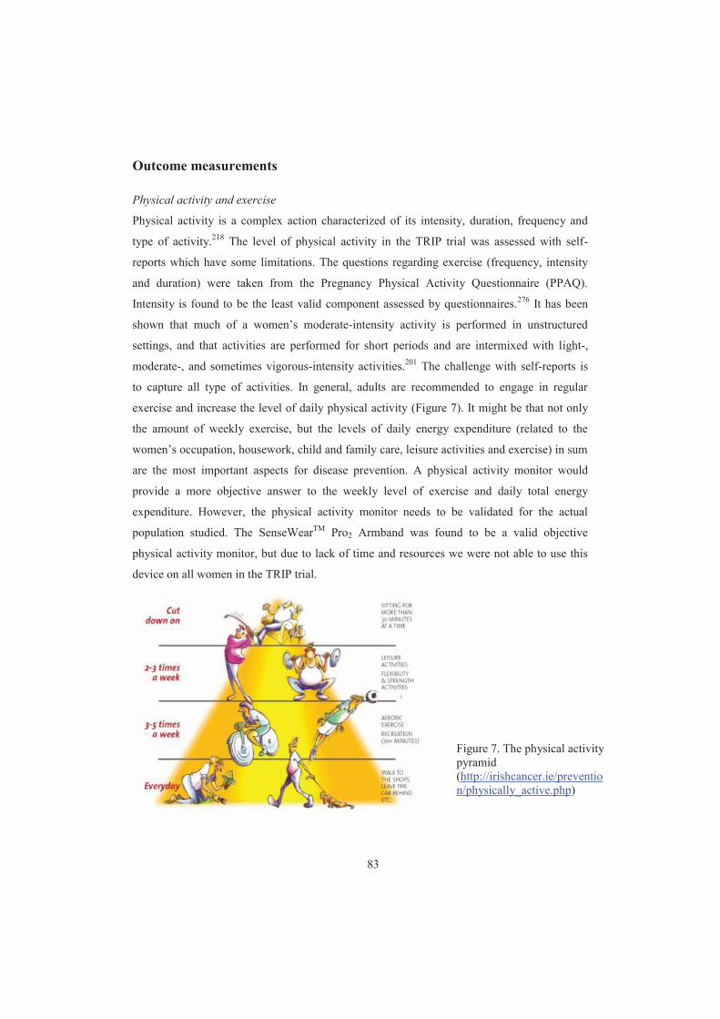
Outcome measurements
Physical activity and exercise
Physical activity is a complex action characterized of its intensity, duration, frequency and
type of activity.218 The level of physical activity in the TRIP trial was assessed with self-
reports which have some limitations. The questions regarding exercise (frequency, intensity
and duration) were taken from the Pregnancy Physical Activity Questionnaire (PPAQ).
Intensity is found to be the least valid component assessed by questionnaires.276 It has been
shown that much of a women’s moderate-intensity activity is performed in unstructured
settings, and that activities are performed for short periods and are intermixed with light-,
moderate-, and sometimes vigorous-intensity activities.201 The challenge with self-reports is
to capture all type of activities. In general, adults are recommended to engage in regular
exercise and increase the level of daily physical activity (Figure 7). It might be that not only
the amount of weekly exercise, but the levels of daily energy expenditure (related to the
women’s occupation, housework, child and family care, leisure activities and exercise) in sum
are the most important aspects for disease prevention. A physical activity monitor would
provide a more objective answer to the weekly level of exercise and daily total energy
expenditure. However, the physical activity monitor needs to be validated for the actual
population studied. The SenseWearTM Pro2 Armband was found to be a valid objective
physical activity monitor, but due to lack of time and resources we were not able to use this
device on all women in the TRIP trial.
Figure 7. The physical activity pyramid (http://irishcancer.ie/prevention/physically_active.php)
83
Gestational diabetes
Fasting and 2-hour glucose levels were measured in serum by the routine methods used by the
hospital laboratory the same day as taken. The level of insulin was measured in serum after
being stored in a freezer (-80 °C). GDM was diagnosed according to the WHO criteria
(fasting glucose level in fasting whole blood 6.1 mmol/L, or plasma glucose 7.0 mmol/L,
or 2-hour value 7.8 mmol/L).11 Comparison between different studies can be difficult
because the OGTT procedure and diagnostic criteria vary.70 We chose to use HOMA-IR as a
measure of insulin sensitivity, but HOMA-IR may not be the most sensitive way of assessing
the impact of exercise on insulin resistance. HOMA-IR is regarded as a good measure of
overall insulin resistance in pregnancy, but it may provide a better reflection of liver rather
than peripheral insulin resistance.169 Exercise most likely exerts its effect peripherally by
reducing peripheral muscle insulin resistance.59
Lumbopelvic pain
The prevalence estimate of LPP was based on self-reports, and was not verified with clinical
tests. Some authors argue that it is important to distinguish between LBP and PGP,326 but this
was not done because it was not within the scope of this thesis to classify pain into different
types, or according to location. It has been argued that because back pain is a subjective
phenomenon, self-report is the most appropriate method of measuring this outcome.173
However, different definitions of back pain may result in different estimates of prevalence
and incidence, and no single definition has been generally accepted in back pain research,
making comparison between studies difficult. We did not ask the women whether they had
LPP before the present pregnancy or if the onset of pain were during the present pregnancy.
Thus, we do not know if we were dealing with old recurrent pain or a new pain. Östgaard et
al.348 have shown that more than one in five (22 %) women had ongoing back pain before
getting pregnant. We assume that this may be true for a number of women included in the
TRIP trial, and this was another reason why the term “pregnancy-related” was not used
together with “lumbopelvic pain”.
The DRI reflects function and disability rather than pain. It was primarily developed for non-
pregnant with LBP, but has been used on a wide range of musculoskeletal problems. DRI
surveys the difficulties in performing specific activities. Hence it is the degree of difficulty
84
that is assessed. The DRI is not optimal for pregnant women since activities such as “lifting
heavy objects” and “running” are included. When the TRIP trial was initiated, no pregnancy-
related instrument for physical ability was found. Later, the Pregnancy Mobility Index321 and
the pelvic girdle questionnaire297 were developed. The Pregnancy Mobility Index321 evaluates
functioning in relation to back and pelvic pain in the following three main areas: daily
mobility in the house, household activities and mobility outdoors. The pelvic girdle
questionnaire297 is a condition-specific instrument for assessing activity limitations in women
with PGP during pregnancy and postpartum.
Pain is multidimensional and the respondents communicate far more than just intensity about
their pain.339 The reliability and validity of measuring pain intensity by VAS have been
discussed.303 Pain intensity measured by VAS is a subjective phenomenon quantifying the
pain. The repeatability of VAS has been found to be good.339 The VAS used for rating pain
intensity in LBP showed good reliability over 24 hours,265 and the VAS also showed
sufficient reliability when used in acute pain.32 A number of studies have demonstrated that
pathology does not correlate well with pain, importantly, the absence of identifiable pathology
does not mean the absence of pain.339
Fear-avoidance refers to the avoidance of specific movements or activities based on fear of
pain or (re)injury. The mFABQ is a modified version of the original FABQ, and consists of
four of the originally five questions regarding general physical activity. The mFABQ was
chosen since it is found that even subjects without pain can answer these questions.188
Both DRI and VAS are continuous variables and intend to provide more graded information
on LPP than merely the presence or absence of LPP. The DRI and VAS has previously been
used in pregnant and postpartum women assessing LPP,228,240,269,299 thus allows for
comparison between studies which is considered important. This was the main reason for
choosing DRI and VAS as outcome measures.
Incontinence
Both UI and AI were measured by self-reports. The severity index280,281 and the St. Marks
score317 are validated and reflect the frequency and amount of leakage and have been used in
85
numerous studies in different populations. The question of UI is in accordance with the
International continence society’s present definition of UI symptoms; “any complaint of
involuntary leakage of urine”.144 In the TRIP trial we wanted to cause minimal discomfort to
healthy pregnant women and therefore did not perform any quantification of the amount of
urine lost with standardized bladder volume. However, Haylen et al.144 emphasize that to
diagnose “urodynamic stress incontinence” the correlation between a woman’s symptom,
sign, and urodynamic investigations should be included
The relationship among different severity measures of UI in women with predominantly SUI
has been found to be weak to moderate, and they appear to measure different aspects of the
incontinence condition.12 Thus, the different severity measures are not interchangeable when
characterizing a patient population or assessing outcomes. To facilitate comparison between
studies a standardized measurement tool should be agreed upon.
AI was measured with the St. Marks score which covers frequency, severity, urgency, pad use
and social restrictions.317 The St. Marks score is a widely used severity score, ranging from 0
(complete continence) to 24 (complete incontinence). In the TRIP trial only the first question
regarding frequency was used (“have you experienced leakage of solid stool / liquid stool /
gas last four weeks”). Women answering rarely / sometimes / weekly / daily were categorized
as having AI.
AI during pregnancy is less prevalent than UI and therefore prone to type II error, i.e. the trial
was too small to detect any possible clinical differences between groups.
Both the severity index and the St. Marks score are validated and reliable measurement tools
widely used in previous research, and therefore chosen for this trial.
86
Main results
Study I – GDM
No previous RCT’s have assessed the effect of regular exercise in the prevention of GDM in a
general population of pregnant women. However, two smaller trials have assessed the effect
on insulin resistance.23,151 The present results on insulin resistance are in accordance with the
results from Hopkins et al.151 Adherence to the exercise protocol in the TRIP trial was 55 %
vs. 75 % in the trial by Hopkins et al.151 Both trials included women with low risk of GDM,
the interventions lasted for 12 weeks and started mid-pregnancy. In contrast, Barakat et al.23
included pregnant women at 6-9 weeks of gestation and women were encouraged to exercise
three times per week throughout pregnancy. The adherence to the exercise protocol was 85 %.
In gestational week 24-28 the glucose levels were lower in the intervention group compared
to the control group. However, that trial had some limitations; glucose levels at baseline were
not measured, the glucose screening test was done non-fasting and there was no control of the
level of physical activity in the control group. Clapp et al.73 assessed whether increasing or
decreasing levels of exercise mid-pregnancy altered fetal growth. The results suggested that
fetoplacental adaptations are dependent on the period of gestation in which exercise training is
initiated and maintained, as well as the intensity or volume of exercise performed.73
A small study of five healthy nulliparous women, who underwent a 100 g OGTT 30 min
following a 30 min exercise bout on 50 % of maximal aerobic capacity in the third trimester,
and a similar OGTT on a day without prior exercise, found no differences in the plasma
glucose response between the control and exercise conditions. Plasma insulin levels were
lower after exercise (23 % lower, p<0.05) indicating an increase in peripheral insulin
sensitivity.344
In non-pregnant women, the acute effect of exercise includes increased glucose uptake in
skeletal muscle via an insulin-independent mechanism that bypasses the insulin signaling
defects associated with insulin resistance.140 The effects of one single exercise session on
insulin sensitivity persist up to 48 hours. Individuals who undertake regular training, may
change the expression or activity of a variety of signaling proteins involved in the regulation
of skeletal muscle glucose uptake,347 and pre-pregnancy physical activity has been
87
consistently associated with a reduced risk of GDM.313 Oken et al.239 found that vigorous
physical activity before pregnancy and continuation of activity into early pregnancy may
reduce a woman’s risk for developing abnormal glucose tolerance and GDM. During
pregnancy, an increase in insulin resistance occurs secondary to the diabetogenic effect of one
or more of the gestational hormones secreted by the placenta.17 The marked reduction in
maternal insulin sensitivity in late pregnancy increases the glucose supply to the fetus.61 The
results from Hopkins et al.,151 Barakat et al.,23 and the TRIP trial suggest that in a normal
healthy pregnancy, maternal insulin sensitivity is regulated persistently to achieve optimal
fetal growth and is not sensitive to the chronic training adaptations in late pregnancy as
previously described in non-pregnant individuals.152 The findings support the safety of
maternal exercise for fetal well-being. Thus, there is a possibility that regular exercise before
pregnancy and in early pregnancy is more important than exercise during the second half of
pregnancy, since chronic changes in the regulation of skeletal muscle glucose uptake are
adapted, and women may be better to handle the metabolic stress of a pregnancy.
In the literature there is agreement that exercise must be undertaken regularly to have
beneficial effect, but there is conflicting evidence whether the intensity or volume of exercise
is the most important determinant for improvement of insulin action in the non-pregnant.139 In
addition, there are unanswered questions regarding gestational endocrine changes and glucose
homeostasis in general, and insulin-stimulated glucose uptake in particular. It is well known
that exercise has an important role both in the prevention and treatment of T2DM. Even
though T2DM and GDM seem to have overlapping causes, it might be that other mechanisms
influence glucose metabolism in GDM than in T2DM. Since only 55 % of the intervention
group women adhered strictly to the exercise protocol, we cannot conclude if exercising at
least three times per week may influence glucose metabolism. If exercise training has an
effect on improving peripheral insulin sensitivity in pregnant women, the differences between
groups in levels of exercise may have been too small to show any differences. In addition, the
intensity of the exercise program was instructed to be moderate (12-14 on Borg’s scale)
according to the recommendations.5 Intensity of activity may have been too low. There is
some indication that vigorous-intensity activities may have greater benefit for reducing
cardiovascular disease and premature mortality than moderate-intensity physical activity.137
88
Controversy still exists about the screening and diagnosis of GDM. In the majority of GDM-
cases, carbohydrate intolerance is asymptomatic and can only be detected by routine
screening challenge tests. The prevalence of GDM among women without risk factors is low.
In the Medical Birth Registry of Norway, 1.3 % of all pregnancies in 2009 were diagnosed
with GDM.307 In a Scandinavian population in Oslo in 2002-2008, the STORK study, a
prevalence of 5.6 % was found, and in the TRIP trial 6.1 % was diagnosed with GDM. In the
TRIP trial 10 % of the population had a pre-pregnancy BMI >27 kg/m2 which according to
current guidelines in Norway is one criteria of screening for GDM. It is questionable whether
the selective screening procedure in Norway captures all women with GDM.
Study II – LPP
The result on reduced need of sick leave due to LPP in the intervention group was in
accordance with three previous trials.120,164,234,350 The intervention in the TRIP trial was from
a public health perspective; including women from a general population rather than women
with LPP. In Norway, 59 % of pregnant women are on sick leave at least once during
pregnancy with an average of 21.5 days off from work,223 and LPP accounts for the majority
of sick leave among pregnant women.121,223,305,350 Sick leave represents considerable
implications for public health costs and productivity. In 1999, SINTEF Unimed reported that
each day on sick leave on average constitutes NOK 1700 in costs for the employer.146 Days on
sick leave were not counted in the TRIP trial, but it is likely that total days on sick leave were
lower in the intervention group, because of the lower percentage on sick leave (22 % vs. 31
%). Myklebø and Thune reported that the number of pregnant women on sick leave increases
with increasing gestation,223 and Myklebø reported that several healthy pregnant women are
sick listed because the work is too demanding or exhausting.222
An interesting question is whether the difference in sick leave in the TRIP trial was caused by
more frequent exercise in the intervention group or simply by closer follow-up. Being
physically active maintain or improve fitness,208 and a recent RCT has shown that exercising
women had an increased perception of health status.25 In the TRIP trial skilled
physiotherapists with knowledge about pregnancy and exercise during pregnancy were
leading the exercise groups. In addition, intervention group women knew that they would be
referred to a midwife or a gynecologist if adverse pregnancy-related conditions occurred. In a
89
qualitative study of 19 Australian women, receiving advice regarding exercise from health
professionals and receiving reassurance that their pregnancy progressed satisfactorily were
two of the motivating factors that facilitated women’s engagement in physical activity.72 The
weekly group training was also an opportunity to get social support by other pregnant women.
Being with other pregnant women is found to be another motivating factor for physical
activity during pregnancy.72
The TRIP trial included 855 healthy pregnant women and less than 10 % of the eligible
women signed up for the trial. It is possible that women with pronounced early onset LPP did
not sign up for this study. However, nearly 60 % of the participants reported experiencing
LPP at the time of inclusion, though mildly affected. The generalizability of the study results
may not be extended to women with severe LPP. No differentiation between LBP and PGP
was done in this study, and we are therefore not able to conclude whether the results on sick
leave are representative for women with PGP or LBP only.
To our knowledge only one RCT has studied the preventive effect of exercise on LPP. The
trial found a reduction of LPP in the intervention group, but no difference in sick leave due to
LPP.228 That study included only nulliparous women, and the intervention was less intensive
with PFMT as main focus. The intervention program in the TRIP trial focused on general
fitness and strength. It was designed for pregnant women, but not specifically for women with
severe LPP. The weekly group training sessions had only few possibilities of individual
adjustments. Research has found that exercise focusing on activation of deep local muscles
promoting motor control and stability is superior to general strength exercises in women with
PGP.298,299 In addition, individualization of the treatment is found necessary.298,299 At
inclusion 60 % reported LPP. However, the TRIP trial was not a treatment trial. If the aim of
the trial had been to treat LPP the intervention program would have been different. Despite
the importance of motor control, muscular strength is still considered a fundamental physical
element necessary for health, functional ability, and enhanced quality of life.174 No women
with LPP, regardless of group allocation, received treatment of the physiotherapists involved
in the trial, but were advised to contact a physiotherapist and/or their general practitioner.
90
Study III – Incontinence
Previous trials have documented the effect of intensive PFMT during pregnancy in prevention
and treatment of UI,92,119,171,226,262,279,282 but the TRIP trial is the first to document a prevention
and treatment effect of PFMT when implemented in a general exercise program for pregnant
women. The role of PFMT in prevention and treatment of AI in pregnancy has been sparsely
studied, and one Cochrane review strongly recommends that all future trials on PFMT in
antenatal and postnatal women collect data on AI.141 The TRIP trial was unfortunately
underpowered to detect any possible differences in AI between groups. Nevertheless, the
TRIP trial is the largest RCT at present assessing prevention and treatment of AI.
General strength training programs for all striated muscles demonstrate an increase in strength
due to neurological and morphological factors.111 The present trial did neither assess strength
nor neurological changes after PFMT. In a previous trial including nulliparous women,
Mørkved et al.226 found reduced prevalence of UI and increased PFM strength in the
intervention group following a 12 week PFMT program. Studies have found that an increase
in strength is found after few weeks of strength training, and the increase in strength continues
at least 12 months after starting strength training.111 The rapid rise in strength at the start of a
training program is primarily due to neurological adaptations such as increased motor-unit
recruitment and firing frequency.111 In MRI-studies an increase in muscle thickness is found
after 8-12 weeks, with a plateau after 6 months.111 During pregnancy the continence
mechanism is continuously challenged due to the increased weight of the fetoplacental unit. In
addition, the TRIP trial included both nulli- and multipara women. Multipara women may
have damage to the pelvic floor from previous births. If the intervention period in the TRIP
trial had been prolonged, muscle hypertrophy would in theory increase and the prevalence of
UI in the intervention group may have been further reduced.
The PFMT followed standard strength training principles and should be performed three days
per week. However, in some rehabilitation situations maximum contraction, or overload, is
not possible and more frequent training sessions need to be used and are often more
desirable.93 Women in the intervention group with existing UI were therefore encouraged to
perform PFMT more frequently than three days per week, most preferable daily.
91
In spite of the favorable result from previous trials in preventing and treating UI during
pregnancy, one study198 casts doubt on whether the success rate reported in previous trials can
be achieved in the “real world”. Mason et al.198 examined the instruction in PFMT given to
women during pregnancy or following delivery, to assess the quality of any instruction
provided, and to consider these in the light of the women’s view of the service. They found
that the routine instructions in PFMT during pregnancy or following childbirth were provided
on an ad hoc basis. They concluded that there is need for the service to be reorganized so that
all women receive high-quality instruction during pregnancy, with a reminder to exercise
following birth. It has also been suggested that in order to maximize compliance with PFMT,
it may be advisable to concentrate on providing women with more information about UI and
developing techniques that help women to remember to perform exercises.95 Another trial by
Mason et al.199 aimed to investigate whether women who experienced UI following childbirth
were provided with information on the condition or whether they seek help and what help
they received. The majority (69 %) had not received any information about UI either during
pregnancy or following birth. Most women thought that information about UI should be
provided during pregnancy to be warned that the condition could occur at this time and
following birth.
Comparison of prevalence estimates between studies is difficult because of methodological
aspects. However, the trial by Mørkved et al.226 was performed in the same geographical area
10 years ago and classified women with or without UI with the same outcome measures and
at the same gestational age as the TRIP trial. The prevalence of UI was halved in the TRIP
trial compared to the previous trial. In recent years following the trial by Mørkved et al.226
there has been an increased awareness on PFMT both among health professionals and
pregnant women which may have resulted in lower prevalence of UI among pregnant women.
The studies from Mason et al.198,199 were performed more than 10 years ago, and the
awareness of PFMT may have changed.
A large proportion of women in the control group reported doing regular PFMT during the
intervention period. Still, the intervention group demonstrated significantly less UI in late
pregnancy. This underscores that for PFMT to be effective, thorough instruction in VPFMC,
intensive PFMT and close follow-up is necessary.
92
Women being physically active are more frequently exposed to higher and more repetitive
increases in abdominal pressure compared to more sedentary women. The prevalence of SUI
in young nulliparous elite athletes is high,52,236 and morphologic and functional differences in
elite athletes has been found.179 A myth still persists among birth attendants that strong PFM
may obstruct labor. In contrast, Salvesen and Mørkved found that women following a PFMT
program during pregnancy reduced the length of the second stage of labor significantly
compared to a control group,278 and other studies have found no negative effect of PFMT on
labor.6,53
General discussion
The current guidelines
There is international consensus about the importance of exercise during pregnancy, and
guidelines for exercise during pregnancy and the postpartum period are worked out by the
ACOG,5 the Society of Obstetricians and Gyaecologists of Canada Clinical Practice
Obstetrics Committee and Canadian Society for Exercise Physiology Board of Directors,83
Sports Medicine Australia,293 The Royal College of Obstetricians and Gynaecologists,308 and
the Norwegian National Council on Nutrition and Physical Activity.230 The ACOG states that
“In the absence of either medical or obstetric complications, 30 minutes or more of moderate
exercise a day on most, if not all, days of the week is recommended for pregnant women.”5
The ACOG guidelines were based on the recommendations for adults presented in 1995.247 In
2007, the ACSM and the American Heart Association issued an updated version of the 1995
recommendations on physical activity in the adult population with the intent to provide a
more comprehensive and explicit public health recommendation for adults based upon
available evidence of the health benefits of physical activity.137 The recommendations
included both aerobic and strength training.137
Since ACOG’s latest version in 2002 new science has emerged that has enhanced our
understanding of the amount of physical-activity expenditure needed and the intensity of
exercise needed to improve health outcomes and quality of life in adults, and this has been
93
taken into account in the new recommendations for adults.137 Today, pregnant women are
recommended to exercise with moderate intensity, and an upper level of safe exercise
intensity has not been established. Salvesen et al.277 found that exercise at intensity above 90
% of maximal maternal heart rate in pregnant elite athletes may compromise fetal wellbeing.
It is now almost a decade since the new guidelines on exercise during pregnancy were
published, and in this period an increasing amount of evidence has been published suggesting
that the recommendations are too conservative. However, the optimal dose of recreational
physical activity (i.e. total volume of energy expenditure and specific frequency, duration and
intensity) for pregnant women remains to be determined.68
There are only few RCT’s assessing the role of exercise in pregnancy, and those published
have involved small numbers of predominantly lean, physically active participants.
Furthermore, most of the studies have provided minimal quantification of exercise
performance.176 Future trials should aim at including sedentary and/or overweight women.
The determination of the optimal dose of physical activity for pregnant women has been
hampered by the complexity of assessing physical activity in general. Valid and reliable
measurements of activity are critical to understand the influence of physical activity on
pregnancy-related outcomes. Today, several physical activity monitors are available and will
provide further knowledge about exercise in pregnancy. However, it is of importance that the
validity of activity monitors need to be carefully examined in the actual research
population.330 It is unknown whether a dose-response or a threshold of physical activity in
pregnancy exists, which would be of major importance for public health policy.68
Barakat et al.24 found that supervised, light intensity resistance exercise training performed
during the second and third trimester of pregnancy does not have a negative impact on the
newborn’s body size. There is evidence that improved muscular strength has beneficial effects
in the prevention of chronic diseases as well as in the ability to cope with daily living
activities in both healthy and diseased people.300,341 Other potential benefits of resistance
training include better posture and independence. The British308 and Australian guidelines293
on exercise during pregnancy include strength training in accordance with the exercise
recommendations for adults. Zavorsky and Longo345 have recently highlighted that there is a
need for an updated version of the exercise guidelines in pregnancy into a more concise
94
format for pregnant women and health professionals. They suggest that specific definition of
moderate-intensity exercise or the recommended amount of weekly physical-activity energy
expenditure together with light strength training should be included in the guidelines.345
Exercise training in antenatal care
Exercise during pregnancy is recommended for healthy pregnant women, but exercise classes
are not included in standard antenatal care. It is important that the interventions found
effective in RCT’s with high methodological quality, are implemented in the ante- and
postnatal health care system. The TRIP trial aimed to assess the effect of the recommended
levels of exercise. Even though the TRIP trial failed to prove any preventive effect on GDM,
the result on reduced sick leave due to LPP and less UI are important findings. In addition, no
serious adverse events related to exercise were seen. Providing accurate information and
encouraging pregnant women to engage in healthy behavior during pregnancy is a necessary
first step toward improving maternal and child care.60 Each pregnancy is a unique opportunity
to educate. Offering women high standard antenatal classes led by skilled physiotherapists
should be given priority. In Norway antenatal care is free of charge, and offering exercise
classes as part of standard care would eliminate any differences for all social groups.
Providing physical activity information to all pregnant women can help reduce disparities in
physical activity knowledge and may increase positive attitudes towards physical activity.60
During pregnancy and after delivery significant changes take place in the body. Studies have
shown that many women experience ill health following pregnancy and childbirth. According
to Glazener et al.116, as many as 17 of 20 women developed health problems of some kind,
whilst Blomquist and Söderman37 found that only 6 % of women had no problems (were
asymptomatic) four months after delivery. Common health problems postpartum include UI
and FI, depression, haemorrhoids, constipation, breast and perineal pain, dyspareunia,
headaches, backache and exhaustion.199 Physiotherapists are trained in pre- and postnatal care,
and postgraduate courses are given all over the world. Thus, physiotherapists should be part
of a multidisciplinary team in hospitals, maternity care centers and health care centers. The
physiotherapist should promote health of the pregnant and postpartum women and on the
motor development of the child. Additionally, this service should offer treatment of problems
95

that women experience in this period, to prevent these problems from being intolerable in
later life.
96
CONCLUSIONS
Study I
There was no evidence that offering women a 12-week standard exercise program during the
second half of pregnancy prevents GDM or improves insulin resistance in healthy pregnant
women with normal BMI.
Study II
Women offered a 12-week exercise program during second half of pregnancy report LPP as
frequently as women in the control group. However, regular exercise reduced the need for
sick leave due to LPP. This supports the general advice that pregnant women should be
encouraged to exercise during pregnancy.
Study III
Women offered a 12-week exercise program including PFMT reported less UI than control
group women. The results document that pregnant women should be advised to do PFMT to
prevent and treat UI. Thorough instruction in correct PFM contraction and PFMT is
important, and specific PFM exercises should be included in exercise classes for pregnant
women. The preventive effect of PFMT on AI should be further explored in future trials.
Study IV
The study showed that SenseWearTM Pro2 Armband provides a valid measure of energy
expenditure during pregnancy. The instrument is feasible to use both in research and in a
clinical setting.
97
IMPLICATIONS FOR FUTURE RESEARCH
Based on the present findings there is a need for further research during pregnancy:
to study gestational endocrine changes and glucose homeostasis in general and insulin-
stimulated glucose uptake in particular.
to study exercise during pregnancy in women with high pre-pregnancy risk of
developing GDM, such as increased BMI, previous GDM and a family history of
T2DM.
to study whether an early intervention including a consultation with a physiotherapist
focusing on ergonomics and exercise instructions can prevent development of LPP and
sick leave due to LPP.
to study the efficacy of identify women at risk of LPP and to offer an individual
follow-up.
to study the etiology and pathophysiology of pregnancy related LPP.
to study if PFMT in pregnancy can reduce AI in pregnancy.
to study whether a dose-response or a threshold of physical activity and exercise
training in pregnancy exists.
In this thesis, only short-term results are reported. Though, it is of importance to study the
effects of regular exercise including PFMT during pregnancy on duration of labor, and long-
term effects for both mother and child. In the TRIP trial there is ongoing postnatal
assessments of growth and development of the children. In addition, the health of the mother
is assessed. The follow-up is planned up to 20 years of age of the offspring and will give
important information about the possible long-term effects of exercise during pregnancy.
98
REFERENCES
1 Proceedings of the 4th International Workshop-Conference on Gestational Diabetes
Mellitus. Chicago, Illinois, USA. 14-16 March 1997. Diabetes Care 1998;21 Suppl
2:B1-167.
2 Abramov Y, Sand PK, Botros SM, Gandhi S, Miller JJ, Nickolov A, et al. Risk factors
for female anal incontinence: new insight through the Evanston-Northwestern twin
sisters study. Obstet Gynecol 2005;106:726-32.
3 Abrams P, Blaivas JG, Stanton SL, Andersen JT. The standardisation of terminology
of lower urinary tract function. The International Continence Society Committee on
Standardisation of Terminology. Scand J Urol Nephrol Suppl 1988;114:5-19.
4 Abrams P, Cardozo L, Fall M, Griffiths D, Rosier P, Ulmsten U, et al. The
standardisation of terminology of lower urinary tract function: report from the
Standardisation Sub-committee of the International Continence Society. Neurourol
Urodyn 2002;21:167-78.
5 ACOG Committee on Obstetric Practice. Committe Opinion no. 267: Exercise During
Pregnancy and the Postpartum Period. Obstet Gynecol 2002;99:171-3.
6 Agur W, Steggles P, Waterfield M, Freeman R. Does antenatal pelvic floor muscle
training affect the outcome of labour? A randomised controlled trial. Int Urogynecol J
Pelvic Floor Dysfunct 2008;19:85-8.
7 Ainsworth BE. Issues in the assessment of physical activity in women. Res Q Exerc
Sport 2000;71:S37-42.
8 Airaksinen O, Brox JI, Cedraschi C, Hildebrandt J, Klaber-Moffett J, Kovacs F, et al.
Chapter 4. European guidelines for the management of chronic nonspecific low back
pain. Eur Spine J 2006;15 Suppl 2:S192-300.
9 Albert H, Godskesen M, Westergaard J. Prognosis in four syndromes of pregnancy-
related pelvic pain. Acta Obstet Gynecol Scand 2001;80:505-10.
10 Albert H, Godskesen M, Westergaard JG, Chard T, Gunn L. Circulating levels of
relaxin are normal in pregnant women with pelvic pain. Eur J Obstet Gynecol Reprod
Biol 1997;74:19-22.
11 Alberti KG, Zimmet PZ. Definition, diagnosis and classification of diabetes mellitus
and its complications. Part 1: diagnosis and classification of diabetes mellitus
provisional report of a WHO consultation. Diabet Med 1998;15:539-53.
99
12 Albo M, Wruck L, Baker J, Brubaker L, Chai T, Dandreo KJ, et al. The relationships
among measures of incontinence severity in women undergoing surgery for stress
urinary incontinence. J Urol 2007;177:1810-4.
13 American College of Obstetricians and Gynecologists. Exercise during pregnancy and
the postpartum period. ACOG Technical Bulletin Number 189--February 1994. Int J
Gynaecol Obstet 1994;45:65-70.
14 American College of Obstetricians and Gynecologists (ACOG). Exercise during
pregnancy and the postnatal period. ACOG home exercise program. Washington DC,
1985.
15 Annuzzi G, Riccardi G, Capaldo B, Kaijser L. Increased insulin-stimulated glucose
uptake by exercised human muscles one day after prolonged physical exercise. Eur J
Clin Invest 1991;21:6-12.
16 Araujo CL, Hallal PC, Nader GA, Neutzling MB, deFatima Vieira M, Menezes AM,
et al. Effect of birth size and proportionality on BMI and skinfold thickness in early
adolescence: prospective birth cohort study. Eur J Clin Nutr 2009;63:634-9.
17 Artal R. Exercise: the alternative therapeutic intervention for gestational diabetes. Clin
Obstet Gynecol 2003;46:479-87.
18 Artal R, O'Toole M. Guidelines of the American College of Obstetricians and
Gynecologists for exercise during pregnancy and the postpartum period. Br J Sports
Med 2003;37:6-12.
19 Ashton-Miller JA, DeLancey JO. Functional anatomy of the female pelvic floor. Ann
N Y Acad Sci 2007;1101:266-96.
20 Atrash H, Jack BW, Johnson K. Preconception care: a 2008 update. Curr Opin Obstet
Gynecol 2008;20:581-9.
21 Baeten JM, Bukusi EA, Lambe M. Pregnancy complications and outcomes among
overweight and obese nulliparous women. Am J Public Health 2001;91:436-40.
22 Ballantyne JW. Antenatal clinics and prematernity practice at the Edinburgh Royal
Maternity Hospital in the years 1909-1915. Br Med J 1916;1:189-92.
23 Barakat R, Cordero Y, Coteron J, Luaces M, Montejo R. Exercise during pregnancy
improves maternal glucose screen at 24-28 weeks: a randomised controlled trial. Br J
Sports Med 2011 (Epub ahead of print).
100
24 Barakat R, Lucia A, Ruiz JR. Resistance exercise training during pregnancy and
newborn's birth size: a randomised controlled trial. Int J Obes (Lond) 2009;33:1048-
57.
25 Barakat R, Pelaez M, Montejo R, Luaces M, Zakynthinaki M. Exercise during
pregnancy improves maternal health perception: a randomized controlled trial. Am J
Obstet Gynecol 2011;204:402.e1-7.
26 Barker DJ. In utero programming of chronic disease. Clin Sci (Lond) 1998;95:115-28.
27 Barker DJ, Eriksson JG, Forsen T, Osmond C. Fetal origins of adult disease: strength
of effects and biological basis. Int J Epidemiol 2002;31:1235-9.
28 Bastiaanssen JM, de Bie RA, Bastiaenen CH, Essed GG, van den Brandt PA. A
historical perspective on pregnancy-related low back and/or pelvic girdle pain. Eur J
Obstet Gynecol Reprod Biol 2005;120:3-14.
29 Bellamy L, Casas JP, Hingorani AD, Williams D. Type 2 diabetes mellitus after
gestational diabetes: a systematic review and meta-analysis. Lancet 2009;373:1773-9.
30 Bewyer KJ, Bewyer DC, Messenger D, Kennedy CM. Pilot data: association between
gluteus medius weakness and low back pain during pregnancy. Iowa Orthop J
2009;29:97-9.
31 Bharucha AE, Zinsmeister AR, Locke GR, Seide BM, McKeon K, Schleck CD, et al.
Prevalence and burden of fecal incontinence: a population-based study in women.
Gastroenterology 2005;129:42-9.
32 Bijur PE, Silver W, Gallagher EJ. Reliability of the visual analog scale for
measurement of acute pain. Acad Emerg Med 2001;8:1153-7.
33 Bjelland E, Eberhard-Gran M, Nielsen C, Eskild A. Age at menarche and pelvic girdle
syndrome in pregnancy: a population study of 74 973 women. BJOG 2011;118:1646-
52.
34 Bjelland EK, Eskild A, Johansen R, Eberhard-Gran M. Pelvic girdle pain in
pregnancy: the impact of parity. Am J Obstet Gynecol 2010;203:146.e1-6.
35 Björklund K, Bergström S. Is pelvic pain in pregnancy a welfare complaint? Acta
Obstet Gynecol Scand 2000;79:24-30.
36 Bland JM, Altman DG. Statistical methods for assessing agreement between two
methods of clinical measurement. Lancet 1986;1:307-10.
37 Blomquist HK, Söderman P. The occurrence of symptoms and the proportion treated
in Swedish infants and their mothers. Scand J Prim Health Care 1991;9:217-23.
101
38 Bols EM, Hendriks EJ, Berghmans BC, Baeten CG, Nijhuis JG, de Bie RA. A
systematic review of etiological factors for postpartum fecal incontinence. Acta Obstet
Gynecol Scand 2010;89:302-14.
39 Boney CM, Verma A, Tucker R, Vohr BR. Metabolic syndrome in childhood:
association with birth weight, maternal obesity, and gestational diabetes mellitus.
Pediatrics 2005;115:e290-6.
40 Borg GA. Psychophysical bases of perceived exertion. Med Sci Sports Exerc
1982;14:377-81.
41 Borghouts LB, Keizer HA. Exercise and insulin sensitivity: a review. Int J Sports Med
2000;21:1-12.
42 Brostrøm S, Lose G. Pelvic floor muscle training in the prevention and treatment of
urinary incontinence in women - what is the evidence? Acta Obstet Gynecol Scand
2008;87:384-402.
43 Brown JS, Grady D, Ouslander JG, Herzog AR, Varner RE, Posner SF. Prevalence of
urinary incontinence and associated risk factors in postmenopausal women. Heart &
Estrogen/Progestin Replacement Study (HERS) Research Group. Obstet Gynecol
1999;94:66-70.
44 Brown SJ, Donath S, MacArthur C, McDonald EA, Krastev AH. Urinary incontinence
in nulliparous women before and during pregnancy: prevalence, incidence, and
associated risk factors. Int Urogynecol J Pelvic Floor Dysfunct 2010;21:193-202.
45 Brækken IH, Majida M, Engh ME, Bø K. Morphological changes after pelvic floor
muscle training measured by 3-dimensional ultrasonography: a randomized controlled
trial. Obstet Gynecol 2010;115:317-24.
46 Bump RC, Hurt WG, Fantl JA, Wyman JF. Assessment of Kegel pelvic muscle
exercise performance after brief verbal instruction. Am J Obstet Gynecol
1991;165:322-7.
47 Burgio KL, Borello-France D, Richter HE, Fitzgerald MP, Whitehead W, Handa VL,
et al. Risk factors for fecal and urinary incontinence after childbirth: the childbirth and
pelvic symptoms study. Am J Gastroenterol 2007;102:1998-2004.
48 Burgio KL, Zyczynski H, Locher JL, Richter HE, Redden DT, Wright KC. Urinary
incontinence in the 12-month postpartum period. Obstet Gynecol 2003;102:1291-8.
49 Butte NF. Carbohydrate and lipid metabolism in pregnancy: normal compared with
gestational diabetes mellitus. Am J Clin Nutr 2000;71:1256S-61S.
102
50 Bø K. Pelvic floor muscle training is effective in treatment of female stress urinary
incontinence, but how does it work? Int Urogynecol J Pelvic Floor Dysfunct
2004;15:76-84.
51 Bø K. Pelvic floor muscle training for stress urinary incontinence. In Bø K,
Berghmans B, Mørkved S, Van Kampen M eds. Evidence-based physical therapy for
the pelvic floor. Philadelphia: Elsevier, 2007:171-87.
52 Bø K, Borgen JS. Prevalence of stress and urge urinary incontinence in elite athletes
and controls. Med Sci Sports Exerc 2001;33:1797-802.
53 Bø K, Fleten C, Nystad W. Effect of antenatal pelvic floor muscle training on labor
and birth. Obstet Gynecol 2009;113:1279-84.
54 Bø K, Haakstad LA. Is pelvic floor muscle training effective when taught in a general
fitness class in pregnancy? A randomised controlled trial. Physiotherapy 2011;97:190-
5.
55 Bø K, Hagen R, Kvarstein B, Jørgensen J, Larsen S, Burgio K. Pelvic floor muscle
exercise for the treatment of female stress urinary incontinence: III. Effects of two
different degrees of pelvic floor muscle exercises. Neurourol Urodyn 1990;9:489-502.
56 Bø K, Larsen S, Oseid S, Kvarstein B, Hagen R, Jørgensen J. Knowledge about and
ability to correct pelvic floor muscle exercises in women with urinary stress
incontinence. Neurourol Urodyn 1988;7:261-2.
57 Bø K, Lilleås F, Talseth T, Hedland H. Dynamic MRI of the pelvic floor muscles in an
upright sitting position. Neurourol Urodyn 2001;20:167-74.
58 Bø K, Sherburn M. Evaluation of female pelvic-floor muscle function and strength.
Phys Ther 2005;85:269-82.
59 Callaway LK, Colditz PB, Byrne NM, Lingwood BE, Rowlands IJ, Foxcroft K, et al.
Prevention of gestational diabetes: feasibility issues for an exercise intervention in
obese pregnant women. Diabetes Care 2010;33:1457-9.
60 Cannella D, Lobel M, Monheit A. Knowing is believing: information and attitudes
towards physical activity during pregnancy. J Psychosom Obstet Gynaecol
2010;31:236-42.
61 Caruso A, Paradisi G, Ferrazzani S, Lucchese A, Moretti S, Fulghesu AM. Effect of
maternal carbohydrate metabolism on fetal growth. Obstet Gynecol 1998;92:8-12.
103
62 Caspersen CJ, Powell KE, Christenson GM. Physical activity, exercise, and physical
fitness: definitions and distinctions for health-related research. Public Health Rep
1985;100:126-31.
63 Castaneda C, Layne JE, Munoz-Orians L, Gordon PL, Walsmith J, Foldvari M, et al.
A randomized controlled trial of resistance exercise training to improve glycemic
control in older adults with type 2 diabetes. Diabetes Care 2002;25:2335-41.
64 Chaliha C, Bland JM, Monga A, Stanton SL, Sultan AH. Pregnancy and delivery: a
urodynamic viewpoint. Bjog 2000;107:1354-9.
65 Chaliha C, Kalia V, Stanton SL, Monga A, Sultan AH. Antenatal prediction of
postpartum urinary and fecal incontinence. Obstet Gynecol 1999;94:689-94.
66 Chaliha C, Sultan AH, Bland JM, Monga AK, Stanton SL. Anal function: effect of
pregnancy and delivery. Am J Obstet Gynecol 2001;185:427-32.
67 Chang HY, Yang YL, Jensen MP, Lee CN, Lai YH. The Experience of and Coping
with Lumbopelvic Pain among Pregnant Women in Taiwan. Pain Med 2011;12:846-
53.
68 Chasan-Taber L, Evenson KR, Sternfeld B, Kengeri S. Assessment of recreational
physical activity during pregnancy in epidemiologic studies of birthweight and length
of gestation: methodologic aspects. Women Health 2007;45:85-107.
69 Chasan-Taber L, Marcus BH, Stanek E, 3rd., Ciccolo JT, Marquez DX, Solomon CG,
et al. A randomized controlled trial of prenatal physical activity to prevent gestational
diabetes: design and methods. J Womens Health 2009;18:851-9.
70 Cheung NW. The management of gestational diabetes. Vasc Health Risk Manag
2009;5:153-64.
71 Chiarelli P, Brown WJ. Leaking urine in Australian women: prevalence and associated
conditions. Women Health 1999;29:1-13.
72 Cioffi J, Schmied V, Dahlen H, Mills A, Thornton C, Duff M, et al. Physical activity
in pregnancy: women's perceptions, practices, and influencing factors. J Midwifery
Womens Health 2010;55:455-61.
73 Clapp JF, 3rd., Kim H, Burciu B, Schmidt S, Petry K, Lopez B. Continuing regular
exercise during pregnancy: effect of exercise volume on fetoplacental growth. Am J
Obstet Gynecol 2002;186:142-7.
104
74 Clark SL, Cotton DB, Pivarnik JM, Lee W, Hankins GD, Benedetti TJ, et al. Position
change and central hemodynamic profile during normal third-trimester pregnancy and
post partum. Am J Obstet Gynecol 1991;164:883-7.
75 Clarke PE, Gross H. Women's behaviour, beliefs and information sources about
physical exercise in pregnancy. Midwifery 2004;20:133-41.
76 Coban A, Arslan GG, Colakfakioglu A, Sirlan A. Impact on quality of life and
physical ability of pregnancy-related back pain in the third trimester of pregnancy. J
Pak Med Assoc 2011;61:1122-4.
77 Colberg SR, Albright AL, Blissmer BJ, Braun B, Chasan-Taber L, Fernhall B, et al.
Exercise and type 2 diabetes: American College of Sports Medicine and the American
Diabetes Association: joint position statement. Exercise and type 2 diabetes. Med Sci
Sports Exerc 2010;42:2282-303.
78 Committee on Advancing Pain Research C, and Education; Institute of Medicine.
Relieving Pain in America: A Blueprint for Transforming Prevention, Care,
Education, and Research. Washington, D.C.: The National Academic Press, 2011.
79 Cota BM, Allen PJ. The developmental origins of health and disease hypothesis.
Pediatr Nurs 2010;36:157-67.
80 Cunningham FG, Lenevo KJ, Bloom SL, Hauth JC, Rouse DJ, Spong CY. Maternal
Physiology. In Cunningham FG, Lenevo KJ, Bloom SL, Hauth JC, Rouse DJ, Spong
CY eds. Williams Obstetrics. 23rd ed. New York: McGraw-Hill, 2010:107-35.
81 Dabelea D, Mayer-Davis EJ, Lamichhane AP, D'Agostino RB, Jr., Liese AD, Vehik
KS, et al. Association of intrauterine exposure to maternal diabetes and obesity with
type 2 diabetes in youth: the SEARCH Case-Control Study. Diabetes Care
2008;31:1422-6.
82 Dabelea D, Snell-Bergeon JK, Hartsfield CL, Bischoff KJ, Hamman RF, McDuffie
RS. Increasing prevalence of gestational diabetes mellitus (GDM) over time and by
birth cohort: Kaiser Permanente of Colorado GDM Screening Program. Diabetes Care
2005;28:579-84.
83 Davies GA, Wolfe LA, Mottola MF, MacKinnon C. Joint SOGC/CSEP clinical
practice guideline: exercise in pregnancy and the postpartum period. Can J Appl
Physiol 2003;28:330-41.
105
84 de Groot M, Pool-Goudzwaard AL, Spoor CW, Snijders CJ. The active straight leg
raising test (ASLR) in pregnant women: differences in muscle activity and force
between patients and healthy subjects. Man Ther 2008;13:68-74.
85 Deindl FM, Vodusek DB, Hesse U, Schüssler B. Pelvic floor activity patterns:
comparison of nulliparous continent and parous urinary stress incontinent women. A
kinesiological EMG study. Br J Urol 1994;73:413-7.
86 Delancey JO. Anatomy. In Cardozo L, Staskin D eds. Textbook of Female Urology
and Urogynecology. Third Edition. London: Informa Healthcare, 2010:162-71.
87 DeLancey JO, Miller JM, Kearney R, Howard D, Reddy P, Umek W, et al. Vaginal
birth and de novo stress incontinence: relative contributions of urethral dysfunction
and mobility. Obstet Gynecol 2007;110:354-62.
88 Devlin JT. Effects of exercise on insulin sensitivity in humans. Diabetes Care
1992;15:1690-3.
89 Dewey KG, McCrory MA. Effects of dieting and physical activity on pregnancy and
lactation. Am J Clin Nutr 1994;59:446S-52S.
90 Dietz HP, Benness CJ. Voiding function in pregnancy and puerperium. Int Urogynecol
J Pelvic Floor Dysfunct 2005;16:151-4.
91 Diez-Itza I, Arrue M, Ibanez L, Murgiondo A, Paredes J, Sarasqueta C. Factors
involved in stress urinary incontinence 1 year after first delivery. Int Urogynecol J
2010;21:439-45.
92 Dinc A, Kizilkaya Beji N, Yalcin O. Effect of pelvic floor muscle exercises in the
treatment of urinary incontinence during pregnancy and the postpartum period. Int
Urogynecol J Pelvic Floor Dysfunct 2009;20:1223-31.
93 DiNubile NA. Strength training. Clin Sports Med 1991;10:33-62.
94 Dishman RK, Washburn RA, Schoeller DA. Measurement of Physical Activity.
QUEST 2001;53:295-309.
95 Dolman M, Chase J. Comparison between the Health Belief Model and Subjective
Expected Utility Theory: predicting incontinence prevention behaviour in post-partum
women. J Eval Clin Pract 1996;2:217-22.
96 Donovan EL, Miller BF. Exercise during pregnancy: developmental origins of disease
prevention? Exerc Sport Sci Rev 2011;39:111.
106
97 Dumas GA, Leger A, Plamondon A, Charpentier KM, Pinti A, McGrath M.
Fatigability of back extensor muscles and low back pain during pregnancy. Clin
Biomech (Bristol, Avon) 2010;25:1-5.
98 Dumas GA, Reid JG, Wolfe LA, Griffin MP, McGrath MJ. Exercise, posture, and
back pain during pregnancy. Part 2: Exercise and back pain. Clin Biomech (Bristol,
Avon) 1995;10:104-9.
99 Dumoulin C, Hay-Smith J. Pelvic floor muscle training versus no treatment, or
inactive control treatments, for urinary incontinence in women. Cochrane Database
Syst Rev 2010:CD005654.
100 Duvekot JJ, Cheriex EC, Pieters FA, Menheere PP, Peeters LH. Early pregnancy
changes in hemodynamics and volume homeostasis are consecutive adjustments
triggered by a primary fall in systemic vascular tone. Am J Obstet Gynecol
1993;169:1382-92.
101 Eason E, Labrecque M, Marcoux S, Mondor M. Anal incontinence after childbirth.
CMAJ 2002;166:326-30.
102 Eliasson K, Nordlander I, Larson B, Hammarström M, Mattsson E. Influence of
physical activity on urinary leakage in primiparous women. Scand J Med Sci Sports
2005;15:87-94.
103 Ezmerli NM. Exercise in pregnancy. Prim Care Update Ob Gyns 2000;7:260-5.
104 Farrell SA, Allen VM, Baskett TF. Parturition and urinary incontinence in primiparas.
Obstet Gynecol 2001;97:350-6.
105 Fell DB, Joseph KS, Armson BA, Dodds L. The impact of pregnancy on physical
activity level. Matern Child Health J 2009;13:597-603.
106 Ferrara A. Increasing prevalence of gestational diabetes mellitus: a public health
perspective. Diabetes Care 2007;30:S141-6.
107 Ferrari P, Friedenreich C, Matthews CE. The role of measurement error in estimating
levels of physical activity. Am J Epidemiol 2007;166:832-40.
108 Fialkow MF, Melville JL, Lentz GM, Miller EA, Miller J, Fenner DE. The functional
and psychosocial impact of fecal incontinence on women with urinary incontinence.
Am J Obstet Gynecol 2003;189:127-9.
109 Flo K, Wilsgaard T, Vårtun Å, Acharya G. A longitudinal study of the relationship
between maternal cardiac output measured by impedance cardiography and uterine
artery blood flow in the second half of pregnancy. Bjog 2010;117:837-44.
107
110 Foldspang A, Hvidman L, Mommsen S, Nielsen JB. Risk of postpartum urinary
incontinence associated with pregnancy and mode of delivery. Acta Obstet Gynecol
Scand 2004;83:923-7.
111 Folland JP, Williams AG. The adaptations to strength training: morphological and
neurological contributions to increased strength. Sports Med 2007;37:145-68.
112 Friedrich M, Cermak T, Maderbacher P. The effect of brochure use versus therapist
teaching on patients performing therapeutic exercise and on changes in impairment
status. Phys Ther 1996;76:1082-8.
113 Fynes M, Donnelly V, Behan M, O'Connell PR, O'Herlihy C. Effect of second vaginal
delivery on anorectal physiology and faecal continence: a prospective study. Lancet
1999;354:983-6.
114 Garshasbi A, Faghih Zadeh S. The effect of exercise on the intensity of low back pain
in pregnant women. Int J Gynaecol Obstet 2005;88:271-5.
115 Genell S. Studies on Insufficientia pelvis (gravidarum et puerperarum). Malmö: The
Women's clinic, 1949.
116 Glazener CM, Abdalla M, Stroud P, Naji S, Templeton A, Russell IT. Postnatal
maternal morbidity: extent, causes, prevention and treatment. Br J Obstet Gynaecol
1995;102:282-7.
117 Goldberg RP, Abramov Y, Botros S, Miller JJ, Gandhi S, Nickolov A, et al. Delivery
mode is a major environmental determinant of stress urinary incontinence: results of
the Evanston-Northwestern Twin Sisters Study. Am J Obstet Gynecol 2005;193:2149-
53.
118 Goldberg RP, Kwon C, Gandhi S, Atkuru LV, Sand PK. Urinary incontinence after
multiple gestation and delivery: impact on quality of life. Int Urogynecol J Pelvic
Floor Dysfunct 2005;16:334-6.
119 Gorbea Chavez V, Velazquez Sanchez Mdel P, Kunhardt Rasch JR. [Effect of pelvic
floor exercise during pregnancy and puerperium on prevention of urinary stress
incontinence]. Ginecol Obstet Mex 2004;72:628-36.
120 Granath AB, Hellgren MS, Gunnarsson RK. Water aerobics reduces sick leave due to
low back pain during pregnancy. J Obstet Gynecol Neonatal Nurs 2006;35:465-71.
121 Grünfeld B, Qvigstad E. [Disease during pregnancy. Sick-listing among pregnant
women in Oslo]. Tidsskr Nor Laegeforen 1991;111:1269-72.
108
122 Guo SS, Wu W, Chumlea WC, Roche AF. Predicting overweight and obesity in
adulthood from body mass index values in childhood and adolescence. Am J Clin Nutr
2002;76:653-8.
123 Gutke A, Östgaard HC, Öberg B. Pelvic girdle pain and lumbar pain in pregnancy: a
cohort study of the consequences in terms of health and functioning. Spine
2006;31:E149-55.
124 Gutke A, Östgaard HC, Öberg B. Association between muscle function and low back
pain in relation to pregnancy. J Rehabil Med 2008;40:304-11.
125 Gutke A, Östgaard HC, Öberg B. Predicting persistent pregnancy-related low back
pain. Spine 2008;33:E386-93.
126 Hall W, McCracken K, Osterweil P, Guise JM. Frequency and predictors for
postpartum fecal incontinence. Am J Obstet Gynecol 2003;188:1205-7.
127 Han S, Crowther CA, Middleton P. Exercise for pregnant women for preventing
gestational diabetes mellitus (Protocol). The Cochrane Library 2011.
128 Handa VL, Harris TA, Ostergard DR. Protecting the pelvic floor: obstetric
management to prevent incontinence and pelvic organ prolapse. Obstet Gynecol
1996;88:470-8.
129 Handa VL, Zyczynski HM, Burgio KL, Fitzgerald MP, Borello-France D, Janz NK, et
al. The impact of fecal and urinary incontinence on quality of life 6 months after
childbirth. Am J Obstet Gynecol 2007;197:636 e1-6.
130 Hannestad YS, Lie RT, Rørtveit G, Hunskaar S. Familial risk of urinary incontinence
in women: population based cross sectional study. BMJ 2004;329:889-91.
131 Hannestad YS, Rørtveit G, Daltveit AK, Hunskaar S. Are smoking and other lifestyle
factors associated with female urinary incontinence? The Norwegian EPINCONT
Study. Bjog 2003;110:247-54.
132 Hannestad YS, Rørtveit G, Sandvik H, Hunskaar S. A community-based
epidemiological survey of female urinary incontinence: the Norwegian EPINCONT
study. Epidemiology of Incontinence in the County of Nord-Trøndelag. J Clin
Epidemiol 2000;53:1150-7.
133 Hansen A, Jensen DV, Larsen EC, Wilken-Jensen C, Kaae BE, Frølich S, et al.
Postpartum pelvic pain--the "pelvic joint syndrome": a follow-up study with special
reference to diagnostic methods. Acta Obstet Gynecol Scand 2005;84:170-6.
109
134 Hansen A, Jensen DV, Wormslev M, Minck H, Johansen S, Larsen EC, et al.
Symptom-giving pelvic girdle relaxation in pregnancy. II: Symptoms and clinical
signs. Acta Obstet Gynecol Scand 1999;78:111-5.
135 Harirah HM, Donia SE, Nasrallah FK, Saade GR, Belfort MA. Effect of gestational
age and position on peak expiratory flow rate: a longitudinal study. Obstet Gynecol
2005;105:372-6.
136 Haskell WL. Dose-response issues. From a biological perspective. In Bouchard C,
Shephard R, Stephens T eds. Physical activity, fitness, and health: International
proceedings and consensus statement. Champaign, IL, England: Human Kinetics
Publishers, 1994:1030-9.
137 Haskell WL, Lee IM, Pate RR, Powell KE, Blair SN, Franklin BA, et al. Physical
activity and public health: updated recommendation for adults from the American
College of Sports Medicine and the American Heart Association. Circulation
2007;116:1081-93.
138 Haugland KS, Rasmussen S, Daltveit AK. Group intervention for women with pelvic
girdle pain in pregnancy. A randomized controlled trial. Acta Obstet Gynecol Scand
2006;85:1320-6.
139 Hawley JA, Gibala MJ. Exercise intensity and insulin sensitivity: how low can you
go? Diabetologia 2009;52:1709-13.
140 Hawley JA, Lessard SJ. Exercise training-induced improvements in insulin action.
Acta Physiol 2008;192:127-35.
141 Hay-Smith J, Mørkved S, Fairbrother KA, Herbison GP. Pelvic floor muscle training
for prevention and treatment of urinary and faecal incontinence in antenatal and
postnatal women. Cochrane Database Syst Rev 2008:CD007471.
142 Hay WWJ. Nutrient delivery and metabolism in the fetus. In Hod M, Jovanovic L, Di
Renzo GC, De Leiva A, Langer O eds. Textbook of Diabetes and Pregnancy. 2nd
edition. London: Informa UK Ltd, 2008:57-70.
143 Hayes DK, Fan AZ, Smith RA, Bombard JM. Trends in selected chronic conditions
and behavioral risk factors among women of reproductive age, behavioral risk factor
surveillance system, 2001-2009. Prev Chronic Dis 2011;8:A120.
144 Haylen BT, de Ridder D, Freeman RM, Swift SE, Berghmans B, Lee J, et al. An
International Urogynecological Association (IUGA)/International Continence Society
110
(ICS) joint report on the terminology for female pelvic floor dysfunction. Neurourol
Urodyn 2010;29:4-20.
145 Hedderson MM, Ferrara A. High blood pressure before and during early pregnancy is
associated with an increased risk of gestational diabetes mellitus. Diabetes Care
2008;31:2362-7.
146 Hem KG. Økonomiske konsekvenser av sykefravær. Oslo: SINTEF Unimed, 1999.
147 Hillier TA, Pedula KL, Schmidt MM, Mullen JA, Charles MA, Pettitt DJ. Childhood
obesity and metabolic imprinting: the ongoing effects of maternal hyperglycemia.
Diabetes Care 2007;30:2287-92.
148 Hilton P, Dolan LM. Pathophysiology of urinary incontinence and pelvic organ
prolapse. Bjog 2004;111 Suppl 1:5-9.
149 Hinton PS, Olson CM. Predictors of pregnancy-associated change in physical activity
in a rural white population. Matern Child Health J 2001;5:7-14.
150 Ho YH, Muller R, Veitch C, Rane A, Durrheim D. Faecal incontinence: an
unrecognised epidemic in rural North Queensland? Results of a hospital-based
outpatient study. Aust J Rural Health 2005;13:28-34.
151 Hopkins SA, Baldi JC, Cutfield WS, McCowan L, Hofman PL. Exercise training in
pregnancy reduces offspring size without changes in maternal insulin sensitivity. J
Clin Endocrinol Metab 2010;95:2080-8.
152 Hopkins SA, Cutfield WS. Exercise in pregnancy: weighing up the long-term impact
on the next generation. Exerc Sport Sci Rev 2011;39:120-7.
153 Hunter S, Robson SC. Adaptation of the maternal heart in pregnancy. Br Heart J
1992;68:540-3.
154 Hvidman L, Foldspang A, Mommsen S, Bugge Nielsen J. Correlates of urinary
incontinence in pregnancy. Int Urogynecol J Pelvic Floor Dysfunct 2002;13:278-83.
155 Hvidman L, Foldspang A, Mommsen S, Nielsen JB. Postpartum urinary incontinence.
Acta Obstet Gynecol Scand 2003;82:556-63.
156 Hägglund D, Walker-Engström ML, Larsson G, Leppert J. Quality of life and seeking
help in women with urinary incontinence. Acta Obstet Gynecol Scand 2001;80:1051-
5.
157 International Association for the Study of Pain. Pain Terms [IASP], 2011. Available
from: http://www.iasp-
pain.org/AM/Template.cfm?Section=Pain_Defi...isplay.cfm&ContentID=1728.
111
158 Jeffreys RM, Stepanchak W, Lopez B, Hardis J, Clapp JF, 3rd. Uterine blood flow
during supine rest and exercise after 28 weeks of gestation. Bjog 2006;113:1239-47.
159 Jovanovic L, Pettitt DJ. Gestational diabetes mellitus. JAMA 2001;286:2516-8.
160 Kajantie E, Osmond C, Barker DJ, Forsen T, Phillips DI, Eriksson JG. Size at birth as
a predictor of mortality in adulthood: a follow-up of 350 000 person-years. Int J
Epidemiol 2005;34:655-63.
161 Kashanian M, Akbari Z, Alizadeh MH. The effect of exercise on back pain and
lordosis in pregnant women. Int J Gynaecol Obstet 2009;107:160-1.
162 Kegel AH. Progressive resistance exercise in the functional restoration of the perineal
muscles. Am J Obstet Gynecol 1948;56:238-48.
163 Kesaniemi YK, Danforth E, Jr., Jensen MD, Kopelman PG, Lefebvre P, Reeder BA.
Dose-response issues concerning physical activity and health: an evidence-based
symposium. Med Sci Sports Exerc 2001;33:S351-8.
164 Kihlstrand M, Stenman B, Nilsson S, Axelsson O. Water-gymnastics reduced the
intensity of back/low back pain in pregnant women. Acta Obstet Gynecol Scand
1999;78:180-5.
165 Kim C. Gestational diabetes: risks, management, and treatment options. Int J Womens
Health 2010;2:339-51.
166 Kim C, Berger DK, Chamany S. Recurrence of gestational diabetes mellitus: a
systematic review. Diabetes Care 2007;30:1314-9.
167 Kim C, Newton KM, Knopp RH. Gestational diabetes and the incidence of type 2
diabetes: a systematic review. Diabetes Care 2002;25:1862-8.
168 King H. Epidemiology of glucose intolerance and gestational diabetes in women of
childbearing age. Diabetes Care 1998;21 Suppl 2:B9-13.
169 Kirwan JP, Huston-Presley L, Kalhan SC, Catalano PM. Clinically useful estimates of
insulin sensitivity during pregnancy: validation studies in women with normal glucose
tolerance and gestational diabetes mellitus. Diabetes Care 2001;24:1602-7.
170 Kluge J, Hall D, Louw Q, Theron G, Grové D. Specific exercises to treat pregnancy-
related low back pain in a South African population. Int J Gynaecol Obstet
2011;113:187-91.
171 Ko PC, Liang CC, Chang SD, Lee JT, Chao AS, Cheng PJ. A randomized controlled
trial of antenatal pelvic floor exercises to prevent and treat urinary incontinence. Int
Urogynecol J Pelvic Floor Dysfunct 2011;22:17-22.
112
172 Kolarzyk E, Szot WM, Lyszczarz J. Lung function and breathing regulation
parameters during pregnancy. Arch Gynecol Obstet 2005;272:53-8.
173 Kopec JA, Sayre EC, Esdaile JM. Predictors of back pain in a general population
cohort. Spine 2004;29:70-7.
174 Kraemer WJ, Adams K, Cafarelli E, Dudley GA, Dooly C, Feigenbaum MS, et al.
American College of Sports Medicine position stand. Progression models in resistance
training for healthy adults. Med Sci Sports Exerc 2002;34:364-80.
175 Kraemer WJ, Ratamess NA, French DN. Resistance training for health and
performance. Curr Sports Med Rep 2002;1:165-71.
176 Kramer MS, McDonald SW. Aerobic exercise for women during pregnancy.
Cochrane Database Syst Rev 2006;3:CD000180.
177 Kristiansson P, Svärdsudd K, von Schoultz B. Back pain during pregnancy: a
prospective study. Spine 1996;21:702-9.
178 Kristiansson P, Svärdsudd K, von Schoultz B. Serum relaxin, symphyseal pain, and
back pain during pregnancy. Am J Obstet Gynecol 1996;175:1342-7.
179 Kruger JA, Dietz HP, Murphy BA. Pelvic floor function in elite nulliparous athletes.
Ultrasound Obstet Gynecol 2007;30:81-5.
180 Kvehaugen AS, Andersen LF, Staff AC. Anthropometry and cardiovascular risk
factors in women and offspring after pregnancies complicated by preeclampsia or
diabetes mellitus. Acta Obstet Gynecol Scand 2010;89:1478-85.
181 Lachin JM. Statistical considerations in the intent-to-treat principle. Control Clin
Trials 2000;21:167-89.
182 Larsen EC, Wilken-Jensen C, Hansen A, Jensen DV, Johansen S, Minck H, et al.
Symptom-giving pelvic girdle relaxation in pregnancy. I: Prevalence and risk factors.
Acta Obstet Gynecol Scand 1999;78:105-10.
183 Lee D. An integrated model of "joint" functioning and its clinical application. 4th
Interdisciplinary World Congress on Low Back Pain & Pelvic Pain. Montreal, Canada,
2001:137-52.
184 Lee D, Vleeming A. Current concepts on pelvic pain. 7th Scientific Conference of the
International Federation of Orthopaedic Manipulative Therapists. Perth, Australia,
2000:118-23.
185 Lee IM, Skerrett PJ. Physical activity and all-cause mortality: what is the dose-
response relation? Med Sci Sports Exerc 2001;33:S459-71.
113
186 Leigh RJ, Turnberg LA. Faecal incontinence: the unvoiced symptom. Lancet
1982;1:1349-51.
187 Liang CC, Chang SD, Lin SJ, Lin YJ. Lower urinary tract symptoms in primiparous
women before and during pregnancy. Arch Gynecol Obstet 2011 (Epub ahead of
print).
188 Linton SJ, Buer N, Vlaeyen J, Hellsing AL. Are fear-avoidance beliefs related to the
inception of an episode of back pain? A prospective study. Psychol Health
2000;14:1051-9.
189 Lo J, Osterweil P, Li H, Mori T, Eden KB, Guise JM. Quality of life in women with
postpartum anal incontinence. Obstet Gynecol 2010;115:809-14.
190 Mabie WC, DiSessa TG, Crocker LG, Sibai BM, Arheart KL. A longitudinal study of
cardiac output in normal human pregnancy. Am J Obstet Gynecol 1994;170:849-56.
191 MacArthur C, Bick DE, Keighley MR. Faecal incontinence after childbirth. Br J
Obstet Gynaecol 1997;104:46-50.
192 MacArthur C, Glazener CM, Wilson PD, Herbison GP, Gee H, Lang GD, et al.
Obstetric practice and faecal incontinence three months after delivery. Bjog
2001;108:678-83.
193 MacLennan AH. The role of the hormone relaxin in human reproduction and pelvic
girdle relaxation. Scand J Rheumatol Suppl 1991;88:7-15.
194 MacLennan AH, Taylor AW, Wilson DH, Wilson D. The prevalence of pelvic floor
disorders and their relationship to gender, age, parity and mode of delivery. Bjog
2000;107:1460-70.
195 Macmillan AK, Merrie AE, Marshall RJ, Parry BR. The prevalence of fecal
incontinence in community-dwelling adults: a systematic review of the literature. Dis
Colon Rectum 2004;47:1341-9.
196 Madoff RD, Williams JG, Caushaj PF. Fecal incontinence. N Engl J Med
1992;326:1002-7.
197 Madsen M, Jørgensen T, Jensen ML, Juhl M, Olsen J, Andersen PK, et al. Leisure
time physical exercise during pregnancy and the risk of miscarriage: a study within the
Danish National Birth Cohort. Bjog 2007;114:1419-26.
198 Mason L, Glenn S, Walton I, Hughes C. The instruction in pelvic floor exercises
provided to women during pregnancy or following delivery. Midwifery 2001;17:55-64.
114
199 Mason L, Glenn S, Walton I, Hughes C. Women's reluctance to seek help for stress
incontinence during pregnancy and following childbirth. Midwifery 2001;17:212-21.
200 Mason L, Roe B, Wong H, Davies J, Bamber J. The role of antenatal pelvic floor
muscle exercises in prevention of postpartum stress incontinence: a randomised
controlled trial. J Clin Nurs 2010;19:2777-86.
201 Masse LC, Ainsworth BE, Tortolero S, Levin S, Fulton JE, Henderson KA, et al.
Measuring physical activity in midlife, older, and minority women: issues from an
expert panel. J Womens Health 1998;7:57-67.
202 Matthews DR, Hosker JP, Rudenski AS, Naylor BA, Treacher DF, Turner RC.
Homeostasis model assessment: insulin resistance and beta-cell function from fasting
plasma glucose and insulin concentrations in man. Diabetologia 1985;28:412-9.
203 McArdle W, Katch F, Katch V. Exercise physiology; energy, nutrition and human
performance. Philadelphia, 1996.
204 McGee SL, Fairlie E, Garnham AP, Hargreaves M. Exercise-induced histone
modifications in human skeletal muscle. J Physiol 2009;587:5951-8.
205 McLean M, Chipps D, Cheung NW. Mother to child transmission of diabetes mellitus:
does gestational diabetes program Type 2 diabetes in the next generation? Diabet Med
2006;23:1213-5.
206 McMurray RG, Mottola MF, Wolfe LA, Artal R, Millar L, Pivarnik JM. Recent
advances in understanding maternal and fetal responses to exercise. Med Sci Sports
Exerc 1993;25:1305-21.
207 Melzer K, Schutz Y, Boulvain M, Kayser B. Physical activity and pregnancy:
cardiovascular adaptations, recommendations and pregnancy outcomes. Sports Med
2010;40:493-507.
208 Melzer K, Schutz Y, Soehnchen N, Othenin-Girard V, Martinez de Tejada B, Irion O,
et al. Effects of recommended levels of physical activity on pregnancy outcomes. Am J
Obstet Gynecol 2010;202:266 e1-6.
209 Mens JM, Snijders CJ, Stam HJ. Diagonal trunk muscle exercises in peripartum pelvic
pain: a randomized clinical trial. Phys Ther 2000;80:1164-73.
210 Metzger BE. 1990 overview of GDM. Accomplishments of the last decade--
challenges for the future. Diabetes 1991;40 Suppl 2:1-2.
211 Metzger BE, Lowe LP, Dyer AR, Trimble ER, Chaovarindr U, Coustan DR, et al.
Hyperglycemia and adverse pregnancy outcomes. N Engl J Med 2008;358:1991-2002.
115
212 Miller JM, Sampselle C, Ashton-Miller J, Hong GR, DeLancey JO. Clarification and
confirmation of the Knack maneuver: the effect of volitional pelvic floor muscle
contraction to preempt expected stress incontinence. Int Urogynecol J Pelvic Floor
Dysfunct 2008;19:773-82.
213 Milsom I. Lower urinary tract symptoms in women. Curr Opin Urol 2009;19:337-41.
214 Mogren I. Perceived health, sick leave, psychosocial situation, and sexual life in
women with low-back pain and pelvic pain during pregnancy. Acta Obstet Gynecol
Scand 2006;85:647-56.
215 Mogren IM. Previous physical activity decreases the risk of low back pain and pelvic
pain during pregnancy. Scand J Public Health 2005;33:300-6.
216 Mogren IM, Pohjanen AI. Low back pain and pelvic pain during pregnancy:
prevalence and risk factors. Spine 2005;30:983-91.
217 Moher D, Hopewell S, Schulz KF, Montori V, Gotzsche PC, Devereaux PJ, et al.
CONSORT 2010 explanation and elaboration: updated guidelines for reporting
parallel group randomised trials. BMJ 2010;340:c869.
218 Montoye HJ. Introduction: evaluation of some measurements of physical activity and
energy expenditure. Med Sci Sports Exerc 2000;32:S439-41.
219 Moon WN, Kim MY, Oh HJ, Suh WS, Kim IC, Choi YH, et al. Incidence and Risk
Factors for Pelvic Pain in Pregnancy. Jornal of Korean Spine Surg. 2000;7:259-63.
220 Mottola MF. The role of exercise in the prevention and treatment of gestational
diabetes mellitus. Curr Diab Rep 2008;8:299-304.
221 Mousavi SJ, Parnianpour M, Vleeming A. Pregnancy related pelvic girdle pain and
low back pain in an Iranian population. Spine 2007;32:E100-4.
222 Myklebø S. Sykefravær og svangerskap (Arbeid og Velferd nr. 3-2007): Arbeids- og
velferdsetaten, 2007.
223 Myklebø S, Thune O. Sykefravær blant gravide (Arbeid og Velferd nr. 2-2010). Oslo:
Arbeids- og velferdsetaten, 2010.
224 Mørkrid K, Jenum A, Sletner L, Vangen S, Birkeland KI. Gestational diabetes
indentified by the IADPSG- and WHO-criteria in a multiethnic population in Oslo,
Norway: the Stork Groruddalen Research Program. Diabetologia 2011;54:S42.
225 Mørkved S, Bø K. Is there evidence to advice pelvic floor muscle training to prevent
and treat urinary incontinence during pregnancy and after childbirth? European
Urology review 2009;2:1-6.
116
226 Mørkved S, Bø K, Schei B, Salvesen KÅ. Pelvic floor muscle training during
pregnancy to prevent urinary incontinence: a single-blind randomized controlled trial.
Obstet Gynecol 2003;101:313-9.
227 Mørkved S, Salvesen KÅ, Bø K, Eik-Nes S. Pelvic floor muscle strength and
thickness in continent and incontinent nulliparous pregnant women. Int Urogynecol J
Pelvic Floor Dysfunct 2004;15:384-9.
228 Mørkved S, Salvesen KÅ, Schei B, Lydersen S, Bø K. Does group training during
pregnancy prevent lumbopelvic pain? A randomized clinical trial. Acta Obstet
Gynecol Scand 2007;86:276-82.
229 Nasjonalt folkehelseinstitutt. Årstabeller for Medisinsk fødselsregister 2009: Fødsler i
Norge. Oslo: Nasjonalt folkehelseinstitutt, 2011.
230 National Council on Nutrition and Physical Activity. Extract from the Norwegian
National Report on Physical Activity and Health. Scand J Med Sci Sports
2001;11:255-7.
231 Nielsen CS, Staud R, Price DD. Individual differences in pain sensitivity:
measurement, causation, and consequences. J Pain 2009;10:231-7.
232 Nilsen RM, Vollset SE, Gjessing HK, Skjærven R, Melve KK, Schreuder P, et al.
Self-selection and bias in a large prospective pregnancy cohort in Norway. Paediatr
Perinat Epidemiol 2009;23:597-608.
233 Nilsson-Wikmar L, Holm K, Oijerstedt R, Harms-Ringdahl K. Effect of three different
physical therapy treatments on pain and activity in pregnant women with pelvic girdle
pain: a randomized clinical trial with 3, 6, and 12 months follow-up postpartum. Spine
2005;30:850-6.
234 Noren L, Östgaard S, Nielsen TF, Östgaard HC. Reduction of sick leave for lumbar
back and posterior pelvic pain in pregnancy. Spine 1997;22:2157-60.
235 Nygaard I. Urinary incontinence: is cesarean delivery protective? Semin Perinatol
2006;30:267-71.
236 Nygaard IE, Thompson FL, Svengalis SL, Albright JP. Urinary incontinence in elite
nulliparous athletes. Obstet Gynecol 1994;84:183-7.
237 Oberwalder M, Connor J, Wexner SD. Meta-analysis to determine the incidence of
obstetric anal sphincter damage. Br J Surg 2003;90:1333-7.
238 Oken E, Gillman MW. Fetal origins of obesity. Obes Res 2003;11:496-506.
117
239 Oken E, Taveras EM, Popoola FA, Rich-Edwards JW, Gillman MW. Television,
walking, and diet: associations with postpartum weight retention. Am J Prev Med
2007;32:305-11.
240 Olsson C, Buer N, Holm K, Nilsson-Wikmar L. Lumbopelvic pain associated with
catastrophizing and fear-avoidance beliefs in early pregnancy. Acta Obstet Gynecol
Scand 2009;88:378-85.
241 Olsson C, Nilsson-Wikmar L. Health-related quality of life and physical ability among
pregnant women with and without back pain in late pregnancy. Acta Obstet Gynecol
Scand 2004;83:351-7.
242 Olsson CB, Nilsson-Wikmar L, Grooten WJ. Determinants for lumbopelvic pain 6
months postpartum. Disabil Rehabil 2012;34:416-22.
243 Oostdam N, van Poppel MN, Eekhoff EM, Wouters MG, van Mechelen W. Design of
FitFor2 study: the effects of an exercise program on insulin sensitivity and plasma
glucose levels in pregnant women at high risk for gestational diabetes. BMC
Pregnancy Childbirth 2009;9:e1-9.
244 Owe KM, Nystad W, Bø K. Correlates of regular exercise during pregnancy: the
Norwegian Mother and Child Cohort Study. Scand J Med Sci Sports 2009;19:637-45.
245 Pandit M, DeLancey JO, Ashton-Miller JA, Iyengar J, Blaivas M, Perucchini D.
Quantification of intramuscular nerves within the female striated urogenital sphincter
muscle. Obstet Gynecol 2000;95:797-800.
246 Panjabi MM. The stabilizing system of the spine. Part I. Function, dysfunction,
adaptation, and enhancement. J Spinal Disord 1992;5:383-9.
247 Pate RR, Pratt M, Blair SN, Haskell WL, Macera CA, Bouchard C, et al. Physical
activity and public health. A recommendation from the Centers for Disease Control
and Prevention and the American College of Sports Medicine. JAMA 1995;273:402-7.
248 Patel DA, Xu X, Thomason AD, Ransom SB, Ivy JS, DeLancey JO. Childbirth and
pelvic floor dysfunction: an epidemiologic approach to the assessment of prevention
opportunities at delivery. Am J Obstet Gynecol 2006;195:23-8.
249 Pennick VE, Young G. Interventions for preventing and treating pelvic and back pain
in pregnancy. Cochrane Database Syst Rev 2007:CD001139.
250 Pereira MA, Rifas-Shiman SL, Kleinman KP, Rich-Edwards JW, Peterson KE,
Gillman MW. Predictors of change in physical activity during and after pregnancy:
Project Viva. Am J Prev Med 2007;32:312-9.
118
251 Perucchini D, DeLancey JO, Ashton-Miller JA, Peschers U, Kataria T. Age effects on
urethral striated muscle. I. Changes in number and diameter of striated muscle fibers
in the ventral urethra. Am J Obstet Gynecol 2002;186:351-5.
252 Pettitt DJ, Jovanovic L. Birth weight as a predictor of type 2 diabetes mellitus: the U-
shaped curve. Curr Diab Rep 2001;1:78-81.
253 Pivarnik JM. Maternal exercise during pregnancy. Sports Med 1994;18:215-7.
254 Pivarnik JM. Cardiovascular responses to aerobic exercise during pregnancy and
postpartum. Semin Perinatol 1996;20:242-9.
255 Pivarnik JM, Chambliss HO, Clapp JF, Dugan SA, Hatch MC, Lovelady CA, et al.
Special communications, roundtable consensus statement: Impact of physical activity
during pregnancy and postpartum on chronic disease risk. Med Sci Sports Exerc
2006;38:989-1006.
256 Pool-Goudzwaard A, van Dijke GH, van Gurp M, Mulder P, Snijders C, Stoeckart R.
Contribution of pelvic floor muscles to stiffness of the pelvic ring. Clin Biomech
(Bristol, Avon) 2004;19:564-71.
257 Pool-Goudzwaard AL, Vleeming A, Stoeckart R, Snijders CJ, Mens JM. Insufficient
lumbopelvic stability: a clinical, anatomical and biomechanical approach to 'a-specific'
low back pain. Man Ther 1998;3:12-20.
258 Pretlove SJ, Thompson PJ, Toozs-Hobson PM, Radley S, Khan KS. Does the mode of
delivery predispose women to anal incontinence in the first year postpartum? A
comparative systematic review. Bjog 2008;115:421-34.
259 Prince SA, Adamo KB, Hamel ME, Hardt J, Gorber SC, Tremblay M. A comparison
of direct versus self-report measures for assessing physical activity in adults: a
systematic review. Int J Behav Nutr Phys Act 2008;5:56.
260 Pritchard JA. Changes in the blood volume during pregnancy and delivery.
Anesthesiology 1965;26:393-9.
261 Raza-Khan F, Graziano S, Kenton K, Shott S, Brubaker L. Peripartum urinary
incontinence in a racially diverse obstetrical population. Int Urogynecol J Pelvic Floor
Dysfunct 2006;17:525-30.
262 Reilly ET, Freeman RM, Waterfield MR, Waterfield AE, Steggles P, Pedlar F.
Prevention of postpartum stress incontinence in primigravidae with increased bladder
neck mobility: a randomised controlled trial of antenatal pelvic floor exercises. Bjog
2002;109:68-76.
119
263 Remple MS, Bruneau RM, VandenBerg PM, Goertzen C, Kleim JA. Sensitivity of
cortical movement representations to motor experience: evidence that skill learning
but not strength training induces cortical reorganization. Behav Brain Res
2001;123:133-41.
264 Risnes KR, Vatten LJ, Baker JL, Jameson K, Sovio U, Kajantie E, et al. Birthweight
and mortality in adulthood: a systematic review and meta-analysis. Int J Epidemiol
2011;40:647-61.
265 Roach KE, Brown MD, Dunigan KM, Kusek CL, Walas M. Test-retest reliability of
patient reports of low back pain. J Orthop Sports Phys Ther 1997;26:253-9.
266 Robinson HS. Pelvic girdle pain and disability during and after pregnancy. A cohort
study (Thesis). The Faculty of Medicine, Institute of Health and Society, Department
of Nursing and Health Sciences. Oslo: University of Oslo, 2010.
267 Robinson HS, Eskild A, Heiberg E, Eberhard-Gran M. Pelvic girdle pain in
pregnancy: the impact on function. Acta Obstet Gynecol Scand 2006;85:160-4.
268 Robinson HS, Mengshoel AM, Bjelland EK, Vøllestad NK. Pelvic girdle pain, clinical
tests and disability in late pregnancy. Man Ther 2010;15:280-5.
269 Robinson HS, Veierød MB, Mengshoel AM, Vøllestad NK. Pelvic girdle pain--
associations between risk factors in early pregnancy and disability or pain intensity in
late pregnancy: a prospective cohort study. BMC Musculoskelet Disord 2010;13:91.
270 Robson SC, Hunter S, Boys RJ, Dunlop W. Serial study of factors influencing changes
in cardiac output during human pregnancy. Am J Physiol 1989;256:H1060-5.
271 Rousham EK, Clarke PE, Gross H. Significant changes in physical activity among
pregnant women in the UK as assessed by accelerometry and self-reported activity.
Eur J Clin Nutr 2006;60:393-400.
272 Rørtveit G, Daltveit AK, Hannestad YS, Hunskaar S. Urinary incontinence after
vaginal delivery or cesarean section. N Engl J Med 2003;348:900-7.
273 Rørtveit G, Hannestad YS, Daltveit AK, Hunskaar S. Age- and type-dependent effects
of parity on urinary incontinence: the Norwegian EPINCONT study. Obstet Gynecol
2001;98:1004-10.
274 Rørtveit G, Hunskaar S. Urinary incontinence and age at the first and last delivery: the
Norwegian HUNT/EPINCONT study. Am J Obstet Gynecol 2006;195:433-8.
120
275 Salén BA, Spangfort EV, Nygren ÅL, Nordemar R. The Disability Rating Index: an
instrument for the assessment of disability in clinical settings. J Clin Epidemiol
1994;47:1423-35.
276 Sallis JF, Saelens BE. Assessment of physical activity by self-report: status,
limitations, and future directions. Res Q Exerc Sport 2000;71:S1-14.
277 Salvesen KÅ, Hem E, Sundgot-Borgen J. Fetal wellbeing may be compromised during
strenuous exercise among pregnant elite athletes. Br J Sports Med 2011 (Epub ahead
of print).
278 Salvesen KÅ, Mørkved S. Randomised controlled trial of pelvic floor muscle training
during pregnancy. BMJ 2004;329:378-80.
279 Sampselle CM, Miller JM, Mims BL, Delancey JO, Ashton-Miller JA, Antonakos CL.
Effect of pelvic muscle exercise on transient incontinence during pregnancy and after
birth. Obstet Gynecol 1998;91:406-12.
280 Sandvik H, Hunskaar S, Vanvik A, Bratt H, Seim A, Hermstad R. Diagnostic
classification of female urinary incontinence: an epidemiological survey corrected for
validity. J Clin Epidemiol 1995;48:339-43.
281 Sandvik H, Seim A, Vanvik A, Hunskaar S. A severity index for epidemiological
surveys of female urinary incontinence: comparison with 48-hour pad-weighing tests.
Neurourol Urodyn 2000;19:137-45.
282 Sangsawang B, Serisathien Y. Effect of pelvic floor muscle exercise programme on
stress urinary incontinence among pregnant women. J Adv Nurs 2011 (Epub ahead of
print).
283 Scholl TO, Sowers M, Chen X, Lenders C. Maternal glucose concentration influences
fetal growth, gestation, and pregnancy complications. Am J Epidemiol 2001;154:514-
20.
284 Schraffordt Koops SE, Vervest HA, Oostvogel HJ. Anorectal symptoms after various
modes of vaginal delivery. Int Urogynecol J Pelvic Floor Dysfunct 2003;14:244-9.
285 Schulz KF, Grimes DA. Sample size slippages in randomised trials: exclusions and the
lost and wayward. Lancet 2002;359:781-5.
286 Schytt E, Lindmark G, Waldenström U. Symptoms of stress incontinence 1 year after
childbirth: prevalence and predictors in a national Swedish sample. Acta Obstet
Gynecol Scand 2004;83:928-36.
121
287 Shim MJ, Lee YS, Oh HE, Kim JS. Effects of a back-pain-reducing program during
pregnancy for Korean women: a non-equivalent control-group pretest-posttest study.
Int J Nurs Stud 2007;44:19-28.
288 Signorello LB, Harlow BL, Chekos AK, Repke JT. Midline episiotomy and anal
incontinence: retrospective cohort study. BMJ 2000;320:86-90.
289 Sihvonen T, Huttunen M, Makkonen M, Airaksinen O. Functional changes in back
muscle activity correlate with pain intensity and prediction of low back pain during
pregnancy. Arch Phys Med Rehabil 1998;79:1210-2.
290 Snijders CJ, Vleeming A, Stoeckart R. Transfer of lumbosacral load to iliac bones and
legs. Part 1: Biomechanics of self-bracing of the sacroiliac joints and its significance
for treatment and exercise. Clin Biomech (Bristol, Avon) 1993;8:285-94.
291 Snowling NJ, Hopkins WG. Effects of different modes of exercise training on glucose
control and risk factors for complications in type 2 diabetic patients: a meta-analysis.
Diabetes Care 2006;29:2518-27.
292 Solans-Domenech M, Sanchez E, Espuna-Pons M. Urinary and anal incontinence
during pregnancy and postpartum: incidence, severity, and risk factors. Obstet
Gynecol 2010;115:618-28.
293 Sport Medicine Australia. SMA statement the benefits and risks of exercise during
pregnancy. Sport Medicine Australia. J Sci Med Sport 2002;5:11-9.
294 Stainton MC, Strahle A, Fethney J. Leaking urine prior to pregnancy: a risk factor for
postnatal incontinence. Aust N Z J Obstet Gynaecol 2005;45:295-9.
295 Stein PK, Hagley MT, Cole PL, Domitrovich PP, Kleiger RE, Rottman JN. Changes
in 24-hour heart rate variability during normal pregnancy. Am J Obstet Gynecol
1999;180:978-85.
296 Steinberger J, Moran A, Hong CP, Jacobs DR, Jr., Sinaiko AR. Adiposity in childhood
predicts obesity and insulin resistance in young adulthood. J Pediatr 2001;138:469-73.
297 Stuge B, Garratt A, Krogstad Jenssen H, Grotle M. The pelvic girdle questionnaire: a
condition-specific instrument for assessing activity limitations and symptoms in
people with pelvic girdle pain. Phys Ther 2011;91:1096-108.
298 Stuge B, Lærum E, Kirkesola G, Vøllestad N. The efficacy of a treatment program
focusing on specific stabilizing exercises for pelvic girdle pain after pregnancy: a
randomized controlled trial. Spine 2004;29:351-9.
122
299 Stuge B, Veierød MB, Lærum E, Vøllestad N. The efficacy of a treatment program
focusing on specific stabilizing exercises for pelvic girdle pain after pregnancy: a two-
year follow-up of a randomized clinical trial. Spine 2004;29:E197-203.
300 Stump CS, Henriksen EJ, Wei Y, Sowers JR. The metabolic syndrome: role of skeletal
muscle metabolism. Ann Med 2006;38:389-402.
301 Sultan AH, Kamm MA, Hudson CN, Thomas JM, Bartram CI. Anal-sphincter
disruption during vaginal delivery. N Engl J Med 1993;329:1905-11.
302 Suputtitada A, Wacharapreechanont T, Chaisayan P. Effect of the "sitting pelvic tilt
exercise" during the third trimester in primigravidas on back pain. J Med Assoc Thai
2002;85 Suppl 1:S170-9.
303 Svensson E. Concordance between ratings using different scales for the same variable.
Stat Med 2000;19:3483-96.
304 Sydsjö A, Sydsjö G, Kjessler B. Sick leave and social benefits during pregnancy--a
Swedish-Norwegian comparison. Acta Obstet Gynecol Scand 1997;76:748-54.
305 Sydsjö G, Sydsjö A. Newly delivered women's evaluation of personal health status
and attitudes towards sickness absence and social benefits. Acta Obstet Gynecol Scand
2002;81:104-11.
306 Sørensen M, Tetzschner T, Rasmussen OO, Bjarnesen J, Christiansen J. Sphincter
rupture in childbirth. Br J Surg 1993;80:392-4.
307 The Medical Birth Registry of Norway. F7d/I1d: Diabetes hos mor, 2011. Available
from: http://mfr-nesstar.uib.no/mfr/.
308 The Royal College of Obstetricians and Gynaecologists (RCOG). Recreational
exercise and pregnancy: information for you, 2006. Available from:
http://www.rcog.org.uk/files/rcog-corp/uploaded-
files/PIRecreationalExercise2006.pdf.
309 The World Medical Association (WMA). WMA Declaration of Helsinki - Ethical
Principles for Medical Research Involving Human Subjects, 2008. Available from:
http://www.wma.net/en/30publications/10policies/b3/.
310 Thom DH, Rørtveit G. Prevalence of postpartum urinary incontinence: a systematic
review. Acta Obstet Gynecol Scand 2010;89:1511-22.
311 Thompson DR, Obarzanek E, Franko DL, Barton BA, Morrison J, Biro FM, et al.
Childhood overweight and cardiovascular disease risk factors: the National Heart,
Lung, and Blood Institute Growth and Health Study. J Pediatr 2007;150:18-25.
123
312 To WW, Wong MW. Factors associated with back pain symptoms in pregnancy and
the persistence of pain 2 years after pregnancy. Acta Obstet Gynecol Scand
2003;82:1086-91.
313 Tobias DK, Zhang C, van Dam RM, Bowers K, Hu FB. Physical activity before and
during pregnancy and risk of gestational diabetes mellitus: a meta-analysis. Diabetes
Care 2011;34:223-9.
314 Torloni MR, Betran AP, Horta BL, Nakamura MU, Atallah AN, Moron AF, et al.
Prepregnancy BMI and the risk of gestational diabetes: a systematic review of the
literature with meta-analysis. Obes Rev 2009;10:194-203.
315 Toulis KA, Goulis DG, Kolibianakis EM, Venetis CA, Tarlatzis BC, Papadimas I.
Risk of gestational diabetes mellitus in women with polycystic ovary syndrome: a
systematic review and a meta-analysis. Fertil Steril 2009;92:667-77.
316 Ueland K, Metcalfe J. Circulatory changes in pregnancy. Clin Obstet Gynecol
1975;18:41-50.
317 Vaizey CJ, Carapeti E, Cahill JA, Kamm MA. Prospective comparison of faecal
incontinence grading systems. Gut 1999;44:77-80.
318 van Brummen HJ, Bruinse HW, van de Pol G, Heintz AP, van der Vaart CH.
Defecatory symptoms during and after the first pregnancy: prevalences and associated
factors. Int Urogynecol J Pelvic Floor Dysfunct 2006;17:224-30.
319 van Brummen HJ, Bruinse HW, van de Pol G, Heintz AP, van der Vaart CH. The
effect of vaginal and cesarean delivery on lower urinary tract symptoms: what makes
the difference? Int Urogynecol J Pelvic Floor Dysfunct 2007;18:133-9.
320 van Brummen HJ, Bruinse HW, van der Bom JG, Heintz AP, van der Vaart CH. How
do the prevalences of urogenital symptoms change during pregnancy? Neurourol
Urodyn 2006;25:135-9.
321 van de Pol G, de Leeuw JR, van Brummen HJ, Bruinse HW, Heintz AP, van der Vaart
CH. The Pregnancy Mobility Index: a mobility scale during and after pregnancy. Acta
Obstet Gynecol Scand 2006;85:786-91.
322 van Raaij JM, Schonk CM, Vermaat-Miedema SH, Peek ME, Hautvast JG. Energy
cost of walking at a fixed pace and self-paced before, during, and after pregnancy. Am
J Clin Nutr 1990;51:158-61.
323 Viktrup L, Lose G. Lower urinary tract symptoms 5 years after the first delivery. Int
Urogynecol J Pelvic Floor Dysfunct 2000;11:336-40.
124
324 Viktrup L, Rørtveit G, Lose G. Risk of stress urinary incontinence twelve years after
the first pregnancy and delivery. Obstet Gynecol 2006;108:248-54.
325 Viktrup L, Rørtveit G, Lose G. Does the impact of subsequent incontinence risk
factors depend on continence status during the first pregnancy or the postpartum
period 12 years before? A cohort study in 232 primiparous women. Am J Obstet
Gynecol 2008;199:73.e1-4.
326 Vleeming A, Albert HB, Östgaard HC, Sturesson B, Stuge B. European guidelines for
the diagnosis and treatment of pelvic girdle pain. Eur Spine J 2008;17:794-819.
327 Vleeming A, Volkers AC, Snijders CJ, Stoeckart R. Relation between form and
function in the sacroiliac joint. Part II: Biomechanical aspects. Spine 1990;15:133-6.
328 Vøllestad NK, Stuge B. Prognostic factors for recovery from postpartum pelvic girdle
pain. Eur Spine J 2009;18:718-26.
329 Wang SM, Dezinno P, Maranets I, Berman MR, Caldwell-Andrews AA, Kain ZN.
Low back pain during pregnancy: prevalence, risk factors, and outcomes. Obstet
Gynecol 2004;104:65-70.
330 Ward DS, Evenson KR, Vaughn A, Rodgers AB, Troiano RP. Accelerometer use in
physical activity: best practices and research recommendations. Med Sci Sports Exerc
2005;37:S582-8.
331 Weidner AC, South MM, Sanders DB, Stinnett SS. Change in urethral sphincter
neuromuscular function during pregnancy persists after delivery. Am J Obstet Gynecol
2009;201:529 e1-6.
332 Wesnes SL, Hunskaar S, Bø K, Rørtveit G. The effect of urinary incontinence status
during pregnancy and delivery mode on incontinence postpartum. A cohort study.
Bjog 2009;116:700-7.
333 Wesnes SL, Hunskaar S, Bø K, Rørtveit G. Urinary incontinence and weight change
during pregnancy and postpartum: a cohort study. Am J Epidemiol 2010;172:1034-44.
334 Wesnes SL, Rørtveit G, Bø K, Hunskaar S. Urinary incontinence during pregnancy.
Obstet Gynecol 2007;109:922-8.
335 Whitaker RC. Predicting preschooler obesity at birth: the role of maternal obesity in
early pregnancy. Pediatrics 2004;114:e29-36.
336 Whitcome KK, Shapiro LJ, Lieberman DE. Fetal load and the evolution of lumbar
lordosis in bipedal hominins. Nature 2007;450:1075-8.
125
337 Whittaker PG, Macphail S, Lind T. Serial hematologic changes and pregnancy
outcome. Obstet Gynecol 1996;88:33-9.
338 WHO. Global recommendations on physical activity for health. Geneva: WHO, 2010.
339 Williamson A, Hoggart B. Pain: a review of three commonly used pain rating scales. J
Clin Nurs 2005;14:798-804.
340 Woldringh C, van den Wijngaart M, Albers-Heitner P, Lycklama a Nijeholt AA,
Lagro-Janssen T. Pelvic floor muscle training is not effective in women with UI in
pregnancy: a randomised controlled trial. Int Urogynecol J Pelvic Floor Dysfunct
2007;18:383-90.
341 Wolfe RR. The underappreciated role of muscle in health and disease. Am J Clin Nutr
2006;84:475-82.
342 Wu WH, Meijer OG, Uegaki K, Mens JM, van Dieen JH, Wuisman PI, et al.
Pregnancy-related pelvic girdle pain (PPP). I: Terminology, clinical presentation, and
prevalence. Eur Spine J 2004;13:575-89.
343 Yajnik CS, Fall CH, Coyaji KJ, Hirve SS, Rao S, Barker DJ, et al. Neonatal
anthropometry: the thin-fat Indian baby. The Pune Maternal Nutrition Study. Int J
Obes Relat Metab Disord 2003;27:173-80.
344 Young JC, Treadway JL. The effect of prior exercise on oral glucose tolerance in late
gestational women. Eur J Appl Physiol Occup Physiol 1992;64:430-3.
345 Zavorsky GS, Longo LD. Adding strength training, exercise intensity, and caloric
expenditure to exercise guidelines in pregnancy. Obstet Gynecol 2011;117:1399-402.
346 Zetterström JP, Lopez A, Anzen B, Dolk A, Norman M, Mellgren A. Anal
incontinence after vaginal delivery: a prospective study in primiparous women. Br J
Obstet Gynaecol 1999;106:324-30.
347 Zierath JR. Invited review: Exercise training-induced changes in insulin signaling in
skeletal muscle. J Appl Physiol 2002;93:773-81.
348 Östgaard HC, Andersson GB, Karlsson K. Prevalence of back pain in pregnancy.
Spine 1991;16:549-52.
349 Östgaard HC, Roos-Hansson E, Zetherström G. Regression of back and posterior
pelvic pain after pregnancy. Spine 1996;21:2777-80.
350 Östgaard HC, Zetherström G, Roos-Hansson E, Svanberg B. Reduction of back and
posterior pelvic pain in pregnancy. Spine 1994;19:894-900.
126

Paper I
127

Is not included due to copyright

128

Paper II
137

Is not included due to copyright

138

Paper III
147

148
Accepted for publication in BJOG
Does regular exercise including pelvic floor muscle training prevent urinary and anal
incontinence during pregnancy? A randomized controlled trial.
Running title: Exercise in pregnancy and prevention of incontinence
Signe N. Stafne, PT, MSc
Department of Public Health and General Practice, Norwegian University of Science and
Technology (NTNU), P.O. Box 8905, 7491 Trondheim, Norway.
Clinical Services, St. Olavs Hospital, Trondheim University Hospital, P.O. Box 3250
Sluppen, 7006 Trondheim, Norway.
Work phone: 0047 73597522/ Email: [email protected]
Kjell Å. Salvesen, Professor, MD, PhD
National Center for Fetal Medicine, St. Olavs Hospital, Trondheim University Hospital, P.O.
Box 3250 Sluppen, 7006 Trondheim, Norway.
Department of Laboratory Medicine, Children's and Women's Health, NTNU, P.O. Box 8905,
7491 Trondheim, Norway.
Department of Obstetrics and Gynaecology, Clinical Sciences, Lund University, P.O. Box
117, 221 00 Lund, Sweden
Work phone: 0046 172550/ Email: [email protected]
Pål R. Romundstad, Professor, PhD
Department of Public Health and General Practice, NTNU, P.O. Box 8905, 7491 Trondheim,
Norway.
Work phone: 0047 73597523 /Email: [email protected]
Irene Hiim Torjusen, PT
Department of Physiotherapy, Stavanger University Hospital, P.O. Box 8100, 4068
Stavanger, Norway.
Work phone: 0047 97598409/ Email: [email protected]
149
Siv Mørkved, Professor, PT, MSc, PhD
Department of Public Health and General Practice, Norwegian University of Science and
Technology (NTNU), P.O. Box 8905, 7491 Trondheim, Norway.
Clinical Services, St. Olavs Hospital, Trondheim University Hospital, P.O. Box 3250
Sluppen, 7006 Trondheim, Norway.
Work phone: 0047 73597584/ Email: [email protected]
Correspondence to:
Signe Nilssen Stafne
Department of Public Health and General Practice
Faculty of Medicine
Norwegian University of Science and Technology
P.O. Box 8905
7491 Trondheim
Norway
Email: [email protected]
Phone: +47 73 59 75 22
Fax: +47 73 59 75 77
150
ABSTRACT
Objective: To assess if pregnant women following a general exercise course including pelvic
floor muscle training (PFMT) were less likely to report urinary and anal incontinence in late
pregnancy than a group of women receiving standard care.
Design: A two-armed, two-center randomized controlled trial.
Setting: Trondheim University Hospital (St. Olavs Hospital) and Stavanger University
Hospital in Norway.
Population: A total of 855 women were included in this trial.
Methods: The intervention was a 12-week exercise program, including PFMT, conducted
between week 20 and 36 of pregnancy. One weekly group session was led by physiotherapists
and home exercises were encouraged at least twice a week. Controls received regular
antenatal care.
Main Outcome Measures: Self-reported urinary- and anal incontinence after the intervention
period (at 32-36 weeks of pregnancy).
Results: Fewer women in the intervention group reported any weekly urinary incontinence
(11% vs. 19%, p = 0.004). Fewer women in the intervention group reported fecal incontinence
(3% vs 5%), but this difference was not statistically significant (p = 0.18).
Conclusions: The present trial indicates that pregnant women should exercise, and in
particular do PFMT to prevent and treat urinary incontinence in late pregnancy. Thorough
instruction is important and specific pelvic floor muscle exercises should be included in
exercise classes for pregnant women. The preventive effect of PFMT on anal incontinence
should be explored in future trials.
Keywords: Exercise, pregnancy, pelvic floor muscle training, incontinence, prevention,
treatment.
Clinical trial registration: www.clinicaltrials.gov, NCT00476567.
151
INTRODUCTION
Pregnancy and delivery are risk factors for urinary and anal incontinence in later life.1,2
Urinary incontinence is defined as “complaint of involuntary loss of urine”,3 while the term
anal incontinence is defined as “complaint of involuntary loss of feces or flatus”.3
The pelvic floor is an important part of the continence mechanisms.4 Contraction of the pelvic
floor muscles (PFM) causes an inward lift and squeeze around the urethra, vagina and
rectum,5 resulting in closure, stabilization, and resistance to downward movement.6
Randomized controlled trials have documented the effects of intensive pelvic floor muscle
training (PFMT) and close follow-up of physiotherapists in prevention and treatment of
pregnancy related urinary incontinence.7-11 Nevertheless, one Cochrane review and two other
systematic reviews conclude that the effect of PFMT during pregnancy to prevent urinary and
anal incontinence still is open to question.12-14 The Cochrane review strongly recommends
that all future trials of PFMT during pregnancy should collect data on fecal incontinence, and
highlights the need for large, pragmatic trials of population-based approaches, using intensive
PFMT and recruiting antenatal women regardless of continence status or parity.12 There is a
lack of trials investigating the effect of implementing PFMT in a more general training
program for pregnant women.
We conducted the present study as one part of a randomized controlled trial aimed to
investigate the effects of regular exercise during pregnancy in prevention of pregnancy-related
diseases and complications during labour. The primary outcome was gestational diabetes and
glucose metabolism.15 Another important research question was whether it was possible to
prevent urinary and anal incontinence by including specific PFM exercises in the general
exercise course, to improve the continence mechanisms before the problem arise (primary
prevention), and to detect incontinence at an early stage and thereby stop the development of
the condition (secondary prevention).
The aim of this study was to assess if pregnant women following a general exercise course
including PFMT, were less likely to report urinary and anal incontinence in late pregnancy
than a group of women receiving standard care.
152
METHODS
We conducted a two-armed, two-center randomized controlled trial of a 12-week regular
exercise program versus standard antenatal care. The procedures followed were in accordance
with ethical standards of research and the Helsinki declaration. The women received written
information about the trial, and they signed informed consent forms. The participants were not
compensated financially. The study was approved by the Regional Committee for Medical
and Health Research Ethics (REK 4.2007.81) and registered in Clinical trial gov (NCT
00476567).
Pregnant women booking for routine ultrasound at Trondheim University Hospital (St. Olavs
Hospital) and Stavanger University Hospital were invited to participate in the trial. Women in
Trondheim were recruited from April 2007 to June 2009, and women in Stavanger were
recruited from October 2007 to January 2009. More than 97% of Norwegian pregnant women
attend a routine scan at around 18 pregnancy weeks, and the exams are free of charge. During
the inclusion period approximately 12 000 pregnant women had routine scans at the two
hospitals. Inclusion criteria were age 18 years and a singleton live fetus. Exclusion criteria
were high-risk pregnancies and/or diseases that could interfere with participation. For
practical reasons we also excluded women who lived too far from the hospitals to attend
weekly training groups (more than 30 minutes drive).
Concealed randomization in blocks of 30 was performed at the Unit for Applied Clinical
Research, Norwegian University of Technology and Science, by a web-based computerized
procedure. The staff involved with training or outcome assessments had no influence on the
randomization procedure. Because of the nature of the study it was not blinded.
Women in the intervention group received a standardized exercise program including aerobic
activity, strength training (including specific PFM exercises) and balance exercises. The
training protocol followed recommendations from The American College of Obstetricians and
Gynecologists and the Norwegian National Report on Physical Activity and Health.16,17
Training sessions of 60 minutes in groups of 8-15 women instructed by a physiotherapist were
offered once a week over a period of 12 weeks (between 20 and 36 pregnancy weeks). Each
group session consisted of three parts:
153
1) 30-35 minutes low impact aerobics (no running or jumping). Step length and body rotations
were reduced to a minimum, and crossing of legs and sharp and sudden changes of position
were avoided. The aerobic dance program was performed at moderate intensity, defined as 13
and 14 on Borg’s rating scale of perceived exertion.18
2) 20-25 minutes strength exercises using body weight as resistance, including exercises for
the upper and lower limbs, back extensors, deep abdominal muscles and pelvic floor muscles.
Three sets of ten repetitions of each exercise were performed.
3) 5-10 minutes of light stretching, body awareness, breathing and relaxation exercises.
In addition, women were encouraged to follow a written 45 minutes home exercise program
including PFMT at least twice a week (30 minutes endurance training and 15 minutes strength
and balance exercises).
Women in the intervention group were individually instructed in pelvic floor anatomy and
how to contract the pelvic floor muscles correctly by a physical therapist. Correct contraction
was controlled by vaginal palpation. The PFMT followed principles for increasing strength of
skeletal muscles.19 Women were encouraged to perform three sets of eight to twelve close to
maximums contractions of the PFM and were encouraged to hold the contraction for six to
eight seconds and if possible to add three fast contractions at the end of the contraction.20
PFMT was performed in different positions with legs apart to emphasise specific strength
training of the pelvic floor muscles and relaxation of other muscles.
Adherence to the protocol was defined as exercising three days per week or more on moderate
to high intensity. Performing the exercise program was strongly emphasized and recorded in
the women's personal training diary and through reports from the physiotherapists leading the
training groups.
Women in the control group received standard antenatal care and the customary information
given by their midwife or general practitioner. They were not discouraged from exercising on
their own. Women in both groups received written information and recommendations on
PFMT, diet and pregnancy related lumbopelvic pain. The PFMT brochure includes detailed
information about the pelvic floor and an evidence-based PFMT program.9
154
Pre-tests were done between 18-22 weeks and post-tests between 32-36 weeks of pregnancy.
Main outcomes were urinary and anal incontinence measured by self-reports. All participants
answered a questionnaire including questions related to urinary (Sandvik’s severity index)21,22
and anal (St. Marks score)23 incontinence at inclusion (18-22 weeks) and at the end of the
intervention period (32-36 weeks). Women were asked to report their urinary leakage when
A) coughing / sneezing / laughing, B) when being physically active (running or jumping), C)
when sudden changes in position / lifting or D) leakage accompanied by a strong urgency to
void. Answer alternatives were “Yes” or “No. Urinary leakage was subclassified according to
the definitions given in the standardized terminology of lower urinary tract symptoms.3
Women confirming any type of urinary leakage (alternative A, B, C or D) were referred to as
having urinary incontinence (UI). Women confirming loss of urine in association with
alternative A, B or C were defined as having stress urinary incontinence (SUI), while women
confirming loss of urine with alternative D were defined as having urge urinary incontinence
(UUI), The outcome measures of UI were further divided into two severity categories in
respect to frequency, “urinary leakage < once per week” or “urinary leakage once per week
(severe UI)”. Anal incontinence was registered as fecal and flatal incontinence during the
previous four weeks based on one question from the St. Marks score.23 Women reporting
leakage of solid and/or liquid stool during the last four weeks were categorized as fecal
incontinent, and women reporting flatus during the last four weeks were categorized as flatal
incontinent.
Frequency, intensity and type of physical activity including PFMT were recorded for both
groups at inclusion and follow-up by self reports in a questionnaire. In addition, women in the
intervention group registered PFMT in a training diary.
Based on a prevalence estimate of gestational diabetes of 9 % with a reduction to 4% (primary
outcome of the trial), a study population of 381 patients in each group was needed in a two-
sample comparison test with a 5% significance level and a power of 0.80. This sample size
was able to detect a 0.2 SD difference on continuous variables with a power of 0.80. Given
the study population, we had a power of 0.79 to detect a risk reduction in urinary incontinence
of 10% from 50% to 40% in the two groups.
155

The statistical analyses were performed with SPSS version 19. The data were analyzed
according to the “intention–to-treat” principle using Chi squared test and binary logistic
regression. The results were presented as crude and baseline adjusted estimates with 95%
confidence intervals.
Women in intervention and control groups were primarily analyzed according to type of
incontinence (UI, SUI, UUI, fecal and flatal incontinence). Subgroup analyses were based on
urinary, fecal and flatal continence status at the time of inclusion, estimating primary and
secondary preventive effects of the intervention.
RESULTS
In all, 875 women consented to participate in the trial. Twenty women were excluded or
withdrew before the first examination: thirteen did not meet the inclusion criteria, five
miscarried and two had twin pregnancies. A total of 855 women were randomly allocated to
an intervention group or a control group (Figure 1). However, 32 women in the intervention
group and 61 in the control group were lost to follow-up. Data from 397 intervention group
women and 365 control group women were included in a complete case analysis.
The groups were similar in baseline characteristics except for severe UI and SUI, which were
more frequent in the control group (p = 0.05 and p = 0.01) (Table 1). After the intervention
period significantly less women in the intervention group reported UI and SUI, irrespective of
severity (Table 2). The findings were consistent when adjusting for baseline values. A lower
proportion of women in the intervention group (3%) than the control group (5%) reported
fecal incontinence, however the difference was not statistically significant (Table 2). There
were no differences in weight or BMI between groups at follow-up.
Subgroup analyses were done for nulliparous and multiparous women. At baseline the
prevalences of severe UI and SUI were lower in the intervention group among nulliparous
women. After the intervention (at 32-36 weeks of pregnancy) the prevalence of UI was 35%
vs. 47% with OR adjusted for baseline values 0.6 (95% CI 0.4, 0.9; p=0.03) in the
intervention and control group among nulliparous women, and severe UI was 6% vs. 14%
156
with OR adjusted for baseline values 0.4 (95% CI 0.2, 0.9; p=0.02). In multiparous women
the prevalence of fecal incontinence at 32-36 weeks of pregnancy was lower in the
intervention group, 3% vs. 8% with OR adjusted for baseline values 0.2 (95% CI 0.1, 0.8;
p=0.03). No other significant differences were seen in a subgroup analysis according to parity.
Baseline characteristics of subgroups based on groups stratified according to urinary
continence status at inclusion are presented in Table 3. Sixty-nine percent of the continent
women and 40% of the incontinent women were nulliparous. The subgroup analyses showed
that in women who were continent for urine at inclusion, the proportions of women reporting
SUI once per week and UUI were significantly lower (p = 0.03 and p = 0.006) in the
intervention group after the intervention period (Table 4). Furthermore, in women in either
group who were incontinent for urine at inclusion, the proportion reporting UI, UI once per
week and SUI after the intervention period was lowest in the intervention group (p = 0.002, p
= 0.03 and p = 0.007). The findings were consistent when adjusting for baseline values (Table
4).
There were no differences in age, parity, weight and BMI based on groups stratified according
to fecal continence status at inclusion. Among fecal continent women at inclusion, 2% (8/359)
in the intervention group and 4% (13/332) in the control group reported fecal incontinence at
follow-up with an OR and 95% CI of 0.6 (0.2, 1.4; p = 0.20). Among women reporting fecal
incontinence at inclusion, 19% (4/21) in the intervention group and 20% (3/15) in the control
group reported fecal incontinence at follow-up with and OR and 95% CI of 0.9 (0.2, 5.0; p =
0.94).
Women reporting flatal incontinence at inclusion were younger than flatal continent women
(30.0 ± 4.1 versus 30.7 ± 4.4) and a larger proportion was nulliparous (65% versus 54%).
Among flatal continent women at inclusion the proportion flatal incontinence in both groups
after the intervention period were 21% (58/275 versus 53/258) with an OR and 95% CI of 1.0
(0.7, 1.6; p = 0.88). Among women reporting flatal incontinence at inclusion, 69% (75/109) in
the intervention group and 74% (69/93) in the control group reported flatal incontinence at
follow-up with and OR and 95% CI of 0.8 (0.4, 1.4; p = 0.40).
157
In the intervention group, 95% reported weekly PFMT at follow-up compared to 79% in the
control group (p < 0.001). The proportion of intervention group women performing PFMT
three times per week or more was 67% compared to 40% in the control group (p < 0.01).
Adherence to the general exercise protocol (exercising three days per week or more at
moderate to high intensity) in the intervention group was 55% (n = 217). For comparison 10%
in the control group exercised three days per week or more at moderate to high intensity at
follow-up (p < 0.001). In the intervention group, 72% exercised at least once per week. UI
and anal incontinence were not more frequent among the protocol adherent intervention group
women (n = 217) compared to the total control group at the end of the intervention.
No serious adverse events related to physical exercise were seen, and the outcomes of
pregnancy were similar in the two groups.15
DISCUSSION
This trial demonstrates that pregnant women following a 12-week regular exercise course
including PFMT were less likely to report UI and SUI in late pregnancy than women given
standard care including written instructions in PFMT. Among pregnant women being
continent for urine at the time of inclusion, significantly fewer women in the intervention
group reported SUI once per week or more and UUI at follow-up, indicating a primary
preventive effect of the intervention. In the subgroup of pregnant women categorised as
incontinent for urine at inclusion, significantly fewer women in the intervention group
reported UI, SUI once per week or more and SUI in late pregnancy compared to women in the
control group (secondary prevention). A lower proportion of women in the intervention group
reported fecal incontinence compared to the control group, however the difference was not
statistically significant. A subgroup analysis of multiparous women showed a significantly
lower prevalence of fecal incontinence in the intervention group after the intervention.
Strengths of the present trial are the large number of participants, the high follow-up rate and
the use of a computerized randomization procedure. Despite the randomized design some of
the baseline characteristics were significantly different between groups. This was probably
158
due to chance and accounted for in the statistical analysis. The investigators were aware of
group allocation. However, since prevalence of incontinence was based on self-reports the
lack of blinding of investigators should not have had any impact on the results. The
prevalence of urinary leakage in pregnancy has been reported to vary between 9% and
74%.24,25 This variation is probably due to different study populations (various proportions of
nulliparous/parous women), different definitions of incontinence (self-reports or standardised
pad tests), and different pregnancy lengths. In the Norwegian Mother and Child Cohort Study
including 43,279 women from 1999-2006, 58% of the women reported any UI and 35% any
UI weekly or more in pregnancy week 30.26 Anal incontinence is less studied in pregnancy,
but a prevalence of 6% in late pregnancy has been reported.27
The prevalence of urinary leakage in mid- and late pregnancy in the present trial was lower
than in a previous trial conducted in the same geographically area ten years ago using the
same outcome measures to classify continent and incontinent women as in the present trial.9
Mørkved et al found that 32% of nulliparous women in the intervention group and 48% in the
control group reported leaking urine once per week or more in pregnancy week 36.9 In the
present trial leaking urine once per week or more in pregnancy week 32-36 was reported by
6% vs. 14% nulliparous women. Since PFMT is known to have a high cure rate,28 the
difference between studies may be due to an increased focus on PFMT in pregnant women
and health care professionals, following the results of the previous trial. In the current trial
60% of the study participants reported doing any PFMT at the time of inclusion, and 25%
reported doing PFMT three times per week or more. In the previous trial 30% performed any
PFMT at inclusion.9
The success of training depends on the ability to effectively contract the PFM. It is estimated
that 30 % of women are unable to contract the PFM on the first attempt.29,30 Bump et al29
found that only 49 % performed an ideal voluntary PFM contraction after brief standardized
verbal instruction. In the present trial all women in the intervention group were individually
instructed in pelvic floor anatomy and correct contraction was controlled with vaginal
palpation. Women in both groups received written information of PFMT including an
evidence-based exercise program, and at follow-up, the number of women performing PFMT
had increased in both groups. At follow-up, a large proportion of women in the control group
reported doing regular PFMT during the intervention period. Still, the intervention group
159
demonstrated significantly less UI in late pregnancy. This finding suggests that thorough
instructions in correct PFM contraction, intensive PFMT and close follow-up are important to
increase the success rate.
Another possible explanation of a low prevalence in the current trial may be differences in
study populations. In the trial by Mørkved et al9 pregnant women were invited to participate
with the explicit goal to study the effects of PFMT on urinary incontinence. The aims of the
current trial were to assess effects of a more general exercise program on several pregnancy-
related conditions. Women experiencing urinary leakage may have been more likely to sign
up in the previous trial aiming to improve continence, and women with severe urinary leakage
may have been less likely to sign up for a general exercise trial including activities or
situations that often cause incontinence. In the previous trial 32 % of the study population
reported weekly UI at inclusion9 compared to 13 % in the current trial. The study population
in the present trial was found to be representative for pregnant women in Norway regarding
age, BMI, parity and level of exercise.15
Three previous trials have addressed primary prevention of UI during pregnancy.8,31,32 Two
trials reported highly significant preventive effects of PFMT.8,32 The third trial had conflicting
results,31 but the response rate of participants was low (ca 50%). The Cochrane review
addressing prevention of urinary- and fecal incontinence included mixed prevention and
treatment trials.12 Data from subgroups of previously continent women7,9 were added to the
analyses of a primary preventive effect of PFMT on UI, and the results documented that for
women having their first baby, antenatal PFMT appears to reduce the prevalence of urinary
incontinence in late pregnancy.12 Thus, the review suggested new large, pragmatic trials using
intensive PFMT and recruiting antenatal women regardless of continence status or parity and
collecting data on both urinary and fecal incontinence.12 Subgroup analyses in the present trial
support the Cochrane review conclusions that the prevention and treatment effects on UI in
late pregnancy were predominantly seen in women having their first baby with a 40-60%
reduction in risk.
Anal incontinence is less prevalent, but this condition is clearly a social and psychological
burden, which reduces the quality of life.33 The preventive effect of PFMT on the
160
development of anal incontinence is sparsely studied. One previous trial included PFMT in a
general fitness program. This trial demonstrated no difference in urinary- or anal incontinence
between intervention and control group. However, this trial was underpowered and included
no control of the women’s ability to contract the PFM correctly.34
In the present trial, the proportion of women reporting fecal incontinence in the intervention
group was reduced from 5% to 3% after the intervention period. In the control group the
proportion reporting fecal incontinence increased from 4% to 5% at follow up. Although this
difference was not statistically significant, it may suggest a possible clinically significant
effect of the intervention. Another interesting finding was the preventive effect on fecal
incontinence in multiparous women. This finding indicates that in women with possible
previous birth related injury to the PFM, PFMT might prevent fecal incontinence in a later
pregnancy.
In the present trial, women in the intervention group were encouraged to follow a general
exercise protocol three times per week or more during the 12-week intervention period.
Fiftyfive percent of the women in the intervention group adhered to the protocol and 72% of
intervention group women exercised at least once per week. For comparison, 10% of control
group women reported exercising three days per week or more and 30% at least once per
week. Women being physically active are more frequently exposed to higher and more
repetitive increases in abdominal pressure compared to more sedentary women. The exercise
protocol in the present trial was physical fitness exercises designed for pregnant women and
did not include running or jumping. However, the aerobic part of the program consisted of
weight bearing exercises causing increased abdominal pressure and increased risk of urinary
leakage. Nevertheless, fewer women in the intervention group reported UI, and women
adhering to the exercise protocol reported less UI than non-adherent women in the
intervention group at follow-up.
In addition to better structural support for the bladder neck with PFMT, the intervention group
may have developed increased awareness and skill of timing the contraction with the event
that elicits leakage. Miller et al35 found that 80% of women with de novo stress incontinence
in pregnancy week 35 were able to reduce leakage during coughing by using the Knack
161
maneuver (i.e. tighten the PFM in preparation for a known leakage-provoking event), with
55% eliminating leakage completely.
An epidemiologic study including 27,936 women reported a prevalence of any urinary
incontinence of 25% in the adult population, with the prevalence of incontinence increasing
with age.36 Among nulliparous women in that study (n = 3,339) 10% reported UI.37 Data
suggest that pregnancy contributes to pelvic floor dysfunction later in life,38 and that the first
pregnancy and delivery is associated with a high prevalence of objective and subjective bowel
dysfunction.39 In a cohort study it was found that most women experiencing postpartum UI
had symptoms of UI during the third trimester.25 Another study found that five years after the
first delivery, 33% of women reported UI.40 Although UI is common, it reduces quality of life
and affects social, physical, occupational and leisure activities.41 The present trial
demonstrates that PFMT during second half of pregnancy reduced the prevalence of UI in late
pregnancy. However, it is important to assess any possible long-term effects postpartum.
When comparing the prevalence of UI in the present trial with one previous trial from the
same area,9 we found a significant reduction over the last ten years. This may be due to
different study populations, but may also be due to increased awareness and knowledge about
prevention and treatment of UI among health care professionals and the general population,
indicating a preventive effect of information, instructions and encouragement to exercise the
PFM.
The prevalence of fecal incontinence during pregnancy is low, and the present trial was
slightly underpowered to assess fecal incontinence properly, as shown by the wide confidence
intervals. One important issue for future research is to assess if PFMT in pregnancy can
reduce fecal incontinence in pregnancy. Further, it is important to assess the effects of regular
exercise including PFMT during pregnancy on duration of labour, the long-term effects on
urinary- and anal continence and to investigate the associations between outcome, prior
continence status and parity.
162

CONCLUSION
The results from the present trial indicate that pregnant women should do PFMT to prevent
and treat UI in late pregnancy. Thorough instruction in correct PFM contraction and PFMT is
important, and specific PFM exercises should be included in exercise classes for pregnant
women. Any possible long-term effects on UI and the preventive effect of PFMT on anal
incontinence should be further explored.
163

Acknowledgements
The authors thank all the physiotherapists (Marit Lindland Ree, Wilma van de Veen, Karen
Schei, Marte Sundby and Henriette Tokvam Larsen) and medical secretaries (Elin Ørndahl
Holthe and Heidi Larsen) at the two hospitals for their effort in performing this study with
exercise classes and testing, as well as the women who participated in this study.
Disclosure of Interests
None of the authors have a conflict of interest.
Contribution to authorship
Signe N. Stafne, physiotherapist, PhD-student. Participated in planning of the main
study, coordinated the data collection, organized the training program, analyzed the
data, wrote the first draft and finalized the manuscript.
Kjell Å. Salvesen, Professor of obstetrics and gynaecology, PhD. Participated in the
planning of the main study, revising and finalizing the manuscript.
Pål R. Romundstad, Professor of epidemiology, PhD. Participated in the data analyses,
revising and finalizing the manuscript.
Irene Hiim Torjusen, physiotherapist. Participated in the interpretation of data,
revising and finalizing the manuscript.
Siv Mørkved, Professor of physiotherapy, PhD. Principal investigator, initiated and
planned the main study, supervised the training program, participated in the
interpretation of the data as well as drafting and finalizing the manuscript.
Details of Ethics Approval
The study was approved by the Regional Committee for Medical and Health Research Ethics
(REK 4.2007.81).
164

Funding
The trial was supported by the Norwegian Fund for Postgraduate Training in Physiotherapy,
and the Liaison Committee for Central Norway Regional Health Authority (RHA) and the
Norwegian University of Science and Technology. The sponsors of the study had no role in
the study design, data collection, data analysis, data interpretation, writing of the manuscript
or decision to submit for publication.
165
REFERENCES
1 Hvidman L, Foldspang A, Mommsen S, Bugge Nielsen J. Correlates of urinary
incontinence in pregnancy. Int Urogynecol J Pelvic Floor Dysfunct 2002;13:278-83.
2 Burgio KL, Borello-France D, Richter HE, Fitzgerald MP, Whitehead W, Handa VL,
et al. Risk factors for fecal and urinary incontinence after childbirth: the childbirth and
pelvic symptoms study. Am J Gastroenterol 2007;102:1998-2004.
3 Haylen BT, de Ridder D, Freeman RM, Swift SE, Berghmans B, Lee J, et al. An
International Urogynecological Association (IUGA)/International Continence Society
(ICS) joint report on the terminology for female pelvic floor dysfunction. Neurourol
Urodyn 2010;29:4-20.
4 Ashton-Miller JA, DeLancey JO. Functional anatomy of the female pelvic floor. Ann
N Y Acad Sci 2007;1101:266-96.
5 Bø K, Lilleås F, Talseth T, Hedland H. Dynamic MRI of the pelvic floor muscles in an
upright sitting position. Neurourol Urodyn 2001;20:167-74.
6 Bø K. Pelvic floor muscle training is effective in treatment of female stress urinary
incontinence, but how does it work? Int Urogynecol J Pelvic Floor Dysfunct
2004;15:76-84.
7 Sampselle CM, Miller JM, Mims BL, Delancey JO, Ashton-Miller JA, Antonakos CL.
Effect of pelvic muscle exercise on transient incontinence during pregnancy and after
birth. Obstet Gynecol 1998;91:406-12.
8 Reilly ET, Freeman RM, Waterfield MR, Waterfield AE, Steggles P, Pedlar F.
Prevention of postpartum stress incontinence in primigravidae with increased bladder
neck mobility: a randomised controlled trial of antenatal pelvic floor exercises. Bjog
2002;109:68-76.
9 Mørkved S, Bø K, Schei B, Salvesen KÅ. Pelvic floor muscle training during
pregnancy to prevent urinary incontinence: a single-blind randomized controlled trial.
Obstet Gynecol 2003;101:313-9.
10 Dinc A, Kizilkaya Beji N, Yalcin O. Effect of pelvic floor muscle exercises in the
treatment of urinary incontinence during pregnancy and the postpartum period. Int
Urogynecol J Pelvic Floor Dysfunct 2009;20:1223-31.
11 Ko PC, Liang CC, Chang SD, Lee JT, Chao AS, Cheng PJ. A randomized controlled
trial of antenatal pelvic floor exercises to prevent and treat urinary incontinence. Int
Urogynecol J Pelvic Floor Dysfunct 2011;22:17-22.
166
12 Hay-Smith J, Mørkved S, Fairbrother KA, Herbison GP. Pelvic floor muscle training
for prevention and treatment of urinary and faecal incontinence in antenatal and
postnatal women. Cochrane Database Syst Rev 2008:CD007471.
13 Brostrøm S, Lose G. Pelvic floor muscle training in the prevention and treatment of
urinary incontinence in women - what is the evidence? Acta Obstet Gynecol Scand
2008;87:384-402.
14 Mørkved S, Bø K. Is there evidence to advice pelvic floor muscle training to prevent
and treat urinary incontinence during pregnancy and after childbirth? European
Urology review 2009;2:1-6.
15 Stafne SN, Salvesen K, Romundstad PR, Eggebø TM, Carlsen SM, Mørkved S.
Regular Exercise During Pregnancy to Prevent Gestational Diabetes: A Randomized
Controlled Trial. Obstet Gynecol 2012;119:29-36.
16 ACOG. Exercise during pregnancy and the postpartum period. Clin Obstet Gynecol
2003;46:496-9.
17 Statens råd for ernæring og fysisk aktivitet. Extract from the Norwegian National
Report on Physical Activity and Health. Scand J Med Sci Sports 2001;11:255-7.
18 Borg GA. Psychophysical bases of perceived exertion. Med Sci Sports Exerc
1982;14:377-81.
19 Pollock ML, Franklin BA, Balady GJ, Chaitman BL, Fleg JL, Fletcher B, et al. AHA
Science Advisory. Resistance exercise in individuals with and without cardiovascular
disease: benefits, rationale, safety, and prescription: An advisory from the Committee
on Exercise, Rehabilitation, and Prevention, Council on Clinical Cardiology,
American Heart Association; Position paper endorsed by the American College of
Sports Medicine. Circulation 2000;101:828-33.
20 Bø K, Talseth T, Holme I. Single blind, randomised controlled trial of pelvic floor
exercises, electrical stimulation, vaginal cones, and no treatment in management of
genuine stress incontinence in women. BMJ 1999;318:487-93.
21 Sandvik H, Hunskaar S, Vanvik A, Bratt H, Seim A, Hermstad R. Diagnostic
classification of female urinary incontinence: an epidemiological survey corrected for
validity. J Clin Epidemiol 1995;48:339-43.
22 Sandvik H, Seim A, Vanvik A, Hunskaar S. A severity index for epidemiological
surveys of female urinary incontinence: comparison with 48-hour pad-weighing tests.
Neurourol Urodyn 2000;19:137-45.
167
23 Vaizey CJ, Carapeti E, Cahill JA, Kamm MA. Prospective comparison of faecal
incontinence grading systems. Gut 1999;44:77-80.
24 Chaliha C, Bland JM, Monga A, Stanton SL, Sultan AH. Pregnancy and delivery: a
urodynamic viewpoint. Bjog 2000;107:1354-9.
25 Raza-Khan F, Graziano S, Kenton K, Shott S, Brubaker L. Peripartum urinary
incontinence in a racially diverse obstetrical population. Int Urogynecol J Pelvic Floor
Dysfunct 2006;17:525-30.
26 Wesnes SL, Rørtveit G, Bø K, Hunskaar S. Urinary incontinence during pregnancy.
Obstet Gynecol 2007;109:922-8.
27 Chaliha C, Kalia V, Stanton SL, Monga A, Sultan AH. Antenatal prediction of
postpartum urinary and fecal incontinence. Obstet Gynecol 1999;94:689-94.
28 Bø K. Pelvic floor muscle training for stress urinary incontinence. In Bø K,
Berghmans B, Mørkved S, Van Kampen M eds. Evidence-based physical therapy for
the pelvic floor. Philadelphia: Elsevier, 2007:171-87.
29 Bump RC, Hurt WG, Fantl JA, Wyman JF. Assessment of Kegel pelvic muscle
exercise performance after brief verbal instruction. Am J Obstet Gynecol
1991;165:322-7.
30 Bø K, Larsen S, Oseid S, Kvarstein B, Hagen R, Jørgensen J. Knowledge about and
ability to correct pelvic floor muscle exercises in women with urinary stress
incontinence. Neurourol Urodyn 1988;7:261-2.
31 Mason L, Roe B, Wong H, Davies J, Bamber J. The role of antenatal pelvic floor
muscle exercises in prevention of postpartum stress incontinence: a randomised
controlled trial. J Clin Nurs 2010;19:2777-86.
32 Gorbea Chavez V, Velazquez Sanchez Mdel P, Kunhardt Rasch JR. [Effect of pelvic
floor exercise during pregnancy and puerperium on prevention of urinary stress
incontinence]. Ginecol Obstet Mex 2004;72:628-36.
33 Ho YH, Muller R, Veitch C, Rane A, Durrheim D. Faecal incontinence: an
unrecognised epidemic in rural North Queensland? Results of a hospital-based
outpatient study. Aust J Rural Health 2005;13:28-34.
34 Bø K, Haakstad LA. Is pelvic floor muscle training effective when taught in a general
fitness class in pregnancy? A randomised controlled trial. Physiotherapy 2011;97:190-
5.
168
35 Miller JM, Sampselle C, Ashton-Miller J, Hong GR, DeLancey JO. Clarification and
confirmation of the Knack maneuver: the effect of volitional pelvic floor muscle
contraction to preempt expected stress incontinence. Int Urogynecol J Pelvic Floor
Dysfunct 2008;19:773-82.
36 Hannestad YS, Rørtveit G, Sandvik H, Hunskaar S. A community-based
epidemiological survey of female urinary incontinence: the Norwegian EPINCONT
study. Epidemiology of Incontinence in the County of Nord-Trøndelag. J Clin
Epidemiol 2000;53:1150-7.
37 Rørtveit G, Daltveit AK, Hannestad YS, Hunskaar S. Urinary incontinence after
vaginal delivery or cesarean section. N Engl J Med 2003;348:900-7.
38 MacLennan AH, Taylor AW, Wilson DH, Wilson D. The prevalence of pelvic floor
disorders and their relationship to gender, age, parity and mode of delivery. Bjog
2000;107:1460-70.
39 Chaliha C, Sultan AH, Bland JM, Monga AK, Stanton SL. Anal function: effect of
pregnancy and delivery. Am J Obstet Gynecol 2001;185:427-32.
40 Viktrup L, Lose G. Lower urinary tract symptoms 5 years after the first delivery. Int
Urogynecol J Pelvic Floor Dysfunct 2000;11:336-40.
41 Hägglund D, Walker-Engström ML, Larsson G, Leppert J. Quality of life and seeking
help in women with urinary incontinence. Acta Obstet Gynecol Scand 2001;80:1051-
5.
169
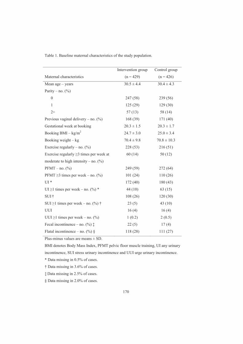
Table 1. Baseline maternal characteristics of the study population.
Intervention group Control group
Maternal characteristics (n = 429) (n = 426)
Mean age – years 30.5 ± 4.4 30.4 ± 4.3
Parity – no. (%)
0 247 (58) 239 (56)
1 125 (29) 129 (30)
2+ 57 (13) 58 (14)
Previous vaginal delivery – no. (%) 168 (39) 171 (40)
Gestational week at booking 20.3 ± 1.5 20.3 ± 1.7
Booking BMI – kg/m2 24.7 ± 3.0 25.0 ± 3.4
Booking weight – kg 70.4 ± 9.8 70.8 ± 10.3
Exercise regularly – no. (%) 228 (53) 216 (51)
Exercise regularly 3 times per week at
moderate to high intensity – no. (%)
60 (14) 50 (12)
PFMT – no. (%) 249 (59) 272 (64)
PFMT 3 times per week – no. (%) 101 (24) 110 (26)
UI * 172 (40) 180 (43)
UI 1 times per week – no. (%) * 44 (10) 63 (15)
SUI † 108 (26) 120 (30)
SUI 1 times per week – no. (%) † 23 (5) 43 (10)
UUI 16 (4) 16 (4)
UUI 1 times per week – no. (%) 1 (0.2) 2 (0.5)
Fecal incontinence – no. (%) ‡ 22 (5) 17 (4)
Flatal incontinence – no. (%) § 118 (28) 111 (27)
Plus-minus values are means ± SD.
BMI denotes Body Mass Index, PFMT pelvic floor muscle training, UI any urinary
incontinence, SUI stress urinary incontinence and UUI urge urinary incontinence.
* Data missing in 0.5% of cases.
† Data missing in 3.6% of cases.
‡ Data missing in 2.5% of cases.
§ Data missing in 2.0% of cases.
170
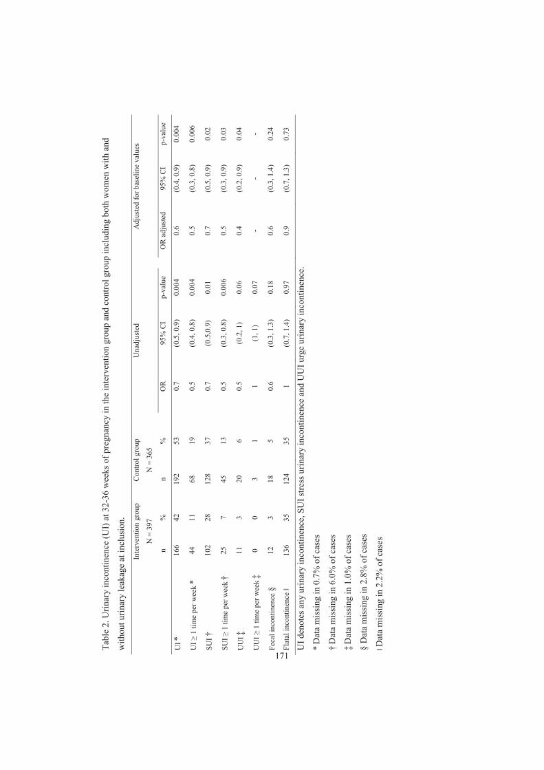
Tabl
e 2.
Urin
ary
inco
ntin
ence
(UI)
at 3
2-36
wee
ks o
f pre
gnan
cy in
the
inte
rven
tion
grou
p an
d co
ntro
l gro
up in
clud
ing
both
wom
en w
ith a
nd
with
out u
rinar
y le
akag
e at
incl
usio
n.
In
terv
entio
n gr
oup
Con
trol g
roup
Una
djus
ted
A
djus
ted
for b
asel
ine
valu
es
N
= 3
97
N =
365
n
%
n %
OR
95
% C
I p-
valu
e
OR
adj
uste
d 95
% C
I p-
valu
e
UI *
166
42
192
53
0.
7 (0
.5, 0
.9)
0.00
4
0.6
(0.4
, 0.9
) 0.
004
UI
1 ti
me
per w
eek *
44
11
68
19
0.
5 (0
.4, 0
.8)
0.00
4
0.5
(0.3
, 0.8
) 0.
006
SUI †
10
2 28
12
8 37
0.7
(0.5
,0.9
) 0.
01
0.
7 (0
.5, 0
.9)
0.02
SUI
1 ti
me
per w
eek †
25
7 45
13
0.5
(0.3
, 0.8
) 0.
006
0.
5 (0
.3, 0
.9)
0.03
UU
I ‡
11
3 20
6
0.
5 (0
.2, 1
) 0.
06
0.
4 (0
.2, 0
.9)
0.04
UU
I 1
tim
e pe
r wee
k ‡ 0
0 3
1
1 (1
, 1)
0.07
- -
-
Feca
l inc
ontin
ence
§
12
3 18
5
0.
6 (0
.3, 1
.3)
0.18
0.6
(0.3
, 1.4
) 0.
24
Flat
al in
cont
inen
ce
136
35
124
35
1
(0.7
, 1.4
) 0.
97
0.
9 (0
.7, 1
.3)
0.73
UI d
enot
es a
ny u
rinar
y in
cont
inen
ce, S
UI s
tress
urin
ary
inco
ntin
ence
and
UU
I urg
e ur
inar
y in
cont
inen
ce.
* Dat
a m
issi
ng in
0.7
% o
f cas
es
† Dat
a m
issi
ng in
6.0
% o
f cas
es
‡ Dat
a m
issi
ng in
1.0
% o
f cas
es
§ D
ata
mis
sing
in 2
.8%
of c
ases
D
ata
mis
sing
in 2
.2%
of c
ases
171
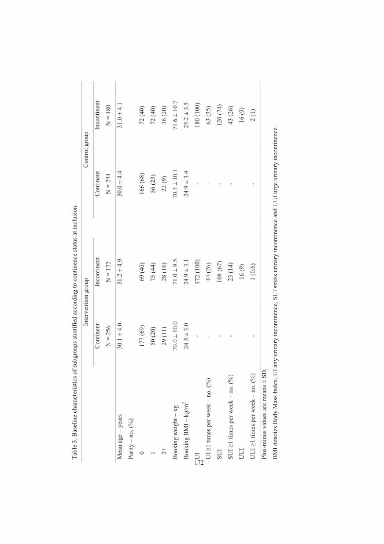
Tabl
e 3.
Bas
elin
e ch
arac
teris
tics o
f sub
grou
ps st
ratif
ied
acco
rdin
g to
con
tinen
ce st
atus
at i
nclu
sion
.
In
terv
entio
n gr
oup
C
ontro
l gro
up
C
ontin
ent
Inco
ntin
ent
C
ontin
ent
Inco
ntin
ent
N
= 2
56
N =
172
N =
244
N
= 1
80
Mea
n ag
e –
year
s 30
.1 ±
4.0
31
.2 ±
4.9
30.0
± 4
.4
31.0
± 4
.1
Parit
y –
no. (
%)
0
177
(69)
69
(40)
166
(68)
72
(40)
1
50 (2
0)
75 (4
4)
56
(23)
72
(40)
2+
29
(11)
28
(16)
22 (9
) 36
(20)
Boo
king
wei
ght –
kg
70
.0 ±
10.
0 71
.0 ±
9.5
70.3
± 1
0.1
71.6
± 1
0.7
Boo
king
BM
I – k
g/m
2 24
.5 ±
3.0
24
.9 ±
3.1
24.9
± 3
.4
25.2
± 3
.5
UI
- 17
2 (1
00)
-
180
(100
)
UI
1 tim
es p
er w
eek
– no
. (%
) -
44 (2
6)
-
63 (3
5)
SUI
- 10
8 (6
7)
-
120
(74)
SUI
1 tim
es p
er w
eek
– no
. (%
) -
23 (1
4)
-
43 (2
6)
UU
I
16 (9
)
16
(9)
UU
I 1
times
per
wee
k –
no. (
%)
- 1
(0.6
)
- 2
(1)
Plus
-min
us v
alue
s are
mea
ns ±
SD
.
BM
I den
otes
Bod
y M
ass I
ndex
, UI a
ny u
rinar
y in
cont
inen
ce, S
UI s
tress
urin
ary
inco
ntin
ence
and
UU
I urg
e ur
inar
y in
cont
inen
ce.
172
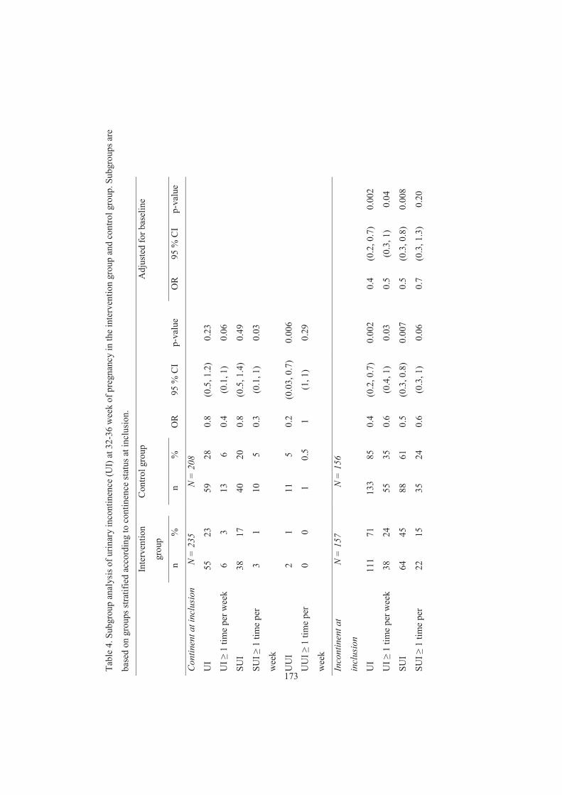
Tabl
e 4.
Sub
grou
p an
alys
is o
f urin
ary
inco
ntin
ence
(UI)
at 3
2-36
wee
k of
pre
gnan
cy in
the
inte
rven
tion
grou
p an
d co
ntro
l gro
up. S
ubgr
oups
are
base
d on
gro
ups s
tratif
ied
acco
rdin
g to
con
tinen
ce st
atus
at i
nclu
sion
.
In
terv
entio
n
grou
p
C
ontro
l gro
up
Adj
uste
d fo
r bas
elin
e
n
%
n
%
OR
95
% C
I p-
valu
e
OR
95
% C
I p-
valu
e
Con
tinen
t at i
nclu
sion
N
= 2
35
N
= 2
08
UI
55
23
59
28
0.
8 (0
.5, 1
.2)
0.23
UI
1 ti
me
per w
eek
6 3
13
6
0.4
(0.1
, 1)
0.06
SUI
38
17
40
20
0.
8 (0
.5, 1
.4)
0.49
SUI
1 ti
me
per
wee
k
3 1
10
5
0.3
(0.1
, 1)
0.03
UU
I 2
1
11
5 0.
2 (0
.03,
0.7
) 0.
006
UU
I 1
tim
e pe
r
wee
k
0 0
1
0.5
1 (1
, 1)
0.29
Inco
ntin
ent a
t
incl
usio
n
N =
157
N =
156
UI
111
71
13
3 85
0.
4 (0
.2, 0
.7)
0.00
2
0.4
(0.2
, 0.7
) 0.
002
UI
1 ti
me
per w
eek
38
24
55
35
0.
6 (0
.4, 1
) 0.
03
0.
5 (0
.3, 1
) 0.
04
SUI
64
45
88
61
0.
5 (0
.3, 0
.8)
0.00
7
0.5
(0.3
, 0.8
) 0.
008
SUI
1 ti
me
per
22
15
35
24
0.
6 (0
.3, 1
) 0.
06
0.
7 (0
.3, 1
.3)
0.20
173

wee
k
UU
I 9
6
9 6
1 (0
.4, 2
.5)
0.97
0.8
(0.3
, 2.5
) 0.
77
UU
I 1
tim
e pe
r
wee
k
0 0
2
1 1
(1, 1
) 0.
15
-
- -
UI d
enot
es a
ny u
rinar
y in
cont
inen
ce, S
UI s
tress
urin
ary
inco
ntin
ence
and
UU
I urg
e ur
inar
y in
cont
inen
ce.
174

Figure legend
Figure 1. Flow chart of study participants.
Word count:
Abstract: 249
Manuscript: 3979
175
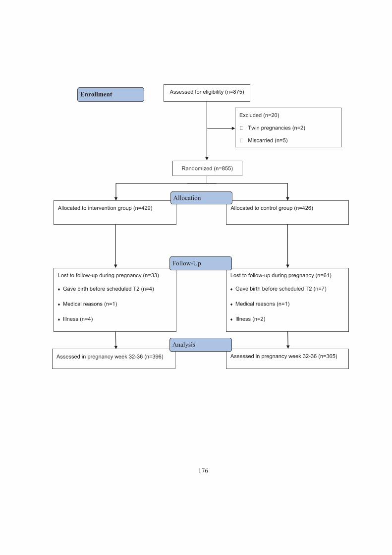
Assessed for eligibility (n=875)
Excluded (n=20)
Twin pregnancies (n=2)
Miscarried (n=5)
Assessed in pregnancy week 32-36 (n=396)
Lost to follow-up during pregnancy (n=33)
Gave birth before scheduled T2 (n=4)
Medical reasons (n=1)
Illness (n=4)
Randomized (n=855)
Enrollment
Allocated to intervention group (n=429) Allocated to control group (n=426)
Lost to follow-up during pregnancy (n=61)
Gave birth before scheduled T2 (n=7)
Medical reasons (n=1)
Illness (n=2)
Assessed in pregnancy week 32-36 (n=365)
Follow-Up
Allocation
Analysis
176

Paper IV
177

Is not included due to copyright

178

184

APPENDICES I-IV
185

186

Appendix I SJEKKLISTE FOR INKLUSJON - ”Effekt av trening under svangerskap”
Spørsmål JA NEI 1. Kvinne >18 år? 2. Bopæl nær sykehuset (<20 km) 3. Kaukasisk, snakker norsk 4. Normal graviditet med lav risiko for
svangerskapskomplikasjoner
5. Informert skriftlig samtykke 6. Diabetes? 7. Tidligere alvorlig
svangerskapskomplikasjoner -preterm fødsel før uke 34 -tidlig innsettende og alvorlig preeklampsi (dvs forløsning før uke 34 eller eklampsi og HELLP syndrom) -alvorlig vekstretardasjon hos foster
8. Alvorlig kroniske sykdommer -astma -hjertesykdom -SLE -hypertoni (medikamentelt behandlet før graviditet) -alvorlig nyresykdom
9. Kjent alkohol- eller medikamentmisbruk
10. Flerlinger 11. Fostermisdannelser 12. Placenta previa 13. Forhøyet blodtrykk (>140/90) i minst
to målinger før sv.uke 20
14. Identifisert høy risiko for preterm fødsel -kort livmorhals -rikelige vaginalblødninger med påvist retroplacentært hematom -vannavgang eller fostervannslekkasje
15. Uegnet til å delta av enhver annen årsak
187

Appendix II
BORGS SKALA
INSTRUKSJON I løpet av treningen vil vi at du skal være oppmerksom i forhold til hvor anstrengende du føler at treningen er. Denne følelsen skal gjenspeile totalsummen av anstrengelse og utmattelse, ved å kombinere alle følelser av fysisk stress, påkjenning og utmattelse. Ikke bry deg om hver enkelt faktor slik som smerte i beina, kortpusthet eller øvelsens intensitet, men forsøk å konsentrere deg om den totale indre følelsen av anstrengelse. Prøv å ikke undervurdere eller overvurdere din følelse av anstrengelse; men vær så presis som du kan.
188

BORGS SKALA 6 7 Meget, meget lett 8 9 Meget lett 10 11 Ganske lett 12 13 Litt anstrengende 14 15 Anstrengende 16 17 Meget anstrengende 18 19 Svært anstrengende 20
189
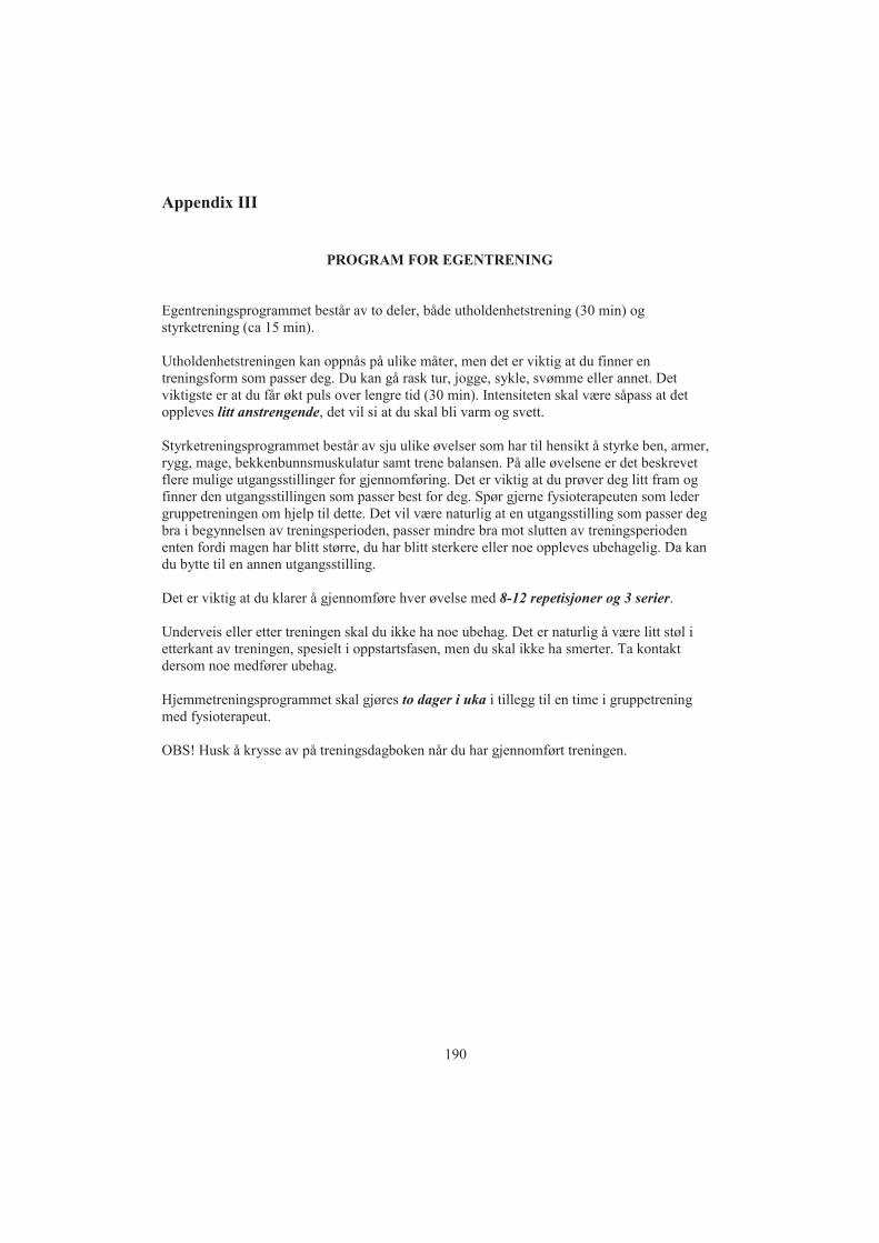
Appendix III
PROGRAM FOR EGENTRENING Egentreningsprogrammet består av to deler, både utholdenhetstrening (30 min) og styrketrening (ca 15 min). Utholdenhetstreningen kan oppnås på ulike måter, men det er viktig at du finner en treningsform som passer deg. Du kan gå rask tur, jogge, sykle, svømme eller annet. Det viktigste er at du får økt puls over lengre tid (30 min). Intensiteten skal være såpass at det oppleves litt anstrengende, det vil si at du skal bli varm og svett. Styrketreningsprogrammet består av sju ulike øvelser som har til hensikt å styrke ben, armer, rygg, mage, bekkenbunnsmuskulatur samt trene balansen. På alle øvelsene er det beskrevet flere mulige utgangsstillinger for gjennomføring. Det er viktig at du prøver deg litt fram og finner den utgangsstillingen som passer best for deg. Spør gjerne fysioterapeuten som leder gruppetreningen om hjelp til dette. Det vil være naturlig at en utgangsstilling som passer deg bra i begynnelsen av treningsperioden, passer mindre bra mot slutten av treningsperioden enten fordi magen har blitt større, du har blitt sterkere eller noe oppleves ubehagelig. Da kan du bytte til en annen utgangsstilling. Det er viktig at du klarer å gjennomføre hver øvelse med 8-12 repetisjoner og 3 serier. Underveis eller etter treningen skal du ikke ha noe ubehag. Det er naturlig å være litt støl i etterkant av treningen, spesielt i oppstartsfasen, men du skal ikke ha smerter. Ta kontakt dersom noe medfører ubehag. Hjemmetreningsprogrammet skal gjøres to dager i uka i tillegg til en time i gruppetrening med fysioterapeut. OBS! Husk å krysse av på treningsdagboken når du har gjennomført treningen.
190
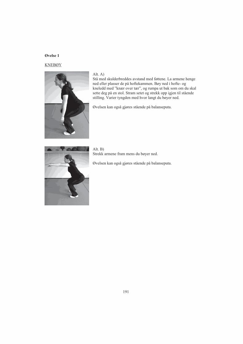
Øvelse 1 KNEBØY
Alt. A) Stå med skulderbreddes avstand med føttene. La armene henge ned eller plasser de på hoftekammen. Bøy ned i hofte- og kneledd med ”knær over tær”, og rumpa ut bak som om du skal sette deg på en stol. Stram setet og strekk opp igjen til stående stilling. Varier tyngden med hvor langt du bøyer ned. Øvelsen kan også gjøres stående på balanseputa.
Alt. B) Strekk armene fram mens du bøyer ned. Øvelsen kan også gjøres stående på balanseputa.
191
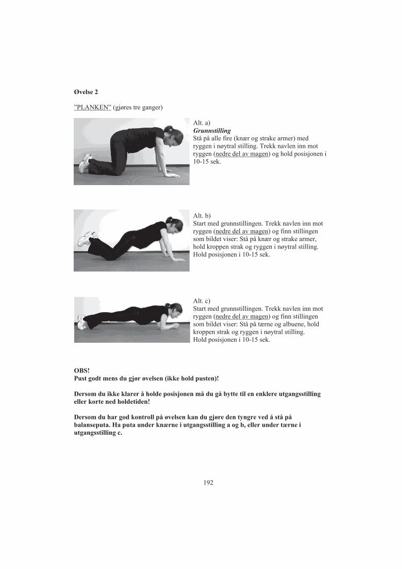
Øvelse 2 ”PLANKEN” (gjøres tre ganger)
Alt. a) Grunnstilling Stå på alle fire (knær og strake armer) med ryggen i nøytral stilling. Trekk navlen inn mot ryggen (nedre del av magen) og hold posisjonen i 10-15 sek.
Alt. b) Start med grunnstillingen. Trekk navlen inn mot ryggen (nedre del av magen) og finn stillingen som bildet viser: Stå på knær og strake armer, hold kroppen strak og ryggen i nøytral stilling. Hold posisjonen i 10-15 sek.
Alt. c) Start med grunnstillingen. Trekk navlen inn mot ryggen (nedre del av magen) og finn stillingen som bildet viser: Stå på tærne og albuene, hold kroppen strak og ryggen i nøytral stilling. Hold posisjonen i 10-15 sek.
OBS! Pust godt mens du gjør øvelsen (ikke hold pusten)! Dersom du ikke klarer å holde posisjonen må du gå bytte til en enklere utgangsstilling eller korte ned holdetiden! Dersom du har god kontroll på øvelsen kan du gjøre den tyngre ved å stå på balanseputa. Ha puta under knærne i utgangsstilling a og b, eller under tærne i utgangsstilling c.
192
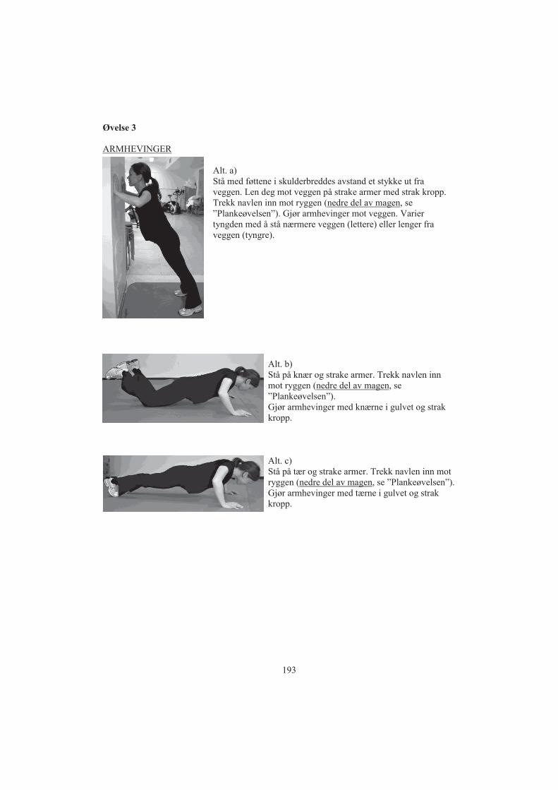
Øvelse 3 ARMHEVINGER
Alt. a) Stå med føttene i skulderbreddes avstand et stykke ut fra veggen. Len deg mot veggen på strake armer med strak kropp. Trekk navlen inn mot ryggen (nedre del av magen, se ”Plankeøvelsen”). Gjør armhevinger mot veggen. Varier tyngden med å stå nærmere veggen (lettere) eller lenger fra veggen (tyngre).
Alt. b) Stå på knær og strake armer. Trekk navlen inn mot ryggen (nedre del av magen, se ”Plankeøvelsen”). Gjør armhevinger med knærne i gulvet og strak kropp.
Alt. c) Stå på tær og strake armer. Trekk navlen inn mot ryggen (nedre del av magen, se ”Plankeøvelsen”). Gjør armhevinger med tærne i gulvet og strak kropp.
193
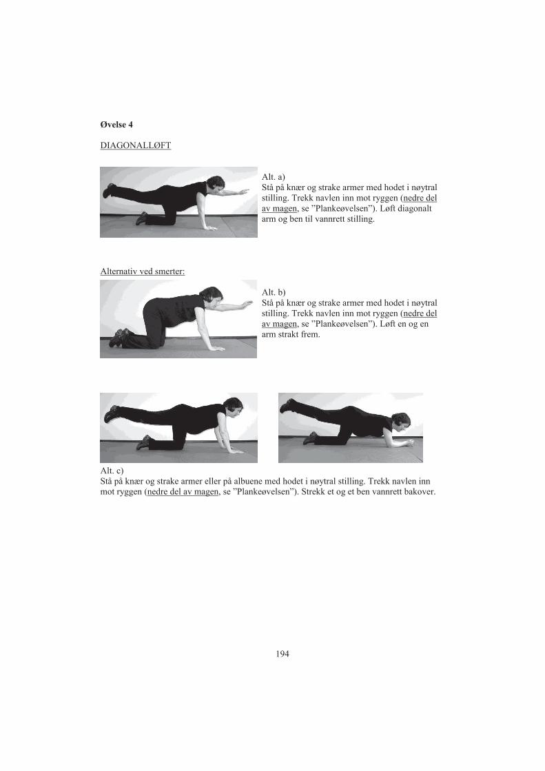
Øvelse 4 DIAGONALLØFT
Alt. a) Stå på knær og strake armer med hodet i nøytral stilling. Trekk navlen inn mot ryggen (nedre del av magen, se ”Plankeøvelsen”). Løft diagonalt arm og ben til vannrett stilling.
Alternativ ved smerter:
Alt. b) Stå på knær og strake armer med hodet i nøytral stilling. Trekk navlen inn mot ryggen (nedre del av magen, se ”Plankeøvelsen”). Løft en og en arm strakt frem.
Alt. c) Stå på knær og strake armer eller på albuene med hodet i nøytral stilling. Trekk navlen inn mot ryggen (nedre del av magen, se ”Plankeøvelsen”). Strekk et og et ben vannrett bakover.
194
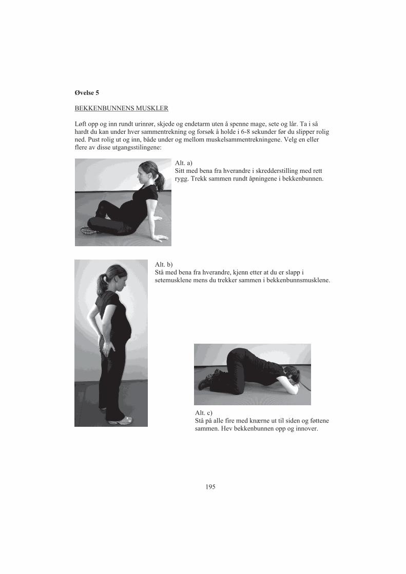
Øvelse 5 BEKKENBUNNENS MUSKLER Løft opp og inn rundt urinrør, skjede og endetarm uten å spenne mage, sete og lår. Ta i så hardt du kan under hver sammentrekning og forsøk å holde i 6-8 sekunder før du slipper rolig ned. Pust rolig ut og inn, både under og mellom muskelsammentrekningene. Velg en eller flere av disse utgangsstilingene:
Alt. a) Sitt med bena fra hverandre i skredderstilling med rett rygg. Trekk sammen rundt åpningene i bekkenbunnen.
Alt. b) Stå med bena fra hverandre, kjenn etter at du er slapp i setemusklene mens du trekker sammen i bekkenbunnsmusklene.
Alt. c) Stå på alle fire med knærne ut til siden og føttene sammen. Hev bekkenbunnen opp og innover.
195
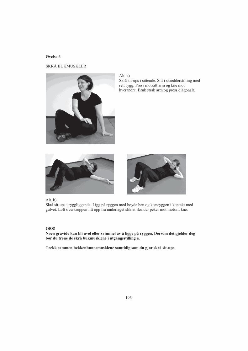
Øvelse 6 SKRÅ BUKMUSKLER
Alt. a) Skrå sit-ups i sittende. Sitt i skredderstilling med rett rygg. Press motsatt arm og kne mot hverandre. Bruk strak arm og press diagonalt.
Alt. b) Skrå sit-ups i ryggliggende. Ligg på ryggen med bøyde ben og korsryggen i kontakt med gulvet. Løft overkroppen litt opp fra underlaget slik at skulder peker mot motsatt kne. OBS! Noen gravide kan bli uvel eller svimmel av å ligge på ryggen. Dersom det gjelder deg bør du trene de skrå bukmusklene i utgangsstilling a. Trekk sammen bekkenbunnsmusklene samtidig som du gjør skrå sit-ups.
196
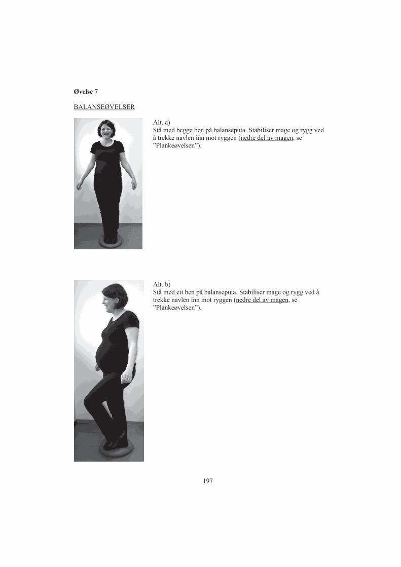
Øvelse 7 BALANSEØVELSER
Alt. a) Stå med begge ben på balanseputa. Stabiliser mage og rygg ved å trekke navlen inn mot ryggen (nedre del av magen, se ”Plankeøvelsen”).
Alt. b) Stå med ett ben på balanseputa. Stabiliser mage og rygg ved å trekke navlen inn mot ryggen (nedre del av magen, se ”Plankeøvelsen”).
197
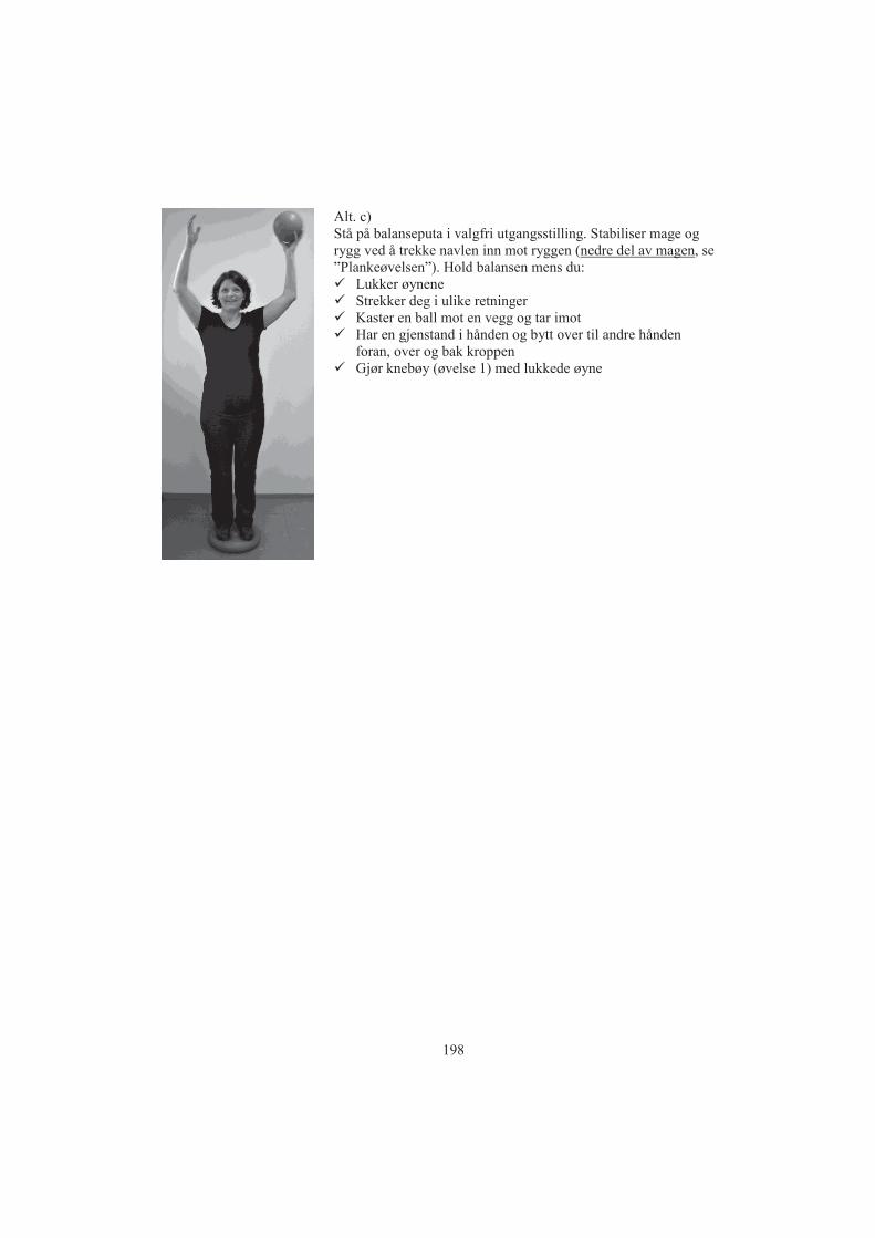
Alt. c) Stå på balanseputa i valgfri utgangsstilling. Stabiliser mage og rygg ved å trekke navlen inn mot ryggen (nedre del av magen, se ”Plankeøvelsen”). Hold balansen mens du:
Lukker øynene Strekker deg i ulike retninger Kaster en ball mot en vegg og tar imot Har en gjenstand i hånden og bytt over til andre hånden
foran, over og bak kroppen Gjør knebøy (øvelse 1) med lukkede øyne
198
14
4
Hvi
s du
øns
ker
åde
lta,
må
du f
ylle
ut
dette
sam
tykk
efor
mul
aret
. Sa
mty
kkef
orm
ular
et l
ever
es t
il pr
osje
ktko
ordi
nato
r ve
d op
pmøt
e fo
r fø
rste
test
.
Jeg
har
lest
info
rmas
jons
skriv
et, o
g ha
r ha
tt an
ledn
ing
til å
still
e sp
ørsm
ål. J
eg
er o
gså
info
rmer
t om
at
jour
nalo
pply
snin
ger
fra
det
aktu
elle
sva
nger
skap
og
føds
el v
il bl
i gje
nnom
gått
og re
gist
rert,
og
sam
tykk
er i
åde
lta i
stud
ien.
Sted
og
dato
………
......
......
......
......
......
-----
-----
-----
-----
------
-----
-----
-----
-U
nder
skrif
t
SAM
TYK
KEE
RK
LÆ
RIN
G F
OR
PR
OSJ
EK
TET
”E
FFE
KT
AV
TR
EN
ING
I SV
AN
GE
RSK
APE
T”
ID-n
umm
er:
(fylle
s ut a
v pr
osje
ktko
ordi
nato
r)
1
Fore
spør
sel o
m å
delta
i en
vi
tens
kape
lig u
nder
søke
lse
EFF
EK
T A
V T
RE
NIN
G I
S
VA
NG
ER
SK
AP
ET
I
FOR
EB
YG
GIN
G O
G
BE
HA
ND
LIN
G A
V
SV
AN
GE
RS
KA
PS
RE
LAT
ER
T
SY
KD
OM
Stud
ien
er e
t sam
arbe
idsp
rosj
ekt m
ello
m K
vinn
eklin
ikke
n ve
d S
t.Ola
vsH
ospi
tal o
g N
TNU
App
endi
x IV
199
15
2
Bak
grun
n og
mål
sett
ing
I løp
et a
v 20
07 o
g 20
08 g
jenn
omfø
res e
t for
skni
ngsp
rosje
kt v
ed S
t. O
lavs
Hos
pita
l, m
ed
mål
setti
ngen
åøk
e ku
nnsk
apen
om
hel
sen
hos g
ravi
de. U
nder
søke
lsen
tar s
ikte
på
åfin
ne
ut m
er o
mef
fekt
er a
v tre
ning
und
er sv
ange
rska
pet,
i for
ebyg
ging
og
beha
ndlin
g av
sv
ange
rska
psre
late
rt sy
kdom
(sva
nger
skap
sdia
bete
s, ov
erve
kt, h
øyt b
lodt
rykk
, ryg
g-og
/elle
r bek
kenr
elat
erte
smer
ter,
urin
-og/
elle
r avf
ørin
gsle
kkas
je).
Des
sute
n vi
l vi
unde
rsøk
e om
tren
inge
n på
virk
er fø
dsel
en.
Hve
m k
an d
elta
, og
hva
inne
bære
r de
ltake
lseA
lle fr
iske
grav
ide
kvin
ner (
18 å
r elle
r mer
), so
m in
nkal
les t
il ru
tinem
essig
ul
traly
dund
ersø
kels
e i 1
8. sv
ange
rska
psuk
e, o
g ha
r bop
el in
nen
20 k
m fr
a sy
kehu
set,
invi
tere
s til
åde
lta i
stud
ien.
For
at v
i ska
l få
vite
mer
om
hel
setil
stan
den
gene
relt,
og
om
he
lsen
i sv
ange
rska
pet s
pesi
elt,
ber v
i om
åfå
ta b
lodp
røve
r av
alle
del
take
rne
i pr
osje
ktet
, og
at a
lle sv
arer
på
spør
resk
jem
a og
gje
nnom
føre
r enk
elte
test
er. T
estin
gen
fore
går p
åsv
ange
rska
pspo
liklin
ikke
n ve
d St
.Ola
vsH
ospi
tal i
sva
nger
skap
suke
20
og 3
2,
sam
t tre
mån
eder
ette
r fød
sele
n. D
u m
åvæ
re p
åsy
kehu
set i
ca
tre ti
mer
ford
i det
skal
gj
øres
en
sukk
erbe
last
ning
stes
t, de
r blo
dprø
vene
skal
tas m
ed to
tim
ers m
ello
mro
m. I
m
ello
mtid
en fy
ller d
u ut
et s
pørr
eskj
ema,
og
en fy
siote
rape
ut u
tføre
r tes
ter p
åde
g av
ry
ggen
, bek
kene
t, ba
lans
en o
g st
yrke
n på
bekk
enbu
nnen
s mus
kula
tur.
Du
møt
er fa
sten
de
til t
estin
gen
og
vil b
li til
budt
mat
ette
rpå.
Des
sute
n be
r vi o
m a
t vi f
år b
enyt
te
info
rmas
jon
om v
ekt o
g bl
odtry
kk fr
a sv
ange
rska
psjo
urna
len,
og
oppl
ysni
nger
om
fø
dsel
sfor
løpe
t og
barn
ets v
ekt o
g le
ngde
fra
føds
elsjo
urna
len.
Enk
elte
tilfe
ldig
utv
algt
e av
del
take
rne
vil b
li sp
urt o
m å
delta
i en
kon
disjo
nste
stin
g og
i en
ultr
alyd
unde
rsøk
else
av
bek
kenb
unns
mus
klen
e og
mag
emus
klen
e.
Når
det
gje
lder
tren
ing
unde
r sva
nger
skap
et, v
et m
an e
nnå
ikke
sikk
ert o
m
svan
gers
kaps
prob
lem
er k
an b
ehan
dles
og
fore
bygg
es, o
g ev
entu
elt h
vilk
e til
tak
som
har
be
st e
ffekt
. Del
take
rne
i den
ne u
nder
søke
lsen
blir
der
for t
ilfel
dig
ford
elt i
to g
rupp
er. E
n da
tam
aski
n av
gjør
ved
lodd
trekn
ing
(ran
dom
iser
ing)
hvi
lken
beh
andl
ing
du sk
al h
a. D
u få
r et t
ilfel
dig
num
mer
i da
taba
sen,
og
bare
pro
sjekt
koor
dina
tor k
jenn
er k
oblin
gen
mel
lom
num
mer
et i
data
base
n og
din
iden
titet
. Del
take
rne
i den
ene
gru
ppen
får d
en
info
rmas
jone
n so
m n
ågi
s rut
inem
essig
til g
ravi
de. D
en a
ndre
gru
ppen
er e
n tre
ning
sgru
ppe,
hvo
r del
take
rne
skal
del
ta i
en ti
mes
felle
stre
ning
lede
t av
fysio
tera
peut
en
gan
g i u
ken,
og
gjen
nom
føre
et e
nkel
t pro
gram
for e
gent
reni
ng p
å45
min
utte
r to
gang
er i
uken
. Tre
ning
en g
jenn
omfø
res m
ello
m u
ke 2
0 og
36
i sva
nger
skap
et.
Ette
r at f
orsk
ning
spro
sjek
tet e
r avs
lutte
t, vi
l alle
del
take
re få
skrif
tlig
info
rmas
jon
om
resu
ltate
ne. H
vis t
reni
ng v
iser s
eg å
ha g
od e
ffekt
som
beh
andl
ing
og fo
reby
ggin
g, v
il de
ltake
rne
i kon
trollg
rupp
en få
info
rmas
jon
om tr
enin
gspr
ogra
mm
et e
tter a
t pro
sjekt
et e
r av
slut
tet.
For å
kunn
e un
ders
øke
lang
tidsv
irkni
ngen
av
treni
ng/ik
ke-tr
enin
g un
der
svan
gers
kape
t, be
r vi o
m sa
mty
kke
til a
t dat
a op
pbev
ares
i 20
år,
slik
at v
i kan
kon
takt
e de
ltake
rne
igje
n fo
r eve
ntue
lle o
ppfø
lgin
gsst
udie
r ved
røre
nde
egen
og
barn
ets h
else
i år
ene
ette
r fød
sele
n.
3
Friv
illig
het o
g sa
mty
kke
•Del
take
lse i
pros
jekt
et e
r friv
illig
.•A
lle d
elta
kere
i pr
osje
ktet
har
rett
til å
trekk
e se
g fra
pro
sjekt
et n
år d
e m
åtte
øns
ke,
uten
at d
ette
får k
onse
kven
ser f
or v
ider
e op
pføl
ging
og
beha
ndlin
g.•A
ll in
form
asjo
n de
ltake
rne
gir i
forb
inde
lse m
ed p
rosje
ktet
, beh
andl
es k
onfid
ensi
elt,
og d
ata
avid
entif
iser
es. A
lle so
m sk
al h
a ko
ntak
t med
de
inns
amle
de d
ata,
er u
nder
lagt
ta
ushe
tspl
ikt i
hen
hold
til F
orva
ltnin
gslo
vens
§13
og
Hel
sepe
rson
ello
vens
§21
. •D
elta
kern
e er
dek
ket a
v Pa
sient
skad
eers
tatn
ings
ordn
inge
n.
Etisk
og
fagl
ig v
urde
ring
•Pro
sjekt
et e
r vur
dert
av R
egio
nal k
omite
for m
edis
insk
fors
knin
gset
ikk,
Reg
ion
Mid
t-N
orge
, og
kom
iteen
har
god
kjen
t at p
rosje
ktet
gje
nnom
føre
s. •P
rosje
ktet
er m
eldt
til p
erso
nver
neom
bude
t for
fors
knin
g, N
orsk
sam
funn
svite
nska
pelig
dat
atje
nest
e.
Ans
varli
ge p
rosje
ktle
dere
er s
eksjo
nsov
erle
ge v
ed F
ødea
vdel
inge
n, S
t. O
lavs
Hos
pita
l, K
jell
Åsm
und
Salv
esen
, for
skni
ngsr
ådgi
ver v
ed K
linik
k fo
r klin
iske
serv
icef
unks
jone
r, St
. Ola
vs h
ospi
tal,
Siv
Mør
kved
og d
okto
rgra
dsst
ipen
diat
ved
Inst
itutt
for
sam
funn
smed
isin
, NTN
U,
Sign
e N
ilsse
nSt
afne
.
HV
IS D
U Ø
NSK
ER Å
DEL
TA
, ELL
ER H
AR
SPØ
RSM
ÅL
OM
PR
OSJ
EK
TET
, BE
S D
U K
ON
TA
KTE
:
Sign
e N
ilsse
nSt
afne
(pro
sjek
tkoo
rdin
ator
)
Du
kan
ta k
onta
kt p
åfø
lgen
de m
åte:
•E-p
ost:
sign
e.n.
staf
ne@
ntnu
.no
•Sen
d SM
S til
46
85 6
2 67
. Skr
iv p
rosj
ekt,
ditt
navn
og
tele
fonn
umm
er•T
elef
on: 7
3 59
75
22 (k
onto
r) /
46 8
5 62
67
(mob
il)
200

201

202
Dissertations at the Faculty of Medicine, NTNU 1977
1. Knut Joachim Berg: EFFECT OF ACETYLSALICYLIC ACID ON RENAL FUNCTION 2. Karl Erik Viken and Arne Ødegaard: STUDIES ON HUMAN MONOCYTES CULTURED IN
VITRO 1978
3. Karel Bjørn Cyvin: CONGENITAL DISLOCATION OF THE HIP JOINT. 4. Alf O. Brubakk: METHODS FOR STUDYING FLOW DYNAMICS IN THE LEFT
VENTRICLE AND THE AORTA IN MAN. 1979
5. Geirmund Unsgaard: CYTOSTATIC AND IMMUNOREGULATORY ABILITIES OF HUMAN BLOOD MONOCYTES CULTURED IN VITRO
1980 6. Størker Jørstad: URAEMIC TOXINS 7. Arne Olav Jenssen: SOME RHEOLOGICAL, CHEMICAL AND STRUCTURAL
PROPERTIES OF MUCOID SPUTUM FROM PATIENTS WITH CHRONIC OBSTRUCTIVE BRONCHITIS
1981 8. Jens Hammerstrøm: CYTOSTATIC AND CYTOLYTIC ACTIVITY OF HUMAN
MONOCYTES AND EFFUSION MACROPHAGES AGAINST TUMOR CELLS IN VITRO 1983
9. Tore Syversen: EFFECTS OF METHYLMERCURY ON RAT BRAIN PROTEIN. 10. Torbjørn Iversen: SQUAMOUS CELL CARCINOMA OF THE VULVA.
1984 11. Tor-Erik Widerøe: ASPECTS OF CONTINUOUS AMBULATORY PERITONEAL
DIALYSIS. 12. Anton Hole: ALTERATIONS OF MONOCYTE AND LYMPHOCYTE FUNCTIONS IN
REALTION TO SURGERY UNDER EPIDURAL OR GENERAL ANAESTHESIA. 13. Terje Terjesen: FRACTURE HEALING AND STRESS-PROTECTION AFTER METAL
PLATE FIXATION AND EXTERNAL FIXATION. 14. Carsten Saunte: CLUSTER HEADACHE SYNDROME. 15. Inggard Lereim: TRAFFIC ACCIDENTS AND THEIR CONSEQUENCES. 16. Bjørn Magne Eggen: STUDIES IN CYTOTOXICITY IN HUMAN ADHERENT
MONONUCLEAR BLOOD CELLS. 17. Trond Haug: FACTORS REGULATING BEHAVIORAL EFFECTS OG DRUGS.
1985 18. Sven Erik Gisvold: RESUSCITATION AFTER COMPLETE GLOBAL BRAIN ISCHEMIA. 19. Terje Espevik: THE CYTOSKELETON OF HUMAN MONOCYTES. 20. Lars Bevanger: STUDIES OF THE Ibc (c) PROTEIN ANTIGENS OF GROUP B
STREPTOCOCCI. 21. Ole-Jan Iversen: RETROVIRUS-LIKE PARTICLES IN THE PATHOGENESIS OF
PSORIASIS. 22. Lasse Eriksen: EVALUATION AND TREATMENT OF ALCOHOL DEPENDENT
BEHAVIOUR. 23. Per I. Lundmo: ANDROGEN METABOLISM IN THE PROSTATE.
1986 24. Dagfinn Berntzen: ANALYSIS AND MANAGEMENT OF EXPERIMENTAL AND
CLINICAL PAIN. 25. Odd Arnold Kildahl-Andersen: PRODUCTION AND CHARACTERIZATION OF
MONOCYTE-DERIVED CYTOTOXIN AND ITS ROLE IN MONOCYTE-MEDIATED CYTOTOXICITY.
26. Ola Dale: VOLATILE ANAESTHETICS. 1987
27. Per Martin Kleveland: STUDIES ON GASTRIN. 28. Audun N. Øksendal: THE CALCIUM PARADOX AND THE HEART. 29. Vilhjalmur R. Finsen: HIP FRACTURES
203
1988 30. Rigmor Austgulen: TUMOR NECROSIS FACTOR: A MONOCYTE-DERIVED
REGULATOR OF CELLULAR GROWTH. 31. Tom-Harald Edna: HEAD INJURIES ADMITTED TO HOSPITAL. 32. Joseph D. Borsi: NEW ASPECTS OF THE CLINICAL PHARMACOKINETICS OF
METHOTREXATE. 33. Olav F. M. Sellevold: GLUCOCORTICOIDS IN MYOCARDIAL PROTECTION. 34. Terje Skjærpe: NONINVASIVE QUANTITATION OF GLOBAL PARAMETERS ON LEFT
VENTRICULAR FUNCTION: THE SYSTOLIC PULMONARY ARTERY PRESSURE AND CARDIAC OUTPUT.
35. Eyvind Rødahl: STUDIES OF IMMUNE COMPLEXES AND RETROVIRUS-LIKE ANTIGENS IN PATIENTS WITH ANKYLOSING SPONDYLITIS.
36. Ketil Thorstensen: STUDIES ON THE MECHANISMS OF CELLULAR UPTAKE OF IRON FROM TRANSFERRIN.
37. Anna Midelfart: STUDIES OF THE MECHANISMS OF ION AND FLUID TRANSPORT IN THE BOVINE CORNEA.
38. Eirik Helseth: GROWTH AND PLASMINOGEN ACTIVATOR ACTIVITY OF HUMAN GLIOMAS AND BRAIN METASTASES - WITH SPECIAL REFERENCE TO TRANSFORMING GROWTH FACTOR BETA AND THE EPIDERMAL GROWTH FACTOR RECEPTOR.
39. Petter C. Borchgrevink: MAGNESIUM AND THE ISCHEMIC HEART. 40. Kjell-Arne Rein: THE EFFECT OF EXTRACORPOREAL CIRCULATION ON
SUBCUTANEOUS TRANSCAPILLARY FLUID BALANCE. 41. Arne Kristian Sandvik: RAT GASTRIC HISTAMINE. 42. Carl Bredo Dahl: ANIMAL MODELS IN PSYCHIATRY.
1989 43. Torbjørn A. Fredriksen: CERVICOGENIC HEADACHE. 44. Rolf A. Walstad: CEFTAZIDIME. 45. Rolf Salvesen: THE PUPIL IN CLUSTER HEADACHE. 46. Nils Petter Jørgensen: DRUG EXPOSURE IN EARLY PREGNANCY. 47. Johan C. Ræder: PREMEDICATION AND GENERAL ANAESTHESIA IN OUTPATIENT
GYNECOLOGICAL SURGERY. 48. M. R. Shalaby: IMMUNOREGULATORY PROPERTIES OF TNF- AND THE RELATED
CYTOKINES. 49. Anders Waage: THE COMPLEX PATTERN OF CYTOKINES IN SEPTIC SHOCK. 50. Bjarne Christian Eriksen: ELECTROSTIMULATION OF THE PELVIC FLOOR IN FEMALE
URINARY INCONTINENCE. 51. Tore B. Halvorsen: PROGNOSTIC FACTORS IN COLORECTAL CANCER.
1990 52. Asbjørn Nordby: CELLULAR TOXICITY OF ROENTGEN CONTRAST MEDIA. 53. Kåre E. Tvedt: X-RAY MICROANALYSIS OF BIOLOGICAL MATERIAL. 54. Tore C. Stiles: COGNITIVE VULNERABILITY FACTORS IN THE DEVELOPMENT AND
MAINTENANCE OF DEPRESSION. 55. Eva Hofsli: TUMOR NECROSIS FACTOR AND MULTIDRUG RESISTANCE. 56. Helge S. Haarstad: TROPHIC EFFECTS OF CHOLECYSTOKININ AND SECRETIN ON
THE RAT PANCREAS. 57. Lars Engebretsen: TREATMENT OF ACUTE ANTERIOR CRUCIATE LIGAMENT
INJURIES. 58. Tarjei Rygnestad: DELIBERATE SELF-POISONING IN TRONDHEIM. 59. Arne Z. Henriksen: STUDIES ON CONSERVED ANTIGENIC DOMAINS ON MAJOR
OUTER MEMBRANE PROTEINS FROM ENTEROBACTERIA. 60. Steinar Westin: UNEMPLOYMENT AND HEALTH: Medical and social consequences of a
factory closure in a ten-year controlled follow-up study. 61. Ylva Sahlin: INJURY REGISTRATION, a tool for accident preventive work. 62. Helge Bjørnstad Pettersen: BIOSYNTHESIS OF COMPLEMENT BY HUMAN ALVEOLAR
MACROPHAGES WITH SPECIAL REFERENCE TO SARCOIDOSIS. 63. Berit Schei: TRAPPED IN PAINFUL LOVE. 64. Lars J. Vatten: PROSPECTIVE STUDIES OF THE RISK OF BREAST CANCER IN A
COHORT OF NORWEGIAN WOMAN.
204
1991 65. Kåre Bergh: APPLICATIONS OF ANTI-C5a SPECIFIC MONOCLONAL ANTIBODIES FOR
THE ASSESSMENT OF COMPLEMENT ACTIVATION. 66. Svein Svenningsen: THE CLINICAL SIGNIFICANCE OF INCREASED FEMORAL
ANTEVERSION. 67. Olbjørn Klepp: NONSEMINOMATOUS GERM CELL TESTIS CANCER: THERAPEUTIC
OUTCOME AND PROGNOSTIC FACTORS. 68. Trond Sand: THE EFFECTS OF CLICK POLARITY ON BRAINSTEM AUDITORY
EVOKED POTENTIALS AMPLITUDE, DISPERSION, AND LATENCY VARIABLES. 69. Kjetil B. Åsbakk: STUDIES OF A PROTEIN FROM PSORIATIC SCALE, PSO P27, WITH
RESPECT TO ITS POTENTIAL ROLE IN IMMUNE REACTIONS IN PSORIASIS. 70. Arnulf Hestnes: STUDIES ON DOWN´S SYNDROME. 71. Randi Nygaard: LONG-TERM SURVIVAL IN CHILDHOOD LEUKEMIA. 72. Bjørn Hagen: THIO-TEPA. 73. Svein Anda: EVALUATION OF THE HIP JOINT BY COMPUTED TOMOGRAMPHY AND
ULTRASONOGRAPHY. 1992
74. Martin Svartberg: AN INVESTIGATION OF PROCESS AND OUTCOME OF SHORT-TERM PSYCHODYNAMIC PSYCHOTHERAPY.
75. Stig Arild Slørdahl: AORTIC REGURGITATION. 76. Harold C Sexton: STUDIES RELATING TO THE TREATMENT OF SYMPTOMATIC NON-
PSYCHOTIC PATIENTS. 77. Maurice B. Vincent: VASOACTIVE PEPTIDES IN THE OCULAR/FOREHEAD AREA. 78. Terje Johannessen: CONTROLLED TRIALS IN SINGLE SUBJECTS. 79. Turid Nilsen: PYROPHOSPHATE IN HEPATOCYTE IRON METABOLISM. 80. Olav Haraldseth: NMR SPECTROSCOPY OF CEREBRAL ISCHEMIA AND REPERFUSION
IN RAT. 81. Eiliv Brenna: REGULATION OF FUNCTION AND GROWTH OF THE OXYNTIC
MUCOSA. 1993
82. Gunnar Bovim: CERVICOGENIC HEADACHE. 83. Jarl Arne Kahn: ASSISTED PROCREATION. 84. Bjørn Naume: IMMUNOREGULATORY EFFECTS OF CYTOKINES ON NK CELLS. 85. Rune Wiseth: AORTIC VALVE REPLACEMENT. 86. Jie Ming Shen: BLOOD FLOW VELOCITY AND RESPIRATORY STUDIES. 87. Piotr Kruszewski: SUNCT SYNDROME WITH SPECIAL REFERENCE TO THE
AUTONOMIC NERVOUS SYSTEM. 88. Mette Haase Moen: ENDOMETRIOSIS. 89. Anne Vik: VASCULAR GAS EMBOLISM DURING AIR INFUSION AND AFTER
DECOMPRESSION IN PIGS. 90. Lars Jacob Stovner: THE CHIARI TYPE I MALFORMATION. 91. Kjell Å. Salvesen: ROUTINE ULTRASONOGRAPHY IN UTERO AND DEVELOPMENT IN
CHILDHOOD. 1994
92. Nina-Beate Liabakk: DEVELOPMENT OF IMMUNOASSAYS FOR TNF AND ITS SOLUBLE RECEPTORS.
93. Sverre Helge Torp: erbB ONCOGENES IN HUMAN GLIOMAS AND MENINGIOMAS. 94. Olav M. Linaker: MENTAL RETARDATION AND PSYCHIATRY. Past and present. 95. Per Oscar Feet: INCREASED ANTIDEPRESSANT AND ANTIPANIC EFFECT IN
COMBINED TREATMENT WITH DIXYRAZINE AND TRICYCLIC ANTIDEPRESSANTS. 96. Stein Olav Samstad: CROSS SECTIONAL FLOW VELOCITY PROFILES FROM TWO-
DIMENSIONAL DOPPLER ULTRASOUND: Studies on early mitral blood flow. 97. Bjørn Backe: STUDIES IN ANTENATAL CARE. 98. Gerd Inger Ringdal: QUALITY OF LIFE IN CANCER PATIENTS. 99. Torvid Kiserud: THE DUCTUS VENOSUS IN THE HUMAN FETUS. 100. Hans E. Fjøsne: HORMONAL REGULATION OF PROSTATIC METABOLISM. 101. Eylert Brodtkorb: CLINICAL ASPECTS OF EPILEPSY IN THE MENTALLY RETARDED. 102. Roar Juul: PEPTIDERGIC MECHANISMS IN HUMAN SUBARACHNOID HEMORRHAGE. 103. Unni Syversen: CHROMOGRANIN A. Phsysiological and Clinical Role.
205
1995 104. Odd Gunnar Brakstad: THERMOSTABLE NUCLEASE AND THE nuc GENE IN THE
DIAGNOSIS OF Staphylococcus aureus INFECTIONS. 105. Terje Engan: NUCLEAR MAGNETIC RESONANCE (NMR) SPECTROSCOPY OF PLASMA
IN MALIGNANT DISEASE. 106. Kirsten Rasmussen: VIOLENCE IN THE MENTALLY DISORDERED. 107. Finn Egil Skjeldestad: INDUCED ABORTION: Timetrends and Determinants. 108. Roar Stenseth: THORACIC EPIDURAL ANALGESIA IN AORTOCORONARY BYPASS
SURGERY. 109. Arild Faxvaag: STUDIES OF IMMUNE CELL FUNCTION in mice infected with MURINE
RETROVIRUS. 1996
110. Svend Aakhus: NONINVASIVE COMPUTERIZED ASSESSMENT OF LEFT VENTRICULAR FUNCTION AND SYSTEMIC ARTERIAL PROPERTIES. Methodology and some clinical applications.
111. Klaus-Dieter Bolz: INTRAVASCULAR ULTRASONOGRAPHY. 112. Petter Aadahl: CARDIOVASCULAR EFFECTS OF THORACIC AORTIC CROSS-
CLAMPING. 113. Sigurd Steinshamn: CYTOKINE MEDIATORS DURING GRANULOCYTOPENIC
INFECTIONS. 114. Hans Stifoss-Hanssen: SEEKING MEANING OR HAPPINESS? 115. Anne Kvikstad: LIFE CHANGE EVENTS AND MARITAL STATUS IN RELATION TO
RISK AND PROGNOSIS OF CANCER. 116. Torbjørn Grøntvedt: TREATMENT OF ACUTE AND CHRONIC ANTERIOR CRUCIATE
LIGAMENT INJURIES. A clinical and biomechanical study. 117. Sigrid Hørven Wigers: CLINICAL STUDIES OF FIBROMYALGIA WITH FOCUS ON
ETIOLOGY, TREATMENT AND OUTCOME. 118. Jan Schjøtt: MYOCARDIAL PROTECTION: Functional and Metabolic Characteristics of Two
Endogenous Protective Principles. 119. Marit Martinussen: STUDIES OF INTESTINAL BLOOD FLOW AND ITS RELATION TO
TRANSITIONAL CIRCULATORY ADAPATION IN NEWBORN INFANTS. 120. Tomm B. Müller: MAGNETIC RESONANCE IMAGING IN FOCAL CEREBRAL
ISCHEMIA. 121. Rune Haaverstad: OEDEMA FORMATION OF THE LOWER EXTREMITIES. 122. Magne Børset: THE ROLE OF CYTOKINES IN MULTIPLE MYELOMA, WITH SPECIAL
REFERENCE TO HEPATOCYTE GROWTH FACTOR. 123. Geir Smedslund: A THEORETICAL AND EMPIRICAL INVESTIGATION OF SMOKING,
STRESS AND DISEASE: RESULTS FROM A POPULATION SURVEY. 1997
124. Torstein Vik: GROWTH, MORBIDITY, AND PSYCHOMOTOR DEVELOPMENT IN INFANTS WHO WERE GROWTH RETARDED IN UTERO.
125. Siri Forsmo: ASPECTS AND CONSEQUENCES OF OPPORTUNISTIC SCREENING FOR CERVICAL CANCER. Results based on data from three Norwegian counties.
126. Jon S. Skranes: CEREBRAL MRI AND NEURODEVELOPMENTAL OUTCOME IN VERY LOW BIRTH WEIGHT (VLBW) CHILDREN. A follow-up study of a geographically based year cohort of VLBW children at ages one and six years.
127. Knut Bjørnstad: COMPUTERIZED ECHOCARDIOGRAPHY FOR EVALUTION OF CORONARY ARTERY DISEASE.
128. Grethe Elisabeth Borchgrevink: DIAGNOSIS AND TREATMENT OF WHIPLASH/NECK SPRAIN INJURIES CAUSED BY CAR ACCIDENTS.
129. Tor Elsås: NEUROPEPTIDES AND NITRIC OXIDE SYNTHASE IN OCULAR AUTONOMIC AND SENSORY NERVES.
130. Rolf W. Gråwe: EPIDEMIOLOGICAL AND NEUROPSYCHOLOGICAL PERSPECTIVES ON SCHIZOPHRENIA.
131. Tonje Strømholm: CEREBRAL HAEMODYNAMICS DURING THORACIC AORTIC CROSSCLAMPING. An experimental study in pigs
1998 132. Martinus Bråten: STUDIES ON SOME PROBLEMS REALTED TO INTRAMEDULLARY
NAILING OF FEMORAL FRACTURES.
206
133. Ståle Nordgård: PROLIFERATIVE ACTIVITY AND DNA CONTENT AS PROGNOSTIC INDICATORS IN ADENOID CYSTIC CARCINOMA OF THE HEAD AND NECK.
134. Egil Lien: SOLUBLE RECEPTORS FOR TNF AND LPS: RELEASE PATTERN AND POSSIBLE SIGNIFICANCE IN DISEASE.
135. Marit Bjørgaas: HYPOGLYCAEMIA IN CHILDREN WITH DIABETES MELLITUS 136. Frank Skorpen: GENETIC AND FUNCTIONAL ANALYSES OF DNA REPAIR IN HUMAN
CELLS. 137. Juan A. Pareja: SUNCT SYNDROME. ON THE CLINICAL PICTURE. ITS DISTINCTION
FROM OTHER, SIMILAR HEADACHES. 138. Anders Angelsen: NEUROENDOCRINE CELLS IN HUMAN PROSTATIC CARCINOMAS
AND THE PROSTATIC COMPLEX OF RAT, GUINEA PIG, CAT AND DOG. 139. Fabio Antonaci: CHRONIC PAROXYSMAL HEMICRANIA AND HEMICRANIA
CONTINUA: TWO DIFFERENT ENTITIES? 140. Sven M. Carlsen: ENDOCRINE AND METABOLIC EFFECTS OF METFORMIN WITH
SPECIAL EMPHASIS ON CARDIOVASCULAR RISK FACTORES. 1999
141. Terje A. Murberg: DEPRESSIVE SYMPTOMS AND COPING AMONG PATIENTS WITH CONGESTIVE HEART FAILURE.
142. Harm-Gerd Karl Blaas: THE EMBRYONIC EXAMINATION. Ultrasound studies on the development of the human embryo.
143. Noèmi Becser Andersen:THE CEPHALIC SENSORY NERVES IN UNILATERAL HEADACHES. Anatomical background and neurophysiological evaluation.
144. Eli-Janne Fiskerstrand: LASER TREATMENT OF PORT WINE STAINS. A study of the efficacy and limitations of the pulsed dye laser. Clinical and morfological analyses aimed at improving the therapeutic outcome.
145. Bård Kulseng: A STUDY OF ALGINATE CAPSULE PROPERTIES AND CYTOKINES IN RELATION TO INSULIN DEPENDENT DIABETES MELLITUS.
146. Terje Haug: STRUCTURE AND REGULATION OF THE HUMAN UNG GENE ENCODING URACIL-DNA GLYCOSYLASE.
147. Heidi Brurok: MANGANESE AND THE HEART. A Magic Metal with Diagnostic and Therapeutic Possibilites.
148. Agnes Kathrine Lie: DIAGNOSIS AND PREVALENCE OF HUMAN PAPILLOMAVIRUS INFECTION IN CERVICAL INTRAEPITELIAL NEOPLASIA. Relationship to Cell Cycle Regulatory Proteins and HLA DQBI Genes.
149. Ronald Mårvik: PHARMACOLOGICAL, PHYSIOLOGICAL AND PATHOPHYSIOLOGICAL STUDIES ON ISOLATED STOMACS.
150. Ketil Jarl Holen: THE ROLE OF ULTRASONOGRAPHY IN THE DIAGNOSIS AND TREATMENT OF HIP DYSPLASIA IN NEWBORNS.
151. Irene Hetlevik: THE ROLE OF CLINICAL GUIDELINES IN CARDIOVASCULAR RISK INTERVENTION IN GENERAL PRACTICE.
152. Katarina Tunòn: ULTRASOUND AND PREDICTION OF GESTATIONAL AGE. 153. Johannes Soma: INTERACTION BETWEEN THE LEFT VENTRICLE AND THE SYSTEMIC
ARTERIES. 154. Arild Aamodt: DEVELOPMENT AND PRE-CLINICAL EVALUATION OF A CUSTOM-
MADE FEMORAL STEM. 155. Agnar Tegnander: DIAGNOSIS AND FOLLOW-UP OF CHILDREN WITH SUSPECTED OR
KNOWN HIP DYSPLASIA. 156. Bent Indredavik: STROKE UNIT TREATMENT: SHORT AND LONG-TERM EFFECTS 157. Jolanta Vanagaite Vingen: PHOTOPHOBIA AND PHONOPHOBIA IN PRIMARY
HEADACHES 2000
158. Ola Dalsegg Sæther: PATHOPHYSIOLOGY DURING PROXIMAL AORTIC CROSS-CLAMPING CLINICAL AND EXPERIMENTAL STUDIES
159. xxxxxxxxx (blind number) 160. Christina Vogt Isaksen: PRENATAL ULTRASOUND AND POSTMORTEM FINDINGS – A
TEN YEAR CORRELATIVE STUDY OF FETUSES AND INFANTS WITH DEVELOPMENTAL ANOMALIES.
161. Holger Seidel: HIGH-DOSE METHOTREXATE THERAPY IN CHILDREN WITH ACUTE LYMPHOCYTIC LEUKEMIA: DOSE, CONCENTRATION, AND EFFECT CONSIDERATIONS.
207
162. Stein Hallan: IMPLEMENTATION OF MODERN MEDICAL DECISION ANALYSIS INTO CLINICAL DIAGNOSIS AND TREATMENT.
163. Malcolm Sue-Chu: INVASIVE AND NON-INVASIVE STUDIES IN CROSS-COUNTRY SKIERS WITH ASTHMA-LIKE SYMPTOMS.
164. Ole-Lars Brekke: EFFECTS OF ANTIOXIDANTS AND FATTY ACIDS ON TUMOR NECROSIS FACTOR-INDUCED CYTOTOXICITY.
165. Jan Lundbom: AORTOCORONARY BYPASS SURGERY: CLINICAL ASPECTS, COST CONSIDERATIONS AND WORKING ABILITY.
166. John-Anker Zwart: LUMBAR NERVE ROOT COMPRESSION, BIOCHEMICAL AND NEUROPHYSIOLOGICAL ASPECTS.
167. Geir Falck: HYPEROSMOLALITY AND THE HEART. 168. Eirik Skogvoll: CARDIAC ARREST Incidence, Intervention and Outcome. 169. Dalius Bansevicius: SHOULDER-NECK REGION IN CERTAIN HEADACHES AND
CHRONIC PAIN SYNDROMES. 170. Bettina Kinge: REFRACTIVE ERRORS AND BIOMETRIC CHANGES AMONG
UNIVERSITY STUDENTS IN NORWAY. 171. Gunnar Qvigstad: CONSEQUENCES OF HYPERGASTRINEMIA IN MAN 172. Hanne Ellekjær: EPIDEMIOLOGICAL STUDIES OF STROKE IN A NORWEGIAN
POPULATION. INCIDENCE, RISK FACTORS AND PROGNOSIS 173. Hilde Grimstad: VIOLENCE AGAINST WOMEN AND PREGNANCY OUTCOME. 174. Astrid Hjelde: SURFACE TENSION AND COMPLEMENT ACTIVATION: Factors
influencing bubble formation and bubble effects after decompression. 175. Kjell A. Kvistad: MR IN BREAST CANCER – A CLINICAL STUDY. 176. Ivar Rossvoll: ELECTIVE ORTHOPAEDIC SURGERY IN A DEFINED POPULATION.
Studies on demand, waiting time for treatment and incapacity for work. 177. Carina Seidel: PROGNOSTIC VALUE AND BIOLOGICAL EFFECTS OF HEPATOCYTE
GROWTH FACTOR AND SYNDECAN-1 IN MULTIPLE MYELOMA. 2001
178. Alexander Wahba: THE INFLUENCE OF CARDIOPULMONARY BYPASS ON PLATELET FUNCTION AND BLOOD COAGULATION – DETERMINANTS AND CLINICAL CONSEQUENSES
179. Marcus Schmitt-Egenolf: THE RELEVANCE OF THE MAJOR hISTOCOMPATIBILITY COMPLEX FOR THE GENETICS OF PSORIASIS
180. Odrun Arna Gederaas: BIOLOGICAL MECHANISMS INVOLVED IN 5-AMINOLEVULINIC ACID BASED PHOTODYNAMIC THERAPY
181. Pål Richard Romundstad: CANCER INCIDENCE AMONG NORWEGIAN ALUMINIUM WORKERS
182. Henrik Hjorth-Hansen: NOVEL CYTOKINES IN GROWTH CONTROL AND BONE DISEASE OF MULTIPLE MYELOMA
183. Gunnar Morken: SEASONAL VARIATION OF HUMAN MOOD AND BEHAVIOUR 184. Bjørn Olav Haugen: MEASUREMENT OF CARDIAC OUTPUT AND STUDIES OF
VELOCITY PROFILES IN AORTIC AND MITRAL FLOW USING TWO- AND THREE-DIMENSIONAL COLOUR FLOW IMAGING
185. Geir Bråthen: THE CLASSIFICATION AND CLINICAL DIAGNOSIS OF ALCOHOL-RELATED SEIZURES
186. Knut Ivar Aasarød: RENAL INVOLVEMENT IN INFLAMMATORY RHEUMATIC DISEASE. A Study of Renal Disease in Wegener’s Granulomatosis and in Primary Sjögren’s Syndrome
187. Trude Helen Flo: RESEPTORS INVOLVED IN CELL ACTIVATION BY DEFINED URONIC ACID POLYMERS AND BACTERIAL COMPONENTS
188. Bodil Kavli: HUMAN URACIL-DNA GLYCOSYLASES FROM THE UNG GENE: STRUCTRUAL BASIS FOR SUBSTRATE SPECIFICITY AND REPAIR
189. Liv Thommesen: MOLECULAR MECHANISMS INVOLVED IN TNF- AND GASTRIN-MEDIATED GENE REGULATION
190. Turid Lingaas Holmen: SMOKING AND HEALTH IN ADOLESCENCE; THE NORD-TRØNDELAG HEALTH STUDY, 1995-97
191. Øyvind Hjertner: MULTIPLE MYELOMA: INTERACTIONS BETWEEN MALIGNANT PLASMA CELLS AND THE BONE MICROENVIRONMENT
208
192. Asbjørn Støylen: STRAIN RATE IMAGING OF THE LEFT VENTRICLE BY ULTRASOUND. FEASIBILITY, CLINICAL VALIDATION AND PHYSIOLOGICAL ASPECTS
193. Kristian Midthjell: DIABETES IN ADULTS IN NORD-TRØNDELAG. PUBLIC HEALTH ASPECTS OF DIABETES MELLITUS IN A LARGE, NON-SELECTED NORWEGIAN POPULATION.
194. Guanglin Cui: FUNCTIONAL ASPECTS OF THE ECL CELL IN RODENTS 195. Ulrik Wisløff: CARDIAC EFFECTS OF AEROBIC ENDURANCE TRAINING:
HYPERTROPHY, CONTRACTILITY AND CALCUIM HANDLING IN NORMAL AND FAILING HEART
196. Øyvind Halaas: MECHANISMS OF IMMUNOMODULATION AND CELL-MEDIATED CYTOTOXICITY INDUCED BY BACTERIAL PRODUCTS
197. Tore Amundsen: PERFUSION MR IMAGING IN THE DIAGNOSIS OF PULMONARY EMBOLISM
198. Nanna Kurtze: THE SIGNIFICANCE OF ANXIETY AND DEPRESSION IN FATIQUE AND PATTERNS OF PAIN AMONG INDIVIDUALS DIAGNOSED WITH FIBROMYALGIA: RELATIONS WITH QUALITY OF LIFE, FUNCTIONAL DISABILITY, LIFESTYLE, EMPLOYMENT STATUS, CO-MORBIDITY AND GENDER
199. Tom Ivar Lund Nilsen: PROSPECTIVE STUDIES OF CANCER RISK IN NORD-TRØNDELAG: THE HUNT STUDY. Associations with anthropometric, socioeconomic, and lifestyle risk factors
200. Asta Kristine Håberg: A NEW APPROACH TO THE STUDY OF MIDDLE CEREBRAL ARTERY OCCLUSION IN THE RAT USING MAGNETIC RESONANCE TECHNIQUES
2002 201. Knut Jørgen Arntzen: PREGNANCY AND CYTOKINES 202. Henrik Døllner: INFLAMMATORY MEDIATORS IN PERINATAL INFECTIONS 203. Asta Bye: LOW FAT, LOW LACTOSE DIET USED AS PROPHYLACTIC TREATMENT OF
ACUTE INTESTINAL REACTIONS DURING PELVIC RADIOTHERAPY. A PROSPECTIVE RANDOMISED STUDY.
204. Sylvester Moyo: STUDIES ON STREPTOCOCCUS AGALACTIAE (GROUP B STREPTOCOCCUS) SURFACE-ANCHORED MARKERS WITH EMPHASIS ON STRAINS AND HUMAN SERA FROM ZIMBABWE.
205. Knut Hagen: HEAD-HUNT: THE EPIDEMIOLOGY OF HEADACHE IN NORD-TRØNDELAG
206. Li Lixin: ON THE REGULATION AND ROLE OF UNCOUPLING PROTEIN-2 IN INSULIN PRODUCING ß-CELLS
207. Anne Hildur Henriksen: SYMPTOMS OF ALLERGY AND ASTHMA VERSUS MARKERS OF LOWER AIRWAY INFLAMMATION AMONG ADOLESCENTS
208. Egil Andreas Fors: NON-MALIGNANT PAIN IN RELATION TO PSYCHOLOGICAL AND ENVIRONTENTAL FACTORS. EXPERIENTAL AND CLINICAL STUDES OF PAIN WITH FOCUS ON FIBROMYALGIA
209. Pål Klepstad: MORPHINE FOR CANCER PAIN 210. Ingunn Bakke: MECHANISMS AND CONSEQUENCES OF PEROXISOME
PROLIFERATOR-INDUCED HYPERFUNCTION OF THE RAT GASTRIN PRODUCING CELL
211. Ingrid Susann Gribbestad: MAGNETIC RESONANCE IMAGING AND SPECTROSCOPY OF BREAST CANCER
212. Rønnaug Astri Ødegård: PREECLAMPSIA – MATERNAL RISK FACTORS AND FETAL GROWTH
213. Johan Haux: STUDIES ON CYTOTOXICITY INDUCED BY HUMAN NATURAL KILLER CELLS AND DIGITOXIN
214. Turid Suzanne Berg-Nielsen: PARENTING PRACTICES AND MENTALLY DISORDERED ADOLESCENTS
215. Astrid Rydning: BLOOD FLOW AS A PROTECTIVE FACTOR FOR THE STOMACH MUCOSA. AN EXPERIMENTAL STUDY ON THE ROLE OF MAST CELLS AND SENSORY AFFERENT NEURONS
2003 216. Jan Pål Loennechen: HEART FAILURE AFTER MYOCARDIAL INFARCTION. Regional
Differences, Myocyte Function, Gene Expression, and Response to Cariporide, Losartan, and Exercise Training.
209
217. Elisabeth Qvigstad: EFFECTS OF FATTY ACIDS AND OVER-STIMULATION ON INSULIN SECRETION IN MAN
218. Arne Åsberg: EPIDEMIOLOGICAL STUDIES IN HEREDITARY HEMOCHROMATOSIS: PREVALENCE, MORBIDITY AND BENEFIT OF SCREENING.
219. Johan Fredrik Skomsvoll: REPRODUCTIVE OUTCOME IN WOMEN WITH RHEUMATIC DISEASE. A population registry based study of the effects of inflammatory rheumatic disease and connective tissue disease on reproductive outcome in Norwegian women in 1967-1995.
220. Siv Mørkved: URINARY INCONTINENCE DURING PREGNANCY AND AFTER DELIVERY: EFFECT OF PELVIC FLOOR MUSCLE TRAINING IN PREVENTION AND TREATMENT
221. Marit S. Jordhøy: THE IMPACT OF COMPREHENSIVE PALLIATIVE CARE 222. Tom Christian Martinsen: HYPERGASTRINEMIA AND HYPOACIDITY IN RODENTS –
CAUSES AND CONSEQUENCES 223. Solveig Tingulstad: CENTRALIZATION OF PRIMARY SURGERY FOR OVARAIN
CANCER. FEASIBILITY AND IMPACT ON SURVIVAL 224. Haytham Eloqayli: METABOLIC CHANGES IN THE BRAIN CAUSED BY EPILEPTIC
SEIZURES 225. Torunn Bruland: STUDIES OF EARLY RETROVIRUS-HOST INTERACTIONS – VIRAL
DETERMINANTS FOR PATHOGENESIS AND THE INFLUENCE OF SEX ON THE SUSCEPTIBILITY TO FRIEND MURINE LEUKAEMIA VIRUS INFECTION
226. Torstein Hole: DOPPLER ECHOCARDIOGRAPHIC EVALUATION OF LEFT VENTRICULAR FUNCTION IN PATIENTS WITH ACUTE MYOCARDIAL INFARCTION
227. Vibeke Nossum: THE EFFECT OF VASCULAR BUBBLES ON ENDOTHELIAL FUNCTION 228. Sigurd Fasting: ROUTINE BASED RECORDING OF ADVERSE EVENTS DURING
ANAESTHESIA – APPLICATION IN QUALITY IMPROVEMENT AND SAFETY 229. Solfrid Romundstad: EPIDEMIOLOGICAL STUDIES OF MICROALBUMINURIA. THE
NORD-TRØNDELAG HEALTH STUDY 1995-97 (HUNT 2) 230. Geir Torheim: PROCESSING OF DYNAMIC DATA SETS IN MAGNETIC RESONANCE
IMAGING 231. Catrine Ahlén: SKIN INFECTIONS IN OCCUPATIONAL SATURATION DIVERS IN THE
NORTH SEA AND THE IMPACT OF THE ENVIRONMENT 232. Arnulf Langhammer: RESPIRATORY SYMPTOMS, LUNG FUNCTION AND BONE
MINERAL DENSITY IN A COMPREHENSIVE POPULATION SURVEY. THE NORD-TRØNDELAG HEALTH STUDY 1995-97. THE BRONCHIAL OBSTRUCTION IN NORD-TRØNDELAG STUDY
233. Einar Kjelsås: EATING DISORDERS AND PHYSICAL ACTIVITY IN NON-CLINICAL SAMPLES
234. Arne Wibe: RECTAL CANCER TREATMENT IN NORWAY – STANDARDISATION OF SURGERY AND QUALITY ASSURANCE
2004 235. Eivind Witsø: BONE GRAFT AS AN ANTIBIOTIC CARRIER 236. Anne Mari Sund: DEVELOPMENT OF DEPRESSIVE SYMPTOMS IN EARLY
ADOLESCENCE 237. Hallvard Lærum: EVALUATION OF ELECTRONIC MEDICAL RECORDS – A CLINICAL
TASK PERSPECTIVE 238. Gustav Mikkelsen: ACCESSIBILITY OF INFORMATION IN ELECTRONIC PATIENT
RECORDS; AN EVALUATION OF THE ROLE OF DATA QUALITY 239. Steinar Krokstad: SOCIOECONOMIC INEQUALITIES IN HEALTH AND DISABILITY.
SOCIAL EPIDEMIOLOGY IN THE NORD-TRØNDELAG HEALTH STUDY (HUNT), NORWAY
240. Arne Kristian Myhre: NORMAL VARIATION IN ANOGENITAL ANATOMY AND MICROBIOLOGY IN NON-ABUSED PRESCHOOL CHILDREN
241. Ingunn Dybedal: NEGATIVE REGULATORS OF HEMATOPOIETEC STEM AND PROGENITOR CELLS
242. Beate Sitter: TISSUE CHARACTERIZATION BY HIGH RESOLUTION MAGIC ANGLE SPINNING MR SPECTROSCOPY
243. Per Arne Aas: MACROMOLECULAR MAINTENANCE IN HUMAN CELLS – REPAIR OF URACIL IN DNA AND METHYLATIONS IN DNA AND RNA
210
244. Anna Bofin: FINE NEEDLE ASPIRATION CYTOLOGY IN THE PRIMARY INVESTIGATION OF BREAST TUMOURS AND IN THE DETERMINATION OF TREATMENT STRATEGIES
245. Jim Aage Nøttestad: DEINSTITUTIONALIZATION AND MENTAL HEALTH CHANGES AMONG PEOPLE WITH MENTAL RETARDATION
246. Reidar Fossmark: GASTRIC CANCER IN JAPANESE COTTON RATS 247. Wibeke Nordhøy: MANGANESE AND THE HEART, INTRACELLULAR MR
RELAXATION AND WATER EXCHANGE ACROSS THE CARDIAC CELL MEMBRANE 2005
248. Sturla Molden: QUANTITATIVE ANALYSES OF SINGLE UNITS RECORDED FROM THE HIPPOCAMPUS AND ENTORHINAL CORTEX OF BEHAVING RATS
249. Wenche Brenne Drøyvold: EPIDEMIOLOGICAL STUDIES ON WEIGHT CHANGE AND HEALTH IN A LARGE POPULATION. THE NORD-TRØNDELAG HEALTH STUDY (HUNT)
250. Ragnhild Støen: ENDOTHELIUM-DEPENDENT VASODILATION IN THE FEMORAL ARTERY OF DEVELOPING PIGLETS
251. Aslak Steinsbekk: HOMEOPATHY IN THE PREVENTION OF UPPER RESPIRATORY TRACT INFECTIONS IN CHILDREN
252. Hill-Aina Steffenach: MEMORY IN HIPPOCAMPAL AND CORTICO-HIPPOCAMPAL CIRCUITS
253. Eystein Stordal: ASPECTS OF THE EPIDEMIOLOGY OF DEPRESSIONS BASED ON SELF-RATING IN A LARGE GENERAL HEALTH STUDY (THE HUNT-2 STUDY)
254. Viggo Pettersen: FROM MUSCLES TO SINGING: THE ACTIVITY OF ACCESSORY BREATHING MUSCLES AND THORAX MOVEMENT IN CLASSICAL SINGING
255. Marianne Fyhn: SPATIAL MAPS IN THE HIPPOCAMPUS AND ENTORHINAL CORTEX 256. Robert Valderhaug: OBSESSIVE-COMPULSIVE DISORDER AMONG CHILDREN AND
ADOLESCENTS: CHARACTERISTICS AND PSYCHOLOGICAL MANAGEMENT OF PATIENTS IN OUTPATIENT PSYCHIATRIC CLINICS
257. Erik Skaaheim Haug: INFRARENAL ABDOMINAL AORTIC ANEURYSMS – COMORBIDITY AND RESULTS FOLLOWING OPEN SURGERY
258. Daniel Kondziella: GLIAL-NEURONAL INTERACTIONS IN EXPERIMENTAL BRAIN DISORDERS
259. Vegard Heimly Brun: ROUTES TO SPATIAL MEMORY IN HIPPOCAMPAL PLACE CELLS
260. Kenneth McMillan: PHYSIOLOGICAL ASSESSMENT AND TRAINING OF ENDURANCE AND STRENGTH IN PROFESSIONAL YOUTH SOCCER PLAYERS
261. Marit Sæbø Indredavik: MENTAL HEALTH AND CEREBRAL MAGNETIC RESONANCE IMAGING IN ADOLESCENTS WITH LOW BIRTH WEIGHT
262. Ole Johan Kemi: ON THE CELLULAR BASIS OF AEROBIC FITNESS, INTENSITY-DEPENDENCE AND TIME-COURSE OF CARDIOMYOCYTE AND ENDOTHELIAL ADAPTATIONS TO EXERCISE TRAINING
263. Eszter Vanky: POLYCYSTIC OVARY SYNDROME – METFORMIN TREATMENT IN PREGNANCY
264. Hild Fjærtoft: EXTENDED STROKE UNIT SERVICE AND EARLY SUPPORTED DISCHARGE. SHORT AND LONG-TERM EFFECTS
265. Grete Dyb: POSTTRAUMATIC STRESS REACTIONS IN CHILDREN AND ADOLESCENTS
266. Vidar Fykse: SOMATOSTATIN AND THE STOMACH 267. Kirsti Berg: OXIDATIVE STRESS AND THE ISCHEMIC HEART: A STUDY IN PATIENTS
UNDERGOING CORONARY REVASCULARIZATION 268. Björn Inge Gustafsson: THE SEROTONIN PRODUCING ENTEROCHROMAFFIN CELL,
AND EFFECTS OF HYPERSEROTONINEMIA ON HEART AND BONE 2006
269. Torstein Baade Rø: EFFECTS OF BONE MORPHOGENETIC PROTEINS, HEPATOCYTE GROWTH FACTOR AND INTERLEUKIN-21 IN MULTIPLE MYELOMA
270. May-Britt Tessem: METABOLIC EFFECTS OF ULTRAVIOLET RADIATION ON THE ANTERIOR PART OF THE EYE
271. Anne-Sofie Helvik: COPING AND EVERYDAY LIFE IN A POPULATION OF ADULTS WITH HEARING IMPAIRMENT
211
272. Therese Standal: MULTIPLE MYELOMA: THE INTERPLAY BETWEEN MALIGNANT PLASMA CELLS AND THE BONE MARROW MICROENVIRONMENT
273. Ingvild Saltvedt: TREATMENT OF ACUTELY SICK, FRAIL ELDERLY PATIENTS IN A GERIATRIC EVALUATION AND MANAGEMENT UNIT – RESULTS FROM A PROSPECTIVE RANDOMISED TRIAL
274. Birger Henning Endreseth: STRATEGIES IN RECTAL CANCER TREATMENT – FOCUS ON EARLY RECTAL CANCER AND THE INFLUENCE OF AGE ON PROGNOSIS
275. Anne Mari Aukan Rokstad: ALGINATE CAPSULES AS BIOREACTORS FOR CELL THERAPY
276. Mansour Akbari: HUMAN BASE EXCISION REPAIR FOR PRESERVATION OF GENOMIC STABILITY
277. Stein Sundstrøm: IMPROVING TREATMENT IN PATIENTS WITH LUNG CANCER – RESULTS FROM TWO MULITCENTRE RANDOMISED STUDIES
278. Hilde Pleym: BLEEDING AFTER CORONARY ARTERY BYPASS SURGERY - STUDIES ON HEMOSTATIC MECHANISMS, PROPHYLACTIC DRUG TREATMENT AND EFFECTS OF AUTOTRANSFUSION
279. Line Merethe Oldervoll: PHYSICAL ACTIVITY AND EXERCISE INTERVENTIONS IN CANCER PATIENTS
280. Boye Welde: THE SIGNIFICANCE OF ENDURANCE TRAINING, RESISTANCE TRAINING AND MOTIVATIONAL STYLES IN ATHLETIC PERFORMANCE AMONG ELITE JUNIOR CROSS-COUNTRY SKIERS
281. Per Olav Vandvik: IRRITABLE BOWEL SYNDROME IN NORWAY, STUDIES OF PREVALENCE, DIAGNOSIS AND CHARACTERISTICS IN GENERAL PRACTICE AND IN THE POPULATION
282. Idar Kirkeby-Garstad: CLINICAL PHYSIOLOGY OF EARLY MOBILIZATION AFTER CARDIAC SURGERY
283. Linn Getz: SUSTAINABLE AND RESPONSIBLE PREVENTIVE MEDICINE. CONCEPTUALISING ETHICAL DILEMMAS ARISING FROM CLINICAL IMPLEMENTATION OF ADVANCING MEDICAL TECHNOLOGY
284. Eva Tegnander: DETECTION OF CONGENITAL HEART DEFECTS IN A NON-SELECTED POPULATION OF 42,381 FETUSES
285. Kristin Gabestad Nørsett: GENE EXPRESSION STUDIES IN GASTROINTESTINAL PATHOPHYSIOLOGY AND NEOPLASIA
286. Per Magnus Haram: GENETIC VS. AQUIRED FITNESS: METABOLIC, VASCULAR AND CARDIOMYOCYTE ADAPTATIONS
287. Agneta Johansson: GENERAL RISK FACTORS FOR GAMBLING PROBLEMS AND THE PREVALENCE OF PATHOLOGICAL GAMBLING IN NORWAY
288. Svein Artur Jensen: THE PREVALENCE OF SYMPTOMATIC ARTERIAL DISEASE OF THE LOWER LIMB
289. Charlotte Björk Ingul: QUANITIFICATION OF REGIONAL MYOCARDIAL FUNCTION BY STRAIN RATE AND STRAIN FOR EVALUATION OF CORONARY ARTERY DISEASE. AUTOMATED VERSUS MANUAL ANALYSIS DURING ACUTE MYOCARDIAL INFARCTION AND DOBUTAMINE STRESS ECHOCARDIOGRAPHY
290. Jakob Nakling: RESULTS AND CONSEQUENCES OF ROUTINE ULTRASOUND SCREENING IN PREGNANCY – A GEOGRAPHIC BASED POPULATION STUDY
291. Anne Engum: DEPRESSION AND ANXIETY – THEIR RELATIONS TO THYROID DYSFUNCTION AND DIABETES IN A LARGE EPIDEMIOLOGICAL STUDY
292. Ottar Bjerkeset: ANXIETY AND DEPRESSION IN THE GENERAL POPULATION: RISK FACTORS, INTERVENTION AND OUTCOME – THE NORD-TRØNDELAG HEALTH STUDY (HUNT)
293. Jon Olav Drogset: RESULTS AFTER SURGICAL TREATMENT OF ANTERIOR CRUCIATE LIGAMENT INJURIES – A CLINICAL STUDY
294. Lars Fosse: MECHANICAL BEHAVIOUR OF COMPACTED MORSELLISED BONE – AN EXPERIMENTAL IN VITRO STUDY
295. Gunilla Klensmeden Fosse: MENTAL HEALTH OF PSYCHIATRIC OUTPATIENTS BULLIED IN CHILDHOOD
296. Paul Jarle Mork: MUSCLE ACTIVITY IN WORK AND LEISURE AND ITS ASSOCIATION TO MUSCULOSKELETAL PAIN
212
297. Björn Stenström: LESSONS FROM RODENTS: I: MECHANISMS OF OBESITY SURGERY – ROLE OF STOMACH. II: CARCINOGENIC EFFECTS OF HELICOBACTER PYLORI AND SNUS IN THE STOMACH
2007 298. Haakon R. Skogseth: INVASIVE PROPERTIES OF CANCER – A TREATMENT TARGET ?
IN VITRO STUDIES IN HUMAN PROSTATE CANCER CELL LINES 299. Janniche Hammer: GLUTAMATE METABOLISM AND CYCLING IN MESIAL
TEMPORAL LOBE EPILEPSY 300. May Britt Drugli: YOUNG CHILDREN TREATED BECAUSE OF ODD/CD: CONDUCT
PROBLEMS AND SOCIAL COMPETENCIES IN DAY-CARE AND SCHOOL SETTINGS 301. Arne Skjold: MAGNETIC RESONANCE KINETICS OF MANGANESE DIPYRIDOXYL
DIPHOSPHATE (MnDPDP) IN HUMAN MYOCARDIUM. STUDIES IN HEALTHY VOLUNTEERS AND IN PATIENTS WITH RECENT MYOCARDIAL INFARCTION
302. Siri Malm: LEFT VENTRICULAR SYSTOLIC FUNCTION AND MYOCARDIAL PERFUSION ASSESSED BY CONTRAST ECHOCARDIOGRAPHY
303. Valentina Maria do Rosario Cabral Iversen: MENTAL HEALTH AND PSYCHOLOGICAL ADAPTATION OF CLINICAL AND NON-CLINICAL MIGRANT GROUPS
304. Lasse Løvstakken: SIGNAL PROCESSING IN DIAGNOSTIC ULTRASOUND: ALGORITHMS FOR REAL-TIME ESTIMATION AND VISUALIZATION OF BLOOD FLOW VELOCITY
305. Elisabeth Olstad: GLUTAMATE AND GABA: MAJOR PLAYERS IN NEURONAL METABOLISM
306. Lilian Leistad: THE ROLE OF CYTOKINES AND PHOSPHOLIPASE A2s IN ARTICULAR CARTILAGE CHONDROCYTES IN RHEUMATOID ARTHRITIS AND OSTEOARTHRITIS
307. Arne Vaaler: EFFECTS OF PSYCHIATRIC INTENSIVE CARE UNIT IN AN ACUTE PSYCIATHRIC WARD
308. Mathias Toft: GENETIC STUDIES OF LRRK2 AND PINK1 IN PARKINSON’S DISEASE 309. Ingrid Løvold Mostad: IMPACT OF DIETARY FAT QUANTITY AND QUALITY IN TYPE
2 DIABETES WITH EMPHASIS ON MARINE N-3 FATTY ACIDS 310. Torill Eidhammer Sjøbakk: MR DETERMINED BRAIN METABOLIC PATTERN IN
PATIENTS WITH BRAIN METASTASES AND ADOLESCENTS WITH LOW BIRTH WEIGHT
311. Vidar Beisvåg: PHYSIOLOGICAL GENOMICS OF HEART FAILURE: FROM TECHNOLOGY TO PHYSIOLOGY
312. Olav Magnus Søndenå Fredheim: HEALTH RELATED QUALITY OF LIFE ASSESSMENT AND ASPECTS OF THE CLINICAL PHARMACOLOGY OF METHADONE IN PATIENTS WITH CHRONIC NON-MALIGNANT PAIN
313. Anne Brantberg: FETAL AND PERINATAL IMPLICATIONS OF ANOMALIES IN THE GASTROINTESTINAL TRACT AND THE ABDOMINAL WALL
314. Erik Solligård: GUT LUMINAL MICRODIALYSIS 315. Elin Tollefsen: RESPIRATORY SYMPTOMS IN A COMPREHENSIVE POPULATION
BASED STUDY AMONG ADOLESCENTS 13-19 YEARS. YOUNG-HUNT 1995-97 AND 2000-01; THE NORD-TRØNDELAG HEALTH STUDIES (HUNT)
316. Anne-Tove Brenne: GROWTH REGULATION OF MYELOMA CELLS 317. Heidi Knobel: FATIGUE IN CANCER TREATMENT – ASSESSMENT, COURSE AND
ETIOLOGY 318. Torbjørn Dahl: CAROTID ARTERY STENOSIS. DIAGNOSTIC AND THERAPEUTIC
ASPECTS 319. Inge-Andre Rasmussen jr.: FUNCTIONAL AND DIFFUSION TENSOR MAGNETIC
RESONANCE IMAGING IN NEUROSURGICAL PATIENTS 320. Grete Helen Bratberg: PUBERTAL TIMING – ANTECEDENT TO RISK OR RESILIENCE ?
EPIDEMIOLOGICAL STUDIES ON GROWTH, MATURATION AND HEALTH RISK BEHAVIOURS; THE YOUNG HUNT STUDY, NORD-TRØNDELAG, NORWAY
321. Sveinung Sørhaug: THE PULMONARY NEUROENDOCRINE SYSTEM. PHYSIOLOGICAL, PATHOLOGICAL AND TUMOURIGENIC ASPECTS
322. Olav Sande Eftedal: ULTRASONIC DETECTION OF DECOMPRESSION INDUCED VASCULAR MICROBUBBLES
323. Rune Bang Leistad: PAIN, AUTONOMIC ACTIVATION AND MUSCULAR ACTIVITY RELATED TO EXPERIMENTALLY-INDUCED COGNITIVE STRESS IN HEADACHE PATIENTS
213
324. Svein Brekke: TECHNIQUES FOR ENHANCEMENT OF TEMPORAL RESOLUTION IN THREE-DIMENSIONAL ECHOCARDIOGRAPHY
325. Kristian Bernhard Nilsen: AUTONOMIC ACTIVATION AND MUSCLE ACTIVITY IN RELATION TO MUSCULOSKELETAL PAIN
326. Anne Irene Hagen: HEREDITARY BREAST CANCER IN NORWAY. DETECTION AND PROGNOSIS OF BREAST CANCER IN FAMILIES WITH BRCA1GENE MUTATION
327. Ingebjørg S. Juel : INTESTINAL INJURY AND RECOVERY AFTER ISCHEMIA. AN EXPERIMENTAL STUDY ON RESTITUTION OF THE SURFACE EPITHELIUM, INTESTINAL PERMEABILITY, AND RELEASE OF BIOMARKERS FROM THE MUCOSA
328. Runa Heimstad: POST-TERM PREGNANCY 329. Jan Egil Afset: ROLE OF ENTEROPATHOGENIC ESCHERICHIA COLI IN CHILDHOOD
DIARRHOEA IN NORWAY 330. Bent Håvard Hellum: IN VITRO INTERACTIONS BETWEEN MEDICINAL DRUGS AND
HERBS ON CYTOCHROME P-450 METABOLISM AND P-GLYCOPROTEIN TRANSPORT 331. Morten André Høydal: CARDIAC DYSFUNCTION AND MAXIMAL OXYGEN UPTAKE
MYOCARDIAL ADAPTATION TO ENDURANCE TRAINING 2008
332. Andreas Møllerløkken: REDUCTION OF VASCULAR BUBBLES: METHODS TO PREVENT THE ADVERSE EFFECTS OF DECOMPRESSION
333. Anne Hege Aamodt: COMORBIDITY OF HEADACHE AND MIGRAINE IN THE NORD-TRØNDELAG HEALTH STUDY 1995-97
334. Brage Høyem Amundsen: MYOCARDIAL FUNCTION QUANTIFIED BY SPECKLE TRACKING AND TISSUE DOPPLER ECHOCARDIOGRAPHY – VALIDATION AND APPLICATION IN EXERCISE TESTING AND TRAINING
335. Inger Anne Næss: INCIDENCE, MORTALITY AND RISK FACTORS OF FIRST VENOUS THROMBOSIS IN A GENERAL POPULATION. RESULTS FROM THE SECOND NORD-TRØNDELAG HEALTH STUDY (HUNT2)
336. Vegard Bugten: EFFECTS OF POSTOPERATIVE MEASURES AFTER FUNCTIONAL ENDOSCOPIC SINUS SURGERY
337. Morten Bruvold: MANGANESE AND WATER IN CARDIAC MAGNETIC RESONANCE IMAGING
338. Miroslav Fris: THE EFFECT OF SINGLE AND REPEATED ULTRAVIOLET RADIATION ON THE ANTERIOR SEGMENT OF THE RABBIT EYE
339. Svein Arne Aase: METHODS FOR IMPROVING QUALITY AND EFFICIENCY IN QUANTITATIVE ECHOCARDIOGRAPHY – ASPECTS OF USING HIGH FRAME RATE
340. Roger Almvik: ASSESSING THE RISK OF VIOLENCE: DEVELOPMENT AND VALIDATION OF THE BRØSET VIOLENCE CHECKLIST
341. Ottar Sundheim: STRUCTURE-FUNCTION ANALYSIS OF HUMAN ENZYMES INITIATING NUCLEOBASE REPAIR IN DNA AND RNA
342. Anne Mari Undheim: SHORT AND LONG-TERM OUTCOME OF EMOTIONAL AND BEHAVIOURAL PROBLEMS IN YOUNG ADOLESCENTS WITH AND WITHOUT READING DIFFICULTIES
343. Helge Garåsen: THE TRONDHEIM MODEL. IMPROVING THE PROFESSIONAL COMMUNICATION BETWEEN THE VARIOUS LEVELS OF HEALTH CARE SERVICES AND IMPLEMENTATION OF INTERMEDIATE CARE AT A COMMUNITY HOSPITAL COULD PROVIDE BETTER CARE FOR OLDER PATIENTS. SHORT AND LONG TERM EFFECTS
344. Olav A. Foss: “THE ROTATION RATIOS METHOD”. A METHOD TO DESCRIBE ALTERED SPATIAL ORIENTATION IN SEQUENTIAL RADIOGRAPHS FROM ONE PELVIS
345. Bjørn Olav Åsvold: THYROID FUNCTION AND CARDIOVASCULAR HEALTH 346. Torun Margareta Melø: NEURONAL GLIAL INTERACTIONS IN EPILEPSY 347. Irina Poliakova Eide: FETAL GROWTH RESTRICTION AND PRE-ECLAMPSIA: SOME
CHARACTERISTICS OF FETO-MATERNAL INTERACTIONS IN DECIDUA BASALIS 348. Torunn Askim: RECOVERY AFTER STROKE. ASSESSMENT AND TREATMENT; WITH
FOCUS ON MOTOR FUNCTION 349. Ann Elisabeth Åsberg: NEUTROPHIL ACTIVATION IN A ROLLER PUMP MODEL OF
CARDIOPULMONARY BYPASS. INFLUENCE ON BIOMATERIAL, PLATELETS AND COMPLEMENT
214
350. Lars Hagen: REGULATION OF DNA BASE EXCISION REPAIR BY PROTEIN INTERACTIONS AND POST TRANSLATIONAL MODIFICATIONS
351. Sigrun Beate Kjøtrød: POLYCYSTIC OVARY SYNDROME – METFORMIN TREATMENT IN ASSISTED REPRODUCTION
352. Steven Keita Nishiyama: PERSPECTIVES ON LIMB-VASCULAR HETEROGENEITY: IMPLICATIONS FOR HUMAN AGING, SEX, AND EXERCISE
353. Sven Peter Näsholm: ULTRASOUND BEAMS FOR ENHANCED IMAGE QUALITY 354. Jon Ståle Ritland: PRIMARY OPEN-ANGLE GLAUCOMA & EXFOLIATIVE GLAUCOMA.
SURVIVAL, COMORBIDITY AND GENETICS 355. Sigrid Botne Sando: ALZHEIMER’S DISEASE IN CENTRAL NORWAY. GENETIC AND
EDUCATIONAL ASPECTS 356. Parvinder Kaur: CELLULAR AND MOLECULAR MECHANISMS BEHIND
METHYLMERCURY-INDUCED NEUROTOXICITY 357. Ismail Cüneyt Güzey: DOPAMINE AND SEROTONIN RECEPTOR AND TRANSPORTER
GENE POLYMORPHISMS AND EXTRAPYRAMIDAL SYMPTOMS. STUDIES IN PARKINSON’S DISEASE AND IN PATIENTS TREATED WITH ANTIPSYCHOTIC OR ANTIDEPRESSANT DRUGS
358. Brit Dybdahl: EXTRA-CELLULAR INDUCIBLE HEAT-SHOCK PROTEIN 70 (Hsp70) – A ROLE IN THE INFLAMMATORY RESPONSE ?
359. Kristoffer Haugarvoll: IDENTIFYING GENETIC CAUSES OF PARKINSON’S DISEASE IN NORWAY
360. Nadra Nilsen: TOLL-LIKE RECEPTOR 2 –EXPRESSION, REGULATION AND SIGNALING 361. Johan Håkon Bjørngaard: PATIENT SATISFACTION WITH OUTPATIENT MENTAL
HEALTH SERVICES – THE INFLUENCE OF ORGANIZATIONAL FACTORS. 362. Kjetil Høydal : EFFECTS OF HIGH INTENSITY AEROBIC TRAINING IN HEALTHY
SUBJECTS AND CORONARY ARTERY DISEASE PATIENTS; THE IMPORTANCE OF INTENSITY,, DURATION AND FREQUENCY OF TRAINING.
363. Trine Karlsen: TRAINING IS MEDICINE: ENDURANCE AND STRENGTH TRAINING IN CORONARY ARTERY DISEASE AND HEALTH.
364. Marte Thuen: MANGANASE-ENHANCED AND DIFFUSION TENSOR MR IMAGING OF THE NORMAL, INJURED AND REGENERATING RAT VISUAL PATHWAY
365. Cathrine Broberg Vågbø: DIRECT REPAIR OF ALKYLATION DAMAGE IN DNA AND RNA BY 2-OXOGLUTARATE- AND IRON-DEPENDENT DIOXYGENASES
366. Arnt Erik Tjønna: AEROBIC EXERCISE AND CARDIOVASCULAR RISK FACTORS IN OVERWEIGHT AND OBESE ADOLESCENTS AND ADULTS
367. Marianne W. Furnes: FEEDING BEHAVIOR AND BODY WEIGHT DEVELOPMENT: LESSONS FROM RATS
368. Lene N. Johannessen: FUNGAL PRODUCTS AND INFLAMMATORY RESPONSES IN HUMAN MONOCYTES AND EPITHELIAL CELLS
369. Anja Bye: GENE EXPRESSION PROFILING OF INHERITED AND ACQUIRED MAXIMAL OXYGEN UPTAKE – RELATIONS TO THE METABOLIC SYNDROME.
370. Oluf Dimitri Røe: MALIGNANT MESOTHELIOMA: VIRUS, BIOMARKERS AND GENES. A TRANSLATIONAL APPROACH
371. Ane Cecilie Dale: DIABETES MELLITUS AND FATAL ISCHEMIC HEART DISEASE. ANALYSES FROM THE HUNT1 AND 2 STUDIES
372. Jacob Christian Hølen: PAIN ASSESSMENT IN PALLIATIVE CARE: VALIDATION OF METHODS FOR SELF-REPORT AND BEHAVIOURAL ASSESSMENT
373. Erming Tian: THE GENETIC IMPACTS IN THE ONCOGENESIS OF MULTIPLE MYELOMA
374. Ole Bosnes: KLINISK UTPRØVING AV NORSKE VERSJONER AV NOEN SENTRALE TESTER PÅ KOGNITIV FUNKSJON
375. Ola M. Rygh: 3D ULTRASOUND BASED NEURONAVIGATION IN NEUROSURGERY. A CLINICAL EVALUATION
376. Astrid Kamilla Stunes: ADIPOKINES, PEROXISOME PROFILERATOR ACTIVATED RECEPTOR (PPAR) AGONISTS AND SEROTONIN. COMMON REGULATORS OF BONE AND FAT METABOLISM
377. Silje Engdal: HERBAL REMEDIES USED BY NORWEGIAN CANCER PATIENTS AND THEIR ROLE IN HERB-DRUG INTERACTIONS
378. Kristin Offerdal: IMPROVED ULTRASOUND IMAGING OF THE FETUS AND ITS CONSEQUENCES FOR SEVERE AND LESS SEVERE ANOMALIES
215
379. Øivind Rognmo: HIGH-INTENSITY AEROBIC EXERCISE AND CARDIOVASCULAR HEALTH
380. Jo-Åsmund Lund: RADIOTHERAPY IN ANAL CARCINOMA AND PROSTATE CANCER 2009 381. Tore Grüner Bjåstad: HIGH FRAME RATE ULTRASOUND IMAGING USING PARALLEL
BEAMFORMING 382. Erik Søndenaa: INTELLECTUAL DISABILITIES IN THE CRIMINAL JUSTICE SYSTEM 383. Berit Rostad: SOCIAL INEQUALITIES IN WOMEN’S HEALTH, HUNT 1984-86 AND
1995-97, THE NORD-TRØNDELAG HEALTH STUDY (HUNT) 384. Jonas Crosby: ULTRASOUND-BASED QUANTIFICATION OF MYOCARDIAL
DEFORMATION AND ROTATION 385. Erling Tronvik: MIGRAINE, BLOOD PRESSURE AND THE RENIN-ANGIOTENSIN
SYSTEM 386. Tom Christensen: BRINGING THE GP TO THE FOREFRONT OF EPR DEVELOPMENT 387. Håkon Bergseng: ASPECTS OF GROUP B STREPTOCOCCUS (GBS) DISEASE IN THE
NEWBORN. EPIDEMIOLOGY, CHARACTERISATION OF INVASIVE STRAINS AND EVALUATION OF INTRAPARTUM SCREENING
388. Ronny Myhre: GENETIC STUDIES OF CANDIDATE TENE3S IN PARKINSON’S DISEASE
389. Torbjørn Moe Eggebø: ULTRASOUND AND LABOUR 390. Eivind Wang: TRAINING IS MEDICINE FOR PATIENTS WITH PERIPHERAL ARTERIAL
DISEASE 391. Thea Kristin Våtsveen: GENETIC ABERRATIONS IN MYELOMA CELLS 392. Thomas Jozefiak: QUALITY OF LIFE AND MENTAL HEALTH IN CHILDREN AND
ADOLESCENTS: CHILD AND PARENT PERSPECTIVES 393. Jens Erik Slagsvold: N-3 POLYUNSATURATED FATTY ACIDS IN HEALTH AND
DISEASE – CLINICAL AND MOLECULAR ASPECTS 394. Kristine Misund: A STUDY OF THE TRANSCRIPTIONAL REPRESSOR ICER.
REGULATORY NETWORKS IN GASTRIN-INDUCED GENE EXPRESSION 395. Franco M. Impellizzeri: HIGH-INTENSITY TRAINING IN FOOTBALL PLAYERS.
EFFECTS ON PHYSICAL AND TECHNICAL PERFORMANCE 396. Kari Hanne Gjeilo: HEALTH-RELATED QUALITY OF LIFE AND CHRONIC PAIN IN
PATIENTS UNDERGOING CARDIAC SURGERY 397. Øyvind Hauso: NEUROENDOCRINE ASPECTS OF PHYSIOLOGY AND DISEASE 398. Ingvild Bjellmo Johnsen: INTRACELLULAR SIGNALING MECHANISMS IN THE INNATE
IMMUNE RESPONSE TO VIRAL INFECTIONS 399. Linda Tømmerdal Roten: GENETIC PREDISPOSITION FOR DEVELOPMENT OF
PREEMCLAMPSIA – CANDIDATE GENE STUDIES IN THE HUNT (NORD-TRØNDELAG HEALTH STUDY) POPULATION
400. Trude Teoline Nausthaug Rakvåg: PHARMACOGENETICS OF MORPHINE IN CANCER PAIN
401. Hanne Lehn: MEMORY FUNCTIONS OF THE HUMAN MEDIAL TEMPORAL LOBE STUDIED WITH fMRI
402. Randi Utne Holt: ADHESION AND MIGRATION OF MYELOMA CELLS – IN VITRO STUDIES –
403. Trygve Solstad: NEURAL REPRESENTATIONS OF EUCLIDEAN SPACE 404. Unn-Merete Fagerli: MULTIPLE MYELOMA CELLS AND CYTOKINES FROM THE
BONE MARROW ENVIRONMENT; ASPECTS OF GROWTH REGULATION AND MIGRATION
405. Sigrid Bjørnelv: EATING– AND WEIGHT PROBLEMS IN ADOLESCENTS, THE YOUNG HUNT-STUDY
406. Mari Hoff: CORTICAL HAND BONE LOSS IN RHEUMATOID ARTHRITIS. EVALUATING DIGITAL X-RAY RADIOGRAMMETRY AS OUTCOME MEASURE OF DISEASE ACTIVITY, RESPONSE VARIABLE TO TREATMENT AND PREDICTOR OF BONE DAMAGE
407. Siri Bjørgen: AEROBIC HIGH INTENSITY INTERVAL TRAINING IS AN EFFECTIVE TREATMENT FOR PATIENTS WITH CHRONIC OBSTRUCTIVE PULMONARY DISEASE
408. Susanne Lindqvist: VISION AND BRAIN IN ADOLESCENTS WITH LOW BIRTH WEIGHT 409. Torbjørn Hergum: 3D ULTRASOUND FOR QUANTITATIVE ECHOCARDIOGRAPHY
216
410. Jørgen Urnes: PATIENT EDUCATION IN GASTRO-OESOPHAGEAL REFLUX DISEASE. VALIDATION OF A DIGESTIVE SYMPTOMS AND IMPACT QUESTIONNAIRE AND A RANDOMISED CONTROLLED TRIAL OF PATIENT EDUCATION
411. Elvar Eyjolfsson: 13C NMRS OF ANIMAL MODELS OF SCHIZOPHRENIA 412. Marius Steiro Fimland: CHRONIC AND ACUTE NEURAL ADAPTATIONS TO STRENGTH
TRAINING 413. Øyvind Støren: RUNNING AND CYCLING ECONOMY IN ATHLETES; DETERMINING
FACTORS, TRAINING INTERVENTIONS AND TESTING 414. Håkon Hov: HEPATOCYTE GROWTH FACTOR AND ITS RECEPTOR C-MET.
AUTOCRINE GROWTH AND SIGNALING IN MULTIPLE MYELOMA CELLS 415. Maria Radtke: ROLE OF AUTOIMMUNITY AND OVERSTIMULATION FOR BETA-CELL
DEFICIENCY. EPIDEMIOLOGICAL AND THERAPEUTIC PERSPECTIVES 416. Liv Bente Romundstad: ASSISTED FERTILIZATION IN NORWAY: SAFETY OF THE
REPRODUCTIVE TECHNOLOGY 417. Erik Magnus Berntsen: PREOPERATIV PLANNING AND FUNCTIONAL
NEURONAVIGATION – WITH FUNCTIONAL MRI AND DIFFUSION TENSOR TRACTOGRAPHY IN PATIENTS WITH BRAIN LESIONS
418. Tonje Strømmen Steigedal: MOLECULAR MECHANISMS OF THE PROLIFERATIVE RESPONSE TO THE HORMONE GASTRIN
419. Vidar Rao: EXTRACORPOREAL PHOTOCHEMOTHERAPY IN PATIENTS WITH CUTANEOUS T CELL LYMPHOMA OR GRAFT-vs-HOST DISEASE
420. Torkild Visnes: DNA EXCISION REPAIR OF URACIL AND 5-FLUOROURACIL IN HUMAN CANCER CELL LINES
2010 421. John Munkhaugen: BLOOD PRESSURE, BODY WEIGHT, AND KIDNEY FUNCTION IN
THE NEAR-NORMAL RANGE: NORMALITY, RISK FACTOR OR MORBIDITY ? 422. Ingrid Castberg: PHARMACOKINETICS, DRUG INTERACTIONS AND ADHERENCE TO
TREATMENT WITH ANTIPSYCHOTICS: STUDIES IN A NATURALISTIC SETTING 423. Jian Xu: BLOOD-OXYGEN-LEVEL-DEPENDENT-FUNCTIONAL MAGNETIC
RESONANCE IMAGING AND DIFFUSION TENSOR IMAGING IN TRAUMATIC BRAIN INJURY RESEARCH
424. Sigmund Simonsen: ACCEPTABLE RISK AND THE REQUIREMENT OF PROPORTIONALITY IN EUROPEAN BIOMEDICAL RESEARCH LAW. WHAT DOES THE REQUIREMENT THAT BIOMEDICAL RESEARCH SHALL NOT INVOLVE RISKS AND BURDENS DISPROPORTIONATE TO ITS POTENTIAL BENEFITS MEAN?
425. Astrid Woodhouse: MOTOR CONTROL IN WHIPLASH AND CHRONIC NON-TRAUMATIC NECK PAIN
426. Line Rørstad Jensen: EVALUATION OF TREATMENT EFFECTS IN CANCER BY MR IMAGING AND SPECTROSCOPY
427. Trine Moholdt: AEROBIC EXERCISE IN CORONARY HEART DISEASE 428. Øystein Olsen: ANALYSIS OF MANGANESE ENHANCED MRI OF THE NORMAL AND
INJURED RAT CENTRAL NERVOUS SYSTEM 429. Bjørn H. Grønberg: PEMETREXED IN THE TREATMENT OF ADVANCED LUNG
CANCER 430. Vigdis Schnell Husby: REHABILITATION OF PATIENTS UNDERGOING TOTAL HIP
ARTHROPLASTY WITH FOCUS ON MUSCLE STRENGTH, WALKING AND AEROBIC ENDURANCE PERFORMANCE
431. Torbjørn Øien: CHALLENGES IN PRIMARY PREVENTION OF ALLERGY. THE PREVENTION OF ALLERGY AMONG CHILDREN IN TRONDHEIM (PACT) STUDY.
432. Kari Anne Indredavik Evensen: BORN TOO SOON OR TOO SMALL: MOTOR PROBLEMS IN ADOLESCENCE
433. Lars Adde: PREDICTION OF CEREBRAL PALSY IN YOUNG INFANTS. COMPUTER BASED ASSESSMENT OF GENERAL MOVEMENTS
434. Magnus Fasting: PRE- AND POSTNATAL RISK FACTORS FOR CHILDHOOD ADIPOSITY
435. Vivi Talstad Monsen: MECHANISMS OF ALKYLATION DAMAGE REPAIR BY HUMAN AlkB HOMOLOGUES
436. Toril Skandsen: MODERATE AND SEVERE TRAUMATIC BRAIN INJURY. MAGNETIC RESONANCE IMAGING FINDINGS, COGNITION AND RISK FACTORS FOR DISABILITY
217
437. Ingeborg Smidesang: ALLERGY RELATED DISORDERS AMONG 2-YEAR OLDS AND ADOLESCENTS IN MID-NORWAY – PREVALENCE, SEVERITY AND IMPACT. THE PACT STUDY 2005, THE YOUNG HUNT STUDY 1995-97
438. Vidar Halsteinli: MEASURING EFFICIENCY IN MENTAL HEALTH SERVICE DELIVERY: A STUDY OF OUTPATIENT UNITS IN NORWAY
439. Karen Lehrmann Ægidius: THE PREVALENCE OF HEADACHE AND MIGRAINE IN RELATION TO SEX HORMONE STATUS IN WOMEN. THE HUNT 2 STUDY
440. Madelene Ericsson: EXERCISE TRAINING IN GENETIC MODELS OF HEART FAILURE 441. Marianne Klokk: THE ASSOCIATION BETWEEN SELF-REPORTED ECZEMA AND
COMMON MENTAL DISORDERS IN THE GENERAL POPULATION. THE HORDALAND HEALTH STUDY (HUSK)
442. Tomas Ottemo Stølen: IMPAIRED CALCIUM HANDLING IN ANIMAL AND HUMAN CARDIOMYOCYTES REDUCE CONTRACTILITY AND INCREASE ARRHYTHMIA POTENTIAL – EFFECTS OF AEROBIC EXERCISE TRAINING
443. Bjarne Hansen: ENHANCING TREATMENT OUTCOME IN COGNITIVE BEHAVIOURAL THERAPY FOR OBSESSIVE COMPULSIVE DISORDER: THE IMPORTANCE OF COGNITIVE FACTORS
444. Mona Løvlien: WHEN EVERY MINUTE COUNTS. FROM SYMPTOMS TO ADMISSION FOR ACUTE MYOCARDIAL INFARCTION WITH SPECIAL EMPHASIS ON GENDER DIFFERECES
445. Karin Margaretha Gilljam: DNA REPAIR PROTEIN COMPLEXES, FUNCTIONALITY AND SIGNIFICANCE FOR REPAIR EFFICIENCY AND CELL SURVIVAL
446. Anne Byriel Walls: NEURONAL GLIAL INTERACTIONS IN CEREBRAL ENERGY – AND AMINO ACID HOMEOSTASIS – IMPLICATIONS OF GLUTAMATE AND GABA
447. Cathrine Fallang Knetter: MECHANISMS OF TOLL-LIKE RECEPTOR 9 ACTIVATION 448. Marit Følsvik Svindseth: A STUDY OF HUMILIATION, NARCISSISM AND TREATMENT
OUTCOME IN PATIENTS ADMITTED TO PSYCHIATRIC EMERGENCY UNITS 449. Karin Elvenes Bakkelund: GASTRIC NEUROENDOCRINE CELLS – ROLE IN GASTRIC
NEOPLASIA IN MAN AND RODENTS 450. Kirsten Brun Kjelstrup: DORSOVENTRAL DIFFERENCES IN THE SPATIAL
REPRESENTATION AREAS OF THE RAT BRAIN 451. Roar Johansen: MR EVALUATION OF BREAST CANCER PATIENTS WITH POOR
PROGNOSIS 452. Rigmor Myran: POST TRAUMATIC NECK PAIN. EPIDEMIOLOGICAL,
NEURORADIOLOGICAL AND CLINICAL ASPECTS 453. Krisztina Kunszt Johansen: GENEALOGICAL, CLINICAL AND BIOCHEMICAL STUDIES
IN LRRK2 – ASSOCIATED PARKINSON’S DISEASE 454. Pål Gjerden: THE USE OF ANTICHOLINERGIC ANTIPARKINSON AGENTS IN
NORWAY. EPIDEMIOLOGY, TOXICOLOGY AND CLINICAL IMPLICATIONS 455. Else Marie Huuse: ASSESSMENT OF TUMOR MICROENVIRONMENT AND
TREATMENT EFFECTS IN HUMAN BREAST CANCER XENOGRAFTS USING MR IMAGING AND SPECTROSCOPY
456. Khalid S. Ibrahim: INTRAOPERATIVE ULTRASOUND ASSESSMENT IN CORONARY ARTERY BYPASS SURGERY – WITH SPECIAL REFERENCE TO CORONARY ANASTOMOSES AND THE ASCENDING AORTA
457. Bjørn Øglænd: ANTHROPOMETRY, BLOOD PRESSURE AND REPRODUCTIVE DEVELOPMENT IN ADOLESCENCE OF OFFSPRING OF MOTHERS WHO HAD PREECLAMPSIA IN PREGNANCY
458. John Olav Roaldset: RISK ASSESSMENT OF VIOLENT, SUICIDAL AND SELF-INJURIOUS BEHAVIOUR IN ACUTE PSYCHIATRY – A BIO-PSYCHO-SOCIAL APPROACH
459. Håvard Dalen: ECHOCARDIOGRAPHIC INDICES OF CARDIAC FUNCTION – NORMAL VALUES AND ASSOCIATIONS WITH CARDIAC RISK FACTORS IN A POPULATION FREE FROM CARDIOVASCULAR DISEASE, HYPERTENSION AND DIABETES: THE HUNT 3 STUDY
460. Beate André: CHANGE CAN BE CHALLENGING. INTRODUCTION TO CHANGES AND IMPLEMENTATION OF COMPUTERIZED TECHNOLOGY IN HEALTH CARE
461. Latha Nrugham: ASSOCIATES AND PREDICTORS OF ATTEMPTED SUICIDE AMONG DEPRESSED ADOLESCENTS – A 6-YEAR PROSPECTIVE STUDY
218
462. Håvard Bersås Nordgaard: TRANSIT-TIME FLOWMETRY AND WALL SHEAR STRESS ANALYSIS OF CORONARY ARTERY BYPASS GRAFTS – A CLINICAL AND EXPERIMENTAL STUDY
Cotutelle with University of Ghent: Abigail Emily Swillens: A MULTIPHYSICS MODEL FOR IMPROVING THE ULTRASONIC ASSESSMENT OF LARGE ARTERIES
2011 463. Marte Helene Bjørk: DO BRAIN RHYTHMS CHANGE BEFORE THE MIGRAINE
ATTACK? A LONGITUDINAL CONTROLLED EEG STUDY 464. Carl-Jørgen Arum: A STUDY OF UROTHELIAL CARCINOMA: GENE EXPRESSION
PROFILING, TUMORIGENESIS AND THERAPIES IN ORTHOTOPIC ANIMAL MODELS 465. Ingunn Harstad: TUBERCULOSIS INFECTION AND DISEASE AMONG ASYLUM
SEEKERS IN NORWAY. SCREENING AND FOLLOW-UP IN PUBLIC HEALTH CARE 466. Leif Åge Strand: EPIDEMIOLOGICAL STUDIES AMONG ROYAL NORWEGIAN NAVY
SERVICEMEN. COHORT ESTABLISHMENT, CANCER INCIDENCE AND CAUSE-SPECIFIC MORTALITY
467. Katrine Høyer Holgersen: SURVIVORS IN THEIR THIRD DECADE AFTER THE NORTH SEA OIL RIG DISASTER OF 1980. LONG-TERM PERSPECTIVES ON MENTAL HEALTH
468. MarianneWallenius: PREGNANCY RELATED ASPECTS OF CHRONIC INFLAMMATORY ARTHRITIDES: DISEASE ONSET POSTPARTUM, PREGNANCY OUTCOMES AND FERTILITY. DATA FROM A NORWEGIAN PATIENT REGISTRY LINKED TO THE MEDICAL BIRTH REGISTRY OF NORWAY
469. Ole Vegard Solberg: 3D ULTRASOUND AND NAVIGATION – APPLICATIONS IN LAPAROSCOPIC SURGERY
470. Inga Ekeberg Schjerve: EXERCISE-INDUCED IMPROVEMENT OF MAXIMAL OXYGEN UPTAKE AND ENDOTHELIAL FUNCTION IN OBESE AND OVERWEIGHT INDIVIDUALS ARE DEPENDENT ON EXERCISE-INTENSITY
471. Eva Veslemøy Tyldum: CARDIOVASCULAR FUNCTION IN PREECLAMPSIA – WITH REFERENCE TO ENDOTHELIAL FUNCTION, LEFT VENTRICULAR FUNCTION AND PRE-PREGNANCY PHYSICAL ACTIVITY
472. Benjamin Garzón Jiménez de Cisneros: CLINICAL APPLICATIONS OF MULTIMODAL MAGNETIC RESONANCE IMAGING
473. Halvard Knut Nilsen: ASSESSING CODEINE TREATMENT TO PATIENTS WITH CHRONIC NON-MALIGNANT PAIN: NEUROPSYCHOLOGICAL FUNCTIONING, DRIVING ABILITY AND WEANING
474. Eiliv Brenner: GLUTAMATE RELATED METABOLISM IN ANIMAL MODELS OF SCHIZOPHRENIA
475. Egil Jonsbu: CHEST PAIN AND PALPITATIONS IN A CARDIAC SETTING; PSYCHOLOGICAL FACTORS, OUTCOME AND TREATMENT
476. Mona Høysæter Fenstad: GENETIC SUSCEPTIBILITY TO PREECLAMPSIA : STUDIES ON THE NORD-TRØNDELAG HEALTH STUDY (HUNT) COHORT, AN AUSTRALIAN/NEW ZEALAND FAMILY COHORT AND DECIDUA BASALIS TISSUE
477. Svein Erik Gaustad: CARDIOVASCULAR CHANGES IN DIVING: FROM HUMAN RESPONSE TO CELL FUNCTION
478. Karin Torvik: PAIN AND QUALITY OF LIFE IN PATIENTS LIVING IN NURSING HOMES
479. Arne Solberg: OUTCOME ASSESSMENTS IN NON-METASTATIC PROSTATE CANCER 480. Henrik Sahlin Pettersen: CYTOTOXICITY AND REPAIR OF URACIL AND 5-
FLUOROURACIL IN DNA 481. Pui-Lam Wong: PHYSICAL AND PHYSIOLOGICAL CAPACITY OF SOCCER PLAYERS:
EFFECTS OF STRENGTH AND CONDITIONING 482. Ole Solheim: ULTRASOUND GUIDED SURGERY IN PATIENTS WITH INTRACRANIAL
TUMOURS 483. Sten Roar Snare: QUANTITATIVE CARDIAC ANALYSIS ALGORITHMS FOR POCKET-
SIZED ULTRASOUND DEVICES 484. Marit Skyrud Bratlie: LARGE-SCALE ANALYSIS OF ORTHOLOGS AND PARALOGS IN
VIRUSES AND PROKARYOTES 485. Anne Elisabeth F. Isern: BREAST RECONSTRUCTION AFTER MASTECTOMY – RISK OF
RECURRENCE AFTER DELAYED LARGE FLAP RECONSTRUCTION – AESTHETIC OUTCOME, PATIENT SATISFACTION, QUALITY OF LIFE AND SURGICAL RESULTS;
219
HISTOPATHOLOGICAL FINDINGS AND FOLLOW-UP AFTER PROPHYLACTIC MASTECTOMY IN HEREDITARY BREAST CANCER
486. Guro L. Andersen: CEREBRAL PALSY IN NORWAY – SUBTYPES, SEVERITY AND RISK FACTORS
487. Frode Kolstad: CERVICAL DISC DISEASE – BIOMECHANICAL ASPECTS 488. Bente Nordtug: CARING BURDEN OF COHABITANTS LIVING WITH PARTNERS
SUFFERING FROM CHRONIC OBSTRUCTIVE PULMONARY DISEASE OR DEMENTIA 489. Mariann Gjervik Heldahl: EVALUATION OF NEOADJUVANT CHEMOTHERAPY IN
LOCALLY ADVANCED BREAST CANCER BASED ON MR METHODOLOGY 490. Lise Tevik Løvseth: THE SUBJECTIVE BURDEN OF CONFIDENTIALITY 491. Marie Hjelmseth Aune: INFLAMMATORY RESPONSES AGAINST GRAM NEGATIVE
BACTERIA INDUCED BY TLR4 AND NLRP12 492. Tina Strømdal Wik: EXPERIMENTAL EVALUATION OF NEW CONCEPTS IN HIP
ARTHROPLASTY 493. Solveig Sigurdardottir: CLINICAL ASPECTS OF CEREBRAL PALSY IN ICELAND. A
POPULATION-BASED STUDY OF PRESCHOOL CHILDREN 494. Arne Reimers: CLINICAL PHARMACOKINETICS OF LAMOTRIGINE 495. Monica Wegling: KULTURMENNESKETS BYRDE OG SYKDOMMENS VELSIGNELSE.
KAN MEDISINSK UTREDNING OG INTERVENSJON HA EN SELVSTENDIG FUNKSJON UAVHENGIG AV DET KURATIVE?
496. Silje Alvestad: ASTROCYTE-NEURON INTERACTIONS IN EXPERIMENTAL MESIAL TEMPORAL LOBE EPILEPSY – A STUDY OF UNDERLYING MECHANISMS AND POSSIBLE BIOMARKERS OF EPILEPTOGENESIS
497. Javaid Nauman: RESTING HEART RATE: A MATTER OF LIFE OR DEATH – PROSPECTIVE STUDIES OF RESTING HEART RATE AND CARDIOVASCULAR RISK (THE HUNT STUDY, NORWAY)
498. Thuy Nguyen: THE ROLE OF C-SRC TYROSINE KINASE IN ANTIVIRAL IMMUNE RESPONSES
499. Trine Naalsund Andreassen: PHARMACOKINETIC, PHARMACODYNAMIC AND PHARMACOGENETIC ASPECTS OF OXYCODONE TREATMENT IN CANCER PAIN
500. Eivor Alette Laugsand: SYMPTOMS IN PATIENTS RECEIVING OPIOIDS FOR CANCER PAIN – CLINICAL AND PHARMACOGENETIC ASPECTS
501. Dorthe Stensvold: PHYSICAL ACTIVITY, CARDIOVASCULAR HEALTH AND LONGEVITY IN PATIENTS WITH METABOLIC SYNDROME
502. Stian Thoresen Aspenes: PEAK OXYGEN UPTAKE AMONG HEALTHY ADULTS – CROSS-SECTIONAL DESCRIPTIONS AND PROSPECTIVE ANALYSES OF PEAK OXYGEN UPTAKE, PHYSICAL ACTIVITY AND CARDIOVASCULAR RISK FACTORS IN HEALTHY ADULTS (20-90 YEARS)
503. Reidar Alexander Vigen: PATHOBIOLOGY OF GASTRIC CARCINOIDS AND ADENOCARCINOMAS IN RODENT MODELS AND PATIENTS. STUDIES OF GASTROCYSTOPLASTY, GENDER-RELATED FACTORS, AND AUTOPHAGY
504. Halvard Høilund-Kaupang: MODELS AND METHODS FOR INVESTIGATION OF REVERBERATIONS IN NONLINEAR ULTRASOUND IMAGING
505. Audhild Løhre: WELLBEING AMONG SCHOOL CHILDREN IN GRADES 1-10: PROMOTING AND ADVERSE FACTORS
506. Torgrim Tandstad: VOX POPULI. POPULATION-BASED OUTCOME STUDIES IN TESTICULAR CANCER
507. Anna Brenne Grønskag: THE EPIDEMIOLOGY OF HIP FRACTURES AMONG ELDERLY WOMEN IN NORD-TRØNDELAG. HUNT 1995-97, THE NORD-TRØNDELAG HEALTH STUDY
508. Kari Ravndal Risnes: BIRTH SIZE AND ADULT MORTALITY: A SYSTEMATIC REVIEW AND A LONG-TERM FOLLOW-UP OF NEARLY 40 000 INDIVIDUALS BORN AT ST. OLAV UNIVERSITY HOSPITAL IN TRONDHEIM 1920-1960
509. Hans Jakob Bøe: LONG-TERM POSTTRAUMATIC STRESS AFTER DISASTER – A CONTROLLED STUDY OF SURVIVORS’ HEALTH 27 YEARS AFTER THE CAPSIZED NORTH SEA OIL RIG
510. Cathrin Barbara Canto, Cotutelle with University of Amsterdam: LAYER SPECIFIC INTEGRATIVE PROPERTIES OF ENTORHINAL PRINCIPAL NEURONS
511. Ioanna Sandvig: THE ROLE OF OLFACTORY ENSHEATHING CELLS, MRI, AND BIOMATERIALS IN TRANSPLANT-MEDIATED CNS REPAIR
220
512. Karin Fahl Wader: HEPATOCYTE GROWTH FACTOR, C-MET AND SYNDECAN-1 IN MULTIPLE MYELOMA
513. Gerd Tranø: FAMILIAL COLORECTAL CANCER 514. Bjarte Bergstrøm: INNATE ANTIVIRAL IMMUNITY – MECHANISMS OF THE RIG-I-
MEDIATED RESPONSE 515. Marie Søfteland Sandvei: INCIDENCE, MORTALITY, AND RISK FACTORS FOR
ANEURYSMAL SUBARACHNOID HEMORRHAGE. PROSPECTIVE ANALYZES OF THE HUNT AND TROMSØ STUDIES
516. Mary-Elizabeth Bradley Eilertsen: CHILDREN AND ADOLESCENTS SURVIVING CANCER: PSYCHOSOCIAL HEALTH, QUALITY OF LIFE AND SOCIAL SUPPORT
517. Takaya Saito: COMPUTATIONAL ANALYSIS OF REGULATORY MECHANISM AND INTERACTIONS OF MICRORNAS
Godkjent for disputas, publisert post mortem: Eivind Jullumstrø: COLORECTAL CANCER AT LEVANGER HOSPITAL 1980-2004
518. Christian Gutvik: A PHYSIOLOGICAL APPROACH TO A NEW DECOMPRESSION ALGORITHM USING NONLINEAR MODEL PREDICTIVE CONTROL
519. Ola Storrø: MODIFICATION OF ADJUVANT RISK FACTOR BEHAVIOURS FOR ALLERGIC DISEASE AND ASSOCIATION BETWEEN EARLY GUT MICROBIOTA AND ATOPIC SENSITIZATION AND ECZEMA. EARLY LIFE EVENTS DEFINING THE FUTURE HEALTH OF OUR CHILDREN
520. Guro Fanneløb Giskeødegård: IDENTIFICATION AND CHARACTERIZATION OF PROGNOSTIC FACTORS IN BREAST CANCER USING MR METABOLOMICS
521. Gro Christine Christensen Løhaugen: BORN PRETERM WITH VERY LOW BIRTH WEIGHT – NEVER ENDING COGNITIVE CONSEQUENCES?
522. Sigrid Nakrem: MEASURING QUALITY OF CARE IN NURSING HOMES – WHAT MATTERS?
523. Brita Pukstad: CHARACTERIZATION OF INNATE INFLAMMATORY RESPONSES IN ACUTE AND CHRONIC WOUNDS
2012 524. Hans H. Wasmuth: ILEAL POUCHES 525. Inger Økland: BIASES IN SECOND-TRIMESTER ULTRASOUND DATING RELATED TO
PREDICTION MODELS AND FETAL MEASUREMENTS 526. Bjørn Mørkedal: BLOOD PRESSURE, OBESITY, SERUM IRON AND LIPIDS AS RISK
FACTORS OF ISCHAEMIC HEART DISEASE 527. Siver Andreas Moestue: MOLECULAR AND FUNCTIONAL CHARACTERIZATION OF
BREAST CANCER THROUGH A COMBINATION OF MR IMAGING, TRANSCRIPTOMICS AND METABOLOMICS
528. Guro Aune: CLINICAL, PATHOLOGICAL, AND MOLECULAR CLASSIFICATION OF OVARIAN CARCINOMA
529. Ingrid Alsos Lian: MECHANISMS INVOLVED IN THE PATHOGENESIS OF PRE-ECLAMPSIA AND FETAL GROWTH RESTRICTION. TRANSCRIPTIONAL ANALYSES OF PLACENTAL AND DECIDUAL TISSUE
530. Karin Solvang-Garten: X-RAY REPAIR CROSS-COMPLEMENTING PROTEIN 1 – THE ROLE AS A SCAFFOLD PROTEIN IN BASE EXCISION REPAIR AND SINGLE STRAND BREAK REPAIR
531. Toril Holien: BONE MORPHOGENETIC PROTEINS AND MYC IN MULTIPLE MYELOMA
532. Rooyen Mavenyengwa: STREPTOCOCCUS AGALACTIAE IN PREGNANT WOMEN IN ZIMBABWE: EPIDEMIOLOGY AND SEROTYPE MARKER CHARACTERISTICS
533. Tormod Rimehaug: EMOTIONAL DISTRESS AND PARENTING AMONG COMMUNITY AND CLINIC PARENTS
534. Maria Dung Cao: MR METABOLIC CHARACTERIZATION OF LOCALLY ADVANCED BREAST CANCER – TREATMENT EFFECTS AND PROGNOSIS
535. Mirta Mittelstedt Leal de Sousa: PROTEOMICS ANALYSIS OF PROTEINS INVOLVED IN DNA BASE REPAIR AND CANCER THERAPY
536. Halfdan Petursson: THE VALIDITY AND RELEVANCE OF INTERNATIONAL CARDIOVASCULAR DISEASE PREVENTION GUIDELINES FOR GENERAL PRACTICE
537. Marit By Rise: LIFTING THE VEIL FROM USER PARTICIPATION IN CLINICAL WORK – WHAT IS IT AND DOES IT WORK?
221
538. Lene Thoresen: NUTRITION CARE IN CANCER PATIENTS. NUTRITION ASSESSMENT: DIAGNOSTIC CRITERIA AND THE ASSOCIATION TO SURVIVAL AND HEALTH-RELATED QUALITY OF LIFE IN PATIENTS WITH ADVANCED COLORECTAL CARCINOMA
539. Berit Doseth: PROCESSING OF GENOMIC URACIL IN MAN AND MOUSE 540. Gro Falkenér Bertheussen: PHYSICAL ACTIVITY AND HEALTH IN A GENERAL
POPULATION AND IN CANCER SURVIVORS – METHODOLOGICAL, OBSERVATIONAL AND CLINICAL ASPECTS
541. Anne Kari Knudsen: CANCER PAIN CLASSIFICATION 542. Sjur Urdson Gjerald: A FAST ULTRASOUND SIMULATOR 543. Harald Edvard Mølmen Hansen: CARDIOVASCULAR EFFECTS OF HIGH INTENSITY
AEROBIC INTERVAL TRAINING IN HYPERTENSITIVE PATIENTS, HEALTHY AGED AND YOUNG PERSONS
544. Sasha Gulati: SURGICAL RESECTION OF HIGH-GRADE GLIOMAS 545. John Chr. Fløvig: FREQUENCY AND EFFECT OF SUBSTANCES AND PSYCHOACTIVE
MEDICATIONS THE WEEK BEFORE ADMISSION TO AN ACUTE PSYCHIATRIC DEPARTMENT
546. Kristin Moksnes Husby: OPTIMIZING OPIOID TREATMENT FOR CANCER PAIN – CLINICAL AND PHARMACOLOGICAL ASPECTS
547. Audun Hanssen-Bauer: X-RAY REPAIR CROSS-COMPLEMENTING PROTEIN 1 ASSOCIATED MULTIPROTEIN COMPLEXES IN BASE EXCISION REPAIR
548. Marit Saunes: ECZEMA IN CHILDREN AND ADOLESCENTS – EPIDEMIOLOGY, COURSE AND IMPACT. THE PREVENTION OF ALLERGY AMONG CHILDREN IN TRONDHEIM (PACT) STUDY, YOUNG-HUNT 1995-97
549. Guri Kaurstad: CARDIOMYOCYTE FUNCTION AND CALCIUM HANDLING IN ANIMAL MODELS OF INBORN AND ACQUIRED MAXIMAL OXYGEN UPTAKE
550. Kristian Svendsen: METHODOLOGICAL CHALLENGES IN PHARMACOEPIDEMIOLOGICAL STUDIES OF OPIOID CONSUMPTION
551. Signe Nilssen Stafne: EXERCISE DURING PREGNANCY
222

Recommended


