DENTAL IMPLANTS
J Oral Maxillofac Surgxx:xxx, 2011
Human Intraoral Harvested MesenchymalStem Cells: Characterization, Multilineage
Differentiation Analysis, and3-Dimensional Migration of Natural
Bone Mineral and TricalciumPhosphate Scaffolds
Birgit Lohberger, PhD, MSc,* Michael Payer, MD,†
Beate Rinner, PhD, MSc,‡ Christina Bartmann, MSc,§
Elke Stadelmeyer, MSc,� Elisabeth Traunwieser, MD,¶
Trevor DeVaney, PhD,# Norbert Jakse, MD,**
Andreas Leithner, MD,†† and Reinhard Windhager, MD‡‡
Purpose: The aim of this study was the establishment of a minimally invasive technique of mesenchy-mal stem cell (MSC) harvesting and a predictable isolation and cultivation method on 2 different bonesubstitutes used as potential scaffolds.
Materials and Methods: Human MSCs isolated from the posterior maxilla were characterized by flowcytometric analysis. After in vitro expansion, cells were cultured and differentiated toward osteogenic,adipogenic, and chondrogenic lineages in 2-dimensional cultures and on natural bone mineral of bovineorigin and �-tricalcium phosphate scaffolds. Three-dimensional growth was analyzed using live cellstaining and confocal laser scanning microscopy.
Results: MSCs from all patients demonstrated the same immunophenotype, with expression ofCD73, CD90, and CD105 but no expression of CD45, CD34, CD14, CD11, and HLA-DR. The potentialof MSCs for multilineage differentiation along osteogenic, adipogenic, and chondrogenic lines wasshown. Based on knowledge of the characteristics of the cells, a method was established to increaseMSC expansion efficiency and seeding conditions on each scaffold. Results of the in vitro charac-terization and laser scanning microscopy visualized the 3-dimensional growth of MSCs on the 2scaffold types.
Received from the Medical University of Graz, Graz, Austria.
*Department of Orthopaedic Surgery.
†Associate Professor, Department of Oral Surgery and Radiology,
School of Dental Medicine.
‡Center for Medical Research.
§Stem Cell Research Unit, Department of Internal Medicine,
Division of Hematology.
�Department of Orthopaedic Surgery and Division of Oncology,
Department of Internal Medicine.
¶Department of Orthopaedic Surgery.
#Institute for Biophysics.
**Associate Professor, Department of Oral Surgery and Radiology,
††Professor, Department of Orthopaedic Surgery.
‡‡Professor, Department of Orthopaedic Surgery, Medical Uni-
versity of Vienna, Vienna, Austria.
Address correspondence and reprint requests to Dr Lohberger:
Department of Orthopaedic Surgery, Medical University of Graz,
Auenbruggerplatz 5, A-8036 Graz, Austria; e-mail: birgit.lohberger@
medunigraz.at
© 2011 American Association of Oral and Maxillofacial Surgeons
0278-2391/11/xx0x-0$36.00/0
doi:10.1016/j.joms.2011.06.216
School of Dental Medicine.
1

sottir
haoBb
2 INTRAORAL HARVESTED MESENCHYMAL STEM CELLS
Conclusions: The present data showed that intraoral MSCs can be cultured predictably under 2- and3-dimensional conditions, have proved multiple potencies, and thus seem to be potential candidates fortissue engineering approaches in maxillofacial reconstructions.© 2011 American Association of Oral and Maxillofacial Surgeons
J Oral Maxillofac Surg xx:xxx, 2011i(CCCaLswap
e((1
Askimpr(w1
The repair and regeneration of bone defects resultingfrom resorption, trauma, cancer, or metabolic disor-ders is a major goal in reconstructive oral surgery.1
Various reconstructive techniques using autologousbone grafts and bone substitutes have been devel-oped and, to greater and lesser degrees, successfullyintroduced as standard procedures in daily practice.2-4
Very frequently, the quality and quantity of the re-maining bone are not suitable to allow complete os-seointegration of dental implants.
Therefore, isolation and in vitro differentiation ofmesenchymal stem cells (MSCs) followed by 3-dimensional (3D) culture on appropriate scaffoldsis one of the most promising approaches in produc-ing adequate bone grafts. The potential of MSCs hasbeen shown previously in bone defects in variousanimal models.5-7 For applied clinical use, it is nec-essary to investigate the optimal culture conditionsfor differentiation and 3D migration behavior of theMSCs that are typically defined as adherent, fibro-blastoid-like cells that differentiate into osteoblasts,adipocytes, and chondrocytes in vitro.8,9 Critical-ize bone defects often demand the transplantationf bone tissue or a substitute for the restoration ofhe integrity of the bone. Existing techniques forhe reconstruction of large mandibular defects us-ng autologous bone grafts raise the problems ofestricted availability and donor site morbidity.
The present study investigated the potential ofuman intraoral MSCs in osteogenic, adipogenic,nd chondrogenic differentiation and 3D migrationn 2 different scaffold types. Bio-Oss (Geistlichiomaterials, Wohlhusen, Switzerland), a naturalone mineral of bovine origin, and �-tricalcium
phosphate (�-TCP; Vitoss, Orthovita, United States)are frequently used materials in oral and maxillofa-cial reconstructions.10-12
Materials and Methods
BONE SAMPLE
Bone samples were harvested from the posteriormaxilla during wisdom tooth removal of 10 patients14 to 20 years of age. The study protocol was ap-proved by the local ethics committee. The harvestedbone samples had a length of 3 to 5 mm and showedcortical or cortical and cancellous structure. After thecleaning procedure, biopsies were transferred into
culture flasks for MSC isolation and expansion. wMSC ISOLATION AND EXPANSION
MSCs were cultured in �-modified minimum essen-tial medium (Sigma Aldrich, Vienna, Austria) supple-mented with 10% pooled human platelet lysate,13 2%penicillin-streptomycin (Gibco Invitrogen, Darmstadt,Germany), 0.5% L-glutamine (Gibco Invitrogen), 0.2%amphotericin B (PAA Laboratory, Pasching, Austria),2.5% HEPES buffer (Sigma Aldrich), and heparin 2U/mL (Biochrom AG, Berlin, Germany) in 75-cm2 cul-ture flasks (TPP, Trasadingen, Switzerland) in a hu-midified atmosphere with 5% carbon dioxide at 37°C.
FLOW CYTOMETRY
For flow cytometric analysis, 1 � 105 MSCs weresuspended in phosphate buffered saline at a finalvolume of 500 �L. Commercial monoclonal antibod-es CD73-phycoerythrin (PE), CD90-allophycocyaninAPC), CD105-PE, CD45-APC-cyanin 7, CD34-APC,D14-fluorescein isothiocyanate, CD11b-Pacific Blue,D19-APC, and HLA-DRAPC (BD Bioscience, San Jose,A) were applied for characterization. Fluorescence-ctivated cell sorting analysis was performed on a BDSR II System, and data were acquired using FACSDivaoftware (BD Bioscience). To exclude debris, a for-ard scatter versus side scatter gate was used and
nalyzed on a linear scale. MSCs were defined by theirhenotype and analyzed on a logarithmic scale.
MULTILINEAGE DIFFERENTIATION ANALYSIS
MSCs were seeded at a density of 104 cells/cm2 inxpansion Dulbecco’s Modified Eagle’s Medium F12Gibco Invitrogen) containing 10% fetal bovine serumLonza, Braine SA, Belgium), 1% penicillin-streptomycin,% L-glutamine, and 0.1% amphotericin B. For the induc-
tion of osteogenic differentiation, the medium was sup-plemented with dexamethasone 10 nmol/L (Sigma Al-drich), ascorbic acid-2-phosphate 0.1 mmol/L (SigmaAldrich), and �-glycerophosphate 10 mmol/L (Sigma
ldrich). Alkaline phosphatase (ALP) activity was as-ayed at days 7 and 14 by histochemical staining (ALPit 85; Sigma Aldrich) according to the manufacturers’nstructions. ALP enzyme activity was calculated after
easuring the absorbance of the p-nitrophenol phos-hate product formed at 405 nm on a microplateeader (Bio-Rad, Vienna, Austria). For Alizarin Red SARS) staining at days 7, 14, and 21, cells were fixedith 10% formaldehyde (Merck) and incubated with
% ARS staining solution. Quantitation of ARS staining
as performed by elution of the fixed cells with 10%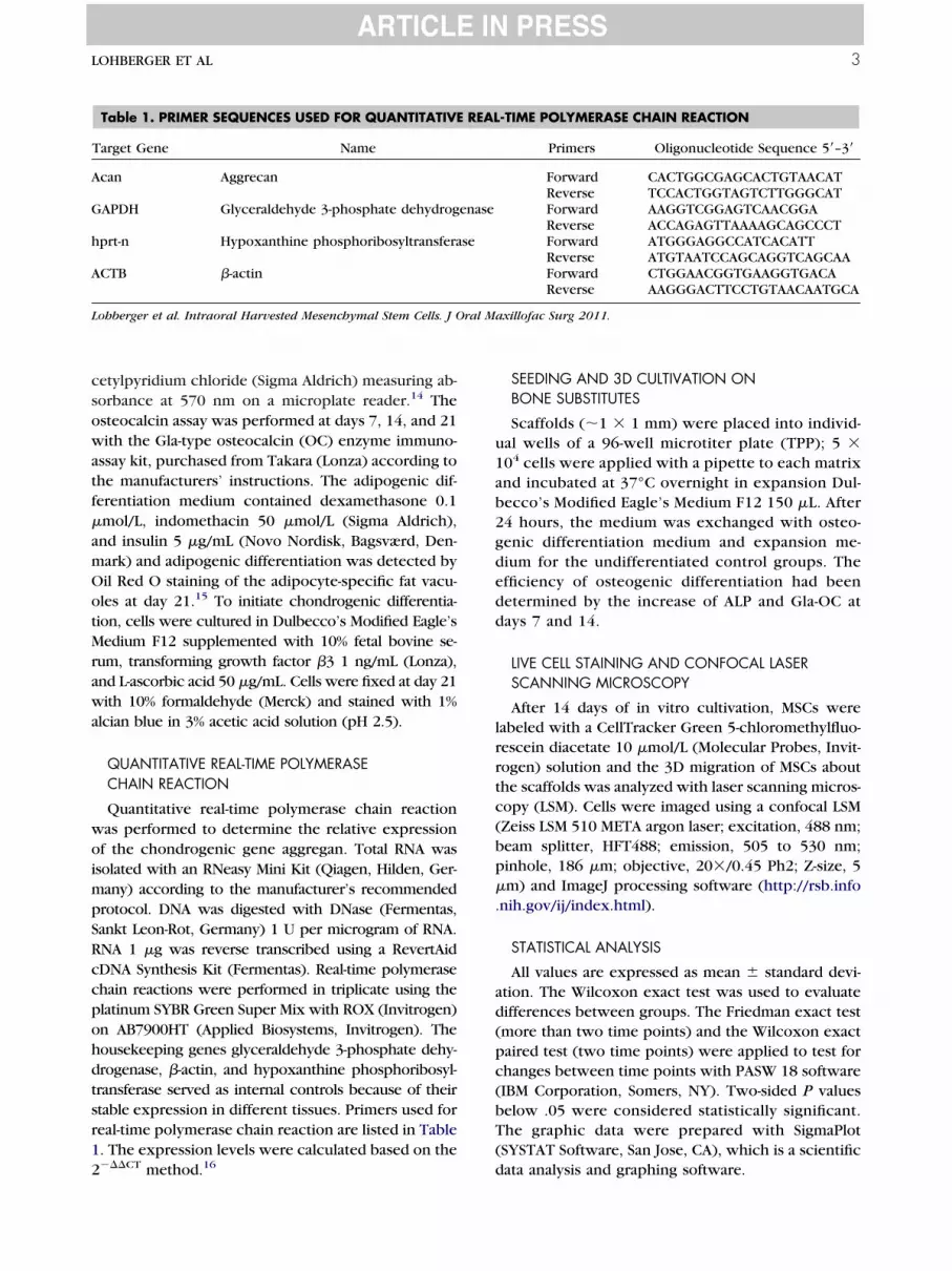
owatf
amOo
a
ccpohd
ab2gdedd
ad(pc(bT(
ral M
LOHBERGER ET AL 3
cetylpyridium chloride (Sigma Aldrich) measuring ab-sorbance at 570 nm on a microplate reader.14 The
steocalcin assay was performed at days 7, 14, and 21ith the Gla-type osteocalcin (OC) enzyme immuno-
ssay kit, purchased from Takara (Lonza) according tohe manufacturers’ instructions. The adipogenic dif-erentiation medium contained dexamethasone 0.1
�mol/L, indomethacin 50 �mol/L (Sigma Aldrich),nd insulin 5 �g/mL (Novo Nordisk, Bagsværd, Den-ark) and adipogenic differentiation was detected byil Red O staining of the adipocyte-specific fat vacu-les at day 21.15 To initiate chondrogenic differentia-
tion, cells were cultured in Dulbecco’s Modified Eagle’sMedium F12 supplemented with 10% fetal bovine se-rum, transforming growth factor �3 1 ng/mL (Lonza),nd L-ascorbic acid 50 �g/mL. Cells were fixed at day 21
with 10% formaldehyde (Merck) and stained with 1%alcian blue in 3% acetic acid solution (pH 2.5).
QUANTITATIVE REAL-TIME POLYMERASECHAIN REACTION
Quantitative real-time polymerase chain reactionwas performed to determine the relative expressionof the chondrogenic gene aggregan. Total RNA wasisolated with an RNeasy Mini Kit (Qiagen, Hilden, Ger-many) according to the manufacturer’s recommendedprotocol. DNA was digested with DNase (Fermentas,Sankt Leon-Rot, Germany) 1 U per microgram of RNA.RNA 1 �g was reverse transcribed using a RevertAidDNA Synthesis Kit (Fermentas). Real-time polymerasehain reactions were performed in triplicate using thelatinum SYBR Green Super Mix with ROX (Invitrogen)n AB7900HT (Applied Biosystems, Invitrogen). Theousekeeping genes glyceraldehyde 3-phosphate dehy-rogenase, �-actin, and hypoxanthine phosphoribosyl-
transferase served as internal controls because of theirstable expression in different tissues. Primers used forreal-time polymerase chain reaction are listed in Table1. The expression levels were calculated based on the
Table 1. PRIMER SEQUENCES USED FOR QUANTITATIVE
Target Gene Name
Acan Aggrecan
GAPDH Glyceraldehyde 3-phosphate dehydrog
hprt-n Hypoxanthine phosphoribosyltransfera
ACTB �-actin
Lohberger et al. Intraoral Harvested Mesenchymal Stem Cells. J O
2���CT method.16 d
SEEDING AND 3D CULTIVATION ONBONE SUBSTITUTES
Scaffolds (�1 � 1 mm) were placed into individ-ual wells of a 96-well microtiter plate (TPP); 5 �104 cells were applied with a pipette to each matrixnd incubated at 37°C overnight in expansion Dul-ecco’s Modified Eagle’s Medium F12 150 �L. After4 hours, the medium was exchanged with osteo-enic differentiation medium and expansion me-ium for the undifferentiated control groups. Thefficiency of osteogenic differentiation had beenetermined by the increase of ALP and Gla-OC atays 7 and 14.
LIVE CELL STAINING AND CONFOCAL LASERSCANNING MICROSCOPY
After 14 days of in vitro cultivation, MSCs werelabeled with a CellTracker Green 5-chloromethylfluo-rescein diacetate 10 �mol/L (Molecular Probes, Invit-rogen) solution and the 3D migration of MSCs aboutthe scaffolds was analyzed with laser scanning micros-copy (LSM). Cells were imaged using a confocal LSM(Zeiss LSM 510 META argon laser; excitation, 488 nm;beam splitter, HFT488; emission, 505 to 530 nm;pinhole, 186 �m; objective, 20�/0.45 Ph2; Z-size, 5�m) and ImageJ processing software (http://rsb.info.nih.gov/ij/index.html).
STATISTICAL ANALYSIS
All values are expressed as mean � standard devi-tion. The Wilcoxon exact test was used to evaluateifferences between groups. The Friedman exact testmore than two time points) and the Wilcoxon exactaired test (two time points) were applied to test forhanges between time points with PASW 18 softwareIBM Corporation, Somers, NY). Two-sided P valueselow .05 were considered statistically significant.he graphic data were prepared with SigmaPlotSYSTAT Software, San Jose, CA), which is a scientific
-TIME POLYMERASE CHAIN REACTION
Primers Oligonucleotide Sequence 5=–3=
Forward CACTGGCGAGCACTGTAACATReverse TCCACTGGTAGTCTTGGGCATForward AAGGTCGGAGTCAACGGAReverse ACCAGAGTTAAAAGCAGCCCTForward ATGGGAGGCCATCACATTReverse ATGTAATCCAGCAGGTCAGCAAForward CTGGAACGGTGAAGGTGACAReverse AAGGGACTTCCTGTAACAATGCA
axillofac Surg 2011.
REAL
enase
se
ata analysis and graphing software.

p0
3u.OGi
4 INTRAORAL HARVESTED MESENCHYMAL STEM CELLS
Results
FLOW CYTOMETRY
The flow cytometric analysis with the monoclonalantibodies CD73-PE, CD90-APC, CD105-PE, CD45-APC-cyanin 7, CD34-APC, CD14-fluorescein isothio-cyanate, CD11b-Pacific Blue, CD19-APC, and HLA-DRAPC and their typical forward/side scattercharacteristics confirmed the phenotype of intraoral
MSCs (Fig 1; online only). The typical forward/sidescatter characteristics of 83.1 � 5.3% MSCs weregated. According to the criteria for defining multipo-tent MSCs,9 intraoral MSCs from all 5 patients demon-strated the same immunophenotype, with expressionof CD73, CD90, and CD105 but no expression ofCD45, CD34, CD14, CD11, and HLA-DR.
MULTILINEAGE DIFFERENTIATION ANALYSIS
Multilineage differentiation potential was achieved.ALP activity was displayed with bright-field micros-copy (Fig 2A-D; online only). A significant increaseover time was observed in cells cultured in osteogenicmedium (1.2 � 0.6 OD at day 7 and 2 � 1.2 OD at day14, P � .001) and compared with cultures with ex-
ression medium (0.4 � 0.2 OD at day 7, P � .001;.6 � 0.4 OD at day 14, P � .001; Fig 2E). Further-
more, values measured at an OD of 570 nm increasedsignificantly over time in cells of osteogenic lineage(0.3 � 0.1 OD at day 7, 1.3 � 0.9 OD at day 14, and.6 � 0.3 OD at day 21, P � .001) compared withndifferentiated controls (0.1 � 0.02 OD at day 7, P �
032; 0.1 � 0.04 OD at day 14, P � .001; 0.15 � 0.02D at day 21, P � .001; Fig 2F). In addition, thela-OC concentrations, measured in 1 � 104 OD cells,
ncreased significantly over time (1.4 � 0.8 ng/mL at
FIGURE 2. Multilineage differentiation analysis of intraoral har-vested human mesenchymal stem cells. Bright-field microscopyshows increasing alkaline phosphatase (ALP) expression in osteo-genic differentiated cultures at A, day 7 and C, day 14. Undiffer-entiated control groups from B, day 7 and D, day 14 showednearly no alkaline phosphatase expression. The images are shownat 100� magnification (A-D, online only). E, Quantitation of alka-line phosphatase enzyme showed a highly significant increaseover time (P � .001) compared with cultures with expressionmedium. F, Quantitation of calcium deposited in cultures showed ahighly significant increase in OD of 570 nm, which could beobserved in osteogenic differentiated mesenchymal stem cells overtime (P � .001) compared with undifferentiated controls. G, Gla-type osteocalcin (OC) concentration of osteogenic differentiatedcells increased over time (P � .005) and compared with undiffer-entiated controls. Oil Red O staining of cells H, in the adipogenesis-induced group and I, the undifferentiated control group after 21days of culturing in adipogenesis-induced medium. Small lipidvacuoles were detected with bright-field microscopy in the treatedcell group. The images are shown at 200� magnification (H-I,online only). J, Human intraoral harvested mesenchymal stem cellswere induced to differentiate along a chondrogenic lineage com-pared with undifferentiated control cells. The interaction of the cationicdye alcian blue and the acid glycosaminoglycans indicated that al-cian blue staining caused a blue coloration. K, Aggregan gene ex-pression level was upregulated significantly after 3 weeks of chondro-genic differentiation. ARS, Alizarin Red S (J-K, online only).
Lohberger et al. Intraoral Harvested Mesenchymal Stem Cells. J Oral
Maxillofac Surg 2011.
0PiOaafptas
4d
B
ral M
LOHBERGER ET AL 5
day 7, 3.4 � 3.7 ng/mL at day 14, and 4 � 4.3 ng/mLat day 21, P � .005) and were compared with undif-ferentiated controls (0.9 � 0.4 ng/mL at day 7, 0.8 �.3 ng/mL at day 14, and 0.7 � 0.1 ng/mL at day 21,
� .029; Fig 2G). Adipogenic differentiation wasndicated by the detection of lipid droplets by Oil Red
staining at day 21 (Fig 2H; online only), whereas nodipogenic expression was detected in undifferenti-ted controls (Fig 2I; online only). Chondrogenic dif-erentiated MSCs augmented blue coloration com-ared with undifferentiated controls depending onhe interaction of the cationic dye alcian blue and thecid glycosaminoglycans (Fig 2J; online only). Analy-
FIGURE 3. Three-dimensional cultivation and live cell imaging onio-Oss scaffolds and C, D, �-tricalcium phosphate (�-TCP) scaffold
mesenchymal stem cells were labeled with the CellTracker Green 5ingrowth in the interior pore regions of the scaffolds of the mesenc(A, B, and D, online only). All images are shown at 200� magnificBio-Oss scaffolds using an enzyme-linked immunosorbent assay teincrease of Gla-type osteocalcin expression at days 7 (d7) and 14determined by histochemical staining of alkaline phosphatase (ALdetermined by the increase of E, F, Gla-type osteocalcin expressio
Lohberger et al. Intraoral Harvested Mesenchymal Stem Cells. J O
is of the expression of aggregan demonstrated a
.7-fold increase (P � .05) because of chondrogenicifferentiation (Fig 2K; online only).
3D CULTIVATION AND LIVE CELL IMAGING ONBONE SUBSTITUTES
Natural bone mineral of bovine origin (Bio-Oss) and�-TCP (Vitoss) scaffolds were successfully colonizedwith human intraoral MSCs; the ingrowth in the inte-rior pore regions of the scaffolds were visualizedusing confocal LSM (Figs 3B; 3A, C, D online only).The efficiency of osteogenic differentiation was deter-mined by the increase of Gla-OC expression (Fig 3E;
ubstitutes. Intraoral mesenchymal stem cells were loaded A, B, onr 14 days of in vitro cultivation, differentiated and undifferentiatedmethylfluorescein diacetate. The 3-dimensional migration and thetem cells were analyzed with confocal laser scanning microscopyE, Quantitative analysis of osteocalcin (OC) protein production one. Efficiency of osteogenic differentiation was determined by an, Efficiency of osteogenic differentiation on Bio-Oss scaffolds was
ity at days 7 and 14. Efficiency of osteogenic differentiation wasG, H, alkaline phosphatase at days 7 and 14 (F, H, online only).
axillofac Surg 2011.
bone ss. Afte-chloro
hymal sation.chniqu
(d14). GP) activn and
3F online only) and ALP (Fig 3G; 3H; online only) at

tga
tbnaspap
oagffco
ip
i
ipptmpvctsy
2wdtamf
aasptdittpvci
A
6 INTRAORAL HARVESTED MESENCHYMAL STEM CELLS
days 7 and 14 compared with the correspondingundifferentiated control groups.
Discussion
With the trend toward minimally invasive surgeryin recent years, the number of bone substitute mate-rials available has increased sharply. However, autol-ogous bone transfer is still considered the gold stan-dard with a good tissue acceptance and a lowinfection risk.17,18 Nonetheless, availability of autolo-gous bone material is limited, and the surgery carriesadditional intra- and postoperative risks for the pa-tient.19 The idea of tissue engineering is based on theransfer of a patient’s own autologous cells. Bonerafting from the iliac region and proximal tibia haslso become widely accepted in oral surgery.20 Rick-
ert et al10 investigated the differences in bone forma-ion after maxillary sinus floor elevation surgery withovine bone mineral (Bio-Oss) mixed with autoge-ous bone or autogenous stem cells. The research forlternatives to conventional augmentation procedureshowed that the application of bone morphogenicrotein to bovine bone material leads to more rapidnd enhanced osseointegration of simultaneouslylaced implants in minipigs.21 De Kok et al22 studied
the capability of MSCs for bone repair in beagles(canines). They showed that bone marrow MSCsseeded on hydroxyapatite and TCP biomaterials canincrease bone formation in dental sockets. Varioustypes of donor tissues of adult MSCs have been iden-tified thus far. Bone marrow MSCs, in most casesharvested from the ileum, intraoral periosteum, orbone ectomesenchymal pulp, have proliferated withproved multiple potencies under in vitro condi-tions.23,24 In an in vitro model, Clausen et al1 investi-gated the characteristics of human maxillary-derivedbone cells and observed clear markers of osteogenicdifferentiation potential. They concluded that maxilla-derived bone cells are of interest for tissue engineer-ing procedures for maxillary defects. Cicconetti etal25 proved the osteogenic potential of human peri-
steal and maxillary bone marrow stromal cells inthymic mice when loaded on a TCP construct. Re-arding donor site morbidity, intraoral MSCs isolatedrom maxillary bone seem to be a promising sourceor clinical use in craniofacial reconstructions be-ause cell harvest procedures from this site representnly minimal trauma to the patient. Trisi et al26
showed successful healing and bone formation of a�-TCP in human artificial mandibular defects. Acil etal27 and Jafarian et al28 described good biocompatibil-ty of Bio-Oss in 3D cell culture systems and theotential in human bone for tissue engineering.In the present study, human maxilla MSCs were
solated and cells were characterized successfully us-
ng flow cytometric analysis. As proof of multipleotencies, the potential concerning osteogenic, adi-ogenic, and chondrogenic differentiation was inves-igated. The increase of the osteogenic differentiationarkers ALP, ARS, and OC indicated the osteogenicotential of the intraoral MSCs, Oil Red O stainingisualized lipid vacuoles, and a highly significant in-rease of mRNA expression of aggregates proved mul-ipotent characteristics. However, 1 factor to be con-idered when interpreting the results is the relativelyoung age of the patients included in this study.As potential carriers for future clinical application,frequently used bone substitutes in oral surgeryere loaded with intraoral harvested MSCs. After 14ays of in vitro cultivation, MSCs were labeled withhe CellTracker Green 5-chloromethylfluorescein di-cetate and, for the first time, the 3D migration ofaxillary MSCs in both scaffolds was evident by con-
ocal LSM.In conclusion, this study showed the expansion
nd differentiation of maxillary human intraoral MSCsnd their 3D migration potential into 2 different boneubstitutes. Natural bone mineral and �-TCP clearlyrovide an appropriate 3D structure for the cultiva-ion and differentiation of human intraoral MSCs. Un-er in vitro conditions, maxillary MSCs may be prom-
sing candidates for tissue engineering approaches forhe replacement of bone in maxillofacial reconstruc-ions. However, the present study was initiated as ailot to a clinical trial on the reconstruction of se-erely resorbed maxillas, which will allow clinicalonclusions to be drawn about this promising initialn vitro data.
cknowledgments
The authors thank Heike Kaltenegger, Tatjana Kueznik, and Al-exandra Novak for excellent technical assistance. Financial supportby the Medical University of Graz is gratefully acknowledged.
References1. Clausen C, Hermund NU, Donatsky O, et al: Characterization of
human bone cells derived from the maxillary alveolar ridge.Clin Oral Implants Res 17:533, 2006
2. Haas R, Donath K, Födinger M, et al: Bovine hydroxyapatite formaxillary sinus grafting: Comparative histomorphometric find-ings in sheep. Clin Oral Implants Res 9:107, 1998
3. Jakse N, Seibert FJ, Lorenzoni M, et al: A modified technique ofharvesting tibial cancellous bone and its use in sinus grafting.Clin Oral Implant Res 12:488, 2001
4. Lee CY: Immediate load protocol for anterior maxilla withcortical bone from mandibular ramus. Implant Dent 15:153,2006
5. Bruder SP, Kraus KH, Goldberg VM, et al: The effect of implantsloaded with autologous mesenchymal stem cells on the healingof canine segmental bone defects. J Bone Joint Surg Am 80:985,1998
6. Kon E, Muraglia A, Corsi A, et al: Autologous bone marrowstromal cells loaded onto porous hydroxyapatite ceramic ac-celerate bone repair in critical-size defects of sheep long bones.
J Biomed Mater Res 49:328, 2000
LOHBERGER ET AL 7
7. Shang Q, Wang Z, Liu W, et al: Tissue-engineered bone repairof sheep cranial defects with autologous bone marrow stromalcells. J Craniofac Surg 12:586, 2001
8. Pittenger MF, Mackay AM, Beck SC, et al: Multilineage potentialof adult human mesenchymal stem cells. Science 284:143,1999
9. Dominici M, Le Blank K, Mueller I: Minimal criteria for definingmultipotent mesenchymal stromal cells. The International So-ciety for Cellular Therapy position statement. Cytotherapie8:315, 2006
10. Rickert D, Sauerbier S, Nagursky H, et al: Maxillary sinus floorelevation with bovine bone mineral combined with eitherautogenous bone or autogenous stem cells: A prospective ran-domized clinical trial. Clin Oral Implant Res 2010
11. Schmelzeisen R, Gutwald R, Oshima T, et al: Making bone II:Maxillary sinus augmentation with mononuclear cells—Casereport with a new clinical method. Br J Oral Maxillofac Surg2010
12. Fickl S, Zuhr O, Wachtel H, et al: Hard tissue alterations aftersocket preservation with additional buccal overbuilding: Astudy in the beagle dog. J Clin Periodontol 36:898, 2009
13. Schallmoser K, Bartmann C, Rohde E, et al: Human plateletlysate can replace fetal bovine serum for clinical-scale expan-sion of functional mesenchymal stromal cells. Transfusion 47:1436, 2007
14. Gori F, Divieti P, Demay MB: Cloning and characterization of anovel WD-40 repeat protein that dramatically accelerates os-teoblastic differentiation. J Biol Chem 276:46515, 2001
15. Kulterer B, Friedl G, Jandrositz A, et al: Gene expression pro-filing of human mesenchymal stem cells derived from bonemarrow during expansion and osteoblast differentiation. BMCGenomics 8:70, 2007
16. Vandesompele J, De Preter K, Pattyn F, et al: Accurate normal-ization of real-time quantitative RT-PCR data by geometric av-eraging of multiple internal control genes. Genome Biol 3:34,2002
17. Delloye C, Cnockaert N, Cornu O: Bone substitutes in 2003: Anoverview. Acta Orthopaed Belg 69:1, 2003
18. Kuebler NR, Wuerzler K: Bone morphogenetic proteins. Im-plantologie 6:177, 2002
19. Behrens E, Kreusch T, Jonas S, et al: Komplikationen bei undnach Knochenentnahmen aus dem Beckenkamm. Dtsch Zah-närztl Z 56:66, 2001
20. Kirmeier R, Payer M, Lorenzoni M, et al: Harvesting of cancel-lous bone from the proximal tibia under local anesthesia:Donor site morbidity and patient experience. J Oral MaxillofacSurg 65:2235, 2007
21. Terheyden H, Jepsen S, Möller B, et al: Sinus floor augmenta-tion with simultaneous placement of dental implants using acombination of deproteinized bone xenografts and recombi-nant human osteogenic protein-1. A histometric study in min-iature pigs. Clin Oral Implants Res 10:510, 1999
22. De Kok IJ, Drapeau SJ, Young R, et al: Evaluation of mesenchy-mal stem cells following implantation in alveolar sockets: Acanine safety study. Int J Oral Maxillofac Implants 20:511, 2005
23. Gronthos S, Mankani M, Brahim J, et al: Postnatal human dentalpulp stem cells (DPSCs) in vitro and in vivo. Proc Natl Acad SciU S A 97:13625, 2000
24. Gronthos S, Brahim J, Li W, et al: Stem cell properties of humandental pulp stem cells. J Dent Res 81:531, 2002
25. Cicconetti A, Saccetti B, Bartoli A, et al: Human maxillarytuberosity and jaw periosteum as source of osteoprogenitorcells for tissue engineering. Oral Surg Oral Med Oral PatholOral Radiol Endod 104:618.e1, 2007
26. Trisi P, Rao W, Rebaudi A, et al: Histologic effect of pure-phasebeta tricalcium phosphate on bone regeneration in humanartificial jawbone defects. Int J Periodont Res Dent 23:69, 2003
27. Acil Y, Springer NG, Broek V, et al: Effects of bone morphoge-netic protein-7 stimulation on osteoblasts cultured on differentbiomaterials. JCB 86:90, 2003
28. Jafarian M, Eslaminejad M, Khojasteh A, et al: Marrow-derivedmesenchymal Stel cell-directed bone regeneration in the dogmandible: A comparison between biphasic calcium phosphate
and natural bone mineral. Oral Surg Oral Med Oral Pathol OralRadiol Endod 105:e14, 2008Recommended


