Embed Size (px)
Citation preview
Journal of
Clinical Medicine
Article
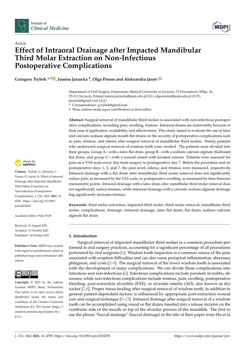
Effect of Intraoral Drainage after Impacted MandibularThird Molar Extraction on Non-InfectiousPostoperative Complications
Grzegorz Trybek *,† , Joanna Jarzecka †, Olga Preuss and Aleksandra Jaron
�����������������
Citation: Trybek, G.; Jarzecka, J.;
Preuss, O.; Jaron, A. Effect of Intraoral
Drainage after Impacted Mandibular
Third Molar Extraction on
Non-Infectious Postoperative
Complications. J. Clin. Med. 2021, 10,
4705. https://doi.org/10.3390/
jcm10204705
Academic Editor: Peter Proff
Received: 26 August 2021
Accepted: 11 October 2021
Published: 14 October 2021
Publisher’s Note: MDPI stays neutral
with regard to jurisdictional claims in
published maps and institutional affil-
iations.
Copyright: © 2021 by the authors.
Licensee MDPI, Basel, Switzerland.
This article is an open access article
distributed under the terms and
conditions of the Creative Commons
Attribution (CC BY) license (https://
creativecommons.org/licenses/by/
4.0/).
Department of Oral Surgery, Pomeranian Medical University in Szczecin, 72 Powstanców Wlkp. St.,70-111 Szczecin, Poland; [email protected] (J.J.); [email protected] (O.P.);[email protected] (A.J.)* Correspondence: [email protected]† These authors made equal contributions as first author.
Abstract: Surgical removal of mandibular third molars is associated with non-infectious postoper-ative complications, including pain, swelling, trismus. Intraoral drains are noteworthy because oftheir ease of application, availability, and effectiveness. This study aimed to evaluate the use of latexand calcium–sodium alginate mouth flat drains on the severity of postoperative complications suchas pain, trismus, and edema after surgical removal of mandibular third molars. Ninety patientswho underwent surgical removal of wisdom teeth were studied. The patients were divided intothree groups. Group A—with a latex flat drain, group B—with a sodium–calcium alginate (Kaltostat)flat drain, and group C—with a wound closed with knotted sutures. Patients were assessed forpain on a VAS scale every day from surgery to postoperative day 7. Before the procedure and onpostoperative days 1, 2, and 7, the pain level, edema, and trismus were measured, respectively.Intraoral drainage with a flat drain after mandibular third molar removal does not significantlyreduce pain, as measured by the VAS scale, or postoperative swelling, as measured by lines betweencraniometric points. Intraoral drainage with a latex drain after mandibular third molar removal doesnot significantly reduce trismus, while intraoral drainage with a calcium–sodium alginate drainagebag significantly increases trismus.
Keywords: third molar extraction; impacted third molar; third molar removal; mandibular thirdmolar; complications; drainage; intraoral drainage; latex flat drain; flat drain; sodium–calciumalginate flat drain
1. Introduction
Surgical removal of impacted mandibular third molars is a common procedure per-formed in oral surgery practices, accounting for a significant percentage of all proceduresperformed by oral surgeons [1]. Wisdom tooth impaction is a common source of the painassociated with eruption difficulties and can also cause periapical inflammation, abscesses,phlegmon, and cysts [2–5]. The surgical removal of the lower wisdom tooth is associatedwith the development of many complications. We can divide these complications intoinfectious and non-infectious [6]. Infectious complications include purulent alveolitis, ab-scesses, while non-infectious complications include trismus, pain, swelling, postoperativebleeding, post-extraction alveolitis (PAE), or alveolar osteitis (AO), also known as drysocket [7,8]. Proper tissue healing after surgical removal of wisdom teeth, in addition togeneral patient-dependent factors, is influenced by appropriate post-extraction woundcare and surgical technique [9–12]. Intraoral drainage after surgical removal of a wisdomtooth can be accomplished using round or flat drains inserted into a release incision on thevestibular side of the mouth or top of the alveolar process of the mandible. The first touse the phrase “buccal drainage” (buccal drainage) in the title of their paper were Hu et al.
J. Clin. Med. 2021, 10, 4705. https://doi.org/10.3390/jcm10204705 https://www.mdpi.com/journal/jcm
J. Clin. Med. 2021, 10, 4705 2 of 16
in 2017, thus defining the location of a latex teat in the floor of the oral vestibule, on thebuccal side, designed to drain buccal swelling and reduce buccal volume after surgicalremoval of the lower wisdom tooth [13]. Drainage after lower third molar extraction hasbeen the subject of many studies and scientific publications. It affects the reduction ofdiscomfort associated with the procedure; however, the results are inconclusive. Differenttypes of incisions used in surgery during wisdom tooth extraction, sites of drain insertion,and materials used to drain secretions have been studied. Latex drainage is routinely usedafter incision of abscesses, causing the evacuation of the purulent contents, preventingtissue approximation and resealing. However, leaving the drain in place for 72 h results inwound epithelialization and the initiation of prolonged secondary healing [14]. The useof a drain after removing the third molar in the mandible is intended to create an outflowroute for excess exudate fluid and minimize the risk of postoperative hematoma infection.It prevents its formation by gravitational drainage of the exudate formed in the first phaseof tissue healing. This study aimed to find an optimal postoperative wound supply thatwould be the least discomforting to the patient and widely available. Conflicting reportson minimizing three parameters, i.e., pain, swelling, and trismus, with the use of a drainafter surgical extraction of the third molar in the mandible inspired a study to answer thequestion: What parameters are affected by the use of a drain inserted in the floor of theoral vestibule after surgical treatment?
2. Materials and Methods
The study was conducted after receiving a positive opinion from the Bioethics Com-mittee of the Medical University, No. KB-0012/38/18 on 12 March 2018.
2.1. Study Group
The study group consisted of 90 patients enrolled by convenience sampling of theDepartment of Oral Surgery of the Medical University who qualified for the plannedremoval of mandibular third molars. The study included adult patients, generally healthynon-smokers with no local or general inflammation symptoms. Exclusion criteria were:patients under 18 years of age, pregnancy and lactation, systemic diseases, clinical signs ofpericoronal inflammation due to the difficult eruption of the eighth tooth, smoking, andallergy to latex. Before the procedure, patients were asked to complete a written consent forthe procedure and participate in the research project. Patients were put into three groupsby simple randomization:
- A (n = 30)—group with latex drain used;- B (n = 30)—group with a sodium–calcium alginate drain applied;- C (n = 30)—control group, in which the wound was secured with standard knotted sutures.
Simple randomization took place before the scheduled surgery; each patient receiveda sealed envelope with allocation to each group. Before the surgical removal of the lowerwisdom tooth, the mandibular third molar’s degree of difficulty, morphology, and positionwere assessed based on a pantographic image. The classification according to Pell andGregory and the surgery difficulty index according to Pederson were used for this purpose.
2.2. Surgery
Before surgical removal of the retained third molar in the mandible, each patientreceived 600 mg of clindamycin in a “one-shot” scheme. The surgical removal of thewisdom tooth was performed by one highly experienced operator with the same repeatablesurgical technique under regional anesthesia of the inferior alveolar, lingual, and buccalnerves (4 mL of 2% lidocaine with norepinephrine). The incision line was carried outon the mandibular rami, distally, and in the gingival sulcus of the mandibular secondmolar. A release cut was then made in the oral vestibule (Cogswell incision). Using drillsand elevator or Meissner forceps, the tooth was removed. After the tooth removal, thewound was revised and cleaned, and the bony margins aligned. The mucoperiosteal flapwas repositioned and secured with single-knotted, non-resorbable 3–0 silk sutures. Each
J. Clin. Med. 2021, 10, 4705 3 of 16
patient received detailed recommendations for postoperative management. Patients wereadvised to rinse the mouth with 0.1% chlorhexidine gluconate solution and to take 100 mgketoprofen orally twice a day for 3 days.
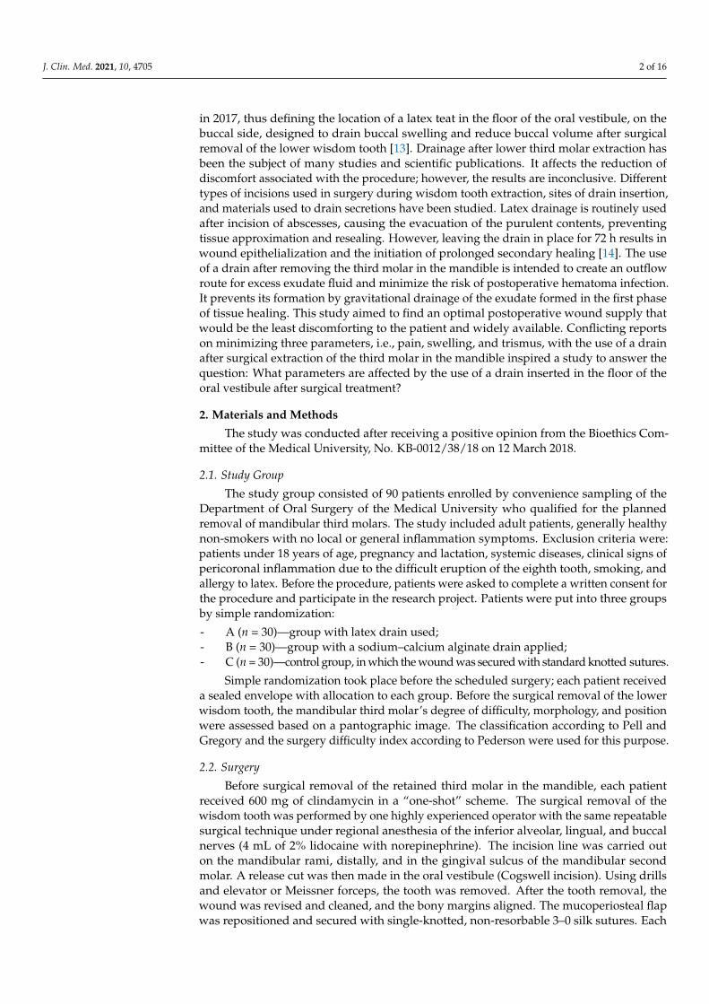
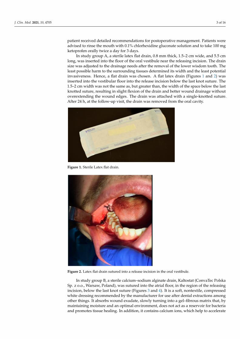
In study group A, a sterile latex flat drain, 0.8 mm thick, 1.5–2 cm wide, and 5.5 cmlong, was inserted into the floor of the oral vestibule near the releasing incision. The drainsize was adjusted to the drainage needs after the removal of the lower wisdom tooth. Theleast possible harm to the surrounding tissues determined its width and the least potentialinvasiveness. Hence, a flat drain was chosen. A flat latex drain (Figures 1 and 2) wasinserted into the vestibular floor into the release incision below the last knot suture. The1.5–2 cm width was not the same as, but greater than, the width of the space below the lastknotted suture, resulting in slight flexion of the drain and better wound drainage withoutoverextending the wound edges. The drain was attached with a single-knotted suture.After 24 h, at the follow-up visit, the drain was removed from the oral cavity.
Figure 1. Sterile Latex flat drain.
Figure 2. Latex flat drain sutured into a release incision in the oral vestibule.
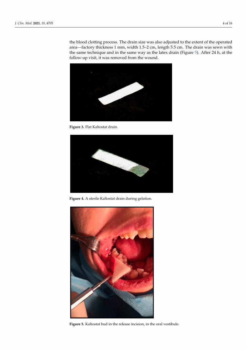
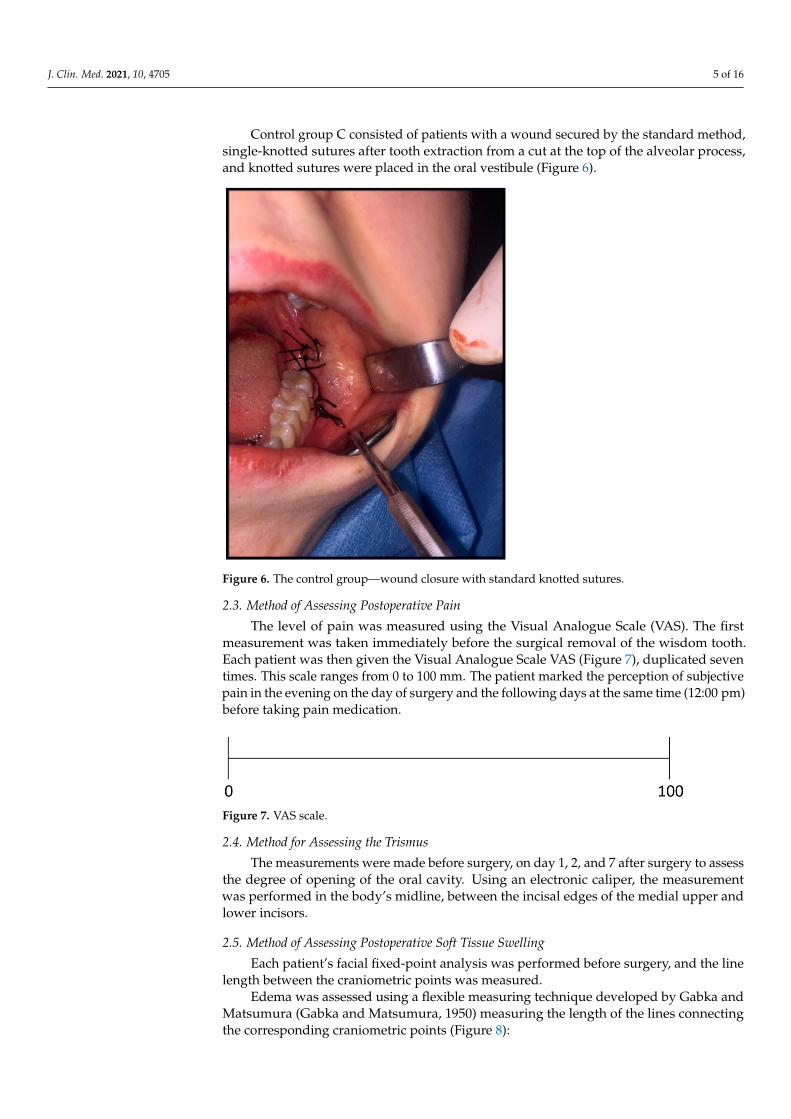
In study group B, a sterile calcium–sodium alginate drain, Kaltostat (ConvaTec PolskaSp. z o.o., Warsaw, Poland), was sutured into the atrial floor, in the region of the releasingincision, below the last knot suture (Figures 3 and 4). It is a soft, nontextile, compressedwhite dressing recommended by the manufacturer for use after dental extractions amongother things. It absorbs wound exudate, slowly turning into a gel–fibrous matrix that, bymaintaining moisture and an optimal environment, does not act as a reservoir for bacteriaand promotes tissue healing. In addition, it contains calcium ions, which help to accelerate
J. Clin. Med. 2021, 10, 4705 4 of 16
the blood clotting process. The drain size was also adjusted to the extent of the operatedarea—factory thickness 1 mm, width 1.5–2 cm, length 5.5 cm. The drain was sewn withthe same technique and in the same way as the latex drain (Figure 5). After 24 h, at thefollow-up visit, it was removed from the wound.
Figure 3. Flat Kaltostat drain.
Figure 4. A sterile Kaltostat drain during gelation.
Figure 5. Kaltostat bud in the release incision, in the oral vestibule.
J. Clin. Med. 2021, 10, 4705 5 of 16
Control group C consisted of patients with a wound secured by the standard method,single-knotted sutures after tooth extraction from a cut at the top of the alveolar process,and knotted sutures were placed in the oral vestibule (Figure 6).
Figure 6. The control group—wound closure with standard knotted sutures.
2.3. Method of Assessing Postoperative Pain
The level of pain was measured using the Visual Analogue Scale (VAS). The firstmeasurement was taken immediately before the surgical removal of the wisdom tooth.Each patient was then given the Visual Analogue Scale VAS (Figure 7), duplicated seventimes. This scale ranges from 0 to 100 mm. The patient marked the perception of subjectivepain in the evening on the day of surgery and the following days at the same time (12:00 pm)before taking pain medication.
Figure 7. VAS scale.
2.4. Method for Assessing the Trismus
The measurements were made before surgery, on day 1, 2, and 7 after surgery to assessthe degree of opening of the oral cavity. Using an electronic caliper, the measurementwas performed in the body’s midline, between the incisal edges of the medial upper andlower incisors.
2.5. Method of Assessing Postoperative Soft Tissue Swelling
Each patient’s facial fixed-point analysis was performed before surgery, and the linelength between the craniometric points was measured.
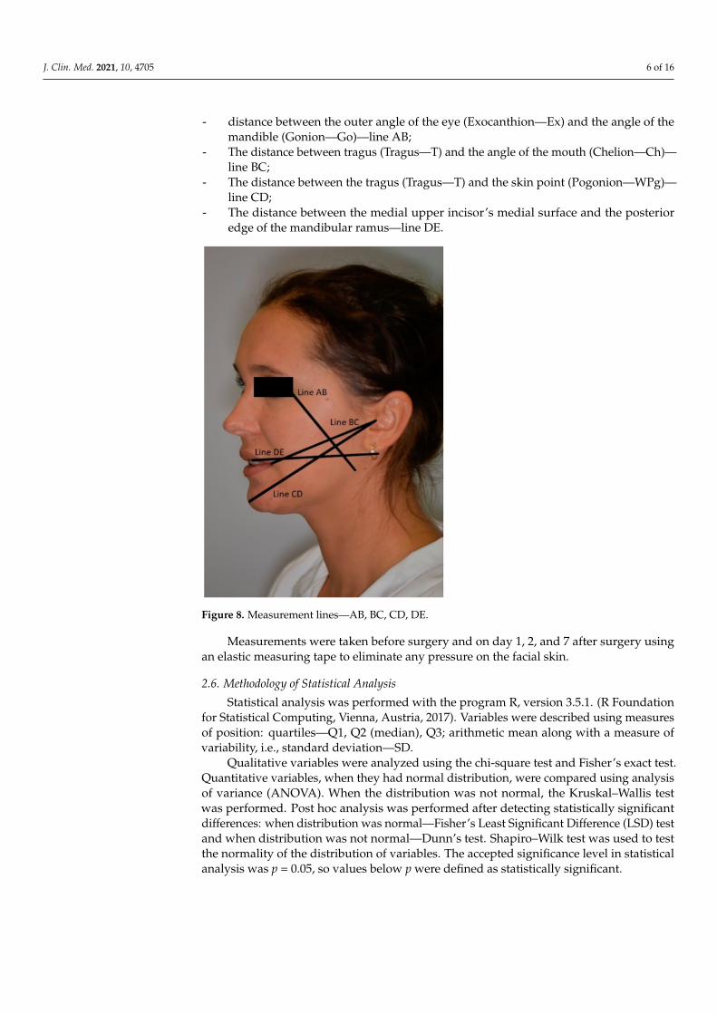
Edema was assessed using a flexible measuring technique developed by Gabka andMatsumura (Gabka and Matsumura, 1950) measuring the length of the lines connectingthe corresponding craniometric points (Figure 8):
J. Clin. Med. 2021, 10, 4705 6 of 16
- distance between the outer angle of the eye (Exocanthion—Ex) and the angle of themandible (Gonion—Go)—line AB;
- The distance between tragus (Tragus—T) and the angle of the mouth (Chelion—Ch)—line BC;
- The distance between the tragus (Tragus—T) and the skin point (Pogonion—WPg)—line CD;
- The distance between the medial upper incisor’s medial surface and the posterioredge of the mandibular ramus—line DE.
Figure 8. Measurement lines—AB, BC, CD, DE.
Measurements were taken before surgery and on day 1, 2, and 7 after surgery usingan elastic measuring tape to eliminate any pressure on the facial skin.
2.6. Methodology of Statistical Analysis
Statistical analysis was performed with the program R, version 3.5.1. (R Foundationfor Statistical Computing, Vienna, Austria, 2017). Variables were described using measuresof position: quartiles—Q1, Q2 (median), Q3; arithmetic mean along with a measure ofvariability, i.e., standard deviation—SD.
Qualitative variables were analyzed using the chi-square test and Fisher’s exact test.Quantitative variables, when they had normal distribution, were compared using analysisof variance (ANOVA). When the distribution was not normal, the Kruskal–Wallis testwas performed. Post hoc analysis was performed after detecting statistically significantdifferences: when distribution was normal—Fisher’s Least Significant Difference (LSD) testand when distribution was not normal—Dunn’s test. Shapiro–Wilk test was used to testthe normality of the distribution of variables. The accepted significance level in statisticalanalysis was p = 0.05, so values below p were defined as statistically significant.
J. Clin. Med. 2021, 10, 4705 7 of 16
3. Results3.1. Baseline Characteristics
Ninety patients classified for surgical removal of mandibular third molars were in-cluded in the study (n = 90). The patients were randomly assigned to one of three groups,each consisting of 30 patients (n = 30): a group that was treated with a latex flat drainsutured—referred to as group A (n = 30), a group that was treated with a Kaltostat suturedafter surgery—referred to as group B (n = 30), and a group that was treated with standardknotted sutures—referred to as group C (n = 30). Fifty-seven women and 33 men partic-ipated in the study. The study also took into account the age of the patients, which alsodid not show statistically significant differences (mean 26.48 ± SD 7.03). There were nosignificant differences between the groups in the numbers of right and left wisdom teethextracted. Forty-eight teeth were removed on the right side, while 42 teeth were removedon the left side of patients.
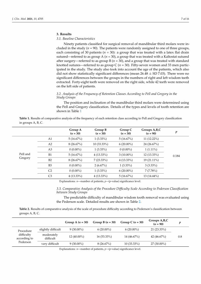
3.2. Analysis of the Frequency of Retention Classes According to Pell and Gregory in theStudy Groups
The position and inclination of the mandibular third molars were determined usingthe Pell and Gregory classification. Details of the types and levels of tooth retention areshown in Table 1
Table 1. Results of comparative analysis of the frequency of each retention class according to Pell and Gregory classificationin groups A, B, C.
Group A(n = 30)
Group B(n = 30)
Group C(n = 30)
Groups A,B,C(n = 90) p
Pell andGregory
A1 5 (16.67%) 1 (3.33%) 5 (16.67%) 11 (12.22%)
0.184
A2 8 (26.67%) 10 (33.33%) 6 (20.00%) 24 (26.67%)
A3 0 (0.00%) 1 (3.33%) 0 (0.00%) 1 (1.11%)
B1 5 (16.67%) 4 (13.33%) 3 (10.00%) 12 (13.33%)
B2 8 (26.67%) 7 (23.33%) 4 (13.33%) 19 (21.11%)
B3 0 (0.00%) 2 (6.67%) 1 (3.33%) 3 (3.33%)
C2 0 (0.00%) 1 (3.33%) 6 (20.00%) 7 (7.78%)
C3 4 (13.33%) 4 (13.33%) 5 (16.67%) 13 (14.44%)
Explanations: n—number of patients, p—(p-value) significance level.
3.3. Comparative Analysis of the Procedure Difficulty Scale According to Pederson Classificationbetween Study Groups
The predictable difficulty of mandibular wisdom tooth removal was evaluated usingthe Pederson scale. Detailed results are shown in Table 2.
Table 2. Results of comparative analysis of the scale of procedure difficulty according to Pederson’s classification betweengroups A, B, C.
Group A (n = 30) Group B (n = 30) Group C (n = 30) Groups A,B,C(n = 90) p
Proceduredifficulty
according toPederson
slightly difficult 9 (30.00%) 6 (20.00%) 6 (20.00%) 21 (23.33%)
0.8moderately
difficult 12 (40.00%) 16 (53.33%) 14 (46.67%) 42 (46.67%)
very difficult 9 (30.00%) 8 (26.67%) 10 (33.33%) 27 (30.00%)
Explanations: n—number of patients, p—(p-value) significance level.
J. Clin. Med. 2021, 10, 4705 8 of 16
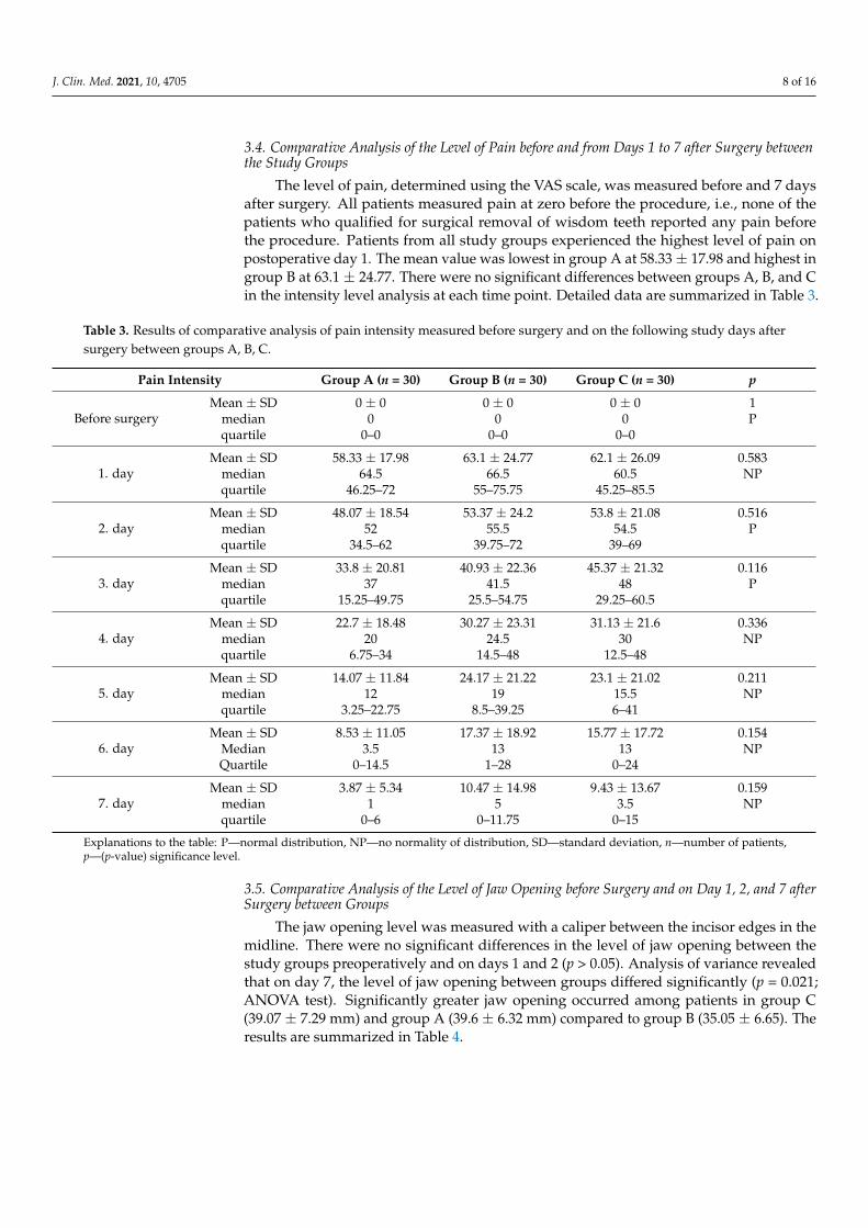
3.4. Comparative Analysis of the Level of Pain before and from Days 1 to 7 after Surgery betweenthe Study Groups
The level of pain, determined using the VAS scale, was measured before and 7 daysafter surgery. All patients measured pain at zero before the procedure, i.e., none of thepatients who qualified for surgical removal of wisdom teeth reported any pain beforethe procedure. Patients from all study groups experienced the highest level of pain onpostoperative day 1. The mean value was lowest in group A at 58.33 ± 17.98 and highest ingroup B at 63.1 ± 24.77. There were no significant differences between groups A, B, and Cin the intensity level analysis at each time point. Detailed data are summarized in Table 3.
Table 3. Results of comparative analysis of pain intensity measured before surgery and on the following study days aftersurgery between groups A, B, C.
Pain Intensity Group A (n = 30) Group B (n = 30) Group C (n = 30) p
Before surgeryMean ± SD 0 ± 0 0 ± 0 0 ± 0 1
median 0 0 0 Pquartile 0–0 0–0 0–0
1. dayMean ± SD 58.33 ± 17.98 63.1 ± 24.77 62.1 ± 26.09 0.583
median 64.5 66.5 60.5 NPquartile 46.25–72 55–75.75 45.25–85.5
2. dayMean ± SD 48.07 ± 18.54 53.37 ± 24.2 53.8 ± 21.08 0.516
median 52 55.5 54.5 Pquartile 34.5–62 39.75–72 39–69
3. dayMean ± SD 33.8 ± 20.81 40.93 ± 22.36 45.37 ± 21.32 0.116
median 37 41.5 48 Pquartile 15.25–49.75 25.5–54.75 29.25–60.5
4. dayMean ± SD 22.7 ± 18.48 30.27 ± 23.31 31.13 ± 21.6 0.336
median 20 24.5 30 NPquartile 6.75–34 14.5–48 12.5–48
5. dayMean ± SD 14.07 ± 11.84 24.17 ± 21.22 23.1 ± 21.02 0.211
median 12 19 15.5 NPquartile 3.25–22.75 8.5–39.25 6–41
6. dayMean ± SD 8.53 ± 11.05 17.37 ± 18.92 15.77 ± 17.72 0.154
Median 3.5 13 13 NPQuartile 0–14.5 1–28 0–24
7. dayMean ± SD 3.87 ± 5.34 10.47 ± 14.98 9.43 ± 13.67 0.159
median 1 5 3.5 NPquartile 0–6 0–11.75 0–15
Explanations to the table: P—normal distribution, NP—no normality of distribution, SD—standard deviation, n—number of patients,p—(p-value) significance level.
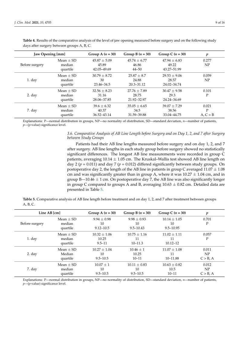
3.5. Comparative Analysis of the Level of Jaw Opening before Surgery and on Day 1, 2, and 7 afterSurgery between Groups
The jaw opening level was measured with a caliper between the incisor edges in themidline. There were no significant differences in the level of jaw opening between thestudy groups preoperatively and on days 1 and 2 (p > 0.05). Analysis of variance revealedthat on day 7, the level of jaw opening between groups differed significantly (p = 0.021;ANOVA test). Significantly greater jaw opening occurred among patients in group C(39.07 ± 7.29 mm) and group A (39.6 ± 6.32 mm) compared to group B (35.05 ± 6.65). Theresults are summarized in Table 4.
J. Clin. Med. 2021, 10, 4705 9 of 16
Table 4. Results of the comparative analysis of the level of jaw opening measured before surgery and on the following studydays after surgery between groups A, B, C.
Jaw Opening [mm] Group A (n = 30) Group B (n = 30) Group C (n = 30) p
Before surgeryMean ± SD 45.87 ± 5.09 45.74 ± 6.77 47.94 ± 6.83 0.277
median 45.89 46.86 49.22 NPquartile 42.05–49.69 44–50 43.27–51.99
1. dayMean ± SD 30.79 ± 8.72 25.87 ± 8.7 29.53 ± 9.06 0.059
median 30 24.88 28.57 NPquartile 23.46–34.5 20.3–31.12 24.02–34.74
2. dayMean ± SD 32.56 ± 8.23 27.76 ± 7.89 30.47 ± 9.58 0.101
median 31.16 28.75 29.3 Pquartile 28.06–37.85 21.92–32.97 24.24–34.69
7. dayMean ± SD 39.6 ± 6.32 35.05 ± 6.65 39.07 ± 7.29 0.021
median 40.37 34.5 38.56 Pquartile 36.52–43.14 31.59–39.88 33.04–44.75 A, C > B
Explanations: P—normal distribution in groups, NP—no normality of distribution, SD—standard deviation, n—number of patients,p—(p-value) significance level.
3.6. Comparative Analysis of AB Line Length before Surgery and on Day 1, 2, and 7 after Surgerybetween Study Groups
Patients had their AB line lengths measured before surgery and on day 1, 2, and 7after surgery. AB line lengths in each study group before surgery showed no statisticallysignificant differences. The longest AB line measurements were recorded in group Cpatients, averaging 10.14 ± 1.05 cm. The Kruskal–Wallis test showed AB line length onday 2 (p = 0.011) and day 7 (p = 0.012) differed significantly between study groups. Onpostoperative day 2, the length of the AB line in patients in group C averaged 11.07 ± 1.08cm and was significantly greater than in group A, where it was 10.27 ± 1.04 cm, and ingroup B—10.46 ± 1 cm. On postoperative day 7, the AB line was also significantly longerin group C compared to groups A and B, averaging 10.63 ± 0.82 cm. Detailed data arepresented in Table 5.
Table 5. Comparative analysis of AB line length before treatment and on day 1, 2, and 7 after treatment between groupsA, B, C.
Line AB [cm] Group A (n = 30) Group B (n = 30) Group C (n = 30) p
Before surgeryMean ± SD 9.94 ± 0.98 9.98 ± 0.93 10.14 ± 1.05 0.701
median 10 10 10 Pquartile 9.12–10.5 9.5–10.43 9.5–10.95
1. dayMean ± SD 10.32 ± 1.06 10.75 ± 1.16 11.02 ± 1.11 0.057
median 10.25 11 11 Pquartile 9.5–11 10–11.3 10.12–12
2. dayMean ± SD 10.27 ± 1.04 10.46 ± 1 11.07 ± 1.08 0.011
Median 10 10.25 11 NPquartile 9.5–10.5 10–11 10–11.88 C > B, A
7. dayMean ± SD 10.07 ± 1 10.11 ± 0.83 10.63 ± 0.82 0.012
median 10 10 10.5 NPquartile 9.5–10.5 9.5–10.5 10–11 C > B, A
Explanations: P—normal distribution in groups, NP—no normality of distribution, SD—standard deviation, n—number of patients,p—(p-value) significance level.
J. Clin. Med. 2021, 10, 4705 10 of 16
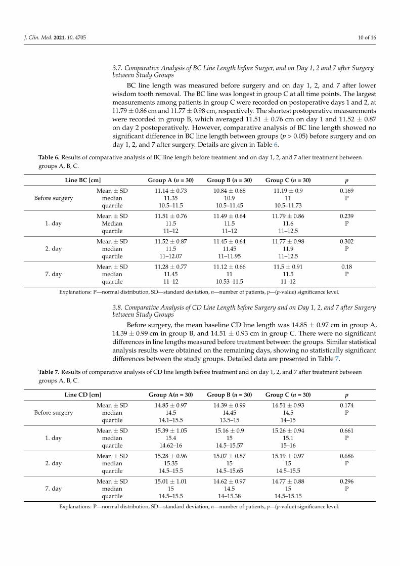
3.7. Comparative Analysis of BC Line Length before Surger, and on Day 1, 2 and 7 after Surgerybetween Study Groups
BC line length was measured before surgery and on day 1, 2, and 7 after lowerwisdom tooth removal. The BC line was longest in group C at all time points. The largestmeasurements among patients in group C were recorded on postoperative days 1 and 2, at11.79 ± 0.86 cm and 11.77 ± 0.98 cm, respectively. The shortest postoperative measurementswere recorded in group B, which averaged 11.51 ± 0.76 cm on day 1 and 11.52 ± 0.87on day 2 postoperatively. However, comparative analysis of BC line length showed nosignificant difference in BC line length between groups (p > 0.05) before surgery and onday 1, 2, and 7 after surgery. Details are given in Table 6.
Table 6. Results of comparative analysis of BC line length before treatment and on day 1, 2, and 7 after treatment betweengroups A, B, C.
Line BC [cm] Group A (n = 30) Group B (n = 30) Group C (n = 30) p
Before surgeryMean ± SD 11.14 ± 0.73 10.84 ± 0.68 11.19 ± 0.9 0.169
median 11.35 10.9 11 Pquartile 10.5–11.5 10.5–11.45 10.5–11.73
1. dayMean ± SD 11.51 ± 0.76 11.49 ± 0.64 11.79 ± 0.86 0.239
Median 11.5 11.5 11.6 Pquartile 11–12 11–12 11–12.5
2. dayMean ± SD 11.52 ± 0.87 11.45 ± 0.64 11.77 ± 0.98 0.302
median 11.5 11.45 11.9 Pquartile 11–12.07 11–11.95 11–12.5
7. dayMean ± SD 11.28 ± 0.77 11.12 ± 0.66 11.5 ± 0.91 0.18
median 11.45 11 11.5 Pquartile 11–12 10.53–11.5 11–12
Explanations: P—normal distribution, SD—standard deviation, n—number of patients, p—(p-value) significance level.
3.8. Comparative Analysis of CD Line Length before Surgery and on Day 1, 2, and 7 after Surgerybetween Study Groups
Before surgery, the mean baseline CD line length was 14.85 ± 0.97 cm in group A,14.39 ± 0.99 cm in group B, and 14.51 ± 0.93 cm in group C. There were no significantdifferences in line lengths measured before treatment between the groups. Similar statisticalanalysis results were obtained on the remaining days, showing no statistically significantdifferences between the study groups. Detailed data are presented in Table 7.
Table 7. Results of comparative analysis of CD line length before treatment and on day 1, 2, and 7 after treatment betweengroups A, B, C.
Line CD [cm] Group A(n = 30) Group B (n = 30) Group C (n = 30) p
Before surgeryMean ± SD 14.85 ± 0.97 14.39 ± 0.99 14.51 ± 0.93 0.174
median 14.5 14.45 14.5 Pquartile 14.1–15.5 13.5–15 14–15
1. dayMean ± SD 15.39 ± 1.05 15.16 ± 0.9 15.26 ± 0.94 0.661
median 15.4 15 15.1 Pquartile 14.62–16 14.5–15.57 15–16
2. dayMean ± SD 15.28 ± 0.96 15.07 ± 0.87 15.19 ± 0.97 0.686
median 15.35 15 15 Pquartile 14.5–15.5 14.5–15.65 14.5–15.5
7. dayMean ± SD 15.01 ± 1.01 14.62 ± 0.97 14.77 ± 0.88 0.296
median 15 14.5 15 Pquartile 14.5–15.5 14–15.38 14.5–15.15
Explanations: P—normal distribution, SD—standard deviation, n—number of patients, p—(p-value) significance level.
J. Clin. Med. 2021, 10, 4705 11 of 16
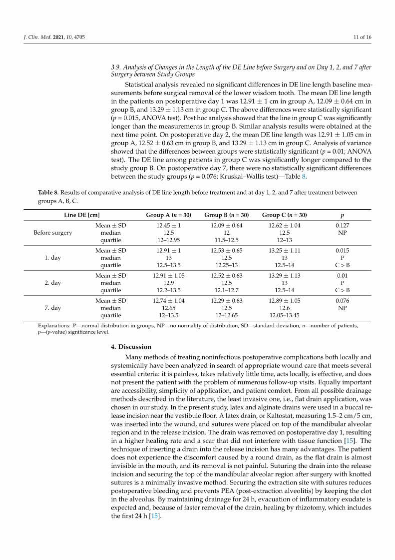
3.9. Analysis of Changes in the Length of the DE Line before Surgery and on Day 1, 2, and 7 afterSurgery between Study Groups
Statistical analysis revealed no significant differences in DE line length baseline mea-surements before surgical removal of the lower wisdom tooth. The mean DE line lengthin the patients on postoperative day 1 was 12.91 ± 1 cm in group A, 12.09 ± 0.64 cm ingroup B, and 13.29 ± 1.13 cm in group C. The above differences were statistically significant(p = 0.015, ANOVA test). Post hoc analysis showed that the line in group C was significantlylonger than the measurements in group B. Similar analysis results were obtained at thenext time point. On postoperative day 2, the mean DE line length was 12.91 ± 1.05 cm ingroup A, 12.52 ± 0.63 cm in group B, and 13.29 ± 1.13 cm in group C. Analysis of varianceshowed that the differences between groups were statistically significant (p = 0.01; ANOVAtest). The DE line among patients in group C was significantly longer compared to thestudy group B. On postoperative day 7, there were no statistically significant differencesbetween the study groups (p = 0.076; Kruskal–Wallis test)—Table 8.
Table 8. Results of comparative analysis of DE line length before treatment and at day 1, 2, and 7 after treatment betweengroups A, B, C.
Line DE [cm] Group A (n = 30) Group B (n = 30) Group C (n = 30) p
Before surgeryMean ± SD 12.45 ± 1 12.09 ± 0.64 12.62 ± 1.04 0.127
median 12.5 12 12.5 NPquartile 12–12.95 11.5–12.5 12–13
1. dayMean ± SD 12.91 ± 1 12.53 ± 0.65 13.25 ± 1.11 0.015
median 13 12.5 13 Pquartile 12.5–13.5 12.25–13 12.5–14 C > B
2. dayMean ± SD 12.91 ± 1.05 12.52 ± 0.63 13.29 ± 1.13 0.01
median 12.9 12.5 13 Pquartile 12.2–13.5 12.1–12.7 12.5–14 C > B
7. dayMean ± SD 12.74 ± 1.04 12.29 ± 0.63 12.89 ± 1.05 0.076
median 12.65 12.5 12.6 NPquartile 12–13.5 12–12.65 12.05–13.45
Explanations: P—normal distribution in groups, NP—no normality of distribution, SD—standard deviation, n—number of patients,p—(p-value) significance level.
4. Discussion
Many methods of treating noninfectious postoperative complications both locally andsystemically have been analyzed in search of appropriate wound care that meets severalessential criteria: it is painless, takes relatively little time, acts locally, is effective, and doesnot present the patient with the problem of numerous follow-up visits. Equally importantare accessibility, simplicity of application, and patient comfort. From all possible drainagemethods described in the literature, the least invasive one, i.e., flat drain application, waschosen in our study. In the present study, latex and alginate drains were used in a buccal re-lease incision near the vestibule floor. A latex drain, or Kaltostat, measuring 1.5–2 cm/5 cm,was inserted into the wound, and sutures were placed on top of the mandibular alveolarregion and in the release incision. The drain was removed on postoperative day 1, resultingin a higher healing rate and a scar that did not interfere with tissue function [15]. Thetechnique of inserting a drain into the release incision has many advantages. The patientdoes not experience the discomfort caused by a round drain, as the flat drain is almostinvisible in the mouth, and its removal is not painful. Suturing the drain into the releaseincision and securing the top of the mandibular alveolar region after surgery with knottedsutures is a minimally invasive method. Securing the extraction site with sutures reducespostoperative bleeding and prevents PEA (post-extraction alveolitis) by keeping the clotin the alveolus. By maintaining drainage for 24 h, evacuation of inflammatory exudate isexpected and, because of faster removal of the drain, healing by rhizotomy, which includesthe first 24 h [15].
J. Clin. Med. 2021, 10, 4705 12 of 16
This study aimed to evaluate the effect of intraoral drainage with flat latex and Kalto-stat drains applied to the buccal side on the severity of complications after surgical removalof the lower wisdom tooth, and we present this method as easy, effective, and likelyto find widespread use. The use of rubber drains for wound care after wisdom toothextraction procedures has been reported in the literature [13,16–18]. It has been notedthat the use of drainage reduces postoperative complications such as pain, swelling, andtrismus [17,19–22]. Based on scientific reports, the primary wound closure technique waschosen for our study of inserting a flat drain into a release cut in the oral vestibule. Thismethod reduces the accumulation of food debris in the wound, minimizes the risk of PEAformation, and minimizes the risk of excessive postoperative bleeding. In addition, astudy by Gay-Escoda et al. [23] demonstrated that suture supply to the site after surgi-cally removing the lower wisdom tooth and securing a single-knotted suture to the oralvestibular incision, without drainage applied, does not reduce pain, swelling, and trismus.According to some authors, the type of flap formed and the incision used do not affect thestatus of postoperative complications [24,25]. Thus, the effect of primary and secondaryhealing on the status of postoperative complications has begun to be studied. It is widelybelieved that an immediate healing process after tight wound care causes more pain andswelling than secondary healing [26–29]. Few are of the opinion that postoperative healingis not significantly different in terms of pain severity [30]. Primary wound healing is rapidand preferable after surgery; however, it results in more swelling, pain, and tendernessdue to lack of evacuation of exudate and pressure on nerves and vessels [31]. It can alsocause hematoma formation, which can become infected. However, keeping the clot in thealveolus and securing it with sutures is the basis of proper healing, as this prevents foodaccumulation in the wound, excessive bleeding, and PEA [13].
Secondary healing by granulation is uncomfortable for the patient because of the longduration, frequent disintegration of the clot leading to PEA, and the accumulation of fooddebris in the alveolus, which can cause tissue infection and a high risk of the formationof a pathological pocket behind the second mandibular molar [30]. Studies have shownthat healing by omitting sutures after surgical extraction of the wisdom tooth [27,28,32],creating a triangular drainage space behind the second molar, and using a drain on topof the alveolar process results in secondary healing that is characterized by little pain andswelling [28,33].
An alternative to secondary healing appears to be primary healing using a drain inthe floor of the oral vestibule to drain wound exudate.
Researchers have focused on keeping the drain in the postoperative wound for 72 h,resulting in secondary healing in the release incision [16]. For this purpose, a rubber drainwith a round cross-section and a diameter of approximately 4 mm was used. Using such adrain was traumatizing to the tissues and uncomfortable for the patient because it caused aforeign body sensation in the mouth. The drain was fixed with knotted sutures. A flat latexdrain was used in study by Hu et al. in 2017 [13]. Patients hardly noticed its presence in theoral cavity; moreover, they were less likely to have a recurrent infection than patients witha round drain. Long-term studies have shown that factors affecting healing quality includethorough wound cleaning, maintenance of a moist environment, and protection frominfection [34]. Local factors that delay wound healing after surgical removal of wisdomteeth include inadequate drainage, edema, hematoma, local tissue anemia due to excessivetissue tension, the method of forming a full-thickness flap, and the type of healing—byprimary healing or granulation [7,34,35].
In addition to using round drains and flat latex drains, calcium–sodium alginate isalso used in the literature after tooth extractions. Turner listed the characteristics of anideal dressing and defined those of an alginate dressing used for wound care, which wasalso used in our study [14,36]. According to the current literature, alginate is used as adressing after extraction and as a drain placed in the cavity of dental abscesses [14,37]. Un-doubted advantages of alginates include permeability to oxygen, maintenance of adequatetissue moisture, absorption of blood and exudate, protection against reinfection, adequate
J. Clin. Med. 2021, 10, 4705 13 of 16
mechanical strength, anti-allergenicity, lack of adhesion to the wound, biodegradability.This material can be a drug carrier [38].
Using flat drains is a form of drainage that is less invasive than inserting rounddrains into the postoperative wound, inducing free flow of exudate; at the same time, itis an alternative to methods minimizing postoperative complications—steroid therapy,cryotherapy, laser therapy, Kinesio Taping [39–43].
Studies by the teams of Deliverska and Petkova and Seymour et al. found that painis most severe in the immediate postoperative period and gradually decreases until itis completely gone [44,45]. A study by Seymour et al. reported that the degree of painafter surgical removal of mandibular molars supplied with sutures without drainage ismost significant on the day of surgery and increases up to 12 h after surgery [45]. In ourstudy, there was no difference in pain between the study groups during the first 24 h,which was confirmed by the analysis of Chukwuneke et al. [17]. They found no statisticallysignificant difference between the control and study groups on postoperative day 1 interms of pain intensity, which was consistent with their study. Additionally, Chukwunekeet al. examined pain complaints after day 3 [17]. After this time, the mean pain levelscore of the control group was significantly lower than that of the study group becausethere was a transient increase in pain in the rubber tube group, probably because of itsirritating effect [18]. A sharp decrease was observed in the level of pain experienced afterremoving the rubber drain after day 3. In the author’s study, no significant reduction inpain was observed after removing the drain, suggesting that its presence did not affect theperception of postoperative discomfort.
Edema can be caused by tissue response to stretch, compression, or trauma associatedwith surgery. Its onset is gradual, with maximum swelling occurring within 48 h of surgery.It increases until the fourth day and completely subsides within the next seven days [46–48].There are different ways to measure facial swelling: the use of the facial arch, cephalostat,ultrasound measurements, photography, and measuring the distance between craniometricpoints [49]. Measuring with a flexible tape measure the corresponding lines defined by thedistances between the various fixed points on the face, the so-called craniometric points, isnon-invasive and easy to perform [21].
Chukwuneke et al. evaluated the effect of drainage after surgical removal of mandibu-lar third molars on facial swelling on day 1 after surgery [17]. It was significantly higher inthe control group (13%) than in the study group (7%). In our study, the most significantswelling was found on day 1 in all study groups. However, it was significantly less inthe latex knot groups and already decreasing from day 2, while in the control group, itpersisted up to 48 h after the procedure.
Like swelling, the trismus usually peaks on the second day and subsides by the end ofthe first week. There is a strong correlation between postoperative pain and maxillo-facialtightness, indicating that pain may be one of the primary causes of reduced jaw dilationafter lower wisdom tooth extraction [50].
In the present study, jaw opening was measured before surgery and after day 1, 2, and7 after surgery, following the scheme given by Handa et al. [51]. Using an electronic caliperand measuring the maximum distance between the incisal edges of the medial incisors inthe maxilla and the incisal edges of the medial incisors in the mandible, it was found thatthe least jaw opening occurred after day 1 in all study groups, which was consistent withthe result of Rakprasitkul and Pairuchvej, Cerqueira et al., and Genc et al. [19,52,53].
As a result of the research conducted by our team, they proved that intraoral drainagewith a flat drain after mandibular third molar removal does not significantly reduce pain,as measured by the VAS scale, or postoperative swelling, as measured by lines betweencraniometric points. Intraoral drainage with a latex drain after mandibular third molarremoval does not significantly reduce trismus, while intraoral drainage with a calcium–sodium alginate drain increases trismus. Intraoral drainage with flat drains, especiallylatex drains, can make the patient’s recovery faster and less traumatic. It may result ina quicker return to activities of daily living and work duties. These results showed the
J. Clin. Med. 2021, 10, 4705 14 of 16
significant clinical implications of our study. Intraoral drainage using flat drains, especiallylatex drains, can make the patient’s recovery faster and less traumatic. This method ischeap, simple, and can be performed in dental offices and oral surgery, even by a generaldentist. It does not require a ready-made preparation—a flat latex drain can be obtainedfrom a sterile latex glove by preparing a drain of the desired dimensions.
Limitations of the study include the small size of the study group. Only 90 peopleparticipated in the study. Moreover, one more group could be added to the study—rounddrains. Furthermore, the assessment of pain on the VAS scale is subjective. In addition, themeasurements assessed in the study were not continuous but taken only at specific timepoints. In the future, more precise tests could also be performed to measure the level ofmaxillary and facial swelling, e.g., evaluation by extraoral scan and comparison of STLfiles [54].
5. Conclusions
Intraoral drainage with a flat drain after mandibular third molar removal does notsignificantly reduce pain, as measured by the VAS scale, or postoperative swelling, asmeasured by lines between craniometric points. Intraoral drainage with a latex drain aftermandibular third molar removal does not significantly reduce trismus, while intraoraldrainage with a calcium–sodium alginate drainage bag significantly increases trismus.Intraoral drainage with flat drains, especially latex drains, can make the patient’s recoveryprocess faster and less traumatic. It may result in a quicker return to activities of dailyliving and work duties.
Author Contributions: Conceptualization, G.T. and J.J.; methodology, G.T. and J.J.; software, A.J., J.J.,O.P.; validation, G.T., O.P.; formal analysis, G.T. and J.J.; investigation, G.T. and J.J.; resources, A.J.,J.J., O.P.; data curation, J.J.; writing—original draft preparation, G.T., J.J., O.P., A.J.; writing—reviewand editing, G.T., A.J.; visualization, J.J., A.J.; supervision, G.T., O.P.; project administration, G.T. Allauthors have read and agreed to the published version of the manuscript.
Funding: This research received no external funding.
Institutional Review Board Statement: The study was conducted according to the guidelines of theDeclaration of Helsinki, and approved by the Institutional Ethics Committee of Pomeranian MedicalUniversity, Szczecin, Poland (No. KB-0012/38/18 on 12 March 2018).
Informed Consent Statement: Informed consent was obtained from all subjects involved in the study.
Data Availability Statement: Data are available on request because of privacy or ethical restrictions.
Conflicts of Interest: The authors declare no conflict of interest.
References1. Waite, P.D.; Reynolds, R.R. Surgical management of impacted third molars. Semin. Orthod. 1998, 4, 113–123. [CrossRef]2. Zawilska, A.; Koszkowski, R.; Waskowska, J. Ocena budowy oraz typów retencji zatrzymanych trzecich trzonowców w obrazie
pantomograficznym. Ann. Acad. Med. Stet. 2007, 53, 165–171.3. Czerniuk, M.R. Zeby madrosci jako potencjalne ognisko infekcji pochodzacej z jamy ustnej—Opis przypadku. Kardiol. Dypl. 2009,
8, 92–96.4. Lorè, B.; Gargari, M.; Ventucci, E.; Cagioli, A.; Nicolai, G.; Calabrese, L. A complication following tooth extraction: Chronic
suppurative osteomyelitis. Oral Implantol. 2013, 6, 43–47. [CrossRef]5. Trybek, G.; Chamarczuk, A.; Falkowska, J.; Grzegorzewska, M.; Preuss, O.; Aniko-Włodarczyk, M. Intraoral odontogenic
abscesses in patients of The Department of Oral Surgery at the Pomeranian Medical University in Szczecin: 7 years of observation.Postep. Hig. Med. Dosw. 2018, 72, 491–498. [CrossRef]
6. Kilinc, A.; Ataol, M. How effective is collagen resorbable membrane placement after partially impacted mandibular third molarsurgery on postoperative morbidity? A prospective randomized comparative study. BMC Oral Health 2017, 17, 1–8. [CrossRef]
7. Kaczmarzyk, T. Poekstrakcyjne zapalenie zebodołu. Med. Prakt. 2012, 2, 45–50.8. Szubert, P.; Jankowski, M.; Krajecki, M.; Jankowska-Wika, A.; Sokalski, J. Analiza czynników predysponujacych do powikłan po
chirurgicznym usunieciu zebów madrosci w zuchwie. Dent. Forum 2015, 63, 45–50.
J. Clin. Med. 2021, 10, 4705 15 of 16
9. de Brabander, E.C.; Cattaneo, G. Effectiveness of cold therapy in reducing pain, trismus, and oedema after impacted mandibularthird molar surgery: A randomized, self-controlled, observer-blind, split-mouth clinical trial. Int. J. Oral Maxillofac. Surg. 2016, 45,118–123.
10. Xavier, R.L.; Vasconcelos, B.C.; Caubi, A.F.; Porto, G.G.; Maurette, M.A. Passive drainage through the vestibular oblique incisionin impacted inferior third molar surgery: A preliminary study. Acta Odontol. Latinoam. 2008, 21, 57–63.
11. Rullo, R.; Addabbo, F.; Papaccio, G.; D’Aquino, R.; Festa, V.M. Piezoelectric device vs. conventional rotative instruments inimpacted third molar surgery: Relationships between surgical difficulty and postoperative pain with histological evaluations. J.Cranio-Maxillofac. Surg. 2013, 41, 33–38. [CrossRef] [PubMed]
12. Uyanik, L.O.; Bilginaylar, K.; Etikan, I. Effects of platelet-rich fibrin and piezosurgery on impacted mandibular third molarsurgery outcomes. Head Face Med. 2015, 11, 1–7. [CrossRef] [PubMed]
13. Hu, T.; Zhang, J.; Ma, J.Z.; Shao, L.N.; Gu, Y.F.; Li, D.Q.; Liang, L.; Yang, Y.Q. A novel method in the removal of impactedmandibular third molar: Buccal drainage. Sci. Rep. 2017, 7, 1–6. [CrossRef] [PubMed]
14. Matthew, I.R.; Browne, R.M.; Frame, J.W.; Millar, B.G. Tissue response to a haemostatic alginate wound dressing in toothextraction sockets. Br. J. Oral Maxillofac. Surg. 1993, 31, 165–169. [CrossRef]
15. Dominiak, M.; Gedrange, T.; Zapała, T. Podstawy Chirurgii Stomatologicznej; Urban & Partner: Wrocław, Poland, 2013.16. Zandi, M. Comparison of corticosteroids and rubber drain for reduction of sequelae after third molar surgery. Oral Maxillofac.
Surg. 2008, 12, 29–33. [CrossRef]17. Chukwuneke, F.N.; Oji, C.; Saheeb, D.B. A comparative study of the effect of using a rubber drain on postoperative discomfort
following lower third molar surgery. J. Oral Maxillofac. Surg. 2008, 37, 341–344. [CrossRef] [PubMed]18. Osunde, O.D.; Adebola, R.A.; Omeje, U.K. Management of inflammatory complications in third molar surgery: A review of the
literature. Afr. Health Sci. 2011, 11, 530–537. [PubMed]19. Rakprasitkul, S.; Pairuchvej, V. Mandibular third molar surgery with primary closure and tube drain. Int. J. Oral Maxillofac. Surg.
1997, 26, 187–190. [CrossRef]20. Saglam, A.A. Effects of tube drain with primary closure technique on postoperative trismus and swelling after removal of fully
impacted mandibular third molars. Quintessence Int. 2003, 34, 143–147.21. Koyuncu, B.O.; Zeytinoglu, M.; Tetik, A.; Gomel, M.M. Effect of tube drainage compared with conventional suturing on
postoperative discomfort after extraction of impacted mandibular third molars. Oral Maxillofac. Surg. 2014, 53, 63–67. [CrossRef]22. Garajei, A.; Emami, A. Effect of surgical drain on the control of swelling in impacted lower third molar surgery. J. Craniomaxillofacial
Res. 2016, 3, 264–267.23. Gay-Escoda, C.; Gómez-Santos, L.; Sánchez-Torres, A.; Herráez-Vilas, J.M. Effect of the suture technique on postoperative pain,
swelling and trismus after removal of lower third molars: A randomized clinical trial. Med. Oral Patol. Oral Cir. Bucal 2015, 20,372–377. [CrossRef] [PubMed]
24. Sortino, F.; Cicciu, M. Strategies used to inhibit postoperative swelling following removal of impacted lower third molar. Dent.Res. J. 2011, 8, 162–171.
25. Dolanmaz, D.; Esen, A.; Isik, K.; Candirli, C. Effect of 2 flap designs on postoperative pain and swelling after impacted thirdmolar surgery. Oral Surg. Oral Med. Oral Pathol. Oral Radiol. 2013, 116, 244–246. [CrossRef] [PubMed]
26. Danda, A.K.; Krishna Tatiparthi, M.; Narayanan, V.; Siddareddi, A. Influence of primary and secondary closure of surgical woundafter impacted mandibular third molar removal on postoperative pain and swelling-A comparative and split mouth study. J. OralMaxillofac. Surg. 2010, 68, 309–312. [CrossRef] [PubMed]
27. Khande, K.; Saluja, H.; Mahindra, U. Primary and secondary closure of the surgical wound after removal of impacted mandibularthird molars. J. Maxillofac. Oral Surg. 2011, 10, 112–117. [CrossRef]
28. Hashemi, H.M.; Beshkar, M.; Aghajani, R. The effect of sutureless wound closure on postoperative pain and swelling afterimpacted mandibular third molar surgery. Br. J. Oral Maxillofac. Surg. 2012, 50, 256–258. [CrossRef]
29. Maria, A.; Malik, M.; Virang, P. Comparison of primary and secondary closure of the surgical wound after removal of impactedmandibular third molars. J. Maxillofac. Oral Surg. 2012, 11, 276–283. [CrossRef]
30. Chaudhary, M.; Singh, M.; Singh, S.; Singh, S.P.; Kaur, G. Primary and secondary closure technique following removal of impactedmandibular third molars: A comparative study. Natl. J. Maxillofac. Surg. 2012, 3, 10–14.
31. Skoracka, J.; Torlinska-Walkowiak, N.; Torlinska, T.; Wozniak, W. Podstawy anatomiczne i fizjologiczne zespołów bólowychukładu stomatognatycznego. Now. Lek. 2006, 75, 80–89.
32. Pathak, H.M.; Kumari, S.; Prasad, S.; Singh, N.; Pathak, P. Suture-less third molar surgery: Review of 30 cases. Int. J. Sci. Res. Publ.2013, 3, 1–5.
33. Osunde, O.D.; Adebola, R.A.; Saheeb, B.D. A comparative study of the effect of suture-less and multiple suture techniques oninflammatory complications following third molar surgery. Int. J. Oral Maxillofac. Surg. 2012, 41, 1275–1279. [CrossRef]
34. Dabrowiecki, S. Fizjologia i patofizjologia procesu gojenia ran. Pol. Med. Paliat. 2003, 2, 283.35. Dominiak, M.; Łysiak, K. Naprawa i/lub regeneracja poresekcyjnych i pocystektomijnych ubytków sródkostnych wyrostka
zebodołowego–ocena uwarunkowan na podstawie pismiennictwa i doswiadczen własnych. Dent. Med. Probl. 2005, 42, 341–350.36. Dawson, C.; Armstrong, M.W.; Fulford, S.C.; Farugi, R.M.; Galland, R.B. Use of calcium alginate to pack abscess cavities: A
controlled clinical trial. J. R. Coll. Physicians Edinb. 1992, 37, 177–179.
J. Clin. Med. 2021, 10, 4705 16 of 16
37. Matthew, I.R.; Browne, R.M.; Frame, J.W.; Millar, B.M. Subperiosteal behaviour of alginate and cellulose wound dressing materials.Biomaterials 1995, 16, 265–274. [CrossRef]
38. Burrow, B.A.; Linday, A. A limited evaluation of alginates and a small scale comparision between Kaltostat and a standardnon-adherent dressing, Ultraplast Alginate, in the treatment of nail avulsion by matrix phenolisation. Chiropodist 1989, 3, 211–218.
39. Jaron, A.; Preuss, O.; Grzywacz, E.; Trybek, G. The Impact of Using Kinesio Tape on Non-Infectious Complications after ImpactedMandibular Third Molar Surgery. Int. J. Environ. Res. Public Health 2021, 18, 399. [CrossRef]
40. Jaron, A.; Jedlinski, M.; Grzywacz, E.; Mazur, M.; Trybek, G. Kinesiology Taping as an Innovative Measure against Post-OperativeComplications after Third Molar Extraction-Systematic Review. J. Clin. Med. 2020, 9, 3988. [CrossRef]
41. Falci, S.G.M.; Lima, T.C.; Martins, C.C.; Santos, C.R.R.D.; Pinheiro, M.L.P. Preemptive Effect of Dexamethasone in Third-MolarSurgery: A Meta-Analysis. Anesth. Prog. 2017, 64, 136–143. [CrossRef]
42. do Nascimento-Júnior, E.M.; Dos Santos, G.M.S.; Tavares Mendes, M.L.; Cenci, M.; Correa, M.B.; Pereira-Cenci, T.; Martins-Filho,P.R.S. Cryotherapy in reducing pain, trismus, and facial swelling after third-molar surgery: Systematic review and meta-analysisof randomized clinical trials. J. Am. Dent. Assoc. 2019, 150, 269–277.e1. [CrossRef] [PubMed]
43. Eshghpour, M.; Ahrari, F.; Takallu, M. Is Low-Level Laser Therapy Effective in the Management of Pain and Swelling AfterMandibular Third Molar Surgery? J. Oral Maxillofac. Surg. 2016, 74, 1322.e1–1322.e8. [CrossRef] [PubMed]
44. Deliverska, E.G.; Petkova, M. Complications after extraction of impacted third molars—Literature review. IMAB 2016, 22,1202–1211. [CrossRef]
45. Seymour, R.A.; Meechan, J.G.; Blair, G.S. An investigation into post-operative pain after third molar surgery under local analgesia.Br. J. Oral Maxillofac. Surg. 1985, 23, 410–418. [CrossRef]
46. Grossi, G.B.; Maiorana, C.; Giarramone, R.A.; Borgonovo, A.; Beretta, M.; Farronato, D. Effects of submucosal injection ofdexamethasone on postoperative discomfort after third molar surgery: A prospective study. J. Oral Maxillofac. Surg. 2007, 65,2218–2226. [CrossRef]
47. Ayaz, H.; Rehman, A.U.; Din, F.U. Post-operative complications associated with impacted mandibular third molar removal. Pak.Oral Dent. J. 2012, 32, 389–392.
48. Darawade, D.A.; Kumar, S.; Mehta, R.; Sharma, A.R.; Reddy, G.S. In search of a better option: Dexamethasone versus methylpred-nisolone in third molar impaction surgery. J. Int. Oral Health 2014, 6, 14–17.
49. Kumar, B.; Bhate, K.; Dolas, R.S.; Kumar, S.; Waknis, P. Comparative evaluation of immediate post-operative sequelae aftersurgical removal of impacted mandibular third molar with or without tube drain—Split-mouth study. J. Clin. Diagn Res. 2016, 10,46–49. [CrossRef]
50. Pedersen, A. Interrelation of complaints after removal of impacted mandibular third molars. Int. J. Oral Surg. 1985, 14, 241–244.[CrossRef]
51. Handa, A.; Agwani, M.K.; Surendra, S.S.; Rana, S.S. Effects of tube drain with primary closure techniques on postoperativetrismus and swelling after removal of impacted mandibular third molars. J. Dent. Med. Sci. 2016, 15, 92–100.
52. Cerqueira, P.R.; Vasconcelos, B.C.; Bessa-Nogueira, R.V. Comparative Study of the Effect of a Tube Drain in Impacted LowerThird Molar Surgery. J. Oral Maxillofac. Surg. 2004, 62, 57–61. [CrossRef]
53. Genc, A.; Cakarer, S.; Yalcin, B.K.; Kilic, B.B.; Isler, S.C.; Keskin, C. A comparative study of surgical drain placement and the useof kinesiologic tape to reduce postoperative morbidity after third molar surgery. Clin. Oral Investig. 2018, 23, 345–350. [CrossRef][PubMed]
54. Metlerski, M.; Grocholewicz, K.; Jaron, A.; Lipski, M.; Trybek, G. Comparison of Presurgical Dental Models Manufactured withTwo Different Three-Dimensional Printing Techniques. J. Healthc. Eng. 2020, 2020, 8893338. [CrossRef] [PubMed]