Identification of KIAA1018/FAN1, a DNARepair Nuclease Recruited to DNADamage by Monoubiquitinated FANCD2Craig MacKay,1 Anne-Cecile Declais,2 Cecilia Lundin,4 Ana Agostinho,3 Andrew J. Deans,6 Thomas J. MacArtney,1
Kay Hofmann,5 Anton Gartner,3 Stephen C. West,6 Thomas Helleday,4,7 David M.J. Lilley,2 and John Rouse1,*1MRC Protein Phosphorylation Unit2CRUK Nucleic Acids Structure Research Group3Wellcome Trust Centre for Gene Regulation and ExpressionCollege of Life Sciences, University of Dundee, Dundee DD1 5EH, Scotland, UK4Gray Institute for Radiation Oncology & Biology, University of Oxford, Oxford OX3 7DQ, UK5Miltenyi Biotec GmbH, D-51429 Bergisch Gladbach, Germany6London Research Institute, Cancer Research UK, Clare Hall Laboratories, South Mimms EN6 3LD, UK7Department of Genetics Microbiology and Toxicology, Stockholm University, S-106 91 Stockholm, Sweden
*Correspondence: [email protected]
DOI 10.1016/j.cell.2010.06.021
SUMMARY
DNA interstrand crosslinks (ICLs) are highly toxicbecause they block the progression of replisomes.The Fanconi Anemia (FA) proteins, encoded bygenes that are mutated in FA, are important for repairof ICLs. The FA core complex catalyzes the monou-biquitination of FANCD2, and this event is essentialfor several steps of ICL repair. However, how mono-ubiquitination of FANCD2 promotes ICL repair atthe molecular level is unknown. Here, we describea highly conserved protein, KIAA1018/MTMR15/FAN1, that interacts with, and is recruited to sitesof DNA damage by, the monoubiquitinated form ofFANCD2. FAN1 exhibits endonuclease activitytoward 50 flaps and has 50 exonuclease activity, andthese activities are mediated by an ancient VRR_nucdomain. Depletion of FAN1 from human cells causeshypersensitivity to ICLs, defects in ICL repair, andgenome instability. These data at least partly explainhow ubiquitination of FANCD2 promotes DNA repair.
INTRODUCTION
DNA interstrand crosslinks (ICLs) are formed when bifunctional
agents covalently link the two strands in a double helix. ICLs
are toxic lesions that prevent strand separation necessary for
transcription and DNA replication. ICLs can be induced by drugs
and also by endogenous metabolites. Crosslinking agents such
as mitomycin-C (MMC) and cisplatin generate a mixture of
monoadducts and ICLs in cells but cellular toxicity correlates
with the number of ICLs. Although ICLs can be repaired in G1,
the major route for ICL repair appears to occur in S phase (Akkari
et al., 2000; Rothfuss and Grompe, 2004; Taniguchi et al., 2002).
Various models for the repair of ICLs have been suggested
(McCabe et al., 2009; Moldovan and D’Andrea, 2009), and recent
studies proposed that ICL repair requires two forks to converge
on the ICL (Raschle et al., 2008) (Figure S1 available online).
Forks that stall at ICLs recruit signaling complexes including
the Fanconi Anemia (FA) proteins and FA-associated proteins
(Moldovan and D’Andrea, 2009) (Figure S1). Fanconi Anemia
is an inherited recessive condition characterized by develop-
mental defects, skeletal abnormalities, bone marrow failure,
and cancer predisposition (Wang, 2007). FA falls into 13 comple-
mentation groups, and the relevant FA genes have been
cloned (Patel and Joenje, 2007; Wang, 2007). Nevertheless, FA
patients exist where mutations in known FA genes could not be
found. The central components of the FA pathway are FANCD2
and its paralogue FANCI, which together form the ‘‘ID’’ complex
(Garcia-Higuera et al., 2001; Smogorzewska et al., 2007). These
two proteins are monoubiquitinated at Lys561 and Lys523,
respectively, in S phase and in response to ICLs (Figure S1)
(Garcia-Higuera et al., 2001; Taniguchi et al., 2002). This reaction
is catalyzed by the E3 ubiquitin ligase FANCL subunit of the
FA core complex, which comprises FANCA, B, C, E, F, G, L,
and M, and also requires the FA-associated proteins FAAP100
and FAAP24 (Ciccia et al., 2007; Collis et al., 2008; Ling
et al., 2007). Furthermore, loss of FANCD2 monoubiquitination
is observed in many FA patients (Moldovan and D’Andrea, 2009).
Monoubiquitination of FANCD2 is necessary for ICL repair but
the underlying molecular mechanisms are unclear. The monoubi-
quitinated form of the ID complex may recruit ICL repair proteins,
but as yet no ligands for ubiquitinated FANCD2 have been re-
ported. It was reported that monoubiquitination of FANCD2 is
required for the ‘‘unhooking’’ of the ICL in a cell-free repair sys-
tem (Knipscheer et al., 2009) (Figure S1). Unhooking involves
incisions on either side of the ICL, one of which is catalyzed
by the structure-specific nuclease MUS81-EME1 (Figure S1)
(Hanada et al., 2007; Hanada et al., 2006). MUS81-EME1 creates
a one-ended double-strand break (DSB) that can be used later to
Cell 142, 65–76, July 9, 2010 ª2010 Elsevier Inc. 65
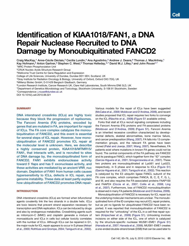
A
B
C
Figure 1. The KIAA1018/MTMR15/FAN1
Family of Proteins
(A) Schematic representation of the domain archi-
tecture of KIAA1018/MTMR15/FAN1 orthologs
from different species. The relevant protein iden-
tification codes are as follows: Homo sapiens
Q9Y2M0, Danio rerio Q1LWH4, Caenorhabditis
elegans P90740, Schizosaccharomyces pombe
Q9Y804, Arabidopsis thaliana Q9SX69, Oryza sativa
B9FRR6, and Pseudomonas aeruginosa Q9I2N0.
(B) Alignment of the VRR_nuc domain of FAN1.
Identical residues are shaded in black, and similar
residues are shaded in gray. The asterisks denote
conserved residues Asp981 and Arg982 mutated
in the FAN1-DR mutant.
(C) Alignment of the UBZ domain of FAN1. Identical
residues are shaded in black, and similar residues
are shaded in gray. The conserved Cys and His resi-
dues that define the two dyads of the UBZ domain
are shaded in red. Asterisks denote the conserved
Cys44 and Cys47 residues in the first dyad.
initiate homologous recombination (HR). The identity of the
nuclease that catalyzes the second incision to enable unhooking
of the ICL is unclear. XPF-ERCC1 has been implicated, but this is
controversial (Bergstralh and Sekelsky, 2008; Bhagwat et al.,
2009). After unhooking, the resulting gap is filled in by translesion
synthesis, which also appears to require FANCD2 ubiquitination
(Knipscheer et al., 2009), and the unhooked lesion is removed by
excision repair. The DSBs generated by unhooking are resected
and one of them initiates HR to complete ICL repair (Figure S1).
Successful HR-mediated repair of the MUS81-generated DSB
depends on processing of DNA repair intermediates by the
SLX4 complex. SLX4 acts as a scaffold for XPF-ERCC1,
MUS81-EME1, and SLX1. Cells lacking, or depleted of, SLX4
(Fekairi et al., 2009; Munoz et al., 2009; Svendsen et al., 2009)
or XPF-ERCC1 (Niedernhofer et al., 2004) cannot efficiently repair
the DSBs created by MUS81 after ICL induction and exhibit
defects in HR-mediated repair of DSBs. In this study, we report
the identification of FAN1, a nuclease recruited to sites of DNA
damage by monoubiquitinated FANCD2 that is important for
repair of ICLs.
66 Cell 142, 65–76, July 9, 2010 ª2010 Elsevier Inc.
RESULTS
Domain Organization of KIAA1018/MTMR15/FAN1We noticed an uncharacterized human
protein, KIAA1018/MTMR15, in the
human sequence databases, that has a
UBZ-type ubiquitin-binding domain do-
main, a SAP-type DNA binding domain,
and a putative nuclease domain termed
the ‘‘VRR_nuc’’ domain (Figure 1A), ini-
tially referred to as ‘‘domain of unknown
function 994’’ (DUF994) (Iyer et al., 2006).
Orthologs of KIAA1018 are found in
prokaryotes and most eukaryotes with
the notable exception of budding yeast
(Figure 1A).
We suspected that KIAA1018 is involved in DNA damage
responses for a number of reasons. KIAA1018 is the only VRR-
nuc domain-containing protein in eukaryotes but many bacteria
and bacteriophages have genes that encode solely VRR_nuc
domains. Although the functions of these genes are unknown,
most of them are located in operons that include known DNA
repair enzymes, hence the name VRR_nuc (virus-type replica-
tion-repair nuclease) (Iyer et al., 2006). The VRR_nuc domains
contain a PD-(D/E)XK motif found in the active site of many
restriction nucleases (Kosinski et al., 2005) (Figure 1B). We thus
suspected that KIAA1018 might act as a repair endonuclease.
A putative role for KIAA1018 in DNA repair is also implied by the
presence of a UBZ4-type ubiquitin-binding domain that belongs
to the RAD18 family of zinc fingers, a domain commonly found
in DNA damage response proteins such as DNA polymerase k
(POL k), RAD18, and WRNIP (Figure 1C) (Hofmann, 2009).
Furthermore, KIAA1018 was also found to interact with the
MLH1 DNA mismatch repair protein in a genome wide screen
(Cannavo et al., 2007). We therefore decided to test whether
KIAA1018, which we refer to hereafter as FAN1 for reasons that
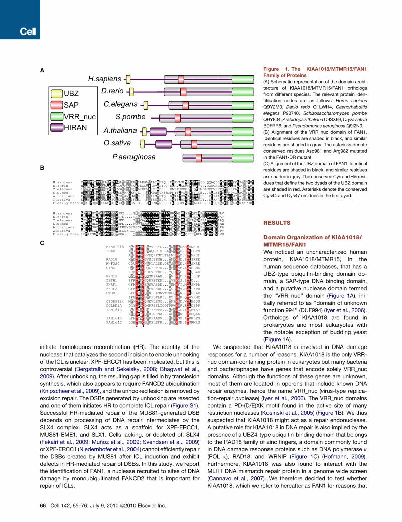
FAN1–DR: FAN1–WT:
– + – – + – – + – – + –
– + – – + – – + – – + –
– + – – + – – + – – + –
– + – – + – – + – – + –
SD 3’F 5’F RF SD 3’F 5’F RF
a3 blab. strand:
substrate:
R R
0 10
20
30
45
60
120
300
0 60
120
300
600
1200
24
00
3600
time:
FAN1–DR FAN1–WT
endo
4 nt
time (s) 200 400 600 0 0
0.2
0.4
0.6
0.8
fract
ion
DN
A cu
t
FAN1–WT FAN1–DR
lane: 1 2 3 4 5 6 7 8 9 10 11 12 1 2 3 4 5 6 7 8 9 10 11 12 lane:
R R (s)
3’ flap (3’F)
a3
b
d3
splayed duplex (SD)
a3
b
5’
3’
5’ flap (5’F)
a3
b c
replication fork (RF)
a3
b c
d3
GACGC G C T G CG C G AC G
GC
TC
C GT AG CC G
C G C T G CG C G A C G
GC
TC
3’
5’ GACGC G C T G CG C G AC G
C GG C
ATG C
C GT AG CC G
C G C T G CG C G A C G
C GG C
A TG C
A
B
DC
Figure 2. FAN1 Has Structure-Specific
Endonuclease Activity
Recombinant human FAN1 was incubated with
synthetic DNA structures: splayed duplex (SD;
oligos a3, b), 30 flap (30F; oligonucleotides a3, b,
d3), 50 flap (50F; oligos a3, b, c), or a replication
fork (RF)-like structure (oligos a3, b, c, d3), each
radioactively 50 32P-labeled on the strands indi-
cated. WT refers to wild-type FAN1, and DR refers
to the Asp981Ala-Arg982Ala FAN1 mutant.
(A) Schematic diagram of the DNA substrates
used in (B). Sites of DNA cleavage are indicated
by arrows.
(B) Reaction products (10 min incubation) were
subjected to denaturing PAGE. Purine-specific
chemical sequencing ladders (R) were derived
from oligonucleotides a3 or b.
(C) FAN1 was incubated with the 50 flap shown in
(A) for the time indicated (s, seconds), and reaction
products were subjected to denaturing PAGE.
(D) Progress curves of cleavage of the 50
flap construct incubated with wild-type (black
squares) and DR (gray triangles) mutant FAN1.
The data have been fitted to a single (DR) or double
(wild-type) exponential functions (lines). From
these data, we have calculated observed rates of
cleavage of >0.2 s�1 and 0.0003 s�1 for wild-
type and DR enzymes, respectively.
See also Figure S2.
will become clear later, has nuclease activity and whether it is
involved in DNA repair.
FAN1 Has Structure-Specific Endonuclease ActivityTo test for nuclease activity, we expressed recombinant FAN1,
fused to an N-terminal NUS-His6 tag, in bacteria and purified
it through three steps of ion exchange chromatography (Fig-
ure S2A). In parallel, we purified a mutant version of FAN1 where
the conserved Asp981 and Arg982 residues (indicated by aster-
isks in Figure 1B) found in the VRR_nuc domain were mutated
to alanine (‘‘DR’’ mutant). We next tested the ability of FAN1 to
cleave a range of branched DNA substrates that resemble DNA
repair and replication intermediates. These included a splayed
duplex, a 30 flap, a 50 flap, and a nicked three-way junction that
mimics a DNA replication fork (Figure 2A). All substrates were32P labeled at the 50 end of the a3 strand or the b strand as indi-
cated in Figure 2A. After incubation with wild-type or mutant
Cell 142,
FAN1, reaction products were separated
by gel electrophoresis under denaturing
conditions.
As shown in Figure 2B, FAN1 displayed
strong endonucleaseactivity toward the 50
flap structure and weaker activity toward
the replication fork model. Cleavage
affected only one strand of these struc-
tures and occurred in the double-
stranded region on the same strand as
the flap, 4 nucleotides (nt) 30 to the branch
point (Figure 2A). Selectivity of FAN1 for
these DNA structures, as opposed to
specificity for DNA sequence, was confirmed by analysis of the
cleavage of an analogous set of branched DNA structures
composed of strands with alternative sequence (Figures 2B
and 2C). The endonuclease activity of the FAN1 DR mutant was
severely reduced compared with wild-type protein (Figure 2B;
Figures S2B and S2C), resulting in cleavage rates approximately
1000-fold lower than those for wild-type protein (Figures 2C and
2D). FAN1 did not exhibit endonuclease activity toward four-way
junctions (data not shown).
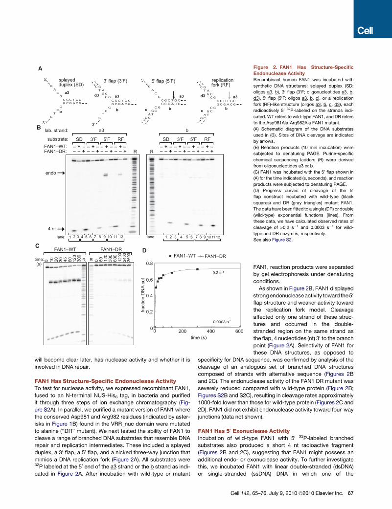
FAN1 Has 50 Exonuclease ActivityIncubation of wild-type FAN1 with 50 32P-labeled branched
substrates also produced a short 4 nt radioactive fragment
(Figures 2B and 2C), suggesting that FAN1 might possess an
additional endo- or exonuclease activity. To further investigate
this, we incubated FAN1 with linear double-stranded (dsDNA)
or single-stranded (ssDNA) DNA in which one of the
65–76, July 9, 2010 ª2010 Elsevier Inc. 67
B
C
A* 3’ * 3’ * 5’ P * P 5’
0 10
20
30
60
120
0 60
120
300
0 15
60
240
960
3480
60
240
960
3480
0 120
300
600
1200
24
00
0 60
300
600
time (s):
FAN1: WT DR WT DR WT WT
4 nt
time (s) 1200 2400 3600 0 0
0.2
0.4
0.6
0.8
fract
ion
DN
A cu
t
1.0
600 1800 3000 3’*-ss, WT
5’*-ds, WT
3’*-ds, WT
5’*-ss, WT
5’*-ds, DR
0 10
20
30
45
60
120
300
time (s):
endo
exo
a3
b *
5’ *
5’
c
R
Figure 3. FAN1 has 50 Exonuclease Activity
Recombinant human FAN1 was incubated for the time indicated (s, seconds)
with dsDNA (oligonucleotide a3, a3-cp), ssDNA (oligonucleotide a3), or a 50
flap (50F; oligonucleotides a3, b, c) radioactively 50 or 30 32P-labeled on the
a3 strand as shown (asterisks). WT refers to wild-type FAN1, and DR refers
to the Asp981Ala-Arg982Ala FAN1 mutant. Reaction products were subjected
to denaturing PAGE.
(A) Cleavage of linear DNA substrates.
(B) The cleavage products were quantitated. ‘‘Fraction DNA cut’’ is the ratio of
the relevant cleavage product to total DNA (cleaved plus uncleaved DNA). The
data are plotted as a function of time and are fitted to single or double expo-
nential functions.
(C) Activity of WT FAN1 on radioactively 30 32P-labeled 50 flap. R refers to a
purine-specific chemical sequencing ladder derived from the labeled strand.
See also Figure S3.
oligonucleotides was radioactively 50 or 30 32P labeled. We
observed a clear 50 to 30 exonuclease activity that initiates 4 nt
from the 50 end and cleaves every phosphate bond thereafter
but with varying intensity (Figure 3A). The exonuclease activity
of FAN1 toward ssDNA required that the 50 end be phosphory-
lated (Figure 3A). FAN1 exhibited potent 50 exonuclease activity
toward DNA substrates with a recessed 50 end, indicating that
a blunt dsDNA end is not required for exonuclease activity
(Figure S3). The exonuclease activity of FAN1 was severely
reduced by mutation of Asp981 and Arg982 in the VRR_nuc
domain (Figures 3A and 3B). Quantitation of these data showed
that the rate of initiation of the exonuclease activity of FAN1
68 Cell 142, 65–76, July 9, 2010 ª2010 Elsevier Inc.
toward dsDNA (0.09 s�1; Figure 3B) was approximately half
that of the endonuclease toward a 50 flap (>0.2 s�1; Figure 2D).
The calculated rates of initiation using wild-type enzyme
(Figure 3B) were 0.09 s�1 for dsDNA (50 32P), 0.002 s�1 for dsDNA
(30 32P), 0.0005 s�1 for ssDNA (50 32P), and too low to measure for
ssDNA (30 32P). The rate of cleavage of dsDNA (50 32P) with the DR
mutant was 0.0003 s�1, around 300-fold lower than that of wild-
type FAN1 (Figure 3B).
These results raised the possibility that the endonuclease
activity of FAN1 on branched substrates might be coupled with
a 50-30 exonuclease activity. We therefore examined FAN1-medi-
ated cleavage of a 50 flap in which the a3 strand containing the
flap was radioactively labeled at the 30 end (Figure 3C). This
experiment clearly revealed that the endonucleolytic incision
described above (Figures 2B and 2C) was followed by a 50-30
exonuclease activity that with time generated ever-shorter prod-
ucts (Figure 3C). Cleavage was observed at each phosphate
bond but with varying intensity. Taken together, these data
show that FAN1 has a 50 to 30 exonuclease activity that initiates
4 nt from the 50 end on single- and double-stranded DNA and
4 nt from the branchpoint on 50 flaps.
FAN1 Interacts with FANCD2 and FANCIIn an attempt to link the endonuclease activity of FAN1 toward
branched DNA structures with known DNA repair pathways,
we aimed to find FAN1-interacting proteins. Plasmids express-
ing GFP-FAN1 or GFP alone, both under the control of a tetracy-
cline-inducible promoter, were stably integrated in HEK293
cells. Cells were lysed after induction either in the presence
of the reversible protein crosslinker dithiobis (succinimidyl
propionate) (DSP) or the deubiquitinase inhibitor N-ethyl malei-
mide (NEM). Extracts were subjected to immunoprecipitation
with GFP-Trap beads, and protein-protein crosslinking was
reversed with dithiothreitol. After SDS-PAGE, strong bands at
the expected molecular weights of GFP-FAN1 and GFP were
observed in the respective lanes (Figure 4A). In addition, a range
of other proteins was found in GFP-FAN1 but not GFP precipi-
tates. Mass fingerprinting revealed that most of these proteins
are involved in DNA repair. Both components of the MLH1-
PMS2 complex involved in mismatch repair were found in
GFP-FAN1 precipitates when cells were lysed in NEM or DSP
(Table S1) (Cannavo et al., 2007). We also found FANCD2 and
FANCI, but only when DSP was included in the lysis buffer.
The presence of ubiquitin in the FANCD2-containing band indi-
cated that the ubiquitinated form of FANCD2 might coprecipitate
with FAN1 (Table S1). The specificity of the FAN1 protein interac-
tions was independently confirmed by analysis of the immuno-
precipitates of FLAG-FAN1 under similar conditions (data not
shown), and only the proteins identified in both experiments
are shown in Table S1.
FAN1 interactors were confirmed by a number of experiments.
First, western blotting detected MLH1, FANCD2, and FANCI in
GFP-FAN1 but not GFP precipitates (Figure 4B). FANCD2 and
FANCI were only found in GFP-FAN1 precipitates when DSP
was present in the lysis buffer. To examine endogenous com-
plexes, we raised antibodies in sheep against human FAN1.
These antibodies recognized a protein of the expected molec-
ular mass (114 kDa) in extracts of HEK293 cells that was
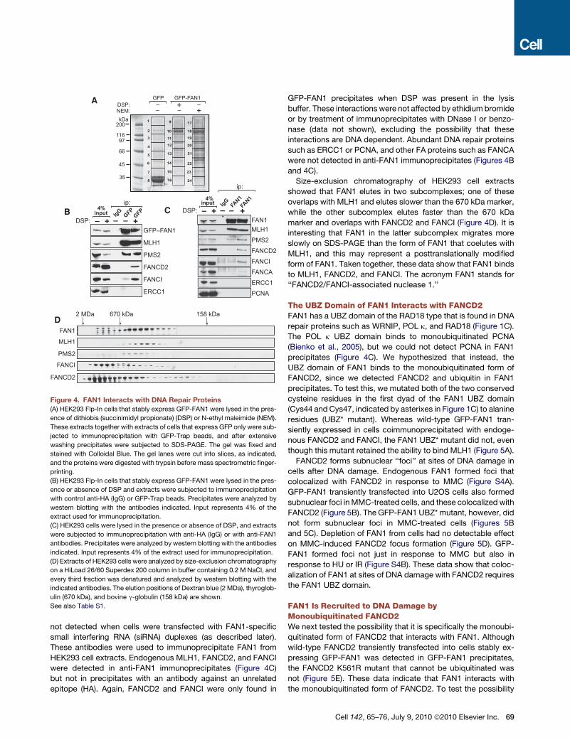
1
2
3
4
5
6
7
8
9
10
11
12
13
14
15
16
17
18
19
20
21
22
23
24
MLH1
PMS2
ERCC1
FANCD2
FANCI
input
GFP–FAN1DSP:
IgG GFPGFP
– + – +
ip:
2 MDa 670 kDa 158 kDa
FANCD2
PMS2
FANCI
MLH1
MLH1 PMS2
FANCA
ERCC1
PCNA
FANCD2 FANCI
inputDSP:
IgG FAN1FAN1
– + – +
ip:
–– .
A GFP-FAN1GFP
B
D
C
200
11697
66
45
35
kDaNEM: – – +DSP: – + –
4%
4%
FAN1
FAN1
Figure 4. FAN1 Interacts with DNA Repair Proteins(A) HEK293 Flp-In cells that stably express GFP-FAN1 were lysed in the pres-
ence of dithiobis (succinimidyl propionate) (DSP) or N-ethyl maleimide (NEM).
These extracts together with extracts of cells that express GFP only were sub-
jected to immunoprecipitation with GFP-Trap beads, and after extensive
washing precipitates were subjected to SDS-PAGE. The gel was fixed and
stained with Colloidal Blue. The gel lanes were cut into slices, as indicated,
and the proteins were digested with trypsin before mass spectrometric finger-
printing.
(B) HEK293 Flp-In cells that stably express GFP-FAN1 were lysed in the pres-
ence or absence of DSP and extracts were subjected to immunoprecipitation
with control anti-HA (IgG) or GFP-Trap beads. Precipitates were analyzed by
western blotting with the antibodies indicated. Input represents 4% of the
extract used for immunoprecipitation.
(C) HEK293 cells were lysed in the presence or absence of DSP, and extracts
were subjected to immunoprecipitation with anti-HA (IgG) or with anti-FAN1
antibodies. Precipitates were analyzed by western blotting with the antibodies
indicated. Input represents 4% of the extract used for immunoprecipitation.
(D) Extracts of HEK293 cells were analyzed by size-exclusion chromatography
on a HiLoad 26/60 Superdex 200 column in buffer containing 0.2 M NaCl, and
every third fraction was denatured and analyzed by western blotting with the
indicated antibodies. The elution positions of Dextran blue (2 MDa), thyroglob-
ulin (670 kDa), and bovine g-globulin (158 kDa) are shown.
See also Table S1.
not detected when cells were transfected with FAN1-specific
small interfering RNA (siRNA) duplexes (as described later).
These antibodies were used to immunoprecipitate FAN1 from
HEK293 cell extracts. Endogenous MLH1, FANCD2, and FANCI
were detected in anti-FAN1 immunoprecipitates (Figure 4C)
but not in precipitates with an antibody against an unrelated
epitope (HA). Again, FANCD2 and FANCI were only found in
GFP-FAN1 precipitates when DSP was present in the lysis
buffer. These interactions were not affected by ethidium bromide
or by treatment of immunoprecipitates with DNase I or benzo-
nase (data not shown), excluding the possibility that these
interactions are DNA dependent. Abundant DNA repair proteins
such as ERCC1 or PCNA, and other FA proteins such as FANCA
were not detected in anti-FAN1 immunoprecipitates (Figures 4B
and 4C).
Size-exclusion chromatography of HEK293 cell extracts
showed that FAN1 elutes in two subcomplexes; one of these
overlaps with MLH1 and elutes slower than the 670 kDa marker,
while the other subcomplex elutes faster than the 670 kDa
marker and overlaps with FANCD2 and FANCI (Figure 4D). It is
interesting that FAN1 in the latter subcomplex migrates more
slowly on SDS-PAGE than the form of FAN1 that coelutes with
MLH1, and this may represent a posttranslationally modified
form of FAN1. Taken together, these data show that FAN1 binds
to MLH1, FANCD2, and FANCI. The acronym FAN1 stands for
‘‘FANCD2/FANCI-associated nuclease 1.’’
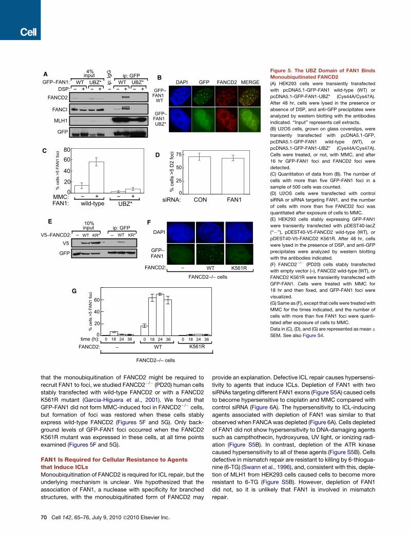
The UBZ Domain of FAN1 Interacts with FANCD2FAN1 has a UBZ domain of the RAD18 type that is found in DNA
repair proteins such as WRNIP, POL k, and RAD18 (Figure 1C).
The POL k UBZ domain binds to monoubiquitinated PCNA
(Bienko et al., 2005), but we could not detect PCNA in FAN1
precipitates (Figure 4C). We hypothesized that instead, the
UBZ domain of FAN1 binds to the monoubiquitinated form of
FANCD2, since we detected FANCD2 and ubiquitin in FAN1
precipitates. To test this, we mutated both of the two conserved
cysteine residues in the first dyad of the FAN1 UBZ domain
(Cys44 and Cys47, indicated by asterixes in Figure 1C) to alanine
residues (UBZ* mutant). Whereas wild-type GFP-FAN1 tran-
siently expressed in cells coimmunoprecipitated with endoge-
nous FANCD2 and FANCI, the FAN1 UBZ* mutant did not, even
though this mutant retained the ability to bind MLH1 (Figure 5A).
FANCD2 forms subnuclear ‘‘foci’’ at sites of DNA damage in
cells after DNA damage. Endogenous FAN1 formed foci that
colocalized with FANCD2 in response to MMC (Figure S4A).
GFP-FAN1 transiently transfected into U2OS cells also formed
subnuclear foci in MMC-treated cells, and these colocalized with
FANCD2 (Figure 5B). The GFP-FAN1 UBZ* mutant, however, did
not form subnuclear foci in MMC-treated cells (Figures 5B
and 5C). Depletion of FAN1 from cells had no detectable effect
on MMC-induced FANCD2 focus formation (Figure 5D). GFP-
FAN1 formed foci not just in response to MMC but also in
response to HU or IR (Figure S4B). These data show that coloc-
alization of FAN1 at sites of DNA damage with FANCD2 requires
the FAN1 UBZ domain.
FAN1 Is Recruited to DNA Damage byMonoubiquitinated FANCD2We next tested the possibility that it is specifically the monoubi-
quitinated form of FANCD2 that interacts with FAN1. Although
wild-type FANCD2 transiently transfected into cells stably ex-
pressing GFP-FAN1 was detected in GFP-FAN1 precipitates,
the FANCD2 K561R mutant that cannot be ubiquitinated was
not (Figure 5E). These data indicate that FAN1 interacts with
the monoubiquitinated form of FANCD2. To test the possibility
Cell 142, 65–76, July 9, 2010 ª2010 Elsevier Inc. 69
BA
ip: I
gG
FANCD2
FANCI
MLH1
GFP
+ + + +DSP: WT UBZ*
input ip: GFP
– – – – – WT UBZ* GFP–FAN1:
25
50
75
CON FAN1siRNA:0
D
% c
ells
>5
D2
foci
GFP– FAN1
WT
GFP– FAN1 UBZ*
GFP DAPI FANCD2 MERGE
UBZ*
C
MMC: wild-type
20
40
60
+–
80
+– 0
FAN1:
% c
ells
>5
FAN
1 fo
ci
G
V5
GFP
V5–FANCD2: – – WT WT KR* KR*
input ip: GFP E
– WT K561R
FANCD2–/– cells
FANCD2:
DAPI
GFP– FAN1
0
20
40
60
% c
ells
>5
FAN
1 fo
ci
– WT K561R
FANCD2–/– cells
FANCD2: 0 18 24 36 0 18 24 36 0 18 24 36 time (h):
F
4%
10%
Figure 5. The UBZ Domain of FAN1 Binds
Monoubiquitinated FANCD2
(A) HEK293 cells were transiently transfected
with pcDNA5.1-GFP-FAN1 wild-type (WT) or
pcDNA5.1-GFP-FAN1-UBZ* (Cys44A/Cys47A).
After 48 hr, cells were lysed in the presence or
absence of DSP, and anti-GFP precipitates were
analyzed by western blotting with the antibodies
indicated. ‘‘Input’’ represents cell extracts.
(B) U2OS cells, grown on glass coverslips, were
transiently transfected with pcDNA5.1-GFP,
pcDNA5.1-GFP-FAN1 wild-type (WT), or
pcDNA5.1-GFP-FAN1-UBZ* (Cys44A/Cys47A).
Cells were treated, or not, with MMC, and after
16 hr GFP-FAN1 foci and FANCD2 foci were
detected.
(C) Quantitation of data from (B). The number of
cells with more than five GFP-FAN1 foci in a
sample of 500 cells was counted.
(D) U2OS cells were transfected with control
siRNA or siRNA targeting FAN1, and the number
of cells with more than five FANCD2 foci was
quantitated after exposure of cells to MMC.
(E) HEK293 cells stably expressing GFP-FAN1
were transiently transfected with pDEST40-lacZ
(‘‘�’’), pDEST40-V5-FANCD2 wild-type (WT), or
pDEST40-V5-FANCD2 K561R. After 48 hr, cells
were lysed in the presence of DSP, and anti-GFP
precipitates were analyzed by western blotting
with the antibodies indicated.
(F) FANCD2�/� (PD20) cells stably transfected
with empty vector (–), FANCD2 wild-type (WT), or
FANCD2 K561R were transiently transfected with
GFP-FAN1. Cells were treated with MMC for
18 hr and then fixed, and GFP-FAN1 foci were
visualized.
(G) Same as (F), except that cells were treated with
MMC for the times indicated, and the number of
cells with more than five FAN1 foci were quanti-
tated after exposure of cells to MMC.
Data in (C), (D), and (G) are represented as mean ±
SEM. See also Figure S4.
that the monoubiquitination of FANCD2 might be required to
recruit FAN1 to foci, we studied FANCD2�/� (PD20) human cells
stably transfected with wild-type FANCD2 or with a FANCD2
K561R mutant (Garcia-Higuera et al., 2001). We found that
GFP-FAN1 did not form MMC-induced foci in FANCD2�/� cells,
but formation of foci was restored when these cells stably
express wild-type FANCD2 (Figures 5F and 5G). Only back-
ground levels of GFP-FAN1 foci occurred when the FANCD2
K561R mutant was expressed in these cells, at all time points
examined (Figures 5F and 5G).
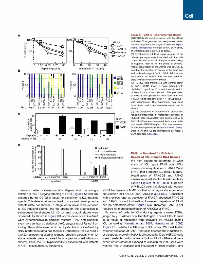
FAN1 Is Required for Cellular Resistance to Agentsthat Induce ICLsMonoubiquitination of FANCD2 is required for ICL repair, but the
underlying mechanism is unclear. We hypothesized that the
association of FAN1, a nuclease with specificity for branched
structures, with the monoubiquitinated form of FANCD2 may
70 Cell 142, 65–76, July 9, 2010 ª2010 Elsevier Inc.
provide an explanation. Defective ICL repair causes hypersensi-
tivity to agents that induce ICLs. Depletion of FAN1 with two
siRNAs targeting different FAN1 exons (Figure S5A) caused cells
to become hypersensitive to cisplatin and MMC compared with
control siRNA (Figure 6A). The hypersensitivity to ICL-inducing
agents associated with depletion of FAN1 was similar to that
observed when FANCA was depleted (Figure 6A). Cells depleted
of FAN1 did not show hypersensitivity to DNA-damaging agents
such as campthothecin, hydroxyurea, UV light, or ionizing radi-
ation (Figure S5B). In contrast, depletion of the ATR kinase
caused hypersensitivity to all of these agents (Figure S5B). Cells
defective in mismatch repair are resistant to killing by 6-thiogua-
nine (6-TG) (Swann et al., 1996), and, consistent with this, deple-
tion of MLH1 from HEK293 cells caused cells to become more
resistant to 6-TG (Figure S5B). However, depletion of FAN1
did not, so it is unlikely that FAN1 is involved in mismatch
repair.
% c
ell v
iabi
lity
cisplatin (mg/ml)0.25 0.50 0.75 1.000
1
10
100
100 20 30 40mitomycin–C (ng/ml)
100
10
1
0
40
80
cisplatin: – – + +
recovery (h): 0 24 48 72 96
pos
itive
cel
ls siRNACON
FAN1
+ + + + + +
siRNACONFAN1–1FAN1–2FANCA
% g
–H2A
X
% c
ells
100
50
0
wild
-type
C01
G5.
8fc
d-2
% c
ells
100
50
0
cisplatin: HN : 2 0 mM 6.6 mM 20 mM 60 mM 0 mM 50 mM 150 mM 450mM
fertileadultssterileadultsL3–L4
L1–L2
wild
-type
C01
G5.
8fc
d-2
wild
-type
C01
G5.
8fc
d-2
wild
-type
C01
G5.
8fc
d-2
wild
-type
C01
G5.
8fc
d-2
wild
-type
C01
G5.
8fc
d-2
wild
-type
C01
G5.
8fc
d-2
wild
-type
C01
G5.
8fc
d-2
0
40
20
60
80
100
% c
ells
with
chr
omos
ome
br
eaks
or r
adia
l for
mat
ion
siRNA: control controlFANCD2 FANCD2 FAN1
untreated MMC
No breaks1 chromatid break1 radial formation>1 break/radial formation
FAN1
A
B
C
D
Figure 6. FAN1 Is Required for ICL Repair
(A) HEK293 cells were transfected with the siRNAs
indicated. Clonogenic survival assays were carried
out with cisplatin or mitomycin-C (see the Experi-
mental Procedures). For each siRNA, cell viability
of untreated cells is defined as 100%.
(B) Synchronized L1 larva stage animals of the
relevant genotype were incubated with the indi-
cated concentrations of nitrogen mustard (HN2)
or cisplatin. After 48 hr, the extent of develop-
mental progression of the worms was scored, by
counting the number of worms in the adult and
various larval stages (L1–L2, L3–L4). Adult worms
were scored as fertile if they contained fertilized
eggs and as sterile if they did not.
(C) HEK293 cells transfected with control siRNA
or FAN1 siRNA (FAN1-1) were treated with
cisplatin (1 mg/ml) for 2 hr and then allowed to
recover for the times indicated. The proportion
of cells in each population with more than two
g-H2AX foci at each time point (‘‘g-H2AX positive’’)
was determined. The experiment was done
three times, and a representative experiment is
shown.
(D) The frequency of chromosome breaks and
radial chromosomes in metaphase spreads of
HEK293 cells transfected with control siRNA or
FAN1-1 siRNA was measured before and after
exposure to MMC (25 ng/ml; 18 hr) was measured
as described previously (Deans and West, 2009).
Data in (A) and (C) are represented as mean ±
SEM. See also Figure S5.
We also tested a Caenorhabditis elegans strain harboring a
deletion in the C. elegans ortholog of FAN1 (Figures 1A and 1B),
encoded by the C01G5.8 locus, for sensitivity to ICL-inducing
agents. This deletion does not lead to any overt developmental
defects (data not shown). L1-stage worm larvae were exposed
to ICL-inducing agents, and the effects on the progression to
subsequent larval stages L2, L3, L4 and to adult stages were
observed. As shown in Figure 6B worms defective in Ce-fan-1
were hypersensitive to nitrogen mustard (HN2) and cisplatin,
even more so than a deletion of the C. elegans fcd-2 Fancd-2 or-
tholog. These data were confirmed by depletion of Ce-fan-1 by
RNA interference (data not shown). Furthermore, the Ce-rend-1
(tm423) deletion resulted in reduced progeny survival when L4
stage animals were exposed to nitrogen mustard (data not
shown). Thus, the ICL hypersensitivity associated with defects
in FAN1 is evolutionarily conserved.
Cell 142,
FAN1 Is Required for EfficientRepair of ICL-Induced DNA BreaksWe next sought to determine at what
stage of ICL repair FAN1 acts. ICLs
causemonoubiquitinationof FANCD2and
FANCI that promotes ICL repair. Monou-
biquitination of FANCD2 and FANCI
causes reduced electrophoretic mobility
(Garcia-Higuera et al., 2001). Exposure
of HEK293 cells transfected with control
siRNA to cisplatin or MMC resulted in damage-induced monou-
biquitination of FANCD2 and FANCI (Figure S5C). Consistent
with previous reports, depletion of FANCA abolished FANCD2
and FANCI monoubiquitination. However, depletion of FAN1
had no detectable effect (Figure S5C). Therefore, FAN1 is not
required for monoubiquitination of FANCD2 or FANCI.
Exposure of cells to ICL-inducing agents causes DSBs,
judged by g-H2AX foci or pulsed field gels. These DSBs, formed
as a result of replication fork cleavage by MUS81 during
ICL unhooking (Hanada et al., 2007; Hanada et al., 2006)
(Figure S1), initiate the HR step of ICL repair. We next tested
whether depletion of FAN1 from cells affected the induction of,
or disappearance of g-H2AX foci induced by ICLs. HEK293 cells
were transfected with control siRNA or FAN1 siRNA and were
either left untreated or exposed to cisplatin for 2 hr. Cells were
washed free of cisplatin and incubated in fresh medium, and
65–76, July 9, 2010 ª2010 Elsevier Inc. 71
CON– +I-SceI:
siRNA:
0
4
8
RAD51 FAN1–1
posi
tive
cells
% G
FP–
– + – + – +FAN1–2
>9
RPA
foci
15
0% c
ells
with
60
30
0 >9
RA
D51
foci
% c
ells
with
30
– – + +recovery (h): 0 24 48 72 96
+ + + + + +cisplatin:
40
20
0 >9
RA
D51
foci
% c
ells
with
IR: – – + +0 4 8 12 24
+ + + + + +recovery (h):
4
8
0RA
D51
foci
/cel
l
6
0RA
D51
foci
/cel
l 12
RPA
foci
/cel
l 8
4
siRNACON
FAN1
– – + +recovery (h): 0 24 48 72 96
+ + + + + +cisplatin:
B
A
D
C
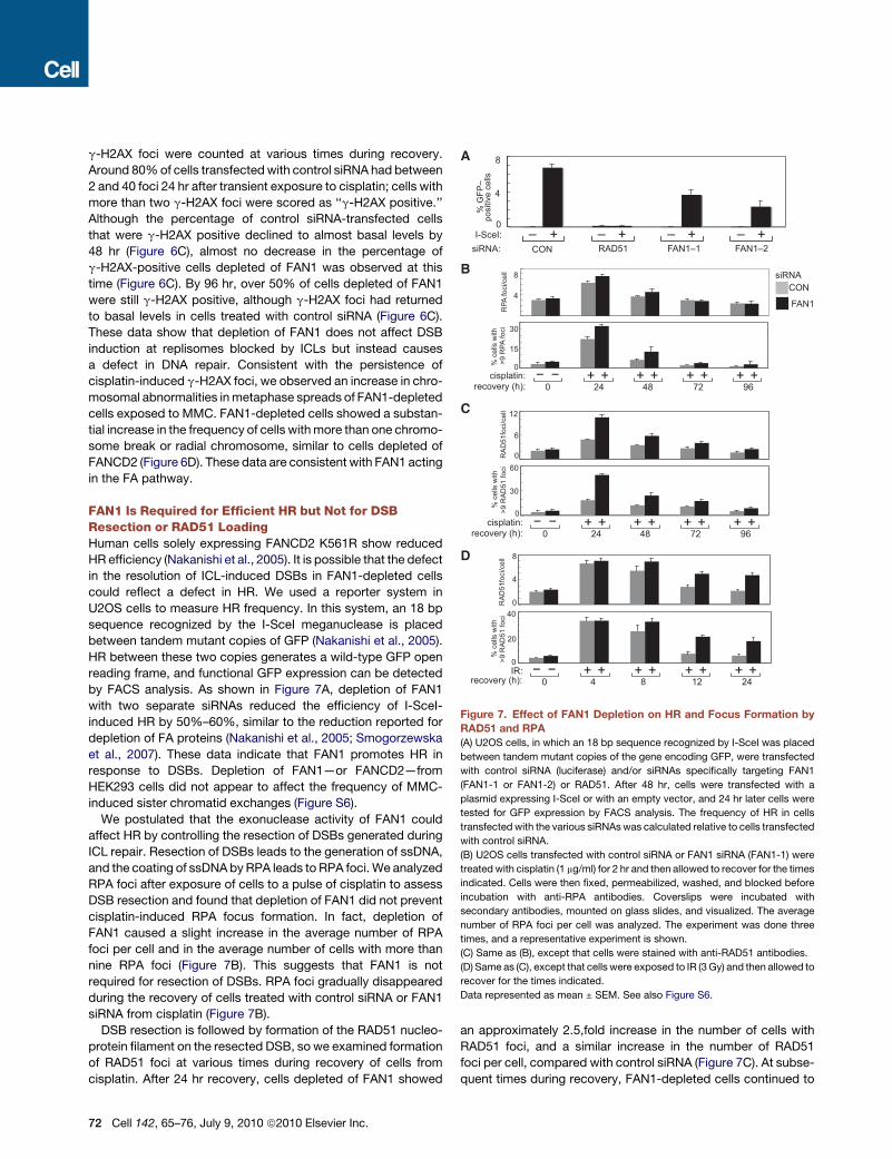
Figure 7. Effect of FAN1 Depletion on HR and Focus Formation by
RAD51 and RPA
(A) U2OS cells, in which an 18 bp sequence recognized by I-SceI was placed
between tandem mutant copies of the gene encoding GFP, were transfected
with control siRNA (luciferase) and/or siRNAs specifically targeting FAN1
(FAN1-1 or FAN1-2) or RAD51. After 48 hr, cells were transfected with a
plasmid expressing I-SceI or with an empty vector, and 24 hr later cells were
tested for GFP expression by FACS analysis. The frequency of HR in cells
transfected with the various siRNAs was calculated relative to cells transfected
with control siRNA.
(B) U2OS cells transfected with control siRNA or FAN1 siRNA (FAN1-1) were
treated with cisplatin (1 mg/ml) for 2 hr and then allowed to recover for the times
indicated. Cells were then fixed, permeabilized, washed, and blocked before
incubation with anti-RPA antibodies. Coverslips were incubated with
secondary antibodies, mounted on glass slides, and visualized. The average
number of RPA foci per cell was analyzed. The experiment was done three
times, and a representative experiment is shown.
(C) Same as (B), except that cells were stained with anti-RAD51 antibodies.
(D) Same as (C), except that cells were exposed to IR (3 Gy) and then allowed to
recover for the times indicated.
Data represented as mean ± SEM. See also Figure S6.
g-H2AX foci were counted at various times during recovery.
Around 80% of cells transfected with control siRNA had between
2 and 40 foci 24 hr after transient exposure to cisplatin; cells with
more than two g-H2AX foci were scored as ‘‘g-H2AX positive.’’
Although the percentage of control siRNA-transfected cells
that were g-H2AX positive declined to almost basal levels by
48 hr (Figure 6C), almost no decrease in the percentage of
g-H2AX-positive cells depleted of FAN1 was observed at this
time (Figure 6C). By 96 hr, over 50% of cells depleted of FAN1
were still g-H2AX positive, although g-H2AX foci had returned
to basal levels in cells treated with control siRNA (Figure 6C).
These data show that depletion of FAN1 does not affect DSB
induction at replisomes blocked by ICLs but instead causes
a defect in DNA repair. Consistent with the persistence of
cisplatin-induced g-H2AX foci, we observed an increase in chro-
mosomal abnormalities in metaphase spreads of FAN1-depleted
cells exposed to MMC. FAN1-depleted cells showed a substan-
tial increase in the frequency of cells with more than one chromo-
some break or radial chromosome, similar to cells depleted of
FANCD2 (Figure 6D). These data are consistent with FAN1 acting
in the FA pathway.
FAN1 Is Required for Efficient HR but Not for DSBResection or RAD51 LoadingHuman cells solely expressing FANCD2 K561R show reduced
HR efficiency (Nakanishi et al., 2005). It is possible that the defect
in the resolution of ICL-induced DSBs in FAN1-depleted cells
could reflect a defect in HR. We used a reporter system in
U2OS cells to measure HR frequency. In this system, an 18 bp
sequence recognized by the I-SceI meganuclease is placed
between tandem mutant copies of GFP (Nakanishi et al., 2005).
HR between these two copies generates a wild-type GFP open
reading frame, and functional GFP expression can be detected
by FACS analysis. As shown in Figure 7A, depletion of FAN1
with two separate siRNAs reduced the efficiency of I-SceI-
induced HR by 50%–60%, similar to the reduction reported for
depletion of FA proteins (Nakanishi et al., 2005; Smogorzewska
et al., 2007). These data indicate that FAN1 promotes HR in
response to DSBs. Depletion of FAN1—or FANCD2—from
HEK293 cells did not appear to affect the frequency of MMC-
induced sister chromatid exchanges (Figure S6).
We postulated that the exonuclease activity of FAN1 could
affect HR by controlling the resection of DSBs generated during
ICL repair. Resection of DSBs leads to the generation of ssDNA,
and the coating of ssDNA by RPA leads to RPA foci. We analyzed
RPA foci after exposure of cells to a pulse of cisplatin to assess
DSB resection and found that depletion of FAN1 did not prevent
cisplatin-induced RPA focus formation. In fact, depletion of
FAN1 caused a slight increase in the average number of RPA
foci per cell and in the average number of cells with more than
nine RPA foci (Figure 7B). This suggests that FAN1 is not
required for resection of DSBs. RPA foci gradually disappeared
during the recovery of cells treated with control siRNA or FAN1
siRNA from cisplatin (Figure 7B).
DSB resection is followed by formation of the RAD51 nucleo-
protein filament on the resected DSB, so we examined formation
of RAD51 foci at various times during recovery of cells from
cisplatin. After 24 hr recovery, cells depleted of FAN1 showed
72 Cell 142, 65–76, July 9, 2010 ª2010 Elsevier Inc.
an approximately 2.5,fold increase in the number of cells with
RAD51 foci, and a similar increase in the number of RAD51
foci per cell, compared with control siRNA (Figure 7C). At subse-
quent times during recovery, FAN1-depleted cells continued to
have around twice as many RAD51 foci as control cells and these
data are consistent with a defect in HR. We also tested the
effects of depleting FAN1 on RAD51 loading after IR. Four hours
after exposure to IR, the number of cells with greater than nine
RAD51 foci, and the average number of RAD51 foci per cell,
was similar in FAN1-depleted and cells treated with control
siRNA (Figure 7D). However, whereas RAD51 foci declined to
basal levels by 12-24 hr after IR in control cells, FAN1-depleted
cells showed a delay in the disappearance of RAD51 foci. These
data suggest that FAN1 depletion leads to failure of a late stage
of HR.
DISCUSSION
It has been known for almost a decade that mono–ubiquitination
of FANCD2 is required for ICL repair (Garcia-Higuera et al.,
2001). However, the molecular role of this ubiquitination event
has remained elusive. Here, we report that the FAN1 nuclease
is recruited to sites of DNA damage by monoubiquitinated
FANCD2 and thus might act as an effector molecule carrying
out one or more nucleolytic steps required for ICL repair. The
phenotypic consequences of depleting FAN1 from human
cells—sensitivity to ICL-inducing agents, chromosome insta-
bility in MMC-treated cells and defects in HR—are consistent
with a role in ICL repair and are similar to those seen in cells
solely expressing FANCD2 K561R (Moldovan and D’Andrea,
2009). These findings might at least in part explain how ubiquiti-
nation of FANCD2 promotes ICL repair.
According to our data, FAN1 is recruited to sites of ICLs by
monoubiquitinated FANCD2. This is supported by the require-
ment of the FAN1 UBZ domain (Figure 5B) and the monoubiqui-
tinated form of FANCD2 (Figure 5F) for FAN1 localization.
Furthermore, FAN1 and FANCD2 proteins coprecipitate in a
manner that depends on FANCD2 K561 and on the FAN1
UBZ domain. Both ubiquitinated and nonubiquitinated FANCD2
were detected in FAN1 immunoprecipitates, even though when
FANCD2 K561 is mutated, no FANCD2 is detected in FAN1
immunoprecipitates. This discrepancy may be explained by deu-
biquitination of a proportion of FANCD2 after cell lysis or by the
association of modified FANCD2 with the unmodified form of the
protein. Even though DNA damage stimulates FANCD2 ubiquiti-
nation, FAN1 interacts with FANCD2 even without exposure of
cells to genotoxins. This is probably a reflection of basal
FANCD2 monoubiquitination that occurs in the absence of DNA
damage in S phase cells (Taniguchi et al., 2002).
Building upon existing models for ICL repair, and what is
already known about the role of FANCD2 monoubiquitination in
this pathway, it is possible to speculate on where on the ICL
repair pathway FAN1 might act. Experiments on the replication
of plasmids bearing single ICLs in Xenopus egg extracts showed
that FANCD2 monoubiquitination is required for ICL unhooking
(Knipscheer et al., 2009), which suggests that FAN1 might act
at this point. In this system, it was proposed that initiation of
ICL repair requires the convergence of two replication forks at
an ICL (Knipscheer et al., 2009). Consequently, an ICL would be
located at the intersection of an X structure shown in Figure S7A.
In this scenario, cleavage of the leading strand template of one of
the forks by MUS81 in concert with cleavage of the same strand
on the opposite side of the ICL (which resembles a 50 flap) by
a second nuclease would unhook the ICL (Figure S7A). This
nuclease could be FAN1 based on our in vitro data showing
that this nuclease preferentially cleaves the double-stranded
portion of a synthetic 50 flap structure (Figure 2A). However, there
are conceptual problems with FAN1 acting at this point of the ICL
repair pathway. First, this hypothesis predicts that MUS81 and
FAN1 are each responsible for 50% of the one-ended DSBs in
cells treated with ICL-inducing agents. Assuming that g-H2AX
foci are representative of DSBs, this is not what we observe—
depletion of FAN1 does not affect formation of cisplatin-induced
DSBs (Figure 6C), whereas deletion of MUS81 abolished all ICL-
induced DSBs (Hanada et al., 2006). Second, this hypothesis
requires that MUS81 acts on the leading strand template of
one of the two stalled replication forks (Figure S7A), but it is diffi-
cult to see why MUS81 would not cleave the leading strand
template of both forks (Figure S7B). Cleavage of both forks by
MUS81 would result in two one-ended DSBs and a linear duplex
containing the ICL, and it is unlikely that unhooking of the ICL from
this linear duplex would require FAN1. Third, it is not yet clear
whether the two-fork model for ICL repair is relevant in vivo, and
it is possible that the collision of a single replication fork with an
ICL is sufficient to initiate ICL repair (Figure S7C). In this scenario,
it is difficult to see how FAN1 could be involved in unhooking
since it would have to cleave linear duplex DNA on the 50 side
of the ICL, a structure that is not flap-like in nature. More experi-
ments are required to test whether the one-fork or two-fork
models for ICL repair, or both, operate in vivo and to test if the
defect in ICL unhooking in the context of the FANCD2 K561R
mutant is due to a defect specifically in recruitment of FAN1.
After unhooking, excision repair removes the crosslink adduct
and translesion synthesis fills in the gap (Figure S7A). Although
FANCD2 ubiquitination appears to be involved in translesion
synthesis in the Xenopus cell-free system, it is difficult to see
how FAN1 could be involved at this stage, and it is likely that
other FANCD2-binding proteins are required. We found that
FAN1 interacts with MLH1 that is involved in mismatch repair,
consistent with a previous report (Cannavo et al., 2007). At
present, the significance of this interaction is not clear, but it
may be that the interaction of FAN1 with MLH1 promotes the
correction of translesion synthesis-induced mismatches during
ICL repair.
Regardless of how exactly MUS81—alone or in conjunction
with FAN1 or an as yet unidentified nuclease—unhooks the
ICL, at least one and possibly two (one-ended) DSBs are gener-
ated (Figure S7). These DSBs are resected and this is a function
that could be fulfilled by FAN1 based on our finding that FAN1
has 50-30 exonuclease activity that is capable of generating 30
overhangs (Figure 3A). However, cytological data showing that
RPA loading is normal in FAN1-depleted cells (Figure 7B) argue
against this role, although potential redundancy between FAN1
and other 50-30 exonucleases would need to be investigated.
One of the overhangs generated by DSB resection invades the
complementary duplex to initiate D loop formation and HR
(Figures 7A and 7B). After extension by DNA synthesis, the
invading strand reanneals to the complementary strand in the
duplex it came from originally. It is possible that continued
DNA synthesis on the parent strand displaces the DNA in front
Cell 142, 65–76, July 9, 2010 ª2010 Elsevier Inc. 73
of it. This would generate a 50 flap that could be cleaved by FAN1.
In this scenario, FAN1 is required at a late step in HR, and several
observations are consistent with this hypothesis. First, there is
a delay in the disappearance of g-H2AX foci induced by cisplatin
or IR in cells treated with FAN1 siRNA compared with control
cells. Similar results were reported recently in FA cells (Leskovac
et al., 2010). At all time points during recovery from cisplatin and
IR, there are more RAD51 foci in FAN1-depleted cells than in
control cells (Figure 7C). It is possible that disappearance of
foci in FAN1-depleted cells reflects inappropriate repair perhaps
by nonhomologous end joining, and this may account for the
increase in radial chromosomes seen in MMC-treated cells
depleted of FAN1 (Figure 6D). Second, depletion of FAN1 from
human cells results in reduced efficiency in I-SceI-induced HR
(Figure 7A), and similar results were reported in human cells ex-
pressing FANCD2 K561R (Nakanishi et al., 2005). Depletion of
FAN1 does not affect RAD51 loading in cisplatin-treated cells
(Figure 7C), suggesting that if FAN1 functions at the HR step of
ICL repair, then it acts independently of RAD51 or after RAD51
focus formation. Similar to FANCD2 null cells, depletion of FAN1
does not affect the frequency of MMC-induced SCEs (Figure S6).
Therefore, if FAN1 does act at the HR step of ICL repair, then its
role may be restricted to a subpathway of HR such as synthesis-
dependent strand annealing that avoids crossing over.
The SLX4 complex of structure-specific nucleases is also
required for the HR step of ICL repair (Fekairi et al., 2009; Munoz
et al., 2009; Svendsen et al., 2009). SLX4-XPF-SLX1-MUS81 can
cleave three-way DNA junctions, 30 flaps, and 50 flaps in vitro,
and so FAN1 specificity overlaps with the SLX4 complex in 50
flap cleavage. We could find no evidence for an interaction of
FAN1 with the SLX4 scaffold (unpublished data). It is not yet clear
why two 50 endonucleases are required during ICL repair, and it
will be important to test redundancy between FAN1 and the
SLX4 complex.
FAN1 is the only VRR_nuc domain-containing protein in
eukaryotic cells. These domains are found in all kingdoms of
life, but the functions of most of them are unknown (Iyer et al.,
2006). Many bacteria and phages have VRR_nuc domain
proteins, and so it appears that the FA repair pathway which
appeared relatively late during evolution was built on a more
ancient VRR_nuc domain nuclease. It will be interesting to follow
up on this hypothesis. Many cytotoxic anticancer agents act by
inducing ICLs, and it is possible that nucleases such as FAN1 are
good targets for sensitizing cancer cells to killing by ICLs. Finally,
although the majority of FA patients have mutations in the known
FA genes, FA patients exist where mutations in known FA genes
could not be found. In this light, it is likely that FAN1 mutations
will be found in some of these patients.
EXPERIMENTAL PROCEDURES
General Methods
Gel filtration and analysis of the resolution of cisplatin-induced DSBs were
carried out as described previously (Munoz et al., 2009). Dithiobis (succini-
midyl propionate) (DSP; Pierce) is a homobifunctional and thiol-cleavable
crosslinker that was used according to the manufacturer’s instructions. DSP
was included in lysis buffer at 2.5 mg/ml, and lysates were incubated for
30 min on ice. Excess DSP was quenched by addition of 1 M Tris-HCl (pH 7.4)
to 0.2 M followed by an additional 30 min incubation. Crosslinks were reversed
74 Cell 142, 65–76, July 9, 2010 ª2010 Elsevier Inc.
by the inclusion of dithiothreitol in SDS-PAGE sample buffer added to cell
extracts or immunoprecipitates before electrophoresis. Details of immunoflu-
orescence are given in the Extended Experimental Procedures.
Antibodies, Cell Lysis, and Immunoprecipiation
The primary antibodies used in this study were the following: FAN1 (this study;
sheep S420C, fourth bleed), MLH1 (BD Pharminigen, 554073), PMS2 (Santa
Cruz, sc-617), PCNA (Santa Cruz, PC10), FANCI (Bethyl, A301-354), FANCD2
(Abcam, ab2187-50), FANCD2 (Novus, NB100-182; immunofluorescence),
FANCA (Cascade Biosciences, abm6202), FANCC (Cascade Biosciences,
abp6305), FANCE (a kind gift from K.J. Patel), FANCF, FANCG (kind gifts
from Johan De Winter), FLAG (Sigma, M2), Ku80 (Cell Signaling, 2753),
RAD51 (Santa Cruz, H-92), RPA70 (Cell Signaling, 2267), and anti-g-H2AX
(Bethyl, A300-081A). The FAN1 antibody, raised in sheep against full-length
FAN1 fused to GST at the Scottish Antibody Production Unit (Carluke, Lanark-
shire), was affinity purified with immobilized antigen. GFP-Trap beads were
from Chromotek. Protein G Sepharose was from GE Healthcare. Cells were
lysed in ice-cold buffer: 40 mM HEPES (pH 7.4), 120 mM NaCl, 1% (v/v) Triton
X-100, and 1 mM EDTA with protease inhibitors (Roche). For visualization of
monoubiquitinated forms of FANCI and FANCD2, 0.5 U/ml of benzonase
(Sigma) was included in the lysis buffer, and lysates were incubated on ice
for 30 min. All immunoprecipitations were carried out in lysis buffer for 1 hr
at 4�C. Endogenous immunoprecipitations were carried out with 2 mg FAN1
antibody coupled to 10 ml protein G Sepharose per 4 mg of whole-cell extract.
Purification of GFP–FAN1 from HEK293 Cells
FlpIn T-Rex cells (Invitrogen) cells stably expressing GFP-FAN1 in a tetracy-
cline-inducible manner were made according to the manufacturer’s instruc-
tions with FAN1 in plasmid pcDNA5-FRT-TO-GFP-FAN1. FAN1 was induced
and purified according to a previously described protocol (Munoz et al., 2009).
siRNA
Cells were transfected with the relevant siRNA duplex (100 nM) via the calcium
phosphate precipitation method. In Figure 7, U2OS cells were transfected in
96-well plates with siRNAs at a concentration of 20 nM and DharmaFECT 1
(Dharmacon) at a 1:1000 concentration. Cells were incubated at 37�C for
48 hr. The messenger RNA target sequences used for siRNAs were as follows:
FANCA (GGGUCAAGAGGGAAAAAUA), FAN1-1 (GUAAGGCUCUUUCAAC
GUA; exon 3), FAN1-2 (GCAGGAAGGCAGAGUGGCU; exon 12), MLH1
(GCAUGUGGCUCAUGUUAC), ATR (GGGAGCCUGUUGAGACAAGAU), and
FANCD2 (siGenome SMARTPool from Dharmacon).
Oligonucleotides
The following oligonucleotides were used:
a3: 50-CCTCGATCCTACCAACCAGATGACGCGCTGCTACGTGCTACCG
GAAGTCG
b: 50-CGACTTCCGGTAGCACGTAGCAGCGGCTCGCCACGAACTGCAC
TCTAGGC
c: 50-GCCTAGAGTGCAGTTCGTGGCGAGC
d3: 50-CGTCATCTGGTTGGTAGGATCGAGG
a3-cp: 50-CGACTTCCGGTAGCACGTAGCAGCGCGTCAACTGGTTGGTA
GGATCGAGG
Preparation of DNA Substrates
All substrates and standards were annealed by slow cooling of one radioac-
tively 50 32P-labeled oligonucleotide with the relevant unlabeled one(s). In
Figure 2, these were splayed duplex (SD), a3, b; 30 flap (30F) a3, b, d3; 50 flap
(50F) a3, b, c; and replication fork analog (RF), a3, b, c, and d3. In Figure 3, these
were a3 and a3-cp (dsDNA), a3 (ssDNA). Synthetic structures were then puri-
fied by electrophoresis on a native 8% polyacrylamide gel and recovered by
the crush and soak method followed by ethanol precipitation.
Nuclease Assays
Purified recombinant FAN1 (35 nM) was preincubated for at least 10 min with
radiolabeled DNA substrates (5 nM) at 37�C in 25 mM Tris-HCl (pH 7.5), 10 mM
NaCl,15 mM KCl, and 0.1 mg/ml BSA to allow binding to occur. The reaction
was started by the addition of 1 mM MnCl2 and stopped by the addition of
2 mM EDTA. The samples were then boiled at 95�C for 10 min and analyzed
by denaturing PAGE (15% polyacrylamide and 8 M urea). Gels were dried,
exposed to storage Phosphor screens, and analyzed with the ImageGauge
software (Fujifilm). For kinetics experiments, the data were plotted as the frac-
tion of DNA in the relevant product bands as a function of time and fitted to
either one or two exponential functions. When two exponential functions
were used, the rate given is the faster of the two.
Clonogenic Survival Assays
HEK293 cells were seeded in 10 cm2 dishes at 25% confluence and allowed to
adhere overnight. Cells were transfected with the relevant siRNA for 48 hr and
cells were split and seeded in 10 cm2 dishes (5000 cells/dish). Cells were
allowed to adhere for a minimum of 8 hr before cisplatin or mitomycin-C
were added at the indicated concentrations for 24 hr. Cells were then washed
free of drugs and incubated in fresh medium for 10–14 days before the number
of colonies of more than 50 cells in each dish were counted.
C. elegans Genotoxin Sensitivity Assays
Worms were maintained at 20�C on NGM (Nematode Growth Media) agar
plates according to standard protocols (Brenner, 1974). Alleles used were
fcd-2 (tm1298) and C01G5.8 (tm423). The C01G5.8 mutant was generated
and kindly provided by Shoehi Mitani of the National Bioresource Project for
the Nematode, Japan. This strain, which has a 411 bp deletion in C01G5.8
that removes exons 6–8, was outcrossed five times with N2 Bristol strain
(wild-type) to eliminate secondary mutations. So that ICL sensitivity could be
accessed, synchronized L1 larval stage animals of the relevant genotype
were incubated at 20�C for 16 hr in 1 ml S-basal buffer (0.1 m NaCl, 0.05M
KH2PO4 [pH 6.0], 5 mg/ml cholesterol) containing E. coli OP50 and the indi-
cated concentration of nitrogen mustard (HN2) or cisplatin. After incubation
worms were transferred to OP50-seeded NGM plates. After 48 hr, the extent
of developmental progression was scored. In each experiment, a minimum
of 60 worms was scored, and the results shown are the average of three inde-
pendent experiments.
GFP HR Assay
Cells were transfected with siRNA in 96-well dishes, and after 48 hr cells were
transfected with 0.25 mg I-Sce-I vector and 0.2 mg PEI in 150 ml OptiMEM/well.
GFP-positive cells were analyzed with FACS 48 hr after I-Sce-I transfection as
previously described (Pierce et al., 1999).
SUPPLEMENTAL INFORMATION
Supplemental Information includes Extended Experimental Procedures, seven
figures, and one table and can be found with this article online at doi:10.1016/
j.cell.2010.06.021.
ACKNOWLEDGMENTS
We are grateful to Katja Kratz and Joe Jiricny for experimental advice, for
helping us with the purification of FAN1, and for communicating results prior
to publication. We thank K.J. Patel, Johan de Winter, and Maureen Hoatlin
for kind gifts of antibodies. pDEST40-V5-FANCD2 wild-type and pDEST40-
V5-FANCD2 K561R plasmids were kind gifts from Niall Howlett and Thomas
Glover. We thank Alan d’Andrea for the kind gifts of FANCD2�/� fibroblasts
(PD20 cells) corrected with vector, FANCD2 wild-type, or FANCD2 K561R.
We are grateful to James Hastie, Hilary MacLauchlan, and the Antibody
Production Team at Division of Signal Transduction Therapy, University of
Dundee, to Bob Gourlay, Sanjay Kothiya, and Nick Morrice for help with
mass spectrometry, and to the DNA Sequencing Service at the College of
Life Sciences (CLS), University of Dundee. We are grateful to the microscopy
facility, CLS, University of Dundee for assistance with microscopy. We thank
Jim Haber and the J.R. lab for critical reading of the manuscript. This work
was funded by the UK Medical Research Council (C.M., T.J.H., and J.R.),
Cancer Research UK (A.C.D, A.G., and D.M.J.L.), and the Fanconi Anemia
Research Fund (A.J.D. and S.C.W.).
Received: March 3, 2010
Revised: May 27, 2010
Accepted: June 15, 2010
Published: July 8, 2010
REFERENCES
Akkari, Y.M.N., Bateman, R.L., Reifsteck, C.A., Olson, S.B., and Grompe, M.
(2000). DNA replication is required To elicit cellular responses to psoralen-
induced DNA interstrand cross-links. Mol. Cell. Biol. 20, 8283–8289.
Bergstralh, D.T., and Sekelsky, J. (2008). Interstrand crosslink repair: can
XPF-ERCC1 be let off the hook? Trends Genet. 24, 70–76.
Bhagwat, N., Olsen, A.L., Wang, A.T., Hanada, K., Stuckert, P., Kanaar, R.,
D’Andrea, A., Niedernhofer, L.J., and McHugh, P.J. (2009). XPF-ERCC1
participates in the Fanconi anemia pathway of cross-link repair. Mol. Cell.
Biol. 29, 6427–6437.
Bienko, M., Green, C.M., Crosetto, N., Rudolf, F., Zapart, G., Coull, B.,
Kannouche, P., Wider, G., Peter, M., Lehmann, A.R., et al. (2005). Ubiquitin-
binding domains in Y-family polymerases regulate translesion synthesis.
Science 310, 1821–1824.
Brenner, S. (1974). The genetics of Caenorhabditis elegans. Genetics 77,
71–94.
Cannavo, E., Gerrits, B., Marra, G., Schlapbach, R., and Jiricny, J. (2007).
Characterization of the interactome of the human MutL homologues MLH1,
PMS1, and PMS2. J. Biol. Chem. 282, 2976–2986.
Ciccia, A., Ling, C., Coulthard, R., Yan, Z., Xue, Y., Meetei, A.R., Laghmani, H.,
Joenje, H., McDonald, N., de Winter, J.P., et al. (2007). Identification of
FAAP24, a Fanconi anemia core complex protein that interacts with FANCM.
Mol. Cell 25, 331–343.
Collis, S.J., Ciccia, A., Deans, A.J., Horejsı, Z., Martin, J.S., Maslen, S.L.,
Skehel, J.M., Elledge, S.J., West, S.C., and Boulton, S.J. (2008). FANCM
and FAAP24 function in ATR-mediated checkpoint signaling independently
of the Fanconi anemia core complex. Mol. Cell 32, 313–324.
Deans, A.J., and West, S.C. (2009). FANCM connects the genome instability
disorders Bloom’s Syndrome and Fanconi Anemia. Mol. Cell 36, 943–953.
Fekairi, S., Scaglione, S., Chahwan, C., Taylor, E.R., Tissier, A., Coulon, S.,
Dong, M.Q., Ruse, C., Yates, J.R., 3rd, Russell, P., et al. (2009). Human
SLX4 is a Holliday junction resolvase subunit that binds multiple DNA repair/
recombination endonucleases. Cell 138, 78–89.
Garcia-Higuera, I., Taniguchi, T., Ganesan, S., Meyn, M.S., Timmers, C.,
Hejna, J., Grompe, M., and D’Andrea, A.D. (2001). Interaction of the Fanconi
anemia proteins and BRCA1 in a common pathway. Mol. Cell 7, 249–262.
Hanada, K., Budzowska, M., Modesti, M., Maas, A., Wyman, C., Essers, J.,
and Kanaar, R. (2006). The structure-specific endonuclease Mus81-Eme1
promotes conversion of interstrand DNA crosslinks into double-strands
breaks. EMBO J. 25, 4921–4932.
Hanada, K., Budzowska, M., Davies, S.L., van Drunen, E., Onizawa, H.,
Beverloo, H.B., Maas, A., Essers, J., Hickson, I.D., and Kanaar, R. (2007).
The structure-specific endonuclease Mus81 contributes to replication restart
by generating double-strand DNA breaks. Nat. Struct. Mol. Biol. 14,
1096–1104.
Hofmann, K. (2009). Ubiquitin-binding domains and their role in the DNA
damage response. DNA Repair (Amst.) 8, 544–556.
Iyer, L.M., Babu, M.M., and Aravind, L. (2006). The HIRAN domain and recruit-
ment of chromatin remodeling and repair activities to damaged DNA. Cell
Cycle 5, 775–782.
Knipscheer, P., Raschle, M., Smogorzewska, A., Enoiu, M., Ho, T.V., Scharer,
O.D., Elledge, S.J., and Walter, J.C. (2009). The Fanconi anemia pathway
promotes replication-dependent DNA interstrand cross-link repair. Science
326, 1698–1701.
Cell 142, 65–76, July 9, 2010 ª2010 Elsevier Inc. 75
Kosinski, J., Feder, M., and Bujnicki, J.M. (2005). The PD-(D/E)XK superfamily
revisited: identification of new members among proteins involved in DNA
metabolism and functional predictions for domains of (hitherto) unknown
function. BMC Bioinformatics 6, 172.
Leskovac, A., Vujic, D., Guc-Scekic, M., Petrovic, S., Joksic, I., Slijepcevic, P.,
and Joksic, G. (2010). Fanconi anemia is characterized by delayed repair
kinetics of DNA double-strand breaks. Tohoku J. Exp. Med. 221, 69–76.
Ling, C., Ishiai, M., Ali, A.M., Medhurst, A.L., Neveling, K., Kalb, R., Yan, Z.,
Xue, Y., Oostra, A.B., Auerbach, A.D., et al. (2007). FAAP100 is essential for
activation of the Fanconi anemia-associated DNA damage response pathway.
EMBO J. 26, 2104–2114.
McCabe, K.M., Olson, S.B., and Moses, R.E. (2009). DNA interstrand crosslink
repair in mammalian cells. J. Cell. Physiol. 220, 569–573.
Moldovan, G.L., and D’Andrea, A.D. (2009). How the fanconi anemia pathway
guards the genome. Annu. Rev. Genet. 43, 223–249.
Munoz, I.M., Hain, K., Declais, A.C., Gardiner, M., Toh, G.W., Sanchez-Pulido,
L., Heuckmann, J.M., Toth, R., Macartney, T., Eppink, B., et al. (2009). Coor-
dination of structure-specific nucleases by human SLX4/BTBD12 is required
for DNA repair. Mol. Cell 35, 116–127.
Nakanishi, K., Yang, Y.G., Pierce, A.J., Taniguchi, T., Digweed, M., D’Andrea,
A.D., Wang, Z.Q., and Jasin, M. (2005). Human Fanconi anemia monoubiquiti-
nation pathway promotes homologous DNA repair. Proc. Natl. Acad. Sci. USA
102, 1110–1115.
Niedernhofer, L.J., Odijk, H., Budzowska, M., van Drunen, E., Maas, A., Theil,
A.F., de Wit, J., Jaspers, N.G., Beverloo, H.B., Hoeijmakers, J.H., and Kanaar,
R. (2004). The structure-specific endonuclease Ercc1-Xpf is required to
resolve DNA interstrand cross-link-induced double-strand breaks. Mol. Cell.
Biol. 24, 5776–5787.
76 Cell 142, 65–76, July 9, 2010 ª2010 Elsevier Inc.
Patel, K.J., and Joenje, H. (2007). Fanconi anemia and DNA replication repair.
DNA Repair (Amst.) 6, 885–890.
Pierce, A.J., Johnson, R.D., Thompson, L.H., and Jasin, M. (1999). XRCC3
promoteshomology-directed repair of DNA damage in mammalian cells.
Genes Dev. 13, 2633–2638.
Raschle, M., Knipscheer, P., Knipsheer, P., Enoiu, M., Angelov, T., Sun, J.,
Griffith, J.D., Ellenberger, T.E., Scharer, O.D., and Walter, J.C. (2008). Mecha-
nism of replication-coupled DNA interstrand crosslink repair. Cell 134,
969–980.
Rothfuss, A., and Grompe, M. (2004). Repair kinetics of genomic interstrand
DNA cross-links: evidence for DNA double-strand break-dependent activation
of the Fanconi anemia/BRCA pathway. Mol. Cell. Biol. 24, 123–134.
Smogorzewska, A., Matsuoka, S., Vinciguerra, P., McDonald, E.R., 3rd, Hurov,
K.E., Luo, J., Ballif, B.A., Gygi, S.P., Hofmann, K., D’Andrea, A.D., and Elledge,
S.J. (2007). Identification of the FANCI protein, a monoubiquitinated FANCD2
paralog required for DNA repair. Cell 129, 289–301.
Svendsen, J.M., Smogorzewska, A., Sowa, M.E., O’Connell, B.C., Gygi, S.P.,
Elledge, S.J., and Harper, J.W. (2009). Mammalian BTBD12/SLX4 assembles
a Holliday junction resolvase and is required for DNA repair. Cell 138, 63–77.
Swann, P.F., Waters, T.R., Moulton, D.C., Xu, Y.Z., Zheng, Q.G., Edwards, M.,
and Mace, R. (1996). Role of postreplicative DNA mismatch repair in the
cytotoxic action of thioguanine. Science 273, 1109–1111.
Taniguchi, T., Garcia-Higuera, I., Andreassen, P.R., Gregory, R.C., Grompe,
M., and D’Andrea, A.D. (2002). S-phase-specific interaction of the Fanconi
anemia protein, FANCD2, with BRCA1 and RAD51. Blood 100, 2414–2420.
Wang, W. (2007). Emergence of a DNA-damage response network consisting
of Fanconi anaemia and BRCA proteins. Nat. Rev. Genet. 8, 735–748.
Recommended


