
TH
EJ
OU
RN
AL
OF
CE
LL
BIO
LO
GY
JCB: ARTICLE
© The Rockefeller University Press $30.00The Journal of Cell Biology, Vol. 180, No. 5, March 10, 2008 1021–1035http://www.jcb.org/cgi/doi/
JCB 102110.1083/jcb.200708213
Correspondence to Leszek Kaczmarek: [email protected]; or Grzegorz M. Wilczynski: [email protected]
Abbreviations used in this paper: ANOVA, analysis of variance; DG, dentate gyrus; DIV, day in vitro; GFAP, glial fi brillary acidic protein; ICAM, intercellular adhesion molecule; IR, immunoreactivity; KA, kainic acid; KO, knockout; MMP, matrix metalloproteinase; NMDAR, N -methyl D -aspartate acid receptor; PSD, postsynaptic density; PTZ, pentylenetetrazole; TG, transgenic; TLE, temporal lobe epilepsy; tPA, tissue plasminogen activator; WT, wild type.
The online version of this paper contains supplemental material.
Introduction Epilepsy is a chronic neurological disorder affecting 1 – 3% of
mankind. It is defi ned by recurrent spontaneous seizures that
may be generated by synchronous fi ring of a localized group of
neurons, the so-called epileptic foci ( Engel, 1996 ; Pitkanen and
Sutula, 2002 ). Temporal lobe epilepsy (TLE) is the most com-
mon type of epilepsy in adults. It is characterized by the pres-
ence of epileptic foci located within the hippocampal formation,
amygdala, or temporal neocortex. Unfortunately, in as many as
70% of patients, TLE is intractable by pharmacologic treatment
and requires brain surgery for seizure control. Accordingly,
there is an urgent need to understand the molecular mechanisms
of this severe disease. Aberrant synaptic plasticity has been postu-
lated to underlie formation of epileptic foci and, thus, molecules
implicated in plasticity offer an interesting clue toward under-
standing the disease.
Matrix metalloproteinases (MMPs) form a large family of
structurally related zinc-dependent secreted or cell membrane –
bound proteinases that are considered to be major executors of
ECM remodeling throughout the body ( Sternlicht and Werb,
2001 ) and of pivotal importance in cancer invasion and metas-
tasis. In the adult brain, MMPs have repeatedly been implicated
in various pathologies, including acute and chronic neuro-
degeneration (e.g., in ischemia, trauma, and Alzheimer ’ s disease),
infl ammation, and cancer (for review see Yong, 2005 ). Recent
studies also suggest that MMP-9 plays an important role in syn-
aptic plasticity, learning, and memory ( Szklarczyk et al., 2002 ;
Meighan et al., 2006 ; Nagy et al., 2006 ; Okulski et al., 2007 ;
Tian et al., 2007 ). As epileptogenic plasticity involves extensive
nervous tissue remodeling ( Ben-Ari, 2001 ), we decided to examine
the potential pathogenic role of MMP-9 in two distinct animal
Temporal lobe epilepsy (TLE) is a devastating disease
in which aberrant synaptic plasticity plays a major
role. We identify matrix metalloproteinase (MMP) 9
as a novel synaptic enzyme and a key pathogenic factor
in two animal models of TLE: kainate-evoked epilepsy and
pentylenetetrazole (PTZ) kindling – induced epilepsy. Nota-
bly, we show that the sensitivity to PTZ epileptogenesis is
decreased in MMP-9 knockout mice but is increased in a
novel line of transgenic rats overexpressing MMP-9.
Immunoelectron microscopy reveals that MMP-9 associates
with hippocampal dendritic spines bearing asymmetrical
(excitatory) synapses, where both the MMP-9 protein
levels and enzymatic activity become strongly increased
upon seizures. Further, we fi nd that MMP-9 defi ciency
diminishes seizure-evoked pruning of dendritic spines
and decreases aberrant synaptogenesis after mossy fi ber
sprouting. The latter observation provides a possible mech-
anistic basis for the effect of MMP-9 on epileptogenesis.
Our work suggests that a synaptic pool of MMP-9 is critical
for the sequence of events that underlie the development
of seizures in animal models of TLE.
Important role of matrix metalloproteinase 9 in epileptogenesis
Grzegorz M. Wilczynski , 1,3 Filip A. Konopacki , 1,2,5 Ewa Wilczek , 4 Zofi a Lasiecka , 1 Adam Gorlewicz , 1 Piotr Michaluk , 2
Marcin Wawrzyniak , 2 Monika Malinowska , 1 Pawel Okulski , 2 Lukasz R. Kolodziej , 2 Witold Konopka , 2
Kamila Duniec , 2,5 Barbara Mioduszewska , 2 Evgeni Nikolaev , 2 Agnieszka Walczak , 1 Dorota Owczarek , 1
Dariusz C. Gorecki , 6 Werner Zuschratter , 7 Ole Petter Ottersen , 8 and Leszek Kaczmarek 2
1 Department of Neurophysiology and 2 Department of Molecular and Cellular Neurobiology, Nencki Institute, 02-093 Warsaw, Poland 3 Department of Histology and Embryology and 4 Department of Pathology, Medical University of Warsaw, 02-005 Warsaw, Poland 5 Postgraduate School of Molecular Medicine, 02-093 Warsaw, Poland 6 Institute of Biomedical and Biomolecular Sciences, University of Portsmouth, PO1 2DT Portsmouth, England, UK 7 Leibniz Institute for Neurobiology, D-39118 Magdeburg, Germany 8 Centre for Molecular Biology and Neuroscience, Institute of Basic Medical Sciences, University of Oslo, N-0317 Oslo, Norway

JCB • VOLUME 180 • NUMBER 5 • 2008 1022
When we performed PTZ kindling on MMP-9 KO mice
and wild-type (WT) controls, no difference was noted in con-
vulsive behavior upon the fi rst injections ( Fig. 1 A ). Sub-
sequently, when WT mice started to respond with seizures,
many MMP-9 KO animals remained insensitive to PTZ. MMP-9 –
defi cient mice eventually developed seizures that progressively
increased in score, but the seizures remained less severe than in
WT mice (repeated measures analysis of variance [ANOVA]:
F(1,9) = 9.9; P < 0.05). Finally, there was also a difference in
survival rate. Thus three out of fi ve WT and one out of six KO
died upon generalized seizures during the last phase of the ex-
periment (sign test; P < 0.05; Fig. 1 A ).
Having identifi ed a protective effect of MMP-9 gene KO,
we then asked whether overexpression of MMP-9 would en-
hance epileptogenesis. To this end, we generated transgenic
(TG) rats with constitutive neuronal MMP-9 overexpression
under the control of the synapsin I promoter (MMP-9 TG). This
procedure resulted in a markedly increased MMP-9 expression
and activity in the brain but not in other organs (Fig. S1, avail-
able at http://www.jcb.org/cgi/content/full/jcb.200708213/DC1).
When we subjected MMP-9 TG rats to PTZ kindling, we ob-
served an increased susceptibility to epileptogenesis as com-
pared with the WT rats (repeated measures ANOVA: F(1,13) = 4.9;
P < 0.05; Fig. 1 B ). Essentially the same results were obtained
in another line of MMP-9 – overexpressing TG rats that we gen-
erated (repeated measures ANOVA: F(1,15) = 5.7; P < 0.05;
not depicted).
MMP-9 is localized to hippocampal synapses and becomes up-regulated therein upon kainate-evoked seizures We hypothesized that MMP-9 facilitated the development of
seizures through an effect on epilepsy-related synaptic plasticity.
As a fi rst step toward the understanding of how MMP-9 exerts
its infl uence on dendritic spines, we investigated its fi ne struc-
tural localization in the dendritic layer of the hippocampal DG
in control and kainate-treated rats, using light and electron micro-
scopic immunocytochemistry.
In the control DG, confocal microscopy revealed punctate
MMP-9 immunostaining throughout the dendritic (molecular)
layer and, to a much lesser extent, throughout the layer of neuro-
nal cell bodies ( Fig. 2, A and B ). A high-power imaging of the
DG dendritic fi eld revealed that the majority (72 ± 7%) of
MMP-9 – immunoreactive foci strictly colocalized with den-
drites (as indicated by their overlap with a dendritic marker,
MAP-2; Fig. 2 C ). Moreover, by labeling MMP-9 in conjunc-
tion with the presynaptic marker synaptophysin ( Fig. 2, D – G ,
circles), the postsynaptic marker N -methyl d -aspartate acid re-
ceptor (NMDAR) subunit NR1 ( Fig. 2, H – J , circles), or the den-
dritic spine protein drebrin (Fig. S2, available at http://www.jcb
.org/cgi/content/full/jcb.200708213/DC1), we found that many
of the dendritic MMP-9 – positive puncta overlapped with, or
were located very close to, synapses. Interestingly, at very high
magnifi cation, synaptic MMP-9 immunoreactivity (IR) appeared
to colocalize more extensively with NR1 than with synaptophy-
sin, which is consistent with an association with postsynaptic
rather than presynaptic domains.
models of TLE: kainate-evoked epilepsy and pentylenetetrazole
(PTZ) kindling – induced epilepsy (for review see Morimoto
et al., 2004 ).
Kainate is a neurotoxic agonist of glutamate receptors that
induces severe status epilepticus, leading to neurodegeneration
in all hippocampal subfi elds except for the dentate gyrus (DG;
Sperk, 1994 ). Instead of dying, the granule neurons of the DG
undergo a sequence of plastic phenomena, including pruning of
dendritic spines ( Suzuki et al., 1997 ) and axonal sprouting that
involves ingrowth of mossy fi bers (granule cell axons) into their
parent dendritic fi eld to make recurrent excitatory connections
( Sutula et al., 1998 ). The sprouting is believed to underlie spon-
taneous seizures that occur in kainate-treated animals after some
latency period (for review see Morimoto et al., 2004 ). It is of in-
terest to note that kainate strongly up-regulates MMP-9 in the
DG ( Szklarczyk et al., 2002 ).
Kindling is a repeated electrical or chemical stimulation
of the brain that initially does not induce seizures but eventually
leads to lowering of the seizure threshold and, fi nally, to the
occurrence of spontaneous seizures that are the hallmark of epi-
lepsy (for review see Morimoto et al., 2004 ). Kindling is not
associated with overt neurodegeneration, yet it induces excessive
sprouting of mossy fi bers and synaptogenesis, mainly within the
mossy fi bers ’ normal terminal fi eld, i.e., at the CA3 pyramidal
cells ’ dendrites ( Chen and Strickland, 1997 ).
Importantly, mechanisms similar to those operating in
kainate- and/or kindling-induced animal epilepsy occur during
the course of TLE (albeit in a different time frame), as demon-
strated by autopsy studies of human epileptic brains ( Proper
et al., 2000 ).
In this paper, using the MMP-9 gene knockout (KO), as
well as a novel model of MMP-9 overexpression, we show
that MMP-9 plays a major role in epileptogenesis. We further
demon strate that MMP-9 protein and enzymatic activity are up-
regulated in hippocampal synapses upon seizures, contributing
to remodeling of dendritic spines and aberrant synaptogenesis.
Our studies have clinical implications, as they identify MMP-9
as a potential pharmacological target in epilepsy.
Results MMP-9 facilitates chemical kindling – induced epilepsy in vivo We fi rst asked whether deletion of the MMP-9 gene affects
epileptogenesis. To avoid any indirect effects of MMP-9 defi -
ciency that could occur in the kainate model because of the well
known role of MMP-9 in neuronal death ( Jourquin et al., 2003 ;
for review see Yong, 2005 ), we decided to investigate a chemi-
cal kindling model that, in contrast to the kainate model, does
not involve neurodegeneration. In the kindling model, animals
are given repeated intraperitoneal injections of a subconvulsive
dose of PTZ for several weeks. During the course of the experi-
ment, progressive lowering of seizure threshold occurs and the
animals start to exhibit seizure behavior, which is aggravated
with time ( Fig. 1 A ). At the end point of such a kindling proce-
dure, the dose that was subconvulsive at the beginning elicits
full-blown generalized tonic-clonic seizures.

1023MATRIX METALLOPROTEINASE 9 IN TEMPORAL LOBE EPILEPSY • WILCZYNSKI ET AL.
showing an increase in MMP-9 IR in the DG after kainate ad-
ministration (maximum at 72 h; Szklarczyk et al., 2002 ).
After 24 h, the punctate MMP-9 IR became accompanied
by MMP-9 labeling of glial cells ( Fig. 3 B ). The glial labeling,
which derived mainly from astrocytes (based on their IR for glial
fi brillary acidic protein [GFAP]; not depicted), increased there-
after, reaching its peak at 7 d after seizure onset ( Fig. 3 C ).
Quantitative evaluation of MMP-9 and MAP-2 co-
localization in the DG molecular layer revealed that the number
(per unit area) and fl uorescence intensity of the dendrite-associ-
ated MMP-9 – positive foci increased 5.6 ± 0.1-fold and 6.7 ±
0.2-fold at 24 and 72 h after seizures, respectively (Kruskall-
Wallis test; P < 0.05). No change was found in the mean area
occupied by dendrites, which indicates that these changes could
not result from the shrinkage of the tissue. To address the status
of MMP-9 IR at the level of synapses, we used preembedding
immunoelectron microscopy with silver enhancement of the
peroxidase reaction product. No major qualitative changes in
the pattern of MMP-9 IR were detected at the ultrastructural
level at the 72-h time point, yet the number of MMP-9 IR –
containing spine profi les per unit area increased by a factor of
2.2 ± 0.1 ( t test; P < 0.05; Fig. 3 M ).
Synaptodendritic MMP-9 enzymatic activity increases after kainate treatment After obtaining the immunocytochemical data, it became neces-
sary to determine whether synaptic MMP-9 is active and whether
this activity is regulated by seizures. To this end, we performed
both in situ and gel zymography of subcellular fractions to com-
pare control brains and brains of kainate-injected animals 24 h
after the onset of seizures. We showed previously that kainate-
induced up-regulation of the bulk gelatinolytic activity peaks at
this time point ( Szklarczyk et al., 2002 ).
To visualize gelatinolytic activity, we established a proto-
col in which 150- μ m thick acutely isolated hippocampal slices
were incubated with a fl uorogenic substrate (DQ gelatin) in the
tissue culture incubator. The experiments demonstrated gelatin-
ase activity to be present in the cell bodies of granule neurons
(not depicted) and in the neuropil of the molecular layer ( Fig. 4,
A, C, D, and F ). In the latter, there were numerous discrete sub-
micrometer foci of enzymatic activity. Double labeling with
anti – MAP-2 antibody revealed that the majority of those foci co-
localized with dendrites ( Fig. 4, C and F , arrows). Kainate caused
To explore the subcellular MMP-9 distribution, we used a
postembedding immunogold procedure that affords preserva-
tion of ultrastructural details as well as protein IR. By this tech-
nique, the majority of gold particles were found to be associated
with a subset ( � 50%) of dendritic spine profi les ( Fig. 2 ). Within
MMP-9 – immunoreactive spines, immunogold particles were
present mainly in the spine head, frequently being located adja-
cent to, or within, postsynaptic densities (PSD) of axospinous
(excitatory) synapses ( Fig. 2, Q, R, T, and U ). Frequently, gold
particles were associated with the spine plasma membrane, with
some particles being superimposed on the synaptic cleft ( Fig. 2,
Q, R, and T ) and/or extracellular space ( Fig. 2, Q and V ). Within
spines, gold particles were often found in the vicinity of vesicu-
lar ( Fig. 2, T and V ) or tubulovesicular ( Fig. 2 U ) membrane
structures. The quantitative analysis of the labeling density of
spine profi les (7.6 × 10 � 6 /nm 2 ; n = 92 spines) confi rmed a sig-
nifi cant association of gold particles with this compartment, as
compared with both background density (0.4 × 10 � 6 /nm 2 ; � 2 =
77.3; P < 0.000001) and overall gold particle density (4.4 ×
10 � 6 /nm 2 ; � 2 = 26.1; P < 0.000001; see Materials and methods
for details) in the section. Interestingly, among spines contain-
ing MMP-9 IR, we found a strong negative correlation between
the density of gold particles and area of spine profi le (Spearman ’ s
rank correlation coeffi cient: r = 0.72; P < 0.05).
Clusters of gold particles were found not only in spines
but also postsynaptic to asymmetrical synapses on dendritic
stems (unpublished data). In contrast, the vast majority of axo-
dendritic symmetric (inhibitory) synapses were devoid of gold
particles, signaling MMP-9 ( Fig. 2 V ). We supported this fi nd-
ing using less precise, albeit statistically much more robust,
immuno fl uorescent confocal analysis, showing virtually no co-
localization of MMP-9 IR with � 2/3 GABA-AR subunits,
which mark inhibitory synapses ( Fig. 2, K – M ). Apart from syn-
aptic sites, MMP-9 IR was also located within vesicular struc-
tures in dendritic shafts ( Fig. 2 V ) and in some small astrocytic
processes (not depicted).
Kainate-induced status epilepticus produced profound
changes in MMP-9 IR throughout the DG molecular layer that
were noticeable over several days after insult. By light micros-
copy, the punctate MMP-9 IR became strongly increased at 24 h
after seizure onset, peaked at 72 h ( Fig. 3, A – C ), and remained
increased at 7 d after insult. Thus, we confi rmed our previous
results, obtained by immunohistochemistry and Western blotting,
Figure 1. MMP-9 involvement in PTZ kindling – induced epilepsy. Seizure scores of PTZ-kindled WT and MMP-9 KO mice (A) and WT and MMP-9 – overexpressing TG rats (B) during the course of the experiment. Note that epilepto-genesis is delayed in MMP-9 KO mice (re-peated measures ANOVA: F(1,9) = 9.9; P < 0.05). In contrast, epileptogenesis is acceler-ated in MMP-9 – overexpressing TG rats (re-peated measures ANOVA: F(1,13) = 4.9; P < 0.05). Note that the differences in the seizure score between WT and TG animals do not show up upon the fi rst few injections but pro-gressively develop thereafter, thus indicating that a plastic process is involved. Error bars represent SEM.

JCB • VOLUME 180 • NUMBER 5 • 2008 1024
Figure 2. MMP-9 IR in the control DG. (A and B) Low-power single-plane confocal images of MMP-9 IR (A) and MMP-9 IR plus MAP-2 IR (B, red and green, respectively) showing predominantly punctate MMP-9 IR in the DG molecular layer. Strong MMP-9 IR is also present within the walls of blood vessels in the stratum lacunosum moleculare (SLM). The anatomical subdivisions of the DG are marked as follows: IML, inner molecular layer; MML, middle molecular layer; OML, outer molecular layer; GL, granular layer. Bar, 75 μ m. (C) High-power confocal image of the DG middle molecular layer double stained for

1025MATRIX METALLOPROTEINASE 9 IN TEMPORAL LOBE EPILEPSY • WILCZYNSKI ET AL.
that MMP-9 KO renders dendritic spines signifi cantly more re-
sistant to kainate-evoked pruning ( Fig. 5 ). Essentially the same
results were obtained with another spine marker, fl uorophore-
conjugated phalloidin (F-actin – specifi c probe), used in conjunc-
tion with the PSD marker ProSAP/Shank ( Fig. 5, G – O ). Again,
in MMP-9 – defi cient mice, the phalloidin staining of spines was
signifi cantly less affected than in WT mice. The spine counts in
untreated WT and KO mice did not differ from each other.
MMP-9 activity is involved in aberrant synaptogenesis after mossy fi ber sprouting Epileptogenesis in the kainate-treated hippocampus begins within
� 1 wk after the insult when sprouting mossy fi bers start to make
recurrent synapses with granule cell dendrites ( Suzuki et al.,
1997 ; Sutula et al., 1998 ; Ben-Ari, 2001 ). To test whether MMP-9
activity may play a role in aberrant synaptogenesis, we fi rst used
rat organotypic hippocampal slices subjected either to kainate
excitotoxicity or picrotoxin stimulation and treated with a spe-
cifi c MMP-9 inhibitor.
Organotypic hippocampal cultures retain the typical ana-
tomical features of the hippocampal formation, thus providing
an excellent model to study the effects of various molecules on
hippocampal structure and function ( Jourquin et al., 2003 ).
We treated such cultures with either kainate or vehicle solution.
The cultures were then allowed to recover for 2 wk, after which
they were fi xed and immunostained for synaptoporin, a marker
of mossy fi ber boutons. Additionally, the cultures were costained
for MAP2 to visualize dendrites. This procedure helped also to
delineate anatomical subdivisions (Fig. S1). In control cultures,
there was an intense synaptoporin IR of the mossy fi ber path-
way (extending from the dentate hilus up to the end of the CA3
area) but little or no synaptoporin labeling in the DG granular
and inner molecular layers ( Fig. 6 A and Fig. S1). Kainate treat-
ment at DIV (day in vitro) 9 caused an extensive neurodegener-
ation (indicated by the loss of MAP2 staining) with a pattern
similar to that found in vivo (CA affected, DG spared). The
damage was fully developed by 24 h. Importantly, when such
cultures were examined at 21 DIV, a dense meshwork of strongly
synaptoporin-positive terminals appeared among the granule
cells bodies and in their dendritic fi eld, which is indicative of
robust mossy fi bers sprouting ( Fig. 6, B and H ; and Fig. S1).
a dramatic increase in the number of dendritic gelatinase spots
( Fig. 4 , compare C and F ).
Gel zymography of subcellular fractions from control hippo-
campi revealed substantial MMP-9 and MMP-2 activities in
total homogenate, but no gelatinase activity was found in the
synaptodendrosomal fraction of this sample ( Fig. 4 G ). Kainate
treatment caused an up-regulation of MMP-9, but not MMP-2,
in total homogenate and the emergence of a single band of an
apparently latent form of MMP-9, but not MMP-2, activity in
synaptodendrosomes. Lack of activated MMP-9 (of smaller
molecular weight) appears surprising; however, it may result from
a washout of the active secreted protein during the biochemical
procedure. It is unlikely that 24 h after the kainate treatment only
pro – MMP-9 is present at the synapses because the activity
clearly shows up in in situ zymography without the SDS treat-
ment that activates the latent MMP-9. However, it should also be
noted that a latent form of MMP-9 has been suggested to have
some gelatinolytic activity in vivo ( Bannikov et al., 2002 ).
Seizure-induced dendritic spine pruning involves MMP-9 To investigate whether MMP-9 is functionally involved in seizure-
induced spine pruning, we decided to compare the degree of
acute spine loss in kainate-treated MMP-9 WT and KO mice.
Because MMP-9 KO mice are resistant to the excitotoxic effect of
intraperitoneally administered kainate (unpublished data), prob-
ably because of their C57BL/6 genetic background ( McKhann
et al., 2003 ), we made unilateral kainate injections into the amyg-
dala (according to Wu et al. [2000] ) that did result in status epi-
lepticus, followed by characteristic hippocampal CA1-CA3 and
hilar degeneration on the injected, but not contralateral, side.
We analyzed in this model the effect of kainate on the
spine density in the DG molecular layer of MMP-9 WT and KO
mice using Golgi silver impregnation. In WT animals, we found
that at 24 h after the onset of seizures, a substantial decrease in
the density of spines occurred at the injected side as compared
with the contralateral side ( Fig. 5 U ). In striking contrast, there
was no statistically signifi cant difference between the injected
and contralateral side in MMP-9 KO mice ( Fig. 5 U ). Impor-
tantly, there were no apparent differences between WT and KO
animals in regard to the intensity of seizures. Thus, it appears
MMP-9 (red) and MAP-2 (green). Bar, 1 μ m. (insets 1 – 5) The majority of MMP-9 – immunoreactive puncta strictly colocalize with dendrites. Bar, 5 μ m. (D – G) High-power triple immunolabeling for MMP-9 (D; and G, red), MAP-2 (E; and G, green), and synaptophysin (F; and G, blue; paraffi n section). Note that dendrite-associated MMP-9 – immunoreactive foci frequently lie adjacent to synaptophysin puncta, which represent presynaptic terminals. Circles indicate the sites of triple colocalization, i.e., putative MMP-9 – immunoreactive synapses. SYN, synaptophysin. Bar, 2 μ m. (H – M) High-power double immunolabeling for MMP-9 (H; J, red; K; and M, red) and either NR1 (I; and J, green) or GABA-R subunits � 2/3 (L; and M, green; paraffi n sections). Although some of the MMP-9 – immunoreactive spots colocalize with NR1 (H – J, circles), virtually none of them colocalize with GABA-R. Bars, 1 μ m. (N – P) Control specimen double immunolabeled for synaptophysin (N; and P, red) and the postsynaptic marker drebrin (O; and P, green), the proteins which are known not to co-localize. DREB, drebrin. Bar, 1 μ m. (Q – V) Electron microscopic immunogold detection (after embedding) of MMP-9 in the DG molecular layer. The following ultrastructural landmarks are shown: SPINE , dendritic spine; B, presynaptic bouton; and SYM, symmetric synapse. Immunogold particles indicating MMP-9 IR are present at dendritic spines (Q – V) and thin shafts (V), where they associate with the plasma membrane (red arrowheads), including the postsynaptic membrane of asymmetrical (excitatory) synapses (R and T, red arrowheads), and with the cytoplasmic vesicular or tubulovesicular structures (T – V, blue arrowheads). Note the immunonegative symmetric (inhibitory) dendritic shaft synapse (V, inset, SYM). Bars: (R and T) 100 nm; (Q,S,V, and inset) 200 nm; (U) 50 nm. (W) A scatterplot demonstrating the relationship between the size of dendritic spine profi le (area of cross section; x axis) and the concentration of MMP-9 IR it contains (approximated by the ratio between the number of gold particles found within the spine cross section and the area of this cross section; y axis). Each spine profi le is represented by dots. Blue dots, synapses that contained at least one gold particle; purple dots, immunonegative spine profi les. Curve fi tting reveals the essentially exponential form of the relationship among immunoreactive profi les (red line). The horizontal green line represents the background labeling (0.4 × 10 � 6 /nm 2 ) estimated over the ultrastructural compartment that had the lowest gold density, i.e., the cytoplasm of large dendritic profi les.

JCB • VOLUME 180 • NUMBER 5 • 2008 1026
tures but the intensity of synaptogenesis was not as pronounced.
There was no loss of MAP-2 staining, indicating absence of
neurodegenerative changes (unpublished data). However, as in the
experiment with kainate, picrotoxin-evoked sprouting was at-
tenuated by administration of the MMP-9 inhibitor ( Fig. 6 S ).
Finally, we asked whether the reduced susceptibility of
MMP-9 KO mice to PTZ kindling in vivo is associated with an
attenuation of aberrant synaptogenesis. We subjected the brains
of the PTZ-kindled mice to synaptoporin immunocytochemistry
( Fig. 6, U and V ) and found that the hippocampi of MMP-9
KO animals displayed signifi cantly less mossy fi ber synapto-
genesis in the CA3 area than did hippocampi of WT mice ( Fig. 6 ,
compare U, V, and W).
Discussion Thus far, ion channels and receptors have been center stage in
epilepsy research. Little attention has been paid to the possibil-
ity that extracellular proteinases are critically involved in the
development of epilepsy. In this paper, we present the novel
fi nding that experimental manipulations of the level and activity
of MMP-9 signifi cantly affect the susceptibility to epileptogen-
esis in two commonly used models of epilepsy. Most notably,
lack of MMP-9 in the KO mice and overexpression of its activ-
ity in the TG rats produce opposite effects on epileptogenesis.
In contrast, when a specifi c MMP-9 inhibitor, S24994 ( Hanessian
et al., 2001 ), was applied after kainate, there was virtually no
synaptoporin immunolabeling and, thus, apparently no sprout-
ing ( Fig. 6, C and I ; and Fig. S1). Quantitative assessment of
the high-resolution images of the DG revealed that MMP-9
inhibition resulted in an � 90% decrease in the density of sprout-
ing mossy fi ber terminals ( Fig. 6 T ). Importantly, in our experi-
mental setup, MMP-9 inhibition had only little (if any) infl uence
on the fi nal extent of kainate-mediated damage (Fig. S1). Thus,
we could rule out possible indirect effects of MMP-9 in-
hibition on sprouting/synaptogenesis, e.g., through promotion
of CA3 cell survival (alleviation of triggering stimuli). Nota-
bly, although there was signifi cant variability of neuronal dam-
age among the cultures, the MMP-9 inhibitor was similarly
effective in all of them in regard to its ability to counteract mossy
fi ber sprouting.
To further avoid potentially confounding effects of excito-
toxic activity of kainate, we studied the effect of S24994 in cul-
tures grown in the presence of picrotoxin (a GABA A receptor
antagonist), a compound known to induce sprouting without
causing cell damage ( Koyama et al., 2004 ). Examined after 10
DIV, the cultures treated with picrotoxin had very strong punc-
tate synaptoporin IR throughout the DG granule cell and mo-
lecular layers ( Fig. 6, K and R ). The overall pattern of aberrant
synaptogenesis was very similar to that of kainate-treated cul-
Figure 3. MMP-9 IR after kainate treatment. (A and B) Low-power confocal images of MMP-9 IR in the DG molecular layer in con-trol brain (A) and 72 h after seizures (B). Bar, 75 μ m. In B, note strongly increased granular IR. In addition, there is labeling of glial cells (mainly astrocytes, as indicated by their GFAP positivity; not de-picted). (C) The densities of MMP-9 – immunoreactive foci colocalizing with dendrites in the DG molecular layer at various time-points after seizures. Quantitative confocal evaluation of the specimens double immunostained for MMP-9 and MAP-2. Asterisk indicates signifi cant difference (Kruskall-Wallis test; P < 0.05). Error bars represent SEM. (D – K) High-power view of the DG neuropil (middle molecular layer) immunostained for MMP-9 (D and H; and G and K, red), NR1 (E and I; and G and K, blue), and synaptophysin (F and J; and G and K, green) in control (D – G) and at 72 h after seizures (H – K). The number of MMP-9 – positive synapses (circles, red-blue-green objects) increases prominently after kainate. The MMP-9 signal in A, B, and D – K was acquired using the photomultiplier sensitivity that was opti-mized to cover the maximum range of signal intensity at the 72-h time point after kainate, resulting in an apparently lower signal in controls (A and D) compared with Fig. 2 . Bars, 5 μ m. (L and M) Electron micro-scopic visualization (L) and quantifi cation (M) of MMP-9 IR in den-dritic spines in control and 72 h after seizures, using preembedding procedure with silver-enhanced diaminobenzidine. There is a promi-nent increase in the frequency of positive dendritic spine profi les in the sections from kainate-treated animals. Asterisk indicates statistical signifi cance ( t test; P < 0.05). Error bars represent SEM.

1027MATRIX METALLOPROTEINASE 9 IN TEMPORAL LOBE EPILEPSY • WILCZYNSKI ET AL.
an important role in the processes that lead to the development
of recurrent epileptic seizures and the associated plasticity.
In a seminal study by Nedivi et al. (1993) , TIMP1, an
endogenous inhibitor of MMPs, was listed among several
genes that were found to be up-regulated by kainate treatment
specifi cally in the DG. We pursued this intriguing fi nding by
demonstrating that TIMP1 was in fact a neuronal target of the
activity-dependent transcription factor AP1 ( Jaworski et al., 1999 )
and was up-regulated by seizures throughout the entire hippo-
campus. Next, while searching for a physiological signifi cance
of TIMP1 up-regulation, we found that kainate also caused a
very prominent increase in neuronal MMP-9 that followed a
similar spatiotemporal pattern and occurred at the levels of
both gene expression and enzymatic activity ( Szklarczyk et al.,
2002 ). Although there is evidence that excitation-driven MMP-9
activation can be deleterious to neurons ( Jourquin et al., 2003 ),
this cannot apply to DG granule cells, which are known to sur-
vive even the very intense kainate-evoked status epilepticus
( Sperk, 1994 ). Thus, we hypothesized that the prolonged MMP-9
up-regulation in the DG after status epilepticus represents an
insult-related plastic response. This brings to the fore the ques-
tion of what effects MMP-9 actually exerts at the cellular and
subcellular levels upon its up-regulation by seizures. Because
the postkainate changes occurring at the DG dendritic tree can
be divided (somewhat arbitrarily) as falling into two phases, the
early spine pruning and subsequent chronic progressive (aber-
rant) synaptogenesis, we searched for possible MMP-9 func-
tions accordingly. Furthermore, after obtaining evidence that
MMP-9 is involved in epileptogenesis in a kainate model, we
investigated another model, pharmacological kindling, using
TG mice and rats. Through this approach, we were able to show
that the pivotal role of MMP-9 in epileptogenesis is not limited
to a particular species or experimental model.
In our studies we have made extensive use of the MMP-9
KO mice. Such a model may be biased by compensatory ef-
fects, such as an overexpression of functionally related protein.
Therefore, we provided additional support for our conclusions
by analysis of MMP-9 – overexpressing TG rats. Notably, the
chemically kindled TG rats displayed a phenotype opposite to
that of the MMP-9 KO mice in regard to epileptogenesis.
One could argue that the resistance of MMP-9 – defi cient
mice to kainate-induced synaptogenesis in the DG results from
their lower sensitivity to the excitotoxic action of kainate. In fact,
in our initial analysis of an MMP-9 – defi cient mouse treated
with intraamygdalar kainate injections, we noticed a consider-
able reduction in the extent of neurodegeneration (as compared
with WT animals) at 3 wk after the kainic acid (KA) injection.
However, using organotypic slice cultures, we were able to con-
clude that MMP-9 directly affects kainate-induced synaptogen-
esis, as the MMP-9 inhibitor had no effect on neuronal damage
in this model yet signifi cantly reduced mossy fi ber sprouting.
Our experiments with picrotoxin (which induces mossy fi ber
sprouting but no cell damage) support the conclusion that the effect
of MMP-9 on sprouting is direct rather than mediated through
an effect on cell damage. Likewise, the effect of MMP-9 on the
development of epilepsy in vivo can be dissociated from any neuro-
pathological consequences of MMP-9 KO or overexpression,
The MMP-9 – overexpressing TG rats are reported herein for the
fi rst time. We also explore the mechanisms involved and demon-
strate that the same changes in MMP-9 gene expression that
regulate epileptogenesis also modify epilepsy-associated syn-
aptic plasticity, including dendritic pruning and mossy fi ber
sprouting. By the use of high-resolution immunogold cyto-
chemistry, we identifi ed a novel synaptic pool of MMP-9 and
showed that this pool increased in response to kainate treat-
ment. Collectively, our data strongly suggest that MMP-9 plays
Figure 4. Zymographic analysis of gelatinase activity. (A – F) High-resolution fl uorescence in situ zymography (A, B, D, and E, single-plane single- channel confocal images; C and F, respective overlays) shows that gelatinase foci (A and D; and C and F, green) frequently colocalize with dendrites (C and F, arrows) and are increased after kainate (compare control [A] and 24 h of kainate [KA 24 h; D]). Bar, 5 μ m. (G) Gel zymog-raphy of hippocampal subcellular fractions. HOMOG, total homogenate; SYNAPT, synaptodendrosomal fraction. Top bands, MMP-9 activity; bot-tom band, MMP-2 activity. No MMP-9 activity is present in control syn-aptodendrosomes. Kainate treatment results in de novo appearance of the prominent band corresponding to synaptodendrosomal (pro – ) MMP-9 activity. MMP-2 activity does not change after kainate and is absent from synaptodendrosomal fractions. The MMP-2 band should be considered as an internal control.

JCB • VOLUME 180 • NUMBER 5 • 2008 1028
To determine the fi ne-structural localization of MMP-9,
we used a variety of complementary approaches, including light
and electron microscopic immunocytochemistry (with the use
of two different MMP-9 – specifi c antibodies), high-resolution
in situ zymography, and subcellular fractionation. These ap-
proaches concurred to support an association of MMP-9 with
dendritic spines in both naive and stimulated brain. Importantly,
our electron microscopic analysis demonstrated that MMP-9
up-regulation after seizures occurred in the very same popula-
tion of spines that underwent pruning (the same population was
protected in MMP-9 KO). These are novel observations that
force us to revise the current concepts regarding the roles of
as the PTZ model (unlike the kainate model) does not show sig-
nifi cant cell damage in the central nervous system.
Our unpublished electrophysiological observations indi-
cate that when used alone, the specifi c MMP-9 inhibitor S24994
does not cause any gross changes in the electrical activity of the
nervous tissue. Therefore, it is very unlikely that the effect of
inhibitor was indirect, e.g., through modifying electrical epilepto-
genic activity. The different experimental approaches provided
consistent results and are in agreement with Reeves et al. (2003) ,
who demonstrated a benefi cial effect of a general MMP inhibi-
tor on the histological and behavioral outcome in an animal
model of trauma-induced aberrant synaptic plasticity.
Figure 5. The analysis and visualization of dendritic spines in MMP-9 KO and WT mice at 24 h after intraamygdalar KA injection. (A – C) Representative fi elds of Golgi-stained hippocampal granule cell dendrites of the DG middle molecular layer in control (A) and kainate-treated (B and C) WT (A and B) and KO (C) mice. Bar, 50 μ m. (D – F) 3D higher-resolution reconstruction of the segments shown in A – C in white rectangles. Note the striking differ-ence in the prevalence of dendritic spines (arrowheads) between control and kainate-treated WT brain (D vs. E). Also note that the density of spines in the kainate-treated KO animal is similar to that of the untreated WT. (G – S) Representative dendritic segments from untreated (G – I) and kainate-treated (J – S) WT (J – L) and KO (M – S) animals stained with rho-damine phalloidin (red) and MAP-2 (green). Bar, 5 μ m. Note that seizure-evoked loss of F-actin – positive dendritic spines (compare I and L) is virtually absent in the MMP-9 KO ani-mal (compare I and O). (P – S) An area from O (white rectangle) showing colocalization of F-actin (red) and PSD-associated ProSAP/Shank protein (blue) that proves the identity of dendritic spines. The purple in the overlay indi-cates colocalization. (T and U) Dendritic spine counts (T) in untreated and injected MMP-9 WT and KO mice and estimates of dendritic spine loss (U) at the injected side (relative to the contralateral side) at 24 h after unilateral intraamygdalar kainate injection. Asterisk indi-cates statistical signifi cance ( t test; P < 0.05). Error bars represent SEM.

1029MATRIX METALLOPROTEINASE 9 IN TEMPORAL LOBE EPILEPSY • WILCZYNSKI ET AL.
Figure 6. MMP-9 involvement in seizure-induced synaptogenesis. (A – S) The effect of the specifi c MMP-9 inhibitor S24994 on mossy fi ber sprouting and aberrant synaptogenesis in the DG dendritic layer of the rat organotypic hippocampal cultures treated with either kainate (B, C, E, F, H, and I) or picro-toxin (K, L, N, O, R, and S) or untreated (A, D, G, J, M, and P). High-power confocal micrographs (A – F and J – O, single channel; G – I and P – S, respective overlays) are taken from cultures immunolabeled for synaptoporin, a mossy fi bers boutons marker (A – C and J – L; and G – I and P – S, red), and MAP-2 (D – F and M – O; and G – I and P – S, green). Pharmacologic MMP-9 inhibition results in a dramatic decrease in the density of ectopic mossy fi bers boutons upon both kainate (compare B and C) and picrotoxin (compare K and L) treatment. (T) Quantitative evaluation of the effect of S24994 on either kainate- (left) or picrotoxin (right)-mediated sprouting, measured as the mean number of synaptoporin-positive boutons per unit volume (30,000 μ m 3 ) of the DG. Asterisks indicate statistical signifi cance ( t test; P < 0.05). Error bars represent SEM. (U and V) Histological effects of PTZ kindling in WT and MMP-9 KO mice. In normal animals (U), kindling is associated with a robust sprouting of mossy fi bers resulting in the formation of new synapses on the basal dendrites (stratum oriens) of CA3 pyramidal cells (white asterisks), as demonstrated using confocal imaging of synaptoporin-stained (red) specimens. In contrast, there is very limited response in MMP-9 – defi cient animals (V). (W) Quantitative analysis of synaptoporin immunofl uorescence in WT versus MMP-9 KO mice. Asterisk indicates statistical signifi cance (Mann-Whitney U test; P < 0.05). Error bars represent SEM.

JCB • VOLUME 180 • NUMBER 5 • 2008 1030
major families of synaptic adhesion proteins that have been
strongly implicated in spine physiology ( Benson et al., 2000 ).
Outside the brain, members of these families are known to func-
tion as either MMP substrates or receptors ( Sternlicht and Werb,
2001 ). The role of integrins in MMP-9 – mediated synaptic
plasticity is strongly supported by the fi nding that MMP-9 –
mediated long-term potentiation depends on intact integrin func-
tion ( Nagy et al., 2006 ). Finally, as found by Tian et al. (2007) ,
ICAM-5 is an MMP-9 substrate involved directly in spine dy-
namics. However, besides the proposed role in structural plastic-
ity, MMP-9 could cleave and thereby activate (or inhibit) other
molecules involved in functional plasticity. This would be simi-
lar to the action of tPA toward pro – brain-derived neurotrophic
factor ( Pang et al., 2004 ). Notably, pro – brain-derived neuro-
trophic factor is an MMP substrate as well ( Sternlicht and Werb,
2001 ). Finally, similar to serine proteinases ( Fernandez-Monreal
et al., 2004 ; Kvajo et al., 2004 ), a proteolytic action of MMP-9
could be directly involved in some aspects of signal transmis-
sion ( Michaluk and Kaczmarek, 2007 ).
The second major phenomenon that we describe in this
paper is MMP-9 action in aberrant synaptogenesis that occurs
in the hippocampal DG days to weeks after status epilepticus
and in the CA3 area upon kindling. Seizure-induced synapto-
genesis is not associated with pruning but with an increase in
spine number ( Suzuki et al., 1997 ; Isokawa, 2000 ) and/or size
( Represa et al., 1993 ). However, because both synapse elimina-
tion and formation likely require the same proteolytic degrada-
tion of extracellular matrix and/or adhesion molecules, it is, in
fact, not unreasonable to hypothesize that MMP-9 is involved in
both aforementioned phenomena. If this is true, blocking MMP-9
at the stage of mossy fi ber sprouting should prevent accompa-
nying spine proliferation, thereby decreasing the effi ciency of
synaptogenesis as, indeed, happens in KO mice ( Fig. 6 ). In con-
trast, MMP-9 overexpression at this stage could facilitate spine
proliferation, thus stimulating formation of the new aberrantly
positioned synapses. Experiments that aim to verify the latter
prediction in TG rats are in progress in our laboratory.
We cannot exclude the possibility that under some condi-
tions, e.g. during sprouting and synaptogenesis, MMP-9 acts on
the presynaptic domain, including axonal growth cones, as sug-
gested by some cell culture studies ( Shubayev and Myers, 2004 ).
Finally, it is also possible that MMP-9 exerts its infl uence on
synaptogenesis after being released from glial cells. Indeed, the
presence of MMP-9 in astrocytes (by double labeling with
GFAP; Szklarczyk et al., 2002 ) and in oligodendrocytes under
nonstimulated conditions ( Oh et al., 1999 ) has been previously
described. Furthermore, our ultrastructural immunolocalization,
as well as electron microscopic in situ hybridization data
( Konopacki et al., 2007 ), consistently indicates the presence of
the enzyme in the tiny astrocytic processes that ensheath the
synapses. This allows for the fascinating possibility that several
pools of MMP-9 contribute to the synaptic effects of this en-
zyme. The specifi c roles of these MMP-9 reservoirs, as well as
the modes of their regulation, are currently a subject of study in
our laboratory. With respect to epileptogenesis, we found the high-
est level of MMP-9 expression in astrocytes at the 7th d after
kainate, i.e., exactly when sprouting is most likely to be initiated
MMP-9 in synaptic plasticity, concepts which are derived mainly
from functional studies and light microscopic data ( Nagy et al.,
2006 ; Bozdagi et al., 2007 ).
Our immunogold analysis of MMP-9 distribution in the
normal hippocampus suggests that the smaller the spine, the
more enzyme it contains ( Fig. 2 ). Interestingly, recent in vivo
imaging studies indicate that small spines are less stable, as they
expand and collapse at much higher rates than the large ones
( Matsuzaki et al., 2004 ). If the MMP is involved in releasing
dendritic spines from constraints imposed by neighboring struc-
tures and pericellular matrix, it would be expected that small
and more dynamic spines have more of it. Furthermore, small
and large spine populations appear to differ from each other in
their content of glutamate receptor subtypes. Large synapses
(on large spines) have a higher density of AMPA than the smaller
ones. In contrast, the density of NMDAR only shows modest
changes with synapse size ( Takumi et al., 1999 ). Accordingly,
long-term synaptic potentiation, which is associated with trans-
formation from silent (NMDAR only – containing) to fully excit-
able (NMDAR- and AMPA-containing) synapses is associated
with the enlargement and/or reshaping of the dendritic spines
( Toni et al., 2001 ; Matsuzaki et al., 2004 ). Notably, if these
changes are precluded by the use of substances disrupting the
F-actin cytoskeleton, potentiation does not occur ( Fukazawa
et al., 2003 ). It is of interest in this regard that MMP-9 appears
to be involved specifi cally in the late phase of long-term poten-
tiation ( Nagy et al., 2006 ; Okulski et al., 2007 ). It is possible
that the failure of MMP-9 – defi cient neurons to develop long-
lasting potentiation refl ected their inability to liberate spines from
the milieu of pericellular matrix and cell adhesion proteins, thus
curbing their expansion.
This concept would be in agreement with recent fi ndings
( Tian et al., 2007 ) suggesting that cleavage of a dendritic spine
adhesion molecule, intercellular adhesion molecule (ICAM) 5
(telencephalin), by MMP-9 is required for activity-dependent
spine enlargement. In line with our electron microscopic obser-
vations, this MMP-9 substrate was found to be present in small
spines. In contrast, one could predict that mechanical constraints
imposed by ECM and/or adhesion molecules (perhaps different
from ICAM-5) should also impede spine pruning. Indeed, our
results clearly indicate that MMP-9 is involved in kainate-
induced spine pruning that shares at least some mechanisms
with physiological spine-size transformations, including actin
depolymerization ( Fig. 1 I ; Hasbani et al., 2001 ). Thus, it is possi-
ble that MMP-9 facilitates elimination, as well as growth of
spines, through a single mechanism, i.e., facilitation of spine re-
modeling. 3D electron microscopic analysis, as well as func-
tional studies in living cells, is needed to establish the precise
relationship between spine size and MMP-9 contents.
At the molecular level, the mechanisms of MMP-9 action
on spine dynamics remain poorly understood. Interestingly,
Oray et al. (2004) have previously demonstrated that degrada-
tion of laminin (a principal component of brain ECM) by tissue
plasminogen activator (tPA) directly impacts the dynamics of
dendritic spines. MMP-9 could be involved because laminin is
one of its substrates and tPA is known to act upstream of MMP-9
( Sternlicht and Werb, 2001 ). Cadherins and integrins are two
1031MATRIX METALLOPROTEINASE 9 IN TEMPORAL LOBE EPILEPSY • WILCZYNSKI ET AL.
see Morimoto et al., 2004 ). Thus, we believe that it is the effect
on pathological synaptogenesis that underlies the role of MMP-9
in the progression of epilepsy ( Fig. 7 ). Our data indicate that the
role of MMP-9 in aberrant synaptogenesis involves its action on
dendritic spines ( Fig. 7 ), although further studies are needed to
confi rm this.
In a wider neurobiological context, our results place MMP-9
in the very small group of extracellular proteinases that function
at the synapses. These are mainly serine proteinases, such as tPA,
neuropsin, neurotrypsin, and possibly also thrombin (for review
see Shiosaka, 2004 ). Among MMPs, only MMP-7 and MMP-24
have been added to this group so far ( Bilousova et al., 2006 ;
( Fig. 3 and not depicted). Although MMP-9 of astrocytic origin
may contribute to epileptogenesis, our studies in animals with
MMP-9 overexpression (in neurons) suggest that the neuronal
pool of MMP-9 is the more important, (Fig. S3, available at
http://www.jcb.org/cgi/content/full/jcb.200708213/DC1).
A key question is whether MMP-9 infl uence on spine dy-
namics and synaptogenesis, can explain how the enzyme facili-
tates the long-term aberrant plastic changes subserving the
development of epilepsy. There is a near consensus that sprout-
ing, even if not absolutely required for the TLE to develop,
powerfully stimulates epileptogenesis by generating recurrent
excitatory circuits within the hippocampal formation (for review
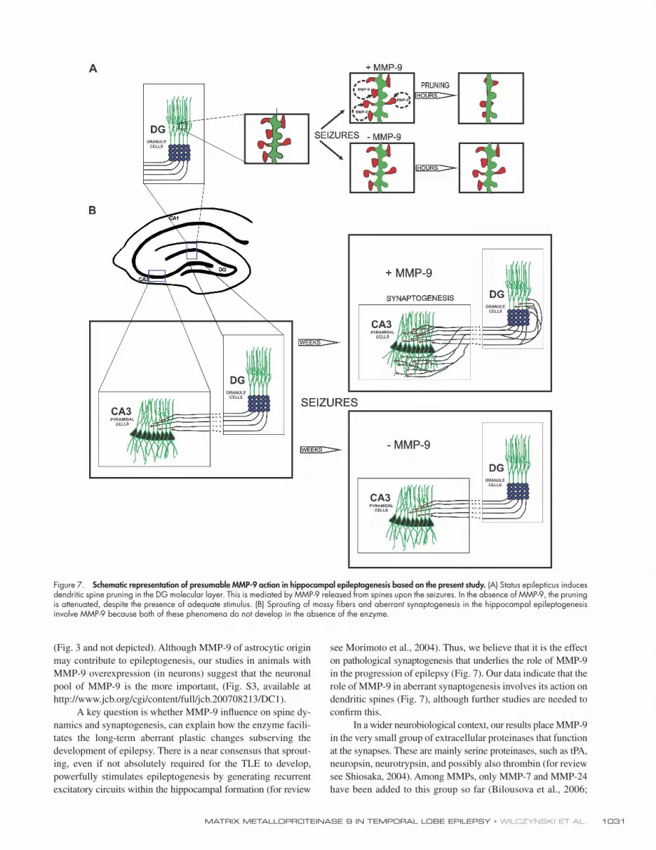
Figure 7. Schematic representation of presumable MMP-9 action in hippocampal epileptogenesis based on the present study. (A) Status epilepticus induces dendritic spine pruning in the DG molecular layer. This is mediated by MMP-9 released from spines upon the seizures. In the absence of MMP-9, the pruning is attenuated, despite the presence of adequate stimulus. (B) Sprouting of mossy fi bers and aberrant synaptogenesis in the hippocampal epileptogenesis involve MMP-9 because both of these phenomena do not develop in the absence of the enzyme.

JCB • VOLUME 180 • NUMBER 5 • 2008 1032
PTZ kindling Mice received intraperitoneal injections of 30 – 40 mg/kg PT every other day consecutively for 6 wk ( De Sarro et al., 2004 ). Behavioral seizures were scored according to a modifi ed scale of Racine: 0, no behavioral changes; 1, facial movements and ear and whisker twitching; 2, myoclonic convulsions without rearing; 3, myoclonic convulsions with rearing; 4, clonic convulsion with loss of posture; and 5, generalized clonic-tonic sei-zures or death. The brains of mice that died during experiments, as well as of those that were killed after the termination of experiments, were immer-sion fi xed in 4% PFA and stored in PBS plus 0.1% sodium azide until further processing. Subsequent immunocytochemical procedures were performed as described in the previous section.
Rats received subcutaneous injections of 40 mg/kg PTZ every other day consecutively for 8 wk and were behaviorally scored using the same scale as for mice. In addition, another group of rats was treated to the intra-peritoneal injections of PTZ.
Organotypic hippocampal culture and kainate and picrotoxin treatment Cultures of hippocampal slices were prepared essentially according to Jourquin et al. (2003) . In brief, 8 – 10-d-old rat pups were decapitated and the hippocampi were rapidly dissected on a cold plate. 400- μ m-thick slices, perpendicular to hippocampal long axis, were cut with a tissue chopper and plated (three to four per well) onto Millicel-CM biomembranes (Millipore). The slices were cultured in a humidifi ed incubator at 37 ° C with 5% CO 2 . Kainate was added to some for 24 h on DIV5 at a 10- μ M con-centration, and the cultures were fi xed on DIV20 and processed for immuno-cytochemical stainings. In the case of picrotoxin, the compound was present in culture medium at concentration of 100 μ M throughout the whole culturing period ( Koyama et al., 2004 ). MMP-9 inhibition was achieved by growing the cultures in the presence of the specifi c MMP-9 in-hibitor S24994 ( Hanessian et al., 2001 ) at a 100-nM concentration. The inhibitor was either present from the beginning (picrotoxin experiment) or added on DIV6 (immediately after removal of kainate).
Immunocytochemical procedures Primary antibodies. Two rabbit polyclonal anti – MMP-9 antibodies were used: an antibody raised against the catalytic domain of the rat MMP-9 (Torrey Pines Biolabs, Inc.; Szklarczyk et al., 2002 ) and an antibody raised against the epitope comprising the catalytic domain of the mouse enzyme (gift from R. Senior, Washington University, St. Louis, MO; Betsuyaku et al., 2000 ; see subsequent sentences for working dilutions). Both antibodies recognize active and latent forms of MMP-9 ( Betsuyaku et al., 2000 ; Szklarczyk et al., 2002 ). The following mouse marker antibodies were used for double and triple immunofl uorescent stainings: anti – MAP-2, a dendritic marker, diluted at 1:200 (Millipore); anti-synaptophysin, a presynaptic marker, diluted at 1:20 (Dako); and anti – NMDAR subunit NR1 and anti – GABA-AR subunits � 2/3, postsynaptic markers, both diluted at 1:50 (Milli-pore). Sprouting mossy fi ber terminals were visualized using a rabbit anti-synaptoporin antiserum (Synaptic Systems GmbH; Singec et al., 2002 ) diluted at 1:200.
Light microscopic immunocytochemistry. Light microscopic immuno-cytochemistry was performed essentially according to Boeckers et al. (1999) in either 50- μ m-thick free-fl oating sections of frozen tissues or 4- μ m-thick paraffi n sections. The latter were used in studies of MMP-9 co-localization with NR1 NMDAR or GABA-AR because of their permeability and mechanical resistance to antigen retrieval techniques. In such experi-ments, antigen retrieval steps using 0.1% pepsin and microwaving were necessary and were done immediately after deparaffi nization. Both kinds of sections were subjected to standard blocking steps, including, if needed, blocking of endogenous biotin (Avidin/Biotin Blocking kit; Vector Labora-tories), followed by overnight incubation in the anti – MMP-9 antibody, diluted at 1:2,000 (R. Senior) or 1:200 (Torrey Pines Biolabs, Inc.). The detection of immunoreactions was performed using either species-specifi c secondary antibodies directly conjugated to fl uorophores or biotinylated secondary antibody (Vector Laboratories) followed by a fl uorophore-conjugated strep-tavidin (Invitrogen). For double or triple stainings, we used antibodies (or streptavidin) conjugated to green Alexa 488 (Invitrogen), red Cy3 (Jackson ImmunoResearch Laboratories), or near-infrared Cy5 fl uorophores (Jackson ImmunoResearch Laboratories). In the case of anti – MMP-9, an amplifi cation step using biotinylated tyramide (PerkinElmer) was included in avidin-biotin – based detection. For triple immunofl uorescence with two marker antibodies originating from the same species, we used Zenon, a custom antibody-(immuno)labeling system (Invitrogen).
For negative controls, the primary antibody was omitted, replaced by a nonimmune IgG, and preabsorbed with the target antigen. In double
Monea et al., 2006 ; for review see Ethell and Ethell, 2007 ).
Notably, all these enzymes appear to be involved in some as-
pects of synaptic plasticity. Besides being engaged in normal
processes, tPA and neuropsin were implicated in the pathogene-
sis of epilepsy ( Wu et al., 2000 ; for review see Shiosaka, 2004 ).
It remains to be determined if, and how, these enzymes interact
with one another in health and disease, e.g., whether presynap-
tic tPA can function as an activator of postsynaptic MMP-9.
We would like to propose that our data have direct impli-
cations for human TLE. In fact, behavioral, anatomical, and
molecular events that are associated with kainate- and kindling-
induced epileptogenesis in rats and mice largely reproduce the
corresponding events in human TLE ( Ben-Ari, 2001 ). An es-
sential feature of the human disease captured by both models is
aberrant synaptic plasticity. In this paper, we show that the for-
mation of aberrant synaptic networks depends on MMP-9.
Because these networks keep progressing, contributing to the
increasing severity of the disease, the inhibition of this process
might be benefi cial at any stage. Hence, this work is not only a
step toward understanding pathophysiology of epilepsy but also
offers an opportunity to search for novel therapeutic approaches
targeting the mechanisms of this disease.
Materials and methods Animals Experiments were performed on adult male Wistar rats and on adult MMP-9 KO mice C57BL/6 strain (provided by Z. Werb, University of California, San Francisco, San Francisco, CA; Vu et al., 1998 ), all according to the rules established by the Ethical Committee on Animal Research of the Nencki Institute, based on national laws that are in full agreement with the European Union directive on animal experimentation.
Generation of MMP-9 – overexpressing rats Expression vector with autoactive MMP-9 gene driven by synapsin-1 pro-moter were constructed in our laboratory. Plasmid DNA for microinjection was purifi ed using the plasmid maxiprep method. The expression cassette was prepared by digesting the vector DNA with PacI and XbaI enzymes and microinjecting it into fertilized eggs of Wistar rats (Fig. S1). All rats were raised and maintained under specifi c pathogen-free conditions. Genomic DNA was extracted from the offspring by ear biopsy and founders were identifi ed by PCR. The primer sequences for the PCR were 5 � -AGG CGC GCT GAC GTC ACT CG-3 � and 5 � -CGC GCT CCACAG TGC GAA-3 � . PCR reactions were performed for 30 cycles at 94 ° C for 45 s, 59 ° C for 60 s, and 72 ° C for 60 s. Two independent lines of TG rats were generated.
Kainate treatment Kainate (Ocean Produce) was used in both rat and mice experiments. Status epilepticus in rats was induced by intraperitoneal injection of 10 mg/kg and the animals were observed for up to 6 h, as previously described ( Szklarczyk et al., 2002 ).
The adult male mice were anesthetized with 1.3 ml/kg ketamine and 1.25 ml/kg xylazine and subjected to stereotactic unilateral injection with 1 nmol kainate in 0.3 μ l PBS into the amygdala ( Wu et al., 2000 ). The coordinates of the injection were the following: bregma, 1.6 mm; medial – lateral, 3.3 mm; and dorsoventral, 4.5 mm. The kainate was delivered over 30 s. The injection needle remained at the injection coordinates for another 2 min afterward to prevent refl ux of fl uid.
For immunocytochemical studies, animals were lethally anesthetized and perfused transcardially with a fi xative (4% formaldehyde, freshly de-polymerized from PFA, in PBS; in some electron microscopic experiments, 0.1% glutaraldehyde was added). Brains were removed, postfi xed in the same fi xative, and either processed for paraffi n embedding or cryopro-tected and snap frozen in isopentane cooled on dry ice.
For biochemical studies, the brains were removed immediately after decapitation. From each brain, hippocampi were dissected, frozen on dry ice, and stored until assayed.
1033MATRIX METALLOPROTEINASE 9 IN TEMPORAL LOBE EPILEPSY • WILCZYNSKI ET AL.
of the dendritic spine linear density (per 1 μ m of the dendritic length) was obtained for each group. Statistical analysis was performed using t test with the level of signifi cance defi ned as P < 0.05.
Electron microscopic immunocytochemistry. Preembedding technique was used according to Boeckers et al. (1999) . In brief, the brains were perfusion fi xed with 4% PFA (plus 0.1% gluteraldehyde in the case of anti – MMP-9) and immunostained free fl oating just as for light microscopy, ex-cept that repeated freezing thawing was applied at the permeabilization step instead of Triton X-100 and peroxidase-based detection was used with DAB as a chromogen. The sections were subjected to silver enhancement and processed for electron microscopy as previously described ( Boeckers et al., 1999 ). The specimens were examined under an electron microscope (912; Carl Zeiss, Inc.) equipped with a spectroscopic fi lter (Omega Optical) and a charge-coupled device camera (2K; Gatan) operating at 80 kV.
Postembedding procedure was performed according to Mathiisen et al. (2006) . In brief, normal Wistar rats were perfused transcardially with a fi xative consisting of 4% formaldehyde plus 0.1% glutaraldehyde. The brains were postfi xed in the same fi xative, cut into 0.5 – 1.0-mm slices, cryoprotected, snap frozen in liquid propane ( � 170 ° C), and subjected to freeze substitution in an apparatus (EM AFS; Leica). Specimens were then embedded in Lowicryl HM20 resin, polymerized by UV light at � 45 – 0 ° C, and cut into ultrathin sections. The immunoreactions consisted of sequential incubations with a rabbit anti – MMP-9 antibody (Torrey Pines Biolabs, Inc.) diluted at 1:50, followed by species-specifi c goat secondary antibodies coupled to 10-nm gold particles (GE Healthcare). The specimens were ex-amined with an electron microscope (CM-10; Philips) at 60 kV. Images were acquired using either a charge-coupled device camera (1K; Soft Im-aging System) or photographic plate camera. In the latter case, negatives were digitized at a 1,200-dpi resolution using a fl atbed scanner (BearPaw; Mustek). Final adjustments of image brightness and contrast were per-formed using Photopaint. The sizes of, and gold particle densities within, various ultrastructural compartments were measured in digital micrographs using ImageJ (either manually or automatically). As a background, we as-sumed the labelling over the cytoplasm of the large dendritic profi les, which was the lowest among all compartments. Such an estimate is likely to be a slight overestimate of the true background labelling ( Mathiisen et al., 2006 ). The statistical evaluation of the labelling was performed using the � 2 test and observed and expected gold counts over the given compart-ments ( Mayhew et al., 2003 ).
Golgi impregnation Mouse brains were perfusion fi xed using 4% PFA in PBS, immersed in 5% potassium dichromate for 4 d, immersed in 1% silver nitrate for 4 d, em-bedded in celloidin, and cut into 200- μ m-thick sections. Dendritic spines were counted manually in image stacks obtained using a microscope equipped with a motorized stage and a 60 × objective lens (1.25 NA). The counting was performed in the DG middle molecular layer (dendritic seg-ments from at least three different cells) of four untreated and six KA-injected animals of either genotype. In the injected mice, both affected and contralateral hippocampi were evaluated. The results were evaluated using t test with the level of signifi cance defi ned as P < 0.05.
Isolation of synaptosomes Isolation of synaptosomes was performed according to Havik et al. (2003) . In brief, frozen tissue was melted, homogenized in a glass homogenizer, and subjected to a series of centrifugations that yielded P1 (cell debris and nuclei), P2 (crude synaptosomes/mitochondria), P3 (microsomes), and S (cytosol) fractions. The P2 fraction was further separated into myelin, mem-branes/Golgi, and synaptosomes using a discontinuous sucrose density gradient (0.8/1.0/1.2 M).
Gel zymography The assay was performed according to Szklarczyk et al. (2002) , using gel-atin – Sepharose 4B (GE Healthcare) and 8% polyacrylamide gel contain-ing 5 mg/ml of gelatin (Loba Feinchemie) for enzyme purifi cation and detection, respectively. Data were collected using the gel documentation program Gene Snap (InGenius) and Bio Imaging System (Syngen, Inc.) and pictures were prepared using Photoshop (Adobe).
In situ zymography We fi rst used established techniques of in situ zymography in which a quenched fl uorescent substrate (DQ gelatin) is applied to fresh-frozen tissue sections ( Oh et al., 1999 ; Jourquin et al., 2003 ). This approach, however, was totally unsatisfactory because tissue integrity was very poorly pre-served, which was evident at the high magnifi cations required to visualize
or triple immunostainings, special care was taken to control for any possi-ble cross-reactivity of the detection systems.
Fluorescent imaging. Fluorescent specimens were examined under a spectral confocal microscope (TCS SP2; Leica), using 488 nm Ar, 543 nm GeNe, and 633 nm HeNe laser lines for the excitation of Alexa 488, Cy3, and Cy5, respectively. The images were acquired through the internal TCS SP2 detectors/photomultipliers. To avoid cross talk between the fl uoro-phores, we carefully adjusted the spectral ranges of detectors and scanned images sequentially. The Plan Apo oil-immersion objective lenses were 40 × (1.25 NA), 63 × (1.32 NA), and 100 × (1.4 NA). The images in Figs. 2 (C and H – P), 5 (G – S), and 6 (A – S) were 3D deconvolved by means of Huygens Professional software (Scientifi c Volume Imaging), using classical maximum likelihood algorithm and theoretical point-spread functions. For fi nal inspec-tion, the images were processed using Photopaint (Corel). Brightness and contrast adjustments and/or Gaussian smoothing were applied, if necessary, to improve image clarity. Quantitative analyses of immunofl uorescence results included unbiased morphometric evaluation using ImageJ software (National Institutes of Health) and are listed in the following paragraphs.
We quantifi ed MMP-9 – positive puncta colocalizing with dendrites. The analysis of MMP-9 (red channel) – MAP-2 (green channel) colocalization in the DG of control and kainate-treated rats was performed in single-plane confocal images acquired using a 63 × objective (oil immersion; 1.3 NA) with a zoom factor of four. The settings of photomultipliers were adjusted to obtain the maximal dynamic ranges of pixels in each channel. Because the intensity of MMP-9 staining (red channel) differed considerably among the time points, several trials were performed before the optimal value was found. However, even with the optimized setting, we could not avoid con-siderable pixel saturation at 72 h after kainate. Thus, at this time point the sensitivity of the photomultiplier was decreased by 10%. Otherwise, identi-cal scanning conditions were maintained during all image acquisition ses-sions, including the constant distance of the focal plane to the surface of the section (7 μ m). In each brain, three different random focal planes were scanned in each of the molecular layer subdivisions, inner, medial, and outer. We designed a macro within ImageJ that automatically segmented areas in which red and green signals colocalized. The colocalization was considered to occur if, at a given pixel, both signals were above the thresh-old values (red, 115; green, 70) and their ratio of signal intensities was > 0.33. Using an unbiased counting frame ( Gundersen, 1978 ), at each time point we estimated the mean number of colocalizing objects per unit area of the tissue, the ratio of the number of colocalizing objects to the number of all MMP-9 – immunoreactive objects, and the mean fl uorescence intensity of the colocalizing objects, defi ned as the product of the mean area of the colocalizing objects times the mean brightness (red channel). At least four animals were analyzed at each time point. Statistical analysis was performed using Kruskall-Wallis ANOVA with the level of signifi cance defi ned as P < 0.05.
We quantifi ed mossy fi ber synapses in hippocampal organotypic cultures. The measurements were performed in cultures (prepared as de-scribed in Organotypic hippocampal culture...) stained with the anti-synapto-porin antibody followed by the anti – rabbit Cy3 (Jackson ImmunoResearch Laboratories) secondary. The fl uorophore was excited with 543 laser light, and stacks of 7 – 25 confocal planes were acquired using a 40 × objective (oil immersion; 1.25 NA). Three different fi elds covering the area of gran-ule cell plus molecular layers were acquired from each culture. The stacks were analyzed using an automatic 3D analysis function of ImageJ at the threshold of 101. The results were calculated as numbers of mossy fi ber boutons per 30,000 μ m 3 . Statistical analysis was performed using t test with the level of signifi cance defi ned as P < 0.05.
We performed confocal analysis of dendritic spine density. Den-dritic spines were identifi ed using a guinea pig anti-ProSAP2/Shank antibody and a PSD marker ( Boeckers et al., 1999 ) together with rhodamine-phalloidin (Sigma-Aldrich), which binds to spine F-actin, or with a guinea pig anti-drebrin antibody (Fitzgerald). The measurements were per-formed in single-plane confocal images acquired using a 100 × objective (oil immersion; 1.4 NA) with a zoom factor of four. Spines (rhodamine, red channel) and dendrites (Cy5, blue channel) were scanned sequentially us-ing laser lines of 543 and 633 nm, respectively. The settings of photomulti-pliers were adjusted to obtain the maximal dynamic ranges of pixel intensities in each channel. Identical scanning conditions were maintained during all image acquisition sessions. In each section the position of focal plane was set at the level of maximum intensity of the blue channel. From each hippocampus, three different random fi elds were scanned within the middle molecular layer. The number of spines and the total length of den-dritic segments within each unbiased counting frame were measured auto-matically using a custom-designed ImageJ macro. As a result, an estimate
JCB • VOLUME 180 • NUMBER 5 • 2008 1034
Engel , J. Jr . 1996 . Epilepsy: structural or functional? AJNR Am. J. Neuroradiol. 17 : 243 – 244 .
Ethell , I.M. , and D.W. Ethell . 2007 . Matrix metalloproteinases in brain develop-ment and remodeling: Synaptic functions and targets. J. Neurosci. Res. 85 : 2813 – 2823 .
Fernandez-Monreal , M. , J.P. Lopez-Atalaya , K. Benchenane , M. Cacquevel , F. Dulin , J.P. Le Caer , J. Rossier , A.C. Jarrige , E.T. Mackenzie , N. Colloc ’ h , C. Ali , and D. Vivien . 2004 . Arginine 260 of the amino-terminal domain of NR1 subunit is critical for tissue-type plasminogen activator-mediated enhancement of N -methyl- d -aspartate receptor signaling. J. Biol. Chem. 279 : 50850 – 50856 .
Frotscher , M. , A. Drakew , and B. Heimrich . 2000 . Role of afferent innervation and neuronal activity in dendritic development and spine maturation of fascia dentata granule cells. Cereb. Cortex . 10 : 946 – 951 .
Fukazawa , Y. , Y. Saitoh , F. Ozawa , Y. Ohta , K. Mizuno , and K. Inokuchi . 2003 . Hippocampal LTP is accompanied by enhanced F-actin content within the dendritic spine that is essential for late LTP maintenance in vivo. Neuron . 38 : 447 – 460 .
Gundersen , H.J. 1978 . Estimators of the number of objects per area unbiased by edge effects. Microsc. Acta . 81 : 107 – 117 .
Hanessian , S. , N. Moitessier , C. Gauchet , and M. Viau . 2001 . N-Aryl sulfonyl homocysteine hydroxamate inhibitors of matrix metalloproteinases: further probing of the S(1), S(1) ’ , and S(2) ’ pockets. J. Med. Chem. 44 : 3066 – 3073 .
Hasbani , M.J. , N.M. Viquez , and M.P. Goldberg . 2001 . NMDA receptors medi-ate hypoxic spine loss in cultured neurons. Neuroreport . 12 : 2731 – 2735 .
Havik , B. , H. Rokke , K. Bardsen , S. Davanger , and C.R. Bramham . 2003 . Bursts of high-frequency stimulation trigger rapid delivery of pre-existing alpha-CaMKII mRNA to synapses: a mechanism in dendritic protein syn-thesis during long-term potentiation in adult awake rats. Eur. J. Neurosci. 17 : 2679 – 2689 .
Isokawa , M. 2000 . Remodeling dendritic spines of dentate granule cells in tem-poral lobe epilepsy patients and the rat pilocarpine model. Epilepsia . 41 : S14 – S17 .
Jaworski , J. , I.W. Biedermann , J. Lapinska , A. Szklarczyk , I. Figiel , D. Konopka , D. Nowicka , R.K. Filipkowski , M. Hetman , A. Kowalczyk , and L. Kaczmarek . 1999 . Neuronal excitation-driven and AP-1-dependent acti-vation of tissue inhibitor of metalloproteinases-1 gene expression in ro-dent hippocampus. J. Biol. Chem. 274 : 28106 – 28112 .
Jourquin , J. , E. Tremblay , N. Decanis , G. Charton , S. Hanessian , A.M. Chollet , T. Le Diguardher , M. Khrestchatisky , and S. Rivera . 2003 . Neuronal activity-dependent increase of net matrix metalloproteinase activity is associated with MMP-9 neurotoxicity after kainate. Eur. J. Neurosci. 18 : 1507 – 1517 .
Konopacki , F.A. , M. Rylski , E. Wilczek , R. Amborska , D. Detka , L. Kaczmarek , and G.M. Wilczynski . 2007 . Synaptic localization of seizure-induced ma-trix metalloproteinase-9 mRNA. Neuroscience . 150 : 31 – 39 .
Koyama , R. , M.K. Yamada , S. Fujisawa , R. Katoh-Semba , N. Matsuki , and Y. Ikegaya . 2004 . Brain-derived neurotrophic factor induces hyperexcitable reentrant circuits in the dentate gyrus. J. Neurosci. 24 : 7215 – 7224 .
Kvajo , M. , H. Albrecht , M. Meins , U. Hengst , E. Troncoso , S. Lefort , J.Z. Kiss , C.C. Petersen , and D. Monard . 2004 . Regulation of brain proteolytic activ-ity is necessary for the in vivo function of NMDA receptors. J. Neurosci. 24 : 9734 – 9743 .
Mathiisen , T.M. , E.A. Nagelhus , B. Jouleh , R. Torp , D.S. Frydenlund , M.-N. Mylonakou , M. Amiry-Moghaddam , L. Covolan , J.K. Utvik , B. Riber , et al . 2006 . Postembedding immunogold cytochemistry of membrane molecules and amino acid transmitters in the central nervous system. In Neuroanatomical Tract-Tracing. L. Zaborszky, F.G. Wouterlood, and J.L. Lanciego, editors. Springer US, New York. 72 – 108.
Matsuzaki , M. , N. Honkura , G.C. Ellis-Davies , and H. Kasai . 2004 . Structural basis of long-term potentiation in single dendritic spines. Nature . 429 : 761 – 766 .
Mayhew , T. , G. Griffi ths , A. Habermann , J. Lucocq , N. Emre , and P. Webster . 2003 . A simpler way of comparing the labelling densities of cellular com-partments illustrated using data from VPARP and LAMP-1 immunogold labelling experiments. Histochem. Cell Biol. 119 : 333 – 341 .
McKhann , G.M. II , H.J. Wenzel , C.A. Robbins , A.A. Sosunov , and P.A. Schwartzkroin . 2003 . Mouse strain differences in kainic acid sensitivity, seizure behavior, mortality, and hippocampal pathology. Neuroscience . 122 : 551 – 561 .
Meighan , S.E. , P.C. Meighan , P. Choudhury , C.J. Davis , M.L. Olson , P.A. Zornes , J.W. Wright , and J.W. Harding . 2006 . Effects of extracellular matrix-degrading proteases matrix metalloproteinases 3 and 9 on spatial learning and synaptic plasticity. J. Neurochem. 96 : 1227 – 1241 .
Michaluk , P. , and L. Kaczmarek . 2007 . Matrix metalloproteinase-9 in gluta-mate-dependent adult brain function and dysfunction. Cell Death Differ. 14 : 1255 – 1258 .
Monea , S. , B.A. Jordan , S. Srivastava , S. DeSouza , and E.B. Ziff . 2006 . Membrane localization of membrane type 5 matrix metalloproteinase by
individual dendrites. To overcome this problem, we took advantage of the technique widely used in electrophysiological studies, in which slices of fresh brain are cut and subsequently maintained at conditions that allow preservation of at least some features of the living tissue for a defi nite time. The refi ned procedure used 150- μ m-thick fresh brain vibratome sections that were placed in tissue culture wells and covered with a solution of 50 mg/ml DQ gelatin (Invitrogen) diluted in artifi cial cerebrospinal fl uid of the following composition: 117 mM NaCl, 1.2 mM MgSO 4 , 4.7 mM KCl, 2.5 mM CaCl 2 , 25 mM NaHCO 3 , 1.2 mM NaH 2 PO 4 , and 10 mM glucose. The specimens were kept for 2 h at 37 ° C with 5% CO 2 in a tissue culture incubator, rinsed twice with PBS for 10 min at RT, and fi xed overnight with 4% PFA in PBS. Subsequent coimmunostaining for visualization of den-drites was performed essentially as described in Light microscopic immuno-cytochemistry, except that the step of the slice dehydration-rehydration using acetone/methanol solution was included at the beginning of the staining to help tissue permeabilization. The examination of the sections un-der the confocal microscope indicated that the procedure gave acceptable results in terms of both retaining enzymatic activity and tissue integrity.
Online supplemental material Fig. S1 presents the outline, as well as the biochemical validation, of the procedure of making TG rats that overexpress MMP-9. Fig. S2 shows confocal micrographs demonstrating colocalization of MMP-9 and the dendritic spine marker drebrin. Fig. S3 shows low power confocal micro-graphs of the hippocampal organotypic cultures demonstrating the effect of pharmacological inhibition of MMP-9 on kainate-induced mossy fi bers sprouting (in both supra- and infragranular layer [ Frotscher et al., 2000 ]). Online supplemental material is available at http://www.jcb.org/cgi/content/full/jcb.200708213/DC1.
We are very grateful to Dr. Z. Werb (University of California, San Francisco, San Francisco, CA) for providing us with MMP-9 KO animals. We are also very grateful to Dr. R. Senior for his generous gift of the anti – MMP-9 antibody. We thank Dr. A. Wrzosek and L. Kilianek for their help with the confocal micro-scope. We also thank Drs. M. Khrestchatisky and S. Rivera (Mediterranean Uni-versity, Marseille, France) for their helpful comments on in situ zymography.
This work was supported by KBN (State Committee for Scientifi c Re-search; grants 3 P05A 022 24 to G.M. Wilczynski and PBZ-MIN-005/P04/2002 to L. Kaczmarek), The Wellcome Trust (D.C. Gorecki and L. Kaczmarek), The International Centre for Genetic Engineering and Biotech-nology (L. Kaczmarek), and Glutamate Receptor Interacting Proteins As Novel Neuroprotective Targets (GRIPANNT; L. Kaczmarek and O.P. Ottersen).
Submitted: 31 August 2007 Accepted: 7 February 2008
References Bannikov , G.A. , T.V. Karelina , I.E. Collier , B.L. Marmer , and G.I. Goldberg .
2002 . Substrate binding of gelatinase B induces its enzymatic activity in the presence of intact propeptide. J. Biol. Chem. 277 : 16022 – 16027 .
Ben-Ari , Y. 2001 . Cell death and synaptic reorganizations produced by seizures. Epilepsia . 42 : 5 – 7 .
Benson , D.L. , L.M. Schnapp , L. Shapiro , and G.W. Huntley . 2000 . Making memories stick: cell-adhesion molecules in synaptic plasticity. Trends Cell Biol. 10 : 473 – 482 .
Betsuyaku , T. , Y. Fukuda , W.C. Parks , J.M. Shipley , and R.M. Senior . 2000 . Gelatinase B is required for alveolar bronchiolization after intratracheal bleomycin. Am. J. Pathol. 157 : 525 – 535 .
Bilousova , T.V. , D.A. Rusakov , D.W. Ethell , and I.M. Ethell . 2006 . Matrix metallo-proteinase-7 disrupts dendritic spines in hippocampal neurons through NMDA receptor activation. J. Neurochem. 97 : 44 – 56 .
Boeckers , T.M. , M.R. Kreutz , C. Winter , W. Zuschratter , K.H. Smalla , L. Sanmarti-Vila , H. Wex , K. Langnaese , J. Bockmann , C.C. Garner , and E.D. Gundelfi nger . 1999 . Proline-rich synapse-associated protein-1/cortactin binding protein 1 (ProSAP1/CortBP1) is a PDZ-domain protein highly enriched in the postsynaptic density. J. Neurosci. 19 : 6506 – 6518 .
Bozdagi , O. , V. Nagy , K.T. Kwei , and G.W. Huntley . 2007 . In vivo roles for matrix metalloproteinase-9 in mature hippocampal synaptic physiology and plasticity. J. Neurophysiol. 98 : 334 – 344 .
Chen , Z.L. , and S. Strickland . 1997 . Neuronal death in the hippocampus is pro-moted by plasmin-catalyzed degradation of laminin. Cell . 91 : 917 – 925 .
De Sarro , G. , G.F. Ibbadu , R. Marra , D. Rotiroti , A. Loiacono , E. Donato Di Paola , and E. Russo . 2004 . Seizure susceptibility to various convulsant stimuli in dystrophin-defi cient mdx mice. Neurosci. Res. 50 : 37 – 44 .

1035MATRIX METALLOPROTEINASE 9 IN TEMPORAL LOBE EPILEPSY • WILCZYNSKI ET AL.
Wu , Y.P. , C.J. Siao , W. Lu , T.C. Sung , M.A. Frohman , P. Milev , T.H. Bugge , J.L. Degen , J.M. Levine , R.U. Margolis , and S.E. Tsirka . 2000 . The tis-sue plasminogen activator (tPA)/plasmin extracellular proteolytic system regulates seizure-induced hippocampal mossy fi ber outgrowth through a proteoglycan substrate. J. Cell Biol. 148 : 1295 – 1304 .
Yong , V.W. 2005 . Metalloproteinases: mediators of pathology and regeneration in the CNS. Nat. Rev. Neurosci. 6 : 931 – 944 .
AMPA receptor binding protein and cleavage of cadherins. J. Neurosci. 26 : 2300 – 2312 .
Morimoto , K. , M. Fahnestock , and R.J. Racine . 2004 . Kindling and status epilep-ticus models of epilepsy: rewiring the brain. Prog. Neurobiol. 73 : 1 – 60 .
Nagy , V. , O. Bozdagi , A. Matynia , M. Balcerzyk , P. Okulski , J. Dzwonek , R.M. Costa , A.J. Silva , L. Kaczmarek , and G.W. Huntley . 2006 . Matrix metal-loproteinase-9 is required for hippocampal late-phase long-term potentia-tion and memory. J. Neurosci. 26 : 1923 – 1934 .
Nedivi , E. , D. Hevroni , D. Naot , D. Israeli , and Y. Citri . 1993 . Numerous can-didate plasticity-related genes revealed by differential cDNA cloning. Nature . 363 : 718 – 722 .
Oh , L.Y. , P.H. Larsen , C.A. Krekoski , D.R. Edwards , F. Donovan , Z. Werb , and V.W. Yong . 1999 . Matrix metalloproteinase-9/gelatinase B is required for process outgrowth by oligodendrocytes. J. Neurosci. 19 : 8464 – 8475 .
Okulski , P. , T. Jay , J. Jaworski , K. Duniec , J. Dzwonek , F. Konopacki , G. Wilczynski , A. Sanchez-Capelo , J. Millet , and L. Kaczmarek . 2007 . TIMP-1 abolishes MMP-9-dependent long-lasting long-term potentiation in the prefrontal cortex. Biol. Psychiatry . 62 : 359 – 362 .
Oray , S. , A. Majewska , and M. Sur . 2004 . Dendritic spine dynamics are regulated by monocular deprivation and extracellular matrix degradation. Neuron . 44 : 1021 – 1030 .
Pang , P.T. , H.K. Teng , E. Zaitsev , N.T. Woo , K. Sakata , S. Zhen , K.K. Teng , W.H. Yung , B.L. Hempstead , and B. Lu . 2004 . Cleavage of proBDNF by tPA/plasmin is essential for long-term hippocampal plasticity. Science . 306 : 487 – 491 .
Pitkanen , A. , and T.P. Sutula . 2002 . Is epilepsy a progressive disorder? Prospects for new therapeutic approaches in temporal-lobe epilepsy. Lancet Neurol. 1 : 173 – 181 .
Proper , E.A. , A.B. Oestreicher , G.H. Jansen , C.W. Veelen , P.C. van Rijen , W.H. Gispen , and P.N. de Graan . 2000 . Immunohistochemical characterization of mossy fi bre sprouting in the hippocampus of patients with pharmaco-resistant temporal lobe epilepsy. Brain . 123 : 19 – 30 .
Reeves , T.M. , M.L. Prins , J. Zhu , J.T. Povlishock , and L.L. Phillips . 2003 . Matrix metalloproteinase inhibition alters functional and structural correlates of deafferentation-induced sprouting in the dentate gyrus. J. Neurosci. 23 : 10182 – 10189 .
Represa , A. , I. Jorquera , G. Le Gal La Salle , and Y. Ben-Ari . 1993 . Epilepsy induced collateral sprouting of hippocampal mossy fi bers: does it in-duce the development of ectopic synapses with granule cell dendrites? Hippocampus . 3 : 257 – 268 .
Shiosaka , S. 2004 . Serine proteases regulating synaptic plasticity. Anat. Sci. Int. 79 : 137 – 144 .
Shubayev , V.I. , and R.R. Myers . 2004 . Matrix metalloproteinase-9 promotes nerve growth factor-induced neurite elongation but not new sprout forma-tion in vitro. J. Neurosci. Res. 77 : 229 – 239 .
Singec , I. , R. Knoth , M. Ditter , C.E. Hagemeyer , H. Rosenbrock , M. Frotscher , and B. Volk . 2002 . Synaptic vesicle protein synaptoporin is differently expressed by subpopulations of mouse hippocampal neurons. J. Comp. Neurol. 452 : 139 – 153 .
Sperk , G. 1994 . Kainic acid seizures in the rat. Prog. Neurobiol. 42 : 1 – 32 .
Sternlicht , M.D. , and Z. Werb . 2001 . How matrix metalloproteinases regulate cell behavior. Annu. Rev. Cell Dev. Biol. 17 : 463 – 516 .
Sutula , T. , P. Zhang , M. Lynch , U. Sayin , G. Golarai , and R. Rod . 1998 . Synaptic and axonal remodeling of mossy fi bers in the hilus and supragranular region of the dentate gyrus in kainate-treated rats. J. Comp. Neurol. 390 : 578 – 594 .
Suzuki , F. , Y. Makiura , D. Guilhem , J.C. Sorensen , and B. Onteniente . 1997 . Correlated axonal sprouting and dendritic spine formation during kainate-induced neuronal morphogenesis in the dentate gyrus of adult mice. Exp. Neurol. 145 : 203 – 213 .
Szklarczyk , A. , J. Lapinska , M. Rylski , R.D. McKay , and L. Kaczmarek . 2002 . Matrix metalloproteinase-9 undergoes expression and activation during dendritic remodeling in adult hippocampus. J. Neurosci. 22 : 920 – 930 .
Takumi , Y. , V. Ramirez-Leon , P. Laake , E. Rinvik , and O.P. Ottersen . 1999 . Different modes of expression of AMPA and NMDA receptors in hippo-campal synapses. Nat. Neurosci. 2 : 618 – 624 .
Tian , L. , M. Stefanidakis , L. Ning , P. Van Lint , H. Nyman-Huttunen , C. Libert , S. Itohara , M. Mishina , H. Rauvala , and C.G. Gahmberg . 2007 . Activation of NMDA receptors promotes dendritic spine development through MMP-mediated ICAM-5 cleavage. J. Cell Biol. 178 : 687 – 700 .
Toni , N. , P.A. Buchs , I. Nikonenko , P. Povilaitite , L. Parisi , and D. Muller . 2001 . Remodeling of synaptic membranes after induction of long-term poten-tiation. J. Neurosci. 21 : 6245 – 6251 .
Vu , T.H. , J.M. Shipley , G. Bergers , J.E. Berger , J.A. Helms , D. Hanahan , S.D. Shapiro , R.M. Senior , and Z. Werb . 1998 . MMP-9/gelatinase B is a key regulator of growth plate angiogenesis and apoptosis of hypertrophic chondrocytes. Cell . 93 : 411 – 422 .
Recommended


