Embed Size (px)
Citation preview

Adult Deletion of SRF Increases Epileptogenesis and DecreasesActivity-Induced Gene Expression
Bozena Kuzniewska & Karolina Nader &
Michal Dabrowski & Leszek Kaczmarek &
Katarzyna Kalita
Received: 21 November 2014 /Accepted: 29 December 2014 /Published online: 31 January 2015# The Author(s) 2015. This article is published with open access at Springerlink.com
Abstract Although the transcription factor serum responsefactor (SRF) has been suggested to play a role in activity-dependent gene expression and mediate plasticity-associatedstructural changes in the hippocampus, no unequivocal evi-dence has been provided for its role in brain pathology, such asepilepsy. A genome-wide program of activity-induced genesthat are regulated by SRF also remains unknown. In the pres-ent study, we show that the inducible and conditional deletionof SRF in the adult mouse hippocampus increases the epilep-tic phenotype in the kainic acid model of epilepsy, reflected bymore severe and frequent seizures. Moreover, we observe arobust decrease in activity-induced gene transcription in SRFknockout mice. We characterize the genetic program con-trolled by SRF in neurons and using functional annotation,we find that SRF target genes are associated with synapticplasticity and epilepsy. Several of these SRF targets functionas regulators of inhibitory or excitatory balance and the struc-tural plasticity of neurons. Interestingly, mutations in thoseSRF targets have found to be associated with such humanneuropsychiatric disorders, as autism and intellectual disabil-ity. We also identify novel direct SRF targets in hippocampus:Npas4,Gadd45g, and Zfp36. Altogether, our data indicate thatproteins that are highly upregulated by neuronal stimulation,identified in the present study as SRF targets, may function as
endogenous protectors against overactivation. Thus, the lackof these effector proteins in SRF knockout animals may leadto uncontrolled excitation and eventually epilepsy.
Keywords SRF . Epilepsy . Gene expression .
Hippocampus . Plasticity
Introduction
Epilepsy is a chronic neurological disorder that affects approx-imately 1 % of the human population [1]. The development ofepilepsy (epileptogenesis) involves progressive alterations insynaptic connections (aberrant plasticity), the molecularmechanisms of which are still poorly understood. The regula-tion of gene transcription by neuronal activity is an integralpart of adaptive plasticity [2]. Therefore, the identification ofgene expression programs that result from and control long-term synaptic changes is crucial for understanding the molec-ular mechanisms that underlie epilepsy [3].
Serum response factor (SRF) is a MADS-box proteinthat binds DNA at the CC(A/T)6GG consensus sequence,known as a CArG box or serum response element (SRE).SREs are found in promoters of actin cytoskeleton genesand immediate-early genes (IEGs; [4–6]). SRF-dependenttranscription is activated by neurotrophins or in responseto calcium influx in neurons [7–11]. The inactivation ofSRF leads to a decrease in the expression of such plasticity-linked genes as Fos, Arc, and Egr1 and produces a deficiencyin hippocampal synaptic plasticity and learning [12, 13]. Theactivity of SRF is also important during the development ofthe nervous system [14–16] and the regulation of structuralplasticity [17, 18]. The molecules that are involved in physi-ological plasticity, such as SRF, may also be engaged in path-ological or aberrant plasticity processes.
Electronic supplementary material The online version of this article(doi:10.1007/s12035-014-9089-7) contains supplementary material,which is available to authorized users.
B. Kuzniewska :K. Nader : L. Kaczmarek :K. Kalita (*)Laboratory of Neurobiology, Nencki Institute, 3 Pasteur Street,Warsaw, Polande-mail: [email protected]
M. DabrowskiLaboratory of Bioinformatics, Neurobiology Center, NenckiInstitute, 3 Pasteur Street, Warsaw, Poland
Mol Neurobiol (2016) 53:1478–1493DOI 10.1007/s12035-014-9089-7

Although SRF has recently been suggested to play a role inplasticity and mediate structural changes in the hippocampus,no solid evidence has been provided for its role in brain pa-thology. Interestingly, the increased binding of SRF to DNAand upregulation of SRF protein levels were found in thehippocampus after pilocarpine-induced status epilepticus,and SRF accumulation and phosphorylation were observedafter kainic acid-induced status epilepticus [19, 20]. Thesefindings indicate that the transcriptional activity of SRF isenhanced during epileptogenesis but provide no explanationfor its precise role in this brain pathology. Furthermore, manySRF-dependent genes that are important for synaptic plasticitylikely still remain unidentified, and no global analysis of SRF-dependent gene expression in response to neuronal stimula-tion in the adult brain has yet been reported.
In the present study, we investigated the role of the SRF-dependent transcriptional program in epileptogenesis usingbrain-specific, inducible SRF gene knockout (KO) in mice.We found that SRF KO mice exhibited an increase in thesusceptibility to spontaneous seizure development and moresevere seizures. We also identified 378 activity-dependentSRF target genes, among which we distinguished a groupwith functions associated with epilepsy and synaptic plas-ticity that may be responsible for the observed phenotype.Furthermore, we identified several novel genes that aredirectly regulated by SRF in the hippocampus in vivo: Npas4,Gadd45g, and Zfp36.
Materials and Methods
Animals
Mice with the conditional deletion of Srf in forebrain neurons(Srf CaMKCreERT2; mutant mice; KO) were generated bycrossing the Srff/f mutant strain (stock nr: 006658; Jackson’sLaboratories, USA; [12]), carrying the loxP flanked Srf, withmice expressing Cre recombinase under the control ofneuron-specific CaMKIIα promoter which is activated onlyin excitatory forebrain neurons (strain nr: EM:02125; TheEuropean Mouse Mutant Archive; [21]). To obtain time-specific control of the genetic process, Cre recombinasewas originally fused with mutated ligand-binding domainof human estrogen receptor to obtain CreERT2 enzymewhich is translocated to the nucleus and active only in re-sponse to tamoxifen (TAM) treatment. CreERT2-negativelittermates were used as a control (Srff/f; control mice; CTR).Adult mice, both genotypes, CTR and KO (2–6 months) wereinjected intraperitoneally with 1 mg of TAM twice dailyfor 5–10 days (Sigma). Experiments were performed atleast 4 weeks after tamoxifen injections. For the ChIPexperiments adult C57BL/6 mice were used. The animalswere treated in accordance with the ethical standards of
European and Polish regulations. All experimental proce-dures were approved by the Local Ethics Committee. Themice were bred in the Animal House of Nencki Institute ofExperimental Biology, Warsaw. Animals were housed in indi-vidual cages under 12-h light/dark cycle with constant tem-perature (23–25 °C) and with food and water available adlibitum.
Immunohistochemistry
Mice were transcardially perfused with 4% paraformaldehydein PBS. Brains were further fixed in the same buffer overnightat 4 °C and cut on vibratome on 40-μm slices. Free floatingbrain slices were permeabilized, blocked in normal goat serumfor 1 h at room temperature, washed, and incubated with anti-SRF antibody (1:500) (H-300; Santa Cruz Biotechnology)overnight at 4 °C. Next, immunochistochemistry was per-formed using standard procedure with Avidin/Biotin Complex(ABC) Kit Vectastain PK-6100 (Vector Labs) and visualizedusing SIGMAFAST™ DAB (Sigma). For Nissl staining,brain sections were air dried on slides. Next, the slides werestained with 0.1 % cresyl violet solution (containing 3 %acetic acid) for 5 min, washed, dehydrated, cleared in xylene,and coverslipped.
Western Blotting
Dentate gyrus of the hippocampus was dissected and homog-enized in a lysis buffer containing 1 mMMgCl2, 5 mMHEPES (pH=7.4), 320 mM sucrose, 1 mM Na2F, and protease in-hibitor cocktail complete (Roche) using Dounce glass homog-enizer. Protein concentration was measured using BCA Pro-tein Assay Kit (Pierce). Fifteen microgram of homogenateswas dissolved on 8 % polyacrylamide gels. Western blotwas performed using a standard procedure using anti-SRFantibody (1:300) (H-300; Santa Cruz Biotechnology). Blotswere reprobed with an anti-α-tubulin antibody (1:5000)(Sigma) to ensure equal total protein levels. Chemilumines-cent detection method was used. For the quantification ofindividual bands, the scan of the photographic film was ana-lyzed by densitometry using GeneTools software (Syngene).
Kainic Acid Treatments
CTR and KO animals were injected with 70 nl of 20 mMsolution of kainic acid (KA, Nanocs) in 0.9 % NaCl or salineat the left CA1 area of the dorsal hippocampus. Forintrahippocampal injection of kainic acid only males wereused. For intraperitoneal injections mice were habituated tohandling and injected with saline for 3 days before the exper-iments. Then, CTR and SRF KO were injected intraperitone-ally with 35–50 mg/kg kainic acid or saline. For the microar-rays profiling experiments only male mice were used.
Mol Neurobiol (2016) 53:1478–1493 1479

Confirmation of microarray results by qRT-PCR was per-formed on males and females. Six hours after kainic acid-induced status epilepticus (SE) or 6 h after saline injection,mice were sacrificed and brain hemispheres were dissectedand incubated overnight in RNAlater solution in 4 °C(Ambion). Next, the dentate gyrus of the hippocampus wasdissected and frozen in −80 °C for further RNA isolation. ForChIP assay only males were used. Animals 2 h afterkainic acid-induced status epilepticus were sacrificedand the hippocampus dissected and used for chromatinimmunoprecipitation.
Surgery and Video-Electroencephalography (EEG)
Surgical Procedures Animals (adult males) were anesthetizedby inhalation of 1.5–2 % isoflurane (Baxter) in oxygen andplaced in a stereotaxic frame and on a heating pad in order tomaintain a constant body temperature. Mice were injectedwith kainic acid (with rapidity of 50 nl/min) or saline (controlanimals) aimed at the left CA1 area of the dorsal hippocampuswith following coordinates: AP −1.8 mm, ML +1.7 mm, andDV −2.3 mm from bregma.
Electrode Preparation, Implantation, Electroencephalogra-phy, and Video Recordings Cortical electrodes were home-made: a screw was joined to socket contact by copper wireand welded (Bilaney Consultants Ltd.). Two electrodes wereplaced bilaterally into the scull over the frontal cortex and nexttwo over the cerebellum (first as reference and second as groundelectrode). Bipolar hippocampal electrode (Bilaney ConsultantsLtd.) was implanted into the injected hippocampus using thecoordinates AP −2.0 mm and ML +1.3 mm with bregma asreference point and DV −1.7 mm below dura. Electrodes wereplaced into pedestal (Bilaney Consultants Ltd.) and securedwithdental acrylate (Duracryl Plus, Spofa Dental, Czech Republic).After operation, mice were connected to a digital acquisitionsystem (TWin Clinical Software for EEG, Grass Technologies)and EEG activity of freely behaving animals was monitored for18 days in an isolated room. The unit consisted of six cages.
Analysis of Video-EEG Monitoring The occurrence of spon-taneous seizures was studied by browsing through EEG re-cord on a computer screen. All motion artifacts were excludedfrom analysis. An encephalographic seizure was specified as ahigh-amplitude (more than 2× baseline) discharge lastingmore than 5 s. Several seizure parameters were characterized:behavioral severity (average score), seizure frequency, andaverage seizure duration. Behavioral severity was analyzedfrommatching video-EEG recording. Seizures were estimatedbased on modified Racine’s scale [22]: score 1—immobility,facial clonus; score 2—Bwet-dog^ shakes, head nodding;score 3—piano pose (bilateral clonus of forepaws); score4—clonic seizures with falling; and score 5—generalized
tonic-clonic seizures. Seizure frequency was specified as atotal number of detected seizures divided by recordingdays, and duration was determined on the basis of EEGrecordings. SE severity was analyzed during first 24 hafter intrahippocampal injection of KA. Spontaneous sei-zures were defined as seizures appearing as early as 24 hafter KA administration.
Gene Expression Profiling Using Microarrays
Total RNAwas isolated from the dentate gyrus using RNeasyMini Kit (Qiagen) as described by the manufacturer. ResidualDNAwas removed by digestion with DNase I (Qiagen). RNAconcentration was calculated from the absorbance at 260 nm.Purity and integrity of RNA was determined using AgilentRNA 6000 Nano Chips on the Agilent 2100 Bioanalyzer Sys-tem (Agilent Technologies). A starting amount of 200-nghigh-quality total RNA was used to generate complementaryDNA (cDNA) and complementary RNA (cRNA) with theIllumina TotalPrep RNA Amplification Kit (Illumina Inc.,San Diego, CA, USA). The obtained cDNA served as a tem-plate for in vitro transcription with T7 RNA polymerase andbiotin UTP to generate multiple copies of biotinylated cRNA.Each cRNA sample (1.5 μg) was hybridized overnight toMouseWG-6 BeadChip array (Illumina); subsequently, chipswere washed, dried, and scanned with the BeadArray Reader(Illumina). A total of 22 microarrays were used (5–6 biologi-cal replicates per group). Analysis and quality control of mi-croarrays were performed using BeadArray R package. Afterbackground subtraction, data was normalized using quantilenormalization and then log2-transformed. The obtained signalwas taken as the measure of mRNA abundance derived fromthe level of gene expression. Statistical analysis of the micro-array results was performed using a two-way ANOVA (for thefactors genotype and treatment) followed by the estimation ofFDR (percent FDR; false discovery rate) using the Benjaminiand Hochberg method. Microarray data are available in theNCBI Gene Expression Omnibus (GEO) under accessionnumber GEO: GSE60772.
Gene ontology enrichment analysis was performed usingDAVID database. Pathway and global functional analysis wasperformed using Ingenuity Pathway Analysis (IPA; Ingenuity®Systems, Qiagen). Additionally, manual analysis of genes’functions and their role in neurons was assigned on the basisof published data (manual search of PubMed).
In Silico Analysis of Putative SRF-Binding Sites
Overrepresentation analysis of transcription factor bindingsites (TFBS) in the group of identified genes was performedusing the cREMaG database with default parameters [23].
Independently, using NGD database [24] we identifiedgenes with at least two putative SRF-binding sites in the
1480 Mol Neurobiol (2016) 53:1478–1493

conserved noncoding sequences between mouse and human,identified with programs AVID-VISTA [25, 26] within the−10 kb/+10 kb from Transcription Start Site. Potential SRF-binding sites were predicted using all the motifs from the$SRFF family (Genomatix MatBase v. 8.4). Moreover, forthe selected genes (Table 3) we compared previously identifiedpotential CArG boxes in the human genome [27] to the mousesequences. Among the putative SRF-binding sites identifiedusing the three above approaches (cREMaG, NGD, CArG),for further analysis, we chose only the sites conforming exactlyto the consensus sequence motif: CC(A/T)6GG or having amaximum two base pair mismatch (only one mismatch in theCC or GG) to the consensus sequence.
RNA Preparation and Quantitative Real-Time PCR
Total RNAwas isolated from the dentate gyrus of the hippo-campus using RNeasy Mini Kit (Qiagen) as described by themanufacturer. Residual DNAwas removed by digestion withDNase I (Qiagen). RNA concentration was calculated fromthe absorbance at 260 nm and the purity of RNA was deter-mined by the 260/280 nm absorbance ratio. RNAwas reversetranscribed with the SuperScript III Reverse Transcriptase(Invitrogen) according to the manufacturer’s instructions.Quantitative real-time PCR reactions were performed usingFast SYBR Green Master Mix (Applied Biosystems) in theApplied Biosystems 7900HT Fast Real-Time PCR Systemusing the following cycling conditions: 50 °C for 2 min,95 °C for 10 min, followed by 40 cycles of 95 °C for 15 sand 60 °C for 1 min. Primer sequences are presented inTable 1. Fold changes in expression were determined usingthe ΔΔCT relative quantification method. Values were nor-malized to the relative amounts of ribosomal protein largeP0 (Arbp) mRNA, which was found to be stable acrossdifferent phases of KA-induced epilepsy in mice [28].
Chromatin Immunoprecipitation
C57BL/6 mice were injected with KA (35–50mg/kg), and 2 hafter KA-induced status epilepticus or saline injection, hippo-campi were dissected, chopped (0.5-mm fragments), and fixedwith 1.5 % formaldehyde for 30 min at room temperature,then glycine was added and incubated for 10 min to quenchformaldehyde (hippocampi from each mouse were processedseparately). Samples were put on ice, washed with ice-coldphosphate-buffered saline (PBS) supplemented with proteaseinhibitor cocktail (Roche) and 1mMPMSF and briefly homog-enized with an Eppendorf fitting pestle. Samples were centri-fuged again, washed with PBS with protease inhibitors, andcentrifuged (1.3 g/5 min/4 °C), and the pellet was resuspendedin 2 ml of lysis buffer (5 mM PIPES, pH 8.0, 85 mM KCl,0.5 % Nonidet P-40, with protease inhibitors), incubated for10 min on ice, and disrupted using a glass–glass homogenizer.
Crude nuclear extract was collected by centrifugation at 1.3 gfor 10 min at 4 °C and frozen in −80 °C. Nuclear extracts werepooled and resuspended (3 hippocampi in 200 μl) in high-saltlysis buffer with protease inhibitors (1× PBS, 1 % NonidetP-40, 0.5 % sodium deoxycholate; 1 % SDS). Samples weresonicated on ice 14 times at 25 % output for 20 s with 30 sbreaks (on ice) using a Branson Sonifier 250 obtaining an av-erage of 300–500 bp DNA fragments. Samples were diluted10× with high-salt lysis buffer without SDS (final concentra-tion of 0.1 % SDS), centrifuged for 15 min to remove celldebris, and precleared with sonicated salmon sperm DNA/protein A agarose (50 % slurry) at 4 °C for 30 min with rota-tion. The DNA content was measured spectrophotometrically,and equal amounts of DNA were immunoprecipitated over-night at 4 °C using 5-μg anti-SRF antibody (H-300, SantaCruz) or 5 μg of normal rabbit IgG as negative control. Nextday, salmon sperm DNA protein A-agarose beads (Milipore)were added for 2 h, then the beads were washed with low-saltwash buffer (0.1 % SDS, 1 % Triton X-100, 2 mM EDTA,20 mM Tris–HCl, pH 8.1, 150 mM NaCl), high-salt washbuffer (0.1 % SDS, 1 % Triton X-100, 2 mM EDTA, 20 mMTris–HCl, pH 8.1, 500 mM NaCl), LiCl wash buffer (0.25 MLiCl, 1 % Nonidet P-40, 1 % deoxycholate, 1 mM EDTA,
Table 1 List of primer sequences used for qRT-PCR validation ofselected microarray results
Gene symbol Primers sequences
Arbp F: 5′ AGATTCGGGATATGCTGTTGGC 3′
R: 5′ TCGGGTCCTAGACCAGTGTTC 3′
Fos F: 5′ CCCATCCTTACGGACTCCC 3′
R: 5′ GAGATAGCTGCTCTACTTTGCC 3′
Npas4 F: 5′ GCTATACTCAGAAGGTCCAGAAGGC 3′
R: 5′ TCAGAGAATGAGGGTAGCACAGC 3′
Bdnf F: 5′ CTGTAGTCGCCAAGGTGGAT 3′
R: 5′ AGAAGTTCGGCTTTGCTCAG 3′
Syt4 F: 5′ TGACCCGTACATCAAAATGACAA 3′
R: 5′ GTGGGGATAAGGGATTCCATAGA 3′
Gadd45g F: 5′ GAAAGCACTGCACGAACTTCT 3′
R: 5′ CTTTGGCGGACTCGTAGACG 3′
Acan F: 5′ GTGGAGCCGTGTTTCCAAG 3′
R: 5′ AGATGCTGTTGACTCGAACCT 3′
Lcn2 F: 5′ GGGAAATATGCACAGGTATCCTC 3′
R: 5′ CATGGCGAACTGGTTGTAGTC 3′
Pcdh8 F: 5′ TTCCCCTTGCAGCTATTTAGTCT 3′
R: 5′ GAACGTGCTGTATCGGACTGT 3′
Elmo1 F: 5′ GGAGAAGATCCAGCCAGAAAT 3′
R: 5′ GCGAGCATTGAGTTTCCTAAAG 3′
Magoh F: 5′ ACTTTTACCTGCGTTACTACGTG 3′
R: 5′ GTTGTTGGCGTATCGCAATTT 3′
Zfp36 F: 5′ TCTCTGCCATCTACGAGAGCC 3′
R: 5′ CCAGTCAGGCGAGAGGTGA 3′
Mol Neurobiol (2016) 53:1478–1493 1481

10 mM Tris–HCl, pH 8.1), and twice in TE. Antibody-DNAcomplexes were eluted with elution buffer (1 % SDS, 0.1 MNaHCO3), crosslinking was reversed, and proteins were re-moved by adding 5 M NaCl and proteinase K and incubationfor 2 h at 42 °C, followed by incubation at 65 °C overnight,next DNA was purified using QIAquick Gel Extraction Kit(Qiagen). Immunoprecipitated chromatin was used as a tem-plate for real-time PCR reactions using Fast SYBRGreenMas-ter Mix (Applied Biosystems) in the Applied Biosystems7900HT Fast Real-Time PCR System with specific primerslocated near the predicted SRF binding sites (Table 2). Obtain-ed data was analyzed using the ΔΔCT relative quantificationmethod and calculated as the percentage of the input.
Statistical Analysis
Data on the graphs are expressed as means±standard errorsof the means (SEM) from at least three independent exper-iments. The statistical analysis of the data was performed
with GraphPad Prism software (GraphPad Software, Inc.).For comparison of two groups either unpaired t test orMann–Whitney test (nonparametric) was used. For compar-ison of multiple groups, a two-way analysis of variance(ANOVA) with post hoc Bonferroni’s multiple comparisonstest was used.
Results
Characterization of Srf f/f;CaMKCreERT2 Mice
To investigate the role of SRF in the development of temporallobe epilepsy (TLE), Srf gene KOmice were used. Because ofembryonic lethality in Srf-null mice, we used conditional mu-tants of Srf that were obtained by crossing mice in which theSrf gene was flanked by loxP sites (Srff/f) with anCaMKCreERT2 line to ablate its expression exclusively in
Table 2 List of putative SRF binding sites within the evolutionaryconserved regions between mouse and human (10/+10 kb fromtranscription start site [TSS]) that were selected for experimentalvalidation. Mismatched nucleotides (compared with SRF consensus
motif) are underlined. The primer sequences used in the ChIP assaysand location of amplicons (relative to TSS) are indicated. The results ofthe ChIP analysis (see also Fig. 5) are presented
Genesymbol
Selected predictedbinding site position
Predicted CArG boxsequence
Ampliconlocation
Primers sequences SRF-binding
Fos −309/−300 CCATATTAGG −391/−260 F: 5′ TCCCCCCCTGCGCTGCACCCTCAGA 3′ YR: 5′ CAACAGGGACCGGCCGTGGAAACCT 3′
Npas4 −960/−951 CCTAATATGG −1130/−1033 F: 5′ AAAGGGTCTTGGGTAGGTGC 3′ YR: 5′ CCTCCGCACAGCTCTAGAAA 3′
−4109/−4100 CCAAATATGG −4207/−4032 F: 5′ TCAGTTGTGTGTGTGCCTGT 3′ YR: 5′ GAGCACCCTTCTCTGGAACC 3′
Gadd45g −31/−22 CTATAAAAGG −177/−86 F: 5′ TTTGAGGCTGTGTCATCCCC 3′ Y−263/−254 CCTTTTAAGG R: 5′ GCCCGCTTTCTGATGCAAAT 3′
Lcn2 −1674/−1665 CCTTTTAAGG −1728/−1605 F: 5′ GTCTCCCATGTGCTGGATAAA 3′ NR: 5′ GTGACCTCTCCACCCTTCTA 3′
Syt4 −1502/−1493 CCATATGAAG −1623/−1542 F: 5′ TCAACTCTGGGCAATCGGTC 3′ NR: 5′ TTTCTCCAAAGCTCAGCCCA 3′
Bdnf +4620/+4629 CCAGAAATGG +4418/+4334 F: 5′ GGGAGGAAGAGAGGGAGAGA 3′ NR: 5′ GCAAGACCAGGGGCTACAAA 3′
−9372/−9363 CCCATTCTGG/CCATTCTGGG −9319/−9154 F: 5′ ACCCCATCTTTGATTTGCAGC 3′ NR: 5′ GCCCCAGTTGCCCTACATTT 3′
−9336/−9327 CCTAATTTGC N
Magoh −2982/−2973 CCACATGTGG −3026/−2876 F: 5′ GCTGAAGGAAGTGTGCTCCA 3′ N−2441/−2432 CTTATTTTGG R: 5′ CCTCCCCACCACCATCAAAT 3′
Zfp36 +3473/+3482 CCATACAAGG +3670/+3867 F: 5′ CCCTCTGTCTCTTAGCCCCT 3′ YR: 5′ TCACAAGGGAGGCAGTTTCC 3′
Pcdh8 −1061/−1052 TCTAAATAGG −1151/−1035 F: 5′ CAGGTTTCAACGTCACGCTG 3′ N−1086/−1077 CCCTATTAGA R: 5′ CTGATGCCTTGTTCCGCCTA 3′
Elmo1 +8069/+8078 CCAATTATGG +7998/+7879 F: 5′ ATGTGGCTTGGGAGGTTGAG 3′ NR: 5′ GTGGGCAGGAGTCAAGTTGA 3′
Arbp exon – – +1509/+1712 F: 5′ AGATTCGGGATATGCTGTTGGC 3′
R: 5′ TCGGGTCCTAGACCAGTGTTC 3′ –
Y yes (SRF binding observed), N no (no SRF binding in the analyzed promoter region detected)
1482 Mol Neurobiol (2016) 53:1478–1493
excitatory forebrain neurons [12, 21]. Additionally, to enablethe time-specific induction of SRF deletion, adult mice(≥8 weeks old) were injected with tamoxifen, which stimu-lates the translocation of recombinant Cre recombinase to thenucleus. Eight weeks after mutation activation, homozygousadult Srff/f mice (CTR) and Srf gene KO mice that carried asingle copy of Cre recombinase Srff/f;CaMKCreERT2 (KO) wereused for the experiments.
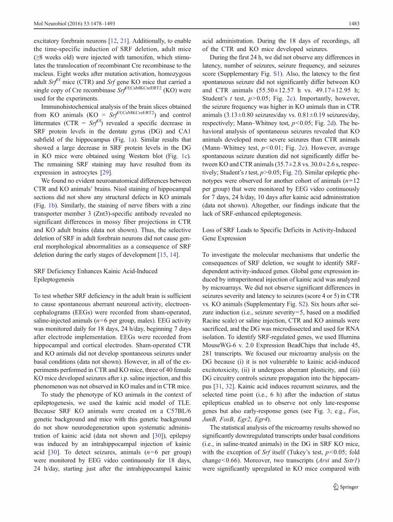
Immunohistochemical analysis of the brain slices obtainedfrom KO animals (KO = Srff/f;CaMKCreERT2) and controllittermates (CTR = Srff/f) revealed a specific decrease inSRF protein levels in the dentate gyrus (DG) and CA1subfield of the hippocampus (Fig. 1a). Similar results thatshowed a large decrease in SRF protein levels in the DGin KO mice were obtained using Western blot (Fig. 1c).The remaining SRF staining may have resulted from itsexpression in astrocytes [29].
We found no evident neuroanatomical differences betweenCTR and KO animals’ brains. Nissl staining of hippocampalsections did not show any structural defects in KO animals(Fig. 1b). Similarly, the staining of nerve fibers with a zinctransporter member 3 (Znt3)-specific antibody revealed nosignificant differences in mossy fiber projections in CTRand KO adult brains (data not shown). Thus, the selectivedeletion of SRF in adult forebrain neurons did not cause gen-eral morphological abnormalities as a consequence of SRFdeletion during the early stages of development [15, 14].
SRF Deficiency Enhances Kainic Acid-InducedEpileptogenesis
To test whether SRF deficiency in the adult brain is sufficientto cause spontaneous aberrant neuronal activity, electroen-cephalograms (EEGs) were recorded from sham-operated,saline-injected animals (n=6 per group, males). EEG activitywas monitored daily for 18 days, 24 h/day, beginning 7 daysafter electrode implementation. EEGs were recorded fromhippocampal and cortical electrodes. Sham-operated CTRand KO animals did not develop spontaneous seizures underbasal conditions (data not shown). However, in all of the ex-periments performed in CTR and KOmice, three of 40 femaleKOmice developed seizures after i.p. saline injection, and thisphenomenonwas not observed in KOmales and in CTRmice.
To study the phenotype of KO animals in the context ofepileptogenesis, we used the kainic acid model of TLE.Because SRF KO animals were created on a C57BL/6genetic background and mice with this genetic backgrounddo not show neurodegeneration upon systematic adminis-tration of kainic acid (data not shown and [30]), epilepsywas induced by an intrahippocampal injection of kainicacid [30]. To detect seizures, animals (n=6 per group)were monitored by EEG video continuously for 18 days,24 h/day, starting just after the intrahippocampal kainic
acid administration. During the 18 days of recordings, allof the CTR and KO mice developed seizures.
During the first 24 h, we did not observe any differences inlatency, number of seizures, seizure frequency, and seizuresscore (Supplementary Fig. S1). Also, the latency to the firstspontaneous seizure did not significantly differ between KOand CTR animals (55.50±12.57 h vs. 49.17±12.95 h;Student’s t test, p>0.05; Fig. 2c). Importantly, however,the seizure frequency was higher in KO animals than in CTRanimals (3.13±0.80 seizures/day vs. 0.81±0.19 seizures/day,respectively; Mann–Whitney test, p<0.05; Fig. 2d). The be-havioral analysis of spontaneous seizures revealed that KOanimals developed more severe seizures than CTR animals(Mann–Whitney test, p<0.01; Fig. 2e). However, averagespontaneous seizure duration did not significantly differ be-tween KO and CTR animals (35.7±2.8 vs. 30.0±2.6 s, respec-tively; Student’s t test, p>0.05; Fig. 2f). Similar epileptic phe-notypes were observed for another cohort of animals (n=12per group) that were monitored by EEG video continuouslyfor 7 days, 24 h/day, 10 days after kainic acid administration(data not shown). Altogether, our findings indicate that thelack of SRF-enhanced epileptogenesis.
Loss of SRF Leads to Specific Deficits in Activity-InducedGene Expression
To investigate the molecular mechanisms that underlie theconsequences of SRF deletion, we sought to identify SRF-dependent activity-induced genes. Global gene expression in-duced by intraperitoneal injection of kainic acid was analyzedby microarrays. We did not observe significant differences inseizures severity and latency to seizures (score 4 or 5) in CTRvs. KO animals (Supplementary Fig. S2). Six hours after sei-zure induction (i.e., seizure severity=5, based on a modifiedRacine scale) or saline injection, CTR and KO animals weresacrificed, and the DG was microdissected and used for RNAisolation. To identify SRF-regulated genes, we used IlluminaMouseWG-6 v. 2.0 Expression BeadChips that include 45,281 transcripts. We focused our microarray analysis on theDG because (i) it is not vulnerable to kainic acid-inducedexcitotoxicity, (ii) it undergoes aberrant plasticity, and (iii)DG circuitry controls seizure propagation into the hippocam-pus [31, 32]. Kainic acid induces recurrent seizures, and theselected time point (i.e., 6 h) after the induction of statusepilepticus enabled us to observe not only late-responsegenes but also early-response genes (see Fig. 3; e.g., Fos,JunB, FosB, Egr2, Egr4).
The statistical analysis of the microarray results showed nosignificantly downregulated transcripts under basal conditions(i.e., in saline-treated animals) in the DG in SRF KO mice,with the exception of Srf itself (Tukey’s test, p<0.05; foldchange<0.66). Moreover, two transcripts (Arsi and Sstr1)were significantly upregulated in KO mice compared with
Mol Neurobiol (2016) 53:1478–1493 1483

CTR mice (Tukey’s test, p<0.05; fold change>1.5). This re-sult was consistent with our EEG recording data, showing nodifference in neuronal activity in CTR vs. KO animals underbasal conditions (data not shown).
In contrast, comparisons of the gene expression profiles ofthe two genotypes (KO and CTR) in response to kainic acid-induced status epilepticus showed robust differences (Fig. 3a).Importantly, however, the loss of SRF in adult neurons did notcause general impairment in activity-dependent gene expres-sion in the DG. Upon seizure induction, 3241 transcripts weresignificantly up- or downregulated in CTR animals (fold
change>1.5 or<0.66; Tukey’s test, p<0.05) (Fig. 3b). Amongthis group, only 729 probes were differentially regulated byKA in KO as compared to CTR (fold change>1.5 or<0.66;Tukey’s test, p<0.05). The remaining 2512 probes (3241−729=2512; 77.5 %) did not meet the criteria (fold changeCTR KA vs. KO KA: >1.5 or <0.66 and Tukey’s test,p<0.05). Thus, SRF deficiency in adult neurons altered onlya subset of genes, suggesting the specificity of the regulation.To further restrict our analysis and obtain a list of the mostsignificantly changed transcripts, we performed a two-wayanalysis of variance (ANOVA) with false discover rate
c
CTR
SRF
KO
-tubulinα
CTR (Srf ) f/f KO (Srf )f/f;CaMKCreERT2
0.0
0.5
1.0
1.5**
CTR KO
SR
F pr
otei
n le
vels
fold
of C
TR
a’ b’
c’ d’
e’ f’
a’ b’
c’ d’
e’ f’
SRF immunoreactivity Nissl staining
a bCTR (Srf ) f/f KO (Srf )f/f;CaMKCreERT2
Fig. 1 Conditional deletion of SRF in the adult hippocampus. aImmunohistochemical staining of SRF in the hippocampus in control(CTR; Srff/f) and knockout (KO; Srff/f;CaMKCreERT2) animals. Serumresponse factor elimination was observed in the dentate gyrus (DG) andCA1 subfield of the hippocampus.bNissl staining of hippocampal sectionsfromCTR andKOmice. No neuroanatomical differences were observed in
the hippocampus in KO mice. Scale bar=a', b' 200 μm, c'–f' 400 μm. cLoss of two SRF isoforms (67 and 62 kDa) in protein extracts fromDG in KO mice compared with CTR (Western blot and quantificationof Western blot results), each line represents a single, independentanimals, **p<0.01 (Mann–Whitney test)
1484 Mol Neurobiol (2016) 53:1478–1493

(FDR) correction at the 1 % level. Using this approach,we identified 431 probes (378 genes) with a significantlyaltered expression profile as a result of SRF knockdownafter kainic acid (ANOVA, genotype × treatment interac-tion, p<0.0005; correction FDR<1 %; multiplicity ofchanges in CTR animals after the administration of kainicacid >1.5 or <0.66, Table S1).
Among those 378 genes, two main groups of genes wereclearly distinguishable: (i) genes upregulated in CTR animalsin response to kainic acid (216 genes) and not changed orupregulated to a lesser extent in KO animals after kainic acidand (ii) genes downregulated in response to kainic acid inCTR animals (162 genes) and not changed or inhibited to alesser extent in KO animals after kainic acid. Genes with themost prominent differences in the expression profile betweenthe two genotypes after kainic acid are shown in the heatmaps(Fig. 3a: 62 transcripts upregulated after kainic acid in CTRanimals >2.5, fold change in KO vs. CTR after kainic acid<0.51; Fig. 3b: 15 transcripts downregulated after kainic acid inCTR animals <0.4; fold change in KO vs. CTR after kainicacid >1.96). The probes are ordered by fold induction in CTRanimals after kainic acid-induced status epilepticus. The genesthat were chosen for further analysis (as described in the nextparagraph; i.e., functions associated with the regulation ofneuronal excitability and structural plasticity) are shown inbold. The values correspond to the indicated fold change (lin-ear scale). To identify the overrepresentation of transcriptionfactor binding sites (TFBSs) in the group of identified genes,we performed an in silico analysis using the cis-regulatoryelements in the mammalian genome (cREMaG) database[23]. We found a significant overrepresentation of SRF bind-ing sites in the group of 216 genes that were upregulated afterkainic acid, suggesting the contribution of genes that are di-rectly regulated by SRF (3.7-fold higher than expected bychance, p=0.0194). Additionally, a significant overrepresen-tation of MEF2A, another MADS-box family transcriptionfactor, was also found (3.7-fold higher than expected bychance, p=0.0184).
Functional Classification of SRF-Dependent Genes
To functionally classify the identified SRF-dependent tran-scripts, lists of downregulated and upregulated genes wereanalyzed by Gene Ontology (GO, DAVID). In the groupof genes with decreased abundance in KO animals, theoverrepresentation of transcripts that are involved in Behavior(4.1-fold enrichment, p=3.48E−6; e.g.,Egr1,Egr2,Bdnf,Ntrk2,Cyr61, Nr4a2, andNr4a3) andMAPK signaling pathway (6.3-fold enrichment, p=9.71E−11; e.g., Ntrk2, Bdnf, Rps6ka3,Gadd45g, Map2k3, Mapkapk3, and Map3k5) was observed.
To further extend the functional analysis, Ingenuity Path-way Analysis (IPA; Ingenuity® Systems, Qiagen) was applied.The Diseases and Bio-Functions analysis revealed Neurological
a
b
electrod imp ntatione laKA injection
18 days
EEG recording24 7/
TAM injection2× daily
10 daysadultmice
8-weeks
d
e f
c
0
24
48
72
96
120
CTR KO
seiz
ure
late
ncy
[hou
rs] ns
0
2
4
6
seiz
ures
freq
uenc
y/da
y
CTR KO
*
0
1
2
3
4
5
CTR KO
**
seiz
ure
scor
e
aver
. sei
zure
dur
atio
n [s
ec]
0
10
20
30
40
50
CTR KO
ns
CTR
h
c
KO
h
c
1 sec1 mV
1 sec1 mV
Fig. 2 Lack of SRF increases number and duration of spontaneousseizures in the kainic acid model of TLE. a Schematic representationof the experimental design. b Representative EEG recordings of aspontaneous seizure in a CTR mouse and KO mouse, with ahippocampal (h) electrode and cortical (c) electrode (CTR, 5 daysafter intrahippocampal kainic acid injection, 33 s duration, behavioralseverity on Racine scale=3; KO, 6 days after intrahippocampal kainicacid injection, 36 s duration, behavioral severity on Racine scale=5).The beginning and end of the seizures are indicated by arrows. c CTRand KO animals did not differ in the latency to the first spontaneousseizure. d Knockout animals developed a greater average number ofseizures per day than CTR animals (Mann–Whitney test, p<0.05). eKnockout animals showed more severe behavioral seizures measuredaccording to Racine’s scale (Mann–Whitney test, p<0.01). f The meanduration of spontaneous seizures was not significantly different in theKO group compared with the CTR group (Student’s t test, p>0.05). AllEEG recordings were conducted in males
Mol Neurobiol (2016) 53:1478–1493 1485

c
d
CTR KOsaline KA saline KA in KO mice
CTR KOsaline KA saline KA
in KO mice
FosSp7C030019I05RikFosbBdnfLcn2Cdkn1aCcl4Socs3Atf3Rcan1AcanYbx3Cdkn1aNpas4Bag3Gadd45gRnd3Cdkn1aIl11Pcdh8Ccl3AcanMapkapk3Mapkapk3Egr2Ccl4Cd14Hmga1Zfp36JunbAdpgkLOC100046232Gadd45gCyr61Gpr34930511J11RikElmo1MagohSdf2l1Ppp2r1bVgfEgr4Errfi1Rrad6030405A18RikAngptl4Dusp1Cd164l2Igfbp3Elmo1Nr4a2Ptgs2Osbpl6AregElmo1Gramd1bSerpina3fElmo1Syt4LOC100048556Map3k5
Bcl11bDspDclk3Bcl11bRapgefl1AI593442Cacna1hBcl11bD430019H16Rik4930488B01RikIl16Bcl11bAI593442Islr2AB112350
a b
fold changein response to KA vs. NaCl
fold changein response to KA vs.NaCl
14,912,211,110,49,49,28,98,38,18,18,07,16,76,46,26,15,85,65,65,45,25,25,05,04,84,44,44,34,14,14,13,93,93,93,93,93,93,63,63,63,63,43,43,33,33,33,33,23,13,13,13,03,02,92,82,72,72,62,62,62,52,5
3,13,64,53,63,72,73,72,92,43,22,51,51,92,41,13,11,81,32,81,42,81,81,32,42,41,61,61,41,31,61,41,71,81,61,51,71,41,51,41,51,41,81,31,51,31,41,21,51,51,11,61,01,51,31,11,31,31,11,31,21,10,8
0,20,30,40,30,40,30,40,40,30,40,30,20,30,40,20,50,30,20,50,30,50,40,30,50,50,40,40,30,30,40,30,40,50,40,40,40,40,40,40,40,40,50,40,50,40,40,40,50,50,40,50,30,50,40,40,50,50,40,50,40,40,3
in CTR mice
in CTR mice
fold changeKO:KA vs CTR:KA
fold changeKO:KA vs CTR:KA
0,20,20,30,30,30,30,30,40,40,40,40,40,40,40,4
0,70,90,60,80,70,80,90,90,80,80,90,90,90,81,0
3,73,22,12,92,12,53,12,22,12,02,12,12,32,12,5
fold
cha
nge
afte
r KA
in K
O
0.1 1 10 1000.1
1
10
100
fold
cha
nge
afte
r KA
in K
O
fold change after KA in CTR0.1 1 10 100
0.1
1
10
100
fold change after KA in CTR
Fig. 3 Microarray analysis showsthat SRF is an important regulatorof gene transcription in response tokainic acid-induced statusepilepticus. The figure shows theresults of gene expression profilingperformed on RNA isolated fromthe dentate gyrus of thehippocampus in control (CTR) andSrf conditional knockout (KO)mice 6 h after kainic acid-inducedstatus epilepticus (KA;intraperitoneal injection) or salineinjection (saline). Twenty-twoanimals (males) were analyzed(5/6 animals per each genotype/treatment combination). a Scatterplot showing fold change valuesfor KA responding genes in CTRand SRF mutants. b Pool of 3241probes which expression wassignificantly changed at least 1.5fold after KA (fold change>1.5 or<0.66; Tukey’s test, p<0.05). Reddots indicate population of genes(729 probes) differentiallyregulated in SRF KO as comparedto CTR (fold change CTR KAvs.KOKA >1.5 or <0.66 and Tukey’stest, p<0.05). The red lines indi-cate microarray probes changed bykainic acid at least 1.5 fold. c, d Inthe heatmaps, each column corre-sponds to one animal with the in-dicated genotype/treatment combi-nation. Rows represent transcriptsas indicated on the right. Colorsindicate normalized expressionvalues as shown by the scale at thebottom (log2 change). The resultsfor the 77 transcripts with thehighest changes after kainic acid-induced status epilepticus in KOvs. CTR are shown (fold change inresponse to kainic acid >2.5 or<0.4, genotype × treatment inter-action, FDR<1 %; fold change inKO vs. CTR after kainic acid>1.96 or <0.51). Transcripts areordered by fold change in CTRanimals treated with kainic acid vs.saline. The values correspond tothe indicated fold change (linearscale). The transcripts that werechosen for further analysis areshown in bold. c Selected genesupregulated in CTR animals inresponse to kainate but notchanged or upregulated to a lesserextent in KO animals after kainicacid. d Genes downregulated inresponse to kainic acid in CTR an-imals but not changed or inhibitedto a lesser extent in KO animals
1486 Mol Neurobiol (2016) 53:1478–1493

Disease as a top-associated category (68molecules; p=4.55E−25
to 3.58E−04). The results of the analysis showed a significantcorrelation with annotations: Epilepsy (35 genes; p=8.51E−19;Neurological Disease category; e.g., Bdnf, Cacna1h, Fos,Gadd45g, Zfp36, Cyr61, Egr1, Egr2, and Egr4), Plasticity ofSynapse (9 genes; p=0.000103; nervous system developmentand function category; e.g., Bdnf, Ntrk2, Pcdh8, and Vgf), andOutgrowth of Neurites (21 genes; p=0.0000411; Nervous Sys-tem Development and Function category; e.g., Npas4, Bdnf,Ntrk2, andGpr3). In addition to GO and IPA, a manual analysisof gene function was performed, and their role in neurons wasassigned based on published data (manual search in PubMed).Several functional groups were found, including (i) transcriptionfactors and other regulatory proteins (e.g., Fos, Npas4, Fosb,Junb, Egr1, Egr2, Egr4, Atf3, Sp7, and Cited2) and (ii) geneswith functions associated with the regulation of neuronal excit-ability and structural plasticity of dendritic spines (e.g., Fos,Npas4, Bdnf, Syt4, Gadd45g, Acan, Lcn2, Pcdh8, Elmo1,Magoh, and Zfp36; Table 3).
The functional analysis of the group of SRF targets wasconsistent with the reported roles of SRF in the regulation ofneurite outgrowth, synaptic plasticity, and behavior [15, 8, 12,13, 48]. Thus, the identified SRF targets could explain theseizure-vulnerable phenotype observed in KO animals.
Identification of SRF Direct Targets
A group of genes with functions associated with the regulationof neuronal excitability and structural plasticity of spines wasselected for validation. Microarray data were verified usingquantitative real-time polymerase chain reaction (qRT-PCR;saline: n≥5 for each genotype; kainic acid: n≥7 for each geno-type) for the selected transcripts. An increase in the expressionlevel 6 h after kainic acid stimulation (intraperitoneal KA in-jection) and dependence on the transcription factor SRF wasconfirmed for FBJ osteosarcoma oncogene (Fos; p<0.001),lipocalin 2 (Lcn2, NGAL; p<0.01), neuronal PAS domain pro-tein 4 (Npas4; p<0.01), brain-derived neurotrophic factor(Bdnf; p<0.05), aggrecan (Acan; p<0.001), protocadherin 8(Pcdh8; p<0.05), zinc finger protein 36 (Zfp36; tristetraprolin[TTP]; p<0.01), mago-nashi homolog, proliferation-associated(Magoh; p<0.05), synaptotagmin IV (Syt4; p<0.05), engulf-ment, and cell motility 1 (Elmo1; p<0.001), and growth arrestand DNA-damage-inducible 45 gamma (Gadd45g; p<0.05)two-way ANOVA genotype×treatment interaction, Bonferronimultiple-comparisons test (Fig. 4).
To determinewhich of the selected SRF-dependent genes aredirect targets of SRF, we analyzed putative SRF binding siteswithin the evolutionary conserved regions between mouse andhuman, using two database tools, namely cREMaG [23] andNGD [24] (see BMaterials and Methods^ section for details).Additionally, we searched the human CArGome (according tothe group of Miano; [27]) for potential CArG boxes that are
conserved in mouse. Among the pool of potential SRF-bindingsites identified with the above methods, only the motifs with amaximum of two mismatches to the CArG box consensus [CC(A/T)6GG] and with at most one mismatch in CC or GG wereselected for the experimental validation.
To identify direct targets of SRF bound in vivo to the genepromoters in the hippocampus, we applied a model of kainicacid-induced status epilepticus. We investigated recruitment ofthe endogenous transcription factor SRF to the identified re-gions of selected genes using chromatin immunoprecipitation.Chromatin from the hippocampus in C57BL/6 mice that weretreated with kainic acid (intraperitoneal kainic acid injection,2 h after seizure onset) or naive mice was immunoprecipitatedusing an anti-SRF antibody or normal immunoglobulin G(IgG) to determine the background, followed by qRT-PCR am-plification with specific primers (for the list of primers andpotential CArG boxes, see Table 2). We observed the in vivobinding of SRF to the promoter of Fos under basal conditions(i.e., in naive animals), whereas a significant in vivo enrich-ment of SRF binding 2 h after seizure induction was observedforNpas4,Gadd45g, and Zfp36 (Fig. 5). The binding of SRF tothe promoters of those genes indicated direct regulation bySRF. They also presented a full CArG box consensus (or onemismatch in the case of Zfp36) in the identified SRE sequences.In contrast, we did not observe significant enrichment in SRFoccupancy at potential CArG box regions of other analyzedgenes (Lcn2, Syt4, Bdnf, Magoh, and Pcdh8). Altogether, weidentified a group of novel SRF targets in neurons, which couldexplain the epilepsy-vulnerable phenotype observed in KO an-imals (Table 3). We showed that three of those genes (Npas4,Gadd45g, and Zfp36) are novel direct targets of SRF in thehippocampus in vivo, whereas the other identified genes arelikely to be indirectly regulated by SRF in neurons.
Discussion
The present study assessed the behavioral and transcriptionaleffects of SRF deficiency in neurons in the context ofepileptogenesis. Animals with adult, neuronal deletion of SRFdeveloped amore severe seizure phenotype comparedwith CTRanimals in a kainic acid model of TLE. Using genome-wideanalysis of activity-induced genes, we identified 378 genes thatare differentially expressed in the hippocampal dentate gyrus ofSRF-deficient mice, including regulators of inhibitory/excitatorybalance and structural plasticity of neurons, together with threenew SRF direct targets (Npas4, Gadd45g, Zfp36).
SRF is an Important Transcription Factor in Epilepsy
Epilepsy is a clinically relevant form of hyperexcitability-associated brain pathology. Genes that are activated during
Mol Neurobiol (2016) 53:1478–1493 1487

seizures may contribute to epileptogenic processes via networkreorganization that leads to hyperexcitability or via compensa-tory or protective mechanisms. In the present study, we uncov-ered SRF as an important transcription factor (TF) that regu-lates epileptogenesis. The deletion of SRF in the adult hippo-campus increased the epileptic phenotype, manifested by moresevere and frequent spontaneous seizures (Fig. 2), despitesimilar severity of acute seizures (Supplementary Fig. S1).
Activity-induced gene expression in neurons might also beregulated by the transcription factors CREB and MEF2.
Despite partially overlapping patterns of gene expression,the inactivation of these TFs leads to divergent consequences.CREB deficiency decreases neuronal excitability and sup-presses epileptogenesis [49–51]. MEF2 was shown to de-crease the number of excitatory synapses and weaken neuro-nal strength [52, 53]. MEF2A binding sites, along with SREs,were identified in the present study as TF binding sites thatwere overrepresented in the promoters of genes regulated bySRF, suggesting that SRF andMEF2 can activate similar tran-scription programs in neurons.
Table 3 SRF-dependent gene candidates that are important for theregulation of neuronal homeostasis. Summary and functionalcategorization of selected genes that represent potential candidatesthat may explain the SRF KO mouse phenotype of enhanced
epileptogenesis. All of the genes are located in the deletion/duplicationregions in human patients and are candidates for mental retardationor neurocognitive disabilities (based on DECIPHER database; http://decipher.sanger.ac.uk/; accessed July 7, 2014)
Gene symbol Function Neurological phenotype associatedwith deletion/duplication
Patient’sID
Fos IEG, regulator of neuronal excitability; expression induced in response to neuronalactivity (e.g., after seizures); activated in human epileptic neocortex [75]; FosKO mice have more severe kainic acid-induced seizures and increased neuronalexcitability [70]
Intellectual disability 290148
Npas4 Transcription factor, IEG, selectively induced by Ca2+ influx; regulateshomeostatic balance between excitation and inhibition in neurons by controllingthe number of γ-aminobutyric acid-releasing synapses on excitatory neurons;Npas4 KO animals are prone to seizures [58, 59]
Intellectual disability, delayed speechand language development
265913
Bdnf Pro-plasticity neurotrophin, expression bi-directionally regulated by neuronal ac-tivity; regulates maturation and function of inhibitory synapses [33] as well aspromotes synaptic transmission and synaptogenesis; activated in human epilep-tic neocortex [75]; lower serum BDNF levels were found in epileptic patientswho suffer more frequent seizures [34]; in contrast, Bdnf KO mice displayreduced epileptogenesis [35, 36], but conditional KOs do not have severelyaltered kindling [37]
Intellectual disability, delayed speechand language development
251208
Syt4 Upregulated in response to depolarization or seizures [38, 39]; modulates synapticfunction by modulating BDNF release [62]; possible involvement in homeostaticplasticity; Syt4 KO mice exhibit enhanced epileptiform responses [62]
Delayed speech and languagedevelopment
250666
Gadd45g Member of the GADD45 family associated with DNA damage repair and DNAdemethylation; other members of GADD45 family (GADD45a and GADD45b)are involved in neurite outgrowth and activity-induced DNA demethylation(e.g., Bdnf and Fgf; [40, 63]); activated in human epileptic neocortex [75]
Autism, intellectual disability 270400
Acan Component of perineuronal nets around parvalbumin interneurons; disruption ofperineuronal nets leads to seizure-like activity in hippocampal cultures [72]; lossof aggrecan staining is observed after status epilepticus (1–2 weeks; [31])
Intellectual disability 261718
Lcn2 Small, inducible, secreted protein, identified as a protein associated with matrixmetalloproteinase-9 [42]; Lcn2 KO animals show increased spine density andneuronal excitability in hippocampus and amygdala [64, 74]
Autism, severe intellectual disability 289308
Pcdh8 Upregulated in response to neuronal activity or seizures; required for induction oflong-term potentiation [43]; regulates dendritic spine number [65]; other membersof Pcdhs family (Pcdh10 and Pcdh19) are associated with neuropsychiatric dis-orders (epilepsy, mental retardation, autism-spectrum disorders; [44, 45]
Intellectual disability 260940
Elmo1 Regulates actin cytoskeleton reorganization; localized to excitatory synapses and isrequired for spine formation in hippocampal neurons [66]
Autism, severe intellectual disability 289704
Magoh Core protein of the exon junction complex that regulates metabolism of splicedmRNA; targets mRNA for nonsense-mediated decay; controls brain size byregulating neural stem cell division [46]; lack of another exon junction complexcomponent, eIF4A3, increases synaptic strength and GLUR1 AMPA receptorabundance at synapses and increases Arc protein levels [47]
Intellectual disability 272313
Zfp36 RNA-binding protein; interacts with AU-rich sequences in the 3′ untranslatedregion of targeted mRNAs and promotes their degradation; activated in humanepileptic neocortex [75]
Global developmental delay 277936
1488 Mol Neurobiol (2016) 53:1478–1493

Possible Role of SRF-Dependent Genes in the Developmentof Epilepsy
Epilepsy is associated with robust synaptic plasticity that oc-curs at the cellular level caused by changes in gene expression[54]. Previous studies on SRF-dependent transcription in neu-rons focused mainly on basal gene expression or were per-formed using in vitro models [55, 56]. Although previous
studies investigated the program downstream of SRF (recentlyreviewed by [57]), SRF-dependent transcription in neuronswas focused mainly on a basal gene expression or were per-formed using in vitro models [55, 56]. SRF is one of the majorregulators of plastic changes [12, 13]. This large-scale study ofSRF-controlled transcription programs under in vivo condi-tions provides more insights into the molecular mechanismsthat lead to the development of pathology. As expected, we
0
10
20
50
100
150
Lcn2
sal. KA
**
Fos
sal. KA
***
Npas4 Bdnf Acan Pcdh8 Zfp36 Magoh Syt4 Elmo1 Gadd45g
sal. KA sal. KA sal. KA sal. KA sal. KA sal. KA sal. KA sal. KA sal. KA
** ****
* ** ** *** *
KOCTR
Fig. 4 SRF is required for the activation of several plasticity genes in kainicacid-induced status epilepticus. The results obtained with microarray analy-sis were verified using qRT-PCR. qRT-PCR amplification of 11 transcriptsfrom control (CTR) and knockout (KO) DG (saline, n≥5 for each genotype;kainic acid, n≥7 for each genotype; males and females) revealed an increase
in mRNA levels in CTR animals in response to kainic acid stimulation (6 hafter intraperitoneal injection of KA), which was abolished in KO animals.*p<0.05, **p<0.01, ***p<0.001 (two-way ANOVA followed byBonferroni post hoc test)
CTR SRF AbKA SRF AbIgG
0.0
0.1
0.2
0.3
0.4
1
2
3
4
5
% o
f inp
ut
Lcn2
sal. KA
Fos
sal. KA
Npas4-960
sal. KA
Bdnf+4.6
sal. KA
Bdnf-9.3
sal. KA
Pcdh8
sal. KA
Zfp36
sal. KA
Magoh
sal. KA
Syt4
sal. KA
Elmo1
sal. KA
Gadd45g
sal. KA
Npas4-4109
sal. KA
Arbpexon
sal. KA
Fig. 5 Endogenous SRF binds to the promoter regions of Fos, Npas4,Gadd45g, and Zfp36. The recruitment of the endogenous transcriptionfactor SRF to the identified regions of selected genes was analyzed usingchromatin immunoprecipitation with SRF-specific antibody, followedby qRT-PCR with primers surrounding predicted SRF-binding sites(for primer sequences and locations, see Table 2). Data are plotted asa percentage of input and are the averages of three independentexperiments±SEM. A fragment of Arbp exon was used as a negative
control. The level of background was determined with normal IgG.Genes with SRF bound to their promoter fragments (Fos, Npas4,Zfp36, and Gadd45g) are marked with frames (>2-fold increase inprecipitation level compared with Arbp exon and IgG controls).Kainic acid-induced status epilepticus (KA, intraperitoneal injection)enhanced the binding of SRF especially to the promoters of theNpas4, Zfp36, and Gadd45g genes. All ChIP analysis were conductedin males
Mol Neurobiol (2016) 53:1478–1493 1489

found a large number of SRF-dependent genes associatedwithsynaptic plasticity and epilepsy, the expression of which wasdecreased in SRF KO mice after seizures.
Functional annotation revealed that many of the SRFtargets we have identified are known as regulators ofinhibitory/excitatory balance, structural plasticity, andmRNA translation (see Table 3). Control of inhibitory/excitatory balance in neurons can be regulated by Npas4,which is an activity-dependent TF that controls inhibitorysynapse development and a number of γ-aminobutyric ac-id-releasing synapses on excitatory neurons [58, 59]; Bdnf,which controls the maturation and function of inhibitorysynapses [60, 61]; Syt4, which regulates synaptic functionand plasticity by modulating BDNF release [62]; andGadd45g, a member of the Gadd45 family that is engagedin the activity-induced demethylation of Bdnf promoters andtranscription [63]. Acan, as a component of perineuronal netsaround inhibitory interneurons, stabilizes synapses and restricttheir reorganization.
Another group of identified genes are those encodingproteins regulating structural and physiological plasticityof excitatory neurons. Lcn2, Pcdh8, and Elmo1 influ-ence the electrophysiological properties of neurons byeither decreasing dendritic spine density or changingtheir morphology [64–66]. These activity-induced pro-teins may suppress the number of spines to dampensynaptic function after elevated neuronal activity, similarto other neural activity-regulated molecules, such as MEF2and PLK2 [53, 67].
A third group of genes are genes that encode RNA-bindingproteins, such as Magoh and Zfp36, which regulate themetabolism of mRNAs by targeting them for degradation[68, 69].
The decreased production of proteins form any of men-tioned above groups may explain the seizure-vulnerable phe-notype observed in SRF KO animals. However, further stud-ies are needed to address the specific role of SRF in the reg-ulation of the balance between inhibition and activation inneurons.
We found an increase in epileptogenesis in SRF KO ani-mals, which is consistent with the results of several studiesthat used animals with individual deletions of SRF-dependentgenes. The ablation of such genes as Npas4, Fos, and Arcleads to increased seizure susceptibility [58, 70, 71]. Thein vitro disruption of Acan causes seizure-like activity in hip-pocampal cultures [72]. Similarly, enhanced epileptiform re-sponses were observed in slices from Syt4 KO animals [73].Moreover, Lcn2-deficient neurons show increased excitability[64, 74]. These proteins are induced by an increase in neuronalactivity and appear to play a role as endogenous inhibitors ofepilepsy; thus, impairment of their expression in SRF KOmice could enhance the epileptic phenotype, as demonstratedby the present results.
SRF-Controlled Genes Associated with Human Pathology
Although no evidence for the role of SRF in humanepilepsy was provided so far, it is important to note thateither deletion or insertion in the regions of SRF targetgenes identified in our study was associated with humanneurological disorders (Table 3). Single-nucleotide poly-morphisms identified within or in close proximity toCArG boxes in humans were shown to be linked withneurological disorders, such as bipolar disorder, amyo-trophic lateral sclerosis, and Alzheimer’s disease [27].Several SRF-controlled genes that were identified inthe present study (e.g., Cyr61, Bdnf, Zfp36, Fos, JunB)are upregulated in the cortex in patients who sufferfrom epilepsy. Statistically significant enrichment ofSREs on the proximal promoters in this group of geneswas observed [75].
In a group of genes that were downregulated in CTR ani-mals after kainic acid-induced status epilepticus but not in KOanimals (Fig. 3b), we identified Cacna1h (Cav3.2). Mutationsin this gene are linked to a wide spectrum of idiopathic gen-eralized epilepsies [76, 77] and influence neuronal excitability[78, 79]. The lack of downregulation ofCacna1hmRNA afterstatus epilepticus in SRF KO mice may contribute to theirenhanced epileptic phenotype.
Molecular Mechanisms of SRF-Dependent Gene Regulation
Our results suggest that SRF can regulate gene expression bytwo possible molecular mechanisms. The first mechanism isdirect binding to the gene promoters. We found in vivo en-richment of SRF occupancy on promoters of Fos, Npas4,Gadd45g, and Zfp36. Although Fos has been previously re-ported to be regulated by SRF, this is the first report of whichwe are aware on the direct regulation ofNpas4,Gadd45g, andZfp36 by SRF in neurons. Significant enrichment of SRFbinding to the new SRF targets was observed upon kainic acidstimulation in the present study. The binding of SRF to DNAcan be constitutive, as observed for Fos, or inducible uponstimulation [80].
The second mechanism involves other transcriptionfactors. Because of the relatively late time point ana-lyzed in the microarray experiments (i.e., 6 h after sei-zure induction), SRF targets may be indirectly regulatedthrough the activation of other genes that encode tran-scription factors (e.g., Fos, Npas4, Egr1), similar to ourprevious study that found that Mmp-9 was regulated inneurons by SRF through Fos [81]. The identification ofseveral transcription factors, among SRF target genes,that are important for neuronal plasticity suggests thatSRF may be a primary hub that can orchestrate theregulation of several aspects of synaptic plasticity.
1490 Mol Neurobiol (2016) 53:1478–1493

Conclusions
Our data show that SRF is an important regulator of activity-induced gene expression in neurons and may be involved inthe development of epilepsy. SRF regulates the expressionof several plasticity genes that together may decreasehyperexcitation in response to a strong neuronal stimula-tion. The lack of these genes may lead to the developmentof a more severe TLE as a consequence of homeostaticimbalance. Still, further studies are needed to determinewhich of the identified SRF target genes are actually in-volved in the development of epilepsy and what is themolecular mechanism underlying this process. To addressthis question, identified genes need to be analyzed indi-vidually and their potential role in the context of epilepsyshould be assessed.
Acknowledgments This work was supported by a Marie Curie Inter-national ReintegrationGrant within the 7th European Community Frame-work Programme under grant agreement no. 230992, EpiTarGene, andthe Polish National Science Center grant (SONATA BIS 2) DEC-2012/07/E/NZ3/01814. The authors wish to thank Prof. Ryszard Przewlocki,Dr. Michal Korostynski, and Dr. Marcin Piechota from GenBioInfo andInstitute of Pharmacology PAN for performing the microarray hybridiza-tion and statistical and bioinformatic analysis; Ewelina Szmajda for thetechnical assistance; the Laboratory of Microarrays Analysis, Institute ofBiochemistry and Biophysics PAN, for bioinformatic analysis using IPA,Qiagen; and Dr. Katarzyna Lukasiuk for the help with EEG recordings.The manuscript was corrected by professional proofreader MichaelArends.
Author Contributions K.K. designed and performed the experimentsand analyzed the data. B.K. performed the experiments and analyzed thedata. K.N. performed the EEG recordings and analyzed the data. M.D.and B.K performed the bioinformatic analysis of SRF binding sites. B.K.and K.K. wrote the paper. K.K. and L.K. supervised the project.
Open Access This article is distributed under the terms of the CreativeCommons Attribution License which permits any use, distribution, andreproduction in any medium, provided the original author(s) and thesource are credited.
References
1. Thurman DJ, Beghi E, Begley CE, Berg AT, Buchhalter JR, Ding D,Hesdorffer DC, HauserWA, Kazis L, Kobau R, Kroner B, Labiner D,Liow K, Logroscino G, Medina MT, Newton CR, Parko K,Paschal A, Preux PM, Sander JW, Selassie A, Theodore W,Tomson T, Wiebe S (2011) Standards for epidemiologic studiesand surveillance of epilepsy. Epilepsia 52(7):2–26. doi:10.1111/j.1528-1167.2011.03121.x
2. Kaczmarek L, Chaudhuri A (1997) Sensory regulation of immediate-early gene expression in mammalian visual cortex: implications forfunctional mapping and neural plasticity. Brain Res Brain Res Rev 23(3):237–256
3. Pitkanen A, Lukasiuk K (2011) Mechanisms of epileptogenesis andpotential treatment targets. Lancet Neurol 10(2):173–186. doi:10.1016/S1474-4422(10)70310-0
4. Treisman R (1987) Identification and purification of a polypeptidethat binds to the c-fos serum response element. EMBO J 6:2711–2717
5. Prywes R, Roeder RG (1987) Purification of the c-fos enhancer-binding protein. Mol Cell Biol 7:3482–3489
6. Miano JM, Long X, Fujiwara K (2007) Serum response factor: mas-ter regulator of the actin cytoskeleton and contractile apparatus. Am JPhysiol Cell Physiol 292(1):C70–81
7. Kalita K, Kharebava G, Zheng JJ, Hetman M (2006) Role ofmegakaryoblastic acute leukemia-1 in ERK1/2-dependent stimula-tion of serum response factor-driven transcription by BDNF or in-creased synaptic activity. J Neurosci 26(39):10020–10032
8. Wickramasinghe SR, Alvania RS, Ramanan N,Wood JN, Mandai K,Ginty DD (2008) Serum response factor mediates NGF-dependenttarget innervation by embryonic DRG sensory neurons. Neuron 58(4):532–545
9. Misra RP, Bonni A, Miranti CK, Rivera VM, Sheng M, GreenbergME (1994) Calcium entry through L-type voltage sensitive calciumchannels can activate transcription via the serum response factor. JBiol Chem 269:25483–25493
10. Morgan JI, Curran T (1986) Role of ion fluxes in the control of c-fosexpression. Nature 322:552–555
11. Miranti CK, Ginty DD, Huang G, Chatila T, Greenberg ME (1995)Calcium activates serum response factor-dependent transcription by aRas- and Elk-1-independent mechanism that involves a Ca2+/calmod-ulin-dependent kinase. Mol Cell Biol 15:3672–3684
12. Ramanan N, Shen Y, Sarsfield S, Lemberger T, Schutz G, Linden DJ,Ginty DD (2005) SRFmediates activity-induced gene expression andsynaptic plasticity but not neuronal viability. Nat Neurosci 8(6):759–767, Epub 2005 May 2008
13. Etkin A, Alarcon JM,Weisberg SP, Touzani K, HuangYY, NordheimA, Kandel ER (2006) A role in learning for SRF: deletion in the adultforebrain disrupts LTD and the formation of an immediate memory ofa novel context. Neuron 50(1):127–143
14. Alberti S, Krause SM, Kretz O, Philippar U, Lemberger T, CasanovaE, Wiebel FF, Schwarz H, Frotscher M, Schutz G, Nordheim A(2005) Neuronal migration in the murine rostral migratory streamrequires serum response factor. PNAS 102(17):6148–6153
15. Knoll B, Kretz O, Fiedler C, Alberti S, Schutz G, Frotscher M,Nordheim A (2006) Serum response factor controls neuronal circuitassembly in the hippocampus. Nat Neurosci 9(2):195–204
16. Li CL, Sathyamurthy A, Oldenborg A, Tank D, Ramanan N (2014)SRF phosphorylation by glycogen synthase kinase-3 promotes axongrowth in hippocampal neurons. J Neurosci 34(11):4027–4042. doi:10.1523/JNEUROSCI. 4677-12.2014
17. Stritt C, Knoll B (2010) Serum response factor regulates hippocampallamination and dendrite development and is connected with reelinsignaling. Mol Cell Biol 30(7):1828–1837
18. Kalita K, Kuzniewska B, Kaczmarek L (2012) MKLs: co-factors ofserum response factor (SRF) in neuronal responses. Int J BiochemCell Biol 44(9):1444–1447
19. Morris TA, Jafari N, Rice AC, Vasconcelos O, DeLorenzo RJ (1999)Persistent increased DNA-binding and expression of serum responsefactor occur with epilepsy-associated long-term plasticity changes. JNeurosci 19(19):8234–8243
20. Herdegen T, Blume A, Buschmann T, Georgakopoulos E, Winter C,Schmid W, Hsieh TF, Zimmermann M, Gass P (1997) Expression ofactivating transcription factor-2, serum response factor and cAMP/Caresponse element binding protein in the adult rat brain followinggeneralized seizures, nerve fibre lesion and ultraviolet irradiation.Neuroscience 81(1):199–212
21. ErdmannG, Schutz G, Berger S (2007) Inducible gene inactivation inneurons of the adult mouse forebrain. BMC Neurosci 8:63
Mol Neurobiol (2016) 53:1478–1493 1491

22. Racine RJ (1972) Modification of seizure activity by electrical stimula-tion I after-discharge threshold. Electroencephalogr Clin Neurophysiol32(3):269–279
23. Piechota M, Korostynski M, Przewlocki R (2010) Identification ofcis-regulatory elements in the mammalian genome: the cREMaGdatabase. PLoS One 5(8):e12465
24. Krystkowiak I, Lenart J, Debski K, Kuterba P, Petas M, Kaminska B,Dabrowski M (2013) Nencki Genomics Database—Ensemblfuncgen enhanced with intersections, user data and genome-wideTFBS motifs. Database (Oxford) 2013:bat069
25. Bray N, Dubchak I, Pachter L (2003) AVID: A global alignmentprogram. Genome Res 13(1):97–102
26. Mayor C, Brudno M, Schwartz JR, Poliakov A, Rubin EM, FrazerKA, Pachter LS, Dubchak I (2000) VISTA: visualizing global DNAsequence alignments of arbitrary length. Bioinformatics 16(11):1046–1047
27. Benson CC, Zhou Q, Long X, Miano JM (2011) Identifying func-tional single nucleotide polymorphisms in the human CArGome.Physiol Genomics 43(18):1038–1048
28. Pernot F, Dorandeu F, Beaup C, Peinnequin A (2010) Selection ofreference genes for real-time quantitative reverse transcription-polymerase chain reaction in hippocampal structure in a murine mod-el of temporal lobe epilepsy with focal seizures. J Neurosci Res 88(5):1000–1008
29. Paul AP, Pohl-Guimaraes F, Krahe TE, Filgueiras CC, Lantz CL,Colello RJ, Wang W, Medina AE (2010) Overexpression of serumresponse factor restores ocular dominance plasticity in a model offetal alcohol spectrum disorders. J Neurosci 30(7):2513–2520. doi:10.1523/JNEUROSCI. 5840-09.2010
30. Schauwecker PE, Steward O (1997) Genetic determinants of suscep-tibility to excitotoxic cell death: implications for gene targeting ap-proaches. PNAS 94(8):4103–4108
31. Nedivi E, Hevroni D, Naot D, Israeli D, Citri Y (1993) Numerouscandidate plasticity-related genes revealed by differential cDNAcloning. Nature 363(6431):718–722
32. Hevroni D, Rattner A, Bundman M, Lederfein D, Gabarah A,Mangelus M, Silverman MA, Kedar H, Naor C, Kornuc M,Hanoch T, Seger R, Theill LE, Nedivi E, Richter-Levin G, Citri Y(1998) Hippocampal plasticity involves extensive gene induction andmultiple cellular mechanisms. J Mol Neurosci 10(2):75–98
33. Rutherford LC, DeWan A, Lauer HM, Turrigiano GG (1997)Brainderived neurotrophic factor mediates the activity-dependentregulation of inhibition in neocortical cultures. J Neurosci 17(12):4527–35
34. Hong Z, Li W, Qu B, Zou X, Chen J, Sander JW, Zhou D Serumbrainderived neurotrophic factor levels in epilepsy. Eur J Neurol 21(1):57–64
35. Kokaia M, Ernfors P, Kokaia Z, Elmer E, Jaenisch R, Lindvall O(1995) Suppressed epileptogenesis in BDNF mutant mice. ExpNeurol 133(2):215–24
36. Croll SD, Suri C, Compton DL, Simmons MV, Yancopoulos GD,Lindsay RM, Wiegand SJ, Rudge JS, Scharfman HE (1999) Brain-derived neurotrophic factor transgenic mice exhibit passive avoid-ance deficits, increased seizure severity and in vitro hyperexcitabilityin the hippocampus and entorhinal cortex. Neuroscience 93(4):1491–506
37. He XP, Kotloski R, Nef S, Luikart BW, Parada LF, McNamara JO(2004) Conditional deletion of TrkB but not BDNF preventsepileptogenesis in the kindling model. Neuron 43(1):31–42
38. Vician L, Lim IK, Ferguson G, Tocco G, Baudry M, Herschman HR(1995) Synaptotagmin IV is an immediate early gene induced bydepolarization in PC12 cells and in brain. PNAS 92(6):2164–8
39. Glisovic S, Glavan G, Saghafi MM, Zivin M (2007) Upregulation ofsynaptotagmin IV protein in kainate-induced seizures. Neuroreport18(8):831–5
40. Yamauchi J, Miyamoto Y, Murabe M, Fujiwara Y, Sanbe A, Fujita Y,Murase S, Tanoue A (2007) Gadd45a, the gene induced by the moodstabilizer valproic acid, regulates neurite outgrowth through JNK andthe substrate paxillin in N1E-115 neuroblastoma cells. Exp Cell Res313(9):1886–96
41. McRae PA, Baranov E, Rogers SL, Porter BE (2012) Persistent de-crease in multiple components of the perineuronal net following sta-tus epilepticus. Eur J Neurosci 36(11):3471–82
42. Kjeldsen L, Bainton DF, Sengelov H, Borregaard N (1993) Structuraland functional heterogeneity among peroxidase-negative granules inhuman neutrophils: identification of a distinct gelatinase-containinggranule subset by combined immunocytochemistry and subcellularfractionation. Blood 82(10):3183–91
43. Yamagata K, Andreasson KI, Sugiura H, Maru E, Dominique M, IrieY, Miki N, Hayashi Y, Yoshioka M, Kaneko K, Kato H, Worley PF(1999) Arcadlin is a neural activity-regulated cadherin involved inlong term potentiation. J Biol Chem 274(27):19473–9
44. Morrow EM, Yoo SY, Flavell SW, Kim TK, Lin Y, Hill RS,Mukaddes NM, Balkhy S, Gascon G, Hashmi A, Al-Saad S, WareJ, Joseph RM, Greenblatt R, Gleason D, Ertelt JA, Apse KA, BodellA, Partlow JN, Barry B, YaoH,Markianos K, Ferland RJ, GreenbergME, Walsh CA (2008) Identifying autism loci and genes by tracingrecent shared ancestry. Science 321(5886):218–23
45. Dibbens LM, Tarpey PS, Hynes K, Bayly MA, Scheffer IE, Smith R,Bomar J, Sutton E, Vandeleur L, Shoubridge C, Edkins S, Turner SJ,Stevens C, O'Meara S, Tofts C, Barthorpe S, Buck G, Cole J,Halliday K, Stevens C, O'Meara S, Tofts C, Barthorpe S, Buck G,Cole J, Halliday K, Jones D, Lee R, Madison M, Mironenko T,Varian J, West S, Widaa S, Wray P, Teague J, Dicks E, Butler A,Menzies A, Jenkinson A, Shepherd R, Gusella JF, Afawi Z, MazaribA, Neufeld MY, Kivity S, Lev D, Lerman-Sagie T, Korczyn AD,Derry CP, Sutherland GR, Friend K, Shaw M, Corbett M, Kim HG,Geschwind DH, Thomas P, Haan E, Ryan S, McKee S, Berkovic SF,Futreal PA, Stratton MR, Mulley JC, Gecz J (2008) Xlinkedprotocadherin 19 mutations cause female-limited epilepsy and cog-nitive impairment. Nat Genet 40(6):776–81
46. Silver DL, Watkins-Chow DE, Schreck KC, Pierfelice TJ, LarsonDM, Burnetti AJ, Liaw HJ, Myung K, Walsh CA, Gaiano N, PavanWJ (2010) The exon junction complex component Magoh controlsbrain size by regulating neural stem cell division. Nat Neurosci 13(5):551–8
47. Giorgi C, Yeo GW, Stone ME, Katz DB, Burge C, Turrigiano G,Moore MJ (2007) The EJC factor eIF4AIII modulates synapticstrength and neuronal protein expression. Cell 130(1):179–91
48. Parkitna JR, Bilbao A, Rieker C, EngblomD, Piechota M, NordheimA, Spanagel R, Schutz G (2010) Loss of the serum responsefactor in the dopamine system leads to hyperactivity. Faseb J24(7):2427–2435
49. Zhou Y, Won J, Karlsson MG, Zhou M, Rogerson T, Balaji J, NeveR, Poirazi P, Silva AJ (2009) CREB regulates excitability and theallocation of memory to subsets of neurons in the amygdala. NatNeurosci 12(11):1438–1443
50. Gruart A, Benito E, Delgado-Garcia JM, Barco A (2012) EnhancedcAMP response element-binding protein activity increases neuronalexcitability, hippocampal long-term potentiation, and classical eye-blink conditioning in alert behaving mice. J Neurosci 32(48):17431–17441
51. Zhu X, Han X, Blendy JA, Porter BE (2012) Decreased CREB levelssuppress epilepsy. Neurobiol Dis 45(1):253–263
52. Barbosa AC, Kim MS, Ertunc M, Adachi M, Nelson ED, McAnallyJ, Richardson JA, Kavalali ET, Monteggia LM, Bassel-Duby R,Olson EN (2008) MEF2C, a transcription factor that facilitates learn-ing and memory by negative regulation of synapse numbers andfunction. PNAS 105(27):9391–9396
53. Flavell SW, Cowan CW, Kim TK, Greer PL, Lin Y, Paradis S,Griffith EC, Hu LS, Chen C, Greenberg ME (2006) Activity-
1492 Mol Neurobiol (2016) 53:1478–1493

dependent regulation of MEF2 transcription factors suppresses excit-atory synapse number. Science 311(5763):1008–1012
54. Scharfman HE (2002) Epilepsy as an example of neural plasticity.Neuroscientist 8(2):154–173
55. Stritt C, Stern S, Harting K, Manke T, Sinske D, Schwarz H, VingronM, Nordheim A, Knoll B (2009) Paracrine control of oligodendro-cyte differentiation by SRF-directed neuronal gene expression. NatNeurosci 12(4):418–427
56. Benito E, Valor LM, Jimenez-MinchanM, Huber W, Barco A (2011)cAMP response element-binding protein is a primary hub of activity-driven neuronal gene expression. J Neurosci 31(50):18237–18250
57. Benito E, Barco A (2014) The neuronal activity-driven tran-scriptome. Mol Neurobiol. doi:10.1007/s12035-014-8772-z
58. Lin Y, Bloodgood BL, Hauser JL, Lapan AD, Koon AC, Kim TK,Hu LS, Malik AN, Greenberg ME (2008) Activity-dependent regu-lation of inhibitory synapse development by Npas4. Nature 455(7217):1198–1204
59. Spiegel I, Mardinly AR, Gabel HW, Bazinet JE, Couch CH, TzengCP, Harmin DA, Greenberg ME (2014) Npas4 regulates excitatory-inhibitory balance within neural circuits through cell-type-specificgene programs. Cell 157(5):1216–1229
60. Marty S (2000) Differences in the regulation of neuropeptide Y, so-matostatin and parvalbumin levels in hippocampal interneurons byneuronal activity and BDNF. Prog Brain Res 128:193–202
61. Vicario-Abejon C, Collin C, McKay RD, Segal M (1998)Neurotrophins induce formation of functional excitatory and inhibi-tory synapses between cultured hippocampal neurons. J Neurosci 18(18):7256–7271
62. Dean C, Liu H, Dunning FM, Chang PY, Jackson MB, Chapman ER(2009) Synaptotagmin-IV modulates synaptic function and long-term potentiation by regulating BDNF release. Nat Neurosci 12(6):767–776
63. MaDK, JangMH, Guo JU, Kitabatake Y, ChangML, Pow-AnpongkulN, Flavell RA, Lu B, Ming GL, Song H (2009) Neuronal activity-induced Gadd45b promotes epigenetic DNA demethylation and adultneurogenesis. Science 323(5917):1074–1077
64. Mucha M, Skrzypiec AE, Schiavon E, Attwood BK, Kucerova E,Pawlak R (2011) Lipocalin-2 controls neuronal excitability and anx-iety by regulating dendritic spine formation and maturation. PNAS108(45):18436–18441
65. Yasuda S, Tanaka H, Sugiura H, Okamura K, Sakaguchi T, Tran U,Takemiya T, Mizoguchi A, Yagita Y, Sakurai T, De Robertis EM,Yamagata K (2007) Activity-induced protocadherin arcadlin regu-lates dendritic spine number by triggering N-cadherin endocytosisvia TAO2beta and p38 MAP kinases. Neuron 56(3):456–471
66. Kim JY, Oh MH, Bernard LP, Macara IG, Zhang H (2011) TheRhoG/ELMO1/Dock180 signaling module is required for spinemorphogenesis in hippocampal neurons. J Biol Chem 286(43):37615–37624
67. Pak DT, Sheng M (2003) Targeted protein degradation and synapseremodeling by an inducible protein kinase. Science 302(5649):1368–1373
68. Gehring NH, Lamprinaki S, Hentze MW, Kulozik AE (2009) Thehierarchy of exon-junction complex assembly by the spliceosomeexplains key features of mammalian nonsense-mediated mRNAdecay. PLoS Biol 7(5):e1000120
69. Singh KK,Wachsmuth L, Kulozik AE, Gehring NH (2013) Twomam-malian MAGOH genes contribute to exon junction complex composi-tion and nonsense-mediated decay. RNA Biol 10(8):1291–1298
70. Zhang J, Zhang D, McQuade JS, Behbehani M, Tsien JZ, Xu M(2002) c-fos regulates neuronal excitability and survival. Nat Genet30(4):416–420
71. Peebles CL, Yoo J, Thwin MT, Palop JJ, Noebels JL, Finkbeiner S(2010) Arc regulates spine morphology and maintains network sta-bility in vivo. PNAS 107(42):18173–18178
72. VedunovaM, Sakharnova T, Mitroshina E, Perminova M, PimashkinA, Zakharov Y, Dityatev A,Mukhina I (2013) Seizure-like activity inhyaluronidase-treated dissociated hippocampal cultures. Front CellNeurosci 7:149
73. Dean C, Liu H, Staudt T, Stahlberg MA, Vingill S, Buckers J, KaminD, Engelhardt J, JacksonMB, Hell SW, Chapman ER (2012) Distinctsubsets of Syt-IV/BDNF vesicles are sorted to axons versus dendritesand recruited to synapses by activity. J Neurosci 32(16):5398–5413
74. Skrzypiec AE, Shah RS, Schiavon E, Baker E, Skene N, Pawlak R,Mucha M (2013) Stress-induced lipocalin-2 controls dendritic spineformation and neuronal activity in the amygdala. PLoS One 8(4):e61046
75. Beaumont TL, Yao B, Shah A, Kapatos G, Loeb JA (2012) Layer-specific CREB target gene induction in human neocortical epilepsy. JNeurosci 32(41):14389–14401
76. Chen Y, Lu J, Pan H, Zhang Y, Wu H, Xu K, Liu X, Jiang Y, Bao X,Yao Z, Ding K, Lo WH, Qiang B, Chan P, Shen Y, Wu X (2003)Association between genetic variation of CACNA1H and childhoodabsence epilepsy. Ann Neurol 54(2):239–243
77. Heron SE, Khosravani H, Varela D, Bladen C, Williams TC, NewmanMR, Scheffer IE, Berkovic SF, Mulley JC, Zamponi GW (2007)Extended spectrum of idiopathic generalized epilepsies associ-ated with CACNA1H functional variants. Ann Neurol 62(6):560–568
78. Vitko I, Chen Y, Arias JM, Shen Y, Wu XR, Perez-Reyes E (2005)Functional characterization and neuronal modeling of the effects ofchildhood absence epilepsy variants of CACNA1H, a T-type calciumchannel. J Neurosci 25(19):4844–4855
79. Eckle VS, Shcheglovitov A, Vitko I, Dey D, Yap CC, Winckler B,Perez-Reyes E (2014) Mechanisms by which a CACNA1Hmutationin epilepsy patients increases seizure susceptibility. J Physiol 592(Pt 4):795–809
80. Esnault C, Stewart A, Gualdrini F, East P, Horswell S, Matthews N,Treisman R (2014) Rho-actin signaling to the MRTF coactivatorsdominates the immediate transcriptional response to serum in fibro-blasts. Genes Dev 28(9):943–958
81. Kuzniewska B, Rejmak E, Malik AR, Jaworski J, Kaczmarek L,Kalita K (2013) Brain-derived neurotrophic factor induces matrixmetalloproteinase 9 expression in neurons via the serum responsefactor/c-Fos pathway. Mol Cell Biol 33(11):2149–2162
Mol Neurobiol (2016) 53:1478–1493 1493


