
Dunlap et al. BMC Genomics 2013, 14:509http://www.biomedcentral.com/1471-2164/14/509
DATABASE Open Access
KEGG orthology-based annotation of thepredicted proteome of Acropora digitifera:ZoophyteBase - an open access and searchabledatabase of a coral genomeWalter C Dunlap1,2, Antonio Starcevic4, Damir Baranasic4, Janko Diminic4, Jurica Zucko4, Ranko Gacesa4,Madeleine JH van Oppen1, Daslav Hranueli4, John Cullum5 and Paul F Long2,3*
Abstract
Background: Contemporary coral reef research has firmly established that a genomic approach is urgently neededto better understand the effects of anthropogenic environmental stress and global climate change on coralholobiont interactions. Here we present KEGG orthology-based annotation of the complete genome sequence ofthe scleractinian coral Acropora digitifera and provide the first comprehensive view of the genome of areef-building coral by applying advanced bioinformatics.
Description: Sequences from the KEGG database of protein function were used to construct hidden Markovmodels. These models were used to search the predicted proteome of A. digitifera to establish complete genomicannotation. The annotated dataset is published in ZoophyteBase, an open access format with different options forsearching the data. A particularly useful feature is the ability to use a Google-like search engine that links querywords to protein attributes. We present features of the annotation that underpin the molecular structure of keyprocesses of coral physiology that include (1) regulatory proteins of symbiosis, (2) planula and early developmentalproteins, (3) neural messengers, receptors and sensory proteins, (4) calcification and Ca2+-signalling proteins,(5) plant-derived proteins, (6) proteins of nitrogen metabolism, (7) DNA repair proteins, (8) stress response proteins,(9) antioxidant and redox-protective proteins, (10) proteins of cellular apoptosis, (11) microbial symbioses andpathogenicity proteins, (12) proteins of viral pathogenicity, (13) toxins and venom, (14) proteins of the chemicaldefensome and (15) coral epigenetics.
Conclusions: We advocate that providing annotation in an open-access searchable database available to the publicdomain will give an unprecedented foundation to interrogate the fundamental molecular structure and interactionsof coral symbiosis and allow critical questions to be addressed at the genomic level based on combined aspects ofevolutionary, developmental, metabolic, and environmental perspectives.
Keywords: Acropora digitifera, KEGG orthology, Database, Annotation, Proteome, Genome, Coral, Symbiosis, Cnidaria
* Correspondence: [email protected] of Pharmaceutical Science, King’s College London, Franklin-WilkinsBuilding, 150 Stamford Street, London SE1 9NH, United Kingdom3Department of Chemistry King’s College London, Franklin-Wilkins Building,150 Stamford Street, London SE1 9NH, United KingdomFull list of author information is available at the end of the article
© 2013 Dunlap et al.; licensee BioMed Central Ltd. This is an Open Access article distributed under the terms of the CreativeCommons Attribution License (http://creativecommons.org/licenses/by/2.0), which permits unrestricted use, distribution, andreproduction in any medium, provided the original work is properly cited.

Dunlap et al. BMC Genomics 2013, 14:509 Page 2 of 59http://www.biomedcentral.com/1471-2164/14/509
BackgroundAll of the reef-building corals (Scleractinia; phylumCnidaria) that create the vast calcium carbonate de-posits of coral reefs have evolved an endosymbioticpartnership with photosynthetic dinoflagellates of thegenus Symbiodinium (Dinophyceae), commonly knownas zooxanthellae, which reside within the gastrodermalcells of their scleractinian host [1-3]. Coral-algal symbiosisis a cooperative metabolic adaptation necessary for sur-vival in the shallow oligotrophic (nutrient-poor) waters oftropical and subtropical marine environments [4,5] thatdrives the productivity of coral reefs [6]. Coral reefs pro-vide habitat and trophic support for many thousands ofmarine species, the richness of which rival the biologicalbiodiversity of tropical rainforests [7]. Underlying the basicrequirements of corals for growth, reproduction and sur-vival are special needs to accommodate symbiont-specifichost recognition, to control innate and responsive im-mune systems, and what is likely to emerge from futureresearch is the extent to which the host is involved indirect regulation of its endosymbiont populations.Much is understood about the cellular biology ofcnidarian-dinoflagellate symbiosis (reviewed in [8]),but less is known at the molecular level of coral symbiology.There is little opposition to the contention that envi-ronmental and anthropogenic disturbances are causingalarming losses to coral reefs ([9] and reference therein).Threats to productivity are being imposed by the disruptionof coral symbiosis (apparent as “coral bleaching”) caused inresponse to increasing thermal stress attributed to globalwarming [10,11], from an increase in stress-related coraldisease [12-14], from the discharge of domestic and indus-trial wastes, pollutants from agricultural development andthe transport of sediments in terrestrial runoff [15,16], andpotentially from imminent declines in coral calcificationowing to rising ocean acidification [17-19]. Accordingly, werequire a better understanding of the molecular stress re-sponses and adaptive potential of corals. Such informationis necessary to predict bleaching events and so better in-form effective management policies for the conservation ofcoral reef ecosystems [20-24].To understand how coral holobionts respond to envi-
ronmental change at the molecular level, the identificationof genes that may respond by transcription to stress is ofprimary importance [25]. Thus, the use of transcriptomicmethodologies to identify stress-responsive genes has beenhighly successful [26-32]. Transcriptome high-throughputprofiling has allowed changes in gene expression acrossthousands of genes to be measured simultaneously. Fuel-led by data-generating power, the number of coral basedstudies utilising transcriptomics to investigate molecularresponses to environmental stressors has expanded greatlyby the acquisition of expressed sequence tag (EST) gene li-braries, the fabrication of microarray biochips used to
estimate levels of mRNA expression, and by direct analysisusing next-generation, high-throughput sequencing. How-ever, much of this work has been conducted using theaposymbiotic state of pre-settlement coral larvae, sotranscribed genes relevant to metamorphosis and thecytobiology of the adult polyp are limited to a few recentstudies [33-36]. The transcriptome additionally does notprovide the structural framework and essential regulatoryelements of the functional genome for comprehensiveevaluation. Recently, deep metatranscriptomic sequencingof two adult coral holobiomes has been made availableon searchable databases: PocilloporaBase for Pocilloporadamicornis [36] and PcarnBase for Platygyra carnosus[37]. In contrast, high-throughput metaproteomic analysesto quantify the product yield of stress-response genes ofthe coral holobiome are yet to be widely adopted by thecoral reef scientific community, despite the proteome be-ing the ultimate measure of the coral phenotype [38,39].The early accumulation of transcriptomic data revealed
that a small proportion of coral ESTs matched genesknown previously only from other kingdoms of life, imply-ing that the ancestral animal genome contained manygenes traditionally regarded as ‘non-animal’ that have beenlost from most animal genomes [40]. Furthermore, an un-expected revelation from EST data is the greater extent towhich coral sequences resemble human genes than thoseof the Drosophila and Caenorhabditis model invertebrategenomes [41,42]. Comparative genomic analysis hasrevealed higher genetic divergence and massive gene losswithin the ecdysozoan lineages. Hence, many genesassumed to have much later evolutionary origins are likelyto have been present in an ancestral or early-divergedmetazoan [43]. While much of the animal kingdomremains yet to be explored, examples of the metazoanphylum Cnidaria provide a unique insight into the deepevolutionary origins of at least some vertebrate gene fa-milies [42]. Thus, the complete genomic sequence of acoral is likely to reveal many genes previously assumed tobe strictly vertebrate innovations. To date, cnidarian ge-nomes have been published for the sea anemone N.vectensis [42] and the hydroid Hydra magnipapillata [44].Only the coral genome of Acropora digitifera is availablewithout restriction on use of its published sequence [45],but the compiled sequence has not been fully annotated.At the time of this writing, the genome assembly ofAcropora millepora has been released to the public do-main [46], also without full annotation, but an embargo isimposed on use of this data that is highly restrictive to theprogress of further studies. Understanding how genomicvariation affects molecular and organismal biology is theultimate justification of genome sequencing, and annota-tion is an essential step in this process. We envisage thatunrestricted access to annotation of the A. digitifera gen-ome will provide an unprecedented foundation to freely

Dunlap et al. BMC Genomics 2013, 14:509 Page 3 of 59http://www.biomedcentral.com/1471-2164/14/509
interrogate the generic molecular structure, possibleendobiotic interactions and the response of coral to en-vironmental stress. Accordingly, we offer annotation ofthe predicted proteome of A. digitifera on the open ac-cess and searchable database, ZoophyteBase [47]. Useof the ZoophyteBase search engines will allow genes ofencoded proteins to be identified that can be examined incontext of the cellular physiology, processes of ecologicalsignificance, the evolutionary and developmental biologyof corals and the functional metabolism of the holobiontthat collectively underpin the health of coral reefs.
Construction and contentZoophyteBase is an open access and searchable databaseof complete annotation of the predicted proteome of thecoral A. digitifera [48]. It was constructed using the MEGGASENSE system, which is a general system for cons-tructing annotation databases with different sorts of in-put data (DNA reads, assembled genomes, predictedproteomes) and the possibility of using different combi-nations of analysis tools to create the annotation (Gacesaet al, in preparation). In the case of ZoophyteBase,hidden Markov model (HMM) profiles [49] were chosenas the annotation tool rather than the more commonBLAST searches [50]. HMM profiles are constructedfrom multiple alignments of protein families and containinformation about conserved differences in amino acidresidues as well as deletions and insertions [49]. This isparticularly important for a coral database, as corals areevolutionarily distant to most other organisms. Thismeans that known homologous sequences present in thedatabases will usually have relatively low similarity, mak-ing BLAST searches inaccurate. The statistical informa-tion in an HMM profile gives more sensitive andaccurate detection of sequence homology. An additionaladvantage of HMM profiles is that the statistical signifi-cance of hits (the expected value) is much more accuratethan that calculated by BLAST programs.The quality of sequence annotation is limited by the
accuracy of information provided in any database used.It is well known that there are many problems withannotation in the large uncurated databases such as theNCBI GenBank nr sequences. Widely accepted, the mostaccurate database for functional annotation is the KEGGdatabase [51]. The KEGG database organises sequencesas groups of KEGG orthologues. These are sets of hom-ologous sequences from as wide a range of organisms aspossible having an assigned molecular function. Thesefunctions are arranged in a hierarchical fashion andgrouped in biological pathways. The sequences belong-ing to KEGG orthologues were used to construct HMMprofiles for annotating the coral sequences. Accordingly,the 23,524 predicted proteins encoded in the coral ge-nome were analysed using HMM profiles. If a protein
showed a highly significant correlation (“hit”) to a singleHMM profile, this was used to create a “trusted” annota-tion of the sequence. Choosing a cut-off for this criterionis not trivial, because longer sequences tend to have moresignificant e-values. For construction of ZoophyteBase thecriterion 1e-5 was used. This resulted in 19,044 predictedproteins giving “trusted” sequence annotation. For manyof these proteins there were two or more highly significanthits to established HMM profiles. In these cases, the mostsignificant correlation was used to construct our “best-fit”annotation file, but other hits can be viewed by the data-base user so that expert knowledge can be employed tooverride the automatic annotation function. In 8,004 outof 19,044 predicted proteins which were annotated, morethan one annotation was assigned based on non-overlapping regions within the protein which were used toconstruct the “best-fit” annotation file. We interpretedthese as “fusion” events generated by the in silico proteinprediction method used, and these proteins were treatedas multiple instead of single encoded proteins. Hence, thisanalysis resulted in the annotation of 33,195 proteins intotal, generated from the original 23,524 predicted coralproteins. This is a very conservative annotation scheme, soit can be assumed that most of the annotations are bio-logically meaningful. Almost 81% (19,044 out of 23,524) ofthe predicted proteome was assigned using this method.
UtilityThe MEGGASENSE system was used to generate a webinterface for ZoophyteBase. The home page (Figure 1A)allows the use of several functions. A text version of theentire annotation can be downloaded for manual inspec-tion. There is a proteome overview that gives statisticsabout the database and a breakdown of the annotatedfunctions into different categories of genes. A particu-larly useful feature of ZoophyteBase is the ability to usetext queries employing a search engine that provides arelevant inquiry in the absence of an exact match be-tween key words of a search and those described for afunctional protein. The search engine uses text from theKEGG-database, PubMed and other sources to establishlinks between query words to access protein data usingan intelligent Google-like search engine implemented bythe search platform Lucene/Solr [52]. This helps to over-come the common problem that different terminology isused by different groups of researchers. The use of thissearch function is illustrated by using the query “pha-gocytosis” (Figure 1B). This inquiry finds 42 hits toKEGG orthologue profiles. One of the hits correspondsto amphiphysin (a synaptic vesicle protein) with annota-tion of two protein homologues encoded in the coralgenome. On the data page there is a brief description ofthe function of amphiphysin together with a PUBMEDliterature reference. The sequences of the predicted coral

Figure 1 Graphical overview of the user-web interface for ZoophyteBase during a typical search. The home page allows several searchfunctions (A). Text queries using an intelligent Google-like search engine is illustrated by using the query “phagocytosis” (B). This finds 42 hits toKEGG orthologue profiles. One of the hits corresponds to amphiphysin with annotation of two protein homologues encoded in the coralgenome. On the data page there is a brief description of the function of amphiphysin together with a PUBMED literature reference. Thesequences of the predicted coral proteins can be retrieved (C).
Dunlap et al. BMC Genomics 2013, 14:509 Page 4 of 59http://www.biomedcentral.com/1471-2164/14/509
proteins (Figure 1C) can be retrieved, and it is also pos-sible to analyse such data with computer aided drugdesign methods [53] to look for conserved domains.There are also two tools for the user to examine matchesto protein sequences. The user can carry out a BLASTsearch against the coral protein sequence or analyse thepredicted sequence against HMM profiles used to anno-tate the coral proteome. These tools require only theuser to paste their queury into the sequence window.In this manuscript we demonstrate the utility of
ZoophyteBase by presenting predicted gene-encodedproteins revealed by annotation of the A. digitifera gen-ome that have physiological, biological and environmentalsignificance. We discuss features of importance in coralphysiology: (1) regulatory proteins of symbiosis, (2) planulaand early developmental proteins, (3) neural messengers,receptors and sensory proteins, (4) calcification and Ca2+-
signalling proteins, (5) plant-derived proteins, (6) proteinsof nitrogen metabolism, (7) DNA repair proteins, (8) stressresponse proteins, (9) antioxidant and redox-protectiveproteins, (10) proteins of cellular apoptosis, (11) microbialsymbioses and pathogenicity proteins, (12) proteins of viralpathogenicity, (13) toxins and venom, (14) proteins of thechemical defencesome and (15) coral epigenetics.
DiscussionRegulatory proteins of symbiosisMetabolic cooperation is a key feature of coral-algalsymbiosis that allows reef-building corals to inhabit theoften nutrient-poor waters of tropical oceans [54]. Inthis phototropic symbiosis, fixed carbon produced byresident algae is released to the host for nutrition, andthe algal symbionts benefit by acquiring the inorganicnutrient wastes of host metabolism [2,55]. The symbiotic

Dunlap et al. BMC Genomics 2013, 14:509 Page 5 of 59http://www.biomedcentral.com/1471-2164/14/509
dinoflagellates reside and proliferate within a specialisedphagosome (the symbiosome) maintained within hostgastrodermal cells. This arrangement requires complexbiochemical coordination by the coral at various metabolicstages that includes endocytosis (phagocytosis) by post-settlement polyps to acquire algal symbionts, accordsymbiosome recognition to arrest phagosomal maturationfor sustained organelle homeostasis, activate symbiophagyor exocytosis to eliminate damaged symbionts [56,57], andregulate apoptotic or exocytotic pathways to removeexcess or impaired populations, all of which have longbeen recognised as essential to preserve the stability ofcoral symbiosis [58]. Although these processes are poorlyunderstood in corals, it has been realised from studies ofthe sea anemone Aiptasia pulchella, a related anthozoanalso containing Symbiodinium sp. endosymbionts, that thepersistence of algal-containing symbiosomes in Cnidariarelies on the exclusion or retention of small Rab GTPasefamily proteins that are key regulatory components ofvesicular trafficking and membrane fusion in eukaryoticcells [59]. Significantly, ApRab3 and ApRab4 accumulatein the biogenesis of maturing symbiosomes of A. pulchella[60,61], and mature symbiosomes enveloping healthy di-noflagellates have tethered ApRab5 [62], a checkpointantagonist of downstream ApRab7 and ApRab11 proteinsthat would otherwise direct autophagy of the symbiontcargo [63,64].Our annotation of the A. digitifera genome reveals
sequences encoding putative Rab homologues of the Rassuperfamily of proteins (Table 1). In a comparison ofcnidarian Rab proteins, eight proteins of A. digitiferamatched homologues of Aiptasia pulchella, twenty-ninematched proteins encoded by the aposymbiotic freshwaterH. magnipapillata and the aposymbiotic anemone N.vectensis genomes, while seven Rab and Rab-interactingproteins of A. digitifera did not match other cnidarian pro-teins (Table 2). Significantly, the eight homologues of A.digitifera that matched exclusively Rab proteins of A.pulchella included homologues of the aforementionedApRab3, ApRab4 and ApRab5 proteins attributed to themaintenance of healthy symbiosomes in Aiptasia, whilehomologues of the autophagic ApRab7 and ApRab11 pro-teins are found also in N. vectensis. While Rab GTPaseproteins and their effector proteins coordinate conse-cutive stages of endocytic vesicular transport [65,66],soluble N-ethylmaleimide-sensitive factor attachment re-ceptor (SNARE) proteins are essential for Rab assembly tocomplete endosomal fusion of vesicle membranes [67], aprocess by which Rab proteins impart specificity by bind-ing distinct Rab and SNARE partner proteins prior tomembrane fusion [68]. Genes encoding syntaxin-likeSNARE proteins have been unambiguously identified[69] from coral EST database libraries constructed fromexpressed mRNA isolated from various early life stages of
Acropora aspera, A. millepora, A. palmata and Orbicellafaveolata (= Monastraea faveolata), as well as from thegenome of the sea anemone N. vectensis [70]. In meta-zoans, vacuolar r-SNARE receptor proteins comprise thesyntaxin, synaptobrevin and VAMP family proteins, ofwhich there are eight syntaxin and syntaxin-binding pro-teins (plus two plant-like syntaxins). Additionally, thereare one t-SNARE target protein to direct vacuolar mor-phogenesis, two synaptosomal proteins, one synaptosomalcomplex ZIP1 protein (yeast homologue), one synaptobrevin membrane protein of secretory vesicles, tenvesicle-associated membrane proteins (VAMPs), a vacu-olar protein-8 regulator of autophagy, four vacuolar-sorting proteins and two SEC22 vesicle trafficking proteinencoded in the genome of A. digitifera (Table 1), manyof which may interact to provide metabolic transportbetween the endoplasmic reticulum and Golgi ap-paratus [71]. Included in this vast but yet unexploredrepertoire of vacuolar-acting proteins are the syntaxin-binding amisyn and tomosyn regulators of SNARE com-plex assembly and disassembly [72,73], which may controlmembrane fusion in the phagocytic establishment and dis-sociation of coral symbiosis.In the final step of exocytosis there is a cytosolic influx
of calcium which binds to synaptotagmin to actuatecompletion of membrane SNARE protein assembly withexocytic docking to form the conducting channel fortrans-membrane vesicular transport on activation byvesicle-fusing ATPase [74]. As synaptotagmin proteinsare not included in the KEGG database, Zoophytebasewas used for BLAST searches with all knownsynaptotagamin sequences [27]. Synaptotagamin pro-teins from A. digitifera were found having similarity tohomologues from diverse invertebrate and vertebrate or-ganisms, including one from the human genome(Table 3). Other Ca2+-sensing proteins of A. digitifera,such as calmodulin and the calcium binding proteinCML, are given with calcification and Ca2+-signallingproteins.Intriguingly, annotation of the A. digitifera genome re-
veals a host cell factor (K14966), but this is not relatedto the elusive “host factor” of symbiosis demonstrated tobe present in tissue homogenates of corals and othermarine invertebrates that harbor Symbiodinium spp. en-dosymbionts [75-77]. Instead, this mammalian transcrip-tional coactivator host cell factor (HFC-1) is known tomediate the enhancer-promoter assemblies of herpessimplex (HSV) and varicella zoster (VZV) viruses for ac-tivation of the latent state for replication [78], such thatthe coral HCF homologue may have similar relevance asa viral checkpoint transcriptional coactivator of viru-lence in A. digitifera. HCF-1 expression is coupled alsoto chromatin modification [79,80] suggesting that thecoral protein homologue may have an additional role in

Table 1 Regulatory proteins of symbiosis in the predicted proteome of A. digitifera
Gene sequence KEGG Orthology Encoded protein description
v1.06849 K06110 Exocyst complex component 3
v1.00063; v1.01826 K06111 Exocyst complex component 4
v1.06336; v1.06337; v1.15354 K07195 Exocyst complex component 7
v1.04340 [+ 4 other sequence copies] K14966 Host cell factor
v1.01629; v1.19166 K12481 Rabenosyn-5
v1.18447 [+ 26 other sequence copies] K07976 Rab family, other (similar to Rab-6B)
v1.02380 K12480 Rab GTPase-binding effector protein-1
v1.01032 K13883 Rab-interacting lysosomal protein
v1.14682; v1.03256; v1.07709 K12484 Rab11 family-interacting protein-1/2/5
v1.13055; v1.13176; v1.16348 K12485 Rab11 family-interacting protein-3/4
v1.01275 K07932 Rab-like protein-2B
v1.17629 [+ 13 other sequence copies] K07933 Rab-like protein-3
v1.03299; v1.09653 K07934 Rab-like protein-4
v1.08498 K07935 Rab-like protein-5
v1.16155 [+5 other sequence copies K07874 Ras-related protein Rab-1A
v1.09098 K07875 Ras-related protein Rab-1B
v1.13558; v1.08983 K07877 Ras-related protein Rab-2A
v1.14260 K07878 Ras-related protein Rab-2B
v1.07500; v1.20532; v1.07498 K07884 Ras-related protein Rab-3D
v1.21242; v1.07502 K07880 Ras-related protein Rab-4B
v1.01341; v1.05619 K07888 Ras-related protein Rab-5B
v1.07125 K07889 Ras-related protein Rab-5C
v1.09239 K07893 Ras-related protein Rab-6A
v1.10443; v1.13335 K07897 Ras-related protein Rab-7A
v1.03086; v1.17122; v1.07231 K07916 Ras-related protein Rab-7 L1
v1.02275 [+ 4 other sequence copies] K07901 Ras-related protein Rab-8A
v1.24612 K07899 Ras-related protein Rab-9A
v1.00411 K07900 Ras-related protein Rab-9B
v1.10697; v1.01515 K07903 Ras-related protein Rab-10
v1.22278; v1.04408; v1.12528 K07905 Ras-related protein Rab-11B
v1.07033; v1.23028 K07881 Ras-related protein Rab-14
v1.02275 K07908 Ras-related protein Rab-15
v1.16455; v1.14911; v1.14959 K07910 Ras-related protein Rab-18
v1.04714 K07911 Ras-related protein Rab-20
v1.01878; v1.12184 K07890 Ras-related protein Rab-21
v1.09930 K06234 Ras-related protein Rab-23
v1.13579; v1.12841 K07912 Ras-related protein Rab-24
v1.10183 K07913 Ras-related protein Rab-26
v1.08199 K07885 Ras-related protein Rab-27A
v1.13978; v1.18893 K07917 Ras-related protein Rab-30
v1.03085; v1.06007; v1.07729 K07918 Ras-related protein Rab-32
v1.24721 K07919 Ras-related protein Rab-33A
v1.18892 K07920 Ras-related protein Rab-33B
Dunlap et al. BMC Genomics 2013, 14:509 Page 6 of 59http://www.biomedcentral.com/1471-2164/14/509

Table 1 Regulatory proteins of symbiosis in the predicted proteome of A. digitifera (Continued)
v1.16060 K07876 Ras-related protein Rab-35
v1.15894 K07922 Ras-related protein Rab-36
v1.03080 K07923 Ras-related protein Rab-38
v1.21391 K07924 Ras-related protein Rab-39A
v1.14786 K07928 Ras-related protein Rab-40
v1.05611 [+ 13 other sequence copies] K08502 Regulator of vacuolar morphogenesis (t-SNARE domain)
v1.18253 K08520 SEC22 vesicle trafficking protein A/C
v1.15499 K13814 t-SNARE domain-containing protein 1
v1.05749 K08516 Synaptobrevin homologue YKT6
v1.13229 K12768 Synaptonemal complex protein ZIP1
v1.16533; v1.17141 K08508 Synaptosomal-associated protein, 23 kDa
v1.05301 K08509 Synaptosomal-associated protein, 29 kDa
v1.19071 K04560 Syntaxin 1A
v1.04614; v1.22747 K08486 Syntaxin 1B/2/3
v1.16462 K08490 Syntaxin 5
v1.20758; v1.21534 K08498 Syntaxin 6
v1.22836; v1.15499 K08488 Syntaxin 7
v1.01959; v1.24227 K08501 Syntaxin 8
v1.02007; v1.06683; v1.12727 K08491 Syntaxin 17
v1.21308; v1.11830; v1.01582 K08492 Syntaxin 18
v1.22100; v1.09457 K08518 Syntaxin binding protein 5 (tomosyn)
v1.18555 K08519 Syntaxin binding protein 6 (amisyn)
v1.12938 K08500 Syntaxin of plants SYP6
v1.06575 K08506 Syntaxin of plants SYP7
v1.14699 K08507 Unconventional SNARE in the endoplasmic reticulum protein 1
v1.23782 [+ 38 other sequence copies] K08332 Vacuolar protein 8
v1.15282; v1.24603; v1.01672 K12196 Vacuolar protein-sorting-associated protein 4
v1.17791 [+ 4 other sequence copies] K12479 Vacuolar protein sorting-associated protein 45
v1.20907 K11664 Vacuolar protein sorting-associated protein 72
v1.15996 [+ 5 other sequence copies] K12199 Vacuolar protein sorting-associated protein VTA1
v1.15614 K08510 Vesicle-associated membrane protein 1 (synaptobrevin)
v1.13353 K13504 Vesicle-associated membrane protein 2 (synaptobrevin)
v1.12458; v1.07528 K13505 Vesicle-associated membrane protein 3 (cellubrevin)
v1.19735; v1.21831; v1.07186 K08513 Vesicle-associated membrane protein 4 (Golgi transport)
v1.05299 K08514 Vesicle-associated membrane protein 5 (exocytosis)
v1.13557; v1.24610 K08515 Vesicle-associated membrane protein 7 (exocytosis)
v1.12279 K08512 Vesicle-associated membrane protein 8 (endobrevin)
v1.00261; v1.08699; v1.04334 K06096 Vesicle-associated membrane protein A
v1.20177 K10707 Vesicle-associated membrane protein B
v1.15472; v1.03568 K06027 Vesicle-fusing ATPase
v1.11431; v1.10487 K08517 Vesicle transport protein SEC22
v1.06393; v1.13003; v1.08735; v1.04261 K08493 Vesicle transport interaction with t-SNAREs 1
Dunlap et al. BMC Genomics 2013, 14:509 Page 7 of 59http://www.biomedcentral.com/1471-2164/14/509

Table 2 Distribution of Rab homologues of Aiptasiapuchella, Hydra magnipapillata and Nematostellavectensis in the predicted proteome of A. digitifera
A. digitifera Rab protein Cnidarian encodingRab homologue
Rab-like protein- 2B, Rab-2B Rab-3D, Rab-4B,Rab-5B, Rab-26, Rab-32, Rab-38
A. puchella
Rab-like protein-3, Rab-36 N. vectensis
Rab-2A, Rab-23 A. puchella, H.magnipapillata
Rab-like protein-6B, Rab-6A, Rab-7 L1, Rab-10,Rab11B, Rab-30, Rab-33B
A. puchella, N. vectensis
Rab effector protein-1, Rab11-interactingprotein-3/4
H. magnipapillata, N.vectensis
Rab-like protein-4, Rab-like protein-5, Rab-1A,Rab5C, Rab-7A, Rab-8A, Rab-9A, Rab-14, Rab-18, Rab-20, Rab-21, Rab-24, Rab-27A, Rab-35
A. puchella, H.magnipapillata, N.vectensis
Rab-interacting lysomal protein, Rab11-interacting protein-1/2/5, Rab-1B, Rab-9B,
Rab-3A, Rab-39A, Rab-40
No match
Table 3 Synaptotagmin proteins in the predictedproteome of A. digitifera
Gene sequence GenBankAccession
Genome encodedhomologue
v1.08623 GI:268530614 Caenorhabditis briggsae:XP_002630433 (worm)
v1.20682; v1.10560;v1.02080; v1.10015
GI:150416761 Platynereis dumerilii:ABR68850 (worm)
v1.10269; v1.04412 GI:288869516 Nasonia vitripennis:NP_001165865 (wasp)
v1.01508 GI:29378331 Lymnaea stagnalis:AA093847 (snail)
v1.18613 GI:391339919 Metaseiulus occidentalis:XP_003744294 (mite)
v1.07402 GI:260834895 Branchiostoma floridae:XP_002612445 (lancelet)
v1.01542 GI:149067023 Rattus norvegicus: EDM16756(rat)
v1.20683 GI:383860584 Megachile rotundata:XP_003705769 (bee)
v1.17688 GI:48529130 Oreochromis niloticus;XP_003452067 (fish)
v1.15777; v1.14902 GI:269785031 Saccoglossus kowalevskii:
Dunlap et al. BMC Genomics 2013, 14:509 Page 8 of 59http://www.biomedcentral.com/1471-2164/14/509
epigenetic reprogramming of the chromatin histone-DNA complex at different stages of development.
NP_001161667 (worm)
v1.17175; v1.11521 GI:11559313 Halocynthia roretzi:BAB18864 (ascidian)
v.1.03344; v1.03345 GI:12658419 Manduca sexta; AF331039(moth)
v1.16152 GI:395729192 Pongo abelii: XP_003780414(orangutan)
v1.10268 GI:327283049 Anolis carolinensis:XP_003226254 (lizard)
v10.2778 GI:125984480 Drosophila pseudoobscuraXP_001356004.1 (fly)
v1.02083; v1.02777 GI:226490194 Schistosoma japonicum:CAX69339.1 (fluke)
v1.04326 GI:167744962 Homo sapiens: 2R83_A(human)
v1.14682; v1.04180 GI:241704658 Ixodes scapularis:XP_002411967 (tick)
Planula and early developmental proteinsIn this section we discuss predicted proteins encoded inthe A. digitifera genome having functional homology toknown proteins are specific to early embryonic develop-ment, planula larvae function and morphogenesis, whichare given in Table 4. Annotation of the coral genome re-veals a large set of homeobox proteins involved in theregulation of anatomical development during morpho-genesis. The homeobox is a highly conserved DNA se-quence (homeodomain) within genes that binds to DNAin a sequence-specific manner [81] often at the pro-moter region of their target gene to affect transcriptionin the developing embryo. Amonst these transcriptionalregulators, Hox genes are essential to metazoan develop-ment as their expressed proteins differentiate embryonicregions along the anterior-posterior axis (the Hox code)and are recognised for their contribution to the evolu-tion of morphological diversity [82]. Hox genes are wellcharacterised in cnidarians and, given their importancein embryonic development, it is not surprising that mo-lecular evidence from the Cnidaria reveal that the gen-etic origins of Hox genes predate the cnidarian-bilateriandivergence [83-85] yet had evolved after divergence ofthe sponge and eumetazoan lineages [86]. Hox genes ofcnidarians are typically located in a conserved genomiccollinear cluster, which is apparent also for A. digitifera,whereby the order of the genes on the chromosome isthe same as that of gene expression in the developingembryo. Included in our annotation are genes encoding
two LIM homeobox proteins and a LIM homeobox tran-scription factor (Lhx) having conserved roles in neuronaldevelopment [87], which in N. vectensis are responsible forthe development of neural networks in developing larvaeand juvenile polyps [88]. Unlike N. vectensis [89], the coralgenome expresses a homeobox BarH-like protein that invertebrates directs neurogenesis [90]. Distinct fromhomeodomain proteins, but serving similar functions,are various protein activators, regulators and receptorsof cellular morphogenesis. Annotation of the coral gen-ome has revealed multiple sequence alignments to a pro-tein homologue of the dishevelled-associated activator of

Table 4 Planula and early developmental proteins in the predicted proteome of A. digitifera
Gene sequence KEGG Orthology Encoded protein description
v1.09797; v1.11180; v1.08414 K03776 Aerotaxis receptor (oxygen sensing)
v1.07838 [+5 other sequence copies] K07822 Archaeal flagellar protein FlaC
v1.14039; v1.11310; v1.11309 K05502 Bone morphogenetic protein 1
v1.01025; v1.17008; v1.15796; v1.23658 K04662 Bone morphogenetic protein 2/4
v1.02299; v1.07696; v1.10675 K04663 Bone morphogenetic protein 5/6/7/8
v1.06335; v1.01763 K04673 Bone morphogenetic protein receptor type-1A
v1.13481 K13578 Bone morphogenetic protein receptor type-1B
v1.10550 [+4 other sequence copies] K04671 Bone morphogenetic protein receptor type-2
v1.00912 [+4 other sequence copies] K13579 Bone morphogenetic protein receptor type-1, invertebrate
v1.19370 K14624 C-C motif chemokine 2
v1.23163 K12499 C-C motif chemokine 5
v1.08576 K05511 C-C motif chemokine 15/23
v1.09229 K05512 C-C motif chemokine 19/21
v1.09305 K08373 C-C chemokine receptor-like 2
v1.04942 K04179 C-C chemokine receptor type 4
v1.02658 K04245 Chemokine-like receptor 1
v1.21300 K12671 C-X-C motif chemokine 10
v1.16396; v1.21991 K10035 C-X-C motif chemokine 16
v1.23712 K11522 Chemotaxis family two-component system response regulator PixG
v1.09435 K13490 Chemotaxis family, histidine kinase sensor response regulator (WspE-like)
v1.14142; v1.05300 K05874 Chemotaxis protein I, serine sensor receptor (MCP family)
v1.07361 K05877 Chemotaxis protein IV, peptide sensor receptor (MCP family)
v1.17411 K03414 Chemotaxis protein CheZ
v1.16104 K00575 Chemotaxis protein methyltransferase CheR
v1.15537 [+ 7 other sequence copies] K08482 Circadian clock protein KaiC
v1.14925 [+ 4 other sequence copies] K02223 Circadian locomoter output cycles kaput protein
v1.06432 [+ 9 other sequence copies] K04512 Dishevelled associated activator of morphogenesis
v1.17637 [+ 70 other sequence copies] K10408 Dynein heavy chain, axonemal
v1.00202 [+5 other sequence copies] K10409 Dynein intermediate chain 1, axonemal
v1.04986; v1.09649; v1.23645 K11143 Dynein intermediate chain 2, axonemal
v1.08695; v1.09481; v1.23153 K10411 Dynein light chain 1, axonemal
v1.11684 K10412 Dynein light chain 4, axonemal
v1.23322; v1.01131; v1.04207 K10410 Dynein light intermediate chain, axonemal
v1.14083 K02401 Flagellar biosynthetic protein FlhB
v1.16997 K02420 Flagellar biosynthetic protein FliQ
v1.02867 K02396 Flagellar hook-associated protein 1 FlgK
v1.18101; v1.13427 K02408 Flagellar hook-basal body complex protein FliE
v1.04339; v1.07633 K06603 Flagellar protein FlaG
v1.17895[+5 other sequence copies] K02383 Flagellar protein FlbB
v1.21111 K02413 Flagellar protein FliJ
v1.17651 [+ 13 other sequence copies] K02415 Flagellar protein FliL
v1.01971 [+ 6 other sequence copies] K02418 Flagellar protein FliO/FliZ
v1.14031 K02423 Flagellar protein FliT
Dunlap et al. BMC Genomics 2013, 14:509 Page 9 of 59http://www.biomedcentral.com/1471-2164/14/509

Table 4 Planula and early developmental proteins in the predicted proteome of A. digitifera (Continued)
v1.08025 K02394 Flagellar P-ring protein precursor FlgI
v1.02396; v1.15777 K02409 Flagellar M-ring protein FliF
v1.20693 K09451 Homeobox protein aristaless-like 4
v1.24732 [+5 other sequence copies] K09452 Homeobox protein aristaless-related
v1.15788; v1.19334; v1.04164 K09313 Homeobox protein cut-like
v1.01801 K09319 Homeobox protein engrailed
v1.16835; v1.06323 K09320 Homeobox even-skipped homologue protein
v1.0412; v1.054771 K09354 Homeobox protein expressed in ES cells 1
v1.13604 K09324 Homeobox protein goosecoid
v1.06346; v1.08163 K09325 Homeobox protein goosecoid-like
v1.17295; v1.17294 K09361 Homeobox protein, BarH-like (vertebrate neurogenesis)
v1.07457 K09316 Homeobox protein DLX, invertebrate
v1.11157; v1.08573; v1.15250 K09317 Homeobox protein EMX
v1.01800 K09321 Homeobox protein GBX
v1.10929; v1.06346; v1.05443; v1.07458 K09310 Homeobox protein GSH
v1.13684; v1.24444 K08025 Homeobox protein HB9
v1.16254; v1.16064 K08024 Homeobox protein HEX
v1.07458; v1.06706; v1.06705 K09339 Homeobox protein HLX1
v1.06347; v1.06348; v1.17294 K09302 Homeobox protein HoxA/B2
v1.06125 K09306 Homeobox protein HoxA/B/C6
v1.19818 K09304 Homeobox protein HoxA/B/C/D4
v1.06706 K09301 Homeobox protein HoxA/B/D1
v1.02056 K09353 Homeobox protein LBX
v1.06347; v1.06348 K09328 Homeobox protein Unc-4
v1.24342; v1.04552 K09318 Homeobox protein ventral anterior
v1.03823; v1.10070; v1.04435 K09309 Homeobox protein Nkx-1
v1.12852 [+ 4 other sequence copies] K08029 Homeobox protein Nkx-2.2
v1.21630 K09345 Homeobox protein Nkx-2.5
v1.10625 K09347 Homeobox protein Nkx-2.8
v1.10625; v1.13865; v1.05476 K09348 Homeobox protein Nkx-3.1
v1.21628; v1.05475; v1.05477 K09995 Homeobox protein Nkx-3.2
v1.06135; v1.10071 K09349 Homeobox protein Nkx-5
v1.14702 K08030 Homeobox protein Nkx-6.1
v1.14917; v1.11907 K09350 Homeobox protein Nkx-6.2
v1.00777; v1.21453 K09322 Homeobox protein MOX
v1.00602 [+ 6 other sequence copies] K09326 Homeobox protein OTX
v1.16722; v1.12785 K09374 LIM homeobox protein 3/4
v1.11281; v1.05135 K09375 LIM homeobox protein 6/8
v1.07988; v1.22037 K09371 LIM homeobox transcription factor 1
v1.09328 [+ 5 other sequence copies] K10394 Kinesin family member 3/17
v1.09196; v1.12479 K11525 Methyl-accepting chemotaxis protein PixJ (MCP family)
v1.17028; v1.13473 K08473 Nematode chemoreceptor
v1.13159; v1.00655 K09330 Paired mesoderm homeobox protein 2
v1.15178; v1.10962; v1.16587; v1.01557 K02633 Period circadian protein
v1.23288; v1.13857 K04627 Pheromone a factor receptor
Dunlap et al. BMC Genomics 2013, 14:509 Page 10 of 59http://www.biomedcentral.com/1471-2164/14/509
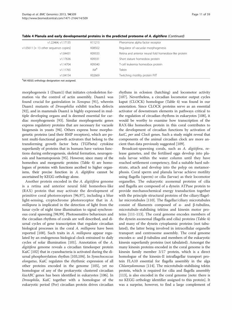
Table 4 Planula and early developmental proteins in the predicted proteome of A. digitifera (Continued)
v1.22464; v1.17135 K11213 Pheromone alpha factor receptor
v1.05611 [+ 13 other sequence copies] K08502 Regulator of vacuolar morphogenesis
v1.04431 K09333 Retina and anterior neural fold homeobox-like protein
v1.17636 K09331 Short stature homeobox protein
v1.14704 K09340 T-cell leukemia homeobox protein
v1.11765 NA1 Tektin
v1.04154 K02669 Twitching motility protein PilT1NA KEGG orthology designation not assigned.
Dunlap et al. BMC Genomics 2013, 14:509 Page 11 of 59http://www.biomedcentral.com/1471-2164/14/509
morphogenesis 1 (Daam1) that initiates cytoskeleton for-mation via the control of actin assembly. Daam1 wasfound crucial for gastrulation in Xenopus [91], whereinDaam1 mutants of Drosophilia exhibit trachea defects[92], and in mammals Daam1 is highly expressed in mul-tiple developing organs and is deemed essential for car-diac morphogenesis [93]. Similar morphogenetic genesexpress regulatory proteins that are necessary for vacuolebiogenesis in yeasts [94]. Others express bone morpho-genetic proteins (and their BMP receptors), which are po-tent multi-functional growth activators that belong to thetransforming growth factor beta (TGFbeta) cytokinesuperfamily of proteins that in humans have various func-tions during embryogenesis, skeletal formation, neurogen-esis and haematopoiesis [95]. However, since many of thehomeobox and morgenetic proteins (Table 4) are homo-logues of proteins with functions ascribed to higher organ-isms, their precise function in A. digitifera cannot beascertained by KEGG orthology alone.Another protein encoded in the A. digitifera genome
is a retina and anterior neural fold homeobox-like(RAX) protein that may activate the development ofprimitive coral photoreceptors [96,97], including a bluelight-sensing, cryptochrome photoreceptor that in A.millepora is implicated in the detection of light from thelunar cycle of night time illumination to signal synchron-ous coral spawning [98,99]. Photosensitive behaviours andthe circadian rhythms of corals are well described, and di-urnal cycles of gene transcription that regulate circadianbiological processes in the coral A. millepora have beenreported [100]. Such traits in A. millepora appear regu-lated by an endogenous biological clock entrained to dailycycles of solar illumination [101]. Annotation of the A.digitifera genome reveals a circadian timekeeper proteinKaiC [102] that in cyanobacteria is activated during the di-urnal phosphorylation rhythm [103,104]. In Synechococcuselongatus, KaiC regulates the rhythmic expression of allother proteins encoded in the genome [105], yet nohomologue of any of the prokaryotic clustered circadiankiaABC genes has been identified in eukaryotes [106]. InDrosophila, KaiC together with a homologue of theeukaryotic period (Per) circadian protein drives circadian
rhythms in eclosion (hatching) and locomotor activity[107]. Nevertheless, a circadian locomotor output cycleskaput (CLOCK) homologue (Table 4) was found in ourannotation. Since CLOCK proteins serve as an essentialactivator of downstream elements in pathways critical tothe regulation of circadian rhythms in eukaryotes [108], itwould be worthy to examine how transcription of theRAX-like homeobox protein in this coral contributes tothe development of circadian functions by activation ofkaiC, per and Clock genes. Such a study might reveal thatcomponents of the animal circadian clock are more an-cient than data previously suggested [109].Broadcast-spawning corals, such as A. digitifera, re-
lease gametes, and the fertilised eggs develop into pla-nula larvae within the water column until they havereached settlement competency, find a suitable hard sub-strate, attach and develop into the polyp on metamor-phosis. Coral sperm and planula larvae achieve motilityusing flagella (sperm) or cilia (larvae) as their locomotororganelles. The eukaryotic axonemal proteins of ciliaand flagella are composed of a dynein ATPase protein toprovide mechanochemical energy transduction togetherwith the principle structural proteins of the ciliary/flagel-lar microtubules [110]. The flagellar/ciliary microtubulesconsist of filaments composed of α- and β-tubulins,microtubule-stabilising tektins and kinesin motor pro-teins [111-113]. The coral genome encodes members ofthe dynein axonemal (flagella and cilia) proteins (Table 4)and many of the dynein cytoplasmic proteins (not tabu-lated), the latter being involved in intracellular organelletransport and centrosome assembly. The coral genomeencodes α- and β-tubulins and members of the eukaryotickinesin superfamily proteins (not tabulated). Amongst themany kinesin proteins encoded in the coral genome is thekinesin family member 3/17 protein, which is a directhomologue of the kinesin-II intraflagellar transport pro-tein FLA10 essential for flagella assembly in the algaChlamydomonas [114]. The microtubule-stabilising tektinprotein, which is required for cilia and flagella assembly[113], is also encoded in the coral genome [note: there isno KEGG orthology identifier assigned to this protein]. Itwas a surprise, however, to find a large complement of

Dunlap et al. BMC Genomics 2013, 14:509 Page 12 of 59http://www.biomedcentral.com/1471-2164/14/509
prokaryotic flagellar proteins encoded in the coral genomeconsisting of archaeal flagellar (FlaC and FlaG), bacterialfilament (FibB, FlliE, FliF, FliJ, FliK, FliO/FliZ, FliQ andFliT) homologue components (Table 4). Included also arethe prokaryote homologues FlgN and FlbB that regulatetranscriptional activation of flagellar assembly [115,116]and FlhB which controls the substrate specificity of the en-tire prokaryotic flagellar apparatus [117]. Encoded in thecoral genome is a flagella-independent Type IV twitchingmobility protein PilT that affords social gliding transloca-tion in many prokaryotic organisms controlled by complexsignal transduction systems that include two-componentsensor regulators [118]. It is unlikely that these genes arederived from contamination from bacterial DNA. Suchcontamination would manifest itself by the random occur-rence of bacterial genes from the whole genome includingmany housekeeping genes. In this case, the genes occur asmembers of groups with specialised functions, suggestingthat multiple horizontal gene transfers between bacteriaand the coral genome have occurred [119]. Their precisefunction in A. digitifera remains unknown; homologues ofthese prokaryotic genes have not been described previ-ously in any other eukaryote genome.Linked closely with flagellar/ciliary proteins are the sen-
sory receptors that signal chemoattraction or avoidance todirect cellular motility. The coral genome reveals a varietyof genes that encode chemoreceptor and chemotaxisproteins (Table 4). The chemoreceptor proteins of A.digitifera include an oxygen-sensing aerotaxis receptorthat in bacteria invokes an avoidance response to anoxicmicro-environments [120]. Encoded also are a nematodesensory chemoreceptor homologue [121], two homo-logous pheromone factor receptor proteins that in fungiactivate a species-specific mating response [122], threechemotaxis protein sensor receptors belonging to themethyl-accepting chemotaxis family of proteins (MCPs) inbacteria and archaea [123], and two proteins (CheZ andCheR) and two regulators (PixG and WspE) of the two-component signal transduction (TCST) system for activa-tion of gene expression. In bacteria and archea, as well assome plants, fungi and protozoa [124], TCST systems me-diate many cellular processes that respond to a broadrange of environmental stimuli via activation of a specifichistidine (or serine) kinase sensor and its cognate responseregulator [125]. There are 77 sequence matches to variouselements of the TCST family of proteins in the A digitiferagenome (data not tabulated). Included also are genes en-coding members of the chemotactic cytokine (chemokine)family of sensory proteins that on secretion directschemotaxis in nearby responsive cells by stimulating targetchemokine receptors; both chemokine and chemokinereceptor proteins are encoded in the coral genome. Sig-nificantly, sensory chemokines/chemokine receptors arefound in all vertebrates, some viruses and some groups of
bacteria, but none have been described previously for in-vertebrates [126].
Neural messengers, receptors and sensory proteinsCorals and other cnidarians are the earliest extant groupof organisms to have a primitive nervous system network[127] thought to be evolved from a eumetazoan an-cestor prior to the divergence of Cnidaria and theBilateria [128,129]. Unlike marine sponges (Porifera)that predate synaptic innovation [130], cnidarians possessa homogenous nerve net that, although lacking any formof cephalization, accommodates fundamental neurosen-sory transmission across the nerve net to end in a moto-neural junction to coordinate tentacle movement requiredfor feeding and predator avoidance [131]. The nervoussystems of cnidarians consist of both ectodermal sensorycells and their effector cells and endodermal multipolarganglions capable of neurotransmission [132]. At the func-tional level, synaptic transmission in cnidarians relies onfast neurotransmitters (glutamate, GABA, glycine) andslow neurotransmitters (catecholamine, serotonin, neuro-peptides) for sensory-signal conduction [133]. At theultrastructural level, many cnidarian neurons have multi-functional traits of sensory, neurosecretory and stimula-tory attributes [134]. Significantly, the genome of A.digitifera encodes the expression of a ciliary neurotrophicfactor, which is a polypeptide hormone and nerve growthfactor that promotes neurotransmitter synthesis, neuriteoutgrowth and regeneration [135]. Additionally, the coralgenome encodes nerve growth factor and neurotrophickinase receptors, a survival motor neuron protein, a sur-vival neuron splicing factor, the neural outgrowth proteinneurotrimin, and a neurotrophin growth factor attributedto signalling neuron survival, differentiation and growth(Table 5). Encoded for neuron regulation and developmentare several neuron cation-gated channels, a neuronal gua-nine nucleotide exchange factor, a neurotransmitter Na+
symporter, several neurogenic differentiation proteins, aneuronal PAS domain transcription factor for activation ofneurogenesis, the axon guidance protein neurophilin-2, aneural crest protein of embryonic neural development,neural ELAV-like transcription proteins of neurogenesis, aNotch protein (79 sequence domain matches) and a neu-tralized protein subset of the Notch signalling pathwaythat promotes neuron proliferation in early neurogenicdevelopment. Structural elements of the coral nerve netinclude neurofilament polypeptides and neuronal adhesionproteins.Cnidarians differentiate highly specialised sensory and
mechanoreceptor cells involved in the capture of preyand for defence against predators. Their stinging cells,termed nematocysts or cnidocytes, are stimulated by ad-jacent chemosensory cells. Nematocysts trigger the re-lease of a stinging barb (cnidae tubule) via ultra-fast

Dunlap et al. BMC Genomics 2013, 14:509 Page 13 of 59http://www.biomedcentral.com/1471-2164/14/509
exocytosis on physical contact with ciliary mechano-receptors of the cnidocyte to deliver the discharge of itsvenom [136]. Despite considerable advances in the sen-sory biology of cnidarians, knowledge of the specificreceptor genes that regulate cnidocyte function remainsincomplete. In Hydra, and perhaps other cnidarians, cni-docyte discharge is controlled by an ancient light-activated, opsin-mediated phototransduction pathway[137] that precedes the evolution of cubozoan (box jelly-fish) eyes [138]; cubozoans are the most basal of animalsto have eyes containing a lens and ciliary-type visual cellssimilar to that of vertebrate eyes [139]. These G-coupledopsin photoreceptors of the retinylidene-forming proteinfamily encoded in the genome of A. digitifera includerhodopsin, bacteriorhodopsin, c-opsin, r-opsin and G0-opsin (Table 5), but not the Gs-subfamily of opsin recep-tors reported to be present in sea anemones, hydra andjellyfish [140], that together with cyclic nucleotide-gated(CNG) ion channel proteins, arrestin (β-adrenergic recep-tor inhibitor) and other retino-protein receptors, are usualcomponents of the bilaterian phototransduction cascade.Present also are genes to express rhodopsin kinase and β-adrenergic receptor kinase which are related members ofthe serine/threonine kinase family of proteins that specif-ically initiate deactivation of G-protein coupled receptors.Additional proteins of retinol metabolism of the photo-transduction pathway encoded in the A. digitifera genomeare retinol dehydrogenase, all-trans-retinol 13,14 reduc-tase and phosphatidylcholine (lichthin)-retinol O-acyltransferase, a neural retina-specific leucine zipper proteinthat is an intrinsic regulator of photoreceptor develop-ment and function, and a retina and anterior neural foldhomeobox-like protein that modulates the expression ofphotoreceptor genes within the rhodopsin promoter.The genome of A. digitifera encodes also a blue light-sensing, cryptochrome photoreceptor thought to signalsynchronous coral spawning by detecting illuminationfrom the lunar cycle [98,99].The A. digitifera genome reveals genes to express a broad
array of neurotransmitter receptor proteins (Table 5), in-cluding glycine and glutamate neuroreceptors, adrenergicreceptors that target non-dopamine catecholamines (i.e.,epinephrine and norepinephrine), dopamine, muscarinicand nicotinic acetylcholine receptors, sensory G protein-coupled receptors and γ-aminobutyric acid (GABA)ligand-gated ion channel and G protein-coupled recep-tors (and inhibitors), several of which are encoded inhigh copy numbers. Cellular trafficking of neurotrans-mitters to presynaptic terminals is essential for neuro-transmission, and significantly the genome of A. digitiferaencodes a wide range of solute carrier neurotransmittertransporters, including a high affinity choline trans-porter and an acetylcholine-specific protein belongingto the major facilitator superfamily (MFS) of secondary
transporters. Encoded also is dopamine β-monooxyge-nase that catalyses the conversion of dopamine to nor-epinephrine in the catecholamine biosynthetic pathway,which is necessary for cross-activation of adrenergicneuroreceptors [141]. Notably, the A. digitifera genomeencodes acetylcholinesterase that is expressed at neuro-muscular junctions and cholinergic synapses where itsprotease activity serves to terminate synaptic transmission.The primitive nervous networks of cnidarians are
strongly peptidergic with at least 35 neuropeptides iden-tified from different cnidarian classes [142]. Our an-notation of the sequenced A. digitifera genome, however,revealed only the neuropeptide FF-amide neurotransmit-ter, a RF amide related peptide, and its neuropeptide FFand Y receptors (Table 5). Neuropeptides are usuallyexpressed as large precursor proteins which comprisemultiple copies of “immature” neuropeptides. Our an-notation did not readily reveal these precursor neuro-peptide proteins, but we did find enzymes required fortheir processing, for example, a variety of carboxypeptidaseenzymes (not tabulated) that remove propeptide carboxylresidues at basic peptidase sites, and the mature peptideneurotransmitters that are finished by consecutive modifi-cation by peptidylglycine (α-hydroxylating) monooxidase(PHM) and peptidyl α-hydroxyglycine α-amidating lyase(PAL) enzymes, both of which are commonly expressed inmammals as a single bifunctional peptidylglycine monooxygenase (K00504/EC 1.14.17.3) [143]. Our extensive cata-logue of animal-like neural and sensory proteins revealedby genome annotation is testament that essential neuro-biological features were developed in the primitive neuralnetworks of early eumetazoan evolution.
Calcification and Ca2+-signalling proteinsThe massive structures of coral reefs evident today are aconstruction of aggregated calcium carbonate depositedover long geological time by scleractinian corals andother calcifying organisms, yet our understanding of themolecular processes that regulate the biological pro-cesses of coral calcification is limited [144]. Ca2+ transferfrom seawater to the calicoblastic site of coral calcifica-tion occurs by passive diffusion through the gastrovas-cular cavity [145] and by active calcium transport [146].Active entry of Ca2+ through the oral epithelial layer isregulated by voltage-dependent calcium channels, suchas demonstrated by the L-type alpha protein clonedfrom the reef-building coral Stylophora pistillata [147].Ca2+ transport across the calioblastic ectoderm to theextracellular calcifying site is facilitated by the plasma-membrane ATP-dependent calcium pump that in S.pistillata resemble the Ca2+-ATPase family of mammalianproteins [148]. By 2H+/Ca2+-exchange at the calioblasticmembrane, Ca2+-ATPase removes H+ (from the net re-action Ca2+ + CO2 + H2O ⇒ CaCO3 + 2H+) thereby

Table 5 Neuronal and sensory proteins in the predicted proteome of A. digitifera
Gene sequence KEGG Orthology Encoded protein description
v1.01918 [+ 5 other sequence copies] K01049 Acetylcholinesterase
v1.18087; v1.14516 K04136 Adrenergic receptor alpha-1B
v1.06394 K04137 Adrenergic receptor alpha-1D
v1.09628; v1.15688; v1.00966 K04140 Adrenergic receptor alpha-2C
v1.19831; v1.20450 K04142 Adrenergic receptor beta-2
v.17293 K00910 beta-Adrenergic-receptor kinase
v1.13740 [+ 5 other sequence copies] K04828 Amiloride-sensitive cation channel 1, neuronal (degenerin)
v1.23541 [+ 6 other sequence copies] K04829 Amiloride-sensitive cation channel 2, neuronal
v1.09323 [+ 4 other sequence copies] K04439 beta-Arrestin
v1.07723; v1.22465 K04641 Bacteriorhodopsin
v1.08062 K05420 Ciliary neurotrophic factor
v1.03288 [+ 5 other sequence copies] K02295 Cryptochrome
v1.20011; v1.20036; v1.20084; v1.18607 K04948 Cyclic nucleotide gated channel alpha 1
v1.21470 K04951 Cyclic nucleotide gated channel alpha 4
v1.21783; v1.01466; v1.01466; v1.01466 K05326 Cyclic nucleotide gated channel, invertebrate
v1.03645 K05391 Cyclic nucleotide gated channel, other eukaryote
v1.21256 K08762 Diazepam-binding inhibitor (GABA receptor, acyl-CoA-binding protein)
v1.22156 [+ 6 other sequence copies] K00503 Dopamine beta-monooxygenase
v1.21775: v1.15989 K04148 Dopamine D1-like receptor
v1.14160; v1.01697 K04144 Dopamine receptor D1
v1.05089; v1.20018 K04145 Dopamine receptor D2
v1.14030; v1.23273 K04146 Dopamine receptor D3
v1.20536 K13088 ELAV-like protein 1
v1.18658 [+ 5 other sequence copies] K13208 ELAV-like protein 2/3/4
v1.05774 [+ 18 other sequence copies] K04313 G protein-coupled receptor 6
v1.00572; v1.18152 K08404 G protein-coupled receptor 17
v1.23842 K04316 G protein-coupled receptor 19
v1.03948 K08411 G protein-coupled receptor 26
v1.09271 K08383 G protein-coupled receptor 34
v1.05595 K04243 G protein-coupled receptor 37 (endothelin receptor type B-like)
v1.04019 K08409 G protein-coupled receptor 45
v1.19913; v1.09821; v1.04291 K08450 G protein-coupled receptor 56
v1.05404 K04321 G protein-coupled receptor 63
v1.02179; v1.10397 K08451 G protein-coupled receptor 64
v1.23269 [+ 5 other sequence copies] K08408 G protein-coupled receptor 68
v1.21091 K08421 G protein-coupled receptor 84
v1.11008 K04302 G protein-coupled receptor 85
v1.21884; v1.01951 K08452 G protein-coupled receptor 97
v1.03243 [+ 13 other sequence copies] K08378 G protein-coupled receptor 103
v1.13790; v1.18939 K08453 G protein-coupled receptor 110
v1.09442; v1.14019 K08455 G protein-coupled receptor 112
v1.24009 K08456 G protein-coupled receptor 113
v1.04290 K08459 G protein-coupled receptor 114
Dunlap et al. BMC Genomics 2013, 14:509 Page 14 of 59http://www.biomedcentral.com/1471-2164/14/509

Table 5 Neuronal and sensory proteins in the predicted proteome of A. digitifera (Continued)
v1.06608; v1.24223 K08457 G protein-coupled receptor 115
v1.10800 [+ 6 other sequence copies] K08458 G protein-coupled receptor 116
v1.07662 [+ 6 other sequence copies] K08462 G protein-coupled receptor 125
v1.09663; v1.08981 K08463 G protein-coupled receptor 126
v1.24252 K08464 G protein-coupled receptor 128
v1.02750 [+ 26 other sequence copies] K08465 G protein-coupled receptor 133
v1.05774 [+ 11 other sequence copies] K08466 G protein-coupled receptor 144
v1.05497; v1.13272; v1.01323 K08436 G protein-coupled receptor 152
v1.08653 [+ 5 other sequence copies] K08467 G protein-coupled receptor 157
v1.11807; v1.10392; v1.10394 K08469 G protein-coupled receptor 158
v1.07294; v1.00247 K08439 G protein-coupled receptor 161
v1.05167 K08442 G protein-coupled receptor 176
v1.08677; v1.23465; v1.19865; v1.06986 K12762 G protein-coupled receptor GPR1
v1.13395 K08291 G protein-coupled receptor kinase
v1.18529; v1.07599; v1.05558 K12487 G protein-coupled receptor kinase interactor 2
v1.02481 K04619 G protein-coupled receptor family C group 5 member B
v1.22242 K04622 G protein-coupled receptor family C group 6 member A
v1.08625; v1.13650; v1.13048; v1.18694 K04599 G protein-coupled receptor Mth (Methuselah protein)
v1.07465; v1.10540 K08341 GABA(A) receptor-associated protein (autophagy-related protein 8)
v1.09831 [+ 30 other sequence copies] K05270 Gamma-aminobutyric acid (GABA) receptor, invertebrate
v1.18702; v1.11701 K05183 Gamma-aminobutyric acid (GABA) A receptor beta-3
v1.04252 [+ 6 other sequence copies] K05185 Gamma-aminobutyric acid (GABA) A receptor epsilon
v1.06325 K05186 Gamma-aminobutyric acid (GABA) A receptor gamma-1
v1.00048 K05188 Gamma-aminobutyric acid (GABA) A receptor gamma-3
v1.07506 [+ 6 other sequence copies] K04615 Gamma-aminobutyric acid (GABA) B receptor 1
v1.07506 [+ 24 other sequence copies] K04616 Gamma-aminobutyric acid (GABA) B receptor 2
v1.06426; v1.10563; v1.01138 K05192 Gamma-aminobutyric acid (GABA) receptor theta
v1.15485 K05198 Glutamate receptor, ionotropic, AMPA 2
v1.09807 K05200 Glutamate receptor, ionotropic, AMPA 4
v1.04764 K05207 Glutamate receptor, ionotropic, delta 2
v1.15247 [+ 12 other sequence copies] K05313 Glutamate receptor, ionotropic, invertebrate
v1.15247 [+ 7 other sequence copies] K05202 Glutamate receptor, ionotropic, kainate 2
v1.00617 K05203 Glutamate receptor, ionotropic, kainate 3
v1.09688 [+ 6 other sequence copies] K05208 Glutamate receptor, ionotropic, N-methyl D-aspartate 1
v1.21204 [+ 4 other sequence copies] K05212 Glutamate receptor, ionotropic, N-methyl-D-aspartate 2D
v1.01622 K05214 Glutamate receptor, ionotropic, N-methyl-D-aspartate 3B
v1.01418 [+ 5 other sequence copies] K05387 Glutamate receptor, ionotropic, other eukaryote
v1.04275 K05194 Glycine receptor alpha-2
v1.10737; v1.06885 K05195 Glycine receptor alpha-3
v1.05488 K05271 Glycine receptor alpha-4
v1.08900; v1.06885 K05196 Glycine receptor beta
v1.18634 K05397 Glycine receptor, invertebrate
v1.14569; v1.14570 K09071 Heart-and neural crest derivatives-expressed protein
v1.16783 [+ 4 other sequence copies] K02168 High-affinity choline transport protein
v1.13837 K07608 Internexin neuronal intermediate filament protein, alpha
Dunlap et al. BMC Genomics 2013, 14:509 Page 15 of 59http://www.biomedcentral.com/1471-2164/14/509
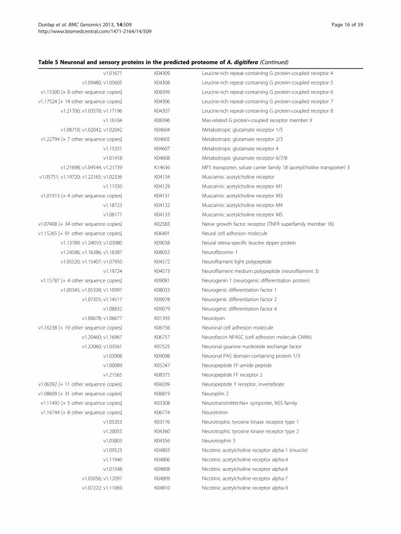
Table 5 Neuronal and sensory proteins in the predicted proteome of A. digitifera (Continued)
v1.01671 K04309 Leucine-rich repeat-containing G protein-coupled receptor 4
v1.09480; v1.05605 K04308 Leucine-rich repeat-containing G protein-coupled receptor 5
v1.15300 [+ 8 other sequence copies] K08399 Leucine-rich repeat-containing G protein-coupled receptor 6
v1.17524 [+ 14 other sequence copies] K04306 Leucine-rich repeat-containing G protein-coupled receptor 7
v1.21700; v1.03578; v1.17196 K04307 Leucine-rich repeat-containing G protein-coupled receptor 8
v1.16104 K08396 Mas-related G protein-coupled receptor member X
v1.08718; v1.02042; v1.02042 K04604 Metabotropic glutamate receptor 1/5
v1.22794 [+ 7 other sequence copies] K04605 Metabotropic glutamate receptor 2/3
v1.15331 K04607 Metabotropic glutamate receptor 4
v1.01418 K04608 Metabotropic glutamate receptor 6/7/8
v1.21698; v1.04544; v1.21739 K14636 MFS transporter, solute carrier family 18 (acetylcholine transporter) 3
v1.05751; v1.19720; v1.22165; v1.02336 K04134 Muscarinic acetylcholine receptor
v1.11550 K04129 Muscarinic acetylcholine receptor M1
v1.01913 [+ 4 other sequence copies] K04131 Muscarinic acetylcholine receptor M3
v1.18723 K04132 Muscarinic acetylcholine receptor M4
v1.08171 K04133 Muscarinic acetylcholine receptor M5
v1.07408 [+ 34 other sequence copies] K02583 Nerve growth factor receptor (TNFR superfamily member 16)
v1.15265 [+ 91 other sequence copies] K06491 Neural cell adhesion molecule
v1.13789; v1.24010; v1.03980 K09038 Neural retina-specific leucine zipper protein
v1.24586; v1.16386; v1.16387 K08052 Neurofibromin 1
v1.05520; v1.15407; v1.07950 K04572 Neurofilament light polypeptide
v1.19724 K04573 Neurofilament medium polypeptide (neurofilament 3)
v1.15787 [+ 4 other sequence copies] K09081 Neurogenin 1 (neurogenic differentiation protein)
v1.00345; v1.05338; v1.10997 K08033 Neurogenic differentiation factor 1
v1.07355; v1.14517 K09078 Neurogenic differentiation factor 2
v1.08832 K09079 Neurogenic differentiation factor 4
v1.06678; v1.06677 K01393 Neurolysin
v1.16238 [+ 19 other sequence copies] K06756 Neuronal cell adhesion molecule
v1.20460; v1.16967 K06757 Neurofascin NFASC (cell adhesion molecule CAMs)
v1.22060; v1.03561 K07525 Neuronal guanine nucleotide exchange factor
v1.03908 K09098 Neuronal PAS domain-containing protein 1/3
v1.00089 K05247 Neuropeptide FF-amide peptide
v1.21565 K08375 Neuropeptide FF receptor 2
v1.06392 [+ 11 other sequence copies] K04209 Neuropeptide Y receptor, invertebrate
v1.08609 [+ 31 other sequence copies] K06819 Neuropilin 2
v1.11492 [+ 5 other sequence copies] K03308 Neurotransmitter:Na+ symporter, NSS family
v1.16744 [+ 8 other sequence copies] K06774 Neurotrimin
v1.05353 K03176 Neurotrophic tyrosine kinase receptor type 1
v1.20055 K04360 Neurotrophic tyrosine kinase receptor type 2
v1.03803 K04356 Neurotrophin 3
v1.09523 K04803 Nicotinic acetylcholine receptor alpha-1 (muscle)
v1.11940 K04806 Nicotinic acetylcholine receptor alpha-4
v1.01548 K04808 Nicotinic acetylcholine receptor alpha-6
v1.05056; v1.12097 K04809 Nicotinic acetylcholine receptor alpha-7
v1.07222; v1.11069 K04810 Nicotinic acetylcholine receptor alpha-9
Dunlap et al. BMC Genomics 2013, 14:509 Page 16 of 59http://www.biomedcentral.com/1471-2164/14/509

Table 5 Neuronal and sensory proteins in the predicted proteome of A. digitifera (Continued)
v1.18231 [+ 32 other sequence copies] K05312 Nicotinic acetylcholine receptor, invertebrate
v1.24404 K04813 Nicotinic acetylcholine receptor beta-2 (neuronal)
v1.06514; v1.23640 K04815 Nicotinic acetylcholine receptor beta-4
v1.18634 K04816 Nicotinic acetylcholine receptor delta
v1.18231 [+ 32 other sequence copies] K05312 Nicotinic acetylcholine receptor, invertebrate
v1.05293 [+ 78 other sequence copies] K02599 Notch protein
v1.15348 [+ 4 other sequence copies] K04256 c-Opsin protein
v1.01972 K08385 G0-Opsin protein
v1.13345 [+ 5 other sequence copies] K04255 r-Opsin protein
v1.00749; v1.03435 K00504 Peptidylglycine monooxygenase
v1.12323 [+ 11 other sequence copies] K00678 Phosphatidylcholine-retinol O-acyltransferase
v1.18340 [+ 6 other sequence copies] K09624 Protease, serine, 12 (neurotrypsin, motopsin)
v1.08030 [+ 9 other sequence copies] K01931 Protein neuralized
v1.04431 K09333 Retina and anterior neural fold homeobox-like protein
v1.01789; v1.06542 K00061 Retinol dehydrogenase
v1.05804 [+ 6 other sequence copies] K11150 Retinol dehydrogenase 8
v1.22340; v1.14029 K11151 Retinol dehydrogenase 10
v1.24399; v1.07017 K11154 Retinol dehydrogenase 16
v1.19667; v1.16885; v1.24371 K00909 Rhodopsin kinase
v1.12432; v1.15302; v1.07505 K09516 all-trans-Retinol 13,14-reductase
v1.09104 [+ 6 other sequence copies] K05613 Solute carrier family 1 (glial high affinity glutamate transporter), member 2
v1.19779; v1.08769; v1.22032 K05617 Solute carrier family 1 (high affinity Asp/glutamate transporter), member 6
v1.19293; v1.19292 K14387 Solute carrier family 5 (high affinity choline transporter), member 7
v1.10901; v1.19493 K05336 Solute carrier family 6 (neurotransmitter transporter), invertebrate
v1.24615 [+ 10 other sequence copies] K05034 Solute carrier family 6 (neurotransmitter transporter, GABA) member 1
v1.07932 K05046 Solute carrier family 6 (neurotransmitter transporter, GABA) member 13
v1.01817 K05036 Solute carrier family 6 (neurotransmitter transporter, dopamine) member 3
v1.20691; v1.16333; v1.15484; v1.02123 K05038 Solute carrier family 6 (neurotransmitter transporter, glycine) member 5
v1.15484; v1.15484 K05042 Solute carrier family 6 (neurotransmitter transporter, glycine) member 9
v1.18461; v1.09068; v1.02237; v1.20880 K05333 Solute carrier family 6 (neurotransmitter transporter) member 18
v1.02239; v1.13836; v1.09067 K05334 Solute carrier family 6 (neurotransmitter transporter) member19
v1.21997 [+ 5 other sequence copies] K12839 Survival of motor neuron-related-splicing factor member 30
v1.21997 [+ 6 other sequence copies] K13129 Survival motor neuron protein
Dunlap et al. BMC Genomics 2013, 14:509 Page 17 of 59http://www.biomedcentral.com/1471-2164/14/509
increasing the saturation state of CaCO3 to sustain cal-cium precipitation [146]. Importantly, located also atthe calicoblastic membrane is carbonic anhydrase [149]which is required to catalyse the intermediate step ofcalcification by the reversible hydration of carbon dio-xide (CO2 + H2O ⇒ HCO3
- + H+). In coral photo-trophic symbiosis, despite numerous studies describingthe well-known phenomenon of light-enhanced calcifica-tion, the relationship linking symbiont photosynthesis tocoral calcification has been elusive [150,151]. Nonetheless,efforts to better understand the calcifying response ofscleractinian corals to environmental change and oceanacidification are gaining traction [149,152,153].
Voltage-gated calcium channels (VGCCs) have beenexamined extensively in mammalian physiology for con-verting membrane potential into intracellular Ca2+ tran-sients for signalling transduction pathways (reviewed in[154]). VGCC signalling affects cellular processes to in-clude muscle contraction, neuronal excitation, genetranscription, fertilisation, cell differentiation and devel-opment, proliferation, hormone release, activation ofcalcium-dependent protein kinases, cell death via ne-crosis and apoptosis pathways, phagocytosis and endo/exocytosis. Remarkably, annotation of the genome of A.digitifera reveals sequences encoding homologues of allthe VGCC (α, αδ, β, and γ) subunits of the molecular (L,

Dunlap et al. BMC Genomics 2013, 14:509 Page 18 of 59http://www.biomedcentral.com/1471-2164/14/509
N, P/Q and R) phenotypes expressed in mammalianphysiology (Table 6). There are multiple sequencesencoding three variants of Ca2+-transporting ATPase, ofwhich at least one is necessary for coral calcification.There is only one sequence match for expressing carbonicanhydrase in the genome of A. digitifera, which mayreflect the high catalytic efficiency of this calcifying en-zyme [155], although a BLAST search of ZoophyteBasedoes reveal scaffolds with low e-values which on futureexperimental inspection might uncover multiple copies ofthis enzyme essential for calcification. There are multiplesequences that express solute carrier Na+/Ca2+- and Na+/K+/Ca2+-exchange families of transport proteins that withexpression of the coral Ca2+/H+-antiporter may regulatecellular pH and Ca2+ homeostasis.Implicit to coral calcification is Ca2+ regulation that
affects signalling of other vital cellular functions. CellularCa2+ is mediated by the calcium-sensing receptorcalmodulin (18 sequence matches) and other messengercalcium-binding effectors (Table 6), including thecalcium-binding protein CML (40 protein domain se-quence matches). Calcium/calmodulin-protein kinase pro-teins are arguably key to Ca2+-signalling in coral symbiosisbut, with the exception of activation of sperm flagellar mo-tility [156], their precise role has not been elaborated.
Plant-derived proteinsEndosymbiosis has contributed greatly to eukaryoticevolution, most notably to the genesis of plastids andmitochondria derived from prokaryotic antecedents.Genetic integration by endosymbiont-to-host transfer(EGT) or replacement (EGR) has been a significant forcein early metazoan innovation, whereby nuclear trans-ferred genes may even adopt novel functions in the hostcell or replace existing versions of the protein that theyencode [157]. Prokaryote-to-eukaryotic gene transfer hasbeen widespread in evolution, but examples of genetic ex-change between unrelated eukaryotes, such as betweenalgal symbionts and their multicellular eukaryote host, areconsidered rare (reviewed by [158,159]). One such ex-ample is aroB (3-dehydroquinate synthase) transferred tothe genome of the sea anemone N. vectensis, which se-quence best fits that of the dinoflagellate Oxyrrhis marina[119]. Close inspection of the amino acid sequence of thearoB gene product, as reported by Shinzato et al. [45],clearly shows this protein to be 2-epi-5-epi-valiolone syn-thase (EVS), a sugar phosphate cyclase orthologue that ca-talyses the conversion of sedoheptulose 7-phosphate to 2-epi-5-epi-valiolone found to be a precursor of themycosporine-like amino acid (MAA) sunscreen shinorinein the cyanobacterium Anabaena variabilis [160]. Add-itionally, the EVS gene of N. vectensis has a distinctive O-methytransferase fusion that is identical in O. marina[161]. The shikimate pathway is essential to apicomplexan
parasites of the genera Plasmodium, Toxoplasma andCryptosporidium and of Tetrahymena ciliates to express apentafunctional aroM gene similar to that of Ascomycetes,which is thought to have been conveyed by fungal genetransfer to a common ancestral progenitor [162]. In a sep-arate example, H. viridis expresses a plant-like ascorbateperoxidase gene (HvAPX1) during oogenesis in both sym-biotic and aposymbiotic individuals [163], whereby perox-idase activity is coincident with oogenesis and embryogenesis that in Hydra acts as a ROS scavenger to protectthe oocyte from apoptotic degradation [164]. The sacog-lossan (sea slug) molluscs Elysia chlorotica and E.viridis (Plakobranchidae) acquire plastids on ingestionof the siphonaceous alga Voucherea litorea (termed“kleptoplasty”) and, by maintaining sequestered plas-tids in an active photosynthetic state, has emerged as amodel organism for the transfer of nuclear-encodedplant genes from algal symbiont to its animal host[165]. In this symbiosis, the family of light-harvestinggenes psbO, prk (phosphoribokinase) and chlorophyll syn-thase (chlG) are entrained in the genome of Elysiachlorotica (reviewed in [166,167]), although there is debatewhether these genes are transcriptionally expressed (com-pare [168] and [169]). Also, phylogenomic analysis of thepredicted proteins of the aposymbiotic unicellular choano-flagellate Monosiga brevicollis, considered to be a stemprogenitor of the animal kingdom [170,171], reveals 103genes having strong algal affiliations arising from multiplephototrophic donors [172]. Such notable examples illus-trate the transfer of algal genes to animal recipients.KEGG orthology-based annotation of the predicted
proteome of A. digitifera reveals a plethora of sequencespresumed to be of algal origin (Table 7). Like E.chlorotica, the coral genome has encoded the photo-system II (PSII) protein PsbO of the oxygen-evolvingcomplex of photosynthesis, as well as the PSII light-harvesting complex protein PsbL that is important inprotecting PSII from photo-inactivation [173]. Encodedalso are the photosystem I subunit proteins PsaI andPsaO. Additionally encoded are the photosystem P840reaction center cytochrome c551 (PscC) protein and thephotosynthetic reaction center M subunit protein, thelight-harvesting proteins complex 1 alpha (PufA), the com-plex II chlorophyll a/b binding protein 6 (LHCB6), thecyanobacterial phycobilisome proteins AcpF and AcpG,the phycocyanin-associated antenna protein CpcD, thephycocyanobilin lyase protein CpcF and the phycoerythrin-associated linker protein CpeS. Like E. chlorotica, the coralgenome encodes chlorophyll synthase (ChlG), a chlorophylltransporter protein PucC, a light-independent nitrogenase-like protochlorophyllide reductase enzyme that is sensitiveto oxygen [174] and a red chlorophyll reductase essentialto the detoxification of photodynamic chlorophyll catabo-lites arising from plant/algal senescence [175]. Three

Table 6 Calcification and Ca2+-signalling proteins in the predicted proteome of A. digitifera
Gene sequence KEGG Orthology Encoded protein description
v1.06452; v1.06451; v1.24424; v1.16923 K07300 Ca2+:H+ antiporter
v1.01669 [+ 9 other sequence copies] K01537 Ca2+−transporting ATPase
v1.22367; v1.22366; v1.22365 K05850 Ca2+ transporting ATPase, plasma membrane
v1.19074 K05853 Ca2+ transporting ATPase, sarcoplasmic/endoplasmic reticulum
v1.22416; v1.22417; v1.15682; v1.00750 K14757 Calbindin D28
v1.24568 [+ 9 other sequence copies] K01672 Carbonic anhydrase
v1.09241 K08272 Calcium binding protein 39
v1.02323 [+ 39 other sequence copies] K13448 Calcium-binding protein CML
v1.05162 [+ 21 other sequence copies] K13412 Calcium-dependent protein kinase
v1.09352 K07359 Calcium/calmodulin-dependent protein kinase kinase
v1.06475; v1.07555;v1.00945; v1.00159; v1.21122 K08794 Calcium/calmodulin-dependent protein kinase I
v1.06475; v1.01061; v1.21150; v1.22443 K04515 Calcium/calmodulin-dependent protein kinase II
v1.00159 K05869 Calcium/calmodulin-dependent protein kinase IV
v1.21927; v1.01218; v1.22226; v1.06623; v1.13703 K06103 Calcium/calmodulin-dependent serine protein kinase
v1.13460 K08284 Calcium channel MID1
v1.20738; v1.01401 K12841 Calcium homeostasis endoplasmic reticulum protein
v1.22794 [+ 11 other sequence copies] K04612 Calcium-sensing receptor
v1.10079 [+ 17 other sequence copies] K02183 Calmodulin
v1.10994 K14734 S100 calcium binding protein G
v1.02488 [+ 14 other sequence copies] K05849 Solute carrier family 8 (sodium/calcium exchanger)
v1.23153 [+ 9 other sequence copies] K13749 Solute carrier family 24 (sodium/potassium/calcium exchanger)
v1.14863 K12304 Soluble calcium-activated nucleotidase 1
v1.18656 [+ 13 other sequence copies] K04858 Voltage-dependent calcium channel alpha-2/delta-1
v1.13222 K04860 Voltage-dependent calcium channel alpha-2/delta-3
v1.08078 [+ 9 other sequence copies] K05315 Voltage-dependent calcium channel alpha 1, invertebrate
v1.03896 [+ 6 other sequence copies] K05316 Voltage-dependent calcium channel alpha-2/delta, invertebrate
v1.04798 K05317 Voltage-dependent calcium channel beta, invertebrate
v1.22788 K04863 Voltage-dependent calcium channel beta-2
v1.09999 K04872 Voltage-dependent calcium channel gamma-7
v1.02505 K04873 Voltage-dependent calcium channel gamma-8
v1.03648[+ 6 other sequence copies] K04850 Voltage-dependent calcium channel L type alpha-1C
v1.03648; v1.17267 K04851 Voltage-dependent calcium channel L type alpha-1D
v1.03648; v1.13219; v1.21895 K04857 Voltage-dependent calcium channel L type alpha-1S
v1.06313; v1.01656; v1.23096 K04344 Voltage-dependent calcium channel P/Q type alpha-1A
v1.08078 [+ 10 other sequence copies] K04849 Voltage-dependent calcium channel N type alpha-1B
v1.07968 K04852 Voltage-dependent calcium channel R type alpha-1E
v1.01364; v1.13467; v1.08705 K04854 Voltage-dependent calcium channel T type alpha-1G
v1.15414; v1.14241; v1.09595 K04855 Voltage-dependent calcium channel T type alpha-1H
Dunlap et al. BMC Genomics 2013, 14:509 Page 19 of 59http://www.biomedcentral.com/1471-2164/14/509
chlorosome proteins of the photosynthetic antenna com-plex of green sulphur bacteria, a bacteriochlorophyllmethyltransferase involved in BChl c biosynthesis [176]and the retinylidene bacteriorhodopsin of phototrophicArchaea are also encoded in the coral genome. Present are
genes encoding subunit 6 of the cytochrome B6f complexthat links PSII and PSI via the plastoquinone pool, to-gether with chloroplast ferredoxin-like NapH and NapGproteins and their 2Fe-2S cluster protein. The coral gen-ome, however, encodes sequences for NAD+-ferredoxin

Table 7 Plant-derived proteins in the predicted proteome of A. digitifera
Gene sequence KEGG Orthology Encoded protein description
v1.14452 K09843 (+)-Abscisic acid 8′-hydroxylase
v1.18868 K14496 Abscisic acid receptor PYR/PYL family (PYL)
v1.21983; v1.05890 K03342 p-Aminobenzoate synthetase / 4-amino-4-deoxychorismate lyase (PabBC)
v1.15436 K02822 Ascorbate-specific IIB component, PTS system (PTS-Ula-EiiB)
v1.11187; v1.13966 K00423 L-Ascorbate oxidase
v1.20081; v1.22465 K13604 Bacteriochlorophyll C20 methyltransferase (BchU)
v1.07723 K04641 Bacteriorhodopsin (BoP)
v1.21858 K04040 Chlorophyll synthase (ChlG)
v1.01742 K08945 Chlorosome envelope protein A (CsmA)
v1.04797; v1.14208 K08946 Chlorosome envelope protein B (CsmB)
v1.18698 K08948 Chlorosome envelope protein D (CamD)
v1.18637 K02642 Cytochrome b6f complex subunit 6 (PetL)
v1.21101; v1.14192; v1.14548 K01735 3-Dehydroquinate synthase (AroB)
v1.05796 K10210 4,4′-Diaponeurosporene oxidase (carotenoid biosynthesis; CrtP)
v1.11730 K04755 Ferredoxin, 2Fe-2S (FdX)
v1.19154; v1.00014 K00532 Ferredoxin hydrogenase
v1.00014 K00534 Ferredoxin hydrogenase small subunit
v1.17698; v1.06031; v1.16647 K02574 Ferredoxin-type protein (NapH)
v1.23058 K02573 Ferredoxin-type protein (NapG)
v1.08414 K08926 Light-harvesting complex 1 alpha chain (PufA)
v1.21458 K08917 Light-harvesting complex II chlorophyll a/b binding protein 6 (LHCB6)
v1.03743 K08226 MFS transporter, BCD family, chlorophyll transporter (PucC)
v1.13030; v1.08678 K13413 Mitogen-activated protein kinase kinase 4/5, plant ((MKK4_5P)
v1.02429; v1.10744; v1.03340 K08929 Photosynthetic reaction center M subunit (PufM)
v1.03631 K02696 Photosystem I subunit VIII (PsaI)
v1.11432 K14332 Photosystem I subunit (PsaO)
v1.17422 K02713 Photosystem II protein (PsbL)
v1.18303 K02716 Photosystem II oxygen-evolving enhancer protein 1 (PsbO)
v1.12300; v1.21136 K08942 Photosystem P840 reaction center cytochrome c551 ((PscC)
v1.00280 K02097 Phycobilisome core component 9 (AcpF)
v1.10967 K02290 Phycobilisome rod-core linker protein (AcpG)
v1.02166 K02287 Phycocyanin-associated, rod protein (CpcD)
v1.19642; v1.07305; v1.19572; v1.01248 K02289 Phycocyanobilin lyase beta subunit (CpcF)
v1.10441 K05382 Phycoerythrin-associated linker protein (CpeS)
v1.13406 K10027 Phytoene dehydrogenase (desaturase; CrtI)
v1.18809; v1.06199 K02291 Phytoene synthase (CrtB)
v1.20411; v1.02037; v1.14064; v1.21095 K09060 Plant G-box-binding factor (GBF)
v1.10035 K00218 Protochlorophyllide reductase [NifEN-like; Por]
v1.21846 K05358 Quinate dehydrogenase (QuiA)
v1.03127 K13545 Red chlorophyll catabolite reductase (ACD2)
v1.05899 K00891 Shikimate kinase (AroK, AroL)
v1.21101; v1.14192; v1.05899 K13829 Shikimate kinase / 3-dehydroquinate synthase (AroKB)
v1.12938 K08500 Syntaxin of plants (SYP6)
Dunlap et al. BMC Genomics 2013, 14:509 Page 20 of 59http://www.biomedcentral.com/1471-2164/14/509

Table 7 Plant-derived proteins in the predicted proteome of A. digitifera (Continued)
v1.06575 K08506 Syntaxin of plants (SYP7)
v1.04929 K09834 Tocopherol cyclase (VTE1, SXD1)
v1.01022 K05928 Tocopherol O-methyltransferase
v1.05457 K09838 Zeaxanthin epoxidase (ZEP, ABA1)
Dunlap et al. BMC Genomics 2013, 14:509 Page 21 of 59http://www.biomedcentral.com/1471-2164/14/509
reductase (HcaD; not tablulated), rather than the requiredNADP+-ferredoxin reductase of photosynthesis. Annotationof the A. digitifera genome revealed genes unexpectedly en-coding ferredoxin hydrogenase [EC:1.12.7.2] and that of itssmall subunit protein (Table 7) involved in light-dependentproduction of molecular hydrogen having its [Fe-Fe]-clustercoupled to the photosynthetic transport chain via a charge-transfer complex with ferredoxin (see [177]).Like N. vectensis and the dinoflagellate Oxyrrhis marina,
the genome of A. digitifera encodes an O-methyltransferasewhich is immediately downstream of EVS, but the twogenes are not fused. Using a ZoophyteBase BlastP search,the O-methyltransferase showed little sequence homologywith the corresponding protein of A. variabilis (e-value of6.972E-2 and Bit score of 34.27), whereas the EVS proteinshared 87% absolute sequence identity to the A. variabilisEVS protein. What role, if any, these two genes play inmycosporine-like amino acid (MAA) biosynthesis in A.digitifera has yet to be determined, although it has been sug-gested from the transcriptome of Acropora microphthalmathat MAA biosynthesis proceeds from a branch point at3-dehydroquinate of the shikimic acid pathway as a sharedmetabolic adaptation between the coral host and its symbi-otic zooxanthellae [40]. The 3-dehydroquinate synthaseenzyme of the shikimic acid pathway, thought to be a keyintermediate in an alternative MAA biosynthetic pathwayin A. variabilis [178], is instead encoded by the fusedaroKB gene of A. digitifera (Table 7). Additional shikimateproteins of the predicted proteome, although not limitedto phototrophs, are shikimate kinase (AroK), quinate de-hydrogenase (QuiA) and the conjoined p-aminobenzoatesynthase and 4-amino-4-deoxychlorismate lysate (PabBC)enzyme necessary for folate biosynthesis [179]. Otherplant-related gene homologues include the phytohormoneabscisic acid receptor protein (PabBC) and its cytochromeP450 monooxygenase abscisic acid 8′-hydroxylase,L-ascorbate oxidase and PTS system degrading enzymes,the unique SYP6 and SYP7 syntaxins of plant vesiculartransport, tocopherol cyclase and a tocopherol O-methyltransferase enzyme that converts γ-tocopherol toα-tocopherol. Essential for carotene biosynthesis arephytoene synthase (CrtB) and phytoene dehydrogenase(CrtI) enzymes. Significantly, encoded within the coralgenome is zeaxanthin epoxidase that is essential forabscisic acid biosynthesis and is a key enzyme in the xan-thophyll cycle of plants and algae to impart oxidativestress tolerance.
Given that viruses often mediate gene transfer processes,it is intriguing that certain bacteriophages of marineSynechococcus and Prochlorococcus cyanobacteria arereported to carry genes encoding the photosynthesis D1(psbA), and D2 (psbD) proteins, a high-light inducibleprotein (HLIP) [180,181] and the photosynthetic elec-tron transport plastocyanin (petE) and ferredoxin (petF)proteins thought to enhance the photosynthetic fitnessof their host [182-184]. Accordingly, it has been sug-gested that the transfer of psbA by viruses associatedwith Symbiodinium could lessen the severity of thermalimpairment to PSII and the response of corals to ther-mal bleaching [185]. It is yet unknown if phages ordinoflagellate-infecting viruses [186], particularly thoseof Symbiodinium [187], may affect gene transfer leadingto complementary (or “shared”) metabolic adaptationsof symbiosis [119,188].
Proteins of nitrogen metabolismIt is well accepted that intracellular Symbiodinium spp.provide reduced carbon for coral heterotrophic metabol-ism by photosynthetic carbon fixation. Because of thismetabolic relationship, light is a critical feature in thebioenergetics of coral symbiosis [189]. The algal photo-synthate translocated to corals, however, is deficient innitrogen at levels necessary to sustain autotrophicgrowth. While corals can assimilate fixed nitrogen fromsurrounding seawater [190], “recycled” nitrogen withinthe symbiosis may account for as much as 90% of thephotosynthetic nitrogen demand [191]. It would not besurprising then that light would have a strong influenceon the uptake and retention of ammonium by symbioticcorals. Consequently, corals excrete excess ammoniumin darkness [192], and in light excretion is induced bytreatment with the photosynthetic electron transport in-hibitor 3-(3,4-diclorophenyl)-1,1-dimethylurea (DCMU)[193]. Since ammonia is the product of nitrogen fixation,these observations suggest that the coral holobiont mayfix nitrogen in the dark, or when photosynthesis is re-pressed, during which coral tissues are hypoxic [194],and nitrogenase activity is not inactivated by molecularoxygen [195].Tropical coral reefs are typically surrounded by low-
nutrient oceanic waters of low productivity but, paradoxic-ally, the waters of coral reefs often have elevated levels ofinorganic nitrogen [196,197] attributed to high rates of ni-trogen fixation. While nitrogen fixation from diazotrophic

Dunlap et al. BMC Genomics 2013, 14:509 Page 22 of 59http://www.biomedcentral.com/1471-2164/14/509
epiphytes of the coral reef substrata and sediments[197,198] and diazotrophic bacterioplankton of the coralreef lagoon [199] provide substantial quantities of fixednitrogen for assimilation by the coral reef, mass-balanceestimates show this input to be less than the community’sannual nitrogen demand [200]. Endolithic nitrogen-fixingbacteria are abundant in the skeleton of living coralswhere they benefit from organic carbon excreted byoverlaying coral tissues to provide a ready source of en-ergy for dinitrogen reduction [201]. Additionally, intra-cellular nitrogen-fixing cyanobacteria are reported tocoexist with dinoflagellate symbionts in the tissues ofMonastraea cavernosa and to functionally express nitro-genase activity [202]. Corals also harbour a diverse as-semblage of heterotrophic microorganisms in their skele-ton, tissues and lipid-rich mucus (reviewed in [203]), andthese communities include large populations of diazo-trophic bacteria [204,205], and archaea [206]. Apart fromnitrogen fixation, the coral microbiota contributes to othernitrogen-cycling processes, such as nitrification, ammoni-fication and denitrification [207,208]. We were surprised tofind several nitrogen fixation and cycling proteins encoded inthe genome of A. digitifera (Table 8), notably a nitrogen fix-ation NifU-like protein, the Nif-specific regulatory protein(NifA), the regulatory NAD(+)-dinitrogen-reductase ADP-D-ribosylastransferase protein, a nitrifying ammonia monooxy-genase enzyme and nitrate reductase, which are usuallyexpressed only by prokaryotic microorganisms.The presence of genes encoding proteins involved in
nitrogen fixation raises speculation that corals may con-tribute directly to, or perhaps co-regulate, certain pro-cesses that catalyse the reduction of dinitrogen (N2) toammonia (NH3) by the enzyme nitrogenase reductase(NifH). The functional NifH enzyme is a binary proteincomposed of a molybdenum-iron (MoFe) protein (NifB/NifDK), or its NifEN homologue, fused with a FeMo-cofactor (FeMoco) protein [209]. While genes encodingNifB, NifDK (or NifEN) and their FeMo-cofactor do notappear in the genome of A. digitifera, a gene encoding theNifEN-like protein protochlorophyllide oxidoreductase(POR) is present (Table 8). POR has all three subunitswith high similarity to the assembled MoFe nitrogenase[210], but this homologue is unlikely to be effective in ni-trogen reduction [211,212] since its activity is lightdependent [213] when tissues are highly oxic [193]. TheNifU protein encoded in the coral genome preassemblesthe metallocatalytic Fe-S clusters for maturation of nitro-genase [214], but its assemblage without NifS, a cysteinedesulfurase needed for [Fe-S] cluster assembly [215],would be incomplete, and its pre-nitrogenase receptor isalso missing. Yet, the coral does have the nifJ gene that en-codes pyruvate:flavodoxin oxidoreductase required forelectron transport in nitrogenase reduction [216]. Theregulatory NifA protein encoded in the coral genome
might activate, on stimulation by the integration host fac-tor (INF), transcription of nitrogen fixation (nif ) operonsof RNA polymerase [217], and both of these proteins areencoded in the coral genome. Additional to this transcrip-tional control, post-translational nitrogenase activity iscontrolled by reversible ADP-ribosylation of a specificarginine residue in the nitrogenase complex [218]. NAD(+)-dinitrogen-reductase ADP-D-ribosyltransferase (DraT)inactivates the nitrogenase complex while ADP-ribosylgly-cohydrolase (DraG) removes the ADP-ribose moiety to re-store nitrogenase activity, and both of these enzymes areencoded in the coral genome. Given that genes encodingessential constituent proteins of nitrogenase assembly ap-pear incomplete, corals are unlikely to fix nitrogen per se,but co-opted elements of the coral genome to regulateprocesses of nitrogen fixation by its diazotrophic consortiais a prospect worthy of exploration [219].Nitrofying/nitrifying bacteria and archaea express the
enzyme ammonia monooxygenase that converts fixedammonia to nitrite (via hydroxylamine) and the enzymenitrite (oxido)reductase completes the oxidation of ni-trite to nitrate, and both of these enzymes are entrainedin the genome of A. digitifera (Table 8). The ammoniamonooxygenase subunit A (amoA) of archaeal consortshas been described in nine species of coral from fourreef locations [220], but the presence of amoA in thecoral genome, together with encoded ammonium trans-port proteins, was not anticipated. Another protein ofprokaryotic origin encoded in the coral genome is ni-trate reductase (periplasmic, assimilatory and respira-tory), the latter being required for anaerobic respirationby bacteria [221], and unlike the nitrate reductase familyof sulphite oxidase enzymes in eukaryotes, the nitrate re-ductases of prokaryotes (K00363) belong to the DMSOreductase family of enzymes. Also encoded in the coralgenome are a nitrite transporter (NirC) and a formate-dependent nitrite reductase (NrfA) required for nitriteammonification [222]. In addition to nitrite reduction,NrfA reduces nitric oxide, hydroxylamine, nitrous oxideand sulphite, the last providing a metabolic link betweennitrogen and sulphur cycling in coral metabolism. Otherenzymes of nitrogen metabolism encoded in the coralgenome are the carbamoyl-phosphate synthase family ofenzymes [223] that catalyses the ATP-dependent syn-thesis of carbamoyl phosphate used for the productionof urea (ornithine cycle) to provide a ready store offixed-N in the urea-nitrogen metabolism of corals [224].Another nitrogen source comes from glutamate de-hydrogenase (GDH) that reversibly converts glutamateto α-ketoglutarate with liberation of ammonia, and asexpected [225], this enzyme is encoded in the coral gen-ome, together with the prokaryotic nitrogen regulatoryprotein PII of glutamine synthase, which in bacteria is acti-vated in response to nitrogen availability. Encoded also is

Table 8 Proteins of nitrogen metabolism in the predicted proteome of A. digitifera
Gene sequence KEGG Orthology Encoded protein description
v1.23444; v1.09133; v1.23443 K05521 ADP-ribosylglycohydrolase (DraG)
v1.09202 K10944 Ammonia monooxygenase subunit A
v1.03645 [+ 8 other sequence copies] K03320 Ammonium transporter, Amt family
v1.12268; v1.12269 K06580 Ammonium transporter Rh
v1.02406 K01954 Carbamoyl-phosphate synthase (CPS)
v1.01524; v1.18283; v1.18284 K01948 Carbamoyl-phosphate synthase (CPS, ammonia)
v1.01615 K04016 Formate-dependent nitrite reductase (NrfA)
v1.16277; v1.23483; v1.13667; v1.22675 K00261 Glutamate dehydrogenase (NAD(P)+)
v1.17166; v1.11089 K01745 Histidine ammonia-lyase
v1.22825; v1.08034;v1.o8520 K05123 Integration host cell factor (INF) subunit beta
v1.11343 K05951 NAD+−dinitrogen-reductase ADP-D-ribosyltransferase (DraT)
v1.00547 K02584 Nif-specific regulatory protein (NifA)
v1.18869 K00371 Nitrate reductase 1, beta subunit
v1.06763 K08346 Nitrate reductase 2, beta subunit
v1.14858; v1.00685; v1.23148 K05916 Nitric oxide dioxygenase
v1.16954 [+ 5 other sequence copies] K02448 Nitric oxide reductase NorD protein
v1.06115 K02164 Nitric oxide reductase NorE protein
v1.17629 [+ 12 other sequence copies] K04748 Nitric oxide reductase NorQ protein
v1.24077 [+ 4 other sequence copies] K13125 Nitric oxide synthase-interacting protein
v1.21801; v1.05719; v1.23577; v1.19464 K13253 Nitric-oxide synthase, invertebrate
v1.05980 K00363 Nitrite reductase (NAD(P)H) small subunit
v1.00101 K02598 Nitrite transporter NirC
v1.02355; v1.18772 K04488 Nitrogen fixation protein NifU
v1.17812 K02589 Nitrogen regulatory protein PII 1
v1.09150 K02570 Periplasmic nitrate reductase NapD
v1.01560 K02571 Periplasmic nitrate reductase NapE
v1.10035 K00218 Protochlorophyllide reductase [NifEN-like]
v1.08939 K03737 Pyruvate-flavodoxin reductase (NifJ)
v1.17373 K00365 Urate oxidase
v1.13217 K01427 Urease
v1.16409 [+ 5 other sequence copies] K03187 Urease accessory protein
v1.13217 K01429 Urease subunit beta
v1.13217 K14048 Urease subunit gamma/beta
v1.12211 [+ 4 other sequence copies] K00106 Xanthine dehydrogenase/oxidase
v1.12212 K13481 Xanthine dehydrogenase small subunit
(Excluding amino acid and pyrimidine/purine nucleotide synthesis or metabolism).
Dunlap et al. BMC Genomics 2013, 14:509 Page 23 of 59http://www.biomedcentral.com/1471-2164/14/509
histidine ammonia-lyase (histidase) that liberates ammonia(and urocanic acid) from cytosolic stores of histidine. It isnow accepted that uric acid deposits accumulated by symbi-otic algae provide a significant store of nitrogen for the coralholobiont [226], so it is noteworthy that the coral genomeencodes urate oxidase (uricase) to catalyse uric acid oxida-tion to allanotoin from which urea and ureidoglycolate areproduced in a reaction catalysed by allantoicase (allantoate
amidinohydrolase), both of which known isoforms arepresent in the coral genome. Encoded in the coral genomeis also urease to catalyse the hydrolysis of urea, presumablyexcreted by its algal symbionts, with the release of carbondioxide and ammonia to meet the nitrogen demand of thecoral holobiont during periods of low nitrogen availability.Similarly, xanthine dehydrogenase (xanthine: NAD+-oxido-reductase) acts by oxidation on a variety of purines,

Dunlap et al. BMC Genomics 2013, 14:509 Page 24 of 59http://www.biomedcentral.com/1471-2164/14/509
including hypoxanthine, to yield urate for the recycling ofnitrogen in coral nutrition. Many of the aforementionedproteins of nitrogen metabolism, including Nif proteins,have been detected in the proteome of an endosymbiont-enriched fraction of the coral S. pistillata [39].Notwithstanding consideration of the rapid diffusion
rate of nitric oxide (NO) or its apparent short biologicalhalf-life [227], there is debate about the provenance ofendogenously produced NO in signalling the bleachingof corals in response to environmental stress. Elevatednitric oxide synthase (NOS) activity and NO productionin algal symbionts has been attributed to the thermalstress response of corals [228,229], whereas the host isascribed to be the major source of NO during exposureto elevated temperature [230,231]. While our annotationmay not resolve this dispute, we show (Table 8) thatnitric oxide synthase enzymes (Nor D, Nor E, Nor Qand an invertebrate NOS protein) are encoded in thegenome of A. digitifera, together with a nitric oxide-interacting protein (NOIP) that in higher animals regu-lates neuronal NOS activity [232]. Nitric oxide is anintermediate of nitrite reduction catalysed by nitrite re-ductase (NIR), which by further reduction produces am-monia. The coral genome also encodes nitric oxidedioxygenase (NOD) that converts nitric oxide to nitrate.Accordingly, enhanced expression of NIR (NO reduc-tion) or NOD (NO oxidation) could ameliorate the NO-signalling response of coral bleaching presumed acti-vated by environmental stress.
DNA repairCellular DNA is prone to damage caused by the productsof normal metabolism and by exogenous agents. Damageto DNA from metabolic processes include the oxidation ofnucleobases and strand interruptions by the production ofreactive oxygen species (ROS), from alkylation of nucleo-tide bases, from the hydrolysis of bases causing dea-mination, depurination and depyrimidination, and fromthe mismatch of base pairs from errors in DNA replica-tion. Damage affected by external agents include exposureto UV light causing pyrimidine dimerization and freeradical-induced damage, exposure to ionising radiationcausing DNA strand breaks, thermal disruption causinghydrolytic depurination and single-strand breaks, and byxenobiotic contamination to cause DNA adduct forma-tion, nucleobase oxidation and DNA crosslinking. Most ofthese lesions affect structural changes to DNA that alteror prevent replication and gene transcription at the site ofDNA damage. Thus, recognition and repair of DNA ab-normalities are vital processes essential to maintain thegenetic integrity of the coral genome. Since there are mul-tiple pathways causing DNA damage at diverse molecularsites, there are likewise diverse and overlapping processesavailable to repair cellular DNA damage. Of the many
nuclear repair processes, photoreactivation (photolyase),base excision repair and nucleotide excision repair are themain elements for the repair of cellular DNA damage.Exposure to sunlight is an absolute requirement for
phototrophic symbiosis, but excessive exposure of coralsto solar ultraviolet radiation can inflict direct damage toDNA by pyrimidine dimerization and 6-4 photoadductformation and cause indirect damage by the productionof ROS to initiate free-radical damage. While there havebeen abundant studies on the sensitivity of corals tosolar ultraviolet radiation, only a few have examined theeffects of solar UV to cause DNA damage. Photoreactiva-tion has been shown to be an important repair pathway forreversing UV-activated DNA damage in adult coral [233]and coral planulae [234]. UV damage to DNA was firstdemonstrated by the detection of unrepaired cyclobutanepyrimidine dimers (CPDs) in the host tissues and algalsymbionts of the coral Porities porites, in which CPDs hadincreased in a UV dose-dependent manner [235], whereasCPDs and 6-4 pyrimidine-pyrimidone photoadducts in thecoral Montipora verrucosa holobiont were correlated in-versely with levels of coral “sunscreen” protection [236].The effects of solar UV radiation causing DNA lesions incoral have been determined by use of the comet assay[237], and UV-induced DNA damage and repair has beenexamined in the symbiotic anemone Aiptasia pallida[238]. The comet assay showed also that DNA lesions incoral planulae had increased on acquiring algal symbionts,presumably from greater ROS production resulting as aby-product of photosynthesis [239]. Iron-induced oxidativestress was found likewise to enhance DNA damage in thecoral Pocillopora damicornis as determined by the oc-currence of DNA apurinic/apyrimidinic sites caused byhydrolytic lesions [240]. Significantly, DNA damage in thehost and algal symbionts of the coral Montastraea faveo-lata was found to occur simultaneously during thermal“bleaching” stress, and DNA damage is further enhancedon exposure to greater irradiances of solar radiation [241].Nevertheless, despite the serious risk of unrepaired DNAdamage to coral survival, the DNA repair processes ofcorals to mitigate the detrimental effects of environmentalstress have not been adequately characterised at the tran-scriptome level of expression [29,242].Our annotation of the sequenced genome of A. digitifera
has revealed genes encoding a large repertoire of DNArepairing enzymes and their adaptor proteins (Table 9).Given strong evidence for DNA photoreactivation incorals having been reported [233,234], it was surprising tofind only one gene in single copy that encodes a solephotolyase enzyme for reversing pyrimidine dimer and 6-4 photoadduct formation. Notably, we found genes encod-ing 6 members of the ERCC family of nucleotide excisionrepair enzymes, together with the UV excision repair pro-tein RAD23, for the repair of UV-induced DNA damage.

Table 9 DNA repair proteins in the predicted proteome of A. digitifera
Gene sequence KEGG Orthology Encoded protein description
v1.02961; v1.13402 K03575 A/G-specific adenine glycosylase (MutY)
v1.11766 K03919 Alkylated DNA repair protein
v1.04821 K10765 Alkylated DNA repair protein alkB homologue 1
v1.02479 K10766 Alkylated DNA repair protein alkB homologue 4
v1.20302 K10767 Alkylated DNA repair protein alkB homologue 5
v1.24450 K10768 Alkylated DNA repair protein alkB homologue 6
v1.02766; v1.09413 K10770 Alkylated DNA repair protein alkB homologue 8
v1.01590 [+ 4 other sequence copies] K10884 ATP-dependent DNA helicase 2 subunit 1
v1.18810; v1.03166; v1.08449 K10885 ATP-dependent DNA helicase 2 subunit 2
v1.08013 K03722 ATP-dependent DNA helicase DinG
v1.03542 K14635 ATP-dependent DNA helicase MPH1
v1.06737 [+ 5 other sequence copies] K15255 ATP-dependent DNA helicase PIF1
v1.17360; v1.21235 K10899 ATP-dependent DNA helicase Q1
v1.01081 [+ 8 other sequence copies] K10730 ATP-dependent DNA helicase Q4
v1.16859 K10902 ATP-dependent DNA helicase Q5
v1.11661 [+ 19 other sequence copies] K03654 ATP-dependent DNA helicase RecQ
v1.20397 K03656 ATP-dependent DNA helicase Rep
v1.18049; v1.07731; v1.05830 K10905 ATR interacting protein
v1.01679 K01669 Deoxyribodipyrimidine photo-lyase
v1.03410; v1.12968; v1.00865; v1.16876 K10887 DNA cross-link repair 1C protein
v1.07474; v1.07473; v1.01809 K10610 DNA damage-binding protein 1
v1.13116; v1.03378; v1.16328 K10140 DNA damage-binding protein 2
v1.17099 [+ 5 other sequence copies] K11885 DNA damage-inducible protein 1
v1.05469 K06663 DNA damage checkpoint protein
v1.02859; v1.14719; v1.21030; v1.10920 K04452 DNA damage-inducible transcript 3
v1.02191 K10844 DNA excision repair protein ERCC-2
v1.19108 [+ 5 other sequence copies] K10843 DNA excision repair protein ERCC-3
v1.22267 [+ 4 other sequence copies] K10848 DNA excision repair protein ERCC-4
v1.15137 [+ 5 other sequence copies] K10846 DNA excision repair protein ERCC-5
v1.18550; v1.02606; v1.14935; v1.08831 K10841 DNA excision repair protein ERCC-6
v1.20045; v1.01844; v1.11724; v1.03203 K10570 DNA excision repair protein ERCC-8
v1.15430; v1.03058 K03658 DNA helicase IV
v1.00228 [+ 4 other sequence copies] K11665 DNA helicase INO80
v1.00136; v1.0678; v1.21529 K10776 DNA ligase 3
v1.23293; v1.19418; v1.23430; v1.15721 K10777 DNA ligase 4
v1.19248 K07458 DNA mismatch endonuclease, patch repair protein
v1.19011 K08739 DNA mismatch repair protein MLH3
v1.11513; v1.11449 K08735 DNA mismatch repair protein MSH2
v1.14781 K08736 DNA mismatch repair protein MSH3
v1.05696; v1.22444; v1.19162 K08740 DNA mismatch repair protein MSH4
v1.04904 K08741 DNA mismatch repair protein MSH5
v1.15360; v1.19426; v1.08585 K08737 DNA mismatch repair protein MSH6
v1.02429 [+ 8 other sequence copies] K03572 DNA mismatch repair protein MutL
Dunlap et al. BMC Genomics 2013, 14:509 Page 25 of 59http://www.biomedcentral.com/1471-2164/14/509

Table 9 DNA repair proteins in the predicted proteome of A. digitifera (Continued)
v1.03990 K03555 DNA mismatch repair protein MutS
v1.14015 K07456 DNA mismatch repair protein MutS2
v1.08443 K10864 DNA mismatch repair protein PMS1
v1.15229 K10858 DNA mismatch repair protein PMS2
v1.08658; v1.14152; v1.01681 K15082 DNA repair protein RAD7
v1.16407 [+ 27 other sequence copies] K10866 DNA repair protein RAD50
v1.22193 K04482 DNA repair protein RAD51
v1.02646; v1.22076 K10958 DNA repair protein RAD57
v1.15671 [+ 4 other sequence copies] K04483 DNA repair protein RadA
v1.16193; v1.19033 K04485 DNA repair protein RadA/Sms
v1.16079; v1.07685 K04484 DNA repair protein RadB
v1.21363; v1.22360; v1.02900 K03584 DNA repair protein RecO (recombination protein O)
v1.18390 K03515 DNA repair protein REV1
v1.04705 K10991 DNA repair protein Swi5/Sae3
v1.13920; v1.03800; v1.16133 K10803 DNA repair protein XRCC1
v1.15052 K10879 DNA repair protein XRCC2
v1.09315 [+ 4 other sequence copies] K10886 DNA repair protein XRCC4
v1.02733; v1.24592 K10868 DNA repair protein XRS2
v1.14551; v1.23176 K10873 DNA repair and recombination protein RAD52
v1.20503 [+ 4 other sequence copies] K10875 DNA repair and recombination protein RAD54
v1.23173; v1.16050 K10877 DNA repair and recombination protein RAD54B
v1.07227; v1.08907; v1.09439; v1.02644 K10847 DNA repair protein complementing XP-A cells
v1.11534 [+ 5 other sequence copies] K10865 Double-strand break repair protein MRE11
v1.07939 K03660 N-glycosylase/DNA lyase
v1.16163 K03652 3-Methyladenine DNA glycosylase
v1.07231 K10726 Replicative DNA helicase Mcm
v1.05482 K04499 RuvB-like protein 1 (pontin 52)
v1.19813 K11338 RuvB-like protein 2
v1.06890 K15080 Single-strand annealing weakened protein 1
v1.17193; v1.14087 K03111 Single-strand DNA-binding protein
v1.15575 K10800 Single-strand monofunctional uracil DNA glycosylase
v1.07134 K10992 Swi5-dependent recombination DNA repair protein 1
v1.13860 K03649 TDG/mug DNA glycosylase family protein
v1.14423; v1.14399; v1.05070 K03648 Uracil-DNA glycosylase
v1.23838 K10791 Three prime repair exonuclease 2
v1.19522 K10839 UV excision repair protein RAD23
Dunlap et al. BMC Genomics 2013, 14:509 Page 26 of 59http://www.biomedcentral.com/1471-2164/14/509
More abundant are the DNA mismatch repair enzymesfrom the MLH, MSH, Mut and PMS protein families andrelated glycosylase/lyase proteins for repairing erroneousinsertion, deletion and mis-incorporation of bases to ariseduring DNA replication and recombination. There is add-itionally a specific gene that encodes a 3′-endonucleaseprotein that has a preference to correct mispaired nucleo-tide sequences. Abundant also are other members of theRAD-family of DNA repair proteins, including 28 sequence
copies of a gene encoding the RAD50 protein for DNAdouble-strand break repair that, together with members ofthe MRE, Rec, REV, Swi5/Sae3, XRCC and XRS families ofrecombination and polymerase proteins, have complemen-tary roles in DNA repair. Apparent also in the genome arethe DNA helicase proteins, including RuvB–like proteins,which are primarily involved in DNA replication and tran-scription, but assist also in the repair of DNA damage byseparating double strands at affected sites of DNA damage

Dunlap et al. BMC Genomics 2013, 14:509 Page 27 of 59http://www.biomedcentral.com/1471-2164/14/509
to facilitate repair. Of the multiple families of ATP-dependent DNA helicase proteins encoded in the coralgenome, RecQ and helicase Q predominate. Encoded inthe coral genome are 5 homologues of the DNA repair alkBproteins that reverse damage to DNA from alkylationcaused by chemical agents by removing methyl groupsfrom 1-methyl adenine and 3-methyl cytosine products insingle-stand DNA. Annotated also are genes encodingDNA ligase 3 for repairing single-strand breaks, DNA ligase4 to repair double-strand breaks, and a DNA cross-link re-pair 1C protein with single-strand specific endonuclease ac-tivity that may serve in a proofreading function for DNApolymerase. Taken together, expressing this arsenal of DNAprotection may provide corals with limited ability to tran-scribe gene-encoded adaptation to a changing globalenvironment.
Stress response proteinsAnnotation of the A. digitifera genome reveals a wideassortment of thermal shock proteins, molecular chaper-ones and other stress response elements that are given in(Table 10), excluding antioxidant and redox-protectiveproteins which are described in the next section. Heatshock proteins 70 kDa, 90 kDa, 110kDA, HspQ and HspX(the last two proteins being homologues of the bacterialheat shock factor sigma32 and α-crystallin, respectively)are encoded in the coral genome, together with severalHSP gene transcription factors. HSPs play a role in variouscellular functioning such as protein folding, intracellularprotein trafficking and resistance to protein denaturation.HSP expression is usually increased on exposure to ele-vated temperatures and other conditions of biotic and abi-otic stress that include infection, inflammation, metabolichyperactivity, exposure to environmental toxicants, ultra-violet light exposure, starvation, hypoxia and desiccation[243]. HSPs and chaperones are transcriptionally regulatedand are induced by heat shock transcription factors [244],of which there are several encoded in the coral genome.Since HSPs are found in virtually all living organisms, it isnot surprising that cnidarian hsp transcription and proteinexpression (HSP60, HSP70 and HSP90) have been profiledas a stress determinant [245-250] and early warning indi-cator of coral bleaching [251-254]. The coral genome re-veals also a cold shock protein encoded by the cspA genefamily, but profiling its expression with other stress re-sponse proteins activated by sub-optimum cold tempera-tures [255] has not been reported. Additionally, the coralgenome encodes transcription of a homologue of the uni-versal stress protein A (UspA), a member of an ancient andconserved group of stress-response proteins [256,257],which have been studied mostly in bacteria [258] but havebeen described also in several plants [259] and animals, in-cluding members of the Cnidaria [260]. Usp transcriptshave been quantified in the thermal stress response of the
coral Montastraea faveolata [261] and its aposymbioticembryos [262]. Another gene product of potential interestis a homologue of the oxidative-stress responsive protein 1(OXSR1) that belongs to the Ser/Thr kinase family of pro-teins, as do other mitogen-stress activated protein kinases(MAPKs), that regulate downstream kinases in response toenvironmental stress [263] by interacting with the Hsp70subfamily of proteins [264]. Another significant responseprotein encoded in the coral genome (Table 10) is ahomologue of the stress-induced phosphoprotein 1 (30 do-main sequence alignments), known also as the Hsp70-Hsp90 organising protein (HOP) belonging to the stress in-ducible (STI1) family of proteins, which is a principleadaptor protein that mediates the functional cooperation ofmolecular chaperones Hsp70 and Hsp90 [265,266]. It is yetto be determined if Hop1 transcription may serve as a pri-mary indicator of environmental stress in corals.Molecular chaperones are a diverse family of proteins
expressed by both prokaryotic and eukaryotic organismsthat serve to maintain correct protein folding in a 3-dimensional functional state, assist in multiprotein com-plex assembly and protect proteins from irreversibleaggregation at synthesis and during conditions of cellu-lar stress [267]. Additionally, heat shock proteins andtheir co-chaperones may regulate cell death pathways byinhibition of apoptosis [268]. The coral genome encodesa large number of DnaJ subfamily (J-domain) chaperones(Hsp40) that with co-chaperone GrpE (Table 10) re-gulates the ATPase activity of Hsp70 (DnaK in bacteria)to enable correct protein folding [269]. The coral gen-ome encodes homologues of the molecular chaperonesHscA (specialised Hsp70), the redox-regulated chape-rone Hsp33, HtpG (high temperature protein G), mem-bers of the calnexin/calreticulin chaperone system of theendoplasmic reticulum, a mitochondrial chaperone BCS1protein necessary for the assembly of the respiratory chaincomplex III and a specific chaperone of trimethyl N-oxidereductase (TorA). The coral genome also encodeshypoxia-inducible factors (HIFs) that moderate the dele-terious effects of hypoxia on cellular metabolism (reviewedin [270]). In the HIF signalling cascade, the alpha subunitsof HIF are hydroxylated at conserved proline residues byHIF prolyl-hydroxylases allowing their recognition forpro-teasomal degradation, which occurs during normoxicconditions but is repressed by oxygen depletion. Hypoxia-stabilised HIF1 upregulates the expression of enzymesprincipally of the oxygen-independent glycolysis pathway,and in higher animals promotes vascularisation, whereasthe mammalian HIF2 paralogue regulates erythropoietincontrol of hepatic erythrocyte production in response tohypoxic stress [271]. The roles of HIF1 and HIF2 homo-logues in corals have been established, with HIF1 regula-tion of glycolysis critical to metabolic function during thedark diurnal anoxic state of coral respiration [193,272].

Table 10 Stress response proteins in the predicted proteome of A. digitifera
Gene sequence KEGG Orthology Encoded protein description
v1.04616; v1.06277 K03694 ATP-dependent Clp protease subunit ClpA
v1.04617; v1.23486; v1.23484; v1.10207 K03695 ATP-dependent Clp protease subunit ClpB
v1.13464 K03697 ATP-dependent Clp protease subunit ClpE
v1.06903; v1.11461 K06891 ATP-dependent Clp protease adaptor protein ClpS
v1.12577; v1.09531; v1.17184 K03544 ATP-dependent Clp protease subunit ClpX
v1.09407 K08054 Calnexin (protein-folding chaperone)
v1.16781 K08057 Calreticulin (Ca2+-binding chaperone)
v1.04005 K10098 Calreticulin 3 (Ca2+-binding chaperone)
v1.02702[+ 5 other sequence copies] K03704 Cold shock protein (beta-ribbon, CspA family)
v1.01907; v1.18998 K07213 Copper chaperone
v1.23457; v1.01713; v1.19228 K04569 Copper chaperone for superoxide dismutase
v1.08719; v1.19128 K09502 DnaJ homologue subfamily A member 1
v1.08719; v1.18432 K09503 DnaJ homologue subfamily A member 2
v1.16210; v1.22054 K09504 DnaJ homologue subfamily A member 3
v1.19128 K09505 DnaJ homologue subfamily A member 4
v1.04818 [+ 6 other sequence copies] K09506 DnaJ homologue subfamily A member 5
v1.02841; v1.02842 K09507 DnaJ homologue subfamily B member 1
v1.00368; v1.13308; v1.16977; v1.03340 K09508 DnaJ homologue subfamily B member 2
v1.11537; v1.09205; v1.08628; v1.02840 K09511 DnaJ homologue subfamily B member 5
v1.24549 [+ 9 other sequence copies] K09512 DnaJ homologue subfamily B member 6
v1.01573 K09513 DnaJ homologue subfamily B member 7
v1.00352; v1.09196; v1.06645 K09514 DnaJ homologue subfamily B member 8
v1.18536 [+ 4 other sequence copies] K09515 DnaJ homologue subfamily B member 9
v1.14710 K09517 DnaJ homologue subfamily B member 11
v1.14959 K09518 DnaJ homologue subfamily B member 12
v1.09205 K09519 DnaJ homologue subfamily B member 13
v1.16242 K09520 DnaJ homologue subfamily B member 14
v1.20109; v1.03468 K09521 DnaJ homologue subfamily C member 1
v1.07111 [+ 5 other sequence copies] K09522 DnaJ homologue subfamily C member 2
v1.21077 [+ 13 other sequence copies] K09523 DnaJ homologue subfamily C member 3
v1.07739; v1.22910 K09524 DnaJ homologue subfamily C member 4
v1.01239 [+ 13 other sequence copies] K09525 DnaJ homologue subfamily C member 5
v1.17629 [+ 29 other sequence copies] K09527 DnaJ homologue subfamily C member 7
v1.18619; v1.08300; v1.23789 K09528 DnaJ homologue subfamily C member 8
v1.13575; v1.04213 K09529 DnaJ homologue subfamily C member 9
v1.05956; v1.05955; v1.21265; v1.21205 K09530 DnaJ homologue subfamily C member 10
v1.13525; v1.04120 K09531 DnaJ homologue subfamily C member 11
v1.09496 [+ 4 other sequence copies] K09533 DnaJ homologue subfamily C member 13
v1.24546 K09534 DnaJ homologue subfamily C member 14
v1.05866 K09536 DnaJ homologue subfamily C member 16
v1.16151; v1.08307; v1.14980 K09537 DnaJ homologue subfamily C member 17
v1.16309 K09539 DnaJ homologue subfamily C member 19
v1.05241; v1.22999; v1.17372 K14258 Facilitated trehalose transporter (anhydrobiosis)
Dunlap et al. BMC Genomics 2013, 14:509 Page 28 of 59http://www.biomedcentral.com/1471-2164/14/509

Table 10 Stress response proteins in the predicted proteome of A. digitifera (Continued)
v1.12967; v1.19789 K14590 FtsJ methyltransferase [heat shock protein]
v1.02247 K09414 Heat shock transcription factor 1
v1.24112 K09416 Heat shock transcription factor 3
v1.05839 K09419 Heat shock transcription factor, other eukaryote
v1.12890 [+ 10 other sequence copies] K03283 Heat shock 70 kDa protein 1/8
v1.07996 K09489 Heat shock 70 kDa protein 4
v1.02854; v1.07452; v1.01623 K09490 Heat shock 70 kDa protein 5
v1.14149; v1.14150 K09487 Heat shock protein 90 kDa beta
v1.07995; v1.07996; v1.16399; v1.11283 K09485 Heat shock protein 110 kDa
v1.08943; v1.05577 K11940 Heat shock protein HspQ
v1.00537; v1.00043 K03799 Heat shock protein HtpX
v1.01623 K04046 Hypothetical chaperone protein
v1.16216 K08268 Hypoxia-inducible factor 1 alpha
v1.08869; v1.15120 K09097 Hypoxia-inducible factor 1 beta
v1.22724 K09095 Hypoxia-inducible factor 2 alpha
v1.23698 [+ 16 other sequence copies] K06711 Hypoxia-inducible factor prolyl 4-hydroxylase
v1.16737; v1.22345 K09486 Hypoxia up-regulated 1 (heat shock protein 70 family)
v1.10188 K08900 Mitochondrial chaperone BCS1
v1.17197; v1.04394 K04445 Mitogen-stress activated protein kinases
v1.16301; v1.21224; v1.19344 K04043 Molecular chaperone DnaK
v1.09682; v1.16748; v1.07471; v1.13624 K03687 Molecular chaperone GrpE
v1.01621; v1.04945; v1.15919 K04044 Molecular chaperone HscA
v1.18210 K04083 Molecular chaperone Hsp33
v1.17478; v1.16977; v1.10289; v1.19907 K04079 Molecular chaperone HtpG
v1.08895; v1.18099 K11416 Mono-ADP-ribosyltransferase sirtuin 6
v1.02024 K11411 NAD-dependent deacetylase sirtuin 1
v1.04813 K11412 NAD-dependent deacetylase sirtuin 2
v1.22049; v1.22211; v1.02221 K11413 NAD-dependent deacetylase sirtuin 3
v1.11849; v1.02221 K11414 NAD-dependent deacetylase sirtuin 4
v1.05495 K11415 NAD-dependent deacetylase sirtuin 5
v1.04868 K11417 NAD-dependent deacetylase sirtuin 7
v1.15070 [+ 4 other sequence copies] K08835 Oxidative-stress responsive protein 1 (OXSR1)
v1.04503 K11875 Proteasome assembly chaperone 1
v1.01531 K11878 Proteasome assembly chaperone 4
v1.01210 K11879 Proteasome chaperone 1
v1.18611 K11880 Proteasome chaperone 2
v1.00599 [+ 29 other sequence copies] K09553 Stress-induced-phosphoprotein 1 (HOP1)
v1.08830 K13057 Trehalose synthase (anhydrobiosis)
v1.22042 K03533 TorA specific chaperone
v1.16986 [+ 7 other sequence copies] K06149 Universal stress protein A
Dunlap et al. BMC Genomics 2013, 14:509 Page 29 of 59http://www.biomedcentral.com/1471-2164/14/509
Heat shock proteins that repair unfolded or misfoldedprotein have a complementary function to the ubiquitin-proteasome system (ubiquitins not tabulated) that selectsdamaged protein for degradation [273], such that HSPchaperones and the proteasome act jointly to preserve
cellular proteostasis [274,275]. Thus, several proteasomechaperones and assembly chaperones are encoded in theA. digitifera genome (Table 10). While proteasome cha-perones serve to target aberrant proteins for ubiquination,the proteasome chaperones facilitates 20S assembly for

Dunlap et al. BMC Genomics 2013, 14:509 Page 30 of 59http://www.biomedcentral.com/1471-2164/14/509
biogenesis of the multiunit 26S proteasome that is acti-vated in response to stress [276,277], possibly by FtsJ (akaRrmJ), a well-conserved heat shock protein having novelribosomal methyltransferase activity that targets methyla-tion of 26S rRNA under heat shock control [278,279]. TheHspQ protein encoded in the coral genome, althoughstudied almost exclusively in bacteria, is known to stimu-late degradation of denatured proteins caused by hyper-thermal stress, particularly DnaA that initiates DNAreplication in prokaryotes [280]. Specifically, HspQ (heatshock factor sigma32) regulates the expression of ClpATPase-dependent protease family enzymes [281,282], ofwhich ClpA, ClpB, ClipE, the protease adaptor proteinClpS [283] and the unfoldase ClpX protein [284] areencoded in the coral genome (Table 10). HspX is a small16 kDa α-crystallin chaperone (Acr) protein belonging tothe Hsp20 family of proteins [285] that suppresses thermaldenaturation and aggregation of proteins [285]. Signifi-cantly, Acr proteins are known to bind with carbonicanhydrase [286] and may have importance in moderatingstress-induced loss of calcium deposition. Thus, HspX/Acr expression may account for differences in the thermalsensitivity of corals to calcification that varies among gen-era [287]. In a different context, HspX is attracting consid-erable attention for its potential to elicit long-termprotective immunity against human Mycobacterium tuber-culosis infection by chaperoning a host-protective antigen[288] that by extension, but yet untested, may likewise re-press virulence in the initiation and progression of micro-bial coral disease [289,290].The coral genome encodes complete membership of the
human sirtuin (SIRT1-7) family of NAD(+)-dependentprotein deacetylases and ADP-ribosyltransferases. Mam-malian SIRT1 (a homologue of yeast Sir2) is an importantregulator of metabolism, cell differentiation, stress re-sponse transcription and pathways of cellular senescence(reviewed in [291]). SIRT proteins regulate chromatinfunction through deacetylation of histones that promotesubsequent alterations in the methylation of histones andDNA to affect, via deactivation of nuclear transcriptionfactors and co-regulators, epigenetic control of nucleartranscription. As NAD+-dependent enzymes, SIRT1 canregulate gene expression in response to cellular NAD+/NADH redox status providing a metabolic template forepigenetic transcriptome reprogramming [292,293]. Inthe human genome repertoire, SIRT1 modulates cellularresponses to hypoxia by deacetylation of HIF1α [294]and inhibits nitric oxide synthesis by suppression ofthe nuclear factor-kappaB (NF-κB) signalling pathway[295], SIRT2 promotes oxidative stress resistance bydeacetylation of forkhead box O (FOXO) proteins [296],SIRT3 decreases ROS production in adipocytes [297],SIRT4 regulates fatty acid metabolism and stress-response elements of mitochondrial gene expression
[298], SIRT5 is a protein lysine desuccinylase anddemalonylase of unknown function [299], SIRT6 acti-vates base-excision repair [300] and SIRT7 inhibitsapoptosis induced by oxidative stress by deacylation ofp53 [301,302]. The significance of coral SIRT proteins,by analogy, to exert stress tolerance is yet to beexamined.Metallochaperones are an important class of enzymes
that transport co-factor metal ions to specific proteins[303]. The copper chaperone protein ATX1 (humanATOX1) delivers cytosolic copper to Cu-ATPase proteinsand serves as a metal homeostasis factor to preventFenton-type production of highly reactive hydroxyl radi-cals. ATX1, which is strongly induced by molecular oxy-gen, functions additionally as an antioxidant to protectcells against the toxicity of both the superoxide anion andhydrogen peroxide [304]. Encoded also is a specific copperchaperone essential to the activation of Cu/Zn superoxidedismutase [305,306] that is enhanced by photooxidativestress in scleractionian corals [307], although reported tobe less pronounced in the host than in symbiotic algae[308]. In addition to high light exposure, reef-buildingcorals of shallow reef flats are occasionally exposed to theatmosphere for periods that can last several hours duringextreme low tides. Hence, species that are adapted towithstand acute desiccation (anhydrobiosis) have a betterchance of surviving such conditions. The disaccharide tre-halose is an osmolyte that in some plants and animals al-lows them to survive prolonged periods of desiccation[309]. The hydrated sugar has high water retention thatforms a gel phase when cells dehydrate, which on rehydra-tion allows normal cellular activity to resume withoutdamage that would otherwise follow a dehydration/rehy-dration cycle. Furthermore, trehalose is highly effective inprotecting enzymes in their native state from inactivationfrom thermal denaturation [310]. Given that A. digitiferais endemic on shallow reef flats prone to exposure at lowtides [311], it is not surprising that the coral genome en-codes trehalose synthase and a facilitated trehalose trans-porter for protection against dehydration.
Antioxidant and redox-protective proteinsOxygen is vital for life, but it can also cause damage tocells, particularly at elevated levels. In coral symbiosis,the photosynthetic endosymbionts of corals typicallyproduce more oxygen than the holobiont is able to con-sume by respiration, so that coral tissues are hyperoxicwith tissue pO2 levels often exceeding 250% of air satur-ation during daylight illumination [193]. Furthermore,because algal symbionts reside within the endodermalcells of their host, coral tissues must be transparent tofacilitate the penetration of downwelling light requiredfor photosynthesis by their algal consorts. In clear shallowwaters this entails concurrent exposure to vulnerable
Dunlap et al. BMC Genomics 2013, 14:509 Page 31 of 59http://www.biomedcentral.com/1471-2164/14/509
molecular sites of both partners to damaging wavelengthsof ultraviolet radiation. The synergistic effects of tissuehyperoxia and UV exposure can cause oxidative damageto the symbiosis via the photochemical production ofcytotoxic oxygen species [312] that are produced also dur-ing normal mitochondrial function [313]. Consequently,protective proteins (antioxidant enzymes) are expressed tomaintain the fine balance between oxygen metabolism andthe production of potentially toxic reactive oxygen species(ROS). If this balance is not maintained by regulation ofoxidative and reductive processes (redox regulation), oxi-dative stress occurs by the generation of excess ROS, caus-ing damage to DNA, proteins, and lipids. Corals elaboratea variety of molecular defences that including the produc-tion of UV-protective sunscreens, (MAAS), antioxidants,antioxidant enzymes, chaperones and heat shock proteins,which are often inducible under conditions of enhancedoxidative stress [307], including conditions that elicit coralbleaching [314,315]. An excellent review on the formationof ROS and the role of antioxidants and antioxidant en-zymes in the field of redox biology is given by Halliwell[316].Annotation of the A. digitifera genome reveals se-
quences encoding two isoforms of the antioxidant enzymesuperoxide dismutase (SOD) from both the Cu/Zn andFe/Mn families of SOD (Table 11). These metalloproteinenzymes catalyse the dismutation of superoxide to yieldmolecular oxygen and hydrogen peroxide, the latter beingless harmful than superoxide. Superoxide can oxidize pro-teins, denature enzymes, oxidize lipids and fragmentDNA. By removing superoxide, SOD protects also againstthe production of reactive peroxynitrite formed by thecombination of superoxide and nitric oxide, which is aprecursor reactant for production of the supra-reactive hy-droxyl radical. Hydrogen peroxide per se is a mild oxidant,but it readily oxidises free cellular ferrous iron to ferriciron with production of hydroxyl radicals via the Fentonreaction. Accordingly, both the removal of hydrogen per-oxide and the expression of proteins, such as transferrin,(bacterio)ferritins and metallothioneins, that bind reactive(transition) metal ions is important to protect cellularcomponents from acute oxidative damage. Oddly, only ametallothionein expression activator was found encodedin the coral genome without finding a sequence to activatetranscription of the actual metallothionein protein gene.As expected from the foregoing, the genome of A.
digitifera encodes the antioxidant enzyme catalase (CAT)that is highly efficient in decomposing hydrogen peroxideto yield molecular oxygen and water. Two isoforms ofCAT are encoded at multiple sites. One is a peroxisomaleukaryotic CAT enzyme that targets the removal of hydro-gen peroxide formed as a by-product of oxidase enzymes,and the other is a related catalase domain-containing pro-tein presumed also to decompose hydrogen peroxide.
Glutathione peroxidise (GPx) reduces both hydrogen per-oxide and lipid hydroperoxides, the latter of which areformed by radical-induced lipid autoxidation. Photo-trophic organisms, including higher plants, utilise ascor-bate peroxidase (APx) as a primary catalyst for thereduction of hydrogen peroxide and lipid hydroperoxides.However, unlike the freshwater cnidarian H. viridis [164],there is no evidence for transfer of APx-encoding genes toA. digitifera. The antioxidant enzymes SOD, CAT, GPxand APx are well characterised in the algal and animalpartners of coral symbiosis (reviewed in [317]). Addition-ally, the coral genome has sequences encoding alkyl hy-droperoxide reductase, hydroperoxide lyase, phospholipid-hydroperoxide glutathione peroxidase, thiol peroxidaseand multiple isoforms of peroxiredoxin, all of which func-tion in the detoxification of organo-hydroperoxides thatare produced as a by-product of aerobic metabolism. Add-itionally, sulfiredoxin (Table 11) repairs peroxiredoxinswhen these enzymes are inhibited by over-oxidation [318].Thioredoxins and glutaredoxins have important second-
ary roles in regulating multiple pathways in manybiological processes, including redox signalling of apop-totic pathways, which have been attributed to processesinvolved in coral bleaching [56]. Other enzymes that regu-late cellular thiol-disulfide homeostasis in this coral aremonothiol glutaredoxin and protein-disulfide reductase.The coral genome encodes the ubiquitous thioredoxin sys-tem of antioxidant proteins (Table 11) that act as electrondonors to peroxidases and ribonucleotide reductase (thelatter not tabulated). By cysteine thiol-disulfide exchange,thioredoxins function as a protein thiol-disulfide oxi-doreductase [319]. In the thioredoxin system, thioredoxinsare maintained in their reduced state by NADPH-depen-dent, flavoenzyme thioredoxin reductase [320]. Peptide-methionine (R)-S-oxide reductase can additionally rescuethioredoxin from oxidative inactivation by disulfide reduc-tion. Related glutaredoxins share many of the functions ofthioredoxins but are reduced directly by glutathione, ra-ther than by a specific reducing enzyme, while in turnglutathione is kept in its native state by NADPH: glutathi-one reductase.In recent years there has been a particular focus on the
role of ROS in coral bleaching, fuelled by dire predictionof future catastrophic episodes caused by environmentalchange affected by global warming [321]. Early predic-tions of coral bleaching were based principally onphysical environmental parameters, rather than on thedetermination of the physiological state of coralpopulations to such conditions. While gene expressionmarkers are being developed to monitor sub-bleachinglevels of stress in situ (e.g., [261]), Kenkel et al. [322]opined that the current challenge for implementingexpression-based methods lies in identifying coralgenes demonstrating the most pronounced and consistent

Table 11 Antioxidant and redox-protective proteins in the predicted proteome of A. digitifera
Gene sequence KEGG Orthology Encoded protein description
v1.10918 K04756 Alkyl hydroperoxide reductase subunit D
v1.11551 K03387 Alkyl hydroperoxide reductase subunit F
v1.07812 K03594 Bacterioferritin
v1.21362 [+ 4 other sequence copies] K00429 Catalase (bacterial)
v1.17525 [+ 4 other sequence copies] K03781 Catalase (peroxisonal)
v1.23457; v1.01713; v1.19228 K04569 Copper chaperone for superoxide dismutase
v1.20153; v1.20154 K10528 Hydroperoxide lyase
v1.19687; v1.19688; v1.18796; v1.18795 K00522 Ferritin heavy chain
v1.06441 K03674 Glutaredoxin 1
v1.19449 K03675 Glutaredoxin 2
v1.14929 [+ 5 other sequence copies] K03676 Glutaredoxin 3
v1.13285; v1.03722; v1.03688; v1.10496 K00432 Glutathione peroxidase
v1.13174; v1.13775; v1.05473 K00383 Glutathione reductase (NADPH)
v1.14344; v1.19399; v1.01421 K01920 Glutathione synthase
v1.02173 K09238 Metallothionein expression activator
v1.09719; v1.16134; v1.18608 K07390 Monothiol glutaredoxin
v1.14890; v1.17685 K07305 Peptide-methionine (R)-S-oxide reductase
v1.14909 K00435 Peroxiredoxin
v1.14106 K13279 Peroxiredoxin 1
v1.08691 K11187 Peroxiredoxin 5, atypical 2-Cys peroxiredoxin
v1.01410 K11188 Peroxiredoxin 6, 1-Cys peroxiredoxin
v1.03688 K05361 Phospholipid-hydroperoxide glutathione peroxidase
v1.05148 K05905 Protein-disulfide reductase
v1.02922; v1.22772; v1.24164 K05360 Protein-disulfide reductase (glutathione)
v1.06810 K12260 Sulfiredoxin
v1.01713 [+ 4 other sequence copies] K04565 Superoxide dismutase, Cu/Zn family
v1.09974; v1.20324 K04564 Superoxide dismutase, Fe/Mn family
v1.02378 K11065 Thiol peroxidase, atypical 2-Cys peroxiredoxin
v1.22324 [+ 7 other sequence copies] K03671 Thioredoxin 1
v1.05148; v1.03230; v1.20699 K03672 Thioredoxin 2
v1.17881 [+ 5 other sequence copies] K13984 Thioredoxin domain-containing protein 5
v1.04532; v1.24501 K09585 Thioredoxin domain-containing protein 10
v1.11551; v1.19049 K00384 Thioredoxin reductase (NADPH)
v1.10930 K14736 Transferrin
Dunlap et al. BMC Genomics 2013, 14:509 Page 32 of 59http://www.biomedcentral.com/1471-2164/14/509
stress response, preferably with a large dynamic range toenable reliable quantification. To this end, we offer inTable 11 the annotation of novel redox-related genes forexamination as potential candidate biomarkers to monitorthe physiological response of A. digitifera to environmen-tal stress.
Proteins of cellular apoptosisApoptosis is the signalling of programmed cell death(PCD) that occurs in multicellular organisms in response
to cellular injury. A key feature of apoptosis is the acti-vation of endogenous endonucleases causing nuclearfragmentation, chromatin condensation and chromo-somal DNA fragmentation, which typically presents inaffected cells by the morphological appearance of plasmamembrane blebbing and cell shrinkage. Caspases and re-lated family member proteases are described as “execu-tioners” of apoptosis that on post-translational activationdegrade the regulatory proteins that prevent DNA deg-radation. Fragmentation of nuclear DNA is one of the

Dunlap et al. BMC Genomics 2013, 14:509 Page 33 of 59http://www.biomedcentral.com/1471-2164/14/509
hallmarks of apoptotic cell death that occurs by PCDstimuli in a wide variety of proliferating cells. NF-κB is aprotein complex that controls the transcription of DNAthat can induce the expression of nitric oxide synthesis(NOS) to produce NO that is a well-known promoter ofthe of the pro-apoptotic transcription factor p53 cell-cycle gatekeeper of the caspase cascade. In contrast tonecrosis, which is the outcome of PCD, apoptosis me-diates the fragmentation of damaged cells, which byphagocytosis are removed or degraded in phagolyso-somes to spare surviving cells from the uncontrolledrelease of cytotoxic agents. Proteins of the caspase-mediated apoptotic cascade are regarded as products ofconstituent housekeeping genes that are necessary tomaintain healthy multicellular function [323]. In the pro-gression of cnidarian bleaching, apoptotic pathways areactivated [322-325], but not all corals that sufferbleaching are destined to die [326,327]. Coral survivalhas been attributed to having a high level of apoptoticprotection at the onset of coral bleaching [328] and dur-ing post-bleaching recovery [329] by specific activationof anti-apoptotic Bcl-2 proteins in surviving cells [330].Cnidarians have a complex apoptotic protein network
that has exceptional ancestral complexity and is com-parable to that of higher vertebrates [331,332]. Cnidarianmetamorphosis is tightly coupled with caspase-dependentapoptosis [333] and subsequent host-symbiont selectionby post-phagocytic winnowing of Symbiodinium geno-types during the establishment of coral-dinoflagellate mu-tualism [334]. As expected, the coral genome of A.digitifera encodes multiple isoforms of genes that tran-scribe the caspase family of apoptotic effectors (Table 12).Included in this signalling pathway are the pro- and anti-apoptotic Bax/Bcl regulators and Bcl-2 athanogene (DNA-binding) activators of apoptosis. Notable in our annota-tion dataset are multiple genes that encode the proteindomains of the apoptotic protease-activating factor(Apaf ) that triggers assembly of the apoptosome leadingto caspase activation [335]. Additional to this arsenal ofcell cycle regulators are the death associated protein-6(DAXX), a Fas-binding adaptor of c-Jun N-terminal kinase(JNK) activation [336], death-associated protein kinase(DAPK), a mediator of calcium/calmodulin-regulated Ser/Thr kinase [337], and the programmed cell death 6-interacting protein (PDCD6IP), which binds to PDCD-6 forexecution of apoptosis via the caspase-3 pathway [338].PDCD6IP activation of apoptosis is an enigma sincePDCD-6 is not encoded in the coral genome, nor iscaspase-3. Other cell cycle regulators are the p53 bindingand p53-associated parkin-like proteins, and the activatingTP53 regulating kinase protein and TP53 apoptosis ef-fector of TP53 gene expression.Our genome annotation reveals 73 sequence matches for
expressing the Apaf protein domain that, in conjunction
with a high copy number for expressing caspase-8 (28 pro-tein sequence matches), may enhance coral survival duringembryogenesis by suppressing receptor-induced proteinkinase (45 sequence matches) during early development[339]. The most conserved function of the CAPS2/RIPKadaptor (45 sequence matches) encoded in the coral gen-ome is its essential regulation of apoptosis [340]. We find awide repertoire of genes that additionally encode proteinsthat mediate apoptosis (Table 12). Amongst these are thecalpain Ca+2-sensing family of proteins that initiate the sig-nalling of apoptotic pathways [341]. There are 79 matchesto sequences that encode the tumor necrosis Fas superfam-ily member 6 (TNFRSF6) receptor, which coupled with thedeath domain (FADD) protein is a cell signalling mediatorfor recruitment of caspase-8 that activates the apoptoticcysteine protease cascade. Coincident in the genome are67 sequences encoding the leucine-rich repeat and deathdomain-containing (LRDD) adaptor that, by interactingwith other p53-inducible death domain-containing (PIDD)proteins such as FADD, induces the caspase-2 pathway ofapoptosis in response to DNA damage [342]. Elements ofthe NF-κB signalling pathway of cnidarians are highly con-served traits [343], which includes the caspase cascade andthe pro-apoptotic and anti-apoptotic Bcl-2 family of pro-teins [344]. The coral genome of A. digitifera encodes thepleiotropic nuclear factor NF-κB p105 subunit, and aston-ishingly there are 212 sequence matches to the NF-κBinhibitor-like protein 2 domain with fewer matches to theNF-κB inhibitor-like protein 1 and NF-κB family inhibitorsalpha, delta and epsilon. Evident in our genome annota-tion is the tumor necrosis factor-alpha induced protein3 (TNFAIP3), a cytokine produced by activated (inflam-matory) macrophages. Although TNF cytokines are amajor extrinsic mediator of cellular apoptotic pathways,the precise function of the superfamily members ofTNF ligands and receptors (Table 12) remains elusive incoral symbiology.
Microbial symbiosis and pathogenicityIt is well established that corals associate with a vastconsortia of microbes, including phototrophic symbionts(Symbiodinium spp.) and other eukaryotic microbionts,cyanophytes, heterotrophic bacteria, archaea and viruses[345]. Corals harbour diverse and abundant prokaryoticcommunities with distinct populations residing in separ-ate habitats of the host skeleton, tissues and surfacemucus layer (reviewed in [203]). Microbial populationsare dominated by a few coral-specific taxonomic traits[346], but the majority of the population comprises ahigh number of taxonomically diverse, low-abundanceribotypes [347] with much of the diversity within thecoral microbiome belonging to the “rare” biosphere[348,349]. The coral microbiome is vital to the nutritionand health of the holobiont [350] and contributes

Table 12 Proteins of cellular apoptosis in the predicted proteome of A. digitifera
Gene sequence KEGG Orthology Encoded protein description
v1.17521; v1.02505; v1.20702; v1.05077 K02159 Apoptosis regulator BAX (BCL2-associated)
v1.05086; v1.20659 K02161 Apoptosis regulator BCL-2
v1.17522; v1.00181; v1.10817; v1.20703 K02163 Apoptosis regulator BCL-W
v1.05147 [+ 6 other sequence copies] K12875 Apoptotic chromatin condensation inducer
v1.22264 [+ 72 other sequence copies] K02084 Apoptotic protease-activating factor (Apaf)
v1.17326; v1.20305; v1.11586 K09555 BCL2-associated athanogene 1
v1.08601 K09558 BCL2-associated athanogene 4
v1.02839 K09559 BCL2-associated athanogene 5
v1.01518 K13087 BCL2-associated transcription factor 1
v1.20278; v1.00172; v1.07858 K14021 BCL-2 homologueous antagonist/killer
v1.09624 K02561 BCL2-related (ovarian) killer protein
v1.17749 K08573 Calpain-3
v1.00595; v1.14671; v1.00040 K08574 Calpain-5
v1.00040 K08575 Calpain-6
v1.19153; v1.17749 K08576 Calpain-7
v1.15226 K04740 Calpain-12
v1.02951 K08582 Calpain-15
v1.11167; v1.06681; v1.20230; v1.01376 K08585 Calpain, invertebrate
v1.0312 7 [+ 6 other sequence copies] K08583 Calpain, small subunit 1
v1.17229; v1.00023; v1.09976 K02186 Caspase 2
v1.11989 [+ 5 other sequence copies] K04397 Caspase 7
v1.02756 [+ 27 other sequence copies] K04398 Caspase 8
v1.01818 K04399 Caspase 9
v1.00817 [+ 4 other sequence copies] K04400 Caspase 10
v1.02005 K04741 Caspase 12
v1.00818 [+ 11 other sequence copies] K04489 Caspase apoptosis-related cysteine protease
v1.13260 K07367 Caspase recruitment domain-containing protein 11
v1.06297 [+ 44 other sequence copies] K02832 CASP2 and RIPK1 adaptor with death domain
v1.21531 K02308 Death-associated protein 6 (DAXX)
v1.09448; v1.15529; v1.20164 K08803 Death-associated protein kinase (DAPK)
v1.23110; v1.14222; v1.03658 K12366 Engulfment and motility protein 1 (phagocytosis/apoptosis)
v1.18448 [+ 78 other sequence copies] K02373 Fas (TNFRSF6)-associated via death domain (FADD)
v1.24288 [+ 66 other sequence copies] K10130 Leucine-rich repeats and death domain-containing protein
v1.20620 K04734 NF-kappa-B inhibitor alpha
v1.01706 K14214 NF-kappa-B inhibitor delta
v1.10378; v1.10729; 1.05609; v1.05609 K05872 NF-kappa-B inhibitor epsilon
v1.17893; v1.22419; v1.00700; v1.08415 K09256 NF-kappa-B inhibitor-like protein 1
v1.04158 [+ 211 other sequence copies] K09257 NF-kappa-B inhibitor-like protein 2
v1.05320; v1.06979; v1.04467; v1.21371 K02580 Nuclear factor NF-kappa-B p105 subunit
v1.20334; v1.22743 K11970 p53-Associated parkin-like cytoplasmic protein
v1.14920; v1.11864; v1.15271; v1.11865 K06643 p53-Binding protein
v1.04289 K06708 Programmed cell death 1 ligand 2
v1.05882 [+ 7 other sequence copies] K12200 Programmed cell death 6-interacting protein (PDCD6!P)
Dunlap et al. BMC Genomics 2013, 14:509 Page 34 of 59http://www.biomedcentral.com/1471-2164/14/509

Table 12 Proteins of cellular apoptosis in the predicted proteome of A. digitifera (Continued)
v1.10959; v1.04994 K04727 Programmed cell death 8 apoptosis-inducing factor
v1.16714 K06875 Programmed cell death protein 5 (PDCD-5)
v1.13112 K03171 Tnfrsf1a-associated via death domain
v1.24655; v1.12385 K10136 TP53 apoptosis effector
v1.09087 K08851 TP53 regulating kinase
v1.05030; v1.07044 K11859 Tumor necrosis factor, alpha-induced protein 3
v1.22799 K04389 Tumor necrosis factor ligand superfamily member 6
v1.05776 K05470 Tumor necrosis factor ligand superfamily member 7
v1.13754 K05472 Tumor necrosis factor ligand superfamily member 9
v1.21776 [+ 6 other sequence copies] K04721 Tumor necrosis factor ligand superfamily member 10
v1.04001 K05473 Tumor necrosis factor ligand superfamily member 11
v1.19776 K05474 Tumor necrosis factor ligand superfamily member 12
v1.09015; v1.14041 K03158 Tumor necrosis factor receptor superfamily member 1A
v1.07010 K05141 Tumor necrosis factor receptor superfamily member 1B
v1.19735 K05142 Tumor necrosis factor receptor superfamily member 4
v1.13754 K03160 Tumor necrosis factor receptor superfamily member 5
v1.22577 K05143 Tumor necrosis factor receptor superfamily member 6B
v1.20003 K05144 Tumor necrosis factor receptor superfamily member 7
v1.23750; v1.17970; v1.19022 K05146 Tumor necrosis factor receptor superfamily member 9
v1.07527 K05148 Tumor necrosis factor receptor superfamily member 11B
v1.10221 K05151 Tumor necrosis factor receptor superfamily member 13C
v1.14826; v1.01054 K05152 Tumor necrosis factor receptor superfamily member 14
v1.09514 K05156 Tumor necrosis factor receptor superfamily member 19
v1.01640 K05161 Tumor necrosis factor receptor superfamily member 26
v1.08207; v1.16237; v1.14824 K10133 Tumor protein p53-inducible protein 3
Dunlap et al. BMC Genomics 2013, 14:509 Page 35 of 59http://www.biomedcentral.com/1471-2164/14/509
significantly to the protection of coral reef ecosystemsagainst the detrimental effects of organic enrichment[351,352]. One emerging threat to coral reefs is the out-break of infectious diseases (reviewed in [353]). Althoughhighly subjective and with little experimental evidence todate, the coral probiotic hypothesis [354] suggests that thecoral prokaryotic microbiome can adapt to changing en-vironmental conditions by selective microbial reorganisa-tion to impart greater resistance to disease and pathogen-mediated bleaching [355]. Whether the coral microbiomecan respond to changing environmental conditions morerapidly than by host genetic mutation and selection basedon contemporary phenotypic evolution on ecologicaltime-scales [356], is a topic of current debate [357].Corals, like other invertebrates, have an innate im-
mune system based on self-histocompatibility recogni-tion (reviewed in [358]), but to date few adaptivecomponents have been identified [359]. Corals do notproduce antibodies and thus lack a true adaptive immunesystem. Nonetheless, corals once susceptible to infectionand bleaching caused by a specific bacterial agent can be-come immune to the invading pathogen by a phenomenon
termed “experience-mediated tolerance”, a precept of thehologenome theory of evolution [360], although how thisprocess occurs is largely unknown. In our annotation ofthe genome sequence of A. digitifera we uncovered genesencoding the expression of disease resistance proteins(Table 13), two of which match the plant RPM1 and RPS2pathogen resistance proteins that guard against disease bybinding with pathogen avirulence receptors [360,361]. Sig-nificant also is a gene to express the pathogenesis-relatedprotein PR-1 (29 sequence domain matches) that is indu-cible in plants for systemic acquired resistance to patho-genic invasion [362]. We uncovered also multiple genesencoding the expression of myeloperoxidase (MPO)enzymes. MPOs produce hypochlorous acid from hydro-gen peroxide and chloride ion (requiring heme as a co-factor), and it oxidizes tyrosine to the tyrosyl radical usinghydrogen peroxide as an oxidizing agent. Hypochlorousacid and tyrosyl radicals are strong cytotoxic agents thatin higher organisms are used as a primary defence byneutrophils to protect against invading pathogens.Phenoloxidase (tyrosinase) activity is reported to contributeto the innate defence system of A. millepora and Porites sp.

Table 13 Microbial symbiosis and pathogenicity proteins in the predicted proteome of A. digitifera
Gene sequence KEGG Orthology Encoded protein description
v1.06126 K13061 Acyl homoserine lactone synthase
v1.19990 K01372 Bleomycin hydrolase
v1.00209; v1.06178 K03587 Cell division protein FtsI (penicillin-binding protein 3)
v1.18860 K13458 Disease resistance protein
v1.16231; v1.00374; v1.08191 K13457 Disease resistance protein RPM1
v1.13482 [+ 4 other sequence copies] K13459 Disease resistance protein RPS2
v1.07889 K12090 Cag pathogenicity island protein 5
v1.24345 K12091 Cag pathogenicity island protein 6
v1.18924; v1.17622 K12093 Cag pathogenicity island protein 8
v1.05278 K12096 Cag pathogenicity island protein 11
v1.02083 K12104 Cag pathogenicity island protein 19
v1.12907 K12109 Cag pathogenicity island protein 24
v1.00209; v1.06178 K03587 Cell division protein FtsI (penicillin-binding protein 3)
v1.13874 K07259 Carboxy/endopeptidase (penicillin-binding protein 4)
v1.12514; v1.09758 K04127 Isopenicillin-N epimerase
v1.21332 K04126 Isopenicillin-N synthase
v1.07742 K02547 Methicillin resistance protein
v1.17478; v1.16977; v1.10289; v1.19907 K04079 Molecular chaperone HtpG (anti-bacterial)
v1.08255 K13651 Motility quorum-sensing regulator, GCU-specific toxin
v1.14792 [+ 7 other sequence copies] K10789 Myeloperoxidase
v1.02333 [+ 26 other sequence copies] K13449 Pathogenesis-related protein 1
v1.05017 K03693 Penicillin-binding protein
v1.17507 K12556 Penicillin-binding protein 2X
v1.13874 K07259 Penicillin-binding protein 4
v1.16655 K02171 Penicillinase repressor
v1.14688 K15126 Type III secretion system cytotoxic effector protein
v1.20647 K03980 Virulence factor, integral membrane protein
v1.18964 K03810 Virulence factor, oxidoreductase domain
Dunlap et al. BMC Genomics 2013, 14:509 Page 36 of 59http://www.biomedcentral.com/1471-2164/14/509
[363] via activation of the melanin-signalling pathway thatis induced in response to coral bleaching and localised dis-ease [364,365]. Three genes of A. digitifera encode tyro-sinase enzymes (data not tabulated) to account for thephenoloxidase activity reported in corals.The genome of A. digitifera also reveals homologues
of genes that promote bacterial pathogenicity (Table 13),including virulence factors that are expressed and ex-creted by invading pathogens (bacteria, viruses, fungiand protozoa) to inhibit certain protective functions ofthe host. Such are the bacterial Type III cytotoxic ef-fector protein and multiple Type IV Cag pathogenicityisland proteins encoded in the coral genome. ManyGram-negative bacteria utilize Type III secretion pro-teins, which are regulated by quorum sensing, to delivercytotoxic effector proteins into eukaryote host cells dur-ing infection. Cag (cytotoxin-associated) pathogenicity
island (PAI) proteins are encoded by mobile genetic ele-ments of the Type IV system secreting both proteins andlarge nucleoprotein complexes [366] that may be trans-ferred between prokaryotes to enhance selected traits ofvirulence [367]. Our annotation reveals genes encodingsix pathogenicity island proteins (Table 13) with similar-ity to the Cag PAI proteins of the human Heliobacterpylori, an infectious bacterium causing peptic ulcers thatmay lead to the development of stomach cancer. Whilemany properties of Type III and IV secretion systemproteins have been well characterized in bacteria, thefunctional purpose of homologous genes in A. digitifera,if expressed, are unknown.The genome of A. digitifera contains genes of bacterial
origin that encode the motility quorum-sensing regulatorof the GCU-specific mRNA interferase toxin and acylhomoserine lactone synthesis used for the communication

Dunlap et al. BMC Genomics 2013, 14:509 Page 37 of 59http://www.biomedcentral.com/1471-2164/14/509
of quorum sensing between bacteria to enable the coord-ination of group behaviour based on collective populationdensity. Apparent in our annotation (Table 13) is a widearray of microbial penicillin-binding proteins (PBPs) thathave an affinity for β-lactam antibiotics that by binding toPBPs prevent bacteria from constructing a cell wall. Thereare genes also to enhance antibiotic resistance, includingpotential expression of a penicillinase repressor, a methi-cillin resistance protein and bleomycin hydrolase (cysteinepeptidase). Additionally, isopenicillin-N synthase and anisopenicillin-N epimerase, both of which catalyse key stepsin the biosynthesis of penicillin and cephalosporin antibi-otics, are encoded in the coral genome. Taken as a whole,we demonstrate an extensive presence of ancient non-metazoan genes that are maintained in the genome of A.digitifera, as is reported in the genomes of A. millepora andthe anemone N. vectensis [368]. Recent thought on genomeevolution places these ancestral conserved domains as ‘or-phan’ or ‘taxonomically restricted’ genes [352,369,370],rather than acquired later by horizontal gene transfer.There is, of course, little knowledge of how or when, if atall, these non-metazoan genes are expressed or even theirfunction to mediate pathogenicity in the coral holobiont.
Proteins of viral pathogenicityMarine viruses were of minor interest until 1989, whenit was realised that virus-like particles (VLPs) are themost abundant biological entities to occupy aquatic en-vironments with variable numbers reaching ~108 VLPsml-1 [371]. Typically, VLPs surpass the number of mar-ine bacteria by an order of magnitude in coastal waters[372]; their diversity is extremely high and many are spe-cific to the marine environment [373,374]. SignificantVLP numbers are reported from the surrounding watersof oceanic coral reef atolls [375], in waters flowingacross the reef substratum [376] and in samples takenwithin the close vicinity of coral colonies [377,378]. Theviral load within the surface microlayer of scleractiniancorals is enumerated as being 107-108 VLPs mL-1 [379]and, based on VLP morphological diversity, is attributedto infecting various microbial hosts (bacteria, archaea,cyanobacteria, fungi and algae) residing within the coralmucus [380]. VLPs have been observed in the epidermaland gastrodermal tissues of corals and occasionally occurin the mesogloea [381]. Latent viruses were found to infectSymbiodinium isolated from several scleractinian corals[382-384] with a preponderance of eukaryotic algae-infecting phycodnaviruses suggested [385]. A wide rangeof bacteriophage and eukaryotic virus families have beenidentified within scleractinians using metagenomic ana-lyses [207,386-388], with bacteriophages being by far themost abundant entities (Wood-Charlson EM, WeynbergKD, Suttle CA, Roux S, van Oppen MJH: Methodologicalbiases in coral viromics, submitted).
The importance of the coral-virus interactome in blea-ching and disease (reviewed in [185,389]) is founded onreports showing that VLP abundances are higher in theseawater immediately surrounding diseased compared tothat of healthy corals [378], that latent viruses are inducedby heat stress in symbiotic dinoflagellates of the sea anem-one Anemonia virdis [382] and the coral Pavona danai[383], and that UV exposure induces a latent virus-likeinfection in cultured Symbiodinium [187]. Quantitative454 pyrosequence analysis of the coral Porites compressaon exposure to reduced pH, elevated nutrients or thermalstress showed that the abundance of its viral consortia var-ied across treatments, but notably a novel herpes-like virusincreased by up to 6 orders of magnitude on exposure toabiotic stress [387], although some caution may bewarranted in assessing the reliability of such determina-tions [Wood-Charlson et al., submitted]. Unexpectedly,the proteome of an endosymbiont-enriched fraction of thecoral Stylophora pistillata showed a significant 114-foldincrease in a viral replication protein on thermal bleaching[39], which is consistent with the finding of VLP inductionin P. compressa by similar treatment [387].General aspects of histocompatibility [390-393] and the
genetic structure of innate immune receptors of theCnidaria [363,394-401], including the immune responseeffected by coral disease and bleaching [364,402], havebeen examined extensively, hence further elaboration hereis unnecessary. Instead, we focus on proteins that directlyregulate the pathogenicity of coral-associated microbesand viruses. The A. digitifera genome encodes proteinhomologues having either putative antiviral and virus-promoting activities (Table 14). These homologues in-clude the antiviral “superkiller” helicase SKI2 proteinthat acts by blocking viral mRNA translation [403] and,together with the superkiller proteins SKI3 (69 se-quence alignments) and SKI8 of the exosome complex,function in a 3′-mRNA degradation pathway [404]. Thecoral genome encodes also three exoribonuclease (RNase)enzymes (XRN, XRN2 and RNB) with antiviral RNA-degrading properties [405,406]. Annotation of the coralgenome reveals homologues to four interferon proteins(IFNB, IFNG, IFNW1 and IFNT1). Interferons are po-tent and selective antiviral cytokines [407], which are in-duced by viral infection or by sensing dsRNA, a by-product of viral replication, leading to the transcription ofinterferon-stimulated genes whose products have antiviralactivities and others having antimicrobial, antiprolifera-tive/antitumor or immumomodulatory effects [408,409].Included in the coral antivirus defence system are threemembers of the interferon regulatory transcription factor(IRF1, IRF2 and IRF8) family proteins. IRF1 and IRF2 aretranscriptional activators of cytokines and other targetgenes [410]; IRF1 is known to trans-activate the tumorsuppressor protein p53 [411] while IRF2 regulates post-

Table 14 Regulatory and related proteins of viral pathogenicity in the predicted proteome of A. digitifera
Gene sequence KEGG Orthology Encoded protein description
v1.20647; v1.06188; v1.21287 K12599 Antiviral helicase SKI2
v1.18443 [+ 40 other sequence copies] K12807 Baculoviral IAP repeat-containing protein 1 (BIRC1)
v1.06263 [+ 6 other sequence copies] K04725 Baculoviral IAP repeat-containing protein 2/3/4 (BIRC2/3/4)
v1.14355 K08731 Baculoviral IAP repeat-containing protein 5 (BIRC5)
v1.04171 [+ 7 other sequence copies] K10586 Baculoviral IAP repeat-containing protein 6 (BIRC6)
v1.12348; v1.01945; v1.16612 K06731 Bone marrow stromal cell antigen 2 (antiviral BST2)
v1.01539 [+ 7 other sequence copies] K04012 Complement component receptor 2 (CR2)
v1.17305 K04462 Ecotropic virus integration site 1 protein (EVI1)
v1.1496 [+ 4 other sequence copies] K12618 5′-3′ Exoribonuclease 1 (antiviral XRN1)
v1.22746; v1.19002; v1.12850; v1.21216 K12619 5′-3′ Exoribonuclease 2 (antiviral XRN2)
v1.09005 K01147 Exoribonuclease II (antiviral RNB)
v1.22793; v1.12978; v1.19008; v1.20838 K09239 HIV virus type I enhancer-binding protein (HIVEP)
v1.02776 [+ 7 other sequence copies] K15046 Influenza virus NS1A-binding protein (NS1A-BP)
v1.09829; v1.13077 K05415 Interferon beta (IFNB)
v1.11946; v1.21512; v1.11221; v1.11927 K04687 Interferon gamma (IFNG)
v1.21512 K14140 Interferon gamma induced GTPase (ITGP)
v1.11946 K05133 Interferon gamma receptor 2 (IFNGR2)
v1.01539 [+ 4 other sequence copies] K04012 Interferon-induced GTP-binding protein Mx1
v1.10782; v1.23797; v1.17119; v1.03221 K12647 Interferon-induced helicase C domain-containing protein 1
v1.06274; v1.15849; v1.05943 K06566 Interferon induced transmembrane protein (IFITM1)
v1.21327; v1.24081 K05440 Interferon, omega 1 (IFNW1)
v1.11817 K09444 Interferon regulatory factor 1 (IRF1)
v1.11816; v1.07639 K10153 Interferon regulatory factor 2 (IRF2)
v1.11421 K10155 Interferon regulatory factor 8 (IRF8)
v1.02158 K12579 Interferon-stimulated gene 20 kDa protein (ISG20)
v1.15947 K05442 Interferon tau-1 (IFNT1)
v1.22825; v1.08034; v1.08520 K05788 Integration host factor subunit beta (IHFB)
v1.14899 K08220 MFS transporter, FLVCR family virus subgroup C receptor
v1.04514; v1.04513; v1.16929 K12648 Mitochondrial antiviral-signalling protein (MAVS)
v1.17718; v1.08002; v1.08001; v1.22382 K06081 Poliovirus receptor-related protein 1 (PVRL1)
v1.21413; v1.06637 K06531 Poliovirus receptor-related protein 2 (PVRL2)
v1.11740; v1.21467; v1.11410; v1.17135 K06592 Poliovirus receptor-related protein 3 (PVRL3)
v1.15077 K06593 Poliovirus receptor-related protein 4 (PVRL4)
v1.04158 [+ 68 other sequence copies] K12600 Superkiller protein 3 (antiviral SHI3)
v1.18238 [+ 4 other sequence copies] K12601 Superkiller protein 8 (antiviral SHI8)
Dunlap et al. BMC Genomics 2013, 14:509 Page 38 of 59http://www.biomedcentral.com/1471-2164/14/509
transcriptional induction of NO synthase [412]. Con-versely, IRF8 is an interferon consensus sequence-binding protein that is a negative (interference) regulatorof enhancer elements common to interferon-induciblegenes [413]. The coral genome additionally includes aninterferon-stimulated 20 kDa protein (ISG20) RNase spe-cific to deactivation of singled-stranded RNA viruses[414]. The coral genome encodes several interferon-inducible proteins, notably interferon gamma induced
GTPase (IGTP) that accumulates in response to IFNB[415], the interferon-induced GTP-binding protein Mx1that is a key element of host antiviral defence [416], theinterferon-induced helicase C domain-containing pro-tein1 (aka MDA-5), which is an immune receptor thatsenses viral dsRNA to activate the interferon antiviral-response cascade [417] and the interferon-induced trans-membrane protein (IFITM1) that suppresses cell growth[418]. The coral genome encodes the interferon-gamma

Dunlap et al. BMC Genomics 2013, 14:509 Page 39 of 59http://www.biomedcentral.com/1471-2164/14/509
receptor 2 (IFNGR2) transmembrane protein that acti-vates downstream signal transduction cascades that con-trol cell proliferation and apoptosis [419]. Encoded alsois a homologue of the human bone marrow stromal cellantigen 2 (BST2) that inhibits retrovirus infection bypreventing VLP release from infected cells [420]. Add-itionally encoded is a mitochondrial antiviral-signallingprotein (MAVS) that triggers the host immune responseby activation of the nuclear transcription factor NF-kBand the interferon regulatory transcription factor IRF3which coordinates the expression of type-1 interferonssuch as IFNB [421].The coral genome encodes a full set of baculoviral IAP
repeat-containing proteins BIRC 1-6 (Table 14). The IAP(inhibitor of apoptosis) family proteins were first identi-fied secreted by baculovirus to protect infected cellsfrom death in the progression of viral replication [422].Expressed by most eukaryotic organisms (reviewed in[423]), their IAP function is presumably conserved incorals. The coral genome encodes a full set of poliovirusreceptor-related proteins (PVRL1-4) of the immuno-globulin superfamily, which bind and transport herpesvi-ruses at the cellular membrane in the establishment oflatent infections (reviewed in [424]). Encoded also is acomplement component (3d/Epstein Barr virus) receptor2 (CR2) protein that binds to the Epstein-Barr virus Her-pes viridae with antigenic activity for disease prevention[425]. Another encoded protein is a homologue of thehuman immunodeficiency virus type 1 (HIV-1) enhancer-binding protein (HIVEP; aka EBP1) that attaches to theHIV long terminal repeat (LTR) region to activate tran-scription via the HIV LTR [426]. Present in the coralgenome is also a homologue of the influenza virus non-structural binding protein NS1A-BP that interacts withthe NS1 virulence factor of the influenza A virus Orthomy-xoviridae to interfere with NS1-inhibition of pre-mRNAsplicing within the host nucleosome [427]. NS1A-BP in-hibits NS1A-mediated disruption of the host immune re-sponse caused by restricting interferon production and theantiviral effects of IFN-induced proteins [428]. The gen-ome of A. digitifera encodes an integration host factorsubunit beta (IHFB), first discovered as a host factor forbacteriophage λ integration of mobile genetic elements,that in E. coli is involved in multiple processes of DNAreplication, site-specific recombination and gene expres-sion [429]. A homologue of the MFS transporter felineleukemia virus subgroup C receptor (FLVCR) cell sur-face protein is encoded in the coral genome, which incats confers susceptibility to FeLV-C infection [430].Encoded also is a viral integration site 1 (EVI1) thatin humans is an oncogenic transcription factor, oftenactivated by viral infection, to cause proliferation of inva-sive tumours [431]. Arguably, these homologue proteinstypically expressed in such distantly related species may
have similar relevance in viral interactions of the coralholobiome.How these regulatory proteins and viral receptors
interact and respond to viral infection in corals is yet tobe realised. The absence of virion-specific sequences(e.g. for nucleic acid replication or capsid structure) sug-gests that proviral DNA is absent from the coral gen-ome, or it may be an artefact of the limited number ofmarine viral sequences deposited in public databases.Discovery of viral activity through proteomics [39] may,therefore, suggest that viral proteins are synthesisedfrom a lytic infection, but this requires confirmation.
Toxins and venomA review of protein sequences deposited in the UniProtdatabase in October 2012 shows that there are 150known cnidarian toxins. These toxins have diverse bio-logical activities (neurotoxins, pore-forming cytolysinsand venom phospholipases) used to capture prey and forprotection against predators [432] that are best charac-terised in sea anemones (Actiniaria) with 141 sequencesdeposited [433,434]. The cytotoxin MCTx-1 isolatedfrom the Net Fire Coral Millepora dichotoma is the onlytoxin from a coral deposited in Uniprot (accession num-ber A8QZJ5). However, our initial examination of thepredicted proteome of A. digitifera shows 18 proteins withsimilarity to bacterial toxins and associated regulatoryproteins (Table 15). Unlike reports from proteomic exam-ination of the coral S. pistillata [39] and nematocysts(stinging organelles) of the jellyfish Olindias samba-quiensis [435],Tamoya haplonema, Chiropsalmus quadru-manus, Chrysaora lactea (PF Long et al., pers comm), bysea anemones [434] and by the highly dangerous box jelly-fish Chironex fleckeri [436,437], no venoms typical ofhigher animals were found in the A. digitifera genome.This was because our annotation was carried out usingthe KEGG database (release v58 [53]) to relate A. digitiferaprotein sequences to KEGG orthologues. The KEGG data-base is a collection of proteins from well characterised andubiquitous biochemical pathways. Animal venoms, how-ever, are highly specialised proteins for which this releaseof the KEGG database does not contain any describedorthologues.KEGG orthology-based annotation of the A. digitifera
genome reveals genes encoding protein homologues of10 bacterial toxins, 7 regulatory toxin proteins and abotulinum protein substrate (Table 15). Of the 9 toxinhomologues, one with similarity to anthrax edema factor(EF) adenylate cyclase (CyaA) is one of three proteinsthat comprise the anthrax toxin of Bacillus anthracis,the other two being a protective antigen (PA) and lethalfactor (LF). Without the LF protein, anthrax CyaA hasno known toxic effects in animals [438], although theEF protein does play an important role in disabling

Table 15 Proteins homologous to bacterial toxins in thepredicted proteome of A. digitifera
Gene sequence KEGG Orthology Encoded protein description
v1.20214 K11029 Anthrax edema toxin adenylatecyclase (CyaA)
v1.17686 K10921 Cholera toxin transcriptionalactivator (ToxR)
v1.13017 K11020 Exotoxin A (ToxA)
v1.23507 K13655 HTH-type transcriptionalregulator (MsqA) antitoxin forMqsR
v1.21184 K11009 Murine toxin (Ymt)
v1.04313 K11033 Non-hemolytic enterotoxin A(NheA)
v1.08011 K11034 Non-hemolytic enterotoxin B/C(NheBC)
v1.08255 K13651 Motility quorum-sensingregulator (MqsR) interferasetoxin
v1.15986 K11059 Probable enterotoxin A (EntA)
v1.13046 K04392 Ras-related C3 botulinum toxinsubstrate 1 (Rac1)
v1.13966 K11007 Shiga toxin subunit B (StxB)
v1.23958 K11063 Toxin A/B (TcdAB)
v1.21174 K10930 Toxin co-regulated pilin (TCP)
v1.05802 K10961 Toxin co-regulated pilusbiosynthesis protein I (TcpI)
v1.21783 K10964 Toxin co-regulated pilusbiosynthesis protein S (TcpS)
v1.14688 K15126 Type III secretion systemcytotoxic effector protein(BteA)
v1.05520 K11028 Vacuolating cytotoxin (VacA)
v1.06590 K10954 Zona occludens toxin (Zot)
Dunlap et al. BMC Genomics 2013, 14:509 Page 40 of 59http://www.biomedcentral.com/1471-2164/14/509
cellular functions vital for microbial host defences[439]. The A. digitifera genome encodes a secretion viru-lence factor exotoxin A-like protein produced by Pseudo-monas aeruginosa, which for this bacterium affects localtissue damage, bacterial invasion and immunosuppressionwithin their eukaryote host [440] with pathogenicity similarto that of the diphtheria toxin [441]. Another encoded pro-tein is a murine-like toxin (Ymt) produced by the entero-bacterium Yersinia pestis, which is the causative agentresponsible for transmission of the notorious bubonicplague [442]. Additionally, two hemolytic enterotoxinssimilar to NheA and NheBC produced by Bacillus cereus[443], an enterotoxin (EntA) similar to that of Staphylococ-cus aureus [444], a Shiga-like enterotoxin (StxB) producedby Shigella dysenteria, the diarrhoea-causing toxin A/B(TcdAB) such as that secreted by Clostridium difficile[445], and a protein similar to the zonula occludens (tightjunction) enterotoxin (Zot) secreted by Vibrio cholera[446] are encoded in the A. digitifera genome. Within thepredicted proteome is also a homologue of the vacuolatingcytotoxin (VacA) produced by Helicobacter pylori that col-onises the gastric mucosa of the human stomach epithe-lium [447].Although a direct homologue of the cholera toxin (CT)
was not found encoded in the A. digitifera genome(Table 15), a protein similar to its transcriptional activatorToxR was. ToxR not only controls the expression of CT inVibrio cholera [448], but also a co-regulated pilin (TcpA)protein that is under control of the ToxR regulon cascade[449]. Bacterial TcpA protein is assembled into toxin-coregulated pili that induce the transfer of DNA by hori-zontal exchange of genetic material during conjugation[450]. TcpA and two toxin co-regulated biosynthetic pro-teins (TcpI and Tcps) of the bacterial virulence-associatedpilus appendage [451] are encoded in the coral genome.Entrained also are the motility quorum-sensing interferenceregulator MsqR and its transcriptional regulator MsqA thatin Eschericia coli controls biofilm formation by inhibitingquorum-sensing motility, and together the MqsR/MqsAcomplex represses the lethal cold shock-like protein cspDgene [452] that on expression impairs DNA replication[453]. The A. digitifera genome likewise encodes a Type IIIsecretion system T3SS cytotoxic effector (BteA) protein[454] that in Gram-negative invasive bacteria is translocatedinto host cells to suppress innate immunity to enhancevirulence [455,456]. However, the ecophysiological signifi-cance of these toxigenic proteins and allied regulators, if in-deed expressed by the coral genome, is unknown.In addition to using the KEGG database, we undertook a
BLAST search of the predicted proteome of A. digitiferaagainst peptide sequences for all animal venoms using theannotated UniProtKB/Swiss-Prot Tox-Prot program [457].This search revealed a large number of accession hits fromthe predicted proteome, although these are unlikely to be
true multiple copies given that the genome sequence hasyet to be completely assembled. However, just taking asingle accession number from each annotation reveals acomplex array of 83 toxins that represents the predictedvenom of A. digitifera (Table 16); UniProt BLAST E-valuesare given in Additional file 1: Table S16b. These venomsare highly diverse and are significantly homologous totoxins from a wide variety of venomous marine and terres-trial creatures such as fish, reptiles, other cnidarians, cone-snails, stinging insects and even a venomous mammal(Shrew), covering the complete range of pharmacologicalproperties known in venoms, including cytolytic, neuro-toxic, haemotoxic, phospholipase, proteinase and protein-ase inhibitor activities. Both the number of toxinspredicted in the venom of A. digitifera and the degreeof homology to such widely divergent phyla is remark-able. Accordingly, cnidarian venoms may possessunique biological properties that might generate new

Table 16 UniProt-predicted homologues of animal venom proteins in the predicted proteome of A. digitifera
Gene sequence UniProt toxin accession Animal with closest homology
v1.01916 [+ 5 other sequencecopies]
Q92035; Acetylcholinesterase Bungarus fasciatus (Banded Krait)
v1.06761; v1.08075; v1.09840;v1.20323
Q9IAM1; Agkisacutacin (subunit anticoagulant protease) Deinagkistrodon acutus (Sharp-nosed Viper)
v1.04809 A8QL52; L-Amino acid oxidase Bungarus fasciatus (Banded Krait)
v1.06380 Q4JHE1; L-Amino acid oxidase Pseudechis australis (Mulga Snake)
v1.10291 P81383; L-Amino acid oxidase Ophiophagus hannah (King Cobra)
v1.14412 A6MFL0; L-Amino acid oxidase Demansia vestigiata (Lesser Black Whipsnake)
v1.16469 P81383; L-Amino acid oxidase Ophiophagus hannah (King Cobra)
v1.23477 P81382; L-Amino acid oxidase Calloselasma rhodostoma (Malayan Pit Viper)
v1.16440 C5NSL2; Bandaporin (haemolysin) Anthopleura asiatica (Sea Anemone)
v1.16571 [+ 10 other sequencecopies]
Q76B45 ; Blarina toxin (vasoactive protease) Blarina brevicauda (Northern Short-Tailed Shrew)
v1.06055 [+ 20 other sequencecopies]
Q593B6; Coagulation factor V Pseudonaja textilis (Eastern Brown Snake)
v1.07831; v1.10094 ; v1.20732 P14530; Coagulation factor IX Protobothrops flavoviridis (Okinawa Habu Snake)
v1.01708 [+ 5 other sequencecopies]
Q4QXT9; Coagulation factor X Tropidechis carinatus (Rough-Scaled Snake)
v1.09601; v1.10410 Q93109; Equinatoxin-5 (cytolysin) Actinia equina (Beadlet Anemone)
v1.06821 Q08169 ; Hyaluronidase Apis mellifera (European Honey Bee)
v1.08924 I0CME7; Hyaluronidase, Conohyal-Cn1 Conus consors (Singed Cone)
v1.06189 [+ 112 other sequencecopies]
Q9XZC0; α-Latrocrustotoxin Lt1a (neurotoxin) Latrodectus tredecimguttatus (Mediterranean BlackWidow Spider)
v1.02942 [+ 8 other sequencecopies]
G0LXV8; α-Latrocrustotoxin Lh1a (neurotoxin) Latrodectus hasseltii (Australian Redback Spider)
v1.00644 [+ 32 other sequencecopies]
Q25338; Δ- Latroinsectotoxin Lt1a (neurotoxin) Latrodectus tredecimguttatus (Mediterranean BlackWidow Spider)
v1.07446 A7X3X3; Lectin, Lectoxin Enh4 (platelet binding) Enhydris polylepis (Macleay’s Water Snake)
v1.20653 A7X3Y6; Lectin, Lectoxin Enh7 (platelet binding) Enhydris polylepis (Macleay’s Water Snake)
v1.02561,v1.11493; v1.16681 A7X3Z4; Lectin, Lectoxin Lio1 (platelet binding) Liophis poecilogyrus (Water Snake)
v1.13597; v1.08696; v1.10757;v1.20654
A7X3Z7; Lectin, Lectoxin Lio2 (platelet binding) Liophis poecilogyrus (Water Snake)
v1.18386, v1.15479 A7X413; Lectin, Lectoxin Lio3 (platelet binding) Liophis poecilogyrus (Water Snake)
v1.06094 A7X406; Lectin, Lectoxin Phi1 (platelet binding) Philodryas olfersii (Green Cobra)
v1.06416; v1.16248; v1.23712 A7X3Z0; Lectin, Lectoxin Thr1 (platelet binding) Thrasops jacksonii (Black Tree Snake)
v1.17681 Q6TPG9; Lectin, Mucrocetin (platelet binding) Protobothrops mucrosquamatus (Brown Spotted PitViper)
v1.00077 [+ 14 other sequencecopies]
Q66S03; Lectin, Nattectin (platelet binding) Thalassophryne nattereri (Toad Fish)
v1.12241; v1.02332; v1.12298 Q71RQ1; Lectin, Stejaggregin-A (platelet binding) Trimeresurus stejnegeri (Bamboo Viper)
v1.02245 [+ 19 other sequencecopies]
A0FKN6; Metalloprotease, Astacin-like toxin Loxosceles intermedia (Recluse Spider)
v1.03638; v1.14772 Q90391; Metalloprotease, Atrolysin Crotalus atrox (Western Diamondback Rattlesnake)
v1.13106 D3TTC2; Metalloproteinase, Atragin Naja atra (Chinese Cobra)
v1.11132 Q7T1T4; Metalloproteinase, BjussuMP-2 Bothrops jararacussu (Jararacussu Pit Viper)
v1.02168 O73795; Metalloproteinase, Disintegrin Gloydius brevicaudus (Chinese Mamushi Snake)
v1.06910 Q7SZE0; Metalloproteinase, Disintegrin Gloydius saxatilis (Rock Mamushi Snake)
v1.22282 P14530; Metalloproteinase, Disintegrin Protobothrops flavoviridis (Okinawa Habu Snake)
Dunlap et al. BMC Genomics 2013, 14:509 Page 41 of 59http://www.biomedcentral.com/1471-2164/14/509

Table 16 UniProt-predicted homologues of animal venom proteins in the predicted proteome of A. digitifera(Continued)
v1.03804 Q2UXQ5; Metalloproteinase, EoVMP2 Echis ocellatus (West African Carpet Viper)
v1.02016 Q91511; Mucrofibrase-5, Hypotensive serine protease Protobothrops mucrosquamatus (Brown Spotted PitViper)
v1.09026 Q7ZZN8; Natrin-2 (neurotoxin) Naja atra (Chinese Cobra)
v1.04153; v1.04595; v1.12730;v1.04157
A0ZSK3; Neoverrucotoxin (haemolysin) Synanceia verrucosa (Reef Stone Fish)
v1.12433 [+ 5 other sequencecopies]
A2VBC4; Phospholipase A1 Polybia paulista (Neotropical Social Wasp)
v1.00019; v1.13757 Q06478; Phospholipase A1 1 Dolichovespula maculata (Bald-Faced Hornet)
v1.09322; v1.09961; v1.13629 P0CH47; Phospholipase A1, Magnifin Vespa magnifica (Giant Hornet)
v1.03556 P53357; Phospholipase A1 2 Dolichovespula maculata (Bald-Faced Hornet)
v1.13015; v1.16921 D2X8K2; Phospholipase A2 Condylactis gigantean (Giant Caribbean Sea Anemone)
v1.18628 Q9TWL9; Phospholipase A2, Conodipine-M Conus magus (Magical Cone)
v1.11796 Q9PUH9; Phospholipase A2, Acidic S9-53 F Austrelaps superbus (Lowland Copperhead Snake)
v1.09883 Q8AXW7; Phospholipase A2, Basic Micrurus corallinus (Painted Coral Snake)
v1.14874 Q90WA8; Phospholipase A2, Basic 2 Bungarus fasciatus (Banded Krait)
v1.11797 P20256; Phospholipase A2, Basic PA-12C Pseudechis australis (Mulga Snake)
v1.07278 [+ 34 other sequencecopies]
Q7SZN0; Prothrombin activator Pseutarin-C Pseudonaja textilis (Eastern Brown Snake)
v1.11045 P83370; Prothrombin activator Hopsarin-D Hoplocephalus stephensii (Stephen’s Branded Snake)
v1.04104 [+ 5 other sequencecopies]
Q58L94; Prothrombin activator Notecarin D2 Notechis scutatus (Tiger Snake)
v1.00387 [+ 9 other sequencecopies]
Q58L90; Prothrombin activator Omicarin C Oxyuranus microlepidotus (Inland Taipan )
v1.02137 [+ 38 other sequencecopies]
Q58L91; Prothrombin activator Omicarin C Oxyuranus scutellatus (Coastal Taipan)
v1.00618 [+ 10 other sequencecopies]
Q58L93; Prothrombin activator Porpharin D Pseudechis porphyriacus (Red-Bellied Black Snake)
v1.09896 P81428; Prothrombin activator Trocarin D Tropidechis carinatus (Rough-Scaled Snake)
v1.13726 A6MFK7; Prothrombin activator Vestarin D1 Demansia vestigiata (Lesser Black Whipsnake)
v1.02129; v1.05362; v1.20273 Q6T269; Protease inhibitor, Bitisilin-3 (neurotoxic) Bitis gabonica (Gaboon Viper)
v1.06980; v1.09028 Q3SB05; Pseudechetoxin (neurotoxin) Pseudonaja textilis (Eastern Brown Snake)
v1.21284 [+ 5 other sequencecopies]
D8VNS7; Ryncolin-1 (haemostasis inhibitor) Cerberus rynchops (Dog-Faced Water Snake)
v1.18895 [+ 20 other sequencecopies]
D8VNS8; Ryncolin-2 (haemostasis inhibitor) Cerberus rynchops (Dog-Faced Water Snake)
v1.14251; v1.10489; v1.14254 D8VNS9; Ryncolin-3 (haemostasis inhibitor) Cerberus rynchops (Dog-Faced Water Snake)
v1.06759 [+ 7 other sequencecopies]
D8VNT0; Ryncolin-4 (haemostasis inhinitor) Cerberus rynchops (Dog-Faced Water Snake)
v1.01273 Q9YGN4; Salmorin toxin (haemostasis inhibitor) Gloydius brevicaudus (Chinese Mamushi Snake)
v1.09855; v1.09856 B2DCR8; SE-Cephalotoxin Sepia esculenta (Golden Cuttlefish)
v1.16247 O13060; Serine protease, 2A Trimeresurus gramineus (Bamboo Viper)
v1.08397; v1.09733 Q9DF66; Serine protease, 3 (haemostasis inhibitor) Protobothrops jerdonii (Jerdon’s Pit Viper)
v1.03275 Q9DG84; Serine protease, Serpentokallikrein-2(haemostasis inhibitor)
Protobothrops mucrosquamatus (Brown Spotted PitViper)
v1.16638 Q7SYF1; Serine protease, Cerastocytin (platelet binding) Cerastes cerastes (Saharan Horned Viper)
v1.22320 P0C5B4; Serine protease, Gloshedobin (platelet binding) Gloydius shedaoensis (Shedao Pit Viper)
v1.15074 [+ 4 other sequencecopies]
B2D0J4; Serine protease, Venom dipeptidyl peptidase 4 Apis mellifera (European Honey Bee)
Dunlap et al. BMC Genomics 2013, 14:509 Page 42 of 59http://www.biomedcentral.com/1471-2164/14/509

Table 16 UniProt-predicted homologues of animal venom proteins in the predicted proteome of A. digitifera(Continued)
v1.05361 B6RLX2; Serine protease inhibitor, TCI (neurotoxin) Ophiophagus hannah (King Cobra)
v1.10994 B7S4N9; Serine protease inhibitor, Taicatoxin (neurotoxin) Oxyuranus scutellatus (Coastal Taipan)
v1.11218; v1.23374 Q90WA0; Serine protease inhibitor, Textilinin-2 (thrombininhibitor)
Pseudonaja textilis (Eastern Brown Snake)
v1.17856; v1.22256 Q8T3S7; Serine protease inhibitor, U1-aranetoxin-Av1a(neurotoxin)
Araneus ventricosus (Devil Spider)
v1.04154 [+ 4 other sequencecopies]
Q98989; Stonustoxin (haemostasis inhibitor) Synanceia horrida (Estuarine Stonefish)
v1.09427; v1.16619; v1.19446 Q76DT2; Toxin AvTX-60A (cytolysin) Actineria villosa (Okinawan Sea Anemone)
v1.12311 Q9GV72; Toxin CrTX-A (haemolysin) Carybdea rastonii (Jimble Jellyfish)
v1.07546 [+ 5 other sequencecopies]
P58911; Toxin PsTX-60 (haemolysin) Phyllodiscus semoni (Night Anemone)
v1.11270; v1.14265 E2IYB3; Veficolin-1 (complement activator) Varanus komodoensis (Komodo Dragon)
v1.02115 Q98993; Verrucotoxin (cytolysin) Synanceia verrucosa (Reef Stonefish)
Dunlap et al. BMC Genomics 2013, 14:509 Page 43 of 59http://www.biomedcentral.com/1471-2164/14/509
leads in the discovery of novel pharmacologically activedrugs. Gene duplication followed by mutation and nat-ural selection is widely held as the key mechanismwhereby the large diversity of toxins found within asingle venom could have evolved [458,459]. Conversely,primary mRNA splicing patterns have been shown toaccount for the diversity of metallopro-teinases in thepit viper Bothrops neuwiedi [460]. Variations in peptideprocessing have also been shown by proteomics andtranscriptomics to explain how a limited set of genestranscripts could generate thousands of toxins in a sin-gle species of cone snail [461]. Despite these variousprocesses that could account for the evolution of toxindiversity, it has never been demonstrated how gene du-plications or variations in transcript or peptide pro-cessing could have radiated across the very differentpoisonous creatures found on Earth. Our data(Table 16) reveal that the predicted toxins of A.digitifera venom are orthologues to all of the most im-portant superfamilies of peptide/protein venoms foundin diverse taxa. We posit that the origins of toxins inthe venoms of higher organisms may have arisen fromdeep eumetazoan innovations and that the molecularevolution of these venom super gene families can nowbe addressed taking an integrated venomics approachusing Cnidaria such as the jellyfish as model systems[462].
Detoxification proteins of the chemical defensomeThere have been considerable advancements made tobetter understand the effects of pollution on coral reefhabitats. The three main categories of environmentalpollutants from anthropogenic sources are nutrientenrichment (eutrophication), hydrocarbon pollution andheavy metal contamination. Eutrophication from terrestrialinputs are a significant threat to coral reefs stemming from
the discharge of treated sewage, the runoff of agriculturalfertilizers (plus herbicides and pesticides), and by sedimen-tation caused by the erosion of organic-rich soils [463].Notwithstanding that eutrophication can shift coral reefcommunities towards macroalgae domination [19], nitro-gen and phosphorus enrichment can diminish coral growthand affect the photosynthetic performance of their algalsymbionts [464]. Nutrient enhancement alters multiplepathways of primary metabolism that in coral is compli-cated by the photosynthetic demands of its symbiotic part-ners. While corals respond to hypertrophic levels ofnutrients by activating general stress-response proteins[465], there are no specific proteins known to mitigate thecellular effects of nutrient enrichment on corals per se, andwe have not attempted to identify such in this study.Gene families and their regulators that defend against
chemical stressors comprise the chemical defensomeencoding a network of detoxifying proteins that allowsan organism to sense, transform and eliminate potentiallytoxic endogenous metabolites and xenobiotic contaminants[466]. Expressed proteins of the chemical defensome in-clude the biotransformation cytochrome P450 (CYP) familyof enzymes, conjugating enzymes, efflux transporters, heavymetal membrane pump exporters and their transcriptionalactivators. Annotation of the genome of A. digitifera revealsmultiple genes encoding 20 hemoproteins belonging to thePhase II cytochrome P450 superfamily of monooxidaseenzymes that catalyse the oxidation of diverse organicsubstances (Table 17). The substrates of CYP enzymes in-clude intermediates of lipid metabolism and sterol/steroidbiosynthesis, and include the detoxification of exogenousxenobiotics. Of significance are the CYP1A-type (arylhydrocarbon hydroxylase) enzymes that have been studiedwidely in the hepatic response of fishes to polycyclic aro-matic hydrocarbon (PAH) contamination (from crude orfuel oil) and exposure to polychlorinated biphenyl and

Table 17 Proteins of the chemical defensome in the predicted proteome of A. digitifera
Gene sequence KEGG Orthology Encoded protein description
v1.06127; v1.06128 K01015 Alcohol sulfotransferase
v1.09267 K00537 Arsenate reductase
v1.24496; v1.24495; v1.03953 K03893 Arsenical pump membrane protein
v1.10691 K07755 Arsenite methyltransferase
v1.20443 K11811 Arsenical resistance protein ArsH
v1.14972 K01551 Arsenite-transporting ATPase
v1.17644; v1.00480; v1.08150; v1.22865 K01014 Aryl sulfotransferase
v1.21535; v1.11835; v1.02456 K01534 Cd2+/Zn2+-exporting ATPase
v1.03485; v1.21926; v1.05686 K01533 Cu2+-exporting ATPase
v1.22646 [+ 8 other sequence copies] K07408 Cytochrome P450, family 1, subfamily A, polypeptide 1
v1.01284 K07421 Cytochrome P450, family 2, subfamily T
v1.10544; v1.02314, v1.17490 K07422 Cytochrome P450, family 2, subfamily U
v1.23039 [+ 13 other sequence copies] K07422 Cytochrome P450, family 3, subfamily A
v1.07750 K07425 Cytochrome P450, family 4, subfamily A
v1.22798; v1.23000 K07426 Cytochrome P450, family 4, subfamily B
v1.02020 [+ 4 other sequence copies] K07427 Cytochrome P450, family 4, subfamily V
v1.19495 K07428 Cytochrome P450, family 4, subfamily X
v1.15382 K15002 Cytochrome P450, family 6
v1.16427 K07430 Cytochrome P450, family 7, subfamily B
v1.17631 K00498 Cytochrome P450, family 11, subfamily A
v1.08074 [+ 4 other sequence copies] K15004 Cytochrome P450, family 12
v1.02478 [+ 5 other sequence copies] K00512 Cytochrome P450, family 17, subfamily A
v1.06713 K07435 Cytochrome P450, family 20, subfamily A
v1.22414 [+ 5 other sequence copies] K07436 Cytochrome P450, family 24, subfamily A
v1.20153 K12665 Cytochrome P450, family 26, subfamily C
v1.08074 [+ 6 other sequence copies] K00488 Cytochrome P450, family 27, subfamily A
v1.06537 K07439 Cytochrome P450, family 39, subfamily A
v1.22302 [+ 5 other sequence copies] K07440 Cytochrome P450, family 46, subfamily A
v1.16335 K09832 Cytochrome P450, family 710, subfamily A
v1.18439; v1.02594; v1.02593 K01016 Estrone sulfotransferase
v1.07758 [+ 5 other sequence copies] K00699 Glucuronosyltransferase
v1.00764 K13299 Glutathione S-transferase kappa 1
v1.17188 K00799 Glutathione S-transferase
v1.04140 K07239 Heavy-metal exporter, HME family
v1.10181 K00481 p-Hydroxybenzoate 3-monooxygenase
v1.16748; v1.07471 K08365 MerR family transcriptional regulator, mercuric resistance
v1.04382; v1.24424 K13638 MerR family transcriptional regulator, Zn(II)-responsive
v1.12760 K08363 Mercuric ion transport protein
v1.04179; v1.01891; v1.00145 K03284 Metal ion transporter, MIT family
v1.21500 [+ 5 other sequence copies] K01253 Microsomal epoxide hydrolase
v1.08005 K08970 Nickel/cobalt exporter
v1.03484 K08364 Periplasmic mercuric ion binding protein
v1.05406 K07245 Putative copper resistance protein D
Dunlap et al. BMC Genomics 2013, 14:509 Page 44 of 59http://www.biomedcentral.com/1471-2164/14/509

Table 17 Proteins of the chemical defensome in the predicted proteome of A. digitifera (Continued)
v1.14635 K08726 Soluble epoxide hydrolase
v1.01929; v1.19296 K05794 Tellurite resistance protein TerC
v1.10880; v1.15709; v1.12348 K07803 Zinc resistance-associated protein
Dunlap et al. BMC Genomics 2013, 14:509 Page 45 of 59http://www.biomedcentral.com/1471-2164/14/509
dibenzodioxin toxicants (reviewed in [467]). CYP450 activ-ity has been detected in the corals Favia fragum [468],Siderastrea siderea [469], Montastraea faveolata [470] andPocillopora damicornis, [471]. Furthermore, CYP encodingsequences have been extracted from the genome of N.vectensis [472] and the transcriptome of A. millepora [29].As well as providing chemical defence, mixed-functionCYPs perform multiple endogenous tasks that are oftentaxon-specific. Hence, the orthology and substrate spe-cificity of coral CYP enzymes cannot be predicted solely onhomology to CYPs of known function assigned to highermetazoans. Similar to the function of CPY enzymes, thereare genes encoding p-hydroxybenzoate 3-monooxygenase,an oxidoreductase catalyzing aryl oxidation and the solubleand microsomal forms of epoxide hydrolase that convertsepoxides, formed by the degradation of aromatic com-pounds, to trans-diols that by conjugation are readilyexcreted. Conjugating enzymes to eliminate hydroxylatedsubstrates are the detoxifying UDP-glucuronosyltransferaseand sulfotransferase families of enzymes. Estronesulfotransferase is significant for inactivation of exogenous(contraceptive) estrogens [473] and similar endocrine-disruptive contaminants released from treated wastewater[474]; their occurrence in marine waters are known to dis-rupt the reproduction and development of fish [475] andcorals [476]. Glutathione S-transferase (GST) enzymescatalyse the addition of reduced glutathione to the reactivesites of electrophilic toxins [477]. Surprisingly, only twoisoforms of GST were detected in the A. digitifera genome(Table 17), whereas 18 distinct GST-encoding genes (6 clas-ses + 1 fungal-type) were classified from genome sequencesof N. vectensis [472]. This unexpected genome reduction ofGST elaboration in A. digitifera begs further examination.Many toxicological studies on the effects of pollution on
cnidarian fitness have focused on their response to heavymetal contamination, including copper, cadmium, mer-cury and zinc [478,479]. In scleractinian corals the uptakeand toxic effects of copper [480-483], cadmium [482] andmercury [484,485] have been studied at the metabolic levelwith specific studies to examine the effects of heavy metaltoxicity on coral fertilisation [486-488], settlement [487],metamorphosis [486] and in coral bleaching [489]. Yet,the identification of molecular markers to monitor the re-sponse of Cnidaria to sub-lethal levels of heavy metalexposure has been elusive [490]. We were delighted to un-cover in our annotation a wide range of genes to expressmetal-specific (arsenic, copper, mercury, nickel/cobalt andtellurium) resistance, transportation and membrane pump
exporting proteins that, together with non-specific heavymetal ion export proteins (Table 17), might prove usefulfor monitoring the environmental response of A. digitiferato heavy metal contamination. Included in the heavy metaldefensome are the Mer-family of transcriptional regulatorsof Hg- and Zn-resistance proteins and a periplasmic ion-binding protein attributed to the Hg detoxification systemof bacteria [491]. Enzymes specific for arsenic detoxifica-tion are an arsenate oxidoreductase for conversion of ar-senate to arsenite [492] and arsenite methyltransferase forconversion of arsenite to the less toxic dimethylarsenitethat is amenable to excretion [493]. Such processes mayenhance the resilience of corals exposed to natural [494]and site-affected [495] levels of arsenic contamination. Incontrast, there were no (organo)cyanide detoxificationgenes apparent in the A. digitifera genome, but one se-quence (v1.01601; K10814) encodes for hydrogen cyanidesynthase of unknown metabolic purpose (data not tabu-lated). Ancillary evidence suggests that the expression ofHCN synthase could be linked to quorum sensing [496]for regulating microbial densities of the coral holobiontcommunity.
Epigenetic and DNA-remodelling proteinsIn all Kingdoms of life, DNA methylation and chromatinremodelling is pivotal to the regulation of gene transcrip-tion independent of underlying allelic variation. One suchprocess mediated by epigenetic changes in eukaryotic biol-ogy is the all-important cellular differentiation during mor-phogenetic development. Epigenetic modifications causethe activation, regulation or silencing of certain genes with-out changing the basic DNA code. Changes in epigeneticregulation can persist during cell division and across mul-tiple generations [497]. In addition, cytosine methylationmay be associated with a higher mutation rate, because de-amination of the methylated base produces thymineresulting in C/T mutations, which on reproduction may betransmitted by the germline to subsequent generations inselective processes of evolution [498]. On the other hand,environmentally induced destabilisation of the epigenomecan produce epigenetic gene variants (epialleles) that acti-vate transcription and mobilization of DNA transposableelements, which may subsequently lead to stable heritabletraits of environmental adaptation, as does occur by geneticimprinting in plants [499]. Transposition has thus the po-tential to direct increased frequencies of permanent geneticmutations for selective adaptation.

Dunlap et al. BMC Genomics 2013, 14:509 Page 46 of 59http://www.biomedcentral.com/1471-2164/14/509
One way by which genes are regulated at the epigenomeis through the remodelling of the chromatin histone-DNAcomplex (the nucleosome), which by post-translationalmodification changes the template structure of DNA as-sociated histone proteins. These modifications are affectedby histone-lysine (and histone-arginine) N-methyltransfer-ase enzymes (Table 18) by which these proteins may befurther modified by acetylation, ADP-ribosylation, ubi-quination, and phosphorylation (annotation not tabulated).
Table 18 Epigenetic and DNA-remodelling proteins in the pre
Gene sequence KEGG Ortholog
v1.04426; v1.02042 K02528
v1.22358; v1.00249 K14191
v1.19400; v1.04238 K00561
v1.05107; v1.05242 K01488
v1.04152; v1.09790 K14857
v1.00197 K13530
v1.12967; v1.19789; v1.07763 K14589
v1.24281 K01489
v1.16211; v1.14952; v1.01094; v1.06983 K00558
v1.19683; v1.05688; v1.04223 K11324
v1.14033; v1.19860; v1.19081; v1.04188 K11420
v1.02068 K01487
v1.02920 K05931
v1.17589 [+ 7 other sequence copies] K11446
v1.07640 K06101
v1.13515; v1.18577; v1.20187; v1.19182 K09186
v1.08381 K09187
v1.24258; v1.19182 K09188
v1.07992; v1.10302; v1.13829 K09189
v1.06939; v1.15255; v1.15254 K11424
v1.05552 K11422
v1.07744 K11423
v1.03190 K11431
v1.21867 K11428
v1.18700 [+ 8 other sequence copies] K11421
v1.07557; v1.11409 K11419
v1.24733; v1.13497 K11429
v1.15405; v1.10291; v1.17601; v1.02845; v1.08629 K11450
v1.23155; v1.09394; v1.17624; v1.05370 K14835
v1.18460 [+ 6 other sequence copies] K03500
v1.07407; v1.03110 K08316
v1.12193 K02427
v1.11499 K11392
v1.16053; v1.12676 K03437
The methylation pattern of histone lysine residues is highlypredictive of the gene expression states of transcriptionalactivation and repression [500]. Necessary epigenomic re-programming of histone modification at different stages ofcell development is affected by the activation of histoneand lysine-specific demethylase enzymes (Table 18). Deter-minants for recognition of the histone code are being re-vealed by a growing body of experimental data providingvaluable information on the molecular tractability of
dicted proteome of A. digitifera
y Encoded protein description
16S rRNA (adenine1518-N6/1519-N6)-dimethyltransferase
18S rRNA (adenine1779-N6/1780-N6)-dimethyltransferase
23S rRNA (adenine2085-N6)-dimethyltransferase
Adenosine deaminase
AdoMet-dependent rRNA methyltransferase SPB1
AraC family transcriptional regulator DNA methyltransferase
Cap-specific mRNA (nucleoside-2′-O-)-methyltransferase 1
Cytidine deaminase
DNA (cytosine-5-)-methyltransferase
DNA methyltransferase 1-associated protein 1
Euchromatic histone-lysine N-methyltransferase
Guanine deaminase
Histone-arginine methyltransferase CARM1
Histone demethylase JARID1
Histone-lysine N-methyltransferase ASH1L
Histone-lysine N-methyltransferase MLL1
Histone-lysine N-methyltransferase MLL2
Histone-lysine N-methyltransferase MLL3
Histone-lysine N-methyltransferase MLL5
Histone-lysine N-methyltransferase NSD1/2
Histone-lysine N-methyltransferase SETD1
Histone-lysine N-methyltransferase SETD2
Histone-lysine N-methyltransferase SETD7
Histone-lysine N-methyltransferase SETD8
Histone-lysine N-methyltransferase SETDB
Histone-lysine N-methyltransferase SUV39H
Histone-lysine N-methyltransferase SUV420H
Lysine-specific histone demethylase 1
Ribosomal RNA methyltransferase Nop2
Ribosomal RNA small subunit methyltransferase B
Ribosomal RNA small subunit methyltransferase D
Ribosomal RNA large subunit methyltransferase E
Ribosomal RNA small subunit methyltransferase F
RNA methyltransferase, TrmH family

Table 18 Epigenetic and DNA-remodelling proteins in the predicted proteome of A. digitifera (Continued)
v1.12453; v1.05459 K13097 Methylcytosine dioxygenase
v1.07692 K07451 5-Methylcytosine-specific restriction enzyme A
v1.21815; v1.17113 K00565 mRNA (guanine-N7-)-methyltransferase
v1.06363; v1.03360; v1.21218 K05925 mRNA (2′-O-methyladenosine-N6-)-methyltransferase
v1.09661 K07442 tRNA (adenine-N1-)-methyltransferase catalytic subunit
v1.08094; v1.04036; v1.18614 K03256 tRNA (adenine-N(1)-)-methyltransferase non-catalytic subunit
v1.11456; v1.00738; v1.04577 K03439 tRNA (guanine-N7-)-methyltransferase
v1.08042 K14864 tRNA methyltransferase
v1.20501 K00557 tRNA (uracil-5-)-methyltransferase
v1.15147 K14964 Set1/Ash2 histone methyltransferase subunit ASH2
v1.08925 K00571 Site-specific DNA-methyltransferase (adenine-specific)
Dunlap et al. BMC Genomics 2013, 14:509 Page 47 of 59http://www.biomedcentral.com/1471-2164/14/509
binding sites involved in epigenetic signalling [501], whichwill enhance further insight to epigenetic function.Direct epigenetic modification of DNA (or mRNA)
occurs by methylation of cytosine, and to a lesser extentadenosine and guanine, by nucleobase-specific DNAmethyltranferases (Table 18) to give 5-methylcytosine (5-meC), 3-methyladenosine (3-meA) and 3-methylguanine(3-meG) nucleotides, respectively. The principal modifica-tion product, 5-methylcytosine behaves much like regularcytosine by pairing with guanine, but in areas of high cyto-sine methylation, genome transcription is strongly re-pressed (reviewed in [502]), together with the repressionof other chromatin-dependent processes, including the in-corporation of transposable elements [503]. Alteration inthe methylation status of the entire genome, individualchromosomes or at specific gene sites is essential for nor-mal cellular function, but processes for reprogrammingmethylated DNA at different stages of cell development,unlike the reversal of histone modifications, is poorlydefined [504]. While there are abundant enzymes to repairDNA damage caused by spurious N-alkylation, direct nu-cleotide C-demethylation (via the hypothetical “DNAdemethylase” [505]) is thermodynamically infeasible. In-stead, removal of epigenetic C-methylated nucleobases oc-curs by several base-repair pathways involving DNAexcision or mismatch repair enzymes. The genome of A.digitifera encodes expression of a specific DNA glycosylaseenzyme [506] for excision of 3-meA, but there are no suchenzymes encoded for the excision of 5-meC and 3-meG,although there is encoded a 5-methylcytosine-specific re-striction enzyme. Another pathway for DNA demethyla-tion requires base-specific deamination by the AID/Apobec family of deaminase enzymes that, for example,converts 5-meC to thymine that is replaced subsequentlyby cytosine by C/T mismatch repair enzymes. These meth-ylated nucleobases are recognized for deamination by thecytosine, adenosine and guanine deaminase enzymes [507]that are encoded in the A. digitifera genome, and theirdeaminated bases are subsequently removed by DNA
mismatch repair enzymes. Additionally, the genome of A.digitifera encodes a methlycytosine dioxygenase enzymethat converts 5-methylcytosine to 5-hydroxymethycytosine(5-hmC), which is recognized for removal by the base exci-sion repair pathway [508] or via its 5-hmC deaminatedintermediate [507]. Combined, these DNA demethylationpathways are able to remodel epigenetic modifications atdifferent stages of cell development.Most current knowledge on DNA and protein methy-
lation comes from studies of mammals and plants, whileour understanding of the extent and roles of DNAmethylation in invertebrates, marine invertebrates inparticular, is still limited [509]. Little is known about theepigenetic potential of corals to acclimatize and adapt tothe thermal and synergistic stressors that cause wide-spreadcoral “bleaching” [510]. Yet, given that acclimatizationoccurs via the generation of epiallele variants that can insome instances lead to stable heritable traits of environ-mental adaptation, there is growing interest in the pro-spect that epigenetic modifications in corals or their algalsymbionts [511] may drive adaptation to defend againstthe damaging threat imposed by rising temperatures fromglobal climate change. It is anticipated that this field ofstudy will rapidly accelerate with the need to better under-stand epigenetic processes that may contribute to the per-sistence of coral reefs.
ConclusionsWe offer ZoophyteBase as an unprecedented foundationto interrogate the molecular structure of the predicted A.digitifera proteome. Some key findings include proteinswith relevance to host-symbiont function, dysfunction andrecovery including those that direct vacuolar traffickingand proteins linking symbiont photosynthesis to coralcalcification. An extensive catalogue of mammalian-likeproteins essential to neural function and venoms related todistant animal phyla suggests their origins lie deep in earlyeumetazoan evolution. Homologues of prokaryotic genesthat have not been described previously in any eukaryote

Dunlap et al. BMC Genomics 2013, 14:509 Page 48 of 59http://www.biomedcentral.com/1471-2164/14/509
genome such as flagella proteins, proteins essential for ni-trogen fixation and photosynthesis point towards lateralgene transfer, perhaps mediated by viruses, that may leadto “shared” metabolic adaptations of symbiosis, and pro-vide corals with limited ability for gene-encoded adaptationto a changing global environment. It is anticipated that un-derstanding how the genome of a coral hosts interacts withthat of its vast array of symbionts, and how it may regulateits metabolic quotient, for example through biochemical orepigenetic modification, will rapidly accelerate our abilityto predict the fate of coral reefs.
Availability and requirementsZoophyteBase was constructed using the Metagenome/Genome Annotated Sequence Natural Language SearchEngine (MEGGASENSE). This is a general system for theannotation of sequence collections and presentation of theresults in a database that can be searched using biologic-ally intuitive search terms. In this implementation, thepredicted proteome of A. digitifera (genome assembly v.1.0 [48]) was used as the source of protein sequences. Theannotation was carried out using the KEGG database (re-lease v58 [51]) to relate A. digitifera protein sequences toKEGG orthologues. The homologous protein sequenceswere used to construct hidden Markov model (HMM)profiles using the HMMER3 package [49]. The predictedproteome sequences of A. digitifera were searched withthe HMM profiles to link proteins to appropriate KEGGorthologues [50,512]. A web interface was developed withvarious tools. The search platform Lucene/Solr [52] wasused to implement natural language searches. Proteinsequences provided by the user can be used for BLAST[50] searches against the coral proteome. Selected se-quences of the coral proteome can be analysed with thirdparty software (e.g. [53]) to interrogate conserved do-mains. ZoophyteBase is deployed using Apache-Tomcat(version 7.0.28 for Linux ×64 [513]) on the Ubuntu Linuxserver of the Section of Bioinformatics at the Faculty ofFood Technology and Biotechnology, University of Zagreb,Croatia and is accessible at our published web address [47].
Additional file
Additional file 1: Table S16b. Predicted (UniProt) homologues ofanimal toxins encoded in the genome of A. digitifera.
Competing interestsThe authors declare no competing interests exist.
Authors’ contributionsWCD and PFL conceived the study, and participated in its design,coordination and drafted the manuscript. AS carried out the annotation. DB,JD, JZ and RG participated in the database design and testing. WCD, MJHvO,AS and PFL performed data analysis. DH and JC participated in andcoordinated the annotation, database design and testing. All authors haveread and approved the final manuscript.
AcknowledgmentsThe authors are indebted to Professor Noriyuki Satoh and his research teamat the Marine Genomics Unit, Okinawa Institute of Science and Technology,Japan for making the protein sequence of A. digitifera publically availablewithout restriction of use. We thank the following scientists from theAustralian Institute of Marine Science for helpful discussions, Dr DavidBourne, Dr Andrew Negri, Dr Elisha Wood-Charlson and Dr Karen Weynberg.We are also grateful to Dr Bill Leggat of the ARC Centre of Excellence forCoral Reef Studies and School of Pharmacy and Molecular Sciences, JamesCook University, Townsville, Australia for critically reviewing this manuscript.Financial support for this work has come from the Biotechnology andBiological Sciences Research Council of the United Kingdom (BBSRC grantBB/H010009/2 to WCD and PFL); from the Australian Institute of MarineScience (WCD and MvO); from a cooperation grant of the German AcademicExchange Service (DAAD) and Ministry of Science, Education and Sports,Republic of Croatia (to JC and DH) and from the Croatian ScienceFoundation (grant 09/5 to DH). The funders had no role in study design,data collection and analysis, decision to publish, or preparation of themanuscript.Initial work (in progress) was presented at the 12th International Coral ReefSymposium, Cairns, Queensland, Australia, 9-13 July 2012.
Author details1Centre for Marine Microbiology and Genetics, Australian Institute of MarineScience, PMB No. 3 Townsville MC, Townsville 4810, Queensland, Australia.2Institute of Pharmaceutical Science, King’s College London, Franklin-WilkinsBuilding, 150 Stamford Street, London SE1 9NH, United Kingdom.3Department of Chemistry King’s College London, Franklin-Wilkins Building,150 Stamford Street, London SE1 9NH, United Kingdom. 4Section forBioinformatics, Department of Biochemical Engineering, Faculty of FoodTechnology and Biotechnology, University of Zagreb, Pierottijeva 6, 10000Zagreb, Croatia. 5Department of Genetics, University of Kaiserslautern,Postfach 3049, 67653 Kaiserslautern, Germany.
Received: 6 March 2013 Accepted: 15 July 2013Published: 26 July 2013
References1. Freudenthal HD: Symbiodinium gen. nov. and Symbiodinium
microadriaticum sp. nov., a zooxanthella: taxonomy, life cycle, andmorphology. J Eukaryot Microbiol 1962, 9:45–52.
2. Muscatine L: The role of symbiotic algae in carbon and energy flux inreef corals. In Coral Reefs: Ecosystems of the World. Edited by Dubinsky Z.Amsterdam: Elsevier; 1990:75–84.
3. Yellowlees D, Rees TAV, Leggat W: Metabolic interactions between algalsymbionts and invertebrate hosts. Plant Cell Environ 2008, 31:679–694.
4. Johannes RE, Wiebe WJ, Crossland CJ, Rimmer DW, Smith SV:Latitudinal limits of coral reef growth. Mar Ecol Prog Ser 1983,11:105–111.
5. Spalding MD, Grenfell AM: New estimates of global and regional coralreef areas. Coral Reefs 1997, 16:225–230.
6. Hatcher BG: Coral reef primary productivity. A hierarchy of pattern andprocess. Trends Ecol Evol 1990, 5:149–155.
7. Reaka-Kudla ML: The global biodiversity of coral reefs: A comparison withrainforests. In Biodiversity II. Understanding and Protecting Our NaturalResources. Edited by Reaka-Kudla ML, Wilson DE, Wilson EO. Washington DC:Joseph Henry/National Academy Press; 1997:83–108.
8. Davy SK, Allemand D, Weis VM: Cell biology of cnidarians-dinoflagellatesymbiosis. Microbiol Mol Biol Rev 2012, 76:229–261.
9. Bellwood DR, Hughes TP, Folke C, Nyström M: Confronting the coral reefcrisis. Nature 2004, 429:827–833.
10. Hoegh-Guldberg O: Climate change, coral bleaching and the future ofthe world’s coral reefs. Mar Freshwat Res 1999, 50:839–866.
11. Hoegh-Guldberg O, Mumby PJ, Hooten AJ, Steneck RS, Greenfield P, Gomez E,Harvell CD, Sale PF, Edwards AJ, Caldeira K, Knowlton N, Eakin CM, Iglesias-Prieto R, Muthiga N, Bradbury RH, Dubi A, Hatziolos ME: Coral reefs underrapid climate change and ocean acidification. Science 2007, 318:1737–1742.
12. Porter JW, Dustan P, Jaap WC, Patterson KL, Kosmynin V, Meier OW,Patterson ME, Parsons M: Patterns of spread of coral disease in the FloridaKeys. Hydrobiologia 2001, 460:1–24.

Dunlap et al. BMC Genomics 2013, 14:509 Page 49 of 59http://www.biomedcentral.com/1471-2164/14/509
13. Bruno JF, Selig ER, Casey KS, Page CA, Willis BL, Harvell CD, Sweatman H,Melendy AM: Thermal stress and coral cover as drivers of coral diseaseoutbreaks. PLoS Biol 2007, 5:e124.
14. Brandt ME, McManus JW: Disease incidence is related to bleaching extentin reef-building corals. Ecology 2009, 90:2859–2867.
15. Devlin MJ, Brodie J: Terrestrial discharge into the Great Barrier ReefLagoon: nutrient behaviour in coastal waters. Mar Pollut Bull 2005,51:9–22.
16. Fabricius KE: Effects of terrestrial runoff on the ecology of corals andcoral reefs: review and synthesis. Mar Pollut Bull 2005, 50:125–146.
17. Orr JC, Fabry VJ, Aumont O, Bopp L, Doney SC, Feely RA, Gnanadesikan A,Gruber N, Ishida A, Joos F, Key RM, Lindsay K, Maier-Reimer E, Matear R,Monfray P, Mouchet A, Najjar RG, Plattner GK, Rodgers KB, Sabine CL,Sarmiento JL, Schlitzer R, Slater RD, Totterdell IJ, Weirig MF, Yamanaka Y,Yool A: Anthropogenic ocean acidification over the twenty-first centuryand its impact on calcifying organisms. Nature 2005, 437:681–686.
18. Anthony KRN, Kline DI, Diaz-Pulido G, Dove S, Hoegh-Guldberg O: Oceanacidification causes bleaching and productivity loss in coral reefbuilders. Proc Natl Acad Sci U S A 2008, 105:17442–17446.
19. De’ath G, Fabricius K: Water quality as a regional driver of coralbiodiversity and macroalgae on the Great Barrier Reef. Ecol Appl 2010,20:840–850.
20. Carpenter KE, Abrar M, Aeby G, Aronson RB, Banks S, Bruckner A, ChiribogaA, Cortés J, Delbeek JC, Devantier L, Edgar GJ, Edwards AJ, Fenner D,Guzmán HM, Hoeksema BW, Hodgson G, Johan O, Licuanan WY,Livingstone SR, Lovell ER, Moore JA, Obura DO, Ochavillo D, Polidoro BA,Precht WF, Quibilan MC, Reboton C, Richards ZT, Rogers AD, Sanciangco J,et al: One third of reef-building corals face elevated extinction risk fromclimate change and local impacts. Science 2008, 321:560–563.
21. Pendleton LH: Valuing coral reef protection. Ocean Coast Manage 1995,26:119–131.
22. Halpern BS, Walbridge S, Selkoe KA, Kappel CV, Micheli F, D’Agrosa C,Bruno JF, Casey KS, Ebert C, Fox HE, Fujita R, Heinemann D, Lenihan HS,Madin EM, Perry MT, Selig ER, Spalding M, Steneck R, Watson R: A globalmap of human impact on marine ecosystems. Science 2008, 319:948–952.
23. Hughes TP, Baird AH, Bellwood DR, Card M, Connolly SR, Folke C, GrosbergR, Hoegh-Guldberg O, Jackson JB, Kleypas J, Lough JM, Marshall P, NyströmM, Palumbi SR, Pandolfi JM, Rosen B, Roughgarden J: Climate change,human impacts, and the resilience of coral reefs. Science 2003,301:929–933.
24. Evans TG, Hofmann GE: Defining the limits of physiological plasticity: howgene expression can assess and predict the consequences of oceanchange. Philos Trans R. Soc B-Biol Sci 2012, 367:1733–1745.
25. Weis VM: The susceptibility and resilience of corals to thermal stress:adaptation, acclimatization or both? Mol Ecol 2010, 19:1515–1517.
26. Edge SE, Morgan MB, Gleason DF, Snell TW: Development of a coral cDNAarray to examine gene expression profiles in Montastraea faveolataexposed to environmental stress. Mar Pollut Bull 2005, 51:507–523.
27. Grasso LC, Maindonald J, Rudd S, Hayward DC, Saint R, Miller DJ, Ball EE:Microarray analysis identifies candidate genes for key roles in coraldevelopment. BMC Genomics 2008, 9:540.
28. Bay LK, Ulstrup KE, Nielsen HB, Jarmer H, Goffard N, Willis BL, Miller DJ,van Oppen MJ: Microarray analysis reveals transcriptional plasticity inthe reef building coral Acropora millepora. Mol Ecol 2009,18:3062–3075.
29. Meyer E, Aglyamova GV, Wang S, Buchanan-Carter J, Abrego D, ColbourneJK, Willis BL, Matz MV: Sequencing and de novo analysis of a coral larvaltranscriptome using 454 GSFlx. BMC Genomics 2009, 10:219.
30. DeSalvo MK, Sunagawa S, Voolstra CR, Medina M: Transcriptomicresponses to heat stress and bleaching in the elkhorn coral Acroporapalmata. Mar Ecol Prog Ser 2010, 402:97–113.
31. Portune KJ, Voolstra CR, Medina M, Szmant AM: Development and heatstress induced transcriptomic changes during embryogenesis of thescleractinian coral Acropora palmata. Mar Genom 2010, 3:51–62.
32. Souter P, Bay LK, Andreakis N, Császár N, Seneca FO, van Oppen MJ: Amultilocus, temperature stress-related gene expression profile inAcropora millepora, a dominant reef-building coral. Mol Ecol Resour 2011,11:328–334.
33. Ladner JT, Barshis DJ, Palumbi SR: Protein evolution in two co-occurringtypes of Symbiodinium: an exploration into the genetic basis of thermaltolerance in Symbiodinium clade D. BMC Evol Biol 2012, 12:217.
34. Barshis DJ, Ladner JT, Oliver TA, Seneca FO, Traylor-Knowles N, Palumbi SR:Genomic basis for coral resilience to climate change. Proc Natl Acad SciUSA 2013, 110:1387–1392.
35. Granados-Cifuentes C, Bellantuono AJ, Ridgway T, Hoegh-Guldberg O,Rodriguez-Lanetty M: High natural gene expression variation in thereef-building coral Acropora millepora: potential for acclimative andadaptive plasticity. BMC Genomics 2013, 14:228.
36. Traylor-Knowles N, Granger BR, Lubinski TJ, Parikh JR, Garamszegi S, Xia Y,Marto JA, Kaufman L, Finnerty JR: Production of a reference transcriptomeand transcriptomic database (PocilloporaBase) for the cauliflower coral,Pocillopora damicornis. BMC Genomics 2011, 12:585.
37. Sun J, Chen Q, Lun JC, Xu J, Qiu JW: PcarnBase: Development of atranscriptomic database for the brain coral Platygyra carnosus. MarBiotechnol 2013, 15:244–451.
38. Cossins A, Fraser J, Hughes M, Gracey A: Post-genomic approaches tounderstanding the mechanisms of environmentally induced phenotypicplasticity. J Exp Biol 2006, 209:2328–2336.
39. Weston AJ, Dunlap WC, Shick JM, Klueter A, Iglic K, Vukelic A, Starcevic A,Ward M, Wells ML, Trick CG, Long PF: A profile of an endosymbiont-enriched fraction of the coral Stylophora pistillata reveals proteinsrelevant to microbial-host interactions. Mol Cell Proteomics 2012, 11:M111.015487.
40. Starcevic A, Dunlap WC, Cullum J, Shick JM, Hranueli D, Long PF:Gene expression in the scleractinian Acropora microphthalma exposed tohigh solar irradiance reveals elements of photoprotection and coralbleaching. PLoS One 2010, 5:e13975.
41. Miller DJ, Ball EE, Technau U: Cnidarians and ancestral genetic complexityin the animal kingdom. Trends Genet 2005, 21:536–539.
42. Putnam NH, Srivastava M, Hellsten U, Dirks B, Chapman J, Salamov A,Terry A, Shapiro H, Lindquist E, Kapitonov VV, Jurka J, Genikhovich G,Grigoriev IV, Lucas SM, Steele RE, Finnerty JR, Technau U, Martindale MQ,Rokhsar DS: Sea anemone genome reveals ancestral eumetazoan generepertoire and genomic organization. Science 2007, 317:86–94.
43. Kortschak RD, Samuel G, Saint R, Miller DJ: EST analysis of the cnidariansAcropora millepora reveals extensive gene loss and rapid sequencedivergence in the model invertebrates. Curr Biol 2003, 13:2190–2195.
44. Chapman JA, Kirkness EF, Simakov O, Hampson SE, Mitros T, Weinmaier T,Rattei T, Balasubramanian PG, Borman J, Busam D, Disbennett K, PfannkochC, Sumin N, Sutton GG, Viswanathan LD, Walenz B, Goodstein DM, HellstenU, Kawashima T, Prochnik SE, Putnam NH, Shu S, Blumberg B, Dana CE, GeeL, Kibler DF, Law L, Lindgens D, Martinez DE, Peng J, Wigge PA, et al:The dynamic genome of the Hydra. Nature 2010, 464:591–596.
45. Shinzato C, Shoguchi E, Kawashima T, Hamada M, Hisata K, Tanaka M,Fujie M, Fujiwara M, Koyanagi R, Ikuta T, Fujiyama A, Miller DJ, Satoh N:Using the Acropora digitifera genome to understand coral responses toenvironmental change. Nature 2011, 476:320–323.
46. Coral genome sequence data usage policy. [http://coralbase.org/accounts/register/]
47. ZoophyteBase coral proteome database. [http://bioserv7.bioinfo.pbf.hr/Zoophyte/index.jsp]
48. Genome Sequencing/Annotation Projects. [http://marinegenomics.oist.jp/genomes/download?%20project_id=3]
49. Finn RD, Clements J, Eddy SR: HMMER web server: interactive sequencesimilarity searching. Nucleic Acids Res 2011, 39(Web Server Issue):W29–W37.
50. Camacho C, Coulouris G, Avagyan V, Ma N, Papadopoulos J, Bealer K,Madden TL: BLAST+: architecture and applications. BMC Bioinforma 2009,10:421.
51. KEGG: Kyoto Encyclopedia of Genes and Genomes. [http://www.genome.jp/kegg/]
52. Apache Solr™. [http://lucene.apache.org/solr/]53. Conserved Domain Database(CDD). [http://www.ncbi.nlm.nih.gov/Structure/
cdd/cdd.shtml]54. Stanley GD Jr: Photosymbiosis and the evolution of modern coral reefs.
Science 2006, 312:857–858.55. Dubinski Z, Falkowski P: Light as a source of information and energy in
zooxanthellate corals. In Coral Reefs: And Ecosystem in Transition. Edited byDubinski Z, Stambler N. Berlin: Springer-Verlag; 2011:107–118.
56. Dunn SR, Schnitzler CE, Wies VM: Apoptosis and autophagy asmechanisms of dinoflagellate symbiont release during cnidarianbleaching: every which way you lose. Proc Biol Soc 2007,274:3079–3085.

Dunlap et al. BMC Genomics 2013, 14:509 Page 50 of 59http://www.biomedcentral.com/1471-2164/14/509
57. Downs CA, Kramarsky-Winter E, Martinez J, Kushmaro A, Woodley CM,Loya Y, Ostrander GK: Symbiophagy as a cellular mechanism for coralbleaching. Autophagy 2009, 5:211–216.
58. Muscatine L, Pool RR: Regulation of numbers of intracellular alage. Proc RSoc B 1979, 204:131–139.
59. Hutagalung AH, Novick PJ: Role of Rab GTPases in membrane traffic andcell physiology. Physiol Rev 2011, 91:119–149.
60. Hong M-C, Huang Y-S, Lin W-W, Fang L-S, Chen M-C: ApRab3, abiosynthetic Rab protein, accumulates on the maturing phagosomesand symbiosomes in the tropical sea anemone, Aiptasia pulchella.Comp Biochem Physiol B Biochem Mol Biol 2009, 152:249–259.
61. Hong M-C, Huang J-S, Song P-C, Lin W-W, Fang L-S, Chen M-C: Cloningand characterization of ApRab4, a recycling Rab protein of Aiptasiapulchella, and its implication in the symbiosome biogenesis.Mar Biotechnol 2009, 11:771–785.
62. Chen MC, Cheng YM, Hong MC, Fang LS: Molecular cloning of Rab5(ApRab5) in Aiptasia pulchella and its retention in phagosomesharbouring live zooxanthellae. Biochem Biophys Res Commun 2004,324:1024–1033.
63. Chen MC, Cheng YM, Sung PJ, Kuo CE, Fang LS: Molecular identification ofRab7 (ApRab7) in Aiptasia pulchella and its exclusion from phagosomesharboring zooxanthellae. Biochem Biophys Res Commun 2003, 308:586–595.
64. Chen MC, Hong MC, Huang YS, Liu MC, Cheng YM, Fang LS: ApRab11, acnidarian homologue of the recycling regulatory protein Rab11,is involved in the establishment and maintenance of theAiptasia-Symbiodinium endosymbiosis. Biochem Biophys Res Commun 2005,338:1607–1616.
65. Collins RN: Rab and ARF GTPase regulation of exocytosis. Mol Membr Biol2003, 20:105–115.
66. Deneka M, Neeft M, van der Sluijs P: Regulation of membrane transportby Rab GTPases. Crit Rev Biochem Mol Biol 2003, 38:121–142.
67. Vassilieva EV, Nusrat A: Vesicular trafficking: molecular tools and targets.Methods Mol Biol 2008, 440:3–14.
68. Ohya T, Miaczynska M, Coskun Ü, Lommer B, Runge A, Drechsel D,Kalaidzidis Y, Zerial M: Reconstruction of Rab- and SNARE-dependentmembrane fusion by synthetic endosomes. Nature 2009, 459:1091–1097.
69. SNARE Database. [http://bioinformatics.mpibpc.mpg.de/snare/snareQueryPage.jsp]
70. Kloepper TH, Kienle CN, Fasshauer D: An elaborate classification of SNAREproteins sheds light on the conservation of the eukaryoticendomembrane system. Mol Biol Cell 2007, 18:3463–3471.
71. Hay JC, Chao DS, Kuo CS, Scheller RH: Protein interactions regulatingvesicle transport between the endoplasmic reticulum and Golgiapparatus in mammalian cells. Cell 1997, 89:149–158.
72. Masuda ES, Huang BC, Fisher JM, Luo Y, Scheller RH: Tomosyn binds t-SNARE proteins via a VAMP-like coiled coli. Neuron 1998, 21:479–480.
73. Scales SJ, Hesser BA, Masuda ES, Scheller RH: Amisyn, a novelsyntaxin-binding protein that may regulate SNARE complex assembly.J Biol Chem 2002, 227:28271–28279.
74. Jahn R, Scheller RH: SNAREs – Engines for membrane fusion. Nat Rev MolCell Biol 2006, 7:631–643.
75. Muscatine L, Pool RR, Cernichiari E: Some factors influencing selectiverelease of soluble organic material by zooxanthellae from reef corals.Mar Biol 1972, 13:298–308.
76. Sutton DC, Hoegh-Guldberg O: Host-zooxanthellae interactions in fourtemperate marine invertebrate symbioses: assessment of the effect ofhost extracts on symbionts. Biol Bull 1990, 178:175–186.
77. Masuda K, Miyachi S, Maruyama T: Sensitivity of zooxanthellae and non-symbiotic microalgae to stimulation of photosynthate excretion by giantclam tissue homogenate. Mar Biol 1994, 118:687–693.
78. Narayanan A, Nogueira ML, Ruyechan WT, Kristie TM: Combinatorialtranscription of Herpes simplex virus and Varicella zoster virusintermediate early genes is strictly determined by cellular coactivatorHCF-1. J Biol Chem 2005, 280:1369–1375.
79. Lee S, Horn V, Julien E, Liu Y, Wysocka J, Bowerman B, Hengartner MO, HerrW: Epigenetic regulation of histone H3 serine 10 phosphorylation statusby HCF-1 proteins in C. elegans and mammalian cells. PLoS One 2007,2:e1213.
80. Kristie TM, Liang Y, Vogel JL: Control of α-herpesvirus IE gene expressionby HCF-1 coupled chromatin modification activities. Biochem Biophys Acta2010, 1799:257–265.
81. Christensen RG, Enuameh MS, Noyes MB, Brodsky MH, Wolfe SA, StormoGD: Recognition models to predict DNA-binding specificities ofhomeodomain proteins. Bioinformatics 2012, 28:i84–i89.
82. Mann RS, Lelli KM, Joshi R: Hox specificity: unique roles for cofactors andcollaborators. Curr Top Dev Biol 2009, 88:66–101.
83. Finnerty JR, Martindale MQ: Ancient origins of axial patterning genes:Hox genes and ParaHox genes in the Cnidaria. Evol Dev 1999, 1:16–23.
84. Hislop NR, de Jong D, Hayward DC, Ball EE, Miller DJ: Tandem organisationof independently duplicated homeobox genes in the basal cnidarianAcropora millepora. Dev Genes Evol 2005, 215:268–273.
85. Ryan JF, Mazza ME, Pang K, Matus DQ, Baxevanis AD, Martindale MQ, FinnertyJR: Pre-bilaterian origins of the Hox cluster and the Hox code: evidencefrom the sea anemone, Nematostella vectensis. PLoS One 2007, 2:e153.
86. Larroux C, Fahey B, Degnan SM, Adamski M, Rokhsar DS, Degnan BM: TheNK homeobox gene cluster predates the origin of Hox genes. Curr Biol2007, 17:706–710.
87. Dowid IB, Chitnis AB: LIM homeobox genes and the CNS: a closerelationship. Neuron 2001, 30:301–303.
88. Srivastava M, Larroux C, Lu CD, Mohanty K, Chapman J, Degnan BM, RokhsarDS: Early evolution of the LIM homeobox gene family. BMC Biol 2010, 8:4.
89. Ryan JF, Burton PM, Mazza ME, Kwong GK, Mullikin JC, Finnerty JR:The cnidarian-bilaterian ancestor possessed at least 56 homeoboxes:evidence from the starlet sea anemone, Nematostella vectensis.Genome Biol 2006, 7:R64.
90. Saito T, Sawamoto K, Okano H, Anderson DJ, Mikoshiba K: MammalianBarH homologue is a potential regulator of neural bHLH genes. Dev Biol1998, 199:216–225.
91. Habas S, Kato Y, He X: Wnt/Frazzled activation of Rho regulatesvertebrate gastrulation and requires a novel Formin homology proteinDaam1. Cell 2001, 107:843–854.
92. Matusek T, Djiane A, Jankovics F, Brunner D, Mlodzik M, Mihály J: TheDrosophila formin DAAM regulates the tracheal cuticle pattern throughorganizing the actin cytoskeleton. Development 2006, 133:957–966.
93. Li D, Hallett MA, Zhu W, Rubart M, Liu Y, Yang Z, Chen H, Haneline LS,Chan RJ, Schwartz RJ, Field LJ, Atkinson SJ, Shou W: Dishevelled-associatedactivator of morphogenesis 1 (Daam1) is required for heartmorphogenesis. Development 2011, 138:303–315.
94. Wada Y, Kitamoto K, Kanbe T, Tanaka K, Anraku Y: The SLP1 gene ofSaccharomyces cerevisiae is essential for vacuolar morphogenesis andfunction. Mol Cell Biol 1990, 10:2214–2223.
95. Bragdon B, Moseychuk O, Saldanha S, King D, Julian J, Nohe A:Bone morphogenetic proteins: a critical review. Cell Signal 2011, 23:609–620.
96. Martin VJ: Photoreceptors of cnidarians. Can J Zool 2002, 80:1703–1722.97. Mason BM, Cohen JH: Long-wavelength photosensitivity in coral planula
larvae. Biol Bull 2012, 222:88–92.98. Gorbunov MY, Falkowski PG: Photoreceptors in the cnidarian host allow
symbiotic corals to sense blue moonlight. Limnol Oceanogr 2002, 47:309–315.99. Levy O, Appelbaum L, Leggat W, Gothlif Y, Hayward DC, Miller DJ,
Hoegh-Guldberg O: Light-responsive cryptochromes from a simplemulticellular animal, the coral Acropora millepora. Science 2007,318:467–470.
100. Vize PD: Transcriptome analysis of the circadian regulatory network inthe coral Acropora millepora. Biol Bull 2009, 216:131–137.
101. Brady AK, Snyder KA, Vize PD: Circadian cycles of gene expression in thecoral, Acropora millepora. PLoS One 2011, 6:e25072.
102. Ishiura M, Kutsuna S, Aoki S, Iwasaki H, Andersson CR, Tanabe A, Golden SS,Johnson CH, Kondo T: Expression of a gene cluster kaiABC as a circadianfeedback process in cyanobacteria. Science 1998, 281:1519–1523.
103. Nishiwaki T, Satomi Y, Nakajima M, Lee C, Kiyohara R, Kageyama H, KitayamaY, Temamoto M, Yamaguchi A, Hijikata A, Go M, Iwasaki H, Takao T, KondoT: Role of KaiC phosphorylation in the circadian clock system ofSynechococcus elongatus PCC 7942. Proc Natl Acad Sci USA 2004,101:13927–13932.
104. Xu Y, Mori T, Qin X, Yan H, Egli M, Johnson CH: Intramolecular regulationof phosphorylation status of the circadian clock protein KaiC. PLoS One2009, 4:e7509.
105. Nakahira Y, Katayama M, Miyashita H, Katsuna S, Iwasaki H, Oyama T, Kondo T:Global gene repression by KaiC as a master process of prokaryotic circadiansystem. Proc Natl Acad Sci USA 2004, 101:881–885.
106. Kondo T, Ishiura M: The circadian clock of cyanobacteria. Bioessays 2000,22:10–15.

Dunlap et al. BMC Genomics 2013, 14:509 Page 51 of 59http://www.biomedcentral.com/1471-2164/14/509
107. Hardin PE, Hall JC, Rosbash M: Feedback of the Drosophila period geneproduct on circadian cycling of its messenger RNA levels. Nature 1990,343:536–540.
108. Dunlap JC: Molecular bases for circadian clocks. Cell 1999, 96:271–290.109. Reitzel AM, Behrendt L, Tarrant AM: Light entrained rhythmic gene
expression in the sea anemone Nematostella vectensis: the evolution ofthe animal circadian clock. PLoS One 2010, 5:e12805.
110. Gibbons IR: Cilia and flagella of eukaryotes. J Cell Biol 1981, 91:107s–124s.111. Linck RW: Tektins and microtubules. Adv Mol Cell Biol 1990, 3:35–63.112. Fox LA, Sawin KE, Sale WS: Kinesin-related proteins in eukaryotic flagella.
J Cell Sci 1994, 107:1545–1550.113. Amos LA: The tektin family of microtubule-stabilizing proteins.
Genome Biol 2008, 9:229.114. Kozminski KG, Beech PL, Rosenbaum JL: The Chlamydomonas kinesin-like
protein FLA10 is involved in motility associated with the flagellarmembrane. J Cell Biol 1995, 131:1517–1527.
115. Bartlett DH, Frantz BB, Matsumura P: Flagellar activators FlbB and FlaI:gene sequences and 5′ consensus sequences of operons under FlbB andFlaI control. J Bacteriol 1988, 170:1575–1581.
116. Aldridge P, Karlinsey J, Hughes KT: The type III secretion chaperone FlgNregulates flagellar assembly via a negative feedback loop containing itschaperone substrates FlgK and FlgL. Mol Microbiol 2003, 49:1333–1345.
117. Ferris HU, Furukawa Y, Minamino T, Kroetz MB, Kihara M, Namba K, MacnabRM: FlhB regulates ordered export of flagellar components viaautocleavage mechanisms. J Biol Chem 2005, 280:41236–41242.
118. Mattick JS: Type IV pili and twitching motility. Annu Rev Microbiol 2002,56:289–314.
119. Starcevic A, Akthar S, Dunlap WC, Shick JM, Hranueli D, Cullum J, Long PF:Enzymes of the shikimate acid pathway encoded in the genome of abasal metazoan, Nematostella vectensis, have microbial origins. Proc NatlAcad Sci U S A 2008, 105:2533–2537.
120. Rebbapragada A, Johnson MS, Harding GP, Zuccarelli AJ, Fletcher HM,Zhulin IB, Taylor BL: The Aer protein and the serine chemoreceptor Tsrindependently sense intracellular energy levels and transduce oxygen,redox, and energy signals for Escherichia coli behavior. Proc Natl Acad SciUSA 1997, 94:10541–10546.
121. Troemei ER, Chou JH, Dwyer ND, Colbert HA, Bargmann CI: Divergentseven transmembrane receptors are candidate chemosensory receptorsin C. elegans. Cell 1995, 83:207–218.
122. Sen M, Shah A, Marsh L: Two types of alpha-factor receptor determinantsfor pheromone specificity in the mating-incompatible yeasts S. cerevisiaeand S. kluyveri. Curr Genet 1997, 31:235–240.
123. Gestwicki JE, Lamanna AC, Harshey RM, McCarter LL, Kiessling LL, Adler J:Evolutionary conservation of methyl accepting chemotaxis proteinlocation in Bacteria and Archaea. J Bacteriol 2000, 182:6499–6502.
124. Capra EJ, Laub MT: Evolution of two-component signal transductionsystems. Annu Rev Microbiol 2012, 66:325–347.
125. Koretke KK, Lupas AN, Warren PV, Rosenberg M, Brown JR: Evolution oftwo-component signal transduction. Mol Biol Evol 2000, 17:1956–1970.
126. DeVries ME, Kelvin AA, Xu L, Ran L, Robertson J, Kelvin DJ: Defining theorigins and evolution of the chemokine/chemokine receptor system.J Immunol 2006, 176:401–415.
127. Holland ND: Early central nervous system evolution: an era of skinbrains? Nat Rev Neurosci 2003, 4:617–627.
128. Galliot B, Quiquand M, Ghila L, de Rosa R, Miljkovic-Licina M, Chera S:Origins of neurogenesis, a cnidarian view. Dev Biol 2009, 332:2–24.
129. Nakanishi N, Renfer E, Technau U, Rentzsch F: Nervous systems of the seaanemone Nematostella vectensis are generated by ectoderm and endodermand shaped by distinct mechanisms. Development 2012, 139:347–357.
130. Sakarya O, Armstrong KA, Adamska M, Adamski M, Wang IF, Tidor B,Degnan BM, Oakley TH, Kosik KS: A post-synaptic scaffold at the origin ofthe animal kingdom. PLoS One 2007, 2:e506.
131. Miljkovik-Licina M, Gauchat D, Galliot B: Neuronal evolution: analysis ofregulatory genes in a first-evolved nervous system, the hydra nervoussystem. Biosystems 2004, 76:75–87.
132. Marlow HQ, Srivastava M, Matus DQ, Rokhsar D, Martindale MQ: Anatomyand development of the nervous system of Nematostella vectensis, ananthozoan cnidarian. Dev Neurobiol 2009, 69:235–254.
133. Kass-Simon G, Pierobon P: Cnidarian chemical neurotransmission, anupdated overview. Comp Biochem Physiol A Mol Intergr Physiol 2007,146:9–25.
134. Grimmelikhuijzen CJP, Westfall JA: The nervous systems of cnidarians. InThe Nervous Systems of Invertebrates. An Evolutionary and ComparativeApproach. Edited by Breidbach O, Kutch W. Basel: Brikhäuser Verlag;1995:7–24.
135. Sleeman MW, Anderson KD, Lambert PD, Yancopoulos GD, Wiegand SJ: Theciliary neurotrophic factor and its receptor, CNFRTA alpha. Pharm ActaHelv 2000, 74:265–272.
136. Kass-Simon G, Scappaticci AA Jr: The behavioural and developmentalphysiology of nematocysts. Can J Zool 2002, 80:1772–1794.
137. Plachetzki DC, Fong CR, Oakley TH: Cnidocyte discharge is regulated bylight and opsin-mediated phototransduction. BMC Biol 2012, 10:17.
138. Coates MM: Visual ecology and functional morphology of Cubozoa(Cnidaria). Integr Comp Biol 2003, 43:542–548.
139. Koyanagi M, Takano K, Tsukamoto H, Ohtsu K, Tokunaga F, Terakita A:Jellyfish vision starts with cAMP signalling mediated by opsin-GS
cascade. Proc Natl Acad Sci U S A 2008, 105:15576–15580.140. Shichida Y, Matsuyama T: Evolution of opsins and phototransduction.
Philos Trans R Soc Lond B Biol Sci 2009, 364:2881–2895.141. Beliaev A, Learmonth DA, Soares-da-Silva P: Synthesis and biological evaluation
of novel, peripherally selective chromanyl imidazolethione-based inhibitors ofdopamine beta-hydroxylase. J Med Chem 2006, 49:1191–1197.
142. Grimmelikhuijzen CJP, Williamson M, Hansen GN: Neuropeptides incnidarians. Can J Zool 2002, 80:1690–1702.
143. Attenborough RM, Hayward DC, Kitahara MV, Miller DJ, Ball EE: A “neural”enzyme in nonbilaterian animals and algae: preneural origins forpeptidylglycine α-amindating monooxygenase. Mol Biol Evol 2012,29:3095–3109.
144. Allemand D, Tambutté E, Zoccola D, Tambutté S: Coral calcification, cellsto reefs. In Coral Reefs: An Ecosystem in Transition. Edited by Dubinsky Z,Stambler N. Dordrecht: Springer; 2011:119–150.
145. Bénazet-Tambutté S, Allemand D, Joubert J: Permeability of the oralepithelial layers in cnidarians. Mar Biol 1996, 126:43–53.
146. Al-Horani FA, Al-Moghrabi SM, de Beer D: The mechanism of calcificationand its relation to photosynthesis and respiration in the scleractiniancoral Galaxea fascicularis. Mar Biol 2003, 142:419–429.
147. Zoccola D, Tambutté E, Sénégas-Balas F, Michiels JF, Failla JP, Jaubert J,Allemand D: Cloning of a calcium channel α1 subunit from thereef-building coral, Stylophora pistillata. Gene 1999, 227:157–167.
148. Zoccola D, Tambutté E, Kulhanek E, Puverel S, Scimeca JC, Allemand D,Tambutté S: Molecular cloning and localisation of a PMCA P-type calciumATPase from the coral Stylophora pistillata. Biochim Biophys Acta 2004,1663:117–126.
149. Moya A, Tambutté S, Bertucci A, Tambutté E, Lotto S, Vullo D, Supuran CT,Allemand D, Zoccola D: Carbonic anydrase in the scleractinian coralStylophora pistillata: characterisation, localisation, and role inbiomineralisation. J Biol Chem 2008, 283:25475–25484.
150. Gattuso J-P, Allemand D, Frankignoulle M: Photosynthesis and calcificationat cellular, organismal and community levels in coral reefs: A review oninteractions and control by carbonate chemistry. Amer Zool 1999,39:160–183.
151. Allemand D, Ferrier-Pagè C, Furla P, Houlbrèque F, Puverel S, Reynaud S,Tambutté É, Tambutté S, Zoccola D: Biomineralisation in reef-buildingcorals: from molecular mechanisms to environmental control. C R Palevol2004, 3:453–467.
152. Venn A, Tambutté E, Holcomb M, Allemand D, Tambutté S: Live tissueimaging shows corals elevate pH under their calcifying tissues relative toseawater. PLoS One 2011, 6:e20013.
153. Kaniewska P, Campbell PR, Kline DI, Rodriguez-Lanetty M, Miller DJ, Dove S,Hoegh-Guldberg O: Major cellular and physiological impacts of oceanacidification on a reef building coral. PLoS One 2012, 7:e34659.
154. Catterall WA: Voltage-gated calcium channels. Cold Spring Harb PerspectBiol 2011, 3:003947.
155. Bertucci A, Tembutté S, Supuran CT, Allemand D, Zoccola D: A new coralcarbonic anhydrase in Stylophora pistillata. Mar Biotechnol 2011, 13:992–1002.
156. Morita M, Iguchi A, Takamura A: Roles of calmodulin and calcium/calmodulin-dependent protein in flagellate motility regulation in thecoral Acropora digitifera. Mar Biotechnol 2009, 11:118–123.
157. Lane CE, Archibald JM: The eukaryotic tree of life: endosymbionts takesits TOL. Trends Ecol Evol 2008, 32:268–275.
158. Keeling PJ: Functional and ecological impacts of horizontal gene transferin eukaryotes. Curr Opin Genet Dev 2009, 19:613–619.

Dunlap et al. BMC Genomics 2013, 14:509 Page 52 of 59http://www.biomedcentral.com/1471-2164/14/509
159. Bock R: The give-and-take of DNA: horizontal gene transfer in plants.Trends Plant Sci 2010, 15:11–22.
160. Balskus EP, Walsh CT: The genetic and molecular basis for sunscreenbiosynthesis in cyanobacteria. Science 2010, 329:1653–1656.
161. Waller RF, Stamovits CH, Keeling PJ: Lateral gene transfer of a multigeneregion from cyanobacteria to dinoflagellates resulting in a novelplastid-targeted fusion protein. Mol Biol Evol 2006, 23:1437–1443.
162. Richards TA, Dacks JB, Campbell SA, Blanchard JL, Foster PG, McLeod R,Roberts CW: Evolutionary origins of the eukaryotic shikimate pathway:gene fusions, horizontal transfer, and endosymbiotic replacement.Eukaryot Cell 2006, 5:1517–1531.
163. Habetha M, Bosch TC: Symbiotic Hydra express a plant-like peroxidasegene during oogenesis. J Exp Biol 2005, 208:2157–2165.
164. Technau U, Miller MA, Bridge D, Steele RE: Arrested apoptosis of nursecells during Hydra oogenesis and embryogenesis. Dev Biol 2003,260:191–206.
165. Rumpho ME, Worful JM, Lee J, Kannan K, Tyler MS, Bhattacharya D,Moustafa A, Manhart JR: Horizontal gene transfer of the algal nucleargene psbO to the photosynthetic sea slug Elysia chlorotica. Proc Natl AcadSci U S A 2008, 105:17867–17871.
166. Rumpho ME, Pelletreau KN, Moustafa A, Bhattacharya D: The making of aphotosynthetic animal. J Exp Biol 2011, 214:303–311.
167. Pierce SK, Curtis NE: Cell biology of the chloroplast symbiosis insacaglossan sea slugs. Int Rev Cell Mol Biol 2012, 293:123–148.
168. Wägele H, Deusch O, Händeler K, Martin R, Schmitt V, Christa G, Pinzger B,Gould SB, Dagan T, Klussmann-Kolb A, Martin W: Transcriptomic evidencethat longevity of acquired plastids in the photosynthetic slugs Elysiatimida and Plakobranchus ocellatus does not entail lateral transfer ofalgal nuclear genes. Mol Biol Evol 2011, 28:699–706.
169. Pierce SK, Fang X, Schwartz JA, Jiang X, Zhao W, Curtis NE, Kocot KM, YangB, Wang J: Transcriptomic evidence for the expression of horizontallytransferred algal nuclear genes in the photosynthetic sea slug, Elysiachlorotica. Mol Biol Evol 2012, 29:1545–1556.
170. Lang BF, O’Kelly C, Nerad T, Gray MW, Burger G: The closest unicellularrelatives of animals. Curr Biol 2002, 12:1773–1778.
171. Ruiz-Trillo I, Roger AJ, Burger G, Gray MW, Lang BF: A phylogenomicinvestigation into the origins of metazoa. Mol Biol Evol 2008, 25:664–672.
172. Sun G, Yang Z, Ishwar A, Huang J: Algal genes in the closest relatives ofanimals. Mol Biol Evol 2010, 27:2879–2889.
173. Ohad I, Dal Bosco C, Herrmann RG, Meurer J: Photosysytem II proteinsPsbL and PsbJ regulate electron flow to the plastoquinone pool.Biochemistry 2004, 43:2297–2308.
174. Yamazaki S, Nomata J, Fujita Y: Differential operation of dualprotochlorophyllide reductases for chlorophyll biosynthesis in responseto environmental oxygen levels in the cyanobacterium Leptolyngbyaboryana. Plant Physiol 2006, 142:911–922.
175. Pruzinská A, Anders I, Aubry S, Schenk N, Tapernoux-Lüthi E, Müller T,Kräutler B, Hörtensteiner S: In vivo participation of red chlorophyllcatabolite reductase in chlorophyll breakdown. Plant Cell 2007,19:369–387.
176. Harada J, Saga Y, Yaeda Y, Oh-Oka H, Tamiaki H: In vitro activity of C-20methyltransferase, BchU, involved in bacteriochlorophyll c biosyntheticpathway in green sulfur bacteria. FEBS Lett 2005, 579:1983–1987.
177. Long H, King PW, Ghirardi ML, Kim K: Hydrogenase/ferredoxincharge-transfer complexes: effect of hydrogenase mutations on thecomplex association. J Phys Chem A 2009, 113:4060–4067.
178. Spence E, Dunlap WC, Shick JM, Long PF: Redundant pathways of sunscreenbiosynthesis in a cyanobacterium. ChemBioChem 2012, 13:531–533.
179. Wegkamp A, van Oorschot W, de Vos WM, Smid EJ: Characterization of therole of para-aminobenzoic acid biosynthesis in folate production byLactococcus lactis. Appl Environ Microbiol 2007, 73:2673–2681.
180. Sharon I, Tzahor S, Williamson S, Shmoish M, Man-Aharonovich D, Rusch DB,Yooseph S, Zeidner G, Golden SS, Mackey SR, Adir N, Weingart U, Horn D,Venter JC, Mandel-Gutfreund YM, Béjà O: Viral photosynthetic reactioncenter genes and transcripts in the marine environment. ISME J 2007,1:492–501.
181. Wang Q, Jantaro S, Lu B, Majeed W, Bailey M, He Q: The high light-induciblepolypeptides stabilize trimeric photosystem I complex under high lightconditions in Synechocystis PCC 6803. Plant Physiol 2008, 147:1239–1250.
182. Mann NH, Cook A, Millard A, Bailey S, Clokie M: Marine ecosystems:bacterial photosynthesis genes in a virus. Nature 2003, 424:741.
183. Lindell D, Sullivan MB, Johnson ZI, Tolonen AC, Rohwer F, Chisholm SW:Transfer of photosynthesis genes to and from Prochlorococcus viruses.Proc Natl Acad Sci U S A 2004, 101:11013–11018.
184. Mann NH, Clokie MRJ, Millard A, Cook A, Wilson WH, Wheatley PJ, Letarov A,Krisch HM: The genome of S-PM2, a “photosynthetic” T4-typebacteriophage that infects marine Synechococcus strains. J Bacteriol 2005,187:3188–3200.
185. van Oppen MJH, Leong J-A, Gates RD: Coral-virus interactions: a double-edged sword? Symbiosis 2009, 47:1–8.
186. Nagasaki K, Tomaru Y, Shirai Y, Takano Y, Mizumoto H: Dinoflagellate-infecting viruses. J Mar Biol Assoc UK 2006, 86:469–474.
187. Lohr J, Munn CB, Wilson WH: Characterization of a latent virus-likeinfection of symbiotic zooxanthellae. Appl Environ Microbiol 2007,73:2976–2981.
188. Moran NA: Symbiosis as an adaptive process and source of phenotypiccomplexity. Proc Natl Acad Sci U S A 2007, 104(Suppl 1):8627–8633.
189. Falkowski PG, Dubinsky Z, Muscatine L, Porter JW: Light and thebioenergentics of a symbiotic coral. Bioscience 1984, 34:705–709.
190. Muscatine L, D’Elia CF: The uptake, retention and release of ammoniumby reef corals. Limnol Oceanogr 1978, 23:725–734.
191. Rahav O, Dubinsky Z, Achituv Y, Falkowski PG: Ammonium metabolism inthe zooxanthellate coral, Stylophora pistillata. Proc R Soc Lond B 1989,236:325–337.
192. Kawaguti S: Ammonium metabolism of the reef corals. Biol J OkayamaUniv 1953, 1:171–176.
193. Kühl M, Cohen Y, Dalsgaard T, Jørgensen BB, Revsbech NP:Microenvironment and photosynthesis of zooxanthellae in scleractiniancorals studied with microsensors for O2, pH and light. Mar Ecol Prog Ser1995, 117:159–172.
194. Gallon JR: The oxygen sensitivity of nitrogenase: a problem forbiochemists and micro-organisms. Trends Biochem Sci 1981, 6:19–23.
195. Webb KL, DuPaul WD, Wiebe W, Sottile W, Johannes RE: Enewetak(Eniwetok) atoll: aspects of the nitrogen cycle on a coral reef. LimnolOceanogr 1975, 20:198–210.
196. Crossland CJ, Barnes DJ: Dissolved nutrients and organic particulates inwater flowing over coral reefs at Lizard Island. Aust J Mar Freshwat Res1983, 34:835–844.
197. Wilkinson CR, Williams DMB, Sommarco PW, Hogg RW, Trott LA: Rates ofnitrogen fixation on coral reefs across the continental shelf of thecentral Great Barrier Reef. Reef Mar Biol 1984, 80:255–262.
198. Capone DG, Dunham SE, Horrigan SG, Duguay LE: Microbial nitrogentransformations in unconsolidated coral reef sediments. Mar Ecol Prog Ser1992, 80:75–88.
199. Hewson I, Moisander PH, Morrison AE, Zehr JP: Diazotrophicbacterioplankton in a coral reef lagoon: phylogeny, diel nitrogenaseexpression and response to phosphate enrichment. ISME J 2007,1:78–91.
200. Larkum AWD, Kennedy IR, Muller WJ: Nitrogen fixation on a coral reef. MarBiol 1988, 98:143–155.
201. Shashar N, Cohen Y, Loya Y, Sar N: Nitrogen fixation (acetylene reduction)in stony corals: evidence for coral-bacteria interactions. Mar Ecol Prog Ser1994, 111:259–264.
202. Lesser MP, Mazel CH, Gorbunov MY, Falkowski PG: Discovery of symbioticnitrogen-fixing cyanobacteria in corals. Science 2004, 305:997–1000.
203. Mouchka ME, Hewson I, Harvell CD: Coral-associated bacterialassemblages: current knowledge and the potential for climate-drivenimpacts. Integr Comp Biol 2010, 50:662–674.
204. Olson ND, Ainsworth TD, Gates RD, Takabayashi M: Diazotrophic bacteriaassociated with Hawaiian Montipora coral: diversity and abundance incorrelation with symbiotic dinoflagellates. J Exp Mar Biol Ecol 2009,371:140–146.
205. Lema KA, Willis BL, Bourne DG: Corals form characteristic associations withsymbiotic nitrogen-fixing bacteria. Appl Environ Microbiol 2012, 78:3136–3144.
206. Kellogg CA: Tropical Archaea: diversity associated with the surfacemicrolayer of corals. Mar Ecol Prog Ser 2004, 273:81–88.
207. Wegley L, Edwards R, Rodriguez-Brito B, Liu H, Rohwer F: Metagenomicanalysis of the microbial community associated with the coral Poritesastreoides. Environ Microbiol 2007, 9:2707–2719.
208. Siboni N, Ben-Dov E, Silvan A, Kushmaro A: Global distribution anddiversity of coral-associated Archaea and their possible role in the coralholobiont nitrogen cycle. Environ Microbiol 2008, 10:2979–2990.

Dunlap et al. BMC Genomics 2013, 14:509 Page 53 of 59http://www.biomedcentral.com/1471-2164/14/509
209. Rubio LM, Ludden PW: Biosynthesis of the iron-molybdenum cofactor ofnitrogenase. Annu Rev Microbiol 2008, 62:93–111.
210. Fujita Y, Bauer CE: Reconstitutuion of the light-independentprotochlorophyllide reductase from purified Bchl and BchN-BchBsubunits. In vitro confirmation of nitrogenase features of abacteriochlorophyll biosynthesis enzyme. J Biol Chem 2000,275:23583–23588.
211. Sarma R, Barney BM, Hamilton TL, Jones A, Seefeld LC, Peters JW: Crystalstructure of the L protein of Rhodobacter sphaeroides light-independentprotochlorophyllide reductase with MgADP bound: a homologue of thenitrogenase Fe protein. Biochemistry 2008, 47:13004–13015.
212. Muraki N, Nomata J, Ebata K, Mizoguchi T, Shiba T, Tamiaki H, Kurisu G,Fujita Y: X-ray crystal structure of the light-independentprotochlorophyllide reductase. Nature 2010, 465:110–114.
213. Heyes DJ, Hunter CN: Making light work of enzyme catalysis:protochlorophyllide oxidoreductase. Trends Biochem Sci 2005, 30:642–649.
214. Dean DR, Bolin JT, Zheng L: Nitrogenase metalloclusters: structure,organisation, and synthesis. J Bacteriol 1993, 175:6737–6744.
215. Zheng L, Dean DR: Catalytic formation of a nitrogenase iron-sulfurcluster. J Biol Chem 1994, 269:18723–18726.
216. Shah VK, Stacey G, Brill WJ: Electron transport to nitrogenase. J Biol Chem1983, 258:12064–12068.
217. Hoover TR, Santero E, Porter S, Kustu S: The integration host factorstimulates interaction of RNA polymerase with FIFA, the transcriptionalactivator for nitrogen fixation operons. Cell 1990, 63:11–22.
218. Merrick MJ, Edwards RA: Nitrogen control in bacteria. Microbiol Mol Biol Rev1995, 59:604–622.
219. Kneip C, Lockhart P, Voss C, Maier UG: Nitrogen fixation in eukaryotes – newmodels for symbiosis. BMC Evol Biol 2007, 7:55.
220. Beman JM, Roberts KJ, Wegley L, Rohwer F, Francis CA: Distribution anddiversity of archaeal ammonia momooxygenase genes associated withcorals. Appl Environ Microbiol 2007, 73:5642–5647.
221. Campbell WH: Nitrate reductase structure, function and regulation:Bridging the gap between biochemistry and physiology. Annu Rev PlantPhysiol Mol Biol 1999, 50:277–303.
222. Einsle O: Structure and function of formate-dependent cytochrome cnitrite reductase, NrfA. Methods Enzymol 2011, 496:399–422.
223. Holden HM, Thoden JB, Raushel FM: Carbamoyl phosphate synthetase: anamazing biochemical odyssey from substrate to product. Cell Mol Life Sci1999, 56:507–522.
224. Crandall JB, Teece MA: Urea is a dynamic pool of bioavailable nitrogen incoral reefs. Coral Reefs 2012, 31:207–214.
225. Catmull J, Yellowlees D, Miller DJ: NADP+-dependent glutamatedehydrogenase from Acropora formosa: purification and properties.Mar Biol 1987, 95:559–563.
226. Clode PL, Saunders M, Maker G, Ludwig M, Atkins CA: Uric acid deposits insymbiotic marine algae. Plant Cell Environ 2009, 32:170–177.
227. Lancaster JR Jr: Simulation of the diffusion and reaction of endogenouslyproduced nitric oxide. Proc Natl Acad Sci USA 1994, 91:8137–8141.
228. Trapido-Rosenthal H, Zielke S, Owen R, Buxon L, Boeing B, Bhagooli R,Archer J: Increased zooxanthellae nitric oxide synthase activity isassociated with coral bleaching. Biol Bull 2005, 208:3–6.
229. Bouchard JN, Yamasaki H: Heat stress stimulates nitric oxide productionin Symbiodinium microadriaticum: a possible linkage between nitricoxide and the coral bleaching phenomenon. Plant Cell Physiol 2008,49:641–652.
230. Perez S, Weis V: Nitric oxide and cnidarians bleaching: an eviction noticemediates breakdown of a symbiosis. J Exp Biol 2006, 209:2804–2810.
231. Safavi-Hemami H, Young ND, Doyle J, Llewellyn L, Klueter A:Characterisation of nitric oxide synthase in three cnidarian-dinoflagellatesymbioses. PLoS One 2010, 5:e10379.
232. Dreyer J, Schleicher M, Tappe A, Schilling K, Kuner T, Kusumawidijaja G,Müller-Esterl W, Oess S, Kuner R: Nitric oxide synthase (NOS)-interactingprotein interacts with neuronal NOS and regulates its distribution andactivity. J Neurosci 2004, 24:10454–10465.
233. Siebeck O: Photoactivation and depth-dependent UV tolerance in reefcoral in the Great Barrier Reef/Australia. Naturwissenschaften 1981,68:426–428.
234. Reef R, Dunn S, Levy O, Dove S, Shemesh E, Brickner I, Leggat W, Hoegh-Guldberg O: Photoreactivation is the main repair pathway forUV-induced DNA damage in coral plenulae. J Exp Biol 2009, 212:2760–2766.
235. Anderson S, Zepp R, Machula J, Santavy D, Hansen L, Mueller E: Indicatorsof UV exposure in corals and their relevance to global climate changeand coral bleaching. Hum Ecol Risk Assess 2001, 7:1271–1782.
236. Torregiani JH, Lesser MP: The effects of short-term exposures toultraviolet radiation in the Hawaiian coral Montipora verrucosa. J Exp MarBiol Ecol 2007, 340:194–203.
237. Baruch R, Avishai N, Rabinowitz C: UV incites diverse levels of DNA breaksin different cellular compartments of a branching coral species. J Exp Biol2005, 208:843–848.
238. Hudson CL, Ferrier MD: Assessing ultraviolet radiation-induced DNAdamage and repair in field-collected Aiptasia pallida using the cometassay. In Proceedings of the 11th International Coral Reef Symposium. Florida;2008:7–11.
239. Nesa B, Baird AH, Harii S, Yakovleva I, Hidaka M: Algal symbionts increase DNAdamage in coral plenulae exposed to sunlight. Zool Stud 2012, 51:112–117.
240. Vijayavel K, Downs CA, Ostrander GK, Richmond RH: Oxidative DNAdamage induced by iron chloride in the larvae of the lace coralPocillopora damicornis. Comp Biochem Physiol C Toxicol Pharmacol 2012,155:275–280.
241. Lesser MP, Farrell JH: Exposure to solar radiation increases damage toboth host tissues and algal symbionts of corals during thermal stress.Coral Reefs 2004, 23:367–377.
242. Polato NR, Vera JC, Baums IB: Gene discovery in the threatened elkhorncoral: 454 sequencing of the Acropora palmata transcriptome. PLoS One2011, 6:e28634.
243. Lindquist S, Craig EA: The heat-shock proteins. Annu Rev Genet 1988,22:631–677.
244. Åkerfelt M, Morimoto RI, Sistonen L: Heat shock factors: Integrators of cellstress, development and lifespan. Nat Rev Mol Cell Biol 2010, 11:545–555.
245. Bosch TCG, Praetzel G: The heat shock response in hydra:immunological relationship of hsp60, the major heat shock protein ofHydra vulgaris, to the ubiquitous hsp70 family. Hydrobiologia 1991,216–217:513–517.
246. Choresh O, Ron E, Loya Y: The 60-kDa hear shock protein (HSP60) of thesea anemone Anemonia virdis; a potential early warning system forenvironmental change. Mar Biotechnol (NY) 2001, 3:501–508.
247. Bromage E, Carpenter L, Kaattari S, Patterson M: Quantification of coralheat shock proteins from individual coral polyps. Mar Ecol Prog Ser 2009,376:123–132.
248. Chow AR, Ferrier-Pagès C, Khalouei S, Reynaud S, Brown IR: Increased lightintensity induces heat shock protein Hsp60 in coral species. Cell StressChaperones 2009, 14:469–476.
249. Venn AA, Quinn J, Jones R, Bodnar A: P-glycoprotein (multi-xenobioticresistance) and heat shock protein gene expression in the coralMontastraea franksi in response to environmental toxicant. Aquat Toxicol2009, 93:188–195.
250. Nakamura M, Morita M, Kurihara H, Mitarai S: Expression of hsp70, hsp90and hsf1 in the reef coral Acropora digitifera under prospective acidifiedconditions over the next several decades. Biol Open 2012, 1:75–81.
251. Hayes RL, King CM: Induction of 70-kD heat shock protein in scleractiniancorals by elevated temperature: significance for coral bleaching. Mol MarBiol Biotechnol 1995, 4:36–42.
252. Fang L-S, Huang S-P, Lin K-L: High temperature induces the synthesis ofheat-shock proteins and the elevation on intracellular calcium in thecoral Acropora grandis. Coral Reefs 1997, 16:127–131.
253. Carpenter LW, Patterson MR, Bromage ES: Water flow influences thespatiotemporal distribution of heat shock protein 70 within colonies ofthe scleractinian coral Montastrea annularis (Ellis and Solander, 1786)following heat stress: Implications for coral bleaching. J Exp Mar Biol Ecol2010, 387:52–59.
254. Rosic NN, Pernice M, Dove S, Dunn S, Hoegh-Guldberg O: Gene expressionprofiles of cytosolic heat shock proteins Hsp70 and Hsp90 fromsymbiotic dinoflagellates in response to thermal stress: possibleimplications for coral bleaching. Cell Stress Chaperones 2011, 16:69–80.
255. Roth MS, Goericke R, Deheyn DD: Cold induces acute stress but heat isultimately more deleterious for the reef-building coral Acropora yongei.Sci Rep 2012, 2:240.
256. Aravind L, Anantharaman V, Koonin EV: Monophyly of class I aminoacyl tRNAsynthase, USPA, ETFP, photolyase and the PP-ATPase nucleotide-bindingdomains: Implications for protein evolution in the RNA world. Proteins 2002,48:1–14.

Dunlap et al. BMC Genomics 2013, 14:509 Page 54 of 59http://www.biomedcentral.com/1471-2164/14/509
257. Kültz D: Molecular and evolutionary basis of the cellular stress response.Annu Rev Physiol 2005, 67:225–257.
258. Kvint K, Nachin L, Diez A, Nyström T: The bacterial universal stress protein:functions and regulation. Curr Opin Microbiol 2003, 6:140–145.
259. Kerk D, Bulgrien J, Smith DW, Gribskov M: Arabidopsis proteins containingsimilarity to the universal stress protein domain of bacteria. Plant Physiol2003, 131:1209–1219.
260. Forêt S, Seneca F, de Jong D, Bieller A, Hemmrich G, Augustin R, HaywardDC, Ball EE, Bosch TC, Agata K, Hassel M, Miller DJ: Phylogenomics revealsan anomalous distribution of USP genes in metazoans. Mol Biol Evol 2011,28:153–161.
261. DeSalvo MK, Voolstra CR, Sunagawa S, Schwartz JA, Stillman JH, CoffrothMA, Szmant AM, Medina M: Differential gene expression during thermalstress and bleaching in the Caribbean coral Montastraea faveolata.Mol Ecol 2008, 17:3952–3971.
262. Voolstra CR, Schnetzer J, Penshkin L, Randall CJ, Szmant AM, Medina M:Effects of temperature on gene expression in embryos of the coralMontastraea faveolata. BMC Genomics 2009, 10:627.
263. Sone H, Akanuma H, Fukuda T: Oxygenomics in environmental stress.Redox Rep 2010, 15:98–114.
264. Pronk TE, Van Someren EP, Stierum RH, Ezendam J, Pennings JLA:Unraveling toxicological mechanisms and predicting toxicity classes withgene dysregulation networks. J Appl Toxicol 2012. doi:10.1002/jat.2800[Epub ahead of print].
265. Hernández MP, Sullivan WP, Toft DO: The assembly and intermolecularproperties of the hsp70-Hop-hsp90 molecular complex. J Biol Chem 2002,277:38294–38304.
266. Song Y, Masison DC: Independent regulation of Hsp70 and Hsp90chaperones by Hsp70/Hsp90-organising protein Sti (Hop1). J Biol Chem2005, 280:34178–34185.
267. Ellis RJ, van der Vies SM: Molecular chaperones. Annu Rev Biochem 1991,60:321–347.
268. Takayama S, Reed JC, Homma S: Heat-shock proteins as regulators ofapoptosis. Oncogene 2003, 2003(22):9041–9047.
269. Qiu XB, Shao YM, Miao S, Wang L: The diversity of the DnaJ/Hsp40 family, thecrucial partners for Hsp70 chaperones. Cell Mol Life Sci 2006, 63:2560–2570.
270. Ke Q, Costa M: Hypoxia-inducible factor-1 (HIF-1). Mol Pharmacol 2006,70:1469–1480.
271. Rankin EB, Biju MP, Liu Q, Unger TL, Rha J, Johnson RS, Simon MC, Keith B,Haase VH: Hypoxia-inducible factor-2 (HIF-2) regulates erythropoietinin vivo. J Clin Invest 2007, 117:1068–1077.
272. Levy O, Kaniewska P, Alon S, Eisenberg E, Karako-Lampert S, Bay LK, Reef R,Rodriguez-Lanetty M, Miller DJ, Hoegh-Guldberg O: Complex diel cycles ofgene expression in coral-algal symbiosis. Science 2011, 331:175.
273. Glickman MH, Ciechanover A: The ubiquitin-proteasome proteolyticpathway: destruction for the sake of construction. Physiol Rev 2002,82:373–428.
274. Parsell DA, Lindquist S: The function of heat-shock proteins in stresstolerance: degradation and reactivation of damaged proteins. Annu RevGenet 1993, 27:437–496.
275. Imai J, Yashiroda H, Maruya M, Yahara I, Tanaka K: Proteasomes andmolecular chaperones: cellular machinery responsible for folding anddestruction of unfolded proteins. Cell Cycle 2003, 2:585–590.
276. Rosenzweig R, Glickman MH: Forging a proteasome α-ring with dedicatedproteasome chaperones. Nat Sruct Mol Biol 2008, 15:218–220.
277. Murata S, Yashiroda H, Tanaka K: Molecular mechanisms of proteasomeassembly. Nat Rev Mol Cell Biol 2009, 10:104–115.
278. Bügl H, Fauman EB, Staker BL, Zheng F, Kushner SR, Saper MA, Bardwell JC,Jakob U: RNA methylation under heat shock control. Mol Cell 2000,6:349–360.
279. Caldas T, Binet E, Bouloc P, Costa A, Desgres J, Richarme G: The FtsJ/RrmJheat shock protein of Escherichia coli is a 23S ribosomal RNA methyltransferase. J Biol Chem 2000, 275:16414–16419.
280. Shimuta T, Nakano K, Yamaguchi Y, Ozaki S, Fujimitsu K, Matsunaga C,Noguchi K, Emoto A, Katayama T: Novel heat shock protein HspQstimulates degradation of mutant DnaA protein in Escherichia coli.Genes Cells 2004, 9:1151–1166.
281. Kitagawa M, Wada C, Yoshioka S, Yura T: Expression of ClpB, an analog ofthe ATP-dependent protease regulatory subunit in Escherichia coli, iscontrolled by a heat shock sigma factor (sigma 32). J Bacteriol 1991,173:4247–4253.
282. Squires C, Squires CL: The Clp proteins: proteolysis or molecularchaperones? J Bacteriol 1992, 174:1081–1085.
283. Lupas AN, Koretke KK: Bioinformatic anatysis of ClpS, a protein moduleinvolved in prokaryotic and eukaryotic protein degradation. J Struct Biol2003, 141:77–83.
284. Maillard RA, Chistol G, Sen M, Righini M, Tan J, Kaiser CM, Hodges C, MartinA, Bustamante C: ClpX(P) generates mechanical force to unfold andtranslocate its protein substrates. Cell 2011, 145:459–469.
285. Horwitz J: Alpha-crystallin can function as a molecular chaperone.Proc Natl Acad Sci U S A 1992, 89:10449–10453.
286. Rao PV, Horwitz J, Zigler JS: α-Crystallin, a molecular chaperone, forms astable complex with carbonic anhydrase upon heat denaturation.Biochem Biophys Res Commun 1993, 190:786–793.
287. Carricart-Ganivet JP, Cabanillas-Terán N, Cruz-Ortega I, Blanchon P:Sensitivity of calcification to thermal stress varies among genera ofmassive reef-building corals. PLoS One 2012, 7:e32859.
288. Taylor JL, Wieczorek A, Keyser AR, Grover A, Flinkstrom R, Karls RK,Bielefeldt-Ohmann H, Dobos KM, Izzo AA: HspX-mediated protectionagainst tuberculosis depends on the chaperoning of a mycobacterialmolecule. Immunol Cell Biol 2012, 90:945–954.
289. Santo Ede O, Alves N Jr, Dias GM, Mazotto AM, Vermelho A, Vora GJ, WilsonB, Beltran VH, Bourne DG, Le Roux F, Thompson FL: Genomic andproteomic analysis of the pathogen Vibrio coralliilyticus reveals a diversevirulence repertoire. ISME J 2011, 5:1471–1483.
290. Kimes NE, Grim CJ, Johnson WR, Hasan NA, Tall BD, Kothary MH, Kiss H,Munk AC, Tapia R, Green L, Detter C, Bruce DC, Brettin TS, Colwell RR, MorrisPJ: Temperature regulation of virulence factors in the pathogen Vibriocoralliilyticus. ISME J 2012, 6:835–846.
291. Zhang T, Kraus WL: SIRT1-dependent regulation of chromatin andtranscription: linking NAD+ metabolism and signaling to the control ofcellular functions. Biochim Biophys Acta 1804, 2010:1666–1675.
292. Liu TF, Yoza BK, El Gazzar M, Vachharajani VT, McCall CE: NAD+-dependentSIRT1 deacetylase participates in epigenetic reprogramming duringendotoxin tolerance. J Biol Chem 2011, 286:9856–9864.
293. Katada S, Imhof A, Sassone-Corsi P: Connecting threads: epigenetics andmetabolism. Cell 2012, 148:24–28.
294. Lim JH, Lee YM, Chun YS, Chen J, Kim JE, Park JW: Sirtuin 1 modulatescellular responses to hypoxia by deacetylating hypoxia-inducible Factor1α. Mol Cell 2010, 38:864–878.
295. Lee JH, Song MY, Song EK, Kim EK, Moon WS, Han MK, Park JW, Kwon KB,Park BH: Overexpression of SIRT1 protects pancreatic beta-cells againstcytokine toxicity by suppressing the nuclear factor-kappaB signallingpathway. Diabetes 2009, 58:344–351.
296. Wang F, Nguyen M, Qin FX, Tong Q: SIRT2 deacetylates FOXO3a inresponse to oxidative stress and caloric restriction. Aging Cell 2007,6:505–514.
297. Shi T, Wang F, Stieren E, Tong Q: SIRT3, a mitochondrial sirtuindeacetylase, regulates mitochondrial function and thermogenesis inbrown adipocytes. J Biol Chem 2005, 280:13560–13567.
298. Nasrin N, Wu X, Fortier E, Feng Y, Baré OC, Chen S, Ren X, Wu Z, Streeper RS,Bordone L: SIRT4 regulates fatty acid oxidation and mitochondrial geneexpression in liver and muscle cells. J Biol Chem 2010, 285:31995–32002.
299. Du J, Zhou Y, Su X, Yu JJ, Khan S, Jiang H, Kim J, Woo J, Kim JH, Choi BH,He B, Chen W, Zhang S, Cerione RA, Auwerx J, Hao Q, Lin H: Sirt5 is aNAD-dependent protein lysine demalonylase and desuccinylase.Science 2011, 334:806–809.
300. Mostoslavsky R, Chua KF, Lombard DB, Pang WW, Fisher MR, Gellon L, Liu P,Mostoslavsky G, Franco S, Murphy MM, Mills KD, Patel P, Hsu JT, Hong AL, FordE, Cheng HL, Kennedy C, Nunez N, Bronson R, Frendewey D, Auerbach W,Valenzuela D, Karow M, Hottiger MO, Hursting S, Barrett JC, Guarente L, MulliganR, Demple B, Yancopoulos GD, et al: Genomic instability and aging-likephenotype in the absence of mammalian SIRT6. Cell 2006, 124:315–329.
301. Luo J, Nikolaev AY, Imai S, Chen D, Su F, Shiloh A, Guarente L, Gu W:Negative control of p53 by Sir2alpha promotes cell survival under stress.Cell 2001, 107:137–148.
302. Vakhrusheva O, Smolka C, Gajawada P, Kostin S, Boettger T, Kubin T,Braun T, Bober E: Sirt7 increases stress resistance of cardiomyocytes andprevent apoptosis and inflammatory cardiomyopathy in mice. Circ Res2008, 102:703–710.
303. O’Halloran TV, Culotta VC: Metallochaperones, an intracellular shuttleservice for metal ions. J Biol Chem 2000, 275:25057–25060.

Dunlap et al. BMC Genomics 2013, 14:509 Page 55 of 59http://www.biomedcentral.com/1471-2164/14/509
304. Lin SJ, Culotta VC: The ATX1 gene of Saccharomyces cerevisiae encodes asmall metal homeostasis factor that protects cells against reactiveoxygen toxicity. Proc Natl Acad Sci U S A 1995, 92:3784–3788.
305. Culotta VC, Klomp LWJ, Strain J, Casareno RLB, Krems B, Gitlin JD:The copper chaperone for superoxide dismutase. J Biol Chem 1997,272:23469–23472.
306. Wong PC, Waggoner D, Subramaniam JR, Tessarollo L, Bartnikas TB,Culotta VC, Price DL, Rothstein J, Gitlin JD: Copper chaperone forsuperoxide dismutase is essential to activate mammalian Cu/Znsuperoxide dismutase. Proc Natl Acad Sci USA 2000, 97:2886–2891.
307. Shick JM, Lesser MP, Dunlap WC, Stochaj WR, Chalker BE, Wu Won J:Depth-dependent responses to solar ultraviolet radiation and oxidative stressin the zooxanthellate coral Acropora microphthalma. Mar Biol 1995, 122:41–51.
308. Lesser MP, Stochaj WR, Tapley DW, Shick JM: Bleaching in coral-reefanthozoans: effects of irradiance, ultraviolet radiation, and temperatureon the activities of protective enzymes against active oxygen. Coral Reefs1990, 8:225–232.
309. Crowe JH, Carpenter JF, Crowe LM: The role of vitrification inanhydrobiosis. Annu Rev Physiol 1998, 60:73–103.
310. Sola-Penna M, Meyer-Fernandes JR: Stabilization against thermal inactivationpromoted by sugars on enzyme structure and function: Why is trehalosemore effective than other sugars? Arch Biochem Biophys 1998, 360:10–14.
311. Veron JEN: Corals of Australia and the Indo-Pacific. Singapore: University ofHawaii Press; 1993:644.
312. Shick JM: Solar UV and oxidative stress in algal-animal symbioses. InFrontiers of Photobiology. Edited by Shima A, Ichihashi M, Fujiwara Y,Takebe H. Amsterdam: Elsevier Science Publishers; 1993:561–564.
313. Turrens JF: Mitochondrial formation of reactive oxygen species. J Physiol2003, 552:335–344.
314. Lesser MP: Oxidative stress causes coral bleaching during exposure toelevated temperatures. Coral Reefs 1997, 16:187–192.
315. Brown BE, Downs CA, Dunn RP, Gibb SW: Exploring the basis ofthermotolerance in the reef coral Goniastrea aspera. Mar Ecol Prog Ser2002, 242:119–120.
316. Halliwell B: Reactive species and antioxidants. Redox biology is afundamental theme of aerobic life. Plant Physiol 2006, 141:312–322.
317. Liñán-Cabello MA, Lesser MP, Flores-Ramírez LA, Zenteno-Savín T,Reyes-Bonilla H: Oxidative stress in coral-photobiont communities. InOxidative Stress in Aquatic Ecosystems. Edited by Abele D, Vázquez-MedinaJP, Zenteno-Savín T. Chichester: Wiley-Blackwell; 2011:127–138.
318. Jönsson TJ, Lowther WT: The peroxiredoxin repair proteins.Subcell Biochem 2007, 44:115–141.
319. Cheng Z, Zhang J, Ballou DP, Williams CH Jr: Reactivity of thioredoxin as aprotein thiol-disulfide oxidoreductase. Chem Rev 2011, 111:5768–5783.
320. Arnér ESJ, Holmgren A: Physiological function of thioredoxin andthioredoxin reductase. Eur J Biochem 2000, 267:6102–6109.
321. Ainsworth TD, Hoegh-Guldberg O: Cellular processes of bleaching in theMediterranean coral Oculina patagonica. Coral Reefs 2008, 27:593–597.
322. Kenkel CD, Aglyamova G, Alamaru A, Bhagooli R, Capper R, Cunning R,De Villers A, Haslun JA, Hédouin L, Keshavmurthy S, Kuehl KA, Mahmoud H,McGinty ES, Montoya-Maya PH, Palmer CV, Pantile R, Sánchez JA, Schils T,Silverstein RN, Squiers LB, Tang PC, Goulet TL, Matz MV: Development ofgene expression markers of acute heat-light stress in reef-building coralsof the genus Porites. PLoS One 2011, 6:e26914.
323. Wang C, Youle RJ: The role of mitochondria in apoptosis. Annu Rev Genet2009, 43:95–118.
324. Richier S, Sabourault C, Courtiade J, Zucchini N, Allemand Furla P: Oxidativestress and apoptotic events during thermal stress in the sea anemone,Anemone viridis. FEBS J 2006, 273:4186–4198.
325. Lasi M, Pauly B, Schmidt N, Cikala M, Stiening B, Käsbauer T, Zenner G, Popp T,Wagner A, Knapp RT, Huber AH, Grunert M, Söding J, David CN, Böttger A: Themolecular cell death machinery in the simple cnidarian Hydra includes anexpanded caspase family and pro- and anti-apoptotic Bcl-2 proteins. Cell Res2010, 20:812–825.
326. Baker AC, Starger CJ, McClanahan TR, Glynn PW: Coral reefs: corals’adaptive response to climate change. Nature 2004, 430:741.
327. McClanahan T: The relationship between bleaching and mortality ofcommon corals. Mar Biol 2004, 144:1239–1245.
328. Ainsworth TD, Wasmund K, Ukani L, Seneca F, Yellowlees D, Miller D,Leggat W: Defining the tipping point. A complex cellular life/deathbalance in corals in response to stress. Sci Rep 2011, 1:160.
329. Tchernov D, Kvitt H, Haramaty L, Bibby TS, Gorbunov MY, Rosenfeld H,Falkowski PG: Apoptosis and the selective survival of host animalsfollowing thermal bleaching in zooxanthellate corals. Proc Natl Acad Sci US A 2011, 108:9905–9909.
330. Pernice M, Dunn SR, Miard T, Dufour S, Dove S, Hoegh-Guldberg O:Regulation of apoptotic mediators reveals dynamic responses to thermalstress in the reef building coral Acropora millepora. PLoS One 2011,6:e16095.
331. David CN, Schmidt N, Schade M, Pauly B, Alexabdrova O, Böttger A:Hydra and the evolution of apoptosis. Integr Comp Biol 2005,45:631–638.
332. Zmasek CM, Zhang Q, Ye Y, Godzik A: Surprising complexity of theancestral apoptosis network. Genome Biol 2007, 8:R226.
333. Siepp S, Wittig K, Stiening B, Böttger A, Leitz T: Morphogenesis inHydractina echinata (Cnidaria) is capase-dependent. Int J Dev Biol 2006,50:63–70.
334. Dunn SR, Weis VM: Apoptosis as a post-phagocytic mechanism in a coral-dinoflagellate mutualism. Environ Microbiol 2009, 11:268–276.
335. Adrian C, Brumatti G, Martin SJ: Apoptosomes: protease activationplatforms to die for. Trends Biochem Sci 2006, 31:243–247.
336. Yang X, Khosravi-Far R, Chang HY, Baltimore D: Daxx, a novel Fas-bindingprotein that activates JNK and apoptosis. Cell 1997, 89:1067–1076.
337. Cohen O, Inbal B, Kissil JL, Raveh T, Berissi H, Spivak-Kroizaman T,Feinstein E, Kimchi A: DAP-kinase participates in TNF-α- and Fas-inducedapoptosis and its function requires the death domain. J Cell Biol 1999,146:141–148.
338. Lee JH, Rho SB, Chun T: Programmed cell death 6 (PDCD6) proteininteracts with death associated protein kinase 1 (DAPk1): additive effecton apoptosis via caspase-3 dependent pathway. Biotechnol Lett 2005,27:1011–1015.
339. Kaiser WJ, Upton JW, Long AB, Livingstone-Rosanoff D, Daley-Bauer LP,Hakem R, Caspary T, Mocarski ES: RIP3 mediates the embryonic lethality ofcaspase-8 deficient mice. Nature 2011, 471:368–372.
340. Krumschnabel G, Sohm B, Bock F, Manzl C, Villunger A: The enigma ofcaspase-2: the layman’s view. Cell Death Differ 2009, 16:195–207.
341. Khorchid A, Ikura M: How calpain is activated by calcium. Nat Struct Biol2002, 9:239–341.
342. Janssens S, Timel A, Lippens T, Tschopp J: PIDD mediates NF-κB activationresponse to DNA damage. Cell 2005, 123:1079–1092.
343. Wolenski FS, Garbati MR, Lubinski TL, Traylor-Knowles N, Dresselhaus E,Stefanik DJ, Goucher H, Finnerty JR, Gilmore TD: Characterisation of thecore elements of the NF-κB signalling pathway of the sea anemoneNematostella vectensis. Mol Cell Biol 2011, 31:1076–1087.
344. Dunn SR, Phillips WS, Spatafora JW, Green DR, Wies VM: Highly conservedcaspase and Bcl-2 homologues from the sea anemone Aptaisia pallida:lower metazoans for the study of apoptosis evolution. J Mol Evol 2006,63:95–107.
345. Knowlton N, Rohwer F: Multispecies microbial mutualisms on coral reefs:the host as a habitat. Am Nat 2003, 162(Suppl):S51–S62.
346. Littman RA, Willis BL, Pfeffer C, Bourne DG: Diversities of coral-associatedbacteria differ with location, but not species, for three acroporid coralson the Great Barrier Reef. FEMS Microbiol Ecol 2009, 68:152–153.
347. Rohwer F, Seguritan V, Azam F, Knowlton N: Diversity and distribution ofcoral-associated bacteria. Mar Ecol Prog Ser 2002, 243:1–10.
348. Sogin ML, Morrison HG, Huber JA, Mark Welch D, Huse SM, Neal PR, ArrietaJM, Herndl GJ: Microbial diversity in the deep sea and the unexplored“rare biosphere”. Proc Natl Acad Sci U S A 2006, 103:12115–12120.
349. Sunagawa S, DeSalvo MK, Voolstra CR, Reyes-Bermudez A, Medina M:Identification and gene expression analysis of a taxonomically restrictedcysteine-rich protein family in reef-building corals. PLoS One 2009,4:e4865.
350. Rosenberg E, Koren O, Reshef L, Efrony R, Zilber-Rosenberg I: The role ofmicroorganisms in coral health, disease, and evolution. Nat Rev Microbiol2007, 5:355–362.
351. Wild C, Huettel M, Klueter A, Kremb SG, Rasheed MY, Jørgensen BB:Coral mucus formation functions as an energy carrier and particle trapin the reef ecosystem. Nature 2004, 428:66–70.
352. Garren M, Azam F: Corals shed bacteria as a potential mechanism ofresilience to organic matter enrichment. ISME J 2012, 6:1159–1165.
353. Bourne DG, Garren M, Work TM, Rosenberg E, Smith GW, Harvell CD: Microbialdisease and the coral holobiont. Trends Microbiol 2009, 17:554–562.

Dunlap et al. BMC Genomics 2013, 14:509 Page 56 of 59http://www.biomedcentral.com/1471-2164/14/509
354. Reshef L, Koren O, Loya Y, Zilber-Rosenberg I, Rosenberg E: The coralprobiotic hypothesis. Environ Microbiol 2006, 8:2068–2073.
355. Vollmer SV, Kline DI: Natural disease resistance in threatened staghorncorals. PLoS One 2008, 3:e3718.
356. Carroll SP, Hendry AP, Reznick DN, Fox CW: Evolution on ecological timescales. Funct Ecol 2007, 21:387–393.
357. van Oppen MJH, Souter P, Howells EJ, Heyward A, Berkelmans R: Novelgenetic diversity through somatic mutations: fuel for adaptation of reefcorals? Diversity 2011, 3:405–423.
358. Mydlarz LD, McGinty ES, Hravell CD: What are the physiological andimmunological responses of coral to climate warming and disease?J Exp Biol 2010, 213:934–945.
359. Hamada M, Shoguchi E, Shinzato C, Kawashima T, Miller DJ, Satoh N:The complex NOD-like receptor repertoire of the coral Acroporadigitifera includes novel domain combinations. Mol Biol Evol 2013,30:167–176.
360. Boyes DC, Nam J, Dangl JL: The Arabidopsis thaliana RPM1 diseaseresistance gene product is a peripheral plasma membrane protein thatis degraded coincident with the hypersensitive response. Proc Natl AcadSci U S A 1998, 95:15849–15854.
361. Axtell MJ, Stakawicz BJ: Initiation of RPS2-specified disease resistance inArabidopsis is coupled to the AvrRpt2-directed elimination of RIN4.Cell 2003, 112:369–377.
362. Edreva A: Pathogenesis-related proteins: research progress in the last 15years. Gen Appl Plant Physiol 2005, 31:105–124.
363. Palmer CV, Mydlarz LD, Willis BL: Evidence of an inflammatory-likeresponse in non-normally pigmented tissues of two scleractinian corals.Proc Biol Sci 2008, 275:2687–2693.
364. Mydlarz LD, Couch CS, Weil E, Smith G, Harvell CD: Immune defenses ofhealthy, bleached and diseased Montastraea faveolata during a naturalbleaching event. Dis Aquat Organ 2009, 87:67–78.
365. Palmer C, Bythwll JC, Willis BL: A comparative study of phenoloxidaseactivity in diseased and bleached colonies of the coral Acroporamillepora. Dev Comp Immunol 2011, 35:1098–1101.
366. Cascales E, Christie PJ: The versatile bacterial type IV secretion systems.Nat Rev Microbiol 2003, 1:137–149.
367. Juhas M, Crook DW, Hood DW: Type IV secretion systems: tools ofbacterial gene transfer and virulence. Cell Microbiol 2008, 10:2377–2386.
368. Technau U, Rudd S, Maxwell P, Gordon PM, Saina M, Grasso LC,Hayward DC, Sensen CW, Saint R, Holstein TW, Ball EE, Miller DJ:Maintenance of ancestral complexity and non-metazoan genes in twobasal cnidarians. Trends Genet 2005, 21:633–639.
369. Khalturin K, Hemmeich G, Fraune S, Augustin R, Bosch TC: More than justorphans: are taxonomically-restricted genes important in evolution?Trends Genet 2009, 25:404–413.
370. Forêt S, Knack B, Houliston E, Momose T, Manuel M, Quéinnec E,Hayward DC, Ball EE, Miller DJ: New tricks with old genes: the geneticbasis of novel cnidarian traits. Trends Genet 2010, 26:154–158.
371. Bergh O, Børsheim KY, Bratbak G, Heldal M: High abundance of virusesfound in aquatic environments. Nature 1989, 340:467–468.
372. Børsheim KY, Bratbak G, Heldal M: Enumeration and biomass estimation ofplanktonic bacteria and viruses by transmission electron microscopy.Appl Environ Microbiol 1990, 56:352–356.
373. Breitbart M, Salamon P, Andressen B, Mahaffy JM, Segall AM, Mead D, AzamF, Rohwer F: Genomic analysis of uncultured marine viral communities.Proc Natl Acad Sci U S A 2002, 99:14250–14255.
374. Williamson SJ, Rusch DB, Yooseph S, Halpern AL, Heidelberg KB, Glass JI,Andrews-Pfannkoch C, Fadrosh D, Miller CS, Sutton G, Frazier M, Venter JC:The Sorcerer II Global Ocean Sampling Expedition: metagenomiccharacterization of viruses within aquatic microbial samples. PLoS One2008, 3:e1456.
375. Bouvy M, Combe M, Bettarel Y, Dupuy C, Rochelle-Newall E, Charpy L:Uncoupled viral and bacterial distributions in coral reef waters of theTuamotu Archipelago (French Polynesia). Mar Pollut Bull 2012, 65:506–515.
376. Dinsdale EA, Pantos O, Smriga S, Edwards RA, Angly F, Wegley L, Hatay M,Hall D, Brown E, Haynes M, Krause L, Sala E, Sandin SA, Thurber RV, Willis BL,Azam F, Knowlton N, Rohwer F: Microbial ecology of four coral atolls inthe Northern Line Islands. PLoS One 2008, 3:e1584.
377. Seymour JR, Patten N, Bourne DG, Mitchell JG: Spatial dynamic of virus-likeparticles and heterotrophic bacteria within a shallow coral reef system.Mar Ecol Prog Ser 2005, 288:1–8.
378. Patten NL, Seymour JR, Mitchell JG: Flow cytometric analysis of virus-likeparticles and heterotrophic bacteria within coral-associated reef water.J Mar Biol Assoc UK 2006, 86:563–566.
379. Leruste A, Bouvier T, Bettarel Y: Enumerating viruses in coral mucus.Appl Environ Microbiol 2012, 78:6377–6379.
380. Davy JE, Patten NL: Morphological diversity of virus-like particles withinthe surface microlayer of scleractinian corals. Aquat Microb Ecol 2007,47:37–44.
381. Patten NL, Harrison PL, Mitchell JG: Prevalence of virus-like particles withinthe staghorn scleractinian coral (Acropora muricata) from the GreatBarrier Reef. Coral Reefs 2008, 27:569–580.
382. Wilson WH, Francis I, Ryan K, Davy SK: Temperature induction of viruses insymbiotic dinoflagellates. Aquat Microb Ecol 2001, 25:99–102.
383. Wilson WH, Dale AL, Davy JE, Davy SK: An enemy within? Observation ofvirus-like particles in reef corals. Coral Reefs 2005, 24:145–148.
384. Davy SK, Burchett SG, Dale AL, Davies P, Davy JE, Muncke C, Hoegh-Guldberg O, Wilson WH: Viruses: agents of coral disease? Dis Aquat Org2006, 69:101–110.
385. Correa AM, Welsh RM, Vega Thurber RL: Unique nucleocytoplasmic dsDNAand +ssRNA viruses are associated with dinoflagellate endosymbionts ofcorals. ISME J 2013, 7:13–27.
386. Marhaver KL, Edwards RA, Rohwer F: Viral communities associated withhealth and bleached corals. Environ Microbiol 2008, 10:2277–2286.
387. Vega Thurber RL, Barott KL, Hall D, Liu H, Rodriguez-Mueller B, Desnues C,Edwards RA, Haynes M, Angly FE, Wegley L, Rohwer FL: Metagenomicanalysis indicates that stressors induce production of herpes-like virusesin the coral Porites compressa. Proc Natl Acad Sci U S A 2008,105:18413–18418.
388. Bettarel Y, Thuy NT, Huy TQ, Hoang PK, Bouvier T: Observation of virus-likeparticles in thin sections of the bleaching scleractinian coral Acroporacytherea. J Mar Biol Assoc UK 2013, 93:909–912.
389. Wilson WH: Coral viruses. In Studies in Viral Ecology, Volume 2. Animal hostsystems. Edited by Hurst CJ. Hoboken: Wiley; 2011:143–152.
390. Kuznetsov SG, Bosch TC: Self/nonself recognition in Cnidaria: contact ofallergenic tissue does not result in elimination of nonself cells in Hydravulgaris. Zool (Jena) 2003, 106:109–116.
391. Rinkevich B: Allorecognition and xenorecognition in reef corals: a decadeof interactions. Hydrobiologia 2004, 530–531:443–450.
392. Rinkevich B: Neglected biological features in cnidarians self-nonselfrecognition. Adv Exp Med Biol 2012, 738:46–59.
393. Bosch TCG: The path less explored: innate immune reactions incnidarians. In Nucleic Acids and Molecular Biology. Volume 21. InnateImmunity of Plants, Animals and Humans. Edited by Heine H. Heidelberg:Springer-Verlag Berlin; 2008:27–42.
394. Wood-Charlson EM, Hollingsworth LL, Krupp DA, Weis VM: Lectin/glycaninteractions play a role in recognition in a coral/dinoflagellate symbiosis.Cell Microbiol 2006, 8:1985–1993.
395. Hemmrich G, Miller DJ, Bosch TCG: The evolution of immunity: a low-lifeperspective. Trends Immunol 2007, 28:449–454.
396. Miller DJ, Hemmrich G, Ball EE, Hayward DC, Khalturin K, Funayama N,Agata K, Bosch TC: The innate immune repertoire in Cnidaria - ancestralcomplexity and stochastic gene loss. Genome Biol 2007, 8:R59.
397. Kvennefors ECE, Leggat W, Hoegh-Guldberg O, Degnan BM, Barnes AC:An ancient and variable mannose-binding lectin from the coral Acroporamillepora binds both pathogens and symbionts. Dev Comp Immunol 2008,32:1582–1592.
398. Bosch TCG, Augustin R, Anton-Erxleben F, Fraune S, Hemmrich G, Zill H,Rosenstiel P, Jacobs G, Schreiber S, Leippe M, Stanisak M, Grötzinger J, Jung S,Podschun R, Bartels J, Harder J, Schröder JM: Uncovering the evolutionaryhistory of innate immunity: the simple metazoan Hydra uses epithelial cellsfor host defence. Dev Comp Immunol 2009, 33:559–569.
399. Dunn SR: Immunorecognition and immunoreceptors in the Cnidaria.Invertebrate Surviv J 2009, 6:7–14.
400. Rosenstiel P, Philipp EER, Schreiber S, Bosch TCG: Evolution and function ofinnate immune receptors - insights from marine invertebrates. J InnateImmun 2009, 1:291–300.
401. Augustin R, Bosch TCG: Cnidarian immunity: a tale of two barriers.Adv Exp Med Biol 2010, 708:1–16.
402. Mydlarz LD, Jones LE, Harvell CD: Innate immunity, environmental drivers,and disease ecology of marine and freshwater invertebrates. Annu RevEcol Evol Syst 2006, 37:251–288.

Dunlap et al. BMC Genomics 2013, 14:509 Page 57 of 59http://www.biomedcentral.com/1471-2164/14/509
403. Widner WR, Wickner RB: Evidence that the SKI antiviral system ofSaccharomyces cerevisiae acts by blocking expression of viral mRNA.Mol Cell Biol 1993, 13:4331–4341.
404. Brown JT, Bai X, Johnson AW: The yeast antiviral proteins Ski2p, Ski3p andSki8p exist as a complex in vivo. RNA 2000, 6:449–457.
405. Houseley J, Tollervey D: The many pathways of RNA degradation.Cell 2009, 136:763–776.
406. Arraiano CM, Matos RG, Barbas A: RNase II: The finer details of the modusoperandi of a molecular killer. RNA Biol 2010, 7:276–281.
407. Samuel CE: Antiviral actions of interferon. Interferon-regulated proteinsand their surprisingly selective antiviral activities. Virology 1991,183:1–11.
408. Haller O, Kochs G, Webe F: The interferon response circuit: Induction andsuppression by pathogenic viruses. Virology 2006, 344:119–130.
409. Fensterl V, Sen GC: Interferons and viral infections. Biofactors 2009,35:14–20.
410. Taniguchi T, Ogasawara K, Takaoka A, Tanaka N: IRF family of transcriptionfactors as regulators of host defense. Annu Rev Immunol 2001,19:623–655.
411. Dornan D, Eckert M, Wallace M, Shimizu H, Ramsay E, Hupp TR, Ball KL:Interferon regulatory factor 1 binding to p300 stimulates DNA-dependentacetylation of p53. Mol Cell Biol 2004, 24:10083–10098.
412. Salkowski CA, Barber SA, Detore GR, Vogel SN: Differential dysregulation ofnitric oxide production in macrophages with targeted disruptions in INFregulatory factor-1 and -2 genes. J Immunol 1996, 156:3107–3110.
413. Weisz A, Marx P, Sharf R, Appella E, Driggers PH, Ozato K, Levi BZ: Humaninterferon consensus sequence binding protein is a negative regulatorof enhancer elements common to interferon-inducible genes. J BiolChem 1992, 267:25589–25596.
414. Espert L, Degols G, Gongora C, Blondel D, Williams BR, Silverman RH, MechtiN: ISG20, a new interferon-induced RNase specific for single-strandedRNA, defines an alternative antiviral pathway against RNA genomicviruses. J Biol Chem 2003, 278:16151–16158.
415. Taylor GA, Jeffers M, Largaespada DA, Jenkins NA, Copeland NG, VandeWoude GF: Identification of a novel GTPase, the inducibly expressedGTPase, that accumulates in response to interferon γ. J Biol Chem 1996,271:20399–20405.
416. Haller O, Staeheli P, Kochs G: Interferon-induced Mx proteins in antiviralhost defense. Biochimie 2007, 89:812–818.
417. Pichlmair A, Schulz O, Tan CP, Rehwinkel J, Kato H, Takeuchi O, Akira S,Way M, Schiavo G, Reise Sousa C: Activation of MDA5 requires higher-orderRNA structures generated during virus infection. J Virol 2009,83:10761–10769.
418. Deblandre GA, Marinx OP, Evans SS, Majjaj S, Leo O, Caput D, Huez GA,Wathelet MG: Expression cloning of an interferon-inducible 17-kDamembrane protein implicated in the control of cell growth. J Biol Chem1995, 270:23860–23866.
419. Farrar MA, Schreiber RD: The molecular cell biology of interferon-gammaand its receptor. Annu Rev Immunol 1993, 11:571–611.
420. Blasius AL, Giurisato E, Cella M, Schreiber RD, Shaw AS, Colonna M: Bonemarrow stromal cell antigen is a specific marker of type 1 IFN-producingcells in the native mouse, but a promiscuous cell surface antigenfollowing IFN stimulation. J Immunol 2006, 177:3260–3265.
421. Seth RB, Sun L, Ea CK, Chen ZJ: Indentification and characterization ofMAVS, a mitochondrial antiviral signaling protein that activatesNF-kappaB and IRF3. Cell 2005, 122:669–682.
422. Crook NE, Clem RJ, Miller LK: An apoptosis-inhibiting baculovirus genewith a zinc finger-like motif. J Virol 1993, 67:2168–2174.
423. Yang YL, Li XM: The IAP family: endogenous caspase inhibitors withmultiple biological activities. Cell Res 2000, 10:169–177.
424. Spear PG, Longnecker R: Herpesvirus entry: an update. J Virol 2003,77:10179–10185.
425. Nemerow GR, Wolfert R, McNaughton ME, Cooper NR: Identification andcharacterization of the Epstein-Barr virus receptor on human Blymphocytes and its relationship to the C3d component receptor (CR2).J Virol 1985, 55:347–351.
426. Wu FK, Garcia JA, Harrich D, Gaynor RB: Purification of the humanimmunodeficiency virus type 1 enhancer and TAR binding proteinsEBP-1 and UBP-1. EMBO J 1988, 7:2117–2130.
427. Lu Y, Qian XY, Krug RM: The influenza virus NS1 protein: a novel inhibitorof pre-mRNA splicing. Genes Dev 1994, 8:1817–1828.
428. Hale BG, Randall RE, Ortín J, Jackson D: The multifunctional NS1 protein ofinfluenza A viruses. J Gen Virol 2008, 89:2359–2376.
429. Freundlich M, Ramani N, Mathew E, Sirko A, Tsui P: The role of intergration hostfactor in gene expression in Escherichia coli. Mol Microbiol 1992, 6:2557–2563.
430. Tailor CS, Willett BJ, Kabat D: A putative cell surface receptor foranemia-inducing feline leukemia virus subgroup C is a member of atransporter superfamily. J Virol 1999, 73:6500–6505.
431. Bard-Chapeau EA, Jeyakani J, Kok CH, Muller J, Chua BQ, Gunaratne J, BatagovA, Jenjaroenpun P, Kuznetsov VA, Wei CL, D’Andrea RJ, Bourque G, Jenkins NA,Copeland NG: Ecotopic viral integration site 1 (EVI1) regulates multiplecellular processes important for cancer and is a synergistic partner for FOSprotein in invasive tumors. Proc Natl Acad Sci U S A 2012, 109:2168–2173.
432. Turk T, Kem WR: The phylum Cnidaria and investigations of its toxins andvenoms until 1990. Toxicon 2009, 54:1031–1037.
433. Anderluh G, Macek P: Cytolytic peptide and protein toxins from seaanemones (Anthozoa: Actiniaria). Toxicon 2002, 40:111–124.
434. Frazão B, Vasconcelos V, Antunes A: Sea anemone (Cnidaria, Anthozoa,Actiniaria) toxins: an overview. Mar Drugs 2012, 10:1812–1851.
435. Weston AJ, Chung R, Dunlap WC, Morandini AC, Marques AC, Moura-da-Silva AM, Ward M, Padilla G, da Silva LF, Andreakis N, Long PF:Proteomic characterisation of toxins isolated from nematocysts of theSouth Atlantic jellyfish Olindias sambaquiensis. Toxicon 2013. doi:10.1016/j.toxicon.2013.05.002
436. Brinkman DL, Burnell JN: Biochemical and molecular characterisation ofcubozoan protein toxins. Toxicon 2009, 54:1162–1173.
437. Brinkman DL, Aziz A, Loukas A, Potriquet J, Seymour J, Mulvenna J: Venomproteome of the Box Jellyfish Chironex fleckeri. PLoS One 2012, 7:e47866.
438. Leppla SH: Anthrax toxin edema factor: a bacterial adenylate cyclase thatincreases cyclic AMP concentrations of eukaryotic cells. Proc Natl Acad SciUSA 1982, 79:3162–3166.
439. Tang WJ, Guo Q: The andenylyl cyclase activity of anthrax edema factor.Mol Aspt Med 2009, 30:423–430.
440. Van Delden C, Iglewski BH: Cell-to-cell signaling and Pseudomonasaeruginosa infections. Emerg Infect Dis 1998, 4:551–560.
441. Yates SP, Merrill AR: Elucidation of eukaryotic elongation factor-2 contactsites within the catalytic domain of Pseudomonas aeruginosa exotoxin A.Biochem J 2004, 379:563–572.
442. Hinnebusch BJ, Rudolph AE, Cherepanov P, Dixon JE, Schwan TG, ForsbergA: Role of Yersinia murine toxin in survival of Yersinia pestis in themidgut of the flea vector. Science 2002, 296:733–735.
443. Fagerlund A, Lindbäck T, Storset AK, Granum PE, Hardy SP: Bacillus cereusNhe is a pore-forming toxin with structural and functional propertiessimilar to the ClyA (HlyE, SheA) family of haemolysins, able to induceosmotic lysis in epithelia. Microbiology 2008, 154:693–704.
444. Marrack P, Kappler J: The staphylococcal enterotoxins and their relatives.Science 1990, 248:705–711.
445. Voth DE, Ballard JD: Clostridium difficile toxins: mechanism of action androle in disease. Clin Microbiol Rev 2005, 18:247–263.
446. Di Pierro M, Lu R, Uzzau S, Wang W, Margaretten K, Pazzani C, Maimone F,Fasano A: Zonula occludens toxin structure-function analysis.Identification of the fragment biologically active on tight junctions andof the zonulin receptor binding domain. J Biol Chem 2001,276:19160–19165.
447. Blaser MJ: Helicobacter pylori and the parthenogenesis of gastroduodenalinflammation. J Infect Dis 1990, 161:626–633.
448. Miller VL, Taylor RK, Mekalanos JJ: Cholera toxin transcriptional activatortoxR is a transmembrane DNA binding protein. Cell 1987, 48:271–279.
449. Childers BM, Klose KE: Regulation of virulence in Vibrio cholerae: the ToxRregulon. Future Microbiol 2007, 2:335–344.
450. Lanka E, Wilkins BM: DNA processing reactions in bacterial conjugation.Annu Rev Biochem 1995, 64:141–169.
451. Iredell JR, Manning PA: The toxin-coregulated pilus of Vibrio cholerae 01: amodel for type A pilus biogenesis? Trends Microbiol 1994, 2:187–192.
452. Kim Y, Wang X, Zhang X-S, Grigoriu S, Page R, Peti W, Wood TK: Escherichiacoli toxin/antitoxin pair MqsR/MqsA regulate toxin CspD. EnvironMicrobiol 2010, 12:1105–1121.
453. Yamanaka K, Zheng W, Crooke E, Wang YH, Inouye M: CspD, a novel DNAreplication inhibitor induced during stationary phase in Escherichia coli.Mol Microbiol 2001, 39:1572–1584.
454. French CT, Panina EM, Yeh SH, Griffith N, Arambula DG, Miller JF: TheBordella type III secretion system effector BteA contains a conserved

Dunlap et al. BMC Genomics 2013, 14:509 Page 58 of 59http://www.biomedcentral.com/1471-2164/14/509
N-terminal motif that guides bacterial virulence factors to lipid rafts.Cell Microbiol 2009, 11:1735–1749.
455. Espinosa A, Alfano JR: Disabling surveillance: bacterial type III secretioneffectors that suppress innate immunity. Cell Microbiol 2004, 6:1027–1040.
456. Deane JE, Roversi P, Cordes FS, Johnson S, Kenjale R, Daniell S, Booy F,Picking WD, Picking WL, Blocker AJ, Lea SM: Molecular model of a type IIIsecretion system needle: implication for host cell sensing. Proc Natl AcadSci U S A 2006, 103:12529–12533.
457. Jungo F, Bougueleret L, Xenarios I, Poux S: The UniProtKB/Swiss-ProtTox-Prot program: a central hub of integrated venom protein data.Toxicon 2012, 60:551–557.
458. Chang D, Duda TF Jr: Extensive and continuous duplication facilitatesrapid evolution and diversification of gene family. Mol Biol Evol 2012,29:2019–2029.
459. Wong ESW, Belov K: Venom evolution through gene duplication. Gene2012, 496:1–7.
460. Moura-da-Silva AM, Furlan MS, Caporrino MC, Grego KF, Portes-Junior JA,Clissa PB, Valente RH, Magalhães GS: Diversity of metalloproteinases inBothrops neuwiedi snake venom transcripts: evidences for recombinationbetween different classes of SVMPs. BMC Genet 2011, 12:94.
461. Dutertre S, Jin AH, Kaas Q, Jones A, Alewood PF, Lewis RJ: Deep venomicsreveals the mechanism for expanded peptide diversity in cone snailvenom. Mol Cell Proteomics 2012, 12:312–329.
462. Starcevic A, Long PF: Diversification of animal venom peptides - werejellyfish amongst the first combinatorial chemists? ChemBiochem 2013.doi:10.1002/cbic.201300305.
463. Fabricius KE: Factors determining the resistance of coral reefs toeutrophication: A review and conceptual model. In Coral Reefs: AnEcosystem in Transition. Edited by Dubinsky Z, Stambler N. Heidelberg:Springer Dordrecht; 2011:493–508.
464. Ferrier-Pagès C, Gatusso J-P, Dallot S, Jaubert J: Effect of nutrientenrichment on growth and photosynthesis of the zooxanthellate coralStylophora pistillata. Coral Reefs 2000, 19:103–113.
465. Kramarsky-Winter E, Downs CA, Downs A, Loya Y: Cellular responses in thecoral Stylophora pistillata exposed to eutrophication from fishmariculture. Evol Ecol Res 2009, 11:1–21.
466. Goldstone JV, Hamdoun A, Cole BJ, Howard-Ashby M, Nebert DW, Scally M,Dean M, Epel D, Hahn ME, Stegeman JJ: The chemical defensome:environmental sensing and response genes in the Strongylocentrotuspurpuratus genome. Dev Biol 2006, 300:366–384.
467. Sarasquete C, Segner H: Cytochrome P4501A (CYP1A) in teleostean fishes. Areview of immunohistochemical studies. Sci Total Environ 2000, 247:313–332.
468. Gassman NJ, Kennedy CJ: Cytochrome P-450 content and xenobioticmetabolizing enzyme activity in the scleractinian coral, Favia fragum(Esper). Bull Mar Sci 1992, 50:320–330.
469. Garcia E, Ramos R, Bastidas C: Presence of cytochrome P450 in theCarribean corals Siderastrea sidereal and Montastaea faveolata. Cinc Mar2005, 31:23–30.
470. Ramos R, Garcia E: Induction of mixed-function oxygenase system andantioxidant enzymes in the coral Montastraea faveolata exposure tobenzo(a)pyrene. Comp Biochem Physiol C. Toxicol Pharmacol 2007,144:348–355.
471. Rougée L, Downs CA, Richmond RH, Ostrander GK: Alteration of normalcellular profiles in the scleractinian coral (Pocillopora damicornis)following laboratory exposure to fuel oil. Environ Toxicol Chem 2006,25:3181–3187.
472. Goldstone JV: Environmental sensing and response genes in Cnidaria: thechemical defensome in the sea anemone Nematostella vectensis. Cell BiolToxicol 2008, 24:483–502.
473. Khanal SK, Xie B, Tompson ML, Sung S, Ong SK, Van Leeuwent J: Fate,transport and biodegradation of natural estrogens in theenvironmental and engineering systems. Environ Sci Technol 2006,40:6537–6546.
474. Snyder SA, Westerhoff P, Yoon Y, Sedlak DL: Pharmaceuticals, personalcare products, and endocrine disruptors in water: Implications for thewater industry. Environ Eng Sci 2003, 20:449–469.
475. Goksøyr A: Endocrine disruptors in the marine environment: mechanismsof toxicity and their influence on reproductive processes in fish. J ToxicolEnviron Health A 2006, 69:175–184.
476. Tarrant AM, Atkinson MJ, Atkinson S: Effects of steroidal estrogens oncoral growth and reproduction. Mar Ecol Prog Ser 2004, 269:121–129.
477. Hayes JD, Flanagan JU, Jowsey IR: Glutathione transferases. Annu RevPharmacol Toxicol 2005, 45:51–88.
478. Karntanut W, Pascoe D: The toxicity of copper, cadmium and zinc to fourdifferent Hydra (Cndaria: Hydrozoan). Chemosphere 2002, 47:1059–1064.
479. Gadelha JR, Ferreira VAM, Abreu SN, Soars AMVM, Morgado FMR:Experimental mercury bioaccumulation trends in sea anemone Actiniaequina exposed to chlor-alkali industry effluent contaminated water. InInterdisciplinary Studies on Environmental Chemistry - Biological Responses toChemical Contaminants: From Molecular to Community Level. Volume 3.Edited by Hamamura N, Suzuki S, Mendo S, Barroso CM, Iwata H, Tanabe S.Tokyo: Terrapub; 2010:149–157.
480. Alutoin S, Boberg J, Nyström M, Tedengren M: Effects of the multiplestressors copper and reduced salinity on the metabolism of thehermatypic coral Porites lutea. Mar Environ Res 2001, 52:289–299.
481. Nyström M, Nordemar I, Tedengren M: Simultaneous and sequential stressfrom increased temperature and copper on the metabolism of thehermatypic coral Porites cylindrica. Mar Biol 2001, 138:1225–1231.
482. Mitchelmore CL, Verde EA, Weis VM: Uptake and partitioning of copper andcadmium in the coral Pocillopora damicornis. Aquat Toxicol 2007, 85:48–56.
483. Bielmyer GK, Grosell M, Bhagooli R, Baker AC, Langdon C, Gillette P, CapoTR: Differential effects of copper on three species of scleractinian coralsand their algal symbionts (Symbiodinium spp.). Aquat Toxicol 2010,97:125–133.
484. Bastidas C, Garcia EM: Sublethal effects of mercury and its distribution inthe coral Porites astreoides. Mar Ecol Prog Ser 2004, 267:133–143.
485. Farina O, Ramos R, Bastidas C, García E: Biochemical response of cnidarianlarvae to mercury and benzo(a)pyrene exposure. Bull Environ ContamToxicol 2008, 81:553–557.
486. Negri AP, Howard AJ: Inhibition of coral fertilisation and larvalmetamorphosis by tributyltin and copper. Mar Environ Res 2001, 51:17–27.
487. Reichelt-Brushett AJ, Harrison PL: The effect of selected trace metals onthe fertilzation success of several scleractinian coral species. Coral Reefs2005, 24:524–534.
488. Victor S, Richmond RH: Effect of copper on fertilisation success in the reefcoral Acropora surculosa. Mar Pollut Bull 2005, 50:1448–1451.
489. Jones RJ: Zooxanthellae loss as a bioassay for assessing stress in corals.Mar Ecol Prog Ser 1997, 149:163–171.
490. Reitzel AM, Sullivan JC, Traylor-Knowles N, Finnerty JR: Genomic survey ofcandidate stress-response genes in the estuarine anemone Nematostellavectensis. Biol Bull 2008, 214:233–254.
491. Steele RA, Opella SJ: Structures of the reduced and mercury-bound formsof MerP, the periplasmic protein from the bacterial mercurydetoxification system. Biochemistry 1997, 36:688–6895.
492. Mukhopadhyay R, Rosen BP: Arsenate reductases in prokaryotes andeukaryotes. Environ Health Perspect 2002, 110:745–748.
493. Hyakawa T, Kobayashi Y, Cui X, Hirano S: A new metabolic pathway ofarsenite:arsenic-glutathione complexes are substrates for human arsenicmethyltransferase Cyt19. Arch Toxicol 2005, 79:183–191.
494. Price RE, London J, Wallaschläger D, Ruiz-Chancho MJ, Pichler T: Enhancedbioaccumulation and biotransformation of As in coral reef organismssurrounding a marine shallow-water hydrothermal vent system.Chem Geol 2012. doi:10.1016/j.chemgeo.2012.02.023.
495. David CP: Heavy metal concentrations in growth bands of corals: arecord of mine tailings input through time (Marinduque Island,Philippines). Mar Poll Bull 2003, 46:187–196.
496. Pessi G, Hass D: Transcriptional control of the hydrogen cyanidebiosynthetic genes hcnABC by the anaerobic regulator ANR and thequorum-sensing regulators LasR and RhlR in Pseudomonas aeruginosa.J Baceriol 2000, 182:6940–6949.
497. Bird A: Perceptions of epigenetics. Nature 2007, 447:396–398.498. Jablonka E, Lamb MJ: Epigenetic inheritance in evolution. J Evol Biol 1998,
11:159–183.499. Mirouze M, Paszkowski J: Epigenetic contribution to stress adaptation in
plants. Curr Opin Plant Biol 2011, 14:267–274.500. Strahl BD, Allis CD: The language of covalent histone modifications.
Nature 2000, 403:41–45.501. Wang M, Mok MW, Harper H, Lee WH, Min J, Knapp S, Oppermann U,
Marsden B, Schapira M: Structural genomics of histone tail recognition.Bioinformatics 2010, 26:2629–2630.
502. Goll MG, Bestor TH: Eukaryotic cytosine methyltransferases. Annu RevBiochem 2005, 74:481–514.

Dunlap et al. BMC Genomics 2013, 14:509 Page 59 of 59http://www.biomedcentral.com/1471-2164/14/509
503. Slokin RK, Martienssen R: Transposable elements and the epigeneticregulation of the genome. Nat Rev Genet 2007, 8:272–285.
504. Wolffe AP, Jones PL, Wade PA: DNA demethylation. Proc Natl Acad SciU S A 1999, 96:5894–5896.
505. Ooi SK, Bestor TH: The colorful history of active DNA demethylation.Cell 2008, 133:1145–1148.
506. Zhu JK: Active DNA demethylation mediated by DNA glycosylases.Annu Rev Genet 2009, 43:143–166.
507. Branco MR, Ficz G, Reik W: Uncovering the role of 5-hydroxymethylcytosine in the epigenome. Nat Rev Genet 2011, 13:7–13.
508. Guo JU, Su Y, Zhong C, Ming GL, Song H: Hydroxylation of 5-methylcytosineby TET1 promotes active DNA demethylation in the adult brain. Cell 2011,145:423–434.
509. Roberts SB, Gavery MR: Is there a relationship between DNA methylationand phenotypic plasticity in invertebrates? Front Physiol 2012, 2:116.
510. Brown BE, Cossins AR: The potential for temperature acclimatisation ofreef corals in the face of climate change. In Coral Reefs: And Ecosystem inTransition. Edited by Dubinski Z, Stambler N. Berlin: Springer-Verlag;2011:421–433.
511. Gilbert SF, McDonald E, Boyle N, Buttino N, Gyi L, Mai M, Prakash N,Robinson J: Symbiosis as a source of selectable epigenetic variation:taking the heat for the big guy. Phil Trans R Soc B 2010, 365:671–678.
512. Sievers F, Wilm A, Dineen DG, Gibson TJ, Karplus K, Li W, Lopez R,McWilliam H, Remmert M, Söding J, Thompson JD, Higgins DG: Fast,scalable generation of high-quality protein multiple sequencealignments using Clustal Omega. Mol Syst Biol 2011, 7:539.
513. Apache-Tomcat. [http://tomcat.apache.org/download-70.cgi]
doi:10.1186/1471-2164-14-509Cite this article as: Dunlap et al.: KEGG orthology-based annotation ofthe predicted proteome of Acropora digitifera: ZoophyteBase - an openaccess and searchable database of a coral genome. BMC Genomics2013 14:509.
Submit your next manuscript to BioMed Centraland take full advantage of:
• Convenient online submission
• Thorough peer review
• No space constraints or color figure charges
• Immediate publication on acceptance
• Inclusion in PubMed, CAS, Scopus and Google Scholar
• Research which is freely available for redistribution
Submit your manuscript at www.biomedcentral.com/submit
Recommended


