Journal of Clinical Pathology, 1978, 31, 1009-1056
Morphology of lymphatic cells and of theirderived tumoursFRANCO RILKE1, SILVANA PILOTTI', ANTONINO CARBONE', ANDLUCIANO LOMBARDI2
From the 'Division of Anatomical Pathology and Cytology and 2Division of Experimental Oncology A,Istituto Nazionale per lo Studio e la Cura dei Tumori, Via Venezian 1, 20133 Milan, Italy
The morphological description of diseases of thelymphatic and reticulum cells, and the classification ofneoplasms derived from these cells, are in a state ofsome confusionwhich arises, in part at least, from thedifferent approaches and terminology used by histo-pathologists and by cytologically orientated haema-tologists. The addition of electron microscopy, ofenzyme histochemistry, and of immunologicalmethods for cell typing has not entirely resolved thedifferences. The subject is still in a state of rapiddevelopment but an analysis of current knowledge isneeded to promote mutual understanding amonghistopathologists, haematologists, immunologists,and many others who may be concerned. Whereasattempts to classify neoplastic disorders of the haemo-poietic system have largely succeeded because thederivation of the tumour cells from the normal cellscould be inferred from morphological study ofmarrow and blood cells, we have had to wait muchlonger for a comparable classification of lymphoreti-cular neoplasms-with the possible exception ofHodgkin's disease.
In non-Hodgkin's malignant lymphoma (ML) amajor advance was made by Rappaport (1966;Rappaport et al., 1956), who moved from a popularterminology (lymphosarcoma, reticular cell sarcoma,and giant follicular lymphoma) to one based onlymph node structure, but the first step in thedirection of a cytological analysis of the variety ofML was the use of haematological staining tech-niques applied to tissue sections in order to bridgethe gap between histological findings and haemato-cytological data. For this purpose the use of theGiemsa stain was proposed by Lennert (1957, 1961).The major indications were, on the one hand, themorphological specificity of germinal centre cells andthe comparability with them of the cells of follicularlymphomas and, on the other hand, the fact that
Received for publication 20 July 1978
follicular neoplasms made up of large and small cellswere not tumours of different cell lines but of cells ofthe same line in different stages of modulation.Details of nuclear structure, nucleolar configuration,and the staining properties of the cytoplasm werealso helpful in distinguishing ML with apparentlysimilar morphology when stained with haematoxylinand eosin only. Further distinctions were derivedfrom enzyme histochemistry applied to haemato-logical and histological material (reviewed by Lederand Stutte, 1975; Harigaya, 1977), the significanceof which was increased by the combination withelectron microscopy (reviewed by MlIler-Hermelinkand Kaiserling, 1975; Kaiserling, 1977a).However, the major recent contribution to the
identification of the linkages between the cells of thenormal lymphoreticular system and their malignantcounterparts comes from immunological typing(reviewed by Preud'homme et al., 1975). The dis-covery of markers that are morphologically un-detectable has permitted the characterisation and, toa great extent, also the function of both normal andmalignant cells.
Since B and T lymphocytes (for reviews see Roittet al., 1969; Henry and Goldman, 1975; Lennert andMuller-Hermelink, 1975) can be characterized by avariety of surface markers, the same techniques havebeen applied extensively to neoplasms of thelymphoreticular system (review, Stein, 1975). How-ever, as has been stressed repeatedly (Brown et al.,1974; Seligmann et al., 1977a), great caution isadvised both in the technical evaluation of theresults and in the transfer of the data obtained fromnormal cells to pathological conditions. The re-markable progress in the immunological characteri-sation of ML is unfortunately occasionally hindered,particularly when detailed immunological analyses ofvaluable case material have been correlated withinadequate morphological investigations and in-appropriate histological diagnoses.The process of revision of the classification of ML
1009
copyright. on January 21, 2022 by guest. P
rotected byhttp://jcp.bm
j.com/
J Clin P
athol: first published as 10.1136/jcp.31.11.1009 on 1 Novem
ber 1978. Dow
nloaded from
Franco Rilke, Silvana Pilotti, Antonino Carbone, and Luciano Lombardi
has brought into focus in recent years the fact thatthese disorders are, in fact, neoplasms of the immunesystem and that the great majority of them arederived from lymphoid cells at various stages offunctional and morphological modulation. Morespecifically, while some classification schemesstressed predominantly nondistinctive morphologicalfeatures (Bennett et al., 1974; Dorfman, 1974),others linked morphological appearances to func-tional phenotype (Lukes and Collins, 1974). A cyto-genealogical approach expressed, however, in purelymorphological terms was preferred for the Kielclassification (Gerard-Marchant et al., 1974), whichalso incorporates a distinction between low- andhigh-grade ML. The existence of a group of ML withintermediate biological behaviour was demonstratedlater on the basis of cell kinetic studies (Silvestriniet al., 1977). Finally, the classification supported bythe World Health Organisation (Mathe et al., 1976)tried to express recent subdivisions while stillrespecting the conventional nomenclature.Although a number of areas of uncertainty still
remain, it seems more meaningful to support thenatural trend towards the separation of nosographicentities. For this purpose, in the following review thestrict adoption of any one classification is avoided,and within the limits of the data at present availablethe known entities are grouped according to the celltype of possible origin. The class of lymphocyticML is heterogeneous, since the only common denom-inator is the small lymphocyte-like size of the cell,but this shortcoming is partlycompensated by theexis-tence of well-defined clinicopathological entities. Forexample, malignant lymphomas of immunoglobulin-secreting cells and of germinal centre cells representexcellent examples of the correlation that is possiblebetween the normal cells and their malignantcounterparts. Immunoblastic ML could, to acertain extent, be considered together with ML ofimmunoglobulin-secreting cells; however, insufficientknowledge of T-type immunoblastic ML and thehigh percentage of large cell ML which are purelymorphologically immunoblastic but 'receptor-silent',suggest a separate categorisation. The last group inwhich lymphoblastic ML and acute lymphoblasticleukaemia (ALL) have been combined depends muchless on morphological and functional analysis thanon immunological and biochemical properties. Thecomparatively exiguous group of neoplasms ofhistiocytic origin should not be classified among ML,since it would be preferable to reserve this term formalignancies of strictly lymphatic origin, except forHodgkin's disease, which in turn deserves a fullyautonomous position. The different cytogenealogy ofreticulum cells in general, and of histiocytes in par-ticular, makes this distinction necessary in spite of
the close functional and anatomical relationshipbetween the respective cell lines of origin.
This classificatory rearrangement (Stein, 1975) isbased mainly on the work presented in recent yearsby Professor Lennert's group at the University ofKiel (Lennert et al., 1975b, 1975c) and has beenterminologically revised (Gerard-Marchant et al.,1974). However, reference is made, whenevernecessary, to other systems and especially to theRappaport classification (Rappaport et al., 1956;Rappaport, 1966) because it has been widelyaccepted in many countries for many years bypathologists and clinicians.The illustrations are from our own material,
observed during the last four years, including over500 cases of ML, 150 of which were also investigatedultrastructurally.
Malignant lymphoma, lymphocytic
CHRONIC LYMPHOCYTIC LEUKAEMIAThis disease, first described by Virchow (1864-65), ischaracterised by an excessive number of smalllymphocytes in the blood and bone marrow, and inmost patients also in lymph nodes, spleen, liver, andother organs (Wintrobe et al., 1974). Although itsmalignant nature is out of the question, its peculiarclinicopathological behaviour has been noted(Galton, 1966; Dameshek, 1967; Wintrobe et al.,1974), and clinical classification and staging systemstake this into consideration (Levin et al., 1973; Raiet al., 1975; Binet et al., 1977a). In the majority ofcases of chronic lymphocytic leukaemia (CLL) theproliferating cell is of B-cell origin (B-CLL), whilein a small percentage it is of T-cell origin (T-CLL).
In sections of lymph nodes, B-CLL consists of adiffuse monomorphic proliferation of lymphocytes,most of which are morphologically not atypical andare almost indistinguishable from normal. The sizeof the latter is quite variable (Wintrobe et al., 1974;Kaung and Ott, 1975), but lymphocytes ofCLL oftengive the impression of being slightly larger. Theirnucleus is round with a regular contour and measures7 to 9 /i in diameter; the chromatin is made up ofcoarse, mosaic-shaped chromocentres, and thenucleoli are either inconspicuous or hardly visible.Mitotic figures are rare. The cytoplasm is scanty,lightly basophilic, and PAS-negative, whereas insmears it occasionally reveals tiny granules anddroplets of glycogen, which show a positive diastase-sensitive periodic acid-Schiff (PAS) reaction. Theglycogen content is higher than in normal lympho-cytes (Astaldi and Verga, 1957; Leder, 1971). Theacid phosphatase reaction is weak to moderate in aminority of cells and less evident than in normallymphocytes (Douglas et al., 1973; Catovsky et al.,
1010
copyright. on January 21, 2022 by guest. P
rotected byhttp://jcp.bm
j.com/
J Clin P
athol: first published as 10.1136/jcp.31.11.1009 on 1 Novem
ber 1978. Dow
nloaded from
Morphology of lymphatic cells and of their derived tumours
1974a). Beta-glucuronidase is also scarce (Zittounet al., 1973), whereas a strong membrane-boundATPase reaction can be visualised (Kaiserling,1977a). Both intracytoplasmic crystalline inclusionsassociated with IgM (Hurez et al., 1972; Clark et al.,1973), IgMk (Mennemeyer et al., 1974), and IgA(Cawley et al., 1973) and non-crystalline PAS-positive intracytoplasmic inclusions related to anexcess of IgM in the neoplastic tissue (Stein et al.,1973) have been reported. It is questionable, how-ever, whether these cases are true CLL or whetherthey should be considered lymphoplasmacytoidimmunocytomas with a CLL-like clinical course.While irregularly shaped lymphocytes of germinal
centre origin (centrocytes) are essentially absent, inthe tissue there is usually an admixture with avariable number of so-called prolymphocytes andlymphoblasts. The latter are up to twice the size ofthe small lymphocytes, and their nuclei containsparse, finely granular chromatin, which is in partadherent to the nuclear membrane and in partarranged in threads that are extended between thenucleolus and the nuclear membrane. The largeamphophilic nucleolus is central, and the cytoplasmshows a moderate degree of basophilia and containsno granules. These cells are also called paraimmuno-blasts, because of their nuclear resemblance toimmunoblasts, by those who reserve the conventionalterm 'lymphoblasts' for the immature lymphoid cellofALL (Lennert, 1976). Prolymphocytes reveal mor-phological features that are intermediate betweenlymphocytes and lymphoblasts, and in sections bothcell types are either irregularly scattered or moreoften grouped in pseudofollicular, clear 'proliferationcentres' (Lennert, 1976) of variable size, which arenot surrounded by condensed reticulin fibres as arethe neoplastic follicles of follicular (nodular) ML. Inthese areas mitotic activity is increased. B-CLLlymphocytes replace the involved lymph nodes com-pletely, and usually no remnants ofnormal structuresare left; the capsule and the subcapsular sinus areoften still recognisable. Other cell types, such asplasma cells, lymphoplasmacytic cells, and follicularcentre cells, are rarely found.
In the blood of the majority of patients withB-CLL, there are usually more than 10 x 109/J andless than 100 x 109/1 lymphocytes at the time ofdiagnosis, with considerable fluctuations during thecourse of the disease. Another haematological defi-nition of B-CLL is a persistent lymphocytosis of atleast 5 x 10⁹/l associated with a lymphocyte countof 25% or more in the bone marrow films (Galton,1966). In blood smears the neoplastic, small lym-phocytes dominate the picture, and there is nomorphological abnormality to distinguish them fromnormal lymphocytes. The nuclei contain dense
chromatin which is subdivided into coarse blocks,nucleoli are therefore unidentifiable. Nuclear abnor-malities are uncommon in B-CLL, but the cells arefragile, as is apparent from the large number ofdamaged cells in the smears (Galton, 1966). Thenarrow cytoplasmic rims are pale blue and usuallydevoid of granules and vacuoles. In a number ofotherwise typical cases of CLL, large (up to 20 ,u indiameter) lymphocytes may predominate,and theirprognostic significance is a matter of debate (Grayet al., 1974; Peterson et al., 1975; Binet et al., 1977b).Immature cells (lymphoblasts) are more com-
monly detected in lymph node aspirates and sectionsthan they are in blood or marrow films or marrowsections prepared at the same time (Galton, 1966).Marrow invasion is always an early event (Galton,1966), as revealed by aspirate smears; however,marrow biopsies show variable pictures of focal,nodular, diffuse, or massive invasion (Duhamel,1974), which correlate to a certain extent with theindolent or active clinical behaviour of the disease(Gray et al., 1974; Carbone et al., 1978).A minority (1-3%) of patients with CLL may
develop an acute leukaemia after a variable, butusually long (more than 5 years), interval (Zarrabiet al., 1977). Either truly undifferentiatied cells orlarge lymphoblasts with vacuolated cytoplasm con-taining rare granules are found to circulate in theblood and to replace to a great extent the marrow.The finding of surface immunoglobulin on theblasts of two cases, and in one of them of the samemonoclonal IgM with an anti-IgG antibody activityon both the leukaemic lymphocytes and the blastcells, is against the possibility of a second malig-nancy and is suggestive of a derivation of the blastsfrom the same clone as the small lymphocytes(Brouet et al., 1973b).
In lymph node sections the numerical increase oflymphoblasts among the monomorphic B-CLLlymphocytic population is usually considered byhistopathologists to be suggestive of neoplastic pro-gression. The development of an anaplastic lym-phoid malignancy ('reticular cell sarcoma') duringthe terminal phases of CLL was described byRichter in 1928. It has distinct clinicopathologicalfeatures (Long and Aisenberg, 1975) and is usuallydiagnosed at necropsy. Rather than a metachronousassociation of two malignant lymphomas, it shouldbe considered an immunoblastic sarcomatousevolution ofCLL with predominant tissue manifesta-tion. The possible role of long-term chemotherapy inthe switch-on of the blastic phase is at present a matterof debate. By contrast, the development of acutemegaloblastic or myelomonocytic leukaemia is anexceedingly rare event (cf. myeloma).
Ultrastructurally, the great majority of the
1011
copyright. on January 21, 2022 by guest. P
rotected byhttp://jcp.bm
j.com/
J Clin P
athol: first published as 10.1136/jcp.31.11.1009 on 1 Novem
ber 1978. Dow
nloaded from
Franco Rilke, Silvana Pilotti, Antonino Carbone, andLuciano Lombardi
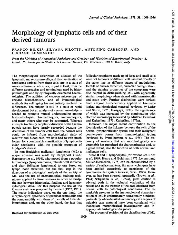
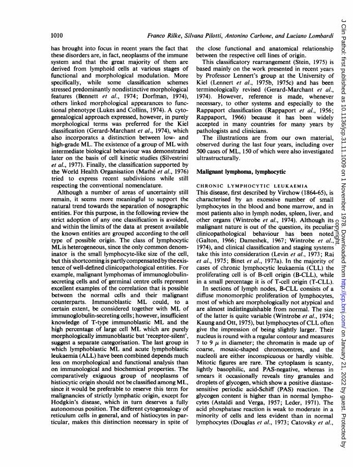
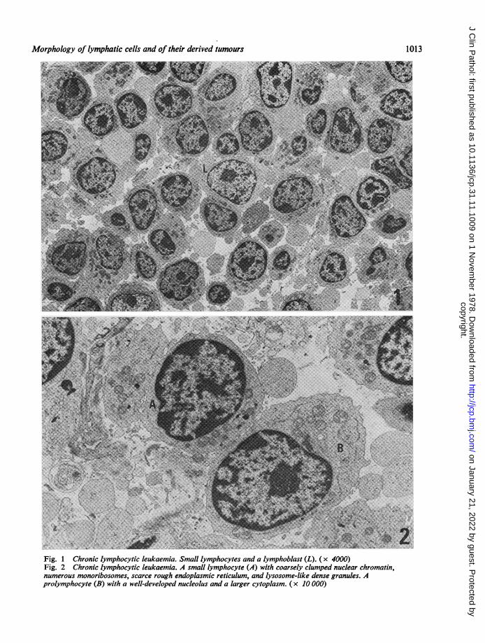
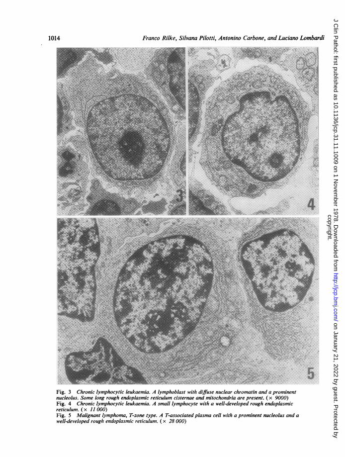
lymphocytes (Figs 1 and 2) have round nuclei withcoarsely clumped chromatin (Mori and Lennert,1969; Cawley and Hayhoe, 1973). There are noindentations of the nuclear membrane; however,occasional nuclear pockets are found. The incon-spicuous nucleoli frequently have a characteristicring-shaped pattern. Conversely, the lymphoblasts(Fig. 3) have diffuse nuclear chromatin and well-developed nucleoli with a prominent nucleolonema.The prolymphocytes (Fig. 2) have a roughly ovalnucleus and a well-developed nucleolus as in thehomonymous cells of Galton's prolymphocyticleukaemia (PL) (Galton et al., 1974), but they havea more abundant cytoplasm and a less clumpednuclear chromatin. Prolymphocytes of both B-CLLand PL are distinguishable from centrocytes, whichhave deeply indented irregular nuclei, inconspicuousnucleoli, and scanty cytoplasm with scarce organelles.The cytoplasm of B-CLL cells is rather scanty in
lymphocytes and is somewhat more conspicuous inlymphoblasts and prolymphocytes. It contains asmall or moderate number of mitochondria,numerous free ribosomes, a small Golgi apparatus,and a few rough endoplasmic reticulum cisternae.Abundant cytoplasmic microfilaments are present insome cells. The previously mentioned inclusions witha regular crystalline-like structure lie within therough endoplasmic reticulum cisternae. A morpho-logical marker that indicates a plasymcytoid trans-formation of rare CLL lymphocytes is represented bysome cisternae of long, flat, and rough, endoplasmicreticulum placed around the nucleus (Fig. 4).Scanning electron microscopy (SEM) does not per-mit the identification of the lymphocytes of B-CLLas neoplastic (Braylan et al., 1976).
Extensive studies of cell surface immunologicalmarkers, such as surface-bound immunoglobulin(Pernis et al., 1970), membrane receptors for theactivated third component of complement (Biancoet al., 1970; Ross et al., 1973), and aggregated IgG(Dickler et al., 1973) on CLL lymphocytes(Johansson and Klein, 1970; Grey et al., 1971;Wilson and Nossal, 1971; Aisenberg and Bloch,1972; Fr0land et al., 1972; Preud'homme andSeligmann, 1972; Piessens et al., 1973; Ross et al.,1973) proved that this B-lymphocyte proliferationis made up of cells in most of which no immuno-globulin secretion takes place but which do bear syn-thesised membrane-bound immunoglobulin. This isof one type only and is usually restricted to one typeof heavy chain (,u, y, or a) and one light chain (K orA), thus indicating the monoclonality of the cell pro-liferation (Aisenberg and Bloch, 1972), even thoughbiclonal processes have rarely been detected(Preud'homme and Seligmann, 1972). In a numberof cases 8 chains are associated with ,t chains, as
may be observed in normal lymphocytes (Fu et al.,1974). Routine immunofluorescent staining pro-cedures for the demonstration of immunoglobulinon the cell surface of leukaemic lymphocytes revealpositivity that is weak (Braylan et al., 1976), par-ticularly when compared with that of the cells oflymphosarcoma-cell leukaemia (Aisenberg andBloch, 1972; Aisenberg et al., 1973a), that is, theleukaemic conterpart of a lymphoma of germinalcentre cell origin. In general, the cells of B-CLL alsohave weakly represented complement receptor sites(Shevach et al., 1972) and membrane receptors foraggregated IgG (Braylan et al., 1976). However, theexpression of binding sites varies considerably fromcase to case. The absence of surface immunoglo-bulin (Piessens et al., 1973; Wilson and Hurdle,1973) has been related to the proliferationof a pre-Ig-synthesising lymphocytic population(Preud'homme and Seligmann, 1972). B-CLL cellseither fail to respond or respond poorly to mitogensin short-term cultures (Quaglino and Cowling,1964). It has also been shown that a high percentageof them form rosettes with mouse erythrocytes(Catovsky et al., 1976; Koziner et al., 1977) incontrast to the cells of lymphosarcoma-cell leukae-mia.
It has been suggested that B-CLL most likelyrepresents the proliferative disease of a 'virgin' (Bi)B-lymphocyte clone, which is still untouched by theantigen, is blocked in its modulation process, and istherefore unable to secrete immunoglobulin; but itsderivation from 'memory' (B2) B-lymphocytes insome cases cannot be ruled out (Salmon andSeligmann, 1974). It has also been suggested that Bi-cell proliferations are those whose cells bear surfaceimmunoglobulin and have complement receptors,whereas B2-cell proliferations also bear surfaceimmunoglobulin but are devoid of complementreceptors (Stein, 1975).When the tissue manifestation of B-CLL is not
accompanied by an excess of cells in the bloodand/or marrow, the disease has been referred to aswell-differentiated, lymphocytic, diffuse ML(Pangalis et al., 1977); a leukaemic manifestationmay appear later (Galton, 1966) or never (Goldbergand Emanuel, 1964). However, the morphology andthe surface characteristics of the cells are the same asthey are in B-CLL (Huber et al., 1974; Peter et al.,1974; Aisenberg and Long, 1975; Brouet et al.,1975b; Braylan et al., 1976). One major differencebetween B-CLL and well-differentiated lymphocyticdiffuse ML was reported to consist of a less evidentdegree of hypogammaglobulinaemia in the latter(Pangalis et al., 1977). In spite of clinical and haema-tological differences, leukaemic and non-leukaemicB-lymphocytic neoplastic proliferations are so
10)12
copyright. on January 21, 2022 by guest. P
rotected byhttp://jcp.bm
j.com/
J Clin P
athol: first published as 10.1136/jcp.31.11.1009 on 1 Novem
ber 1978. Dow
nloaded from
Morphology of lymphatic cells and of their derived tumours.......
1
WR'x'''(.C
4 q .:.5, R a;q ,ic t > fS t
Fig. 1 Chronic lymphocytic leukaemia. Small lymphocytes and a lymphoblast (L). (x 4000)Fig. 2 Chronic lymphocytic leukaemia. A small lymphocyte (A) with coarsely clumped nuclear chromatin,numerous monoribosomes, scarce rough endoplasmic reticulum, and lysosome-like dense granules. Aprolymphocyte (B) with a well-developed nucleolus and a larger cytoplasm. (x 10 000)
1013
..X J1, .11
copyright. on January 21, 2022 by guest. P
rotected byhttp://jcp.bm
j.com/
J Clin P
athol: first published as 10.1136/jcp.31.11.1009 on 1 Novem
ber 1978. Dow
nloaded from
Franco Rilke, Silvana Pilotti, Antonino Carbone, and Luciano Lombardi
...*s.. ... .. ;.p
4
5Fig. 3 Chronic lymphocytic leukaemia. A lymphoblast with diffuse nuclear chromatin and a prominentnucleolus. Some long rough endoplasmic reticulum cisternae and mitochondria are present. ( x 9000)Fig. 4 Chronic lymphocytic leukaemia. A small lymphocyte with a well-developed rough endoplasmicreticulum. ( x 11 000)Fig. 5 Malignant lymphoma, T-zone type. A T-associated plasma cell with a prominent nucleolus and awell-developed rough endoplasmic reticulum. ( x 28 000)
1014
.......
41
Ik,
copyright. on January 21, 2022 by guest. P
rotected byhttp://jcp.bm
j.com/
J Clin P
athol: first published as 10.1136/jcp.31.11.1009 on 1 Novem
ber 1978. Dow
nloaded from
Morphology of lymphatic cells and of their derived tumours
closely related that they are probably just differenthaematopathological expressions of the samedisease.
In large series of cases of CLL, occasional patientsmay be encountered in whom morphological and/orimmunological deviations from the classic pictureare noticed. Morphological deviations are repre-sented by cases in which circulating cells have eithercleft nuclei similar to Rieder cells and to smallfollicular centre cells (centrocytes) of lympho-plasmacytic features (Rudders, 1976) and containcytoplasmic inclusions. In general, lymphocytes ofabnormal appearance have been related to a morerapid course of the disease. Along these lines, im-munological variations are exemplified either bycases whose cells demonstrate a surface markerphenotype, which is comparable to that usuallydisplayed by follicular centre cells (Braylan et al.,1976) or by so-called 'atypical CLL' cases whosecells contain intracytoplasmic IgG (Rudders, 1976).On the basis of these descriptions one might suspectthat these atypical cases of CLL are, in fact, leukae-mic maifestations of ML, respectively of follicularcentre cell origin or of lymphoplasmacytoid type. Inrare instances, both B- and T-cell surface markershave been found on leukaemic cells (Chin et al.,1973); however, their true B + T nature has beenquestioned (Brouet et al., 1975c; Siegal et al., 1976).Shifts of markers have also been noticed during thecourse of the disease (Kay et al., 1974).T-CLL (Bentwich and Kunkel, 1973; Dickler
et al., 1973; Lille et al., 1973; Wilson and Hurdle,1973) is a rare entity (less than 2% of all CLL), withonset in adulthood, frequent massive splenomegaly,skin lesions, and variable lymphocyte count, whichis characterised cytologically by somewhat largerlymphocytes than those of B-CLL (Brouet et al.,1975a). The cells have fairly large, occasionallydeeply basophilic, PAS-negative cytoplasm whichcontains large azurophilic granules and shows apositive beta-glucuronidase and acid-phosphatasereaction (Catovsky, 1975). The nuclei containcoarse chromatin granules and a small nucleolus.Prolymphocytes and lymphoblasts are absent.On electron microscopy the nuclei are irregular
and there is a high content of lysosomal enzymes.Transmission and SEM cannot detect any mor-phological difference either between normal B and Tlymphocytes (Alexander et al., 1976; Newell et al.,1976; Polliack, 1977) or between CLL with T or Bsurface immunological markers. Immunologically,the presence of receptors for sheep erythrocytes(Lay et al., 1971; Jondal et al., 1972) and thereactivity with heterologous anti-T-cell antiserum(Aisenberg et al., 1973b) indicate the T-derived natureofthe cells.
Additional cases of this or of a closely relateddisease with a poor prognosis have been reportedin Japan. The circulating lymphocytes, however,showed a higher degree of nuclear pleomorphismand fewer cytoplasmic granules (Uchiyama et al.,1977) as compared with the T-CLL described inwestern countries. As far as we are aware, detailedhistological descriptions of lymph nodes involved byT-CLL are lacking.
T-ZONE MALIGNANT LYMPHOMAWhether T-CLL bears any relationship to the T-zone lymphocytic ML has not yet been established(Lennert et al., 1975b). It seems, however, that thisrecently described entity (Lennert et al., 1975b;Lennert, 1976) is a well-defined one and that itrepresents neoplastic malignant proliferation of thelymphocytes of the nodal paracortical area. In fact,in cases that are not too far advanced, uninvolvedsecondary follicles which are widely dissociated bythe neoplastic growth remain recognisable. Theneoplastic cell population is somewhat pleomorphicand is made up of irregularlyshaped lymphocytes, thenuclei of which are on average less hyperchromaticthan are those of B-CLL. The chromocentres aresmaller and not clumped, and a small prominentnucleolus is usually visible. The mitotic index is low.The cells display a strong acid phosphatase activity(Kaiserling, 1977b). The histological picture is alsocharacterised by the presence of the normal com-ponents of the T-dependent paracortical area(Kaiserling, 1977b), that is, postcapillary venuleslined by hob-nailed endothelial cells and surroundedby small lymphocytes, a network of reticulin fibres,and interdigitating reticulum cells, the clear, highlyirregular nuclei of which may be recognised even onlight microscopy (Veldman, 1970). These cells areconsidered to be the characteristic reticulum cells ofthe paracortical area of the lymph nodes. They donot usually display phagocytic activity. In addition,so-called T-associated plasma cells are part ofthe picture (Lennert et al., 1975a).The intermingled rare large blast cells are similar
to Hodgkin's mononuclear cells and may possiblybe interpreted as T immunoblasts. Multinucleatecells of the same type simulate Reed-Sternberg cells.Six cases of a very similar entity, defined as ML ofperipheral T-lymphocyte origin, were recentlyreported by Waldron et al. (1977) in elderly patients.The majority of the malignant cells of these caseswere identified immunologically as T lymphocytes.On electron microscopy the cellular population of
the T-zone ML diffusely infiltrates the lymph nodesand consists of pleomorphic lymphoid cells, asmaller number of non-neoplastic macrophages,rare plasma cells, and granulocytes (Waldron et al.,
1015
copyright. on January 21, 2022 by guest. P
rotected byhttp://jcp.bm
j.com/
J Clin P
athol: first published as 10.1136/jcp.31.11.1009 on 1 Novem
ber 1978. Dow
nloaded from
Franco Rilke, Silvana Pilotti, Antonino Carbone, and Luciano Lombardi
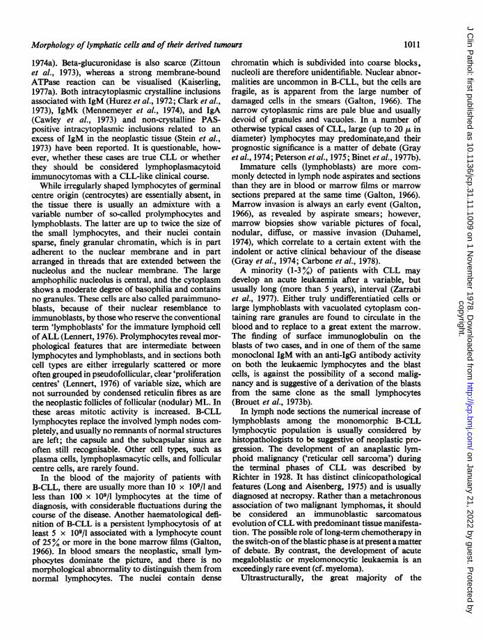
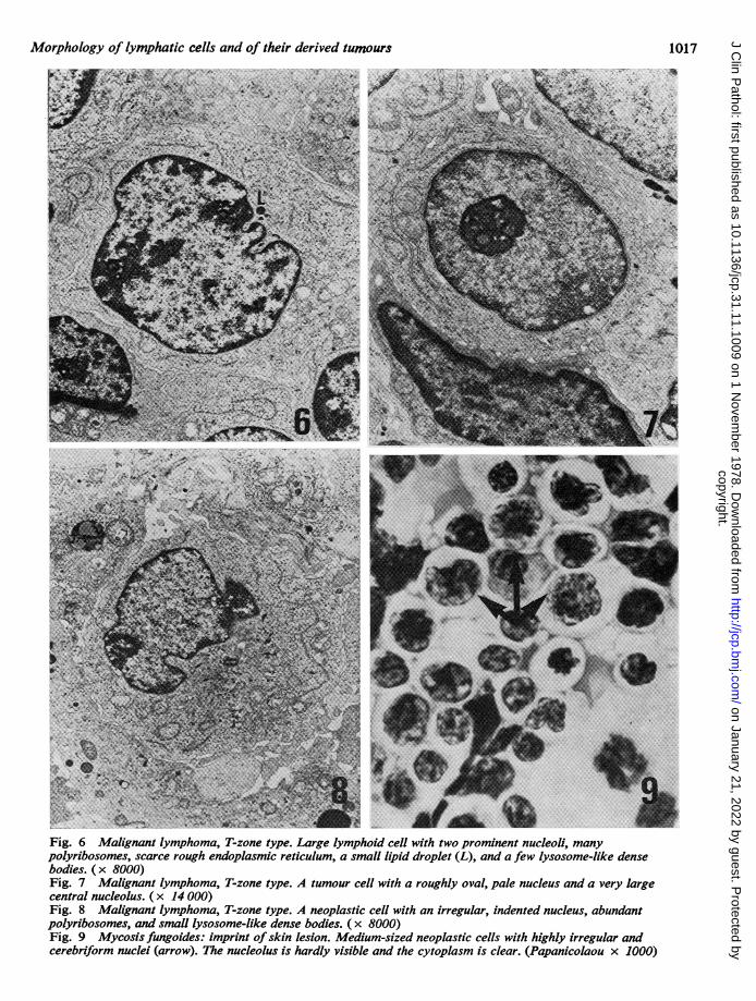
1977). T-associated plasma cells may be numerous(Fig. 5). The size and the nuclear morphology of thelymphoid cells vary considerably. The small lym-phoid cells are similar to small circulating lympho-cytes, whereas large cells (Fig. 6) look like trans-formed lymphocytes with pale oval nuclei, scantyheterochromatin, and prominent nucleoli. Theabundant cytoplasm contains many polyribosomes,scarce rough endoplasmic reticulum cisternae, avariable number of mitochondria, and occasionallysosome-like dense granules located in the Golgiarea. A few abnormal cells (Fig. 7) may show eithera huge central nucleolus with a very prominentnucleolonema or an irregularly indented nucleus(Fig. 8) with dispersed chromatin; these cells recallthe descriptions of mononuclear Hodgkin's cells(Dorfman et al., 1973; Glick et al., 1976). Theidentification within the tumour tissue of inter-digitating reticulum cells can be considered adiagnostic marker of the T-zone lymphoma andother T-derived lymphomas (Kaiserling, 1977b).Interdigitating reticulum cells have a clear nucleuswith scarce heterochromatin placed along thenuclear membrane and with a regular shape or,occasionally, with deep indentations. A smallnucleolus is observed. The abundant cytoplasm isextended into interdigitating processes. No junc-tional differentiation is evident among the plasmamembranes of two contiguous reticulum cells. In thecytoplasm a complex of vesicles and sacs, markedlycontrasted by silver-methenamine staining, is found.This electron microscope cytochemical method isconsidered specific for glycoproteins (Veldman,1970). The Golgi apparatus also shows a positivesilver-methenamine reaction, which supports afunctional correlation between the two structures. Afew rough endoplasmic reticulum cisternae, somemitochondria, bundles of filaments, and a smallnumber of mono- and polyribosomes are alsofound. No digestive vacuoles are seen.
PROLYMPHOCYTIC LEUKAEMIA (PL)This disease (Galton et al., 1974) is a rare variant ofCLL with distinctive features; lymphadenopathy isinconspicuous, whereas the spleen and liver aremarkedly enlarged. There is usually a high WBCcount, and the proliferating cell has been designated'prolymphocyte', with a different meaning, how-ever, from that used in the WHO classification ofneoplastic lymphoid diseases. The leukaemic cellsare larger than the lymphocytes of B-CLL, have afairly large basophilic cytoplasm, a nucleus withcondensed chromatin, and a single vesicular andprominent nucleolus. A variable number of lympho-cytes as well as larger blast-like cells are also present.The latter are different from the lymphoblasts of
CLL because of the larger size and the greateramount of chromatin. Abnormal looking cells areoften found. Most of the reported cases have been ofB-cell origin, but a few have been of T-cell origin.The former reveal a stronger expression of surfacemembrane-bound immunoglobulin and of comple-ment receptors than the lymphocytes of B-CLL(Catovsky et al., 1973), while the T-cell prolympho-cytes lack those markers but yield a high spon-taneous E-rosette count. In smears, a high percentageof the cells of B-type PL contain glycogen(Stathopoulos et al., 1974) in the form of granuleswhich are coarser than those found in B-CLLlymphocytes. Acid phosphatase content is weak butoccasionally tartrate-resistant (Catovsky et al.,1974a). On the other hand, T-cell type PL cellsreveal a higher content of acid phosphatase and arePAS negative.
Essential differences between the ultrastructuralfeatures of prolymphocytes and those of B-CLLlymphocytes consist of the larger size and the moreprominent nucleolus, with the well-developednucleolonema of the former. Ring-shaped nucleoliare seldom seen. In the cytoplasm a variable numberof mitochondria and some rough endoplasmicreticulum cisternae are found. The blasts of PL havelarge nucleoli and dispersed chromatin. In compari-son to prolymphocytes of B-CLL, prolymphocytesof PL are characterised by a clumped chromatinpattern and may therefore be placed morphologicallyin an intermediate position between small lympho-cytes and the intermediate cells of CLL. No mor-phological differences have been reported betweenT- and B-type PL.
MYCOSIS FUNGOIDES AND SEIZARYSYNDROMEMycosis fungoides is an uncommon (less than 1% ofall ML) but distinctive histopathological and clinicalentity which involves primarily the skin (Edelson etal., 1974; Cline, 1975) and later, in over two-thirds ofcases, the lymph nodes and other organs (Long andMihm, 1974; Rappaport and Thomas, 1974). Thecellular composition of the infiltrates of mycosisfungoides is lymphocytic in nature (Crossen et al.,1971); the cells are unique and different from those ofother ML, and are considered to originate from T-dependent lymphocytes.
Histological diagnosis is usually made on skinbiopsies of the second (indurated neoplasticcutaneous plaque) and of the third (mycotic orneoplastic) stage of the disease. The neoplasticcellular population is identical in the cutaneous andextracutaneous sites. In the skin there is an infiltra-tion by mononuclear atypical cells, which invadethe papillary dermis and the basal layers of the
1016
copyright. on January 21, 2022 by guest. P
rotected byhttp://jcp.bm
j.com/
J Clin P
athol: first published as 10.1136/jcp.31.11.1009 on 1 Novem
ber 1978. Dow
nloaded from
Morphology of lymphatic cells and of their derived tumours
j.2. .
At
a.is : , .s;.
°~~~~~~~~OFig. 6 Malignant lymphoma, T-zone type. Large lymphoid cell with two prominent nucleoli, manypolyribosomes, scarce rough endoplasmic reticulum, a small lipid droplet (L), and a few lysosome-like densebodies. (x 8000)Fig. 7 Malignant lymphoma, T-zone type. A tumour cell with a roughly oval, pale nucleus and a very largecentral nucleolus. ( x 14 000)Fig. 8 Malignant lymphoma, T-zone type. A neoplastic cell with an irregular, indented nucleus, abundantpolyribosomes, and small lysosome-like dense bodies. (x 8000)Fig. 9 Mycosis fungoides: imprint of skin lesion. Medium-sized neoplastic cells with highly irregular andcerebriform nuclei (arrow). The nucleolus is hardly visible and the cytoplasm is clear. (Papanicolaou x 1000)
1017copyright.
on January 21, 2022 by guest. Protected by
http://jcp.bmj.com
/J C
lin Pathol: first published as 10.1136/jcp.31.11.1009 on 1 N
ovember 1978. D
ownloaded from
Franco Rilke, Silvana Pilotti, Antonino Carbone, and Luciano Lombardi
epidermis, in which the so-called Darier-Pautrier'sabscesses are often formed within epidermolyticspaces. The neoplastic infiltrate consists of cells witha wide range of sizes. The, most common cells, how-ever, are medium-sized and measure 10 to 20 K. indiameter; they have a scanty, faintly stained cyto-plasm, which shows a focal acid phosphataseactivity (Schwarze, 1975) and contains perinucleardiastase-resistant, PAS-positive granules. Theirnuclei are pleomorphic, with dense chromocentricchromatin and a small nucleolus.The atypical cells may show a certain variation in
size and shape with marked atypicalities. The largerthe cells the more evident are the irregularities of thenuclear membrane, particularly in imprints (Fig. 9),with hyperconvolution and cerebriform patterns. Inthe very small cells nuclear details are barely visible.The so-called mycosis cells are a rare finding; theyare the largest of the whole neoplastic cell popula-tion and show marked hyperconvolution and hyper-chromasia of the nucleus and amphophilic cyto-plasm. In addition, a large number of abnormallooking histiocytes may be present (Robinowitz etal., 1976).
In lymph nodes, the neoplastic proliferation ofmycosis fungoides seems to involve initially the'thymic-dependent' paracortical area and later, to avariable extent, the remaining node (Thomas andRappaport, 1975). All cell types present in thecutaneous infiltrate can be encountered in extra-cutaneous sites of invasion. In the spleen, eitherscattered foci in both the red and the white pulp ora diffuse infiltration may be seen (Variakojis et al.,1974). The selective involvement of the thymic-dependent periarteriolar lymphatic sheath has beenstressed (Thomas and Rappaport, 1975).
Sezary's syndrome (Sezary and Bouvrain, 1938), achronic leukaemia associated with erythroderma, isconsidered the leukaemicvariant ofmycosis fungoides(Lutzner et al., 1975; Robinowitz et al., 1976). Infact, the cutaneous infiltrate in Sezary syndrome isvery similar to that of mycosis fungoides, and, on theother hand, in a high percentage of cases of mycosisfungoides circulating Sezary cells were found(Moran et al., 1977). In addition, in Sezary syndromethe earliest infiltration in lymph nodes was reportedto be found histologically in tertiary follicles(Lennert, 1974). It was therefore proposed thatmycosis fungoides and Sezary syndrome should begrouped together under the term 'cutaneous T-celllymphoma' (Schein et al., 1976).The circulating cells, which represent the diag-
nostic marker of the disease-even though not anabsolute one (Lutzner et al., 1975)-are variable innumber (between 10 and 50% of the nucleated cells inblood smears) and reveal nuclear and cytoplasmic
features that are closely related to those described formycosis cells. In marrow smears Sezary cells are rarerthan expected in a leukaemic disease. The cellsusually measure between 15 and 20 ,u in diameter andhave a high nuclear:cytoplasmic ratio. The scantycytoplasm is well defined, appears light blue with theGiemsa stain, and does not contain granules, whileempty vacuoles may be present. In thin blood smearsthe nuclei disclose their highly characteristic struc-ture: the nuclear membrane shows various furrows,indentations, folds, and convolutions. Nucleoli arebarely visible. The same finding is revealed byimprints of skin lesions as well as of lymph nodesthat are involved by mycosis fungoides and bySezary syndrome. Sezary syndrome also has a moremarkedly leukaemic small cell variant that is madeup of lymphocytes measuring about 8 ,u in diameterwhich are morphologically more closely similar tosmall lymphocytes even though nuclear indentationsare present (Lutzner et al., 1973). The large cells havenear-tetraploid DNA values and near-tetraploidchromosome counts, whereas the small cells havediploid DNA values and pseudodiploid or hyper-diploid chromosome counts.
Sezary cells reveal a granular acid phosphatasecontent, are moderately positive for ,B-glucuronidaseand cx-naphthyl acetate esterase, and are negative forperoxidase, naphthol-AS-D-chloroacetate esterase,alkaline phosphatase, adenosine-triphosphatase, Oil-Red 0, and Sudan black-B (Loffler, 1972). PASstains cytoplasmic granules of neutral mucopoly-saccharides in a variable number without diffusebackground staining (Crossen et al., 1971). Largeand small Sezary cells have T-cell markers; theyform spontaneous E rosettes, react with antihumanT-cell antisera, respond to phytohaemagglutinin(Brouet et al., 1973a; Lutzner et al., 1973), and lackB-cell markers; they do not bear complementreceptor sites and are devoid of surface-boundimmunoglobulin (Broome et al., 1973). In addition,evidence was given that Sezary cells represent aneoplastic proliferation of T-helper cells (Broder etal., 1976). A few cases of Sezary syndrome in whichthe neoplastic cells failed to form E rosettes have alsobeen reported (Goldstone et al., 1976).
Ultrastructurally, tissue-bound mycosis fungoidescells (Lutzner et al., 1971) are also very similar tocirculating Sezary cells (Lutzner and Jordan, 1968;Zucker-Franklin et al., 1974). The nuclear:cyto-plasmic ratio is high, and the nucleus is charac-teristically irregular, serpentine, indented, andlobed. However, this degree of irregularity varieslargely from cell to cell and from case to case (Rosas-Uribe et al., 1974). Heterochromatin is abundant andconcentrated along the nuclear membrane. One ortwo nucleoli with nucleolonema or ring-shaped
1018
copyright. on January 21, 2022 by guest. P
rotected byhttp://jcp.bm
j.com/
J Clin P
athol: first published as 10.1136/jcp.31.11.1009 on 1 Novem
ber 1978. Dow
nloaded from
Morphology of lymphatic cells and of their derived tumours
nucleoli and occasional nuclear inclusions are
observed. The cytoplasm contains glycogen and aprominent network of microfilaments (Zucker-Franklin et al., 1974). The number of mitochondria,multivesicular bodies, rough endoplasmic reticulumcisternae, and free mono- and polyribosomes variesfrom case to case. Cytoplasmic pseudopods andlysosomes are found, whereas phagocytosis is never
seen.At SEM most of the Sezary cells show a moderate
to markedly villous surface and do not displayruffled membranes (Polliack et al., 1977). SinceSezary-like cells are found in patients with a variety ofnonlymphomatous dermatoses, it is of diagnosticimportance that atypical cells should be demon-strated in clusters or sheets in the affected tissues.
HAIRY CELL LEUKAEMIAThis uncommon form of a chronic leukaemiclymphoproliferative disorder is characterised clini-cally in most cases by splenomegaly, hepatomegaly,and pancytopenia. Lymphadenopathy is a rare andlate symptom. In sections of spleens there is adiffuse proliferation of monomorphic cells whichmeasure between 12 and 20 tk in diameter. Thenucleus is roundish or oval, occasionally kidney-shaped, often indented and wrinkled, and eccentric.It occupies about half of the cell area. Mitoticfigures are rarely seen. The chromatin is delicateand loose, and nucleoli (if discernible) are small andpale. The cytoplasm is quite large, ill-defined,irregular, and lightly basophilic (grey-blue inWright-Giemsa smears) and does not containgranules. Large blast-like cells are never found. Inordinary blood smears in which the cell morphologyis best observed, the hairy finger-like projections ofthe cytoplasm may be recognised. However, theyappear to be better preserved and defined in phase-contrast microscopy. In thin, well-stained smears andsplenic imprints, small, cigar-shaped or roundishpyroninophilic cytoplasmic inclusions may bedetected that correspond to the ribosome-lamellarcomplexes (Katayama et al., 1973).The cytological marker is the presence of a pro-
minent focal acid-phosphatase activity, which isresistant to degradation by tartaric acid (isoenzyme5) (Yam et al., 1972). Even though hairy cells mayoccasionally be tartaric-acid sensitive, this isozymeshould always be sought, preferably in blood smears
(Katayama and Schneider, 1977); it is absent innormal lymphocytes but may be found in some CLLandPL lymphocytes, and its appearance in hairy cellsshould be considered as a newly acquired propertythat is related to the malignant status of the cells.Hairy cells are negative for Sudan black-B, peroxi-dase, and PAS, even though in some cells a PAS-
positive granulation has occasionally been seen(Haak et al., 1974). A moderate granular sodiumfluoride-resistant naphthol AS-D-acetate esteraseactivity, allegedly stronger than that of lympho-cytes and weaker than that of histiocytes, has beenreported (Flandrin et al., 1973). Recent extensiveenzymohistochemical investigations revealed thathairy cells show only occasionally a-naphthyl-acetate esterase activity, while they are negative foralkaline phosphatase, a-naphthyl-butyrate esterase,sodium fluoride-resistant naphthol AS-D-acetateesterase, 5'-nucleotidase, and N-acetyl-fl-glucosa-minidase (Nanba et al., 1977b).The earliest and most striking proliferation of cells
takes place in the spleen, which usually becomesmarkedly enlarged. Grossly, the spleen is dark red incolour, firmer than normal, and does not revealdiscrete tumour masses. In the early stages, how-ever, hairy cells appear first in the cords of the redpulp, and subsequently the sinusoidal structure andthe Malpighian corpuscles of the white pulp tend todisappear because of compression and atrophy. Inaddition, an increase of actively phagocytosinghistiocytes has been described around the arteries ofthe red pulp (Nanba et al., 1977b). Plasma cells, bothnormal and of atypical appearance, are foundamong the hairy cells. Their purely reactive naturehasbeen questioned. Distended spaces filled witherythrocytes and lined by hairy cells and not byendothelial cells, which show a strong fluoride-sensitive naphthol-AS-D-acetate esterase activity,were recently described as pseudosinuses (Nanba etal., 1977c).Marrow invasion may be either massive or focal.
The marked and diffuse increase in reticulin fibres(Burke et al., 1974) explains the 'dry tap' that is mostoften found; however, imprints of marrow biopsiesyield adequate diagnostic cytological material(Krause et al., 1977). In the liver, the invasionusually follows the sinusoidal structure, and pseudo-angiomatous lesions similar to those of the spleenand lined by hairy cells have also been described. Inthe lymph nodes, the neoplastic proliferation appearslate in the course of the disease and begins in the B-cell region of the outer cortex with early infiltrationof the marginal sinus. The spread to the paracorticaland medullary areas develops later (Lennert, 1974).Lymph node invasion is often only partial, withresidual follicles and foci of lymphoid cells.The large number of synonyms used so far for the
definition of this disease, which is still also calledleukaemic reticuloendotheliosis (Ewald, 1923;Bouroncle et al., 1958), indicates the uncertaintiesabout the proliferating cell type, the normal counter-part of which has not yet been identified. It has beenquestioned whether hairy cell leukaemia is really a
1019
copyright. on January 21, 2022 by guest. P
rotected byhttp://jcp.bm
j.com/
J Clin P
athol: first published as 10.1136/jcp.31.11.1009 on 1 Novem
ber 1978. Dow
nloaded from
Franco Rilke, Silvana Pilotti, Antonino Carbone, and Luciano Lombardi
completely homogeneous disease rather than agroup of subentities (Golde et al., 1977b), and itsorigin from histiocytic, endothelial, or lymphoidcells was and still is a matter of controversy.The moderate nonspecific esterase activity, the
occasional and inconsistent ability of phagocyticactivity in vitro (latex particles and complement-coated zymogen particles) (Boldt et al., 1977) and invivo (Catovsky et al., 1975a; Nanba et al., 1977b;Utsinger et al., 1977), and adhesion of the cells toglass and nylon (Flandrin et al., 1973; Boldt et al.,1977) are weak arguments, indicating that hairy cellsmay be monocytic in nature; and the severe mono-cytopenia associated with the disease has beeninterpreted in the same way (Seshadri et al., 1976).However, none of these is a compelling reason andfurthermore, hairy cells do not produce muramidase(Catovsky et al., 1975a). An origin from endo-thelial cells also seems unlikely because of thetartaric acid-resistant acid phosphatase activity andthe absence of naphthol AS-D-acetate esterasereaction.On the other hand, arguments identfying the
hairy cell as a neoplastic B-modified lymphocyte, inaddition to the morphological findings are: frequentsurface-bound ATPase activity (as in CLL) and fi-glucuronidase activity (as in malignant plasma cells)(Nanba et al., 1977b); the finding of surface immuno-globulin (Catovsky et al., 1974c; Stein andKaiserling, 1974; Leech et al., 1975b; Boldt et al.,1977), probably IgM (Debusscher et al., 1975), and ofintracellular immunoglobulin (Leech et al., 1975b;Golde et al., 1977a), probably IgMA (Debusscheret al., 1975); and reactivity with the Merritt B-cellalloantibodies (Naeim et al., 1977). However,surface immunoglobulin was demonstrated occa-sionally to be polyclonal on hairy cells, and receptorsfor cytophilic antibodies were identified (Jaffe et al.,1974b), while the search for complement receptorsgave predominantly negative results (Burns et al.,1977). The responsiveness to mitogens was found tobe impaired but less than it is in CLL (Haak et al.,1974). On the hairy cells surface immunoglobulinredistribution was induced by antibodies; polar capformation occurs as with other B cells and was foundto be very active and resistant to various agents,such as low temperature and sodium azide(Salsano et al., 1978). No evidence for a T-cellorigin has been found so far.
Ultrastructural investigations also confirm thatin lymph nodes the neoplastic cell proliferationbegins in the outer cortex. The tumour populationconsists of one cell type only: the hairy cell shows thesame features in all sites and is characterised by theabundant hairy-like cytoplasmic projections inter-digitating in a complex way and giving to the
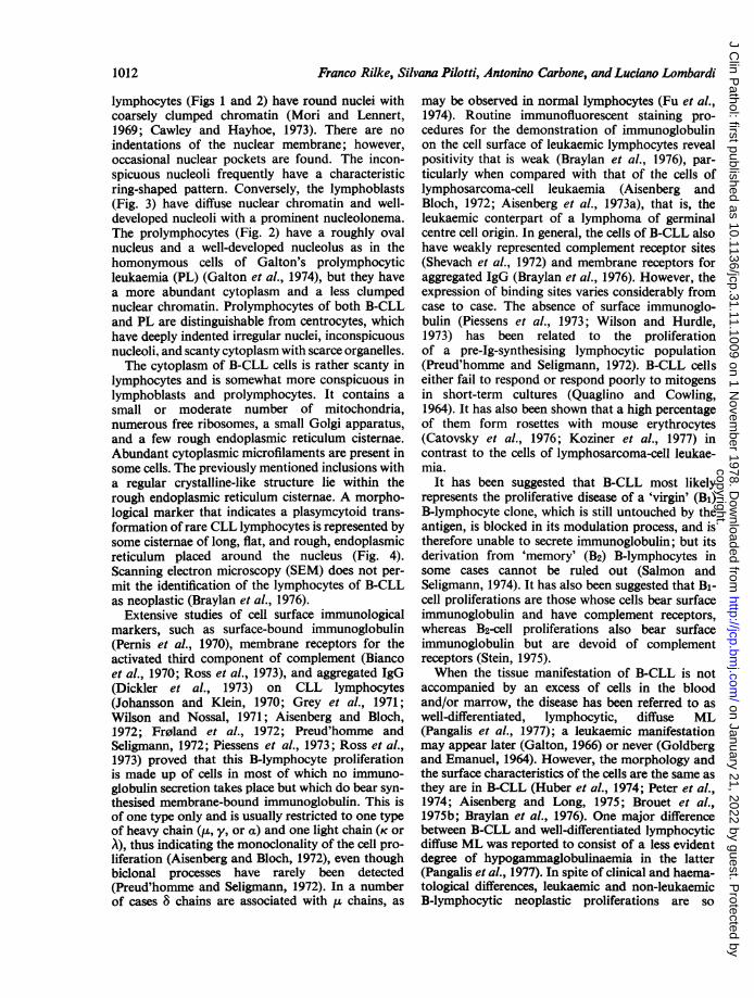
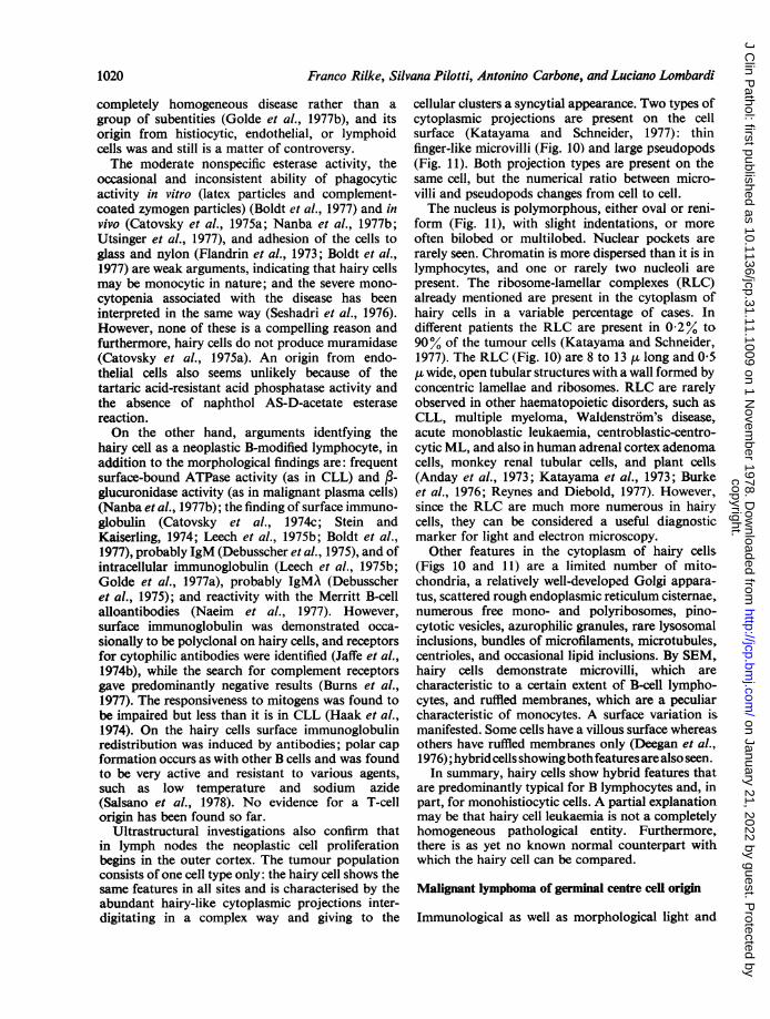
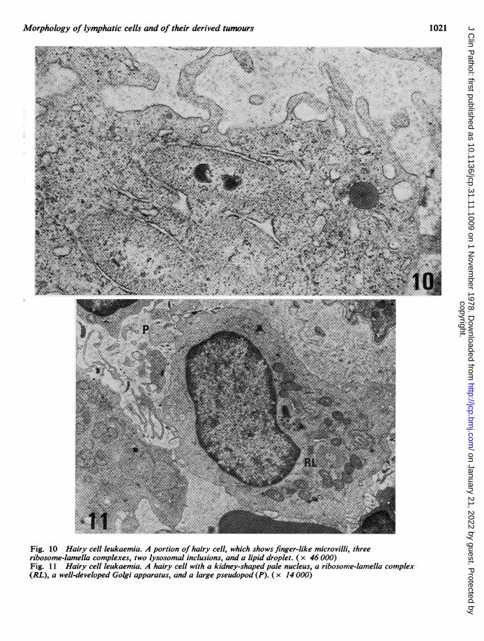
cellular clusters a syncytial appearance. Two types ofcytoplasmic projections are present on the cellsurface (Katayama and Schneider, 1977): thinfinger-like microvilli (Fig. 10) and large pseudopods(Fig. 11). Both projection types are present on thesame cell, but the numerical ratio between micro-villi and pseudopods changes from cell to cell.The nucleus is polymorphous, either oval or reni-
form (Fig. 11), with slight indentations, or moreoften bilobed or multilobed. Nuclear pockets arerarely seen. Chromatin is more dispersed than it is inlymphocytes, and one or rarely two nucleoli arepresent. The ribosome-lamellar complexes (RLC)already mentioned are present in the cytoplasm ofhairy cells in a variable percentage of cases. Indifferent patients the RLC are present in 0-2% to9000 of the tumour cells (Katayama and Schneider,1977). The RLC (Fig. 10) are 8 to 13 p long and 0 5,u wide, open tubular structures with a wall formed byconcentric lamellae and ribosomes. RLC are rarelyobserved in other haematopoietic disorders, such asCLL, multiple myeloma, Waldenstr6m's disease,acute monoblastic leukaemia, centroblastic-centro-cytic ML, and also in human adrenal cortex adenomacells, monkey renal tubular cells, and plant cells(Anday et al., 1973; Katayama et al., 1973; Burkeet al., 1976; Reynes and Diebold, 1977). However,since the RLC are much more numerous in hairycells, they can be considered a useful diagnosticmarker for light and electron microscopy.
Other features in the cytoplasm of hairy cells(Figs 10 and 11) are a limited number of mito-chondria, a relatively well-developed Golgi appara-tus, scattered rough endoplasmic reticulum cisternae,numerous free mono- and polyribosomes, pino-cytotic vesicles, azurophilic granules, rare lysosomalinclusions, bundles of microfilaments, microtubules,centrioles, and occasional lipid inclusions. By SEM,hairy cells demonstrate microvilli, which arecharacteristic to a certain extent of B-cell lympho-cytes, and ruffled membranes, which are a peculiarcharacteristic of monocytes. A surface variation ismanifested. Some cells have a villous surface whereasothers have ruffled membranes only (Deegan et al.,1976); hybrid cells showingbothfeatures are also seen.
In summary, hairy cells show hybrid features thatare predominantly typical for B lymphocytes and, inpart, for monohistiocytic cells. A partial explanationmay be that hairy cell leukaemia is not a completelyhomogeneous pathological entity. Furthermore,there is as yet no known normal counterpart withwhich the hairy cell can be compared.
Malignant lymphoma of germinal centre celi origin
Immunological as well as morphological light and
1020
copyright. on January 21, 2022 by guest. P
rotected byhttp://jcp.bm
j.com/
J Clin P
athol: first published as 10.1136/jcp.31.11.1009 on 1 Novem
ber 1978. Dow
nloaded from
Morphology of lymphatic cells and of their derived tumours
Fig. 10 Hairy cell leukaemia. A portion of hairy cell, which shows finger-like microvilli, threeribosome-lamella complexes, two lysosomal inclusions, and a lipid droplet. ( x 46 000)Fig. 11 Hairy cell leukaemia. A hairy cell with a kidney-shaped pale nucleus, a ribosome-lamella complex(RL), a well-developed Golgi apparatus, and a large pseudopod (P). ( x 14 000)
1021copyright.
on January 21, 2022 by guest. Protected by
http://jcp.bmj.com
/J C
lin Pathol: first published as 10.1136/jcp.31.11.1009 on 1 N
ovember 1978. D
ownloaded from
Franco Rilke, Silvana Pilotti, Antonino Carbone, and Luciano Lombardi
electron microscopic studies (Sordat et al., 1970)indicate that lymphatic follicles and their germinalcentres are, at least in their early phase, the site ofmultiplication of B-lymphocyte clones, which reactspecifically to antigenic stimulation. In fact, largedeposits of immunoglobulin are accumulated withingerminal centres (Nossal et al., 1968). Cytologically,in secondary stimulated follicles, several cell typesare identifiable. There are specific large cells namedgerminoblasts (Lennert, 1957; Lennert and Remmele,1958), renamed centroblasts' (Gerard-Marchant etal., 1974), and small cells named germinocytes (Len-nert, 1964) renamed centrocytes', and, in addition,dendritic reticulum cells (Milanesi, 1965a, 1965b),'tingible body' macrophages (histiocytic reticulumcells), a few plasma cells and precursors, andoccasional immunoblasts (ie, large basophilic trans-formed lymphocytes). In germinal centres mitoticactivity is always very intense.Normal centroblasts are round cells measuring
between 12 and 15,u with basophilic cytoplasm and arather clear nucleus in which two to three nucleoli arelocated against the nuclear membrane. Normalcentrocytes are small cells with little cytoplasm, anindented nucleus, and very small nucleoli. These cellsare easily identified in thin and well-stained sectionsof properly fixed material. They are also readilyidentified in imprints. The relationship betweencentroblasts and centrocytes is interpreted as thetransformation of the former into the latter on thebasis of cytophotometric and autoradiographicinvestigations which showed that the cellular proli-feration in germinal centres is primarily a functionof centroblasts (Lennert et al., 1969). Opposing viewshave been expressed by Lukes and Collins (1974). Itappears, however, that non-stimulated primaryfollicles do not contain centroblasts and are made uppredominantly of centrocytes, some of which carrynatural antibody on the cell surface. It has been stres-sed that centroblasts and centrocytes are specific trans-formed lymphoid cells of the germinal centres whichhave unique distinctive properties. It has also beenemphasised that it seems inadvisable to use for theiridentification inappropriate names, such as histio-cytes or poorly differentiated lymphocytes (Lennert,1973). It also seems unrealistic to define the cells ofgerminal centres by use of the recommended terms,such as large lymphoid cells and medium-sizedlymphocytes (Cottier et al., 1973), since the sameare also applied to other cell types seen in histologicalsections of lymph nodes that have different stainingproperties of the cytoplasm, structure of the nucleus,and size and number of nucleoli.
'Greek: xyv&pov = point, centre; PA(Xwrrov= offspring, germ;
,Cvroa = cavity, container.
The derivation of ML from the cells of the germinalcentres has been the subject of a long-standingdebate. Morphological light microscopic and ultra-structural data (Lennert et al., 1966; Mori andLennert, 1969; Kojima et al., 1973; Lennert, 1973;Kaiserling, 1977a), immunological findings (Shevachetal., 1973; Jaffe etal., 1974a), and cytochemical data(Lennert, 1964, 1968) were accumulated essentially infavour of a derivation of ML from the cells that arethe constituents of the primary and secondarylymphatic follicles (Lennert et al., 1975c).
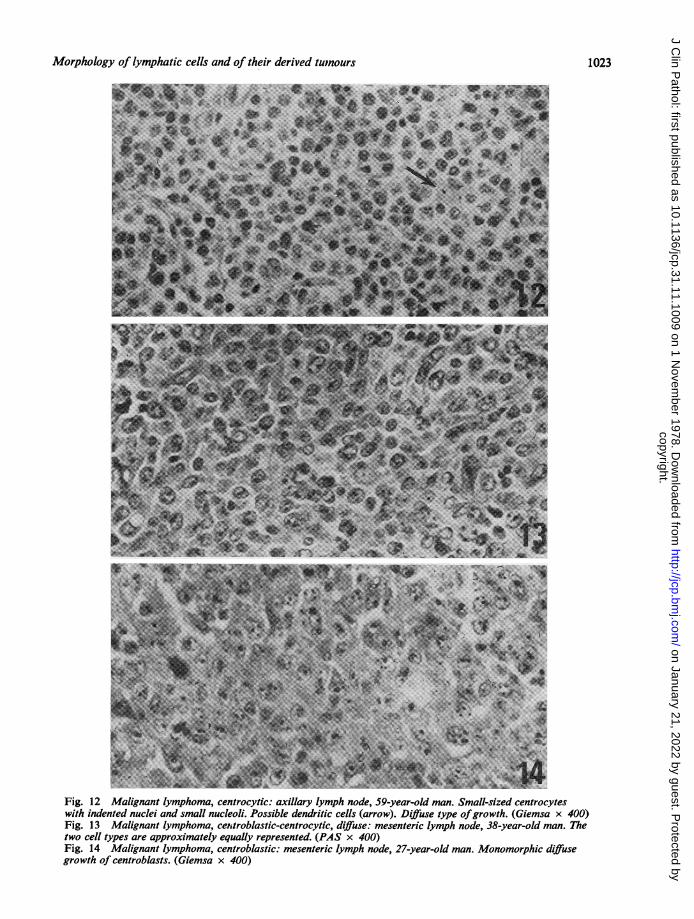
CENTROCYTIC MALIGNANT LYMPHOMA(GERMINOCYTOMA)Lymphomas made up of cells that correspond to thenormal centrocyte have also been defined as poorlydifferentiated, lymphocytic ML. In lymph nodes,these tumours always reveal a diffuse growthpattern, even though they may also display avaguely nodular aspect (Fig. 12). In such cases thereare ill-defined, small, compact nodules, whichclosely resemble primary follicles, but a definitefollicular architecture with compression of theperiphery of the reticulin fibre network and of thevessels is never seen. The centrocytes reveal a ratherwide range of sizes. In general, they are small ormedium-sized (Kaiserling, 1977a), have cleaved andindented nuclei, usually with one or rarely two verysmall nucleoli that are distant from the nuclearmembrane, and a small amount of slightly baso-philic PAS-negative cytoplasm. The main differencesfrom lymphocytes ofB-CLL in sections consist of theslightly larger size, the nuclear indentations, and thechromatin structure; however, the nuclear irregulari-ties are less impressive in imprints. In those cases inwhich the cells are predominantly small and notheavily indented, the diagnosis of lymphocyticintermediate type ML has been applied (Dick et al.,1974). However, even small indented cells are differ-ent from the small lymphocytes of CLL, as has alsobeen shown in viable cells (Schrek and Donnelly,1961).There are cases in which the centrocytes are quite
large, with a size that is double that of the small ones.ML made up of large centrocytes is a separate sub-group termed large, cleaved, follicular centre cell MLof Lukes and Collins' (1974) classification. Large-centrocytes have prominent nucleoli and a baso-philic cytoplasm, show marked atypia, and reveal ahigh mitotic index. Cell kinetic studies indicate thatlarge cell centrocytic ML has a higher degree of pro-liferation than do the small cell types (Silvestrini et-al., 1977). For these cases centrocytic sarcoma wasthe diagnosis proposed by Kaiserling (1977a).Of the accompanying cells centroblasts are usually
recognisable in sections but centroblast-like cells may
1022
copyright. on January 21, 2022 by guest. P
rotected byhttp://jcp.bm
j.com/
J Clin P
athol: first published as 10.1136/jcp.31.11.1009 on 1 Novem
ber 1978. Dow
nloaded from
Morphology of lymphatic cells and of their derived tuinoursiisW;- !........... ... |;>. iiU
Fig. 12 Malignant lymphoma, centrocytic: axillary lymph node, 59-year-old man. Small-sized centrocyteswith indented nuclei and small nucleoli. Possible dendritic cells (arrow). Diffuse type ofgrowth. (Giemsa x 400)Fig. 13 Malignant lymphoma, cetitroblastic-centrocytic, diffuse: mesenteric lymph node, 38-year-old man. Thetwo cell types are approximately equally represented. (PAS x 400)Fig. 14 Malignant lymphoma, centroblastic: mesenteric lymph node, 27-year-old man. Monomorphic diffusegrowth of centroblasts. (Giemsa x 400)
1023
copyright. on January 21, 2022 by guest. P
rotected byhttp://jcp.bm
j.com/
J Clin P
athol: first published as 10.1136/jcp.31.11.1009 on 1 Novem
ber 1978. Dow
nloaded from
Franco Rilke, Silvana Pilotti, Antonino Carbone, and Luciano Lombardi
occasionally be identified in imprint preparations ofaffected lymph nodes. Furthermore, a small numberof plasma cells and precursors may be found. Insome cases there is a variable amount of sclerosis.Interspersed dendritic reticulum cells are a frequentfinding even on light microscopy which is confirmedon electron microscopy (Lennert et al., 1975b). Inimprints of lymph nodes and smears of extranodalcentrocytic ML, centrocytes are quite typical becauseof their nuclear shape and the lightly basophilic, smallcytoplasm (Rilke et al., 1978b). Small cell centro-cytic ML seems to be among those tumours the cellsof which have been shown to have immunologicalproperties intermediate between CLL and follicularcentroblastic-centrocytic ML (Jaffe et al., 1977) andfrequently to show a strong alkaline phosphataseactivity on their cellular membranes, thus reflectingsimilarities to the cells of primary follicles (Nanbaet al., 1977a). In imprints, centrocytes reveal amoderate acid phosphatase reaction and a cellmembrane-bound ATPase activity (Kaiserling,1977a).
CENTROBLASTIC-CENTROCYTIC MALIGNANT
LYMPHOMA (GERMINOBLASTOMA)Even if Brill et al. (1925) and Symmers (1927) arecommonly given credit for the first description offollicular ML, the first clear documentation of a MLwith a germinal centre-like structure was given in1916 by Ghon and Roman. This entity, which ispractically unknown in childhood (Butler, 1969;Lennert, 1973; Hausner et al., 1977), is most com-monly, at least in the initial stages, made up offollicular structures of variable size, which are verylikely to be the malignant counterpart of thesecondary lymphatic follicle, since they contain twoproliferating cell types which are present in thenormal follicles in various proportions, depending ontheir functional activity (Lennert and Muller-Hemerlink, 1975). De novo diffuse centroblastic-centrocytic ML is uncommon, while in a number ofcases follicular and diffuse patterns are present at thesame time. Cytologically, the neoplastic populationis made up of centrocytes, which are identical withthose described previously, and of centroblasts whichmay also show some variation in size. Their nucleusis round and contains a delicate chromatin networkwith two or three prominent nucleoli, which areadjacent to the nuclear membrane. The cytoplasm isbasophilic and pyroninophilic and does not containPAS-positive material. In imprints, centrocytes andcentroblasts reveal a moderate acid phosphataseactivity. 5'-Nucleotidase and adenosinetriphos-phatase activities were found at the cellular mem-brane of centroblasts and centrocytes (Lennert andRinnerberg, 1961; Muller-Hermelink, 1974;
Kaiserling, 1977a). A variant of this lymphoma witha variable degree of sclerosis was described byBennett and Millett (1969) and has a better prognosis(Bennett, 1975).
Proteinaceous, eosinophilic, and PAS-positiveprecipitate is present in some of the neoplasticfollicles (Rosas-Uribe et al., 1973). Intercellularaccumulation of immunoglobulin and/or antigen-antibody complexes in neoplastic follicles is muchless evident than it is in benign hyperplastic germinalcentres (Braylan and Rappaport, 1973). The presenceof dendritic reticulum cells may already be suspectedon light microscopy in sections and in smears, but itcan be better demonstrated ultrastructurally (Lennertand Niedorf, 1969; Glick et al 1975; Lennert andMuiller-Hermelink, 1975; Levine and Dorfman,1975). When the centrocytes predominate, as is morecommonly the case, the histological diagnosis ofnodular, poorly differentiated lymphocytic ML isusually applied. When centrocytes and centroblastsare present in a similar proportion, then the diagnosisof nodular mixed lymphocytic and histiocytic ML isusually made, whereas when the large cells pre-dominate, the diagnosis of nodular histiocytic ML isapplied. The most common combinations are thefirst and the second. The mitotic index is usually low,but it is increased when the centroblasts predominate.Follicular lymphomas may maintain their follicularpattern for a long time, even until the death of thepatient (Warnke et al., 1977); however, they mayalso tend to transform into the diffuse form(Fig. 13), while the reverse has never been observed(Rappaport et al., 1956). The morphological steps ofthe transformation from the follicular to the diffusetype of growth pattern have already been illustrated(Rappaport etal., 1956; Rappaport, 1966; Lukes andCollins, 1975). The evolution to the diffuse growthmay either not produce changes in the cellular popu-lation or accompany an abrupt increase eitherof centroblasts or, rarely, of anaplastic centrocytes.The increase of the centroblastic component isparalleled by the increase of the cellular prolifer-ation rate, as has been shown by cell kineticstudies (Silvestrini et al., 1977). Centroblastic-centrocytic ML may eventually undergo an ana-plastic transformation, and in that case identificationof the histological type becomes difficult. Differen-tial diagnosis between centrocytic and centroblastic-centrocytic, diffuse ML has been discussed in detail(Kaiserling, 1977a).
Malignant lymphomas made up exclusively oralmost exclusively of cells identifiable as malignantcentroblasts, that is, large, round cells with baso-philic cytoplasm and a vesicular nucleus with two tothree nucleoli adjacent or adeherent to the nuclearmembrane (Fig. 14), are usually diagnosed as histio-
1024
copyright. on January 21, 2022 by guest. P
rotected byhttp://jcp.bm
j.com/
J Clin P
athol: first published as 10.1136/jcp.31.11.1009 on 1 Novem
ber 1978. Dow
nloaded from
Morphology of lymphatic cells and of their derived tumours
16d.:..A..wx...
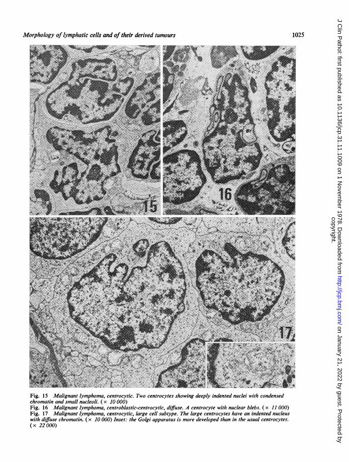
7.Li: yFig. 15 Malignant lymphoma, centrocytic. Two centrocytes showing deeply indented nuclei with condensedchromatin and small nucleoli. ( x 10 000)Fig. 16 Malignant lymphoma, centroblastic-centrocytic, diffuse. A centrocyte with nuclear blebs. ( x 11 000)Fig. 17 Malignant lymphoma, centrocytic, large cell subtype. The large centrocytes have an indented nucleuswith diffuse chromatin. ( x 10 000) Inset: the Golgi apparatus is more developed than in the usual centrocytes.(x 22000)
1025
..W.:I
copyright. on January 21, 2022 by guest. P
rotected byhttp://jcp.bm
j.com/
J Clin P
athol: first published as 10.1136/jcp.31.11.1009 on 1 Novem
ber 1978. Dow
nloaded from
Franco Rilke, Silvana Pilotti, Antonino Carbone, and Luciano Lombardi
cytic ML. In sequential biopsies of centroblastic-centrocytic ML and at necropsy (Lennert, 1976) offormer centroblastic-centrocytic ML, the transfor-mation into a pure centroblastic ML (germino-blastic sarcoma) may become evident and should beconsidered as the transition to a high-grade MLwith a poor prognosis. Although infrequently,primary centroblastic malignant lymphomas doexist as de novo malignancies and may occasionallybe follicular (Kaiserling, 1977a), even if more oftenthey appear with a diffuse growth pattern. The mostprominent feature of centroblastic ML is the uniformproliferation of highly malignant-looking cells,which retain the morphological and immunologicalcharacteristics of the centroblast. Cytologically, thesecells are larger and different from those of Burkitttype ML, in which the nucleoli are more variable innumber and size and commonly not adherent to thenuclear membrane and the cytoplasm is more deeplybasophilic and contains lipidic droplets. Quite oftenthe proliferating cell population is also made up, inaddition to the malignant centroblasts, of some im-munoblasts and centrocytes. Immunological studieson six cases of large cell ML, which developed inpatients with a previous diagnosis of 'nodular'lymphoma, revealed the persistence of B-lymphocytemarkers (Jaffe et al., 1977).
Ultrastructurally, the centrocytes have a diameter(5-10 ,t) and cytoplasmic characteristics thatapproach those of the small circulating lymphocytes,from which they differ mainly in the morphology oftheir nuclei. Centrocytes (Fig. 15) have a deeplycleaved nuclear membrane, which confers on thenucleus a characteristic lobed appearance with lobesconnected by narrow bridges. Heterochromatin isless dense than it is in normal small lymphocytes, andnucleoli are inconspicuous or absent. Nucleolarpockets (blebs) are sometimes found (Fig. 16). Thecytoplasmic areas surrounded by the nuclearpockets occasionally contain strictly interlacedtubules of smooth endoplasmic reticulum (Fig. 16).Their scanty cytoplasm contains a few small mito-chondria and rough endoplasmic reticulum cisternae,a small Golgi apparatus, and numerous monori-bosomes. Centrioles, lipid droplets, small micro-filament bundles, and lysosome-like dense granulesare observed in some cells. Neoplastic centrocytesare similar to normal cells but their nuclei are morewrinkled and show a larger number of blebs than dothose of normal cells.Large centrocytes (Fig. 17) have similar twisted
nuclei but are larger (7-14 ,u in diameter) and showmore signs of morphological activation than do thesmall centrocytes. The chromatin is more dispersed,the nucleoli, the Golgi apparatus (Fig. 17, inset), andthe rough endoplasmic reticulum are more promi-
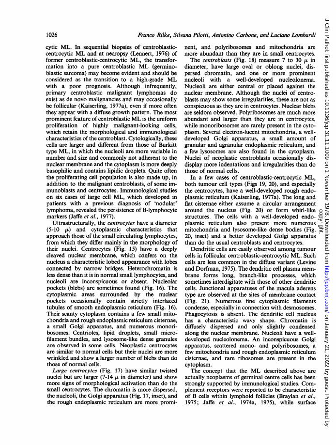
nent, and polyribosomes and mitochondria aremore abundant than they are in small centrocytes.The centroblasts (Fig. 18) measure 7 to 30 ,u in
diameter, have large oval or oblong nuclei, dis-persed chromatin, and one or more prominentnucleoli with a well-developed nucleolonema.Nucleoli are either central or placed against thenuclear membrane. Although the nuclei of centro-blasts may show some irregularities, these are not asconspicuous as they are in centrocytes. Nuclear blebsare seldom observed. Polyribosomes are much moreabundant and larger than they are in centrocytes,while monoribosomes are rarely present in the cyto-plasm. Several electron-lucent mitochondria, a well-developed Golgi apparatus, a small amount ofgranular and agranular endoplasmic reticulum, anda few lysosomes are also found in the cytoplasm.Nuclei of neoplastic centroblasts occasionally dis-display more indentations and irregularities than dothose of normal cells.
In a few cases of centroblastic-centrocytic ML,both tumour cell types (Figs 19, 20), and especiallythe centrocytes, have a well-developed rough endo-plasmic reticulum (Kaiserling, 1977a). The long andflat cisternae either assume a circular arrangementaround the nucleus (Fig. 20) or form whirl-likestructures. The cells with a well-developed endo-plasmic reticulum also present more numerousmitochondria and lysosome-like dense bodies (Fig.20, inset) and a better developed Golgi apparatusthan do the usual centroblasts and centrocytes.
Dendritic cells are easily observed among tumourcells in follicular centroblastic-centrocytic ML. Suchcells are less common in the diffuse variant (Levineand Dorfman, 1975). The dendritic cell plasma mem-brane forms long, branch-like processes, whichsometimes interdigitate with those of other dendriticcells. Junctional apparatuses of the macula aderenstype are observed at the sites of membrane contact(Fig. 21). Numerous fine cytoplasmic filamentscondense, especially in connection with desmosomes.Phagocytosis is absent. The dendritic cell nucleushas a characteristic wavy shape. Chromatin isdiffusely dispersed and only slightly condensedalong the nuclear membrane. Nucleoli have a well-developed nucleolonema. An inconspicuous Golgiapparatus, scattered mono- and polyribosomes, afew mitochondria and rough endoplasmic reticulumcisternae, and rare ribosomes are present in thecytoplasm.The concept that the ML described above are
actually neoplasms of germinal centre cells has beenstrongly supported by immunological studies. Com-plement receptors were reported to be characteristicof B cells within lymphoid follicles (Braylan et al.,1975; Jaffe et al., 1974a, 1975), while surface
1026
copyright. on January 21, 2022 by guest. P
rotected byhttp://jcp.bm
j.com/
J Clin P
athol: first published as 10.1136/jcp.31.11.1009 on 1 Novem
ber 1978. Dow
nloaded from
Morphology of lymphatic cells and of their derived i
C
.j. I
..cV v
tumours 1027
'~~~~~~.Si< , + ; '
4 0+1W
;y°>> >tp-¢'ot;4'aA '
r~~a_ E G w., 4 -\S^ $,~~~X_ 1KaiZ; g e~~bsg ^>L jj= -Zt~~~'04 e tyasfle
HA
_,,aS;twWD~~'4* ;
Fig. 18 Malignant lymphoma, centroblastic-centrocytic. A centroblast presenting a roughly oval nucleus withdispersed chromatin and three well-developed nucleoli adjacent to the nuclear membrane. ( x 9000)Fig. 19 Malignant lymphoma, centroblastic-centrocytic, diffuse. A centroblast with well-developed roughendoplasmic reticulum. (x 12 000)Fig. 20 Malignant lymphoma, centroblastic-centrocytic, diffuse. A centrocyte with abundant flatendoplasmic reticulum cisternae and some lipid droplets. ( x 12 000) Inset: numerous lysosome-like dense bodiesin a centrocyte with abundant rough endoplasmic reticulum. ( x 9000)
- *\
:- 'oe. JS
*Si e; s;
copyright. on January 21, 2022 by guest. P
rotected byhttp://jcp.bm
j.com/
J Clin P
athol: first published as 10.1136/jcp.31.11.1009 on 1 Novem
ber 1978. Dow
nloaded from
Franco Rilke, Silvana Pilotti, Antonino Carbone, and Luciano Lombardi
immunoglobulin-bearing B cells are distributed inboth follicles and the medullary cords. It was shownthat follicular lymphomas are of B-cell type originand that all their cells have complement receptors andthe majority bear surface immunoglobulin (Gajl-Peczalska et al., 1973; Shevach et al., 1973; Jaffe etal., 1974a; Aisenberg and Long, 1975; Leech et al.,1975a; Bloomfield et al., 1976). The same applies tocentrocytic ML (Stein, 1975). The cells of follicularlymphomas have neither receptors for cytophilicantibody nor do they form spontaneous E rosettes.The presence of the complement receptor is un-
related to the cytological subtype of follicularlymphoma, although a loss of these receptors was
reported during the transition from the nodular tothe diffuse pattern (Crossman et al., 1977). Except fora few IgG-containing cells, none of the tumour cellscontains cytoplasmic immunoglobulin (Johanssonet al., 1976). In contrast to B-CLL lymphocytes, thesmall follicular centre cells of cases with leukaemicmanifestations and either a nodular or diffusestructure show a lesser tendency to form rosettes withmouse erythrocytes (Koziner et al., 1977). Quiteconceivably, the cells of diffuse centroblastic-centrocytic ML with anaplasia may behave as'receptor-silent' cells or show immature receptorprofiles, as has been reported (Habeshaw et al., 1977)for some diffuse, mixed, and histiocytic ML, whichcorrespond to large cleaved and non-cleaved follicu-lar centre cell ML described by Lukes and Collins(1974).
It has been postulated that for the characterisationof centroblastic-centrocytic ML, further investiga-tion of the tissue between the neoplastic follicles mayyield significant additional information. In fact,morphologically on light and electron microscopy,the interfollicular tissue contains many structures(postcapillary venules with recirculating lympho-cytes, T-associated plasma cells, interdigitatingreticulum cells) that are characteristic of the paracor-tical area of lymph nodes (Kaiserling, 1977a). Inaddition, immunologically, at the periphery of theneoplastic follicles, an unforeseen accumulation of Tlymphocytes has been detected (Jaffe et al., 1977).Whether this finding should be interpreted as a hostdefence or as a process relevant to the folliculartumorigenesis remains a challenging question.
In a number of cases, ML of germinal centre cellorigin presents with leukaemic spread, the firstdescription of which may be identified in the reportof Isaacs (1937). He reported a type of lymphocyticleukaemia associated with lymphosarcoma in thelymph nodes, which he believed to be different fromother types of lymphocytic leukaemia. Lymphosar-coma cell leukaemia may occur either as an earlyphenomenon (early leukaemic lymphosarcoma) or
may appear as a late manifestation (late leukaemiclymphosarcoma) (Mathe et al., 1976). In all cases ofblood involvement the marrow is invariably infil-trated, but whether or not marrow infiltrationwithout blood involvement should be consideredleukaemic conversion is debatable (Wintrobe et al.,1974).Owing to the use of the Rappaport classification,
in which nodular, poorly differentiated lymphocyticML comprise pseudonodular centrocytic as well asfollicular centroblastic-centrocytic ML, and diffuse,poorly differentiated lymphocytic ML comprisecentrocytic and centroblastic-centrocytic diffuse ML,it is difficult to reinterpret previous reports, sincequite often nodular and diffuse ML cases are reportedtogether. The impression that one gains, however, isthat in both instances centrocytes are the mostfrequently circulating cell type (Figs 22 and 23). Inany case, centrocytes in blood smears and in marrowsmears bear striking similarities to the 'notched-nucleus' cells described by Anday and Schmitz(1952) in follicular lymphomas, to the 'haemato-gones' described by Rosenthal et al. (1952) also infollicular lymphomas, and to the cells observed insome of the cases described by Schwartz et al.(1965).The same cell type corresponds to the third type ofleukaemic lymphosarcomas described by Mathe etal. (1975b) and to the notched-nucleus lymphocytesdescribed by Spiro et al. (1975). The cytoplasm maycontain vacuoles, azurophilic granules, and baso-philic granules, which appear structureless onelectron microscopy (Wintrobe et al., 1974). In aseries of 16 cases with lymphosarcoma cell leukaemia(Schnitzer et at., 1970) lymph node biopsy revealeda poorly differentiated lymphocytic ML, which wasdiffuse in six and nodular in 10 cases. In the majorityof diffuse ML leukaemia was present at diagnosis butwas a later manifestation in the nodular cases. Thechanges in the course of the disease were also demon-strated by the fact that, at the time of necropsy, onecase only had maintained the nodular structure ofthe lymphoma, while the others had changed towardsa diffuse pattern.
In the blood of centroblastic-centrocytic follicularML, mostly centrocytes are recognisable, whereascentroblasts may be found only occasionally(Lennert, 1969). The circulating centrocytes revealthe same previously described immunological surfacemarkers as do the cells in the solid tumour, thusconfirming their B-type nature and germinal centreorigin. There are, however, reports on dual markersfor B and T cells detected in cases of lymphosarcomacell leukaemia (Hsu et al., 1975; Lin and Hsu, 1976).
In a survey of 75 cases of follicular lymphoma,among which all cytological subtypes (small andlarge cells) were represented, 25 cases were found to
1028
copyright. on January 21, 2022 by guest. P
rotected byhttp://jcp.bm
j.com/
J Clin P
athol: first published as 10.1136/jcp.31.11.1009 on 1 Novem
ber 1978. Dow
nloaded from
Morphology of lymphatic cells and of their derived tumours
have abnormal cells in the blood (Spiro et al., 1975).In 17 the blood lymphocyte count ranged between5 and 30 x 109/l, and in another eight cases it wasbelow 5 x 10⁹/l; in all these cases, however, themarrow aspirate films showed neoplastic lymphocyticinfiltration made up of abnormal notched-nucleuscells. These were also found in blood films of cases ofnodular lymphoma with large cell type predomi-nance (histiocytic). The most important definingcharacteristic of the circulating cells, in addition tothe nuclear clefts of varying depth, was the chro-matin pattern, which was more homogeneous thanwas that of the small lymphocytes and did notreveal any structure made up of coarse blocks ofdensely stained chromocentres. In addition to thecells with the indented nuclei, other circulating cellswith scanty basoplilic cytoplasm and barely per-ceptible nuclear notches were identified in theblood.
Examination of marrow trephine biopsy sectionsat the time of initial diagnosis of follicular lym-phomas reveals a high percentage with involvement(Dick et al., 1974; McKenna et al., 1975; Castellaniet al., 1977), although the degree of infiltration andthe pattern of distribution is variable (Jones et al.,1972, 1973; Dick et al., 1974). Marrow films aresomewhat less reliable. For diffuse, poorly differ-entiated lymphocytic ML, marrow invasion wasfound by trephine in about 30% of cases. Marrowinvolvement by nodular lymphomas with predomi-nantly small cells (nodular lymphocytic, poorlydifferentiated) wasmuch more frequently encounteredthan in those with large cells (nodular histiocytic)(Jones et al., 1972, 1973).With regard to the cell types encountered in
marrow invasion, the morphological description of asmall cell (7-10 pz in diameter) with an irregularlycleaved nucleus, small inconspicuous nucleoli, andscanty, lightly basophilic cytoplasm corresponds tothat of a centrocyte, small cleaved follicular centrecell (Lukes and Collins, 1974), small nodularlymphoma cell (McKenna et al., 1975), 'haemato-gone' (Rosenthal et al., 1952), and the lymphocyticcell of poorly differentiated lymphocytic ML (Dicket al., 1974). If this is the cell most commonly foundin these instances, it is also not uncommon toidentify both large cells (which measure up to 20 ILin diameter with a round nucleus, multiple nucleoli,and basophilic cytoplasm) that correspond to centro-blasts and cells with features intermediate betweenthe centrocytes and the centroblasts (Dick et al.,1974; McKenna et al., 1975). The rare circulatinglarge cells are cytologically different from immuno-blasts inasmuch as their cytoplasm is less baso-philic and their large nuclei contain multiple nucleolirather than a single huge central nucleolus.
The morphological differences between haemato-gones found in the marrow of children and adults inhealth and in various pathological conditions and theneoplastic haematogones (Rosenthal et al., 1952) havebeen noted by McKenna et al. (1975). On the otherhand, the occasional finding of cells similar to smallcentrocytes in marrow films in various non-neoplastichaematological conditions has also been reported(McKenna et al., 1975). It seems, therefore, that thiscell type is sufficiently distinct to be differentiated oncytological grounds alone from the small lympho-cyte.
In summary, almost all the reports on the leu-kaemic manifestations of poorly differentiated lym-phocytic ML, both nodular and diffuse, and ofnodular ML of other cell types clearly indicate thatthe haematological, morphological, and immuno-logical features observed are different from those ofCLL. It appears that in these cases the circulatingcells are exclusively or predominantly centrocytes,which may reveal more or less accentuated features ofatypia, whereas the presence of centroblasts is muchless common. The need for a better correlation in thefuture between histological and haematological cellmorphology of leukaemic, poorly differentiatedlymphocytic ML is justified, since immunologicaldifferences between diffuse and nodular types of MLhave been reported (Aisenberg and Long, 1975;Aisenberg and Wilkes, 1976). In fact, clinical andhaematological differences between the purely centro-cytic diffuse and the follicular centroblastic-centro-cytic ML do exist. The greater tendency towardsgeneralised dissemination and marrow invasion(Castellani et al., 1977), and leukaemic change insome cases with a high WBC and a fulminant course,in addition to the existence of the large cell variant(Silvestrini et al., 1977), are some of the distin-guishing features of centrocytic ML versus follicularcentroblastic-centrocytic ML, which in turn aremore frequently associated with a chronic leukaemicmanifestation. At the same time it is fair to add thatthe importance of these distinctions has been doubted(Dorfman, 1977).
BURKITT LYMPHOMAIn spite of clinical (Levine et al., 1975), epidemio-logical, immunological (Epstein et al., 1976), andvirological (Zur Hausen, 1975; Andersson et al.,1976; Ziegler et al., 1976) differences betweenendemic African (Burkitt, 1958) and sporadicAfrican and non-African (O'Conor et al., 1965;Burkitt, 1967) Burkitt lymphomas, their morpho-logical-cytological expressions are very similar(Berard et al., 1969). This tumour has beenconsidered as the most classic example of anundifferentiated ML because of its uniform and
1029
copyright. on January 21, 2022 by guest. P
rotected byhttp://jcp.bm
j.com/
J Clin P
athol: first published as 10.1136/jcp.31.11.1009 on 1 Novem
ber 1978. Dow
nloaded from
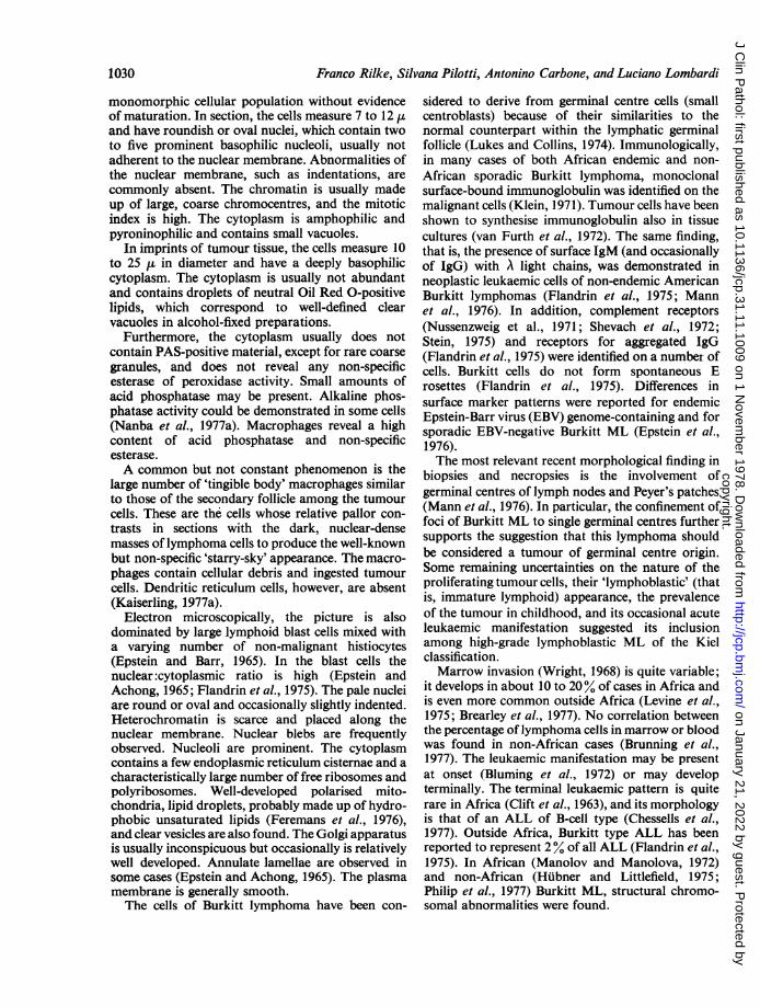
Franco Rilke, Silvana Pilotti, Antonino Carbone, and Luciano Lombardi
monomorphic cellular population without evidenceof maturation. In section, the cells measure 7 to 12 ,and have roundish or oval nuclei, which contain twoto five prominent basophilic nucleoli, usually notadherent to the nuclear membrane. Abnormalities ofthe nuclear membrane, such as indentations, arecommonly absent. The chromatin is usually madeup of large, coarse chromocentres, and the mitoticindex is high. The cytoplasm is amphophilic andpyroninophilic and contains small vacuoles.
In imprints of tumour tissue, the cells measure 10to 25 p1 in diameter and have a deeply basophiliccytoplasm. The cytoplasm is usually not abundantand contains droplets of neutral Oil Red O-positivelipids, which correspond to well-defined clearvacuoles in alcohol-fixed preparations.
Furthermore, the cytoplasm usually does notcontain PAS-positive material, except for rare coarsegranules, and does not reveal any non-specificesterase of peroxidase activity. Small amounts ofacid phosphatase may be present. Alkaline phos-phatase activity could be demonstrated in some cells(Nanba et al., 1977a). Macrophages reveal a highcontent of acid phosphatase and non-specificesterase.A common but not constant phenomenon is the
large number of 'tingible body' macrophages similarto those of the secondary follicle among the tumourcells. These are the cells whose relative pallor con-trasts in sections with the dark, nuclear-densemasses of lymphoma cells to produce the well-knownbut non-specific 'starry-sky' appearance. The macro-phages contain cellular debris and ingested tumourcells. Dendritic reticulum cells, however, are absent(Kaiserling, 1977a).
Electron microscopically, the picture is alsodominated by large lymphoid blast cells mixed witha varying number of non-malignant histiocytes(Epstein and Barr, 1965). In the blast cells thenuclear :cytoplasmic ratio is high (Epstein andAchong, 1965; Flandrin et al., 1975). The pale nucleiare round or oval and occasionally slightly indented.Heterochromatin is scarce and placed along thenuclear membrane. Nuclear blebs are frequentlyobserved. Nucleoli are prominent. The cytoplasmcontains a few endoplasmic reticulum cisternae and acharacteristically large number of free ribosomes andpolyribosomes. Well-developed polarised mito-chondria, lipid droplets, probably made up of hydro-phobic unsaturated lipids (Feremans et al., 1976),and clear vesicles are also found. The Golgi apparatusis usually inconspicuous but occasionally is relativelywell developed. Annulate lamellae are observed insome cases (Epstein and Achong, 1965). The plasmamembrane is generally smooth.The cells of Burkitt lymphoma have been con-
sidered to derive from germinal centre cells (smallcentroblasts) because of their similarities to thenormal counterpart within the lymphatic germinalfollicle (Lukes and Collins, 1974). Immunologically,in many cases of both African endemic and non-African sporadic Burkitt lymphoma, monoclonalsurface-bound immunoglobulin was identified on themalignant cells (Klein, 1971). Tumour cells have beenshown to synthesise immunoglobulin also in tissuecultures (van Furth et al., 1972). The same finding,that is, the presence of surface IgM (and occasionallyof IgG) with A light chains, was demonstrated inneoplastic leukaemic cells of non-endemic AmericanBurkitt lymphomas (Flandrin et al., 1975; Mannet al., 1976). In addition, complement receptors(Nussenzweig et al., 1971; Shevach et al., 1972;Stein, 1975) and receptors for aggregated IgG(Flandrin et al., 1975) were identified on a number ofcells. Burkitt cells do not form spontaneous Erosettes (Flandrin et al., 1975). Differences insurface marker patterns were reported for endemicEpstein-Barr virus (EBV) genome-containing and forsporadic EBV-negative Burkitt ML (Epstein et al.,1976).The most relevant recent morphological finding in
biopsies and necropsies is the involvement ofgerminal centres of lymph nodes and Peyer's patches(Mann et al., 1976). In particular, the confinement offoci of Burkitt ML to single germinal centres furthersupports the suggestion that this lymphoma shouldbe considered a tumour of germinal centre origin.Some remaining uncertainties on the nature of theproliferating tumour cells, their 'Iymphoblastic' (thatis, immature lymphoid) appearance, the prevalenceof the tumour in childhood, and its occasional acuteleukaemic manifestation suggested its inclusionamong high-grade lymphoblastic ML of the Kielclassification.Marrow invasion (Wright, 1968) is quite variable;
it develops in about 10 to 20% of cases in Africa andis even more common outside Africa (Levine et al.,1975; Brearley et al., 1977). No correlation betweenthe percentage oflymphoma cells in marrow or bloodwas found in non-African cases (Brunning et al.,1977). The leukaemic manifestation may be presentat onset (Bluming et al., 1972) or may developterminally. The terminal leukaemic pattern is quiterare in Africa (Clift et al., 1963), and its morphologyis that of an ALL of B-cell type (Chessells et al.,1977). Outside Africa, Burkitt type ALL has beenreported to represent 2% of all ALL (Flandrin et al.,1975). In African (Manolov and Manolova, 1972)and non-African (Hubner and Littlefield, 1975;Philip et al., 1977) Burkitt ML, structural chromo-somal abnormalities were found.
1030
copyright. on January 21, 2022 by guest. P
rotected byhttp://jcp.bm
j.com/
J Clin P
athol: first published as 10.1136/jcp.31.11.1009 on 1 Novem
ber 1978. Dow
nloaded from
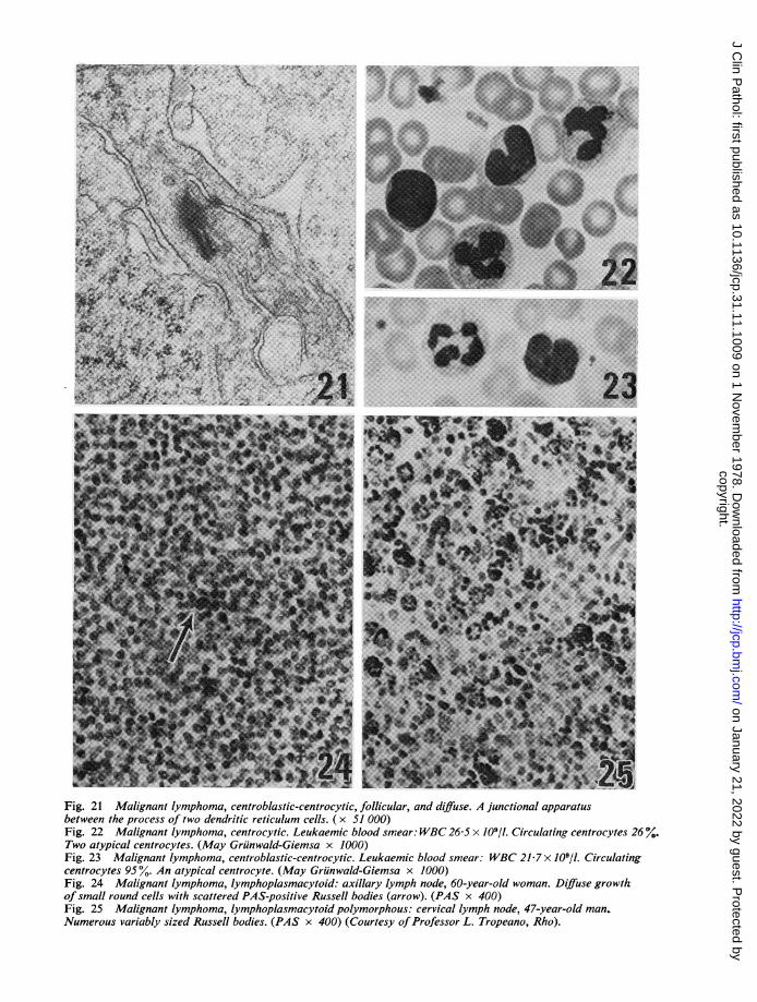
... ... ..._
...::....:...:
Fig. 21 Malignant lymphoma, centroblastic-centrocytic, follicular, and diffuse. A junctional apparatusbetween the process of two dendritic reticulum cells. ( x 51 000)Fig. 22 Malignant lymphoma, centrocytic. Leukaemic blood smear: WBC 26.5 x 10⁹/l. Circulating centrocytes 26%.Two atypical centrocytes. (May Grunwald-Giemsa x 1000)Fig. 23 Malignant lymphoma, centroblastic-centrocytic. Leukaemic blood smear: WBC 21-7 x 1O9/1. Circulatingcentrocytes 95%. An atypical centrocyte. (May Grunwald-Giemsa x 1000)Fig. 24 Malignant lymphoma, lymphoplasmacytoid: axillary lymph node, 60-year-old woman. Diffuse growthof small round cells with scattered PAS-positive Russell bodies (arrow). (PAS x 400)Fig. 25 Malignant lymphoma, lymphoplasmacytoid polymorphous: cervical lymph node, 47-year-old man.Numerous variably sized Russell bodies. (PAS x 400) (Courtesy ofProfessor L. Tropeano, Rho).
copyright. on January 21, 2022 by guest. P
rotected byhttp://jcp.bm
j.com/
J Clin P
athol: first published as 10.1136/jcp.31.11.1009 on 1 Novem
ber 1978. Dow
nloaded from
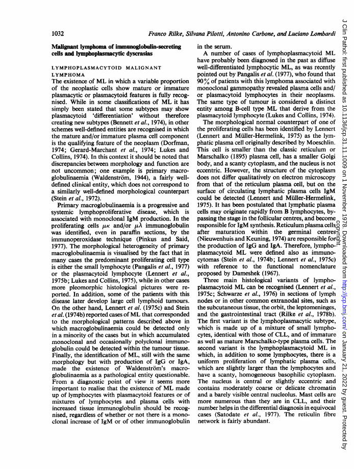
Franco Rilke, Silvana Pilotti, Antonino Carbone, and Luciano Lombardi
Malignant lymphoma of immunoglobulin-secretingcells and lymphoplasmacytic dyscrasias
LYMPHOPLASMACYTOID MALIGNANTLYMPHOMAThe existence of ML in which a variable proportionof the neoplastic cells show mature or immatureplasmacytic or plasmacytoid features is fully recog-nised. While in some classifications of ML it hassimply been stated that some subtypes may showplasmacytoid 'differentiation' without thereforecreating new subtypes (Bennett et al., 1974), in otherschemes well-defined entities are recognised in whichthe mature and/or immature plasma cell componentis the qualifying feature of the neoplasm (Dorfman,1974; Gerard-Marchant et al., 1974; Lukes andCollins, 1974). In this context it should be noted thatdiscrepancies between morphology and function arenot uncommon; one example is primary macro-globulinaemia (Waldenstr6m, 1944), a fairly well-defined clinical entity, which does not correspond toa similarly well-defined morphological counterpart(Stein et al., 1972).Primary macroglobulinaemia is a progressive and
systemic lymphoproliferative disease, which isassociated with monoclonal IgM production. In theproliferating cells PK and/or HtA immunoglobulinwas identified, even in paraffin sections, by theimmunoperoxidase technique (Pinkus and Said,1977). The morphological heterogeneity of primarymacroglobulinaemia is visualised by the fact that inmany cases the predominant proliferating cell typeis either the small lymphocyte (Pangalis et al., 1977)or the plasmacytoid lymphocyte (Lennert et al.,1975b; Lukes and Collins, 1975), while in other casesmore pleomorphic histological pictures were re-ported. In addition, some of the patients with thisdisease later develop large cell lymphoid tumours.On the other hand, Lennert et al. (1975c) and Steinet al. (1974b) reported cases ofML that correspondedto the morphological patterns described above inwhich macroglobulinaemia could be detected onlyin a minority of the cases but in which accumulatedmonoclonal and occasionally polyclonal immuno-globulin could be detected within the tumour tissue.Finally, the identification of ML, still with the samemorphology but with production of IgG or IgA,made the existence of Waldenstrom's macro-globulinaemia as a pathological entity questionable.From a diagnostic point of view it seems moreimportant to realise that the existence of ML madeup of lymphocytes with plasmacytoid features or ofmixtures of lymphocytes and plasma cells withincreased tissue immunoglobulin should be recog-nised, regardless of whether or not there is a mono-clonal increase of IgM or of other immunoglobulin
in the serum.A number of cases of lymphoplasmacytoid ML
have probably been diagnosed in the past as diffusewell-differentiated lymphocytic ML, as was recentlypointed out by Pangalis et al. (1977), who found that90% of patients with this lymphoma associated withmonoclonal gammopathy revealed plasma cells and/or plasmacytoid lymphocytes in their neoplasms.The same type of tumour is considered a distinctentity among B-cell type ML that derive from theplasmacytoid lymphocyte (Lukes and Collins, 1974).The morphological normal counterpart of one of
the proliferating cells has been identified by Lennert(Lennert and Miiller-Hermelink, 1975) as the lym-phatic plasma cell originally described by Moeschlin.This cell is smaller than the classic reticulum orMarschalko (1895) plasma cell, has a smaller Golgibody, and a scanty cytoplasm, and the nucleus is noteccentric. However, the structure of the cytoplasmdoes not differ qualitatively on electron microscopyfrom that of the reticulum plasma cell, but on thesurface of circulating lymphatic plasma cells IgMcould be detected (Lennert and Mililer-Hermelink,1975). It has been postulated that lymphatic plasmacells may originate rapidly from B lymphocytes, by-passing the stage in the follicular centres, and becomeresponsible for IgM synthesis. Reticulum plasma cellsafter maturation within the germinal centres(Nieuwenhuis and Keuning, 1974) are responsible forthe production of IgG and IgA. Therefore, lympho-plasmacytoid ML were defined also as immuno-cytomas (Stein et al., 1974b; Lennert et al., 1975c)with reference to the functional nomenclatureproposed by Dameshek (1967).Three main histological variants of lympho-
plasmacytoid ML can be recognised (Lennert et al.,1975c; Schwarze et al., 1976) in sections of lymphnodes or in other common extranodal sites, such asthe subcutaneous tissue, the orbit, the leptomeninges,and the gastrointestinal tract (Rilke et al., 1978b).The first variant is the lymphoplasmacytic subtype,which is made up of a mixture of small lympho-cytes, identical with those of CLL, and of immatureas well as mature Marschalko-type plasma cells. Thesecond variant is the lymphoplasmacytoid ML inwhich, in addition to some lymphocytes, there is auniform proliferation of lymphatic plasma cells,which are slightly larger than the lymphocytes andhave a scanty, homogeneous basophilic cytoplasm.The nucleus is central or slightly eccentric andcontains moderately coarse or delicate chromatinand a barely visible central nucleolus. Mast cells aremore numerous than they are in CLL, and theirnumber helps in the differential diagnosis in equivocalcases (Satodate et al., 1977). The reticulin fibrenetwork is fairly abundant.
1032
copyright. on January 21, 2022 by guest. P
rotected byhttp://jcp.bm
j.com/
J Clin P
athol: first published as 10.1136/jcp.31.11.1009 on 1 Novem
ber 1978. Dow
nloaded from
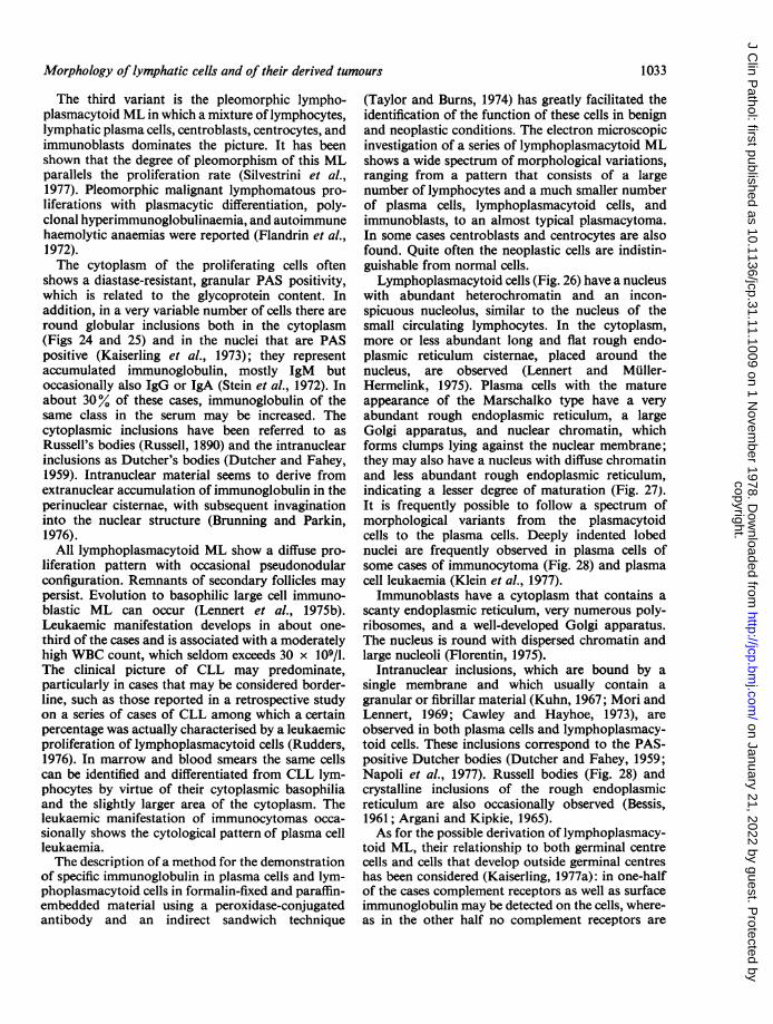
Morphology of lymphatic cells and of their derived tumours
The third variant is the pleomorphic lympho-plasmacytoid ML in which a mixture of lymphocytes,lymphatic plasma cells, centroblasts, centrocytes, andimmunoblasts dominates the picture. It has beenshown that the degree of pleomorphism of this MLparallels the proliferation rate (Silvestrini et al.,1977). Pleomorphic malignant lymphomatous pro-liferations with plasmacytic differentiation, poly-clonal hyperimmunoglobulinaemia, and autoimmunehaemolytic anaemias were reported (Flandrin et al.,1972).The cytoplasm of the proliferating cells often
shows a diastase-resistant, granular PAS positivity,which is related to the glycoprotein content. Inaddition, in a very variable number of cells there areround globular inclusions both in the cytoplasm(Figs 24 and 25) and in the nuclei that are PASpositive (Kaiserling et al., 1973); they representaccumulated immunoglobulin, mostly IgM butoccasionally also IgG or IgA (Stein et al., 1972). Inabout 30% of these cases, immunoglobulin of thesame class in the serum may be increased. Thecytoplasmic inclusions have been referred to asRussell's bodies (Russell, 1890) and the intranuclearinclusions as Dutcher's bodies (Dutcher and Fahey,1959). Intranuclear material seems to derive fromextranuclear accumulation of immunoglobulin in theperinuclear cisternae, with subsequent invaginationinto the nuclear structure (Brunning and Parkin,1976).
All lymphoplasmacytoid ML show a diffuse pro-liferation pattern with occasional pseudonodularconfiguration. Remnants of secondary follicles maypersist. Evolution to basophilic large cell immuno-blastic ML can occur (Lennert et al., 1975b).Leukaemic manifestation develops in about one-third of the cases and is associated with a moderatelyhigh WBC count, which seldom exceeds 30 x 109/1.The clinical picture of CLL may predominate,particularly in cases that may be considered border-line, such as those reported in a retrospective studyon a series of cases of CLL among which a certainpercentage was actually characterised by a leukaemicproliferation of lymphoplasmacytoid cells (Rudders,1976). In marrow and blood smears the same cellscan be identified and differentiated from CLL lym-phocytes by virtue of their cytoplasmic basophiliaand the slightly larger area of the cytoplasm. Theleukaemic manifestation of immunocytomas occa-sionally shows the cytological pattern of plasma cellleukaemia.The description of a method for the demonstration
of specific immunoglobulin in plasma cells and lym-phoplasmacytoid cells in formalin-fixed and paraffin-embedded material using a peroxidase-conjugatedantibody and an indirect sandwich technique
(Taylor and Burns, 1974) has greatly facilitated theidentification of the function of these cells in benignand neoplastic conditions. The electron microscopicinvestigation of a series of lymphoplasmacytoid MLshows a wide spectrum of morphological variations,ranging from a pattern that consists of a largenumber of lymphocytes and a much smaller numberof plasma cells, lymphoplasmacytoid cells, andimmunoblasts, to an almost typical plasmacytoma.In some cases centroblasts and centrocytes are alsofound. Quite often the neoplastic cells are indistin-guishable from normal cells.Lymphoplasmacytoid cells (Fig. 26) have a nucleus
with abundant heterochromatin and an incon-spicuous nucleolus, similar to the nucleus of thesmall circulating lymphocytes. In the cytoplasm,more or less abundant long and flat rough endo-plasmic reticulum cisternae, placed around thenucleus, are observed (Lennert and Muller-Hermelink, 1975). Plasma cells with the matureappearance of the Marschalko type have a veryabundant rough endoplasmic reticulum, a largeGolgi apparatus, and nuclear chromatin, whichforms clumps lying against the nuclear membrane;they may also have a nucleus with diffuse chromatinand less abundant rough endoplasmic reticulum,indicating a lesser degree of maturation (Fig. 27).It is frequently possible to follow a spectrum ofmorphological variants from the plasmacytoidcells to the plasma cells. Deeply indented lobednuclei are frequently observed in plasma cells ofsome cases of immunocytoma (Fig. 28) and plasmacell leukaemia (Klein et al., 1977).Immunoblasts have a cytoplasm that contains a
scanty endoplasmic reticulum, very numerous poly-ribosomes, and a well-developed Golgi apparatus.The nucleus is round with dispersed chromatin andlarge nucleoli (Florentin, 1975).
Intranuclear inclusions, which are bound by asingle membrane and which usually contain agranular or fibrillar material (Kuhn, 1967; Mori andLennert, 1969; Cawley and Hayhoe, 1973), areobserved in both plasma cells and lymphoplasmacy-toid cells. These inclusions correspond to the PAS-positive Dutcher bodies (Dutcher and Fahey, 1959;Napoli et al., 1977). Russell bodies (Fig. 28) andcrystalline inclusions of the rough endoplasmicreticulum are also occasionally observed (Bessis,1961; Argani and Kipkie, 1965).As for the possible derivation of lymphoplasmacy-
toid ML, their relationship to both germinal centrecells and cells that develop outside germinal centreshas been considered (Kaiserling, 1977a): in one-halfof the cases complement receptors as well as surfaceimmunoglobulin may be detected on the cells, where-as in the other half no complement receptors are
1033
copyright. on January 21, 2022 by guest. P
rotected byhttp://jcp.bm
j.com/
J Clin P
athol: first published as 10.1136/jcp.31.11.1009 on 1 Novem
ber 1978. Dow
nloaded from
Fig. 26 Malignant lymphoma, lymphoplasmacytoid polymorphous. A lymphoplasmacytoid cell with abundantJheterochromatin, an inconspicuous nucleolus, and flat endoplasmic reticulum cisternae placed around thenucleus. ( x 12 000)Fig. 27 Malignant lymphoma, lymphoplasmacytic. An immature plasma cell that shows a nucleus with diffusechromatin and a small nucleolus. Dense inclusions are observed in the mitochondria. ( x 12 000)Fig. 28 Malignant lymphoma, lymphoplasmacytoid polymorphous. A plasma cell with a lobed nucleus andRussell bodies within the rough endoplasmic reticulum cisternae. ( x 11 000)Fig. 29 Malignant lymphoma, immunoblastic: axillary lymph node, 66-year-old man. Diffuseproliferation of large cells with basophilic cytoplasm and large central nucleolus. A few cells revealplasmacytoid features. (Giemsa x 400)
copyright. on January 21, 2022 by guest. P
rotected byhttp://jcp.bm
j.com/
J Clin P
athol: first published as 10.1136/jcp.31.11.1009 on 1 Novem
ber 1978. Dow
nloaded from
Morphology of lymphatic cells and of their derived tumours
found (Stein, 1975). Lymphoplasmacytoid ML havetherefore been considered neoplasms of B cells, therange of which goes from cells that are halfwaybetween the non-secreting and the secreting stage tothose that are capable of secretary activity but in amajority of the cases are unable to discharge theproduct into the blood (Stein, 1975). In fact, less thanone-third of the cases of lymphoplasmacytoid MLare diagnosed clinically as Waldenstr6m's disease.Owing to the variable morphological pattern,Waldenstrdm's disease has also been defined as alymphoplasmacytic dyscrasia with abnormal secre-tion of monoclonal immunoglobulin. The samedefinition may also be applied to heavy chaindiseases, which are characterised by the secretion ofsubunits of immunoglobulin.
HEAVY CHAIN DISEASESHeavy chain diseases comprise a group of conditionsin which populations of lymphoid cells synthesiseand secrete immunoglobulin heavy chains that arestructurally defective (Buxbaum, 1976). They havealso been defined as immunoproliferative disorderswhich are characterised by the presence in the serumof a monoclonal population of molecules composedof incomplete heavy polypeptide chains whichbelong to a given class or subclass of immunoglo-bulin and are devoid of light chains (Warner et al.,1974). Three types have been recognised so far:alpha chain disease (Seligmann et al., 1968), gammachain disease (Franklin et al., 1964), and mu chaindisease (Ballard et al., 1970; Forte et al., 1970).The diagnosis is based exclusively on non-mor-
phological investigations; the clinical pattern isdifferent from that of multiple myeloma and appearsto be extremely variable. Even though in some casesno morphological evidence of abnormal cellularproliferation is found, in most cases mixed patternssimilar to those found in macroglobulinaemia areseen in marrow smears and in lymph nodes.Except for a few cases with a respiratory form of
the disease (Florin-Christensen et al., 1974), alphachain disease affects predominantly the digestivetract, producing malabsorption (Rambaud et al.,1968). Even though alpha chain disease mayoccasionally be associated with immunoblastic MLof the intestine, in the majority of cases it isassociated with what has been defined as Mediter-ranean lymphoma or immunoproliferative smallintestinal disease of Mediterranean type (Rambaudand Matuchansky, 1973). There is a diffuse andmassive infiltration of the lamina propria of theintestine by plasma cells and/or lymphocytes(Rappaport et al., 1972; Seligmann, 1975) withoutevidence of cellular atypia. The proliferation may alsobe present in mesenteric nodes, but it never spreads
to either the marrow or to extra-abdominal sites;it usually begins in the upper small intestine andsubsequently involves it completely. The cellularcomposition varies from completely mature plasmacells to a mixture of plasma cells and lymphocytes orto intermediate lymphoplasmacytoid cells. Themature cell proliferation may occasionally bemoderately invasive with atrophy of the intestinalvilli. In other cases the proliferating plasma cells aremore obviously malignant, and they invade themuscular layer of the intestine. Finally, there arecases in which there is a highly malignant prolifera-tion of immunoblasts, which may or may not beassociated with a still evident benign lympho-plasmacytoid proliferation (Ala et al., 1976). A wordof caution is needed concerning the neoplastic natureof the most benign types, since the condition is rarelyreversible either spontaneously or after antibiotictreatment with disappearance of the plasmacyteexcess and of the abnormal alpha chain (Ala et al.,1976). It has been proved that the lymphomas arisinglate in the course of the intestinal form of alphachain disease derive from the same B-cell clone as doesthe initial plasma cell proliferation (Brouet et al.,1975b, 1977;Pangalis and Rappaport, 1977; Ramotet al., 1977). By contrast, 'Western type' ML of thesmall bowel have clinical and morphologicaldifferences and seem to be made up of a differentcell type (Lewin et al., 1976).Gamma chain disease often shows intermediate
features between multiple myeloma and ML. It hasin common with the former anaemia, frequency ofinfections, and a variable clinical course, often withsevere renal disease. However, there are no lyticbone lesions. In common with ML there is oftenenlargement of lymph nodes, splenomegaly, andhepatomegaly. It may be associated with a lympho-plasmacytic ML, either leukaemic or non-leukaemic,or with pleomorphic cells in transition to immuno-blastic ML. It may occasionally be associated withextra-osseous plasmacytoma and may show plasmacell leukaemia in the terminal phase.Mu chain disease is rare and has been sometimes
found in association with an immunocytoma (Len-nert, personal communication, 1978). In marrowsamples, in addition to lymphocytosis, a certaindegree of plasmacytosis is found. The plasma cellshave been reported to be vacuolated. In both gammaand mu chain disease, cases without evidence ofabnormal cell proliferation have also been reported(Seligmann, 1972; Danon et al., 1975).
PLASMACYTIC MALIGNANT LYMPHOMAPure plasmacytic ML of extra-osseous sites was,
added later to the low-grade ML of the Kiel classifi-cation (Gerard-Marchant et al., 1974). It is equiva-
1035
copyright. on January 21, 2022 by guest. P
rotected byhttp://jcp.bm
j.com/
J Clin P
athol: first published as 10.1136/jcp.31.11.1009 on 1 Novem
ber 1978. Dow
nloaded from
Franco Rilke, Silvana Pilotti, Antonino Carbone, and Luciano Lombardi
lent to the extramedullary plasmacytoma (Wiltshaw,1976), which may arise in lymph nodes, in the upperand lower air passages and the lung, in the spleen, inthe skin and subcutaneous tissues, in the gastro-intestinal tract (stomach, small intestine, largeintestine), and less frequently in other sites. It hasbeen shown that extramedullary plasmacytoma iscomposed of an autonomous and irreversible pro-liferation of plasma cells of varying degrees ofdifferentiation and has characteristic features, whichdistinguish it from myelomatosis and from solitarymyeloma of the bone (Wiltshaw, 1976). The tumourmasses are made up of mature plasma cells of theMarschalko reticular type with an eccentric nucleusand typical arrangement of the chromatin; thenucleus is surrounded by a clear halo, and thecytoplasm is intensely eosinophilic and pyronino-philic and shows diastase-resistant PAS positivity.In addition, less well-differentiated and poorlydifferentiated plasma cells may be present to avariable extent, and in some cases the tumour ismade up of very pleomorphic plasma cells. In thesethe nuclei are more centrally located and may bemultiple, the nucleolus is more prominent, and thecytoplasm is more scarce; however, the perinuclearhalo is often still present.
In lymph nodes the plasmacytic ML may be eitherthe primary or the metastasis of an extramedullaryplasmacytoma (Fishkin and Spiegelberg, 1976).Extranodal extramedullary plasmacytomas show ahigh incidence of metastatic spread to soft tissues(Wiltshaw, 1976). It has been pointed out that inspite of the anatomical distribution of plasma cells,which is similar to that of lymphocytes, plasmacytictumours are rare in lymph nodes and much morecommon in the bone marrow. An unsuspected pro-portion of plasma cell tumours among the MLarising in the gastrointestinal tract has been reportedrecently (Henry and Farrer-Brown, 1977). At theirpresentation, extramedullary plasmacytic ML arerarely associated with serum protein abnormalities,whereas at a later stage paraproteinaemia mayappear (Wiltshaw, 1976).
Immunoblastic malignant lymphoma
The identification of this entity among the hetero-genous group of more or less differentiated reticulumcell sarcomas (Oberling, 1928; Roulet, 1930; Robb-Smith, 1938; Rossle, 1939) depends upon electronmicroscopy and immunological methods (Kaiserlinget al., 1973; Stein et al., 1972, 1974a). ImmunoblasticML is characterised by a monomorphic prolifera-tion of large cells (15-30 ,t in diameter) with paleeosinophilic but deeply pyroninophilic cytoplasm, amoderate or large amount of endoplasmic reticulum,
round or oval clear nuclei, one or two large, centrallylocated eosinophilic nucleoli, and, in some cases, thepresence of condensed IgM in the cytoplasm.Mitotic figures are common. The similarities of theneoplastic cells to transformed lymphocytes led tothe introduction of the term immunoblastic MLderived from the original terminology of Dameshek(1963).These neoplasms have previously been classified
either as undifferentiated non-Burkitt ML or asdiffuse poorly differentiated histiocytic ML. Im-munoblastic ML are high-grade malignancies andhave a very poor prognosis, allowing only a shortsurvival of the patient. Although in many instancesthe neoplasm appears rapidly de novo, in a numberof cases it represents the late transformation oflymphoplasmacytoid ML, of immunoblastic lym-phadenopathy (Lukes and Tindle, 1975), of plasmacell proliferations of various types, and even offollicular lymphomas (Dick et al., 1977). In theselatter cases the B-cell type origin of the tumour isvery likely, since the common clonal origin of thelymphoplasmacytic proliferation in alpha chaindisease and of immunoblasts in the supervening MLcould be ascertained.The cytoplasm contains diastase-resistant PAS-
positive granules but no naphthyl esterase activity(Stein et al., 1972; Glick et al., 1975), or acid phos-phatase (except for a few granules), whereas theseenzymatic activities are highly evident in phago-cytosing reticulum cells (Carr, 1973). Membrane-bound ATPase activity is strong, while 5'-nucleo-tidase is absent (Kaiserling, 1977a).The identification of these cells in imprints of
lymph nodes, as well as in smears of blood andmarrow, is based upon the above-mentionedfeatures, and differential diagnosis has to take intoconsideration Burkitt type ML, centroblastic ML,true histiocytic neoplastic proliferations, and mye-loid sarcoma. In particular, in haematoxylin-eosin-stained sections, the cells of the latter may bedifficult to identify as myeloid in nature unless thereare myelocytes and eosinophilic cells among themyeloblasts. When the degree of differentiation islow (Brugo et al., 1975; Carmichael and Lee, 1977),additional stains, such as Giemsa for eosinophilicgranules, PAS for neutrophilic intracytoplasmicgranules, and naphthol AS-D chloracetate esterase,may be very helpful (Leder and Stutte, 1975).Reactions for peroxidase and the use of Sudan blackB are also recommended. Cytoplasmic basophilia,ascertained with methyl green-pyronine and Giemsa,is lower in immature myeloid cells than it is inimmunoblasts.Immunoblastic ML is not always cytologically
monomorphic but may show two main types of
1036
copyright. on January 21, 2022 by guest. P
rotected byhttp://jcp.bm
j.com/
J Clin P
athol: first published as 10.1136/jcp.31.11.1009 on 1 Novem
ber 1978. Dow
nloaded from
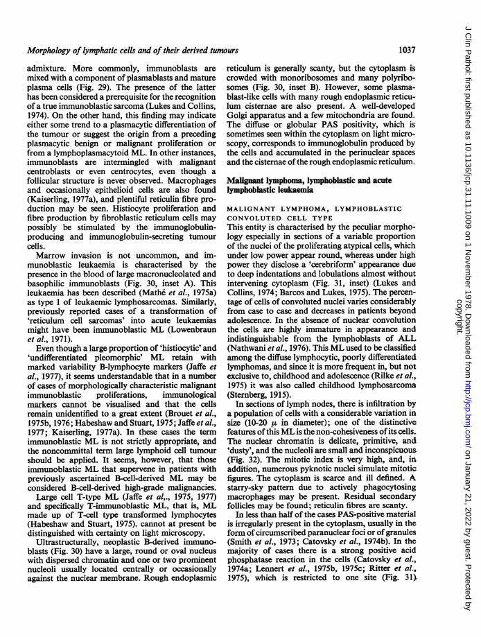
Morphology of lymphatic cells and of their derived tumours
admixture. More commonly, immunoblasts aremixed with a component of plasmablasts and matureplasma cells (Fig. 29). The presence of the latterhas been considered a prerequisite for the recognitionof a true immunoblastic sarcoma (Lukes and Collins,1974). On the other hand, this finding may indicateeither some trend to a plasmacytic differentiation ofthe tumour or suggest the origin from a precedingplasmacytic benign or malignant proliferation orfrom a lymphoplasmacytoid ML. In other instances,immunoblasts are intermingled with malignantcentroblasts or even centrocytes, even though afollicular structure is never observed. Macrophagesand occasionally epithelioid cells are also found(Kaiserling, 1977a), and plentiful reticulin fibre pro-duction may be seen. Histiocyte proliferation andfibre production by fibroblastic reticulum cells maypossibly be stimulated by the immunoglobulin-producing and immunoglobulin-secreting tumourcells.Marrow invasion is not uncommon, and im-
munoblastic leukaemia is characterised by thepresence in the blood of large macronucleolated andbasophilic immunoblasts (Fig. 30, inset A). Thisleukaemia has been described (Mathe et al., 1975a)as type 1 of leukaemic lymphosarcomas. Similarly,previously reported cases of a transformation ofreticulumm cell sarcomas' into acute leukaemiasmight have been immunoblastic ML (Lowenbraunet al., 1971).Even though a large proportion of 'histiocytic' and
'undifferentiated pleomorphic' ML retain withmarked variability B-lymphocyte markers (Jaffe etal., 1977), it seems understandable that in a numberof cases of morphologically characteristic malignantimmunoblastic proliferations, immunologicalmarkers cannot be visualised and that the cellsremain unidentified to a great extent (Brouet et al.,1975b, 1976; Habeshaw and Stuart, 1975; Jaffe et al.,1977; Kaiserling, 1977a). In these cases the termimmunoblastic ML is not strictly appropriate, andthe noncommittal term large lymphoid cell tumourshould be applied. It seems, however, that thoseimmunoblastic ML that supervene in patients withpreviously ascertained B-cell-derived ML may beconsidered B-cell-derived high-grade malignancies.
Large cell T-type ML (Jaffe et al,., 1975, 1977)and specifically T-immunoblastic ML, that is, MLmade up of T-cell type transformed lymphocytes(Habeshaw and Stuart, 1975). cannot at present bedistinguished with certainty on light microscopy.
Ultrastructurally, neoplastic B-derived immuno-blasts (Fig. 30) have a large, round or oval nucleuswith dispersed chromatin and one or two prominentnucleoli usually located centrally or occasionallyagainst the nuclear membrane. Rough endoplasmic
reticulum is generally scanty, but the cytoplasm iscrowded with monoribosomes and many polyribo-somes (Fig. 30, inset B). However, some plasma-blast-like cells with many rough endoplasmic reticu-lum cisternae are also present. A well-developedGolgi apparatus and a few mitochondria are found.The diffuse or globular PAS positivity, which issometimes seen within the cytoplasm on light micro-scopy, corresponds to immunoglobulin produced bythe cells and accumulated in the perinuclear spacesand the cisternae of the rough endoplasmic reticulum.
Malignant lymphoma, lymphoblastic and acutelymphoblastic leukaemia
MALIGNANT LYMPHOMA, LYMPHOBLASTICCONVOLUTED CELL TYPEThis entity is characterised by the peculiar morpho-logy especially in sections of a variable proportionof the nuclei of the proliferating atypical cells, whichunder low power appear round, whereas under highpower they disclose a 'cerebriform' appearance dueto deep indentations and lobulations almost withoutintervening cytoplasm (Fig. 31, inset) (Lukes andCollins, 1974; Barcos and Lukes, 1975). The percen-tage of cells of convoluted nuclei varies considerablyfrom case to case and decreases in patients beyondadolescence. In the absence of nuclear convolutionthe cells are highly immature in appearance andindistinguishable from the lymphoblasts of ALL(Nathwani et al., 1976). This ML used to be classifiedamong the diffuse lymphocytic, poorly differentiatedlymphomas, and since it is more frequent in, but notexclusive to, childhood and adolescence (Rilke et al.,1975) it was also called childhood lymphosarcoma(Sternberg, 1915).
In sections of lymph nodes, there is infiltration bya population of cells with a considerable variation insize (10-20 ,t in diameter); one of the distinctivefeatures of this ML is the non-cohesiveness of its cells.The nuclear chromatin is delicate, primitive, and'dusty', and the nucleoli are small and inconspicuous(Fig. 32). The mitotic index is very high, and, inaddition, numerous pyknotic nuclei simulate mitoticfigures. The cytoplasm is scarce and ill defined. Astarry-sky pattern due to actively phagocytosingmacrophages may be present. Residual secondaryfollicles may be found; reticulin fibres are scanty.
In less than half of the cases PAS-positive materialis irregularly present in the cytoplasm, usually in theform of circumscribed paranuclear foci or of granules(Smith et al., 1973; Catovsky et al., 1974b). In themajority of cases there is a strong positive acidphosphatase reaction in the cells (Catovsky et al.,1974a; Lennert et al., 1975b, 1975c; Ritter et al.,1975), which is restricted to one site (Fig. 31}
1037
copyright. on January 21, 2022 by guest. P
rotected byhttp://jcp.bm
j.com/
J Clin P
athol: first published as 10.1136/jcp.31.11.1009 on 1 Novem
ber 1978. Dow
nloaded from
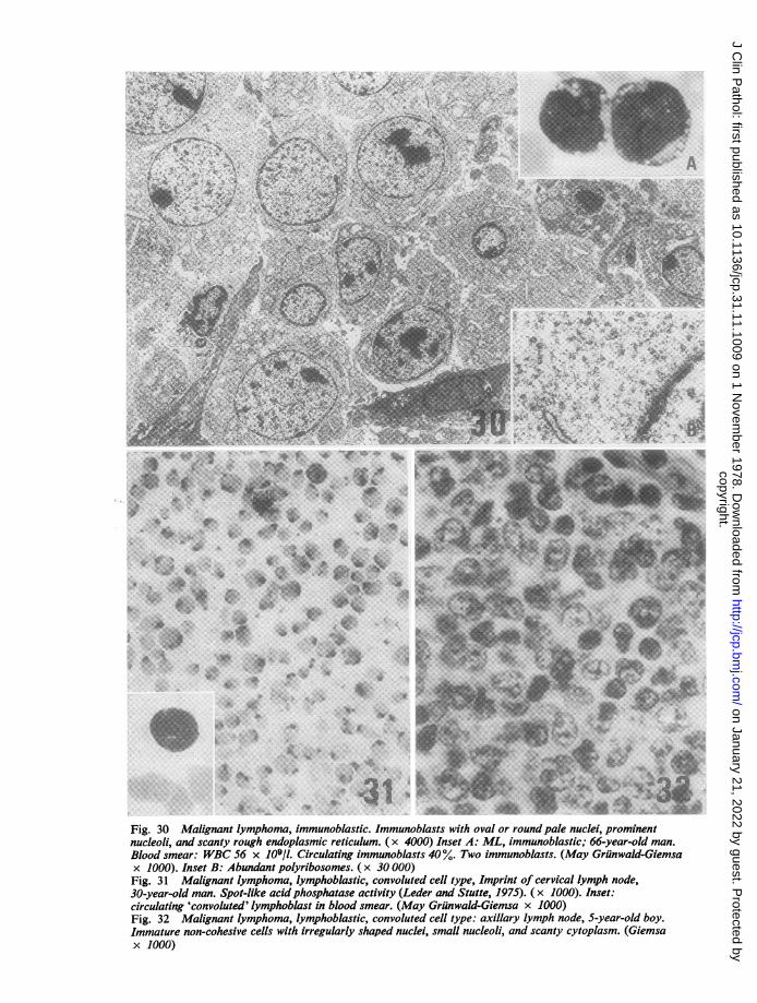
t :Or*.:.|R :.:0:i.E:-, .: id F Nf} ......................... :.:::: 8e wZ e ls ft*; i>, W...... q | | 4,2 '' .................. : 4^ . >. .><. w... 2. ! :' .t * '-w:t
Fig. 30 Malignant lymphoma, immunoblastic. Immunoblasts with oval or round pale nuclei, prominentnucleoli, and scanty rough endoplasmic reticulum. (x 4000) Inset A: ML, immunoblastic; 66-year-old man.Blood smear: WBC 56 x 109/l. Circulating immunoblasts 40%. Two immunoblasts. (May Griinwald-Giemsax 1000). Inset B: Abundant polyribosomes. ( x 30 000)Fig. 31 Malignant lymphoma, lymphoblastic, convoluted cell type, Imprint of cervical lymph node,30-year-old man. Spot-like acidphosphatase activity (Leder and Stutte, 1975). (x 1000). Inset:circulating 'convoluted' lymphoblast in blood smear. (May Griinwald-Giemsa x 1000)Fig. 32 Malignant lymphoma, lymphoblastic, convoluted cell type: axillary lymph node, 5-year-old boy.Immature non-cohesive cells with irregularly shaped nuclei, small nucleoli, and scanty cytoplasm. (Giemsax 1000)
.-,4A:1".,.v "I".....
copyright. on January 21, 2022 by guest. P
rotected byhttp://jcp.bm
j.com/
J Clin P
athol: first published as 10.1136/jcp.31.11.1009 on 1 Novem
ber 1978. Dow
nloaded from
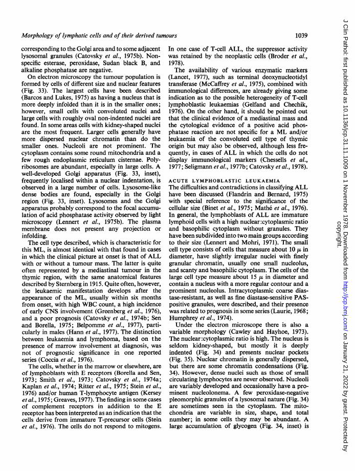
Morphology of lymphatic cells and of their derived tumours
corresponding to the Golgi area and to some adjacentlysosomal granules (Catovsky et al., 1975b). Non-specific esterase, peroxidase, Sudan black B, andalkaline phosphatase are negative.On electron microscopy the tumour population is
formed by cells of different size and nuclear features(Fig. 33). The largest cells have been described(Barcos and Lukes, 1975) as having a nucleus that ismore deeply infolded than it is in the smaller ones;however, small cells with convoluted nuclei andlarge cells with roughly oval non-indented nuclei arefound. In some areas cells with kidney-shaped nucleiare the most frequent. Larger cells generally havemore dispersed nuclear chromatin than do thesmaller ones. Nucleoli are not prominent. Thecytoplasm contains some round mitochondria and afew rough endoplasmic reticulum cisternae. Poly-ribosomes are abundant, especially in large cells. Awell-developed Golgi apparatus (Fig. 33, inset),frequently localised within a nuclear indentation, isobserved in a large number of cells. Lysosome-likedense bodies are found, especially in the Golgiregion (Fig. 33, inset). Lysosomes and the Golgiapparatus probably correspond to the focal accumu-lation of acid phosphatase activity observed by lightmicroscopy (Lennert et al., 1975b). The plasmamembrane does not present any projection orinfolding.The cell type described, which is characteristic for
this ML, is almost identical with that found in casesin which the clinical picture at onset is that of ALLwith or without a tumour mass. The latter is quiteoften represented by a mediastinal tumour in thethymic region, with the same anatomical featuresdescribed by Sternberg in 1915. Quite often, however,the leukaemic manifestation develops after theappearance of the ML, usually within six monthsfrom onset, with high WBC count, a high incidenceof early CNS involvement (Greenberg et al., 1976),and a poor prognosis (Catovsky et al., 1974b; Senand Borella, 1975; Belpomme et al., 1977), parti-cularly in males (Hann et al., 1977). The distinctionbetween leukaemia and lymphoma, based on thepresence of marrow involvement at diagnosis, wasnot of prognostic significance in one reportedseries (Coccia et al., 1976).The cells, whether in the marrow or elsewhere, are
of lymphoblasts with E receptors (Borella and Sen,1973; Smith et al., 1973; Catovsky et al., 1974a;Kaplan et al., 1974; Ritter et al., 1975; Stein et al.,1976) and/or human T-lymphocyte antigen (Kerseyet al., 1975; Greaves, 1977). The finding in some casesof complement receptors in addition to the Ereceptor has been interpreted as an indication that thecells derive from immature T-precursor cells (Steinet al., 1976). The cells do not respond to mitogens.
In one case of T-cell ALL, the suppressor activitywas retained by the neoplastic cells (Broder et al.,1978).The availability of various enzymatic markers
(Lancet, 1977), such as terminal deoxynucleotidyltransferase (McCaffrey et al., 1975), combined withimmunological differences, are already giving someindication as to the possible heterogeneity of T-celllymphoblastic leukaemias (Gelfand and Chechik,1976). On the other hand, it should be pointed outthat the clinical evidence of a mediastinal mass andthe cytological evidence of a positive acid phos-phatase reaction are not specific for a ML and/orleukaemia of the convoluted cell type of thymicorigin but may also be observed, although less fre-quently, in cases of ALL in which the cells do notdisplay immunological markers (Chessells et al.,1977; Seligmann et al., 1977b; Catovsky et al., 1978).
ACUTE LYMPHOBLASTIC LEUKAEMIAThe difficulties and contradictions in classifying ALLhave been discussed (Flandrin and Bernard, 1975)with special reference to the significance of thecellular size (Binet et al., 1975; Mathe et al., 1976).In general, the lymphoblasts of ALL are immaturelymphoid cells with a high nuclear :cytoplasmic ratioand basophilic cytoplasm without granules. Theyhave been subdivided into two main groups accordingto their size (Lennert and Mohri, 1971). The smallcell type consists of cells that measure about 10 ,u indiameter, have slightly irregular nuclei with finelygranular chromatin, usually one small nucleolus,and scanty and basophilic cytoplasm. The cells of thelarge cell type measure about 15 u in diameter andcontain a nucleus with a more regular contour and aprominent nucleolus. Intracytoplasmic coarse dias-tase-resistant, as well as fine diastase-sensitive PAS-positive granules, were described, and their presencewas related to prognosis in some series (Laurie, 1968;Humphrey et al., 1974).Under the electron microscope there is also a
variable morphology (Cawley and Hayhoe, 1973).The nuclear :cytoplasmic ratio is high. The nucleus isseldom kidney-shaped, but mostly it is deeplyindented (Fig. 34) and presents nuclear pockets(Fig. 35). Nuclear chromatin is generally dispersed,but there are some chromatin condensations (Fig.34). However, dense nuclei such as those of smallcirculating lymphocytes are never observed. Nucleoliare variably developed and occasionally have a pro-minent nucleolonema. A few peroxidase-negativepleomorphic granules of a lysosomal nature (Fig. 34)are sometimes seen in the cytoplasm. The mito-chondria are variable in size, shape, and totalnumber; in some cells they may be abundant. Alarge accumulation of glycogen (Fig. 34, inset) is
1039
copyright. on January 21, 2022 by guest. P
rotected byhttp://jcp.bm
j.com/
J Clin P
athol: first published as 10.1136/jcp.31.11.1009 on 1 Novem
ber 1978. Dow
nloaded from
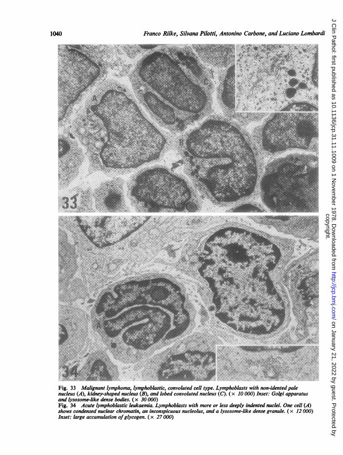
1040 Franco Rilke, Silvana Pilotti, Antonino Carbone, and Luciano Lombi;! . * tb; it g . ~4 * ,ge ofQ (x+ 27000)1
Z ;; W uvi * Ds ~~~~~~||; e~#,'^' @ dCi~~~~~~~~~~~~~~s9X¢GeeSF< S ti #Wi~~~~~~~~~~~~. s ~~~~~~~~~~~~~~~~~~~~~~~~~~~~~~~~~~~~. .. ii. ... .... ........ :;_
" X v.ffi.;a. ...... . '~~~~~~~~~~~~~~~~~~~~~~~~~~~~~~~~~~~~: < S :9. . z z_~~~~~~~~~~~........
..~~~~~~~~~~~~~~~~~~~~~~~~~~~~~~~~~~~~~~~~~~~~~ '.
[X.S't~~~~~~~~~~~~~~~~~~~~~~~~~~~j jw\ j> , , E tA i^<trJm
andIyooelk dnebde. 0 000
Fig. 34 Acute Iymphoblastic leukaemia. Lymphoblasts with more or less deeply inented nuclei. One cell (A)shows condensed nuclear chromatin, an inconspicuous nucleolus, and a lysosome-like dense granule. ( x 12 000)Inset: large accumulation ofglycogen. ( x 27 000)
rardi
copyright. on January 21, 2022 by guest. P
rotected byhttp://jcp.bm
j.com/
J Clin P
athol: first published as 10.1136/jcp.31.11.1009 on 1 Novem
ber 1978. Dow
nloaded from
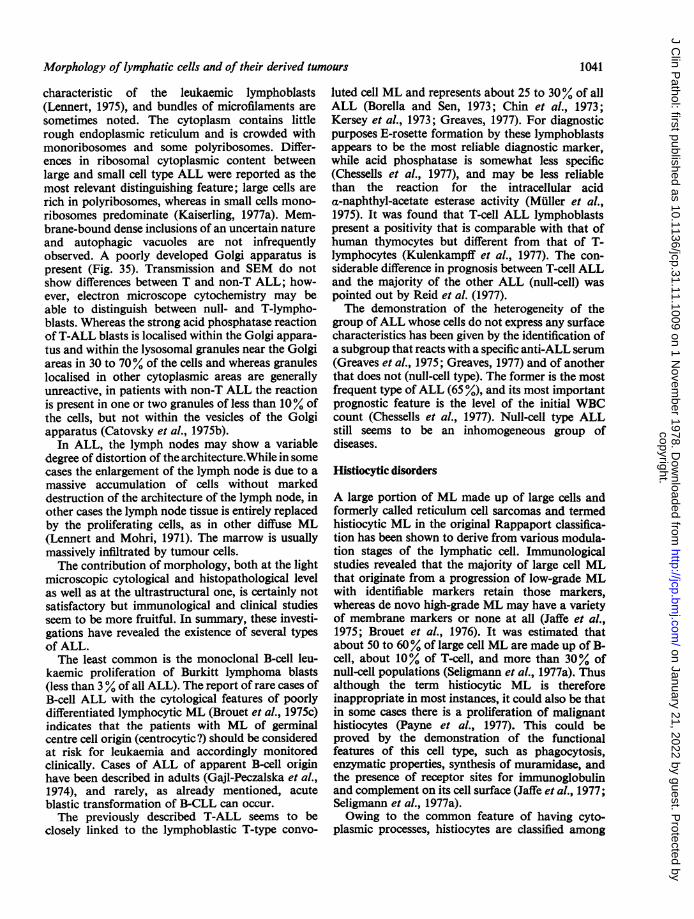
Morphology of lymphatic cells and of their derived tumours
characteristic of the leukaemic lymphoblasts(Lennert, 1975), and bundles of microfilaments aresometimes noted. The cytoplasm contains littlerough endoplasmic reticulum and is crowded withmonoribosomes and some polyribosomes. Differ-ences in ribosomal cytoplasmic content betweenlarge and small cell type ALL were reported as themost relevant distinguishing feature; large cells arerich in polyribosomes, whereas in small cells mono-ribosomes predominate (Kaiserling, 1977a). Mem-brane-bound dense inclusions of an uncertain natureand autophagic vacuoles are not infrequentlyobserved. A poorly developed Golgi apparatus ispresent (Fig. 35). Transmission and SEM do notshow differences between T and non-T ALL; how-ever, electron microscope cytochemistry may beable to distinguish between null- and T-lympho-blasts. Whereas the strong acid phosphatase reactionof T-ALL blasts is localised within the Golgi appara-tus and within the lysosomal granules near the Golgiareas in 30 to 70% of the cells and whereas granuleslocalised in other cytoplasmic areas are generallyunreactive, in patients with non-T ALL the reactionis present in one or two granules of less than 10% ofthe cells, but not within the vesicles of the Golgiapparatus (Catovsky et al., 1975b).
In ALL, the lymph nodes may show a variabledegree of distortion of the architecture.While in somecases the enlargement of the lymph node is due to amassive accumulation of cells without markeddestruction of the architecture of the lymph node, inother cases the lymph node tissue is entirely replacedby the proliferating cells, as in other diffuse ML(Lennert and Mohri, 1971). The marrow is usuallymassively infiltrated by tumour cells.The contribution of morphology, both at the light
microscopic cytological and histopathological levelas well as at the ultrastructural one, is certainly notsatisfactory but immunological and clinical studiesseem to be more fruitful. In summary, these investi-gations have revealed the existence of several typesof ALL.The least common is the monoclonal B-cell leu-
kaemic proliferation of Burkitt lymphoma blasts(less than 3% of all ALL). The report of rare cases ofB-cell ALL with the cytological features of poorlydifferentiated lymphocytic ML (Brouet et al., 1975c)indicates that the patients with ML of germinalcentre cell origin (centrocytic?) should be consideredat risk for leukaemia and accordingly monitoredclinically. Cases of ALL of apparent B-cell originhave been described in adults (Gajl-Peczalska et al.,1974), and rarely, as already mentioned, acuteblastic transformation of B-CLL can occur.The previously described T-ALL seems to be
closely linked to the lymphoblastic T-type convo-
luted cell ML and represents about 25 to 30% of allALL (Borella and Sen, 1973; Chin et al., 1973;Kersey et al., 1973; Greaves, 1977). For diagnosticpurposes E-rosette formation by these lymphoblastsappears to be the most reliable diagnostic marker,while acid phosphatase is somewhat less specific(Chessells et al., 1977), and may be less reliablethan the reaction for the intracellular acida-naphthyl-acetate esterase activity (Muller et al.,1975). It was found that T-cell ALL lymphoblastspresent a positivity that is comparable with that ofhuman thymocytes but different from that of T-lymphocytes (Kulenkampff et al., 1977). The con-siderable difference in prognosis between T-cell ALLand the majority of the other ALL (null-cell) waspointed out by Reid et al. (1977).The demonstration of the heterogeneity of the
group ofALL whose cells do not express any surfacecharacteristics has been given by the identification ofa subgroup that reacts with a specific anti-ALL serum(Greaves et al., 1975; Greaves, 1977) and of anotherthat does not (null-cell type). The former is the mostfrequent type ofALL (65 %), and its most importantprognostic feature is the level of the initial WBCcount (Chessells et al., 1977). Null-cell type ALLstill seems to be an inhomogeneous group ofdiseases.
Histiocytic disorders
A large portion of ML made up of large cells andformerly called reticulum cell sarcomas and termedhistiocytic ML in the original Rappaport classifica-tion has been shown to derive from various modula-tion stages of the lymphatic cell. Immunologicalstudies revealed that the majority of large cell MLthat originate from a progression of low-grade MLwith identifiable markers retain those markers,whereas de novo high-grade ML may have a varietyof membrane markers or none at all (Jaffe et al.,1975; Brouet et al., 1976). It was estimated thatabout 50 to 60% of large cell ML are made up of B-cell, about 10% of T-cell, and more than 30% ofnull-cell populations (Seligmann et al., 1977a). Thusalthough the term histiocytic ML is thereforeinappropriate in most instances, it could also be thatin some cases there is a proliferation of malignanthistiocytes (Payne et al., 1977). This could beproved by the demonstration of the functionalfeatures of this cell type, such as phagocytosis,enzymatic properties, synthesis of muramidase, andthe presence of receptor sites for immunoglobulinand complement on its cell surface (Jaffe et al., 1977;Seligmann et al., 1977a).Owing to the common feature of having cyto-
plasmic processes, histiocytes are classified among
1041
copyright. on January 21, 2022 by guest. P
rotected byhttp://jcp.bm
j.com/
J Clin P
athol: first published as 10.1136/jcp.31.11.1009 on 1 Novem
ber 1978. Dow
nloaded from
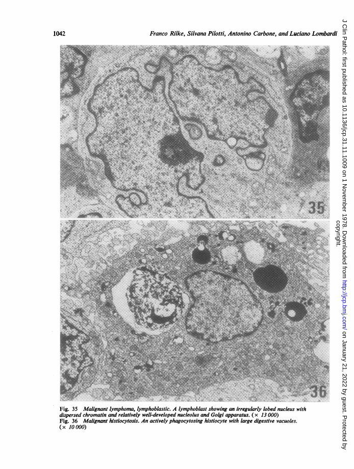
Franco Rilke, Silvana Pilotti, Antonino Carbone, and Luciano Lombardi
Fig. 35 Malignant lymphoma, lymphoblastic. A lymphoblast showing an irregularly lobed nucleus withdispersed chromatic and relatively well-developed nucleolus and Golgi apparatus. ( x 13 000)Fig. 36 Malignant histiocytosis. An actively phagocytosing histiocyte with large digestive vacuoles.(x 10000)
1042
copyright. on January 21, 2022 by guest. P
rotected byhttp://jcp.bm
j.com/
J Clin P
athol: first published as 10.1136/jcp.31.11.1009 on 1 Novem
ber 1978. Dow
nloaded from
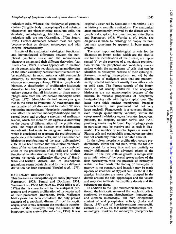
Morphology of lymphatic cells and of their derived tumours
reticulum cells. Whereas the histiocytes of germinalcentres ('tingible body macrophages') and subsinusphagocytes are phagocytosing reticulum cells, thedendritic, interdigitating, fibroblastic, and darkreticulum cells are not (Kaiserling, 1977a; Stuart,1975). Further differences between these cell typesbecome evident on electron microscopy and withenzyme histochemistry.
In spite of the anatomical, cytological, functional,and immunological differences between the peri-pheral lymphatic tissue and the mononuclearphagocyte system and their different derivation (vanFurth et al., 1972), it seems appropriate to mentionin this context also the neoplastic malignant disordersof the latter, essentially because their true nature canbe established, in most instances with reasonablecertainty, by morphology alone using light andelectron miscroscopy (Henry, 1975) on lymph nodebiopsies. A classification of proliferative histiocyticdisorders has been proposed on the basis of theunitary concept that all histiocytes or tissue macro-phages arise from the BM-derived monocytic series(Cline, 1975). Monoblast-derived monocytes giverise in the tissue to immature 'A' macrophages thatare capable of cell division and to mature 'B' non-replicating macrophages. Neoplastic transformationcan affect the normal monocyte-histiocyte line atseveral levels and produce a spectrum of malignantlesions, which are more or less aggressive accordingto the degree of differentiation of the proliferatingcells. The range of the diseases extends from acutemonoblastic leukaemia to malignant histiocytosis,which is considered to represent the proliferation ofmoderately differentiated cells, and to circumscribedhistiocytic proliferations of the most differentiatedcells. It has been stressed that the clinical manifesta-tions of the various diseases result from a combinedeffect of the proliferation of the cells and of theirfunctional manifestations (Cline, 1978). The positionamong histiocytic proliferative disorders of Hand-Schuller-Christian disease and of eosinophilicgranuloma seems debatable, and for them a differentderivation has been suggested (Nezelof et al., 1973).
MALIGNANT HISTIOCYTOSJSThis disease is a clinicopathological entity (Byrne andRappaport, 1973; Berard and Dorfman, 1974;Wamke et al., 1975; Mathe et al., 1976; Rilke et al.,1978a) that is characterised by the malignant pro-gressive and systemic proliferation of histiocytes andof their precursors (Rappaport, 1966). Malignanthistiocytosis has been considered the only provedexample of a neoplastic disease of 'true' histiocyticorigin, since it may represent the neoplastic transfor-mation of the histiocytes lining the sinuses of thelymphoreticular system (Berard et al., 1976). It was
originally described by Scott and Robb-Smith (1939)as histiocytic medullary reticulosis. The anatomicalareas predominantly involved by the disease are thelymph nodes, spleen, liver, marrow, and skin (Byrneand Rappaport, 1973; Warnke et al., 1975). Thediagnosis is made by histology on biopsy materialbut may sometimes be apparent in bone marrowsmears.The most important histological criteria for the
diagnosis on lymph nodes, which are the electivesite for the identification of the disease, are repre-sented (a) by the presence of a neoplastic prolifera-tion within the peripheral and medullary sinusesand/or within the parenchyma of cells that can beidentified as histiocytes because of their cytologicalfeatures, including phagocytosis, and (b) by thedistribution of malignant cells that are predomi-nantly isolated and do not usually form either cordsor solid nests. The fibrous capsule of the lymphnodes is not usually infiltrated. The neoplastichistiocytes are not monomorphic because of themixture in variable proportions of deceptivelybenign-looking cells with highly atypical ones. Thelatter have thick nuclear membranes, irregularheterochromatin, and prominent but not verylarge nucleoli. Phagocytosis is a constant finding,even though quantitatively variable; within thecytoplasm of the histiocytes, erythrocytes, leucocytes,platelets, fat droplets, cellular debris, and PAS-positive granules may be found. Erythrophagocytosisin particular may be massive and as such is diag-nostic. The number of mitotic figures is variable.Plasma cells and eosinophilic granulocytes are oftenbut not constantly found in a variable amount.
In the spleen, neoplastic proliferation occurs pre-dominantly within the red pulp, while the folliclesmay persist for a long time and are partially ortotally obliterated in the advanced phase of thedisease. In the liver, cellular growth is recognisableas an infiltration of the portal spaces and/or of theliver parenchyma with the presence of histiocyteswithin the liver cords. The finding of histiocytes inmarrow is not constant, and invasion may be madeup only of small foci of atypical cells. In the skin theatypical histiocytes are more often grouped in thedermis around the skin appendages and the vesselsand may also infiltrate the papillary dermis and thesubcutaneous tissue.
In addition to the light microscopic findings men-tioned, the histiocytic nature of the neoplastic cells isconfirmed by enzyme histochemistry, immunology,and electron microscopy. The high cytoplasmiccontent of acid phosphatase activity (Leder andStutte, 1975) and of fluoride-resistant non-specificesterase (Li et al., 1972) is easily demonstrated. Im-munological markers for monocytes (receptors for
1043
copyright. on January 21, 2022 by guest. P
rotected byhttp://jcp.bm
j.com/
J Clin P
athol: first published as 10.1136/jcp.31.11.1009 on 1 Novem
ber 1978. Dow
nloaded from
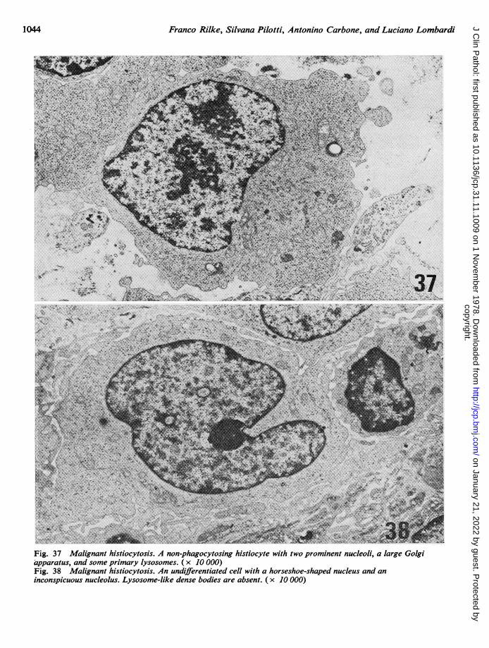
1044 Franco Rilke, Silvana Pilotti, Antonino Carbone, and Luciano Lombardi~~~~~~~~~~W.~~~a~~~~~~~~~~~~~~~~~~~~~~~~
~~~A ~~~~ ~ ~ ~
4.C
-~~~~~~~~~~~~~~~~~~~~~~~~~~~~~-
It~~~~~~~~~~~~~~~~~~~~~~~~~~~~~~~~~~Ik
<V~~~~~~~~~~~~~
Fig. 37 Malignant histiocytosis. A non-phagocytosing histiocyte with two prominent nucleoli, a large Golgiapparatus, and some primary lysosomes. ( x 10 000)Fig. 38 Malignant histiocytosis. An undifferentiated cell with a horseshoe-shaped nucleus and aninconspicuous nucleolus. Lysosome-like dense bodies are absent. ( x 10 000)
lk
copyright. on January 21, 2022 by guest. P
rotected byhttp://jcp.bm
j.com/
J Clin P
athol: first published as 10.1136/jcp.31.11.1009 on 1 Novem
ber 1978. Dow
nloaded from
Morphology of lymphatic cells and of their derived tumours 1045
the Fc portion ofIgG and for complement) have beenidentified on the cells of malignant histiocytosis(Jaffe et al., 1975).
In our ultrastructural study of six cases of malig-nant histiocytosis (Lombardi et al., 1978), weobserved three types of proliferating cells: activelyphagocytosing histiocytes (Fig. 36) with largedigestive vacuoles and numerous primary lysosomes;non-phagocytosing histiocytes (Fig. 37) with pri-mary lysosomes only; and undifferentiated cells(Fig. 38) with a few small primary lysosomes.Non-phagocytosing and phagocytosing histiocytes(Figs 36, 37), which show ultrastructural featuressimilar to those of the A and B macrophagesdescribed by Cline (1975), have an oval or deeplyindented lobed nucleus with well-developed nucleoliand scarce and marginated heterochromatin. Un-differentiated cells (Fig. 38) have a horseshoe-shaped or occasionally a lobed pale nucleus with aninconspicuous nucleolus. All these cell types generallyhave a scarce, smooth, and rough endoplasmicreticulum and abundant free ribosomes. Somephagocytosing and non-phagocytosing histiocyteswith a well-developed rough and smooth endo-plasmic reticulum were observed in two cases only.Some multinucleated phagocytosing histiocytes andsome cells with features intermediate either betweenundifferentiated and non-phagocytosing histiocytesor between non-phagocytosing and phagocytosinghistiocytes are also observed.
In some cases of circumscribed histiocyticcutaneous and/or nodal neoplasm, similar pro-liferating cells were observed. Histologically at highmagnification the tumour cells could not be dis-tinguished from those found in the sinuses of clear-cut cases of malignant histiocytosis. At low power,however, the higher degree of cohesiveness and thelower degree of atypia represented the main,although qualitatively not well-defined, differencesbetween localised histiocytic tumours and malignanthistiocytosis.
Histological differential diagnosis takes into con-sideration familial haemaphagocytic reticulosis(Farquhar et al., 1958), immunoblastic hyperplasia,sinus histiocytosis with massive lymphadenopathy(Rosai and Dorfman, 1969), other histiocyticdisorders, high-grade non-Hodgkin's ML, andHodgkin's disease (Byrne and Rappaport, 1973;Warnke et al., 1975; Rilke et al., 1978a).
This study was supported in part by grants no.76.01081 and no. 77.01849 from the ConsiglioNazionale delle Ricerche, Rome. We thank Ms. B.Johnston for editing and preparing the manuscript.
References
Aisenberg, A. C., and Bloch, K. J. (1972). Immuno-globulins on the surface of neoplastic lymphocytes.New England Journal of Medicine, 287, 272-276.
Aisenberg, A. C., Bloch, K. J., and Long, J. C. (1973a).Cell-surface immunoglobulins in chronic lymphocyticleukemia and allied disorders. American Journal ofMedicine, 55, 184-191.
Aisenberg, A. C., Bloch, K. J., Long, J. C., and Colvin,R. B. (1973b). Reaction of normal human lymphocytesand chronic lymphocytic leukaemia cells with anantithymocyte antiserum. Blood, 41, 417-423.
Aisenberg, A. C., Long, J. C. (1975). Lymphocyte surfacecharacteristics in malignant lymphoma. AmericanJournal of Medicine, 58, 300-306.
Aisenberg, A. C., and Wilkes, B. (1976). Lymphosarcomacell leukemia: the contribution of cell surface study todiagnosis. Blood, 48, 707-715.
Ala, F., et al. (1976). Alpha-chain disease and relatedsmall-intestinal lymphoma: a memorandum. Bulletinof the World Health Organization, 54, 615-624.
Alexander, E., Sanders, S., and Braylan, R. (1976).Purported difference between human T- and B-cellsurface morphology is an artefact. Nature, 261, 239-241.
Anday, G. J., Goodman, J. R., and Tishkoff, G. H.(1973). An unusual cytoplasmic ribosomal structure inpathologic lymphocytes. Blood, 41, 439-449.
Anday, G. J., and Schmitz, H. L. (1952). Follicularlymphoma with transient leukemic phase: brief reviewof literature and report of a case. Archives of InternalMedicine, 89, 621-634.
Andersson, M., Klein, G., Ziegler, J. L., and Henle, W.(1976). Association of Epstein-Barr viral genomes withAmerican Burkitt lymphoma. Nature, 260, 357-359.
Argani, I., and Kipkie, G. F. (1965). The cellular originof macroglobulins. A study of the protein secretingcells in Waldenstrbm's disease. Laboratory Investiga-tion, 14, 720-728.
Astaldi, G., and Verga, L. (1957). The glycogen contentof the cells of lymphatic leukaemia. Acta Haematolo-gica, 17, 129-135.
Ballard, H. S., Hamilton, L. M., Marcus, A. J., and Illes,C. H. (1970). A new variant of heavy-chain disease (g-chain disease). New England Journal of Medicine, 282,1060-1062.
Barcos, M. P., and Lukes, R. J. (1975). Malignantlymphoma of convoluted lymphocytes: a new entity ofpossible T-cell type. In Conflicts in Childhood Cancer:An Evaluation of Current Management, volume 4,edited by L. F. Sinks and J. 0. Godden, pp. 147-178.A. Liss, New York.
Belpomme, D., Mathe, G., and Davies, A. J. S. (1977).Clinical significance and prognostic value of the T-Bimmunological classification of human primary acutelymphoid leukaemias. Lancet, 1, 555-558.
Bennett, M. H. (1975). Sclerosis in non-Hodgkin'slymphomata. British Journal of Cancer, 31, Supplement2, 44-52.
Bennett, M. H., Farrer-Brown, G., Henry, K., and Jelliffe,A. M. (1974). Classification of non-Hodgkin's lym-
copyright. on January 21, 2022 by guest. P
rotected byhttp://jcp.bm
j.com/
J Clin P
athol: first published as 10.1136/jcp.31.11.1009 on 1 Novem
ber 1978. Dow
nloaded from
1046 Franco Rilke, Silvana Pilotti, Antonino Carbone, and Luciano Lombardi
phomas (Letter). Lancet, 2, 405-406.Bennett, M. H., and Millett, Y. L. (1969). Nodular
sclerotic lymphosarcoma. A possible new clinico-pathological entity. Clinical Radiology, 20, 339-343.
Bentwich, Z., and Kunkel, H. G. (1973). Specific pro-perties of human B and T lymphocytes and alterationsin disease. Transplantation Reviews, 16, 29-50.
Berard, C. W. (1975). Reticuloendothelial system: anoverview of neoplasia. In The ReticuloendothelialSystem, Chapter 15, edited by J. W. Rebuck, C. W.Berard, and M. R. Abell, pp. 301-317. Williams &Wilkins, Baltimore.
Berard, C. W., and Dorfman, R. F. (1974). Histo-pathology of malignant lymphomas. Clinics in Haema-tology, 3, 39-76.
Berard, C. W., Gallo, R. C., Jaffe, E. S., Green, I., andDe Vita, V. T., Jr. (1976). Current concepts of leukemiaand lymphoma: etiology, pathogenesis and therapy.Annals of Internal Medicine, 85, 351-366.
Berard, C. W., O'Conor, G. T., Thomas, L. B., andTorloni, H. (Eds.) (1969). Histopathological definitionof Burkitt's tumour. Bulletin of the World HealthOrganization, 40, 601-607.
Bessis, M. C. (1961). Ultrastructure of lymphoid andplasma cells in relation to globulin and antibody forma-tion. Laboratory Investigation, 10, 1040-1067.
Bianco, C., Patrick, R., and Nussenzweig, V. (1970). Apopulation of lymphocytes bearing a membranereceptor for antigen-antibody-complement complexes.I. Separation and characterization. Journal of Experi-mental Medicine, 132, 702-720.
Binet, J. L., Dighiero, G., D'Athis, P., Feinermann, M.,de Montaut, F., and Seroude, J. (1975). Volumetricdeterminations of blood cells in acute lymphoblasticleukemia. In Unclassifiable Leukemias, edited by M.Bessis and G. Brecher, pp. 95-97. Springer, Berlin.
Binet, J. L., Leporrier, M., Dighiero, G., Charron, D.,D'Athis, P., Vaugier, G., Beral, H. M., Natali, J. C.,Raphael, M., Nizet, B., and Follezou, J. Y. (1977a). Aclinical staging system for chronic lymphocyticleukemia. Cancer, 40, 855-864.
Binet, J. L., Vaugier, G., Dighiero, G., d'Athis, P., andCharron, D. (1977b). Investigation of a new parameterin chronic lymphocytic leukemia: the percentage oflarge peripheral lymphocytes determined by theHemalog D. Prognostic significance. American Journalof Medicine, 63, 683-688.
Bloomfield, C. D., Kersey, J. H., Brunning, R. D., andGajl-Peczalska, K. J. (1976). Prognostic significance oflymphocyte markers in adult non-Hodgkin's malignantlymphoma. Lancet, 2, 1330-1333.
Bluming, A. Z., Ziegler, J. L., and Carbone, P. P. (1972).Bone marrow involvement in Burkitt's lymphoma.Results of a prospective study. British Journal ofHaematology, 22, 369-376.
Boldt, D. H. Speckart, S. F., MacDermott, R. P., Nash,G. S., and Valeski, J. E. (1977). Leukemic reticulo-endotheliosis: 'hairy cell leukemia,' functional andstructural features of the abnormal cell in a patientwith profound leukocystosis. Blood, 49, 745-757.
Borella, L., and Sen, L. (1973). T cell surface markers onlymphoblasts from acute lymphocytic leukemia.
Journal of Immunology, 111, 1257-1260.Bouroncle, B. A., Wiseman, B. K., and Doan, C. A.
(1958). Leukemic reticuloendotheliosis. Blood, 13,609-630.
Braylan, R. C., Jaffe, E. S., and Berard, C. W. (1975).Malignant lymphomas: current classification and newobservations. Pathology Annual, 10, 213-270.
Braylan, R. C., Jaffe, E. S., Burbach, J. W., Frank, M. M.,Johnson, R. E., and Berard, C. W. (1976). Similaritiesof surface characteristics of neoplastic well-differen-tiated lymphocytes from solid tissues and fromperipheral blood. Cancer Research, 36, 1619-1625.
Braylan, R. C., and Rappaport, H. (1973). Tissue im-munoglobulins in nodular lymphomas as comparedwith reactive follicular hyperplasias. Blood, 42, 579-589.
Brearley, R. L., Lister, T. A., Whitehouse, J. M. A., andStansfeld, A. G. (1977). Burkitt's lymphoma in Britishadults: clinical features and response to chemotherapy.British Journal of Cancer, 35, 484-487.
Brill, N. E., Baehr, G., and Rosenthal, N. (1925).Generalized giant lymph follicle hyperplasia of lymphnodes and spleen; a hitherto undescribed type. Journalof the American Medical Association, 84, 668-671.
Broder, S., Edelson, R. L., Lutzner, M. A., Nelson, D. L.,MacDermott, R. P., Durm, M. E., Goldman, C. K.,Meade, B. D., and Waldmann, T. A. (1976). TheS&ary syndrome. A malignant proliferation of helperT cells. Journal of Clinical Investigation, 58, 1297-1306.
Broder, S., Poplack, D., Whang-Peng, J., Durm, M.,Goldman, C., Muul, L., and Waldmann, T. A. (1978).Characterization of a suppressor-cell leukemia.Evidence for the requirement of an interaction of two T-cells in the development of human suppressor effectorcells. New England Journal of Medicine, 298, 66-72.
Broome, J. D., Zucker-Franklin, D., Weiner, M. S.,Bianco, C., and Nussenzweig, V. (1973). Leukemic cellswith membrane properties of thymus-derived (T)lymphocytes in a case of Sezary's syndrome: morpho-logic and immunologic studies. Clinical Immunologyand Immunopathology, 1, 319-329.
Brouet, J. C., Flandrin, G., Sasportes, M., Preud'homme,J. L., and Seligmann, M. (1975a). Chronic lympho-cytic leukaemia of T-cell origin. Immunological andclinical evaluation in eleven patients. Lancet, 2, 890-893.
Brouet, J. C., Flandrin, G., and Seligmann, M. (1973a).Indications of the thymus-derived nature of the pro-liferating cells in six patients with Sezary's syndrome.New England Journal of Medicine, 289, 341-344.
Brouet, J. C., Labaume, S., and Seligmann, M. (1975b).Evaluation of T and B lymphocyte membrane markersin human non-Hodgkin malignant lymphomata.British Journal of Cancer, 31, Supplement 2, 121-127.
Brouet, J. C., Mason, D. Y., Danon, F., Preud'homme,J. L., Seligmann, M., Reyes, F., Navab, F., Galian,A., Rene, E., and Rambaud, J. C. (1977). Alpha-chaindisease: evidence for common clonal origin of intestinalimmunoblastic lymphoma and plasmacytic prolifera-tion (Letter). Lancet, 1, 861.
Brouet, J. C., Preud'homme, J. L., Flandrin, G., Chelloul,N., and Seligmann, M., (1976). Membrane markers in'histiocytic' lymphomas (reticulum cell sarcomas).
copyright. on January 21, 2022 by guest. P
rotected byhttp://jcp.bm
j.com/
J Clin P
athol: first published as 10.1136/jcp.31.11.1009 on 1 Novem
ber 1978. Dow
nloaded from
Morphology of lymphatic cells and of their derived tumours 1047
Journal of the National Cancer Institute, 56, 631-633.Brouet, J. C., Preud'homme, J. L., and Seligmann, M.
(1975c). The use of B and T membrane markers in theclassification of human leukemias, with specialreference to acute lymphoblastic leukemia. In Un-classifiable Leukemias, edited by M. Bessis and G.Brecher, pp. 81-90. Springer, Berlin.
Brouet, J. C., Preud'homme, J. L., Seligmann, M., andBernard, J. (1973b). Blast cells with monoclonalsurface immunoglobulin in two cases of acute blastcrisis supervening on chronic lymphocytic leukaemia.British Medical Journal, 4, 23-24.
Brown, G., Greaves, M. F., Lister, T. A., Rapson, N.,and Papamichael, M. (1974). Expression of human Tand B lymphocyte cell-surface markers on leukaemiccells. Lancet, 2, 753-755.
Brugo, E. A., Larkin, E., Molina-Escobar, J., andCostanzi, J. (1975). Primary granulocytic sarcoma ofthe small bowel, Cancer, 35, 1333-1340.
Brunning, R. D., McKenna, R. W., Bloomfield, C. D.,Coccia, P., and Gajl-Peczalska, K. J. (1977). Bonemarrow involvement in Burkitt's lymphoma. Cancer,40, 1771-1779.
Brunning, R. D., and Parkin, J. (1976). Intranuclearinclusions in plasma cells and lymphocytes frompatients with monoclonal gammopathies. AmericanJournal of Clinical Pathology, 66, 10-21.
Burke, J. S., Byrne, G. E., Jr., and Rappaport, H. (1974).Hairy cell leukemia (leukemic reticuloendotheliosis).I. A clinical pathologic study of 21 patients. Cancer, 33,1399-1410.
Burke, J. S., Mackay, B., and Rappaport, H. (1976).Hairy cell leukemia (leukemic reticuloendotheliosis).II. Ulstrastructure of the spleen. Cancer, 37, 2267-2274.
Burkitt, D. (1958). A sarcoma involving the jaws inAfrican children. British Journal of Surgery, 46, 218-223.
Burkitt, D. (1967). Burkitt's lymphoma outside theknown endemic areas of Africa and New Guinea.International Journal of Cancer, 2, 562-565.
Burns, G. F., Cawley, J. C., Barker, C. R., and Hayhoe,F. G. J. (1977). Absence of a receptor for fixed C3 on thehairy cells of leukaemic reticuloendotheliosis. Clinicaland Experimental Immunology, 29, 442-446.
Butler, J. J., (1969). Non-neoplastic lesions of lymphnodes of man to be differentiated from lymphomas.National Cancer Institute Monographs, 32, 233-255.
Buxbaum, J. N. (1976). Heavy chain diseases in man.Ricerca in Clinica e in Laboratorio, 6, 301-317.
Byrne, G. E., Jr., and Rapport, H. (1973). Malignanthistiocytosis. GANN Monographs on Cancer Research,15, 145-162.
Carbone, A., Santoro, A., Pilotti, S., and Rilke, F. (1978).Bone-marrow patterns and clinical staging in chroniclymphocytic leukaemia (Letter). Lancet, 1, 606.
Carmichael, G. P., Jr., and Lee, Y. T. (1977). Granulo-cytic sarcoma simulating 'nonsecretory' multiplemyeloma. Human Pathology, 8, 697-700.
Carr, I. (1973). The Macrophage: A Review of Ultra-structure and Function. Academic Press, London andNew York.
Castellani, R., Bonadonna, G., Spinelli, P., Bajetta, E.,
Galante, E., and Rilke, F. (1977). Sequential pathologicstaging of untreated non-Hodgkin's lymphomas bylaparoscopy and laparotomy combined with marrowbiopsy. Cancer, 40, 2322-2328.
Catovsky, D. (1975). T-cell origin of acid-phosphatase-positive lymphoblasts (Letter). Lancet, 2, 327-328.
Catovsky, D., Cherchi, M., Greaves, M. F., Janossy, G.,Pain, C., and Kay, H. E. M. (1978). Acid phosphatasereaction in acute lymphoblastic leukaemia. Lancet, 1,749-751.
Catovsky, D., Cherchi, M., Okos, A., Hegde, U., andGalton, D. A. G. (1976). Mouse red-cell rosettes in B-lymphoproliferative disorders. British Journal ofHaematology, 33, 173-177.
Catovsky, D., Frisch, B., Okos, A., and van Noorden, S.(1975a). Scanning electron microscopy and the natureof the hairy cell (Letter). Lancet, 1, 462-463.
Catovsky, D., Frisch, B., and van Noorden, S. (1975b).B, T, and 'null' cell leukaemias. Electron cytochemistryand surface morphology. In Unclassifiable Leukemias,edited by M. Bessis and G. Brecher, pp. 115-124.Springer, Berlin.
Catovsky, D., Galetto, J., Okos, A., Galton, D. A. G.,Wiltshaw, E., and Stathopoulos, G. (1973). Prolym-phocytic leukaemia of B and T cell type. Lancet, 2,232-234.
Catovsky, D., Galetto, J., Okos, A., Miliani, E., andGalton, D. A. G. (1974a). Cytochemical profile of Band T leukaemic lymphocytes with special reference toacute lymphoblastic leukaemia. Journal of ClinicalPathology, 27, 767-771.
Catovsky, D., Goldman, J. M., Okos, A., Frisch, B.,and Galton, D. A. G. (1974b). T-lymphoblasticleukaemia: a distinct variant of acute leukaemia.British Medical Journal, 2, 643-646.
Catovsky, D., Pettit, J. E., Galetto, J., Okos, A., andGalton, D. A. G. (1974c). The B-lymphocyte nature ofthe hairy cell of leukaemic reticuloendotheliosis.British Journal of Haematology, 26, 29-37.
Cawley, J. C., Barker, C. R., Britchford, R. D., andSmith, J. L. (1973). Intracellular IgA immunoglobulincrystals in chronic lymphocytic leukaemia. Clinical andExperimental Immunology, 13, 407-416.
Cawley, J. C., and Hayhoe, F. G. J. (1973). Ultra-structure of Haemic Cells: A Cytological Atlas ofNormal and Leukaemic Blood and Bone Marrow. W. B.Saunders Co., Ltd., Philadelphia and London.
Chessells, J. M., Hardisty, R. M., Rapson, N. T., andGreaves, M. F. (1977). Acute lymphoblastic leukaemiain children: classification and prognosis. Lancet, 2,1307-1309.
Chin, A. H., Saiki, J. H., Trujillo, J. M., and Williams,R. C., Jr. (1973). Peripheral blood T- and B-lympho-cytes in patients with lymphoma and acute leukemia.Clinical Immunology and Immunopathology, 1, 499-5 10.
Clark, C., Rydell, R. E., and Kaplan, M. E. (1973).Frequent association of IgM with crystalline inclusionsin chronic lymphatic leukemic lymphocytes. NewEngland Journal of Medicine, 289, 113-117.
Clift, R. A., Wright, D. H., and Clifford, P. (1963).Leukemia in Burkitt's lymphoma. Blood, 22, 243-251.
Cline, M. J. (1975). The White Cell. Harvard University
copyright. on January 21, 2022 by guest. P
rotected byhttp://jcp.bm
j.com/
J Clin P
athol: first published as 10.1136/jcp.31.11.1009 on 1 Novem
ber 1978. Dow
nloaded from
1048 Franco Rilke, Silvana Pilotti, Antonino Carbone, and Luciano Lombardi
Press, Cambridge, Massachusetts.Cline, M. J. (1978). Malignant diseases of the mono-
nuclear phagocyte system, pp. 85-86. In Cline, M. J.(moderator), Monocytes and macrophages: functionand diseases. Annals of Internal Medicine, 88, 78-88.
Coccia, P. F., Kersey, J. H., Gajl-Peczalska, K. J., Krivit,W., and Nesbit, M. E. (1976). Prognostic significance ofsurface marker analysis in childhood non-Hodgkin'slymphoproliferative malignancies. American Journal ofHaematology, 1, 405-417.
Cossman, J., Schnitzer, B., and Deegan, M. J. (1977).Immunologic surface markers in non-Hodgkin'slymphomas. American Journal ofPathology, 87, 19-32.
Cottier, H., Turk, J., and Sobin, L. (1973). A proposal fora standardized system of reporting human lymph nodemorphology in relation to immunological function.Journal of Clinical Pathology, 26, 317-33 1.
Crossen, P. E., Mellor, J. E. L., Finley, A. G., Ravich,R. B. M., Vincent, P. C., and Gunz, F. W. (1971). TheS&zary syndrome. Cytogenetic studies and identificationof the S6zary cell as an abnormal lymphocyte. AmericanJournal of Medicine, 50, 24-34.
Dameshek, W. (1963). 'Immunoblasts' and 'immuno-cytes.' An attempt at functional nomenclature. Blood,21, 243-245.
Dameshek, W. (1967). Chronic lymphocytic leukemia.An accumulative disease of immunologically incom-petent lymphocytes. Blood, 29, 566-584.
Danon, F., Mihaesco, C., Bouvry, M., Clerc, M., andSeligmann, M. (1975). A new case of heavy ,u-chaindisease. Scandinavian Journal of Haematology, 15, 5-9.
Debusscher, L., Bernheim, J. L., Collard-Ronge, E.,Govaerts, A., Hooghe, R., Lejeune, F. J., Zeicher, M.,and Stryckmans, P. A. (1975). Hairy cell leukemia:functional, immunologic, kinetic, and ultrastructuralcharacterization. Blood, 46, 495-507.
Deegan, M. J., Cossman, J., Chosney, B. T., andSchnitzer, B. (1976). Hairy cell leukemia. An immuno-logic and ultrastructural study. Cancer, 38, 1952-1961.
Dick, F., Bloomfield, C. D., and Brunning, R. D. (1974).Incidence, cytology, and histopathology of non-Hodgkin's lymphomas in the bone marrow. Cancer, 33,1382-1398.
Dick, F. R., Parks, W., and O'Connor, M. (1977). Trans-formation of nodular lymphoma to an immunoblasticIgM-producing tumor. American Journal of ClinicalPathology, 68, 611-615.
Dickler, H. B., Siegal, F. P., Bentwich, Z. H., and Kunkel,H. G. (1973). Lymphocyte binding of aggregated IgGand surface Ig staining in chronic lymphocytic leukae-mia. Clinical and Experimental Immunology, 14, 97-106.
Dorfman, R. F. (1974). Classification of non-Hodgkin'slymphomas (Letter). Lancet, 1, 1295-1296.
Dorfman, R. F. (1977). Pathology of the non-Hodgkin'slymphomas: new classifications. Cancer TreatmentReports, 61, 945-951.
Dorfman, R. F., Rice, D. F., Mitchell, A. D., Kempson,R. L., and Levine, G. (1973). Ultrastructural studies ofHodgkin's disease. National Cancer Institute Mono-graphs, 36, 221-238.
Douglas, S. D., Cohnen, G., Konig, E., and Brittinger,G. (1973). Lymphocyte lysosomes and lysosomal
enzymes in chronic lymphocytic leukaemia. Blood, 41,511-518.
Duhamel, G. (1974). Histopathologie Clinique de laMoelle Osseuse, pp. 71-77. Masson, Paris.
Dutcher, T. F., and Fahey, J. L. (1959). The histo-pathology of the macroglobulinemia of Waldenstrom.Journal of the National Cancer Institute, 22, 887-917.
Edelson, R. L., Kirkpatrick, C. H., Shevach, E. M.,Schein, P. S., Smith, R. W., Green, I., and Lutzner, M.(1974). Preferential cutaneous infiltration by neoplasticthymus-derived lymphocytes. Annals of InternalMedicine, 80, 685-692.
Epstein, A. L., Henle, W., Henle, G. Hewetson, J. F., andKaplan, H. S. (1976). Surface marker characteristicsand Epstein-Barr virus studies of two established NorthAmerican Burkitt's lymphoma cell lines. Proceedings ofthe National Academy of Sciences of the United Statesof America, 73, 228-232.
Epstein, M. A., and Achong, B. G. (1965). Fine structuralorganization of human lymphoblasts of a tissue culturestrain (EBI) from Burkitt's lymphoma. Journal of theNational Cancer Institute, 34, 241-253.
Epstein, M. A., and Barr, Y. M. (1965). Characteristicsand mode of growth of a tissue culture strain (EBI) ofhuman lymphoblasts from Burkitt's lymphoma.Journal of the National Cancer Institute, 34, 231-240.
Ewald, 0. (1923). Die leukamische Reticuloendotheliose.Deutsches Archiv fur klinische Medizin, 142, 222-228.
Farquhar, J. W., MacGregor, A. R., and Richmond, J.(1958). Familial haemophagocytic reticulosis. BritishMedical Journal, 2, 1561-1564.
Feremans, W. W., Demaertelaere, E., Delalieux, G., andNeve, P. (1976). Clinical and ultrastructural study ofBurkitt-like leukaemia. Pathologia Europaea, 11, 137-146.
Fishkin, B. G., and Spiegelberg, H. L. (1976). Cervicallymph node metastasis as the first manifestation oflocalized extramedullary plasmacytoma. Cancer, 38,1641-1644.
Flandrin, G., and Bernard, J. (1975). Cytological classifi-cation of acute leukemias. A survey of 1400 cases. InUnclassiflable Leukemias, edited by M. Bessis and G.Brecher, pp. 7-16. Springer, Berlin.
Flandrin, G., Brouet, J. C., Daniel, M. T., andPreud'homme, J. L. (1975). Acute leukemia withBurkitt's tumor cells: a study of six cases with specialreference to lymphocyte surface markers. Blood, 45,183-188.
Flandrin, G., Daniel, M. T., El Yafi, G., and Chelloul, N.(1972). Sarcomatoses ganglionnaires diffuses a diff6r-enciation plasmocytaire avec anemie hemolytique auto-immune. Actualites HImatologiques, 6, 24-41.
Flandrin, G., Daniel, M. T., Fourcade, M., and Chelloul,N. (1973). Leuckmie a 'tricholeucocyte' (hairy cellleukemia), etude clinique et cytologique de 55 observa-tions. Nouvelle Revue Fran!aise d'He~matologie, 13,609-640.
Florentin, I. (1975). The immunoblast. Biomedicine, 22,457-460.
Florin-Christensen, A., Doniach, D., and Newcomb,P. B. (1974). Alpha-chain disease with pulmonarymanifestations. British Medical Journal, 2, 413-415.
copyright. on January 21, 2022 by guest. P
rotected byhttp://jcp.bm
j.com/
J Clin P
athol: first published as 10.1136/jcp.31.11.1009 on 1 Novem
ber 1978. Dow
nloaded from

Morphology of lymphatic cells and of their derived tumours 1049
Forte, F. A., Prelli, F., Yount, W. J., Jerry, L. M.,Kochwa, S., Franklin, E. C., and Kunkel, H. G. (1970).Heavy chain disease of the ,u (yM) type: report of thefirst case. Blood, 36, 137-144.
Franklin, E. C., Lowenstein, J., Bigelow, B., and Meltzer,M. (1964). Heavy chain disease-a new disorder ofserum y-globulins. Report of the first case. AmericanJournal of Medicine, 37, 332-350.
Fr0land, S. S., Natvig, J. B., and Stavem, P. (1972).Immunological characterization of lymphocytes inlymphoproliferative diseases. Restriction of classes,subclasses and Gm allotypes of membrane-bound lg.Scandinavian Journal of Immunology, 1, 351-360.
Fu, S. M., Winchester, R. J., and Kunkel, H. G. (1974).Occurrence of surface IgM, IgD, and free light chainson human lymphocytes. Journal of ExperimentalMedicine, 139, 451-456.
van Furth, R., Cohn, Z. A., Hirsch, J. G., Humphrey,J. H., Spector, W. G., and Langevoort, H. L. (1972).The mononuclear phagocyte system: a new classifica-tion of macrophages, monocytes, and their precursorcells. Bulletin of the World Health Organization, 46,845-852.
Gajl-Peczalska, K. J., Bloomfield, C. D., Nesbit, M. E.,and Kersey, J. H. (1974). B-cell markers on lympho-blasts in acute lymphoblastic leukemia. Clinical andExperimental Immunology, 17, 561-569.
Gajl-Peczalska, K. J., Hansen, J. A., Bloomfield, C. D.,and Good, R. A. (1973). B lymphocytes in untreatedpatients with malignant lymphoma and Hodgkin'sdisease. Journal of Clinical Investigation, 52, 3064-3073.
Galton, D. A. G. (1966). The pathogenesis of chroniclymphocytic leukemia. Canadian Medical AssociationJournal, 94, 1005-1010.
Galton, D. A. G., Goldman, J. M., Wiltshaw, E.,Catovsky, D., Henry, K., and Goldenberg, G. J.(1974). Prolymphocytic leukaemia. British Journal ofHaematology, 27, 7-23.
Garrett, J. V., Newton, R. K., Scarffe, J. H. (1977).Abnormal blood lymphocytes in non-Hodgkin'slymphoma (Letter). Lancet, 1, 542.
Gelfand, E. W., and Chechik, B. E. (1976). Unmaskingthe heterogeneity of acute lymphoblastic leukemia.New England Journal of Medicine, 294, 275-276.
Gerard-Marchant, R., Hamlin, I., Lennert, K., Rilke, F.,Stansfeld, A. G., and van Unnik, J. A. M. (1974).Classification of non-Hodgkin's lymphomas (Letter).Lancet, 2, 406-408.
Ghon, A., and Roman, B. (1916). Ober das Lympho-sarkom. Frankfurter Zeitschrift fur Pathologie, 19, 1-138.
Glick, A. D., Leech, J. H., Flexner, J. M., and Collins,R. D. (1976). Ultrastructural study of Reed-Sternbergcells. Comparison with transformed lymphocytes andhistiocytes. American Journal ofPathology, 85, 195-208.
Glick, A. D., Leech, J. H., Waldron, J. A., Flexner, J. M.,Horn, R. G., and Collins, R. D. (1975). Malignantlymphomas of follicular center cell origin in man. II.Ultrastructural and cytochemical studies. Journal ofthe National Cancer Institute, 54, 23-36.
Goldberg, G. M., and Emanuel, B. (1964). A study of
malignant lymphomas and leukemias. VII. Lympho-genous leukemia and lymphosarcoma involvement ofthe lymphatic and hemic bed, with reference to differ-entiating criteria. Cancer, 17, 277-287.
Golde, D. W., Saxon, A., and Stevens, R. H. (1977a).Macroglobulinemia and hairy-cell leukemia. NewEngland Journal of Medicine, 296, 92-93.
Golde, D. W., Saxon, A., and Stevens, R. H. (1977b).Letter to the Editor (on hairy-cell leukemia). NewEngland Journal of Medicine, 296, 882.
Goldstone, A. H., Cawley, J. C., Roberts, S. 0., Leven-tine, A., and Barker, C. R. (1976). A case of small-cellSezary's syndrome with null-cell features. Journal ofClinical Pathology, 29, 848-851.
Gray, J. L., Jacobs, A., and Block, M. (1974). Bonemarrow and peripheral blood lymphocytosis in theprognosis of chronic lymphocytic leukemia. Cancer, 33,1169-1178.
Greaves, M. F. (1977). Immunodiagnosis of leukaemia.In Immunodiagnosis of Cancer, edited by R. Herbermanand K. McIntyre. Marcel Dekker Inc, New York.
Greaves, M. F., Brown, G., Rapson, N. T., and Lister,T. A. (1975). Antisera to acute lymphoblastic leukemiacells. Clinical Immunology and Immunopathology, 4,67-84.
Greenberg, B. R., Peter, C. R., Glassy, F., andMacKenzie, M. R. (1976). A case of T-cell lymphomawith convoluted lymphocytes. Cancer, 38, 1602-1607.
Grey, H. M., Rabellino, E., and Pirofsky, B. (1971).Immunoglobulins on the surface of lymphocytes. IV.Distribution in hypogammaglobulinemia, cellularimmune deficiency, and chronic lymphatic leukemia.Journal of Clinical Investigation, 50, 2368-2375.
Haak, H. L., De Man, J. C. H., Hijmans, W., Knapp, W.,and Speck, B. (1974). Further evidence for the lympho-cytic nature of leukaemic reticuloendotheliosis (hairycell leukaemia). British Journal of Haematology, 27,31-38.
Habeshaw, J. A., Macaulay, R. A. A., and Stuart, A. E.(1977). Correlation of surface receptors with histo-logical appearance in 29 cases of non-Hodgkinlymphoma. British Journal of Cancer, 35, 858-867.
Habeshaw, J. A., and Stuart, A. E. (1975). Cell receptorstudies on seven cases of diffuse histiocytic malignantlymphoma (reticulum cell sarcoma). Journal of ClinicalPathology, 28, 289-297.
Hann, H. L., London, W. T., and Evans, A. E. (1977).Lymphoblasts with T-cell markers in five girls withacute lymphocytic leukemia. Cancer, 39, 2001-2003.
Harigaya, K. (1977). Enzyme histochemical charac-teristics of non-Hodgkin's lymphomas. Acta Patholo-gica Japonica, 27, 345-358.
Hausner, R. J., Rosas-Uribe, A., Wickstrum, D. A., andSmith, P. C. (1977). Non-Hodgkin's lymphoma in thefirst two decades of life. A pathological study of 30cases. Cancer, 40, 1533-1547.
Henry, K. (1975). Electron miscroscopy in the non-Hodgkin's lymphomata. British Journal of Cancer, 31(Supplement 2), 73-93.
Henry, K., and Farrer-Brown, G. (1977). Primarylymphomas of the gastrointestinal tract. I. Plasma celltumors. Histopathology, 1, 53-76.
copyright. on January 21, 2022 by guest. P
rotected byhttp://jcp.bm
j.com/
J Clin P
athol: first published as 10.1136/jcp.31.11.1009 on 1 Novem
ber 1978. Dow
nloaded from
1050 Franco Rilke, Silvana Pilotti, Antonino Carbone, and Luciano Lombardi
Henry, K., and Goldman, J. M. (1975). The lymphocyte.In Recent Advances in Pathology, no. 9, edited by C. V.Harrison and K. Weinbren, pp. 30-72. ChurchillLivingstone, Edinburgh and London.
Hsu, C. C. S., Marti, G. E., Schrek, R., and Williams,R. C., Jr. (1975). Lymphocytes bearing B- and T-cellmarkers in patients with lymphosarcoma cell leukemia.Clinical Immunology and Immunopathology, 3, 385-395.
Huber, C., Dworzak, E., Fink, U., Michlmayr, G.,Braunsteiner, H., and Huber, H. (1974). Receptor sitesfor aggregated gammaglobulin (AGG) on lympho-cytes in lymphoproliferative diseases. British Journal ofHaematology, 27, 643-654.
Hubner, K. F., and Littlefield, L. G. (1975). Burkittlymphoma in three American children. AmericanJournal of Diseases of Children, 129, 1219-1223.
Humphrey, G. B., Nesbit, M. E., and Brunning, R. D.(1974). Prognostic value of the periodic acid-Schiff(PAS) reaction in acute lymphoblastic leukemia.American Journal of Clinical Pathology, 61, 393-397.
Hurez, D., Flandrin, G., Preud'homme, J. L., andSeligmann, M. (1972). Unreleased intracellular mono-clonal macroglobulin in chronic lymphocytic leukae-mia. Clinical and Experimental Immunology, 10, 223-234.
Isaacs, R. (1973). Lymphosarcoma cell leukemia. Annalsof Internal Medicine, 11, 657-662.
Jaffe, E. S., Braylan, R. C., Nanba, K., Frank, M. M.,and Berard, C. W. (1977). Functional markers: a newperspective on malignant lymphomas. Cancer Treat-ment Reports, 61, 953-962.
Jaffe, E. S., Shevach, E. M., Frank, M. M., Berard,C. W., and Green, I. (1974a). Nodular lymphoma-evidence for origin from follicular B lymphocytes. NewEngland Journal ofMedicine, 290, 813-819.
Jaffe, E. S., Shevach, E. M., Frank, M. M., and Green, I.(1974b). Leukemic reticuloendotheliosis: presence of areceptor for cytophilic antibody. American Journal ofMedicine, 57, 108-114.
Jaffe, E. S., Shevach, E. M., Sussman, E. H., Frank,M. M., Green, I., and Berard, C. W. (1975). Membranereceptor sites for the identification of lymphoreticularcells in benign and malignant conditions. British Journalof Cancer, 31 (Supplement 2), 107-120.
Johansson, B., and Klein, E. (1970). Cell surface localizedIgM-kappa immunoglobulin reactivity in a case ofchronic lymphocytic leukaemia. Clinical and Experi-mental Immunology, 6, 421-428.
Johansson, B., Klein, E., and Haglund, S. (1976).Correlation between the presence of surface localizedimmunoglobulin (Ig) and the histological type ofhuman malignant lymphomas. Clinical Immunology andImmunopathology, 5, 119-132.
Jondal, M., Holm, G., and Wigzell, H. (1972). Surfacemarkers on human T and B lymphocytes. I. A largepopulation of lymphocytes forming nonimmunerosettes with sheep red blood cells. Journal of Experi-mental Medicine, 136, 207-215.
Jones, S. E., Fuks, Z., Bull, M., Kadin, M. E., Dorfman,R. F., Kaplan, H. S., Rosenberg, S. A., and Kim, H.(1973). Non-Hodgkin's lymphomas. IV. Clinicopath-
ological correlation in 405 cases. Cancer, 31, 806-823.Jones, S. E., Rosenberg, S. A., and Kaplan, H. S. (1972).Non-Hodgkin's lymphomas. I. Bone marrow involve-ment. Cancer, 29, 954-960.
Kaiserling, E. (1977a). Non-Hodgkin-Lymphome: Ultra-struktur und Cytogenese. G. Fischer, Stuttgart.
Kaiserling, E. (1977b). Morphologische Befunde zurMicrookologie des T-Zonen-Lymphoms und descentroblastisch/centrocytischen Lymphoms (Abstract).Verhandlungen der Deutschen Gesellschaft fur Patho-logie, 61, 362.
Kaiserling, E., Stein, H., and Lennert, K. (1973). IgM-producing malignant lymphomas without macro-globulinemia. Morphological and immunochemicalfindings. Virchow's Archiv, B: Zellpathologie, 14, 1-18.
Kaplan, J., Mastrangelo, R., and Peterson, W. D., Jr.(1974). Childhood lymphoblastic lymphoma, a cancerof thymus-derived lymphocytes. Cancer Research, 34,521-525.
Katayama, I., Nagy, G. K., and Balogh, K., Jr. (1973).Light microscopic identification of the ribosome-lamella complex in 'hairy cells' of leukemic reticulo-endotheliosis. Cancer, 32, 843-846.
Katayama, I., and Schneider, G. B. (1977). Further ultra-structural characterization of hairy cells of leukemicreticuloendotheliosis. American Journal of Pathology,86, 163-182.
Kaung, D. T., and Ott, M. (1975). Are normal lympho-cytes small? (Letter). Lancet, 2, 414.
Kay, N. E., Douglas, S. D., and Estren, S. (1974). T-cellsin chronic lymphocytic leukaemia (Letter). Lancet, 2,1326.
Kersey, J., Nesbit, M., Hallgren, H., Sabad, A., Yunis, E.,and Gajl-Peczalska, K. J. (1975). Evidence for origin ofcertain childhood acute lymphoblastic leukemias andlymphomas in thymus-derived lymphocytes. Cancer,36, 1348-1352.
Kersey, J. H., Sabad, A., Gajl-Peczalska, K. J., Hallgren,H. M., Yunis, E. J., and Nesbit, M. E. (1973). Acutelymphoblastic leukemic cells with T (thymus-derived)lymphocyte markers. Science, 182, 1355-1356.
Klein, B., Lewinski, U., Shabtai, F., Freidin, N., andDjaldetti, M. (1977). Transmission and scanningelectron microscopy study on plasma cell leukemia.Blut, 35, 11-19.
Klein, G. (1971). Immunological aspects of Burkitt'slymphoma. Advances in Immunology, 14, 187-250.
Kojima, M., Imai, Y., and Mori, N. (1973). A concept offollicular lymphoma. A proposal for the existence of aneoplasm originating from the germinal center. GANNMonographs on Cancer Research, 15, 195-207.
Koziner, B., Filippa, D. A., Mertelsmann, R., Gupta, S.,Clarkson, B., Good, R. A., and Siegal, F. P. (1977).Characterization of malignant lymphomas in leukemicphase by multiple differentiation markers of mono-nuclear cells. Correlation with clinical features andconventional morphology. American Journal ofMedicine, 63, 556-567.
Krause, J. R., Srodes, C., and Lee, R. E. (1977). Use of thebone marrow imprint in the diagnosis of leukemicreticuloendotheliosis ('hairy cell leukemia'). AmericanJournal of Clinical Pathology, 68, 368-371.
copyright. on January 21, 2022 by guest. P
rotected byhttp://jcp.bm
j.com/
J Clin P
athol: first published as 10.1136/jcp.31.11.1009 on 1 Novem
ber 1978. Dow
nloaded from
Morphology of lymphatic cells and of their derived tumours 1051
Kuhn, C. (1967). Nuclear bodies and intranuclearglobulin inclusions in Waldenstrom's macroglo-bulinemia. Laboratory Investigation, 17, 404-415.
Kulenkampff, J., Janossy, G., and Greaves, M. F. (1977).Acid esterase in human lymphoid cells and leukaemicblasts: a marker for T lymphocytes. British Journal ofHaematology, 36, 231-240.
Lancet (1977). Enzyme markers in leukaemia. (Editorial).Lancet, 2, 539-540.
Laurie, H. C. (1968). Duration of remissions in lympho-blastic leukaemia of childhood. British MedicalJournal,2, 95-97.
Lay, W. H., Mendes, N. F., Bianco, C., and Nussenzweig,V. (1971). Binding of sheep red blood cells to a largepopulation of human lymphocytes. Nature, 230, 531-532.
Leder, L. D. (1971). Zytochemische Differenzierungnormaler und pathologischer mononuklarer Blut- undKnochenmarkszellen. Acta Histochemica, Supplement9, 141-151.
Leder, L., D., and Stutte, H. J. (1975). Seminar furhamatologisch-zytochemische Techniken. Verhand-lungen der Deutschen Gesellschaft fur Pathologie, 59,503-509.
Leech, J. H., Glick, A. D., Waldron, J. A., Flexner,J. M., Horn, R. G., and Collins, R. D. (1975a).Malignant lymphomas of follicular center cell originin man. I. Immunologic studies. Journal of the NationalCancer Institute, 54, 11-21.
Leech, J., Roy, R. M., Flexner, J. M., Glick, A. D.,Waldron, J. A., and Collins, R. D. (1975b). Evidencefor synthesis of surface immunoglobulin in leukemicreticuloendotheliosis (Abstract) Blood, 46, 1037.
Lennert, K. (1957). Ober die Erkennung von Keim-zentrumszellen in Lymphknotenausstrich. KlinischeWochenschrift, 35, 1130-1132.
Lennert K. (1961). Lymphknoten Diagnostik in Schnittund Ausstrich. A. Cytologie und Lymphadenitis (Hand-buch der speziellen pathologischen Anatomie undHistologie, Bd. 1, Th. 3). Springer, Berlin.
Lennert, K. (1964). Pathologie der Halslymphknoten.Springer, Berlin.
Lennert, K. (1968). Classification of the malignantlymphomas (European concept). In Progress inLymphology: Proceedings of the International Sym-posium on Lymphology, Zurich, edited by A. Ruttimann,pp. 103-109. Thieme, Stuttgart.
Lennert, K. (1969). Pathologisch-anatomische Klassifika-tion der malignen Lymphome. Strahlentherapie,Supplement 69, 1-7.
Lennert, K. (1973). Follicular lymphoma. A tumor of thegerminal centers. GANN Monograph on CancerResearch, 15, 217-231.
Lennert, K. (1974). Origin of malignant lymphomas(Letter). Lancet, 2, 586.
Lennert, K. (1975). Morphology and classification ofmalignant lymphomas and so-called reticuloses. ActaNeuropathologica, Supplement 6, 1-16.
Lennert, K. (1976). Klassifikation und Morphologie derNon-Hodgkin-Lymphome. Blut, Supplement, 18,145-166.
Lennert, K., Caesar, R., and Muller, H. K. (1966).
Electron miscroscopic studies of germinal centers inman. In Germinal Centers in Immune Responses, editedby H. Cottier, N. Odartchenko, R. Schindler andC. C. Congdon, pp. 49-59. Springer, Berlin.
Lennert, K., Kaiserling, E., and Miuller-Hermelink, H. K.(1975a). T-associated plasma-cells. Lancet, 1, 1031-1032.
Lennert, K., Mitrou, P., and Miiller-Hermelink, H. K.(1969). Cytophotometric and autoradiographic investi-gations on germinal center cells in man. In LymphaticTissue and Germinal Centers in Immune Response,edited by L. Fiore-Donati and M. G. Hanna, Jr. pp.65-71. Plenum Press, New York.
Lennert, K., and Mohri, N. (1971). Zur Pathologie derLeukamien und malignen Lymphome im Kindesalter(Referat). Verhandlungen der Deutschen Gesellschaftfur Pathologie, 55, 216-269.
Lennert, K., Mohri, N., Stein, H., and Kaiserling, E.(1975b). The histopathology of malignant lymphoma.British Journal of Haematology, Supplement 31, 193-203.
Lennert, K., and Muller-Hermelink, H. K. (1975).Lymphocyten und ihre Funktionsformen. Morpho-logie, Organisation und immunologische Bedeutung.Verhandlungen der Anatomischen Gesellschaft, 69,19-62.
Lennert, K., and Niedorf, H. R. (1969). Nachweis vondesmosomal verkniipften Reticulumzellen im folliku-karen Lymphom (Brill-Symmers). Virchows Archiv, B:Zellpathologie, 4, 148-150.
Lennert, K., and Remmele, W. (1958). KaryometrischeUntersuchungen an Lymphknotenzellen des Menschen.I. Germinoblasten, Lymphoblasten und Lymphozyten.Acta Haematologica, 19, 99-113.
Lennert, K., and Rinneberg, H. (1961). Fermenthisto-chemische Untersuchungen des Lymphknotens. II.Adenosintriphosphatase und 5-Nucleotidase imLymph-knotenschnitt. Klinische Wochenschrift, 39, 923-924.
Lennert, K., Stein, H., and Kaiserling, E. (1975c).Cytological and functional criteria for the classificationof malignant lymphomata. British Journal ofCancer, 31,Supplement 2, 29-43.
Levin, W. C., Gehan, E., Griffith, K., Huguley, C. M.,Silver, R. T., Steinfeld, J. L., Weiner, L., and Cahn,E. L. (1973). Proposed guidelines for protocol studies.III. Chronic lymphocytic leukemia. Cancer Chemo-therapy Reports, 4 (3), 159-165.
Levine, G. D., and Dorfman, R. F. (1975). Nodularlymphoma: an ultrastructural study of its relationshipto germinal centers and a correlation of light andelectron microscopic findings. Cancer, 35, 148-164.
Levine, P. H., Cho, B. R., Connelly, R. R., Berard, C. W.,O'Conor, G. T., Dorfman, R. F., Easton, J. M., andDeVita, V. T. (1975). The American Burkitt lymphomaregistry: a progress report. Annals ofInternal Medicine,83, 31-36.
Lewin, K. J., Kahn, L. B., and Novis, B. H. (1976).Primary intestinal lymphoma of 'Western' and'Mediterranean' type, alpha chain disease and massiveplasma cell infiltration. Cancer, 38, 2511-2528.
Li, C. Y., Yam, L. T., and Crosby, W. H. (1972). Histo-chemical characterization of cellular and structuralelements of the human spleen. Journal of Histo-
copyright. on January 21, 2022 by guest. P
rotected byhttp://jcp.bm
j.com/
J Clin P
athol: first published as 10.1136/jcp.31.11.1009 on 1 Novem
ber 1978. Dow
nloaded from
1052 Franco Rilke, Silvana Pilotti, Antonino Carbone, and Luciano Lombardi
chemistry and Cytochemistry, 20, 1049-1058.Lille, I., Desplaces, A., Meeus, L., Saracino, R. T., and
Brouct, J. C. (1973). Thymus-derived proliferatinglymphocytes in chronic lymphocytic leukaemia(Letter). Lancet, 2, 263-264.
Lin, P. S., and Hsu, C. C. S. (1976). Human leukaemic Tcells with complement receptors. Clinical and Experi-mental Immunology, 23, 209-213.
Loffier, H. (1972). Cytologische Befunde beim Sezary-Syndrom. Verhandlungen der Deutschen Gesellschaftfur innere Medizin, 78, 285-287.
Lombardi, L., Carbone, A., Pilotti, S., and Rilke, F.(1978). Malignant histiocytosis: a histological andultrastructural study of lymph nodes in six cases.Histopathology (In press).
Long, J. C., and Aisenberg, A. C. (1975). Richter'ssyndrome. A terminal complication of chronic lympho-cytic leukemia with distinct clinicopathologic features.American Journal of Clinical Pathology, 63, 786-795.
Long, J. C., and Mihm, M. C. (1974). Mycosis fungoideswith extra-cutaneous dissemination: a distinct clinico-pathologic entity. Cancer, 34, 1745-1755.
Lowenbraun, S., Sutherland, J. C., Feldman, M. J., andSerpick, A. A. (1971). Transformation of reticulumcell sarcoma to acute leukemia. Cancer, 27, 579-585.
Lukes, R. J. (1968). The pathologic picture of the malig-nant lymphomas. In Proceedings of the InternationalConference on Leukemia-Lymphoma, edited by C. J. D.Zarafonetis, pp. 331-354. Lea and Febiger, Phila-delphia.
Lukes, R. J., Butler, J. J., and Hicks, E. B. (1966).Natural history of Hodgkin's disease as related to itspathological picture. Cancer, 19, 317-344.
Lukes, R. J., and Collins, R. D. (1974). Immunologiccharacterization of human malignant lymphomas.Cancer, 34, 1488-1503.
Lukes, R. J., and Collins, R. D. (1975). A functionalclassification of malignant lymphomas. In The Reticu-loendothelial System (International Academy ofPathology Monograph), edited by J. W. Rebuck, C. W.Berard and M. R. Abell, pp. 213-242. Williams andWilkins Co., Baltimore.
Lukes, R. J., and Tindle, B. H. (1975). Immunoblasticlymphadenopathy. A hyperimmune entity resemblingHodgkin's disease. New England Journal of Medicine,292, 1-8.
Lutzner, M., Edelson, R., Schein, P., Green, I.,Kirkpatrick, C., and Ahmed, A. (1975). Cutaneous T-cell lymphomas: the Sezary syndrome, mycosisfungoides, and related disorders. Annals of InternalMedicine, 83, 534-552.
Lutzner, M. A., Emerit, I., Durepaire, R., Flandrin, G.,Grupper, C., and Prunieras, M. (1973). Cytogenetic,cytophotometric, and ultrastructural study of largecerebriform cells of the Sezary syndrome and descrip-tion of a small-cell variant. Journal of the NationalCancer Institute, 50, 1145-1162.
Lutzner, M. A., Hobbs, J. W., and Horvath, P. (1971).Ultrastructure of abnormal cells in Sezary syndrome,mycosis fungoides, and parapsoriasis en plaque.Archives of Dermatology, 103, 375-386.
Lutzner, M. A., and Jordan, H. W. (1968). The ultra-
structure of an abnormal cell in Sezary's syndrome.Blood, 31, 719-726.
McCaffrey, R., Harrison, T. A., Parkman, R., andBaltimore, D. (1975). Terminal deoxyncleotidyltransferase activity in human leukemic cells and innormal human thymocytes. New England Journal ofMedicine, 292, 775-780.
McKenna, R. W., Bloomfield, C. D., and Brunning, R. D.(1975). Nodular lymphoma: bone marrow and bloodmanifestations. Cancer, 36, 428-440.
Mann, R. B., Jaffe, E. S., Braylan, R. C., Nanba, K.,Frank, M. M., Ziegler, J. L., and Berard, C. W. (1976).Non-endemic Burkitt's lymphoma: a B-cell tumorrelated to germinal centers. New England Journal ofMedicine, 295, 685-691.
Manolov, G., and Manolova, Y. (1972). Marker band inone chromosome 14 from Burkitt lymphomas. Nature,237, 33-34.
Marschalko, T. (1895). Ober die sogenannten Plasma-zellen; ein Beitrag zur Kenntnis der Herkunft derentzundlichen Infiltrationszellen. Archiv fir Derma-tologie und Syphilis, 30, 241-282.
Mathe, G., Belpomme, D., Dantchev, D., Khalil, A.,Afifi, A. M., Taleb, N., Pouillart, P., Schwarzenberg,L., Hayat, M., De Vassal, F., Jasmin, C., Misset, J. L.,and Musset, M. (1975a). Immunoblastic lymphosar-coma, a cytological and clinical entity? Biomedicine, 22,473488.
Mathe, G., Belpomme, D., Dantchev, D., Pouillart, P.,Schlumberger, J. R., and Lafleur, M. (1975b). Leukae-mic lymphosarcomas: respective prognosis of the threetypes: prolymphocytic, lymphoblastic (or lympho-blastoid) and immunoblastic. Blood Cells, 1, 25-36.
Mathe, G., Rappaport, H., O'Conor, G. T., and Torloni,H. (1976). Histological and Cytological Typing of Neo-plastic Diseases of Haematopoietic and LymphoidTissues, p. 28. World Health Organization, Geneva.
Matter, A., Lisowska-Bemstein, B., Ryser, J. E., Lamelin,J. P., and Vassalli, P. (1972). Mouse thymus-indepen-dent and thymus-derived lymphoid cells. Journal ofExperimental Medicine, 136, 1008-1030.
Mennemeyer, R., Hammar, S. P., and Cathey, W. J.(1974). Malignant lymphoma with intracytoplasmicIgM crystalline inclusions. New England Journal ofMedicine, 291, 960-963.
Milanesi, S. (1965a). Sulla ultrastruttura dei follicolilinfatici del linfonodo. Bollettino della Societa Italianadi Biologia Sperimentale, 41, 1221-1223.
Milanesi, S. (1965b). Sulla presenza di dispositivi digiunzione tra le cellule dendritiche dei follicoli linfaticidel linfonodo. Bollettino della Societa Italiana diBiologia Sperimentale, 41, 1223-1225.
Moran, E. M., Walther, J. R., Aronson, I. K., andVariakojis, D. (1977). Clinical significance ofcirculatingSezary cells in mycosis fungoides (Abstract). Pro-ceedings of the American Society of Clinical Oncology,18, 276.
Mori, Y., and Lennert, K. (1969). Electron MicroscopicAtlas ofLymph Node Cytology andPathology. Springer,Berlin, Heidelberg.
Muller, J., Brun del Re, G., Burki, H., Keller, H. U.,Hess, M. W., and Cottier, H. (1975). Nonspecific acid
copyright. on January 21, 2022 by guest. P
rotected byhttp://jcp.bm
j.com/
J Clin P
athol: first published as 10.1136/jcp.31.11.1009 on 1 Novem
ber 1978. Dow
nloaded from
Morphology of lymphatic cells and of their derived tumours 1053
esterase activity: a criterion for differentiation ofT andB lymphocytes in mouse lymph nodes. EuropeanJournal ofImmunology, 5, 270-274.
Miiller-Hermelink, H. K. (1974). Characterization of theB-cell and T-cell regions of human lymphatic tissuethrough enzyme histochemical demonstration ofATPase and 5'-nucleotidase activities. VirchowsArchiv, B: Zellpathologie, 16, 371-378.
Muiller-Hermelink, H. K., and Kaiserling, E. (1975).Seminar fur elektronenmikroskopisch-histochemischeTechniken. Verhandlungen der Deutschen Gesellschaftfur Pathologie, 59, 522-527.
Naeim, F., Gossett, T., and Walford, R. L. (1977).Reactivity of the Merrit B-cell alloantiserums withhairy cells (Letter). New England Journal of Medicine,296, 882.
Nanba, K., Jaffe, E. S., Braylan, R. C., Soban, E. J., andBerard, C. W. (1977a). Alkaline phosphatase-positivemalignant lymphoma. A subtype of B-cell lymphomas.American Journal of Clinical Pathology, 68, 535-542.
Nanba, K., Jaffe, E. S., Soban, E. J., Braylan, R. C., andBerard, C. W. (1977b). Hairy cell leukemia. Enzymehistochemical characterization, with special reference tosplenic stromal changes. Cancer, 39, 2323-2336.
Nanba, K._ Soban, E. J., Bowling, M. C., and Berard,C. W. (1977c). Splenic pseudosinuses and hepaticangiomatous lesions. Distinctive features of hairy cellleukemia. American Journal of Clinical Pathology, 67,415-426.
Napoli, V. M., Nichols, C. W., and Wright, R. E. (1977).Malignant lymphoma of the nasopharynx in Walden-strom's macroglobulinemia: report of a case with severeepistaxis. Southern Medical Journal, 70, 508-509.
Nathwani, B. N., Kim, H., and Rappaport, H. (1976).Malignant lymphoma, lymphoblastic. Cancer, 38, 964-983.
Newell, D. G., Roath, S., and Smith, J. L. (1976). Thescanning electron microscopy of normal human peri-pheral blood lymphocytes. British Journal of Haema-tology, 32, 309-316.
Nezelof, C., Basset, F., and Rousseau, M. F. (1973).Histiocytosis X. Histogenetic arguments for a Langer-hans cell origin. Biomedicine, 18, 365-371.
Nieuwenhuis, P., and Keuning, F. J. (1974). Germinalcentres and the origin of the B-cell system. II. Germinalcentres in the rabbit spleen and popliteal lymph nodes.Immunology, 26, 509-519.
Nossal, G. J. V., Abbot, A., Mitchell, J., and Lummus, Z.(1968). Antigens in immunity. XV. Ultrastructuralfeatures of antigen capture in primary and secondarylymphoid follicles. Journal of Experimental Medicine,127, 277-289.
Nussenzweig, V., Bianco, C., Dukor, P., and Eden, A.(1971). Receptors for C3 on B lymphocytes. Possiblerole in the immune response. In Progress in Im-munology, edited by B. Amos, pp. 73-82. AcademicPress, New York.
Oberling, C. (1928). Les reticulosarcomes et les reticulo-endoth6liosarcomes de la moelle osseuse (sarcomesd'Ewing). Bulletin de l'Association Fran!aise pour£tude du Cancer, 17, 259-296.
O'Conor, G. T., Rappaport, H., and Smith, E. B. (1965).
Childhood lymphoma resembling 'Burkitt tumor' inthe United States. Cancer, 18, 411-417.
Pangalis, G. A., Nathwani, B. N., and Rappaport, H.(1977). Malignant lymphoma, well differentiatedlymphocytic. Its relationship with chronic lymphocyticleukemia and macroglobulinemia of Waldenstrom.Cancer, 39, 999-1010.
Pangalis, G. A., and Rappaport, H. (1977). Commonclonal origin of lymphoplasmacytic proliferation andimmunoblastic lymphoma in intestinal a-chain disease(Letter). Lancet, 2, 880.
Payne, S. V., Smith, J. L., Jones, D. B., and Wright,D. H. (1977). Lymphocyte markers in non-Hodgkin'slymphomas. British Journal of Cancer, 36, 57-64.
Pemis, B., Forni, L., and Amante, L. (1970). Immuno-globulin spots on the surface of rabbit lymphocytes.Journal of Experimental Medicine, 132, 1001-1018.
Peter, C. R., MacKenzie, M. R., and Glassy, F. J. (1974).T or B cell origin of some non-Hodgkin's lymphomas.Lancet, 2, 686-689.
Peterson, L. C., Bloomfield, C. D., Sundberg, R. D.,Gajl-Peczalska, K. J., and Brunning, R. D. (1975).Morphology of chronic lymphocytic leukemia and itsrelationship to survival. American Journal of Medicine,59, 316-324.
Philip, P., Jensen, M. K., and Pallesen, G. (1977). Markerchromosome 14q + in non-endemic Burkitt's lym-phoma. Cancer, 39, 1495-1499.
Piessens, W. F., Schur, P. H., Moloney, W. C., andChurchill, W. H. (1973). Lymphocyte surface immuno-globulins. Distribution and frequency in lymphopro-liferative diseases. New England Journal of Medicine,288, 176-180.
Pinkus, G. S., and Said, J. W. (1977). Specific identifica-tion of intracellular immunoglobulin in paraffinsections of multiple myeloma and macroglobulinemiausing an immunoperoxidase technique. AmericanJournal ofPathology, 87, 47-58.
Polliack, A. (1977). Surface morphology of leukemiccells: application of scanning electron microscopy tothe study ofhuman leukemias. IsraelJournal ofMedicalSciences, 13, 701-709.
Polliack, A., Djaldetti, M., Reyes, F., Biberfeld, P.,Daniel, M. T., and Flandrin, G. (1977). Surfacefeatures of Sezary cells: a scanning electron microscopystudy of 5 cases. Scandinavian Journal ofHaematology,18, 207-213.
Preud'homme, J. L., Brouet, J. C., and Seligmann, M.(1975). Lymphocyte membrane markers in humanlymphoproliferative diseases. In Membrane Receptorsof Lymphocytes, edited by M. Seligmann, J. L.Preud'homme, and F. M. Kourilsky, pp. 417429.North-Holland Publ. Co., Amsterdam.
Preud'homme, J. L., and Seligmann, M. (1972). Surfacebound immunoglobulins as a cell marker in humanlymphoproliferative diseases. Blood, 40, 777-794.
Quaglino, D., and Cowling, D. C. (1964). Cytochemicalstudies on cells from chronic lymphocytic leukaemiaand lymphosarcoma cultured with phytohaemag-glutinin. British Journal ofHaematology, 10, 358-364.
Rai, K. R., Sawitsky, A., Cronkite, E. P., Chanana, A. D.,Levy, R. N., and Pasternack, B. S. (1975). Clinical
copyright. on January 21, 2022 by guest. P
rotected byhttp://jcp.bm
j.com/
J Clin P
athol: first published as 10.1136/jcp.31.11.1009 on 1 Novem
ber 1978. Dow
nloaded from
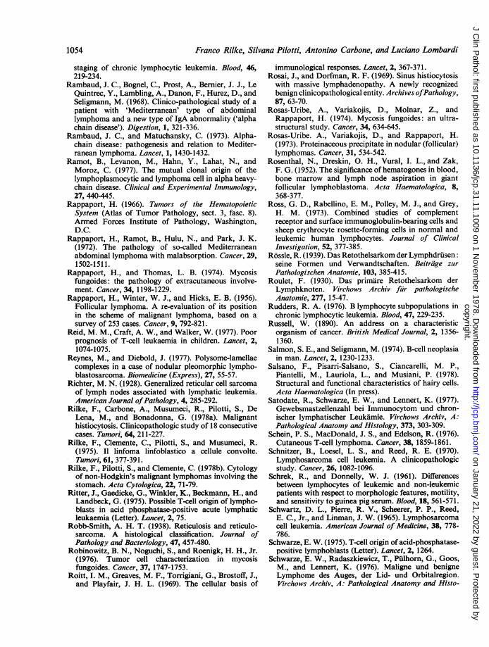
1054 Franco Rilke, Silvana Pilotti, Antonino Carbone, and Luciano Lombardi
staging of chronic lymphocytic leukemia. Blood, 46,219-234.
Rambaud, J. C., Bognel, C., Prost, A., Bernier, J. J., LeQuintrec, Y., Lambling, A., Danon, F., Hurez, D., andSeligmann, M. (1968). Clinico-pathological study of apatient with 'Mediterranean' type of abdominallymphoma and a new type of IgA abnormality ('alphachain disease'). Digestion, 1, 321-336.
Rambaud, J. C., and Matuchansky, C. (1973). Alpha-chain disease: pathogenesis and relation to Mediter-ranean lymphoma. Lancet, 1, 1430-1432.
Ramot, B., Levanon, M., Hahn, Y., Lahat, N., andMoroz, C. (1977). The mutual clonal origin of thelymphoplasmocytic and lymphoma cell in alpha heavy-chain disease. Clinical and Experimental Immunology,27, 440-445.
Rappaport, H. (1966). Tumors of the HematopoieticSystem (Atlas of Tumor Pathology, sect. 3, fasc. 8).Armed Forces Institute of Pathology, Washington,D.C.
Rappaport, H., Ramot, B., Hulu, N., and Park, J. K.(1972). The pathology of so-called Mediterraneanabdominal lymphoma with malabsorption. Cancer, 29,1502-1511.
Rappaport, H., and Thomas, L. B. (1974). Mycosisfungoides: the pathology of extracutaneous involve-ment. Cancer, 34, 1198-1229.
Rappaport, H., Winter, W. J., and Hicks, E. B. (1956).Follicular lymphoma. A re-evaluation of its positionin the scheme of malignant lymphoma, based on asurvey of 253 cases. Cancer, 9, 792-821.
Reid, M. M., Craft, A. W., and Walker, W. (1977). Poorprognosis of T-cell leukaemia in children. Lancet, 2,1074-1075.
Reynes, M., and Diebold, J. (1977). Polysome-lamellaecomplexes in a case of nodular pleomorphic lympho-blastosarcoma. Biomedicine (Express), 27, 55-57.
Richter, M. N. (1928). Generalized reticular cell sarcomaof lymph nodes associated with lymphatic leukemia.American Journal ofPathology, 4, 285-292.
Rilke, F., Carbone, A., Musumeci, R., Pilotti, S., DeLena, M., and Bonadonna, G. (1978a). Malignanthistiocytosis. Clinicopathologic study of 18 consecutivecases. Tumori, 64, 211-227.
Rilke, F., Clemente, C., Pilotti, S., and Musumeci, R.(1975). Il linfoma linfoblastico a cellule convolte.Tumori, 61, 377-391.
Rilke, F., Pilotti, S., and Clemente, C. (1978b). Cytologyof non-Hodgkin's malignant lymphomas involving thestomach. Acta Cytologica, 22, 71-79.
Ritter, J., Gaedicke, G., Winkler, K., Beckmann, H., andLandbeck, G. (1975). Possible T-cell origin of lympho-blasts in acid phosphatase-positive acute lymphaticleukaemia (Letter). Lancet, 2, 75.
Robb-Smith, A. H. T. (1938). Reticulosis and reticulo-sarcoma. A histological classification. Journal ofPathology and Bacteriology, 47, 457-480.
Robinowitz, B. N., Noguchi, S., and Roenigk, H. H., Jr.(1976). Tumor cell characterization in mycosisfungoides. Cancer, 37, 1747-1753.
Roitt, I. M., Greaves, M. F., Torrigiani, G., Brostoff, J.,and Playfair, J. H. L. (1969). The cellular basis of
immunological responses. Lancet, 2, 367-371.Rosai, J., and Dorfman, R. F. (1969). Sinus histiocytosis
with massive lymphadenopathy. A newly recognizedbenign clinicopathological entity. Archives ofPathology,87, 63-70.
Rosas-Uribe, A., Variakojis, D., Molnar, Z., andRappaport, H. (1974). Mycosis fungoides: an ultra-structural study. Cancer, 34, 634-645.
Rosas-Uribe. A., Variakojis, D., and Rappaport, H.(1973). Proteinaceous precipitate in nodular (follicular)lymphomas. Cancer, 31, 534-542.
Rosenthal, N., Dreskin, 0. H., Vural, I. L., and Zak,F. G. (1952). The significance of hematogones in blood,bone marrow and lymph node aspiration in giantfollicular lymphoblastoma. Acta Haematologica, 8,368-377.
Ross, G. D., Rabellino, E. M., Polley, M. J., and Grey,H. M. (1973). Combined studies of complementreceptor and surface immunoglobulin-bearing cells andsheep erythrocyte rosette-forming cells in normal andleukemic human lymphocytes. Journal of ClinicalInvestigation, 52, 377-385.
Rossle, R. (1939). Das Retothelsarkom der Lymphdrusen:seine Formen und Verwandtschaften. Beitrage zurPathologischen Anatomie, 103, 385-415.
Roulet, F. (1930). Das primare Retothelsarkom derLymphknoten. Virchows Archiv fur pathologischeAnatomie, 277, 15-47.
Rudders, R. A. (1976). B lymphocyte subpopulations inchronic lymphocytic leukemia. Blood, 47, 229-235.
Russell, W. (1890). An address on a characteristicorganism of cancer. British Medical Journal, 2, 1356-1360.
Salmon, S. E., and Seligmann, M. (1974). B-cell neoplasiain man. Lancet, 2, 1230-1233.
Salsano, F., Pisarri-Salsano, S., Ciancarelli, M. P.,Piantelli, M., Lauriola, L., and Musiani, P. (1978).Structural and functional characteristics of hairy cells.Acta Haematologica (In press).
Satodate, R., Schwarze, E. W., and Lennert, K. (1977).Gewebsmastzellenzahl bei Immunocytom und chron-ischer lymphatischer Leukamie. Virchows Archiv, A:Pathological Anatomy and Histology, 373, 303-309.
Schein, P. S., MacDonald, J. S., and Edelson, R. (1976).Cutaneous T-cell lymphoma. Cancer, 38, 1859-1861.
Schnitzer, B., Loesel, L. S., and Reed, R. E. (1970).Lymphosarcoma cell leukemia. A clinicopathologicstudy. Cancer, 26, 1082-1096.
Schrek, R., and Donnelly, W. J. (1961). Differencesbetween lymphocytes of leukemic and non-leukemicpatients with respect to morphologic features, motility,and sensitivity to guinea pig serum. Blood, 18, 561-571.
Schwartz, D. L., Pierre, R. V., Scheerer, P. P., Reed,E. C., Jr., and Linman, J. W. (1965). Lymphosarcomacell leukemia. American Journal of Medicine, 38, 778-786.
Schwarze, E. W. (1975). T-cell origin of acid-phosphatase-positive lymphoblasts (Letter). Lancet, 2, 1264.
Schwarze, E. W., Radaszkiewicz, T., Piilhorn, G., Goos,M., and Lennert, K. (1976). Maligne und benigneLymphome des Auges, der Lid- und Orbitalregion.Virchows Archiv, A: Pathological Anatomy and Histo-
copyright. on January 21, 2022 by guest. P
rotected byhttp://jcp.bm
j.com/
J Clin P
athol: first published as 10.1136/jcp.31.11.1009 on 1 Novem
ber 1978. Dow
nloaded from
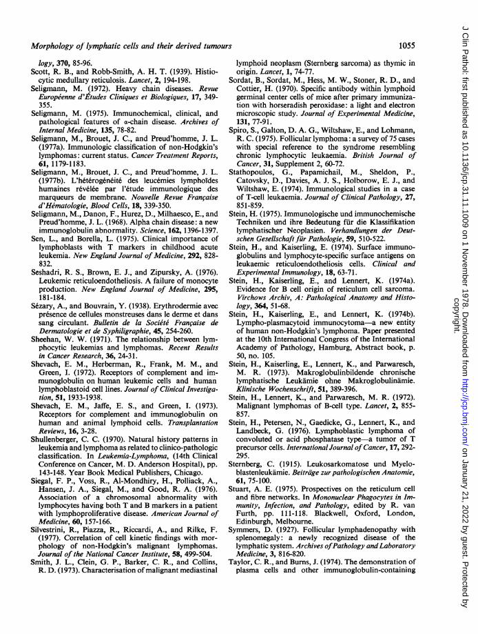
Morphology of lymphatic cells and their derived tumours 1055
logy, 370, 85-96.Scott, R. B., and Robb-Smith, A. H. T. (1939). Histio-
cytic medullary reticulosis. Lancet, 2, 194-198.Seligmann, M. (1972). Heavy chain diseases. RevueEuropeenne d'Atudes Cliniques et Biologiques, 17, 349-355.
Seligmann, M. (1975). Immunochemical, clinical, andpathological features of a-chain disease. Archives ofInternal Medicine, 135, 78-82.
Seligmann, M., Brouet, J. C., and Preud'homme, J. L.(1977a). Immunologic classification of non-Hodgkin'slymphomas: current status. Cancer Treatment Reports,61, 1179-1183.
Seligmann, M., Brouet, J. C., and Preud'homme, J. L.(1977b). L'heterogeneite des leucemies lymphoideshumaines r6velee par I'etude immunologique desmarqueurs de membrane. Nouvelle Revue Francaised'Hematologie, Blood Cells, 18, 339-350.
Seligmann, M., Danon, F., Hurez, D., Milhaesco, E., andPreud'homme, J. L. (1968). Alpha chain disease: a newimmunoglobulin abnormality. Science, 162, 1396-1397.
Sen, L., and Borella, L. (1975). Clinical importance oflymphoblasts with T markers in childhood acuteleukemia. New England Journal of Medicine, 292, 828-832.
Seshadri, R. S., Brown, E. J., and Zipursky, A. (1976).Leukemic reticuloendotheliosis. A failure of monocyteproduction. New England Journal of Medicine, 295,181-184.
Sezary, A., and Bouvrain, Y. (1938). Erythrodermie avecpresence de cellules monstreuses dans le derme et danssang circulant. Bulletin de la Societe Francaise deDermatologie et de Syphiligraphie, 45, 254-260.
Sheehan, W. W. (1971). The relationship between lym-phocytic leukemias and lymphomas. Recent Resultsin Cancer Research, 36, 24-31.
Shevach, E. M., Herberman, R., Frank, M. M., andGreen, I. (1972). Receptors of complement and im-munoglobulin on human leukemic cells and humanlymphoblastoid cell lines. Journal of Clinical Investiga-tion, 51, 1933-1938.
Shevach, E. M., Jaffe, E. S., and Green, I. (1973).Receptors for complement and immunoglobulin onhuman and animal lymphoid cells. TransplantationReviews, 16, 3-28.
Shullenberger, C. C. (1970). Natural history patterns inleukemia and lymphoma as related to clinico-pathologicclassification. In Leukemia-Lymphoma, (14th ClinicalConference on Cancer, M. D. Anderson Hospital), pp.143-148. Year Book Medical Publishers, Chicago.
Siegal, F. P., Voss, R., Al-Mondhiry, H., Polliack, A.,Hansen, J. A., Siegal, M., and Good, R. A. (1976).Association of a chromosomal abnormality withlymphocytes having both T and B markers in a patientwith lymphoproliferative disease. American Journal ofMedicine, 60, 157-166.
Silvestrini, R., Piazza, R., Riccardi, A., and Rilke, F.(1977). Correlation of cell kinetic findings with mor-phology of non-Hodgkin's malignant lymphomas.Journal of the National Cancer Institute, 58, 499-504.
Smith, J. L., Clein, G. P., Barker, C. R., and Collins,R. D. (1973). Characterisation of malignant mediastinal
lymphoid neoplasm (Sternberg sarcoma) as thymic inorigin. Lancet, 1, 74-77.
Sordat, B., Sordat, M., Hess, M. W., Stoner, R. D., andCottier, H. (1970). Specific antibody within lymphoidgerminal center cells of mice after primary immuniza-tion with horseradish peroxidase: a light and electronmicroscopic study. Journal of Experimental Medicine,131, 77-91.
Spiro, S., Galton, D. A. G., Wiltshaw, E., and Lohmann,R. C. (1975). Follicular lymphoma: a survey of 75 caseswith special reference to the syndrome resemblingchronic lymphocytic leukaemia. British Journal ofCancer, 31, Supplement 2, 60-72.
Stathopoulos, G., Papamichail, M., Sheldon, P.,Catovsky, D., Davies, A. J. S., Holborow, E. J., andWiltshaw, E. (1974). Immunological studies in a caseof T-cell leukaemia. Journal of Clinical Pathology, 27,851-859.
Stein, H. (1975). Immunologische und immunochemischeTechniken und ihre Bedeutung fur die Klassifikationlymphatischer Neoplasien. Verhandlungen der Deut-schen Gesellschaft fur Pathologie, 59, 510-522.
Stein, H., and Kaiserling, E. (1974). Surface immuno-globulins and lymphocyte-specific surface antigens onleukaemic reticuloendotheliosis cells. Clinical andExperimental Immunology, 18, 63-71.
Stein, H., Kaiserling, E., and Lennert, K. (1974a).Evidence for B cell origin of reticulum cell sarcoma.Virchows Archiv, A: Pathological Anatomy and Histo-logy, 364, 51-68.
Stein, H., Kaiserling, E., and Lennert, K. (1974b).Lympho-plasmacytoid immunocytoma-a new entityof human non-Hodgkin's lymphoma. Paper presentedat the 10th International Congress of the InternationalAcademy of Pathology, Hamburg, Abstract book, p.50, no. 105.
Stein, H., Kaiserling, E., Lennert, K., and Parwaresch,M. R. (1973). Makroglobulinbildende chronischelymphatische Leukamie ohne Makroglobulinamie.Klinische Wochenschrift, 51, 389-396.
Stein, H., Lennert, K., and Parwaresch, M. R. (1972).Malignant lymphomas of B-cell type. Lancet, 2, 855-857.
Stein, H., Petersen, N., Gaedicke, G., Lennert, K., andLandbeck, G. (1976). Lymphoblastic lymphoma ofconvoluted or acid phosphatase type-a tumor of Tprecursor cells. International Journal ofCancer, 17, 292-295.
Sternberg, C. (1915). Leukosarkomatose und Myelo-blastenleukamie. Beitrage zur pathologischen Anatomie,61, 75-100.
Stuart, A. E. (1975). Prospectives on the reticulum celland fibre networks. In Mononuclear Phagocytes in Im-munity, Infection, and Pathology, edited by R. vanFurth, pp. 111-118. Blackwell, Oxford, London,Edinburgh, Melbourne.
Symmers, D. (1927). Follicular lymphadenopathy withsplenomegaly: a newly recognized disease of thelymphatic system. Archives ofPathology andLaboratoryMedicine, 3, 816-820.
Taylor, C. R., and Burns, J. (1974). The demonstration ofplasma cells and other immunoglobulin-containing
copyright. on January 21, 2022 by guest. P
rotected byhttp://jcp.bm
j.com/
J Clin P
athol: first published as 10.1136/jcp.31.11.1009 on 1 Novem
ber 1978. Dow
nloaded from
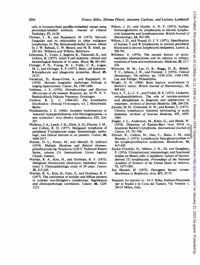
1056 Franco Rilke, Silvana Pilotti, Antonino Carbone, and Luciano Lombardi
cells in formalin-fixed paraffin-embedded tissues usingperoxidase-labelled antibody. Journal of ClinicalPathology, 27, 14-20.
Thomas, L. B., and Rappaport, H. (1975). Mycosisfungoides and its relationship to other malignantlymphomas. In The Reticuloendothelial System, editedby J. W. Rebuck, C. W. Berard, and M. R. Abell, pp.243-261. Williams and Wilkins, Baltimore.
Uchiyama, T., Yodoi, J., Sagawa, K., Takatsuki, K., andUchino, H. (1977). Adult T-cell leukemia: clinical andhematological features of 16 cases. Blood, 50, 481-492.
Utsinger, P. D., Young, W. J., Fuller, C. R., Logue,M. J., and Orringer, E. P. (1977). Hairy cell leukemia:B-lymphocyte and phagocytic properties. Blood, 49,19-27.
Variakojis, D., Rosas-Uribe, A., and Rappaport, H.(1974). Mycosis fungoides: pathologic findings instaging laparotomies. Cancer, 33, 1589-1600.
Veldman, J. E. (1970). Histophysiology and ElectronMicroscopy of the Immune Response, pp. 42-79. N. V.Boekdrukkerij Dijkstra Niemeyer, Groningen.
Virchow, R. L. C. (1864-65). Die krankhaftenGeschwulste: Dreissig Vorlesungen, vol. 2. Hirschwald,Berlin.
Waldenstrom, J. G. (1944). Incipient myelomatosis or'essential' hyperglobulinemia with fibrinogenopenia-anew syndrome? Acta Medica Scandinavica, 117, 216-247.
Waldron, J. A., Leech, J. H., Glick, A. D., Flexner, J. M.,and Collins, R. D. (1977). Malignant lymphoma ofperipheral T-lymphocyte origin. Immunologic, patho-logic, and clinical features in six patients. Cancer, 40,1604-1617.
Warner, N. L., Potter, M., and Metcalf, D. (editors)(1974). Multiple Myeloma and Related Immuno-globulin-producing Neoplasms (UICC Technical ReportSeries, volume 13). International Union AgainstCancer, Geneva.
Warnke, R. A., Kim, H., and Dorfman, R. F. (1975).Malignant histiocytosis (histiocytic medullary reticu-losis). I. Clinicopathologic study of 29 cases. Cancer,35, 215-230.
Warnke, R. A., Kim, H., Fuks, Z., and Dorfman, R. F.(1977). The coexistence of nodular and diffuse patternsin nodular non-Hodgkin's lymphomas. Significanceand clinicopathologic correlation. Cancer, 40, 1229-1233.
Wilson, J. D., and Hurdle, A. D. F. (1973). Surfaceimmunoglobulins on lymphocytes in chronic lympho-cytic leukaemia and lymphosarcoma. British Journal ofHaematology, 24, 563-569.
Wilson, J. D., and Nossal, G. J. V. (1971). Identificationof human T and B lymphocytes in normal peripheralblood and in chronic lymphocytic leukaemia. Lancet, 2,788-791.
Wiltshaw, E. (1976). The natural history of extra-medullary plasmacytoma and its relation to solitarymyeloma of bone and myelomatosis. Medicine, 55, 217-238.
Wintrobe, M. M., Lee, G. R., Boggs, D. R., Bithell,T. C., Athens, J. W., and Forester, J. (1974). ClinicalHematology, 7th edition, pp. 1520-1534, 1568-1598.Lea and Febiger, Philadelphia.
Wright, D. H. (1968). Bone marrow involvement inBurkitt's tumor. British Journal of Haematology, 15,409-416.
Yam, L. T., Li, C. Y., and Finkel, H. E. (1972). Leukemicreticuloendotheliosis. The role of tartrate-resistantacid phosphatase in diagnosis and splenectomy intreatment. Archives ofInternal Medicine, 130, 248-256.
Zarrabi, M. H., Grunwald, H. W., and Rosner, F. (1977).Chronic lymphocytic leukemia terminating in acuteleukemia. Archives of Internal Medicine, 137, 1059-1064.
Ziegler, J. L., Andersson, M., Klein, G., and Henle, W.(1976). Detection of Epstein-Barr virus DNA inAmerican Burkitt's lymphoma. International Journal ofCancer, 17, 701-706.
Zittoun, R., Cadiou, M., Dao, C., Blanc, J. M., andBousser, J. (1973). Lymphocytic beta-glucuronidase inthe lymphoproliferative syndromes. Biomedicine, 18,415-420.
Zucker-Franklin, D., Melton, J. W., III, and Quagliata,F. (1974). Ultrastructural, immunologic, and functionalstudies on Sezary cells: a neoplastic variant of thymus-derived (T) lymphocytes. Proceedings of the NationalAcademy of Sciences of the United States of America,71, 1877-1881.
Zur Hausen, H. (1975). Oncogenic herpes viruses.Biochimica et Biophysica Acta, 417, 25-53.
Requests for reprints to: Dr F. Rilke, InstitutoNazionaleper lo Studio e la Cuva dei Tumori, Via Venezia 1,20133 Milan, Italy.
copyright. on January 21, 2022 by guest. P
rotected byhttp://jcp.bm
j.com/
J Clin P
athol: first published as 10.1136/jcp.31.11.1009 on 1 Novem
ber 1978. Dow
nloaded from
Recommended


