Mother’s Milk-Induced Hsp70 Expression Preserves IntestinalEpithelial Barrier Function in an Immature Rat Pup Model
JENNIFER L. LIEDEL, YUEE GUO, YUEYUE YU, SHENG-RU SHIOU, SANGZI CHEN, ELAINEO. PETROF, SHIEN HU, MARK W. MUSCH, and ERIKA C. CLAUDDepartments of Pediatrics [J.L.L., Y.G., Y.Y., S.-R.S., S.C., E.C.C.] and Medicine [S.H., M.W.M.,E.C.C.], The University of Chicago, Chicago, Illinois 60637; Department of Medicine [E.O.P.],Queen’s University, Kingston, Ontario K7L 2V7, Canada
AbstractPreterm infants face many challenges in transitioning from the in utero to extrauterineenvironment while still immature. Failure of the preterm gut to successfully mature toaccommodate bacteria and food substrate leads to significant morbidity such as neonatalnecrotizing enterocolitis. The intestinal epithelial barrier plays a critical role in gut protection.Heat shock protein 70 (Hsp70) is an inducible cytoprotective molecule shown to protect theintestinal epithelium in adult models. To investigate the hypothesis that Hsp70 may be importantfor early protection of the immature intestine, Hsp70 expression was evaluated in intestine ofimmature rat pups. Data demonstrate that Hsp70 is induced by exposure to mother’s milk. Hsp70is found in mother’s milk, and increased Hsp70 transcription is induced by mother’s milk. ThisHsp70 colocalizes with the tight junction protein ZO-1. Mother’s milk-induced Hsp70 maycontribute to maintenance of barrier function in the face of oxidant stress. Further understandingof the means by which mother’s milk increases Hsp70 in the ileum will allow potential means ofstrengthening the intestinal barrier in at-risk preterm infants.
The single layer of enterocytes lining the intestinal epithelium forms a functional barrierbetween the luminal contents of the gut and the host. This critical barrier is responsible forallowing nutrients and beneficial macromolecules to cross while preventing translocation ofbacteria and bacterial products (1). The intestinal barrier comprises a mucin layer coveringthe intestinal epithelial cells, the epithelial cell plasma membrane, and the apical junctionalcomplex between cells (1). The apical junctional complex consists of a network of tightjunction proteins and the adherens junction, both anchored by a perijunctional actomyosinring (2). The tight junction is a principal determinant of mucosal permeability.
The requirements for the intestinal barrier change over the course of development (3–6). Inutero, the gut lumen is bathed in amniotic fluid and has no contact with bacteria or foodsubstrates. Thus, the intestine has high permeability to allow passage of macromoleculescritical for development from the amniotic fluid (6,7). After birth, the barrier mustappropriately accommodate bacteria and products of digestion. The preterm infant,expecting continuation of the in utero environment, carries much of the immature phenotypedesigned for in utero life into the extrauterine world. This includes a porous intestinal barriernot prepared for sudden exposure to intestinal bacteria. Thus, the immature gut of preterminfants has increased translocation of intestinal bacteria, predisposing the preterm infant tosepsis and neonatal necrotizing enterocolitis (NEC) (8–10). The immature gut has been
Copyright © 2011 International Pediatric Research Foundation, Inc.Correspondence: Erika C. Claud, M.D., Department of Pediatrics, Neonatology, The University of Chicago, 5841 S. Maryland AveMC6060, Chicago, IL 60637; [email protected].
NIH Public AccessAuthor ManuscriptPediatr Res. Author manuscript; available in PMC 2011 May 5.
Published in final edited form as:Pediatr Res. 2011 May ; 69(5 Pt 1): 395–400. doi:10.1203/PDR.0b013e3182114ec9.
NIH
-PA Author Manuscript
NIH
-PA Author Manuscript
NIH
-PA Author Manuscript
shown to have an exaggerated inflammatory response to intestinal bacteria (9,11–13).Permeability across the tight junction can be increased by inflammatory cytokines such asIFNγ and TNFα, thus further increasing bacterial translocation and the cycle of injury(1,14,15).
Inducible heat shock protein (Hsp) 70 is a cytoprotective protein shown to have importantroles in intestinal protection and regeneration both in vitro and in vivo (16–19). Hsp70 hasspecifically been shown to maintain barrier function, in part, by stabilizing the tightjunctions between intestinal epithelial cells (20,21). This intestinal epithelial protection isassociated with restricted bacterial translocation and a reduction in inflammation (22). Therole of Hsps in the immature gut has not been previously explored. Only one study hasinvestigated the temporal pattern of Hsp expression. This study in newborn pigs investigateda number of Hsps following weaning (23). We hypothesized that Hsp70 may be importantfor early protection, before weaning, of the immature intestine in the face of stressorsleading to inflammation.
METHODSNeonatal rat pups
Animal experiments were approved by the animal care committee at the University ofChicago. All rat pups were allowed to spontaneously deliver and then remain with themother for rearing (mother-fed) or separated from mother for stresses. Stressed pups werehoused in a humidified incubator maintained at 37°C. Formula-fed pups were gavaged every3 h with Esbilac puppy formula via an orogastric tube. For 24 h, animals were fed 0.1 mLevery 3 h, which was increased daily by 50 μL every 3 h until killing.
Immunohistochemistry stainingIntestinal segments were removed, opened longitudinally, washed three times in saline, andfixed in formalin. Paraffin-embedded tissues were cut into 4-μm-thick sections. The sectionswere deparaffinized at 56°C, immersed in xylene three times, and hydrated with ethanol(two times with 100%, two times with 95%, and one time with 75% ethanol) for 5 min. Forantigen unmasking, slides were heated in 10-mM sodium citrate buffer (pH 6.0) for 15 minbefore treatment with 0.3% hydrogen peroxide for 30 min. The specimens were treated with5% BSA in TBST (Tris-buffered saline with 0.05% vol/vol Tween-20) for 30 min at roomtemperature followed by overnight incubation with mouse anti-Hsp70 antibody (MAB1663;R&D Systems, Minneapolis, MN) at 4°C. After washing, the sections were incubated withpolymer-HRP anti-mouse (Dako, Carpinteria, CA) for 30 min at room temperature. Positivestaining was visualized with DAB chromogen, and nuclei counterstain was performed withhematoxylin (HHS32; Sigma Chemical Co., St. Louis, MO).
Quantitative measurement of Hsp70 in rat dam milkRat milk was collected postmortem from lactating dams on postnatal d 10, using gentlesuction. Whole milk was centrifuged (700×g for 10 min at 4°C) and supernatant Hsp70measured by ELISA (EKS-700B; Assay Designs, Boulder, CO) as per manufacturer’sinstructions.
Real-time PCR for Hsp mRNATotal RNA was extracted from ileal tissue by TRIzol (Invitrogen, Carlsbad, CA) accordingto the manufacturer’s instruction. cDNA was synthesized using SuperScript II (Invitrogen)and random hexonucleotide primer. The rat Hsp70 sequences were downloaded fromGenBank. The forward and reverse primers were for rat Hsp70 (NM_031971, bases 25–139)and rat GAPDH (NM_017008, bases 154–223). Real-time PCR was performed with an
LIEDEL et al. Page 2
Pediatr Res. Author manuscript; available in PMC 2011 May 5.
NIH
-PA Author Manuscript
NIH
-PA Author Manuscript
NIH
-PA Author Manuscript
iCycler using iQSYBR Green PCR Supermix (Bio-Rad, Hercules, CA). The two-stepquantification cycling protocol was used. The Ct value is defined as the cycle number atwhich the fluorescence crosses a fixed threshold above the baseline. As a relativequantitation, fold changes were measured using the ΔΔCt method (24).
Ex vivo intestinal loops and assessment of barrier functionSections of ileum 1–1.5 cm in length were taken beginning 1 cm above the ileocecaljunction. The segments were flushed with warm PBS to remove stool. The ends weresecured with silk ties and the lumen filled with 10 mg/mL of 10 kD FITC-dextran (SigmaChemical Co.) with or without freshly prepared 0.1 mM monochloramine) (NH2Cl) untilmoderate distension was achieved (75μL) (20). The loops were placed in the inner well oforgan culture dishes (Falcon; Becton Dickinson Labware, Franklin Lakes, NJ) filled withRPMI 1640 medium with 10% vol/vol heat-inactivated fetal bovine serum. These wereincubated for 1 h at 37°C in a 5% CO2 incubator. At 30 and 60 min, 100 μL of the bathingmedia was removed for measurement of fluorescence to determine translocation across theloops. Translocated FITC-dextran was quantified by fluorescence and concentrationdetermined by a standard curve of known amounts of FITC-dextran, normalized to intestinallength in millimeters, and expressed as fold change over baseline. The middle section ofthese loops was removed, placed in 10% buffered formalin, and embedded for sectioningand immunohistochemistry.
Hsp70 and ZO-1 immunofluorescence stainingSections were prepared as described for immunohistochemistry staining. The specimenswere treated with 5% BSA in TBST for 30 min at room temperature followed by overnightincubation with mouse anti-Hsp70 antibody (R&D Systems) or rabbit anti-ZO-1(Invitrogen) at 4°C. After overnight incubation with primary antibody and washing, sectionswere incubated with Cy2-anti-mouse and Cy5 anti-rabbit (Jackson ImmunoResearch, WestGrove, PA) overnight at 4°C. Slides were washed with saline five times, incubated brieflywith 4′,6-diamidino-2-phenylindole (DAPI; final concentration 1 μg/mL for 5 min;Molecular Probes, Eugene, OR) to stain nuclei, washed three times in saline, and coverslipsmounted using Slow Fade mounting medium (Molecular Probes) and visualized. Confocalmicroscopy was performed using a Leica SP2AOBS system (Leica, Wetzlar, Germany) ofthe Light Microscopy Core Center of the University of Chicago.
In vivo model of intestinal injuryAn in vivo model of intestinal stress was performed using a protocol modified frompublished protocols designed to model NEC (25,26). Rat pups were delivered by cesareansection on the 21st d of gestation. Pups were maintained in an incubator at 37°C and gavage-fed as described above. Pups were colonized with bacteria 107 colony forming units each ofSerratia marcescens, Klebsiella pneumoniae, and Streptococci viridans once daily in 100 μLformula via the orogastric feeding catheter. In addition, pups were stressed with hypoxia(5% oxygen and 95% nitrogen for 10 min) three times a day. Pups were killed at indicatedtime points. Hematoxylin and eosin (H&E)-stained sections were scored by a blindedpathologist using a validated NEC scoring system with scores ranging from “0” to “4”indicating increasing severity of injury (26).
RESULTSMother’s milk feeding maintains small intestinal Hsp70 expression
To determine whether the intestine of neonatal rat pups expresses Hsp70, ileal sections ofsmall intestine were analyzed by immunostaining. Hsp70 was present in the epithelial cell
LIEDEL et al. Page 3
Pediatr Res. Author manuscript; available in PMC 2011 May 5.
NIH
-PA Author Manuscript
NIH
-PA Author Manuscript
NIH
-PA Author Manuscript
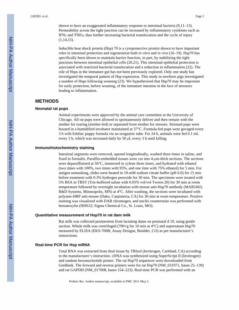
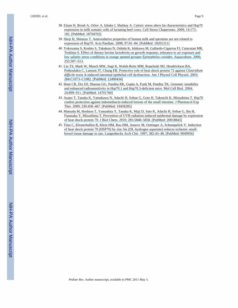
compartment to a greater degree in mother’s milk-fed pups compared with pups receivingonly formula (Fig. 1A, arrow denotes brown staining indicative of Hsp70 expression).Formula-fed pups demonstrated a time-dependent decrease in Hsp70 staining, whereas theexpression of Hsp70 increased in the mother’s milk-fed pups.
Rat milk was obtained and analyzed for Hsp70 by ELISA. Hsp70 was found atconcentrations of 1.48–2.78 ng/mL in whole milk (n = 3). To determine whether smallintestinal Hsp70 expression was additionally due to epithelial expression, mRNA wasextracted from extensively washed (three times) tissue from pups at d 3. Hsp70 mRNA wasmeasured and compared by the ΔΔCt method. Hsp70 mRNA was readily detected in thewashed tissues, demonstrating intestinal epithelial expression. Hsp70 mRNA thresholdvalues were significantly higher for the pups on mother’s milk. When ΔΔCt value for thepups on mother’s milk was set to 1, Hsp70 mRNA values in formula-fed pups were ~50%lower on d 3 (Fig. 1B), corresponding to the decreased Hsp70 immunostaining shown in Fig.1.
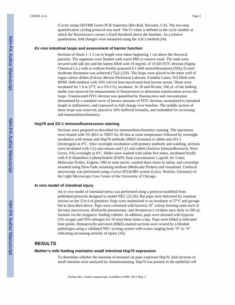
Permeability of the small intestine is protected from oxidants in mother’s milk-fed pupsPermeability of the small intestine was assessed in ileal loops. The basal permeability wasnot different between the mother’s milk-fed and formula-fed ileum (Fig. 2). The oxidantmonochloramine (NH2Cl) was used to increase permeability. Loops were filled with a 10-kD fluorescent dextran and movement of this marker to the external bathing mediummeasured. NH2Cl (0.1 mM) did not stimulate increased flux of FITC-dextran in mother’smilk-fed pups, demonstrating resistance to oxidant-induced injury. However, the sameNH2Cl concentration stimulated a large increase in FITC-dextran movement out of the ilealloops of formula-fed pups (Fig. 2).
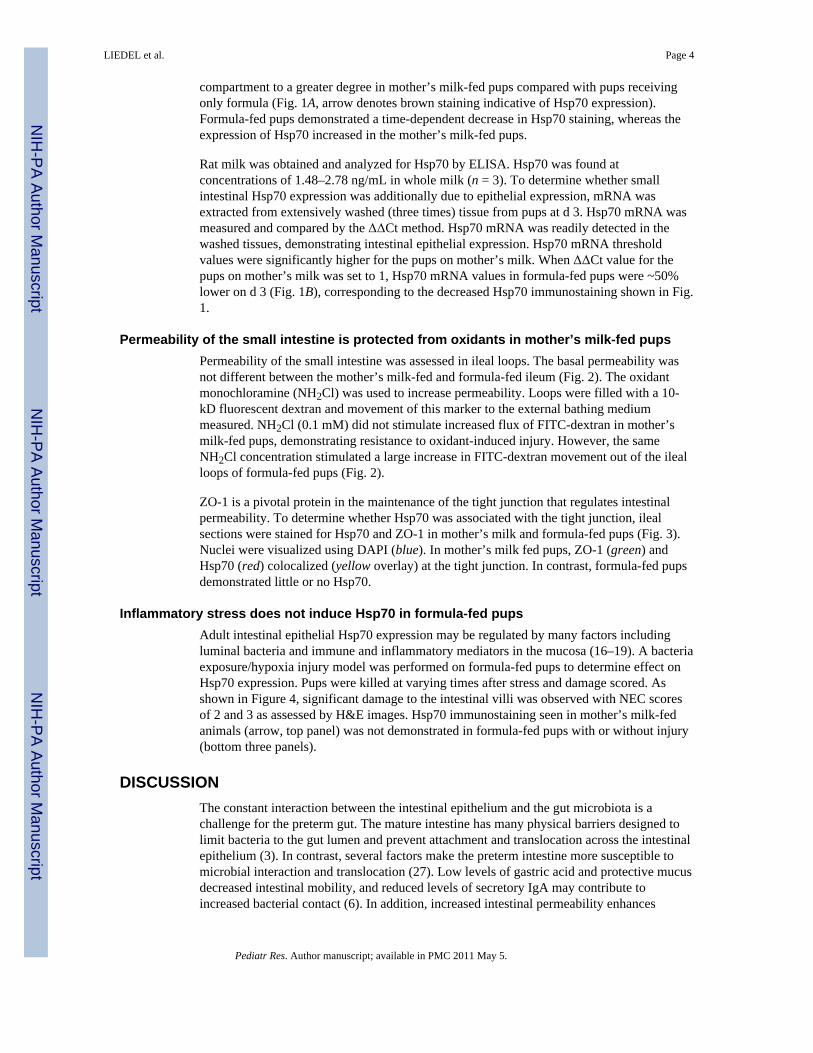
ZO-1 is a pivotal protein in the maintenance of the tight junction that regulates intestinalpermeability. To determine whether Hsp70 was associated with the tight junction, ilealsections were stained for Hsp70 and ZO-1 in mother’s milk and formula-fed pups (Fig. 3).Nuclei were visualized using DAPI (blue). In mother’s milk fed pups, ZO-1 (green) andHsp70 (red) colocalized (yellow overlay) at the tight junction. In contrast, formula-fed pupsdemonstrated little or no Hsp70.
Inflammatory stress does not induce Hsp70 in formula-fed pupsAdult intestinal epithelial Hsp70 expression may be regulated by many factors includingluminal bacteria and immune and inflammatory mediators in the mucosa (16–19). A bacteriaexposure/hypoxia injury model was performed on formula-fed pups to determine effect onHsp70 expression. Pups were killed at varying times after stress and damage scored. Asshown in Figure 4, significant damage to the intestinal villi was observed with NEC scoresof 2 and 3 as assessed by H&E images. Hsp70 immunostaining seen in mother’s milk-fedanimals (arrow, top panel) was not demonstrated in formula-fed pups with or without injury(bottom three panels).
DISCUSSIONThe constant interaction between the intestinal epithelium and the gut microbiota is achallenge for the preterm gut. The mature intestine has many physical barriers designed tolimit bacteria to the gut lumen and prevent attachment and translocation across the intestinalepithelium (3). In contrast, several factors make the preterm intestine more susceptible tomicrobial interaction and translocation (27). Low levels of gastric acid and protective mucusdecreased intestinal mobility, and reduced levels of secretory IgA may contribute toincreased bacterial contact (6). In addition, increased intestinal permeability enhances
LIEDEL et al. Page 4
Pediatr Res. Author manuscript; available in PMC 2011 May 5.
NIH
-PA Author Manuscript
NIH
-PA Author Manuscript
NIH
-PA Author Manuscript
translocation (27). As a counter balance, our data reveal an inducible intestinal protectivemechanism that is unique to the immature intestine. In contrast to adults without smallintestinal Hsp70 expression, we found that the immature intestine has basal expression ofthe cytoprotective protein Hsp70 in the ileum. Increased Hsp70 transcription was induced bymother’s milk and was noted to colocalize with the tight junction protein ZO-1.
It has been shown in clinical studies, using enteral lactulose/mannitol administration toevaluate intestinal permeability, that infants receiving formula had increased permeabilityover the 1st mo of life compared with infants receiving breast milk (28). Furthermore,clinical studies have documented a decline in the incidence of NEC in human milk-fedinfants compared with formula-fed infants, suggesting a protective effect of human milk(29–31). Human milk contains several antibacterial and antiinflammatory factors that maybe protective in addition to growth factors that may stimulate intestinal repair throughincreased cell restitution, growth, and inhibition of apoptosis (32–39). Our findings add tothe list of beneficial effects of mother’s milk by suggesting that mother’s milk containsHsp70 protein and has factors to promote Hsp70 induction.
Our study does not confirm whether supply of intact Hsp70 present in the milk or inductionof Hsp70 in enterocytes is of greater importance. Measurable amounts of secreted Hsp70were found in expressed mother’s milk obtained by gentle suction. It is unknown whetherthis method of obtaining milk altered milk composition and Hsp70 amounts. Suckling pupsalso had increased Hsp70 in stained intestinal sections. This is consistent with other studiesdemonstrating the presence of Hsp70 in bovine milk-producing cells and secreted milk (38).Examination of epithelial RNA demonstrated increased Hsp70 mRNA in ileum from pupsfed with mother’s milk, suggesting increased transcription. Tissues for mRNA analysis werecarefully washed before analysis to remove any adherent mother’s milk and potentialcontaminating mRNA that may have been secreted into the milk itself.
Hsp70 from mother’s milk and epithelial production may both play protective roles;however, our study does not determine the specificity of Hsp70 in preserving barrierfunction. It is likely that other factors also induced by mother’s milk have a role inpreservation of the barrier. Future studies examining the effect of mother’s milk notcontaining Hsp70 or exogenous Hsp70 added to formula are necessary to determine thespecificity of the role of Hsp70 in intestinal protection. Hsp70 transcription may bestimulated by a mother’s milk factor. Lactoferrin, which is known to be found in mother’smilk, has been shown to increase Hsp70 in other models and is one potential candidate (40).Additional studies beyond the scope of work are necessary to identify the factors in milk thatstimulate Hsp70 production.
Inducible Hsps belong to a family of highly conserved proteins, which play an importantrole in protecting cells against stressors such as heat, ischemia/reperfusion injury, oxidativestress, or exposure to radiation and toxins (20,41–44). In the adult rat intestine, induction ofHsps before ischemia-reperfusion preserves mucosal integrity, attenuating mucosal injuryand neutrophilic infiltration (45). Hsp70, in particular, protects intestinal epithelial cellsagainst oxidant injury in vitro (16,20). In vivo, commensal bacteria are responsible forinducing the expression of Hsps in the adult intestine (19,21), which likely provideprotection against the hostile environment normally found in the gut. Therefore, induction ofHsp70 expression, which results from these bacterial-epithelial cell interactions, plays animportant role in maintaining intestinal homeostasis.
Intestinal immaturity of preterm infants increases susceptibility to intestinal injury. NEC isthe most common gastrointestinal injury in preterm infants, and human milk has been shownto be protective against this disease. It has been suggested that intestinal barrier disruption
LIEDEL et al. Page 5
Pediatr Res. Author manuscript; available in PMC 2011 May 5.
NIH
-PA Author Manuscript
NIH
-PA Author Manuscript
NIH
-PA Author Manuscript
may contribute to the pathogenesis of NEC, leading to enhanced translocation of luminalbacteria across the intestinal epithelium, which triggers an inflammatory response resultingin the clinical signs and symptoms of this disease. Several inflammatory mediators includingplatelet-activating factor, TNFα, and IL-1β are known to be elevated in NEC and are able todisrupt the intestinal epithelial barrier. However, it is not known if it is the presence of aharmful mediator or the absence of a protective mediator that increases susceptibility todisease. In our ex vivo model, formula feeding alone did not result in an alteration in barrierfunction as measured by FITC-dextran translocation in the formula-fed loops. Oxidantinjury significantly increased permeability only in loops from formula-fed pups, which didnot have Hsp70. Injured intestinal loops from mother’s milk-fed pups with abundantepithelial Hsp70 did not have increased permeability. Shoji and Shimizu (39) havepreviously shown a protective effect of Hsp against oxidant stress in rat jejunal IEC-6 cells.In that study, a specific fraction of human colostrum administered in vitro for 24 h did notinduce Hsp70. Our in vivo model demonstrates that whole milk does induce Hsp70 withhighest levels beginning at d 3 suggesting that longer exposure or a factor present in wholemilk and secreted over time may be necessary. The complex structure of the tight junctionsuggests that maintenance of appropriate intestinal permeability is multifactorial. The lackof change in baseline permeability suggests that Hsp70 may not be involved in basalregulation of tight junction function. Hsp70 is known to be cytoprotective; thus, it is underconditions of stress that we expect Hsp70 to play a role. We used NH2Cl oxidant-inducedstress as a well-described in vitro model of intestinal damage. Our confocal imagesdemonstrate Hsp70 localization to the ZO-1 containing tight junctional region, suggestingthat protection may be due to association with ZO-1. However, Hsp70 may also bind otherproteins that form the junctional complex.
In adult models, Hsp have been shown to be increased under conditions of stress such asbacteria exposure. We attempted to induce Hsp production in the formula-fed pups withoutbaseline Hsp70 expression by exposure to bacteria and hypoxia in the in vivo model ofintestinal injury. These clinically relevant stresses did not increase Hsp70 proteinexpression, possibly because of induction of inflammation. Other studies have shown thatinflammation decreases Hsp70 transcription (17). Our hypothesis is that the Hsp70 needs tobe present before inflammatory injury to be protective.
Mother’s milk provides many benefits to infants including intestinal protection. Our studiesadd to the building knowledge base of means by which protection of the intestinal barrieroccurs. These results demonstrate that expression of Hsp70 in the neonatal intestine isregulated by exposure to mother’s milk. Although Hsp70 is present in mothers milk,additional factors that are absent from formula must be present for the induction of Hsp70 tooccur. Further understanding of the means by which mother’s milk increases Hsp70 in theileum will allow potential means of strengthening the intestinal barrier in at-risk preterminfants.
AcknowledgmentsSupported by NIH grants NIDDK K08 HD049514 [J.L.], HD 59123 [E.C.C.], HD 55237 [E.C.C.], and AT 00404[E.C.C., E.O.P.] and Digestive Disease Research Core Center DK42086.
We thank Drs. Amy Noffsinger and Maria Westerhoff from the Department of Pathology for their assistance inscoring of the intestinal tissue sections.
Abbreviations
H&E hematoxylin and eosin
LIEDEL et al. Page 6
Pediatr Res. Author manuscript; available in PMC 2011 May 5.
NIH
-PA Author Manuscript
NIH
-PA Author Manuscript
NIH
-PA Author Manuscript
Hsp heat shock protein
NEC neonatal necrotizing enterocolitis
References1. Nusrat A, Turner JR, Madara JL. Molecular physiology and pathophysiology of tight junctions. IV.
Regulation of tight junctions by extracellular stimuli: nutrients, cytokines, and immune cells. Am JPhysiol Gastrointest Liver Physiol. 2000; 279:G851–G857. [PubMed: 11052980]
2. Mitic LL, Anderson JA. Molecular architecture of tight junctions. Annu Rev Physiol. 1998; 60:121–142. [PubMed: 9558457]
3. Udall JN, Pang K, Fritze L, Kleinman R, Walker WA. Development of gastrointestinal mucosalbarrier. I. The effect of age on intestinal permeability to macromolecules. Pediatr Res. 1981;15:241–244. [PubMed: 7220146]
4. Israel EJ. Neonatal necrotizing enterocolitis, a disease of the immature intestinal mucosal barrier.Acta Paediatr Suppl. 1994; 396:27–32. [PubMed: 8086678]
5. Pácha J. Development of intestinal transport function in mammals. Physiol Rev. 2000; 80:1633–1667. [PubMed: 11015621]
6. Walker WA. Development of the intestinal mucosal barrier. J Pediatr Gastroenterol Nutr. 2002;34:S33–S39. [PubMed: 12082386]
7. Rouwet EV, Heineman E, Buurman WA, ter Riet G, Ramsay G, Blanco CE. Intestinal permeabilityand carrier-mediated monosaccharide absorption in preterm neonates during the early postnatalperiod. Pediatr Res. 2002; 51:64–70. [PubMed: 11756641]
8. Deitch EA. Role of bacterial translocation in necrotizing enterocolitis. Acta Paediatr Suppl. 1994;396:33–36. [PubMed: 8086679]
9. Halpern MD, Dominguez JA, Dvorakova K, Houbec H, Williams CS, Meza YG, Ruth MC, DvorakB. Ileal cytokine dysregulation in experimental necrotizing enterocolitis is reduced by epidermalgrowth factor. J Pediatr Gastroenterol Nutr. 2003; 36:126–133. [PubMed: 12500008]
10. Hackam DJ, Upperman JS, Grishin A, Ford HR. Disordered enterocyte signaling and intestinalbarrier dysfunction in the pathogenesis of necrotizing enterocolitis. Semin Pediatr Surg. 2005;14:49–57. [PubMed: 15770588]
11. Caplan MS, MacKendrick W. Necrotizing enterocolitis: a review of pathogenic mechanisms andimplications for prevention. Pediatr Pathol. 1993; 13:357–369. [PubMed: 8516229]
12. Claud EC, Lu L, Anton PM, Savidge T, Walker WA, Cherayil BJ. Developmentally regulatedIkappaB expression in intestinal epithelium ans susceptibility to flagellin-induced inflammation.Proc Natl Acad Sci U S A. 2004; 101:7404–7408. [PubMed: 15123821]
13. Claud EC, Zhang X, Petrof EO, Sun J. Developmentally regulated tumor necrosis factor alphainduced nuclear factor kappaB activation in intestinal epithelium. Am J Physiol Gastrointest LiverPhysiol. 2007; 292:G1411–G1419. [PubMed: 17307728]
14. Clark JA, Doelle SM, Halpern MD, Saunders TA, Holubec H, Dvorak K, Boitano SA, Dvorak B.Intestinal barrier failure during experimental necrotizing enterocolitis: protective effect of EGFtreatment. Am J Physiol Gastrointest Liver Physiol. 2006; 291:G938–G949. [PubMed: 16798726]
15. Ismail AS, Hooper LV. Epithelial cells and their neighbors. IV. Bacterial contributions to intestinalepithelial barrier integrity. Am J Physiol Gastrointest Liver Physiol. 2005; 289:G779–G784.[PubMed: 16227525]
16. Musch MW, Kaplan B, Chang EB. Role of increased basal expression of heat shock protein 72 incolonic epithelial c2BBE adenocarcinoma cells. Cell Growth Differ. 2001; 12:419–426. [PubMed:11504707]
17. Hu S, Zhu X, Triggs JR, Tao Y, Wang Y, Lichtenstein L, Bissonnette M, Musch MW, Chang EB.Inflammation induced 3′UTR dependent translational inhibition of Hsp70 mRNA impairsintestinal homeostasis. Am J Physiol Gastrointest Liver Physiol. 2009; 296:G1003–G1011.[PubMed: 19299581]
LIEDEL et al. Page 7
Pediatr Res. Author manuscript; available in PMC 2011 May 5.
NIH
-PA Author Manuscript
NIH
-PA Author Manuscript
NIH
-PA Author Manuscript
18. Petrof EO, Ciancio MJ, Chang EB. Role and regulation of intestinal epithelial heat shock proteinsin health and disease. Chin J Dig Dis. 2004; 5:45–50. [PubMed: 15612656]
19. Arvans DL, Vavricka SR, Ren H, Musch MW, Kang L, Rocha FG, Lucioni A, Turner JR, AlverdyJ, Chang EB. Luminal bacterial flora determines physiological expression of intestinal epithelialcytoprotective heat shock proteins 25 and 72. Am J Physiol Gastrointest Liver Physiol. 2005;288:G696–G704. [PubMed: 15528251]
20. Musch MW, Sugi K, Straus D, Chang EB. Heat-shock protein 72 protects against oxidant-inducedinjury of barrier function of human colonic epithelial Caco2/bbe cells. Gastroenterology. 1999;117:115–122. [PubMed: 10381917]
21. Kojima K, Musch MW, Ren H, Boone DL, Hendrickson BA, Ma A, Chang EB. Enteric flora andlymphocyte-derived cytokines determine expression of heat shock proteins in mouse colonicepithelial cells. Gastroenterology. 2003; 124:1395–1407. [PubMed: 12730879]
22. Eaves-Pyles T, Wong HR, Alexander JW. Sodium arsenite induces the stress response in the gutand decreases bacterial translocation in a burned mouse model with gut-derived sepsis. Shock.2000; 13:314–319. [PubMed: 10774621]
23. David JC, Grongnet JF, Lalles JP. Weaning affects the expression of heat shock proteins indifferent regions of the gastrointestinal tract of piglets. J Nutr. 2002; 132:2551–2561. [PubMed:12221208]
24. Schmittgen TD, Zakrajsek BA, Mills AG, Gorn V, Singer MJ, Reed MW. Quantitative reversetranscription polymerase chain reaction to study mRNA decay: comparison of end point and realtime methods. Anal Biochem. 2000; 285:194–204. [PubMed: 11017702]
25. Nadler EP, Dickinson E, Knisely A, Zhang X, Boyle P, Beer-Stolz D, Watkins SC, Ford HR.Expression of inducible nitric oxide synthase and interleukin-12 in experimental necrotizingenterocolitis. J Surg Res. 2000; 92:71–77. [PubMed: 10864485]
26. Jilling T, Simon D, Lu J, Meng FJ, Li D, Schy R, Thomson RB, Soliman A, Arditi M, Caplan MS.The roles of bacteria and TLR4 in rat and murine models of necrotizing enterocolitis. J Immunol.2006; 177:3273–3282. [PubMed: 16920968]
27. Go LL, Albanese CT, Watkins SC, Simmons RL, Rowe MI. Breast milk protects the neonate frombacterial translocation. J Pediatr Surg. 1994; 29:1059–1063. [PubMed: 7965506]
28. Weaver LT, Laker MF, Nelson R. Intestinal permeability in the newborn. Arch Dis Child. 1984;59:236–241. [PubMed: 6424583]
29. Pitt J, Barlow B, Heird WC. Protection against experimental necrotizing enterocolitis by maternalmilk. I. Role of milk leukocytes. Pediatr Res. 1977; 11:906–909. [PubMed: 329204]
30. Dvorak B, Halpern MD, Holubec H, Dovrakova K, Dominquez JA, Williams CS, Meza YG,Kozakova H, McCuskey RS. Maternal milk reduces severity of necrotizing enterocolitis andincreases intestinal IL-10 in a neonatal rat model. Pediatr Res. 2003; 53:426–433. [PubMed:12595590]
31. Lucas A, Cole TJ. Breast milk and neonatal necrotizing enterocolitis. Lancet. 1990; 336:1519–1523. [PubMed: 1979363]
32. Chandan RC, Parry RM, Shahani KM. Lysozyme, lipase, and ribonuclease in milk of variousspecies. J Dairy Sci. 1968; 51:606–607.
33. Ronayne de Ferrer PA, Baroni A, Sambucetti ME, López NE, Ceriani Cernadas JM. Lactoferrinlevels in term and preterm milk. J Am Coll Nutr. 2000; 19:370–373. [PubMed: 10872899]
34. Moya FR, Eguchi H, Zhao B, Furukawa M, Sfeir J, Osorio M, Ogawa Y, Johnston JM. Platelet-activating factor acetylhydrolase in term and preterm human milk: a preliminary report. J PediatrGastroenterol Nutr. 1994; 19:236–239. [PubMed: 7815247]
35. Buescher ES, Malinowska I. Soluble receptors and cytokine antagonists in human milk. PediatrRes. 1996; 40:839–844. [PubMed: 8947960]
36. Cummins AG, Thompson FM. Effect of breast milk and weaning on epithelial growth of the smallintestine in humans. Gut. 2002; 51:748–754. [PubMed: 12377819]
37. Leaphart CL, Cavallo J, Gribar SC, Cetin S, Li J, Branca MF, Dubowski TD, Sodhi CP, HackamDJ. A critical role for TLR4 in the pathogenesis of necrotizing enterocolitis by modulatingintestinal injury and repair. J Immunol. 2007; 179:4808–4820. [PubMed: 17878380]
LIEDEL et al. Page 8
Pediatr Res. Author manuscript; available in PMC 2011 May 5.
NIH
-PA Author Manuscript
NIH
-PA Author Manuscript
NIH
-PA Author Manuscript
38. Eitam H, Brosh A, Orlov A, Izhake I, Shabtay A. Caloric stress alters fat characteristics and Hsp70expression in milk somatic cells of lactating beef cows. Cell Stress Chaperones. 2009; 14:173–182. [PubMed: 18704763]
39. Shoji H, Shimizu T. Antioxidative properties of human milk and spermine are not related toexpression of Hsp70. Acta Paediatr. 2008; 97:81–84. [PubMed: 18201311]
40. Yokoyama S, Koshio S, Takakura N, Oshida K, Ishikawa M, Gallardo-Cigarroa FJ, Catacutan MR,Teshima S. Effect of dietary bovine lactoferrin on growth response, tolerance to air exposure andlow salinity stress conditions in orange spotted grouper Epinephelus coioides. Aquaculture. 2006;255:507–513.
41. Liu TS, Mark W, Musch MW, Sugi K, Walsh-Reitz MM, Ropeleski MJ, Hendrickson BA,Pothoulakis C, Lamont JT, Chang EB. Protective role of heat shock protein 72 against Clostridiumdifficile toxin A-induced intestinal epithelial cell dysfunction. Am J Physiol Cell Physiol. 2003;284:C1073–C1082. [PubMed: 12490434]
42. Hunt CR, Dix DJ, Sharma GG, Pandita RK, Gupta A, Funk M, Pandita TK. Genomic instabilityand enhanced radiosensitivity in Hsp70.1 and Hsp70.3-deficient mice. Mol Cell Biol. 2004;24:899–911. [PubMed: 14701760]
43. Asano T, Tanaka K, Yamakawa N, Adachi H, Sobue G, Goto H, Takeuchi K, Mizushima T. Hsp70confers protection against indomethacin-induced lesions of the small intestine. J Pharmacol ExpTher. 2009; 330:458–467. [PubMed: 19458285]
44. Matsuda M, Hoshion T, Yamashita Y, Tanaka K, Maji D, Sato K, Adachi H, Sobue G, Ihn H,Funasaka Y, Mizushima T. Prevention of UVB radiation-induced epidermal damage by expressionof heat shock protein 70. J Biol Chem. 2010; 285:5848–5858. [PubMed: 20018843]
45. Töns C, Klosterhalfen B, Klein HM, Rau HM, Anurov M, Oettinger A, Schumpelick V. Inductionof heat shock protein 70 (HSP70) by zinc bis (DL-hydrogen aspartate) reduces ischemic small-bowel tissue damage in rats. Langenbecks Arch Chir. 1997; 382:43–48. [PubMed: 9049956]
LIEDEL et al. Page 9
Pediatr Res. Author manuscript; available in PMC 2011 May 5.
NIH
-PA Author Manuscript
NIH
-PA Author Manuscript
NIH
-PA Author Manuscript
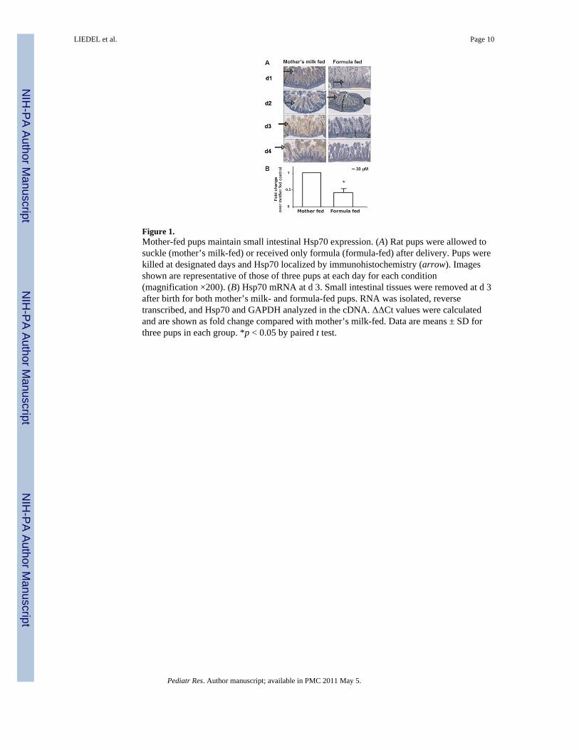
Figure 1.Mother-fed pups maintain small intestinal Hsp70 expression. (A) Rat pups were allowed tosuckle (mother’s milk-fed) or received only formula (formula-fed) after delivery. Pups werekilled at designated days and Hsp70 localized by immunohistochemistry (arrow). Imagesshown are representative of those of three pups at each day for each condition(magnification ×200). (B) Hsp70 mRNA at d 3. Small intestinal tissues were removed at d 3after birth for both mother’s milk- and formula-fed pups. RNA was isolated, reversetranscribed, and Hsp70 and GAPDH analyzed in the cDNA. ΔΔCt values were calculatedand are shown as fold change compared with mother’s milk-fed. Data are means ± SD forthree pups in each group. *p < 0.05 by paired t test.
LIEDEL et al. Page 10
Pediatr Res. Author manuscript; available in PMC 2011 May 5.
NIH
-PA Author Manuscript
NIH
-PA Author Manuscript
NIH
-PA Author Manuscript
Figure 2.Small intestine of mother’s milk-fed pups are resistant to increased permeability byoxidants. Permeability of loops of small intestine of d 3 pups on mother’s milk- vs formula-fed was measured using 10 kD FITC-dextran. Loops were treated with monochloramine(NH2Cl, 0.1 mM) to increase permeability. Data are means ± SD for three separate pups ineach group. *p < 0.05 by analysis of variance.
LIEDEL et al. Page 11
Pediatr Res. Author manuscript; available in PMC 2011 May 5.
NIH
-PA Author Manuscript
NIH
-PA Author Manuscript
NIH
-PA Author Manuscript

Figure 3.ZO-1 and Hsp70 colocalization in small intestine of mother’s milk-fed pups. Frozen sectionsof small intestine of (A) mother’s milk or (B) formula-fed d 3 pups were stained with rabbitpolyclonal anti-ZO-1, mouse monoclonal anti-Hsp70, and DAPI to localize nuclei. Imagesshown are representative of those of three separate pups, two sections were observed foreach pup. Blue is nuclear DAPI staining, green for ZO-1, red for Hsp70, and yellow foroverlay colocalization (magnification ×200 for formula-fed cross section, all others ×400).
LIEDEL et al. Page 12
Pediatr Res. Author manuscript; available in PMC 2011 May 5.
NIH
-PA Author Manuscript
NIH
-PA Author Manuscript
NIH
-PA Author Manuscript
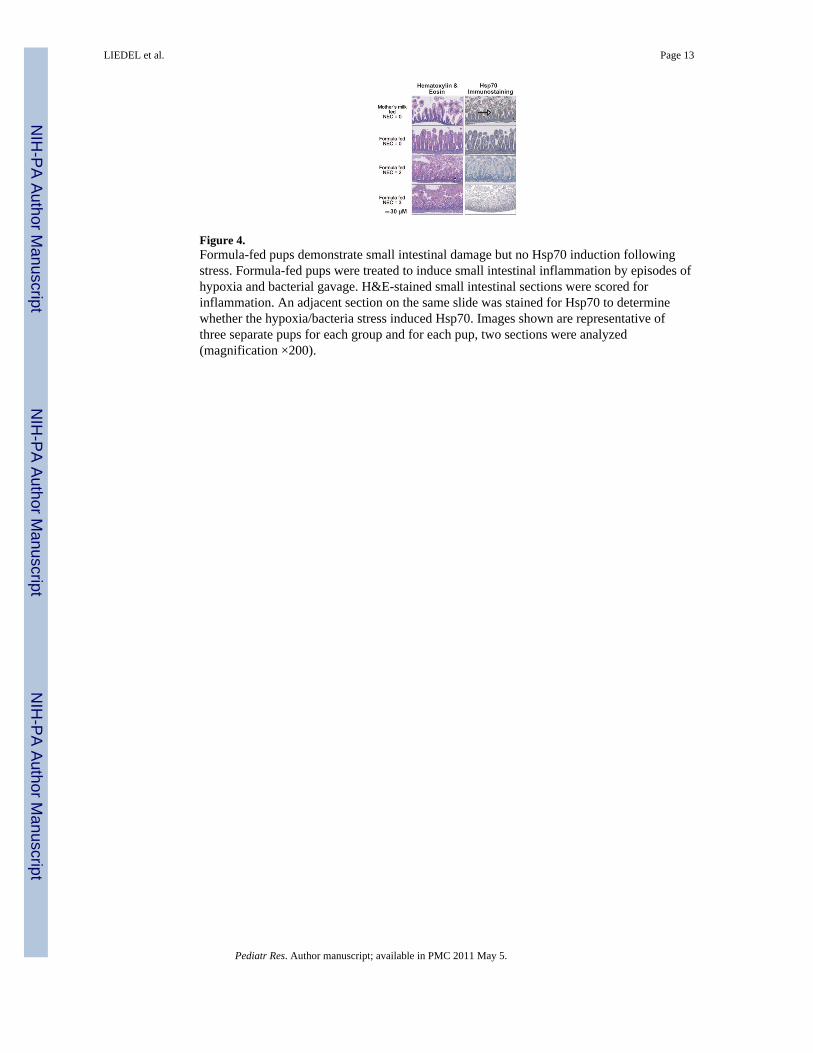
Figure 4.Formula-fed pups demonstrate small intestinal damage but no Hsp70 induction followingstress. Formula-fed pups were treated to induce small intestinal inflammation by episodes ofhypoxia and bacterial gavage. H&E-stained small intestinal sections were scored forinflammation. An adjacent section on the same slide was stained for Hsp70 to determinewhether the hypoxia/bacteria stress induced Hsp70. Images shown are representative ofthree separate pups for each group and for each pup, two sections were analyzed(magnification ×200).
LIEDEL et al. Page 13
Pediatr Res. Author manuscript; available in PMC 2011 May 5.
NIH
-PA Author Manuscript
NIH
-PA Author Manuscript
NIH
-PA Author Manuscript
Recommended


