Embed Size (px)
Citation preview

Allosteric Transitions Direct Protein Tagging by PafA, theProkaryotic Ubiquitin-like Protein (Pup) Ligase*□S
Received for publication, November 12, 2012, and in revised form, February 14, 2013 Published, JBC Papers in Press, March 7, 2013, DOI 10.1074/jbc.M112.435842
Naomi Ofer‡, Nadav Forer‡, Maayan Korman‡, Marina Vishkautzan§, Isam Khalaila¶, and Eyal Gur‡§1
From the ‡Department of Life Sciences, the §National Institute for Biotechnology in the Negev, and the ¶Avram and StellaGoldstein-Goren Department of Biotechnology Engineering, Ben-Gurion University of the Negev, Beer-Sheva 84105, Israel
Background:The interaction of PafA, the prokaryotic ubiquitin-like ligase, with its protein substrates is poorly understood.Results: Measurements of PafA kinetics reveal cooperative substrate binding and experiments with engineered substratessuggest that PafA forms dimers.Conclusion: The PafA enzymatic mechanism involves allosteric transitions.Significance: PafA interaction with its target substrates is regulated at the enzyme level.
Protein degradation via prokaryotic ubiquitin-like protein(Pup) tagging is conserved in bacteria belonging to the phylaActinobacteria and Nitrospira. The physiological role of thisnovel proteolytic pathway is not yet clear, although inMycobac-terium tuberculosis, the world’s most threatening bacterialpathogen, Pup tagging is important for virulence. PafA, the Pupligase, couples ATP hydrolysis with Pup conjugation to lysineside chains of protein substrates. PafA is the solePup ligase inM.tuberculosis and apparently, in other bacteria. Thus, whereasPafA is a key player in the Pup tagging (i.e. pupylation) system,control of its activity and interactions with target protein sub-strates remain poorly understood. In this study, we examinedthemechanismof protein pupylationbyPafA inMycobacteriumsmegmatis, a model mycobacterial organism. We report thatPafA is an allosteric enzyme that binds its target substratescooperatively and find that PafA allostery is controlled by thebinding of target protein substrates, yet is unaffected by Pupbinding. Analysis of PafA pupylation using engineered sub-strates differing in the number of pupylation sites points to PafAacting as a dimer. These findings suggest that protein pupyla-tion can be regulated at the level of PafA allostery.
Protein degradation via ubiquitin tagging was long thoughtto be a uniquely eukaryotic proteolytic pathway. In the last fewyears, however, it has become clear that bacterial speciesbelonging to the phyla Nitrospira and Actinobacteria possess atagging and degradation pathway that is highly analogous to theeukaryotic ubiquitin-proteasome pathway (1). Initially discov-ered in Mycobacterium tuberculosis, the bacterial ubiquitin-like tagging pathway conjugates Pup2 to lysine side chains of
protein substrates, thereby targeting them for proteasomal deg-radation (2).M. tuberculosis, like other actinobacterial species,contains a proteasome in addition to other smaller proteases,such as ClpXP and FtsH (3–5). Because Pup is recognized byMpa, the proteasomal regulatory subunit, tagged (i.e. pupy-lated) proteins are degraded (6–10). Puppylation-deficient M.tuberculosis mutants are viable, but their virulence is compro-mised. Specifically, such mutants are sensitized to reactivenitrogen intermediates, antimicrobial compounds that arereleased into phagosomes by macrophages (11, 12). Notewor-thy, the vast majority of Pup/proteasome-containing bacteriaare nonpathogenic, suggesting a fundamental role of this pro-teolytic pathway in these species (13, 14).Despite apparent functional similarity between Pup and
ubiquitin, these degradation tags are not homologous. Unlikeubiquitin, Pup is a small, natively unstructured protein of 64amino acids (7 kDa) that presents aGGEmotif at its C terminus(2, 15, 16). Conjugation of Pup to protein substrates occursthrough formation of an isopeptide bond between the �-car-boxylate of the C-terminal glutamate of Pup and an �-aminegroup of a substrate lysine (2, 17). Like ubiquitin, Pup is acti-vated by ATP hydrolysis prior to its conjugation (18, 19). How-ever, the enzymatic mechanism of Pup activation and conjuga-tion is altogether different from that of ubiquitin (1, 20). ThePup ligase, an enzyme termed PafA (proteasomal accessory fac-tor A), both activates and conjugates Pup to protein substratesin a two-step reaction that is typical of glutamine synthetasesand �-glutamyl cysteine synthetases (2, 14, 19). Indeed, PafAwas identified as a distant homologue of this group of enzymes(14). The first step in the PafA-catalyzed reaction is the phos-phorylation of Pup on the �-carboxylate of its C-terminal glu-tamate. Next, PafA catalyzes a nucleophilic attack on the phos-phorylated carboxylate by the �-amine group of a substratelysine (19). For pupylation and PafA binding, only the C-termi-nal region of Pup is required, and whereas the N-terminalregion of the protein is required for interaction with Mpa, it isexpandable for pupylation (8, 9). In M. tuberculosis and manyother related species, Pup is translated with a glutamine, ratherthan a glutamate at its C terminus. In these bacteria, a PafA-homologous enzyme termed Dop (deamidase of Pup) converts
* This work was supported in part by the German-Israeli Foundation GrantI-2230-2055.13/2009.
□S This article contains supplemental Figs. S1 and S2 and supplemental Exper-imental Procedures.
1 To whom correspondence should be addressed: Life Sciences Departmentand the National Institute of Biotechnology in the Negev, Ben-Gurion Uni-versity, Rager Blvd., Beer-Sheva 84105, Israel. Tel.: 972-3-6472930; Fax: 972-3-6479114; E-mail: [email protected].
2 The abbreviations used are: Pup, prokaryotic ubiquitin-like protein; Ni2�-NTA, nickel-nitrilotriacetic acid; PupE, deamidated Pup; TEV, tobacco etchvirus.
THE JOURNAL OF BIOLOGICAL CHEMISTRY VOL. 288, NO. 16, pp. 11287–11293, April 19, 2013© 2013 by The American Society for Biochemistry and Molecular Biology, Inc. Published in the U.S.A.
APRIL 19, 2013 • VOLUME 288 • NUMBER 16 JOURNAL OF BIOLOGICAL CHEMISTRY 11287
by guest on August 13, 2016
http://ww
w.jbc.org/
Dow
nloaded from
by guest on August 13, 2016
http://ww
w.jbc.org/
Dow
nloaded from
by guest on August 13, 2016
http://ww
w.jbc.org/
Dow
nloaded from
by guest on August 13, 2016
http://ww
w.jbc.org/
Dow
nloaded from
by guest on August 13, 2016
http://ww
w.jbc.org/
Dow
nloaded from
by guest on August 13, 2016
http://ww
w.jbc.org/
Dow
nloaded from

this glutamine to a glutamate, thereby rendering Pup availablefor PafA conjugation (21).Despite remarkable progress in understanding the pupyla-
tion system, it remains poorly understood how PafA interactswith its protein substrates and how such interactions are regu-lated. PafA is the only Pup ligase identified to date; and in M.tuberculosis, no pupylation is detected in pafA mutants (2).Understanding the enzymaticmechanism by which PafA inter-acts with its substrates is central to the understanding of thisnovel proteolytic pathway. Furthermore, PafA is important forM. tuberculosis virulence, and, as such, detailed mechanisticanalysis of this enzyme may prove useful in the global combatagainst this pathogen.In this study, we analyzed protein pupylation by PafA from
Mycobacterium smegmatis. M. smegmatis is a commonly usedmycobacterial model bacterium, with a PafA that is 94% iden-tical to that ofM. tuberculosis.We find that PafA is an allostericenzyme that binds its target substrates cooperatively. In con-trast, binding of deamidated Pup (PupE) by PafA follows simplekinetics and is, therefore, not involved in the allosteric transi-tions of PafA. We also find that Pup and target substrate bind-ing by PafA are independent events. Analysis of engineered chi-meric substrates that differ in the number of pupylation sitessuggested that PafA is active as a dimer. In view of these find-ings, we propose that protein pupylation can be regulated at theenzyme level via an allosteric mechanism.
EXPERIMENTAL PROCEDURES
Protein Purification—All proteins, except a nontagged PanB,carried N-terminal polyhistidine tags and were expressed fromplasmid pSH21 under the transcriptional control of the T7 pro-moter. Expression was at 37 °C, unless stated otherwise, inEscherichia coli ER2566 (New England Biolabs). Cells werelysed by sonication, and purification using Ni2�-NTA-agarose(Qiagen) was carried out according to a standard protocol. ForPafA and PanB, purification Ni2�-NTA buffers contained 10%glycerol (v/v). As a consequent purification step, proteins wereloaded onto a Superdex 75 size exclusion column (GE Health-care) equilibratedwith 25mMTris-HCl, pH 8.0 (hereafter, T25)for the titin-I27 variants, with pupylation buffer (50 mM Tris-HCl, pH7.5, 100mMKCl, 20mMMgCl2, and 10% (v/v) glycerol)for PafA and with T25, 50 mMNaCl, and 10% glycerol for poly-histidine-tagged PanB.For PupE purification, pupE was cloned into plasmid pSH21
in fusion with DNA encoding titin-I27 and a TEV protease rec-ognition sequence (His6-I27-TEV-PupE). Expression was at30 °C, and Ni2�-NTA purification was carried out as above.FollowingTEVcleavage, a buffer exchange stepwas carried out,and the His6-I27-TEV portion of the chimera was removed byloading the solution onto a Ni2�-NTA column. The flow-through was collected, and PupE was further purified on a C18reverse phase column, lyophilized, and resuspended in T25.For purification of Fl-PupE, a variant encoding a N-terminal
cysteine was cloned (i.e. his66 i27-tev-cys-pupE). Labeling with5-iodoacetamidofluorescein was carried out following bindingof the protein to the Ni2�-NTA column. After excess 5-iodo-acetamidofluorescein was washed, purification proceeded asfor PupE.
To purify a PanB variant that does not carry a polyhistidinetag, panBwas cloned into plasmid pET11a, and the protein wasexpressed at 37 °C in E. coli ER2566. The cell pellet was sus-pended in T25 containing 10% glycerol (v/v), and the cells weredisrupted by sonication. Following centrifugation (12,000 � g,15 min), streptomycin sulfate (1% (w/v)) was added to the clearlysate for precipitation of nucleic acids; and, following centrif-ugation as above, the supernatant was loaded onto an anionicexchange column (HiPrep Q FF; GE Healthcare) pre-equili-brated with T25 containing 10% glycerol (v/v). ANaCl gradient(0–1 M) was used for elution. PanB-containing fractions werepooled, concentrated, and loaded onto a size exclusion column(Superdex 200; GE Healthcare) pre-equilibrated with T25, 50mM NaCl, 10% glycerol (v/v). As a final purification step, aMono Q column was used (GE Healthcare), and following abuffer exchange step into T25, 50 mM NaCl, 10% glycerol (v/v)using a PD10 column (GE Healthcare), protein aliquots werestored at �80 °C until use.Assays—Pupylation assays were carried out at 30 °C by mix-
ing PafA, PupE, and a target substrate in pupylation buffer.Reactions were initiated with the addition of ATP to a finalconcentration of 2mM.Quantitative pupylation assays relied onthe use of a polyhistidine-tagged target substrate and the use ofFl-PupE instead of PupE. Initial rates were measured by collect-ing 10-�l aliquots at intervals during the reactions into tubescontaining 50 �l of T25 and 6 M guanidine HCl (hereafter,T25G). Next, samples were diluted such that each containedequal concentrations of a polyhistidine-tagged target protein.50 �l of each diluted sample was transferred into filter micro-centrifuge tubes (Corning ZQ-VW-8169) containing 50 �l ofNi2�-NTA-agarose beads equilibrated with 500 �l of T25G.Following a 2-min incubation, the liquid was removed by cen-trifugation (7000� g, 3min), and beadswerewashed twicewith500 �l of T25G. Elution was carried out by resuspension of thebeads in T25G, 250mM imidazole, and centrifugation as above.The fluorescence intensity of each sample wasmeasured with aSynergy 2 microplate reader (Biotek instruments) at 485 nm(excitation) and 528 nm (emission).For Fl-PupE-PafA binding assays, the proteins were mixed in
pupylation buffer without ATP or target substrates. Fluores-cence anisotropy was measured as above.LC/MS analysis, bioinformatics, and pupylation site identifi-
cation are described in supplemental Experimental Procedures.
RESULTS
Cooperative Substrate Binding by PafA—To study the inter-action of M. smegmatis PafA with protein substrates, pupyla-tion of a bona fide target substratewas examined. Earlier studiesindicated that M. tuberculosis PanB (ketopantoate hydroxym-ethyltransferase), a homodecameric protein complex, is pupy-lated efficiently by theM. tuberculosis PafA both in vitro and invivo (2, 9, 22). To test in vitro pupylation by the M. smegmatissystem, the M. smegmatis panB, pafA, and pupE genes werecloned, and their encoded proteins were purified to homogene-ity. In a reaction including ATP and PupE, the formation of apupylation productwith amolecularmass approximately 7 kDaheavier than PanB suggested that each PanBmonomer was sin-gly pupylated by PafA (Fig. 1A, left). MS/MS analysis indicated
PafA Allostery
11288 JOURNAL OF BIOLOGICAL CHEMISTRY VOLUME 288 • NUMBER 16 • APRIL 19, 2013
by guest on August 13, 2016
http://ww
w.jbc.org/
Dow
nloaded from

that PanB is pupylated primarily on either one of two lysines,namely Lys34 or Lys36. Pupylation on Lys137 was also detected,although less frequently (Fig. 1B and supplemental Fig. S1).Next, a quantitative assay able to examine M. smegmatis PafApupylation kinetics was sought. Although the pupylation kinet-ics of M. tuberculosis PafA have been previously examined bymonitoring the first half of the pupylation reaction cycle (i.e.ATP hydrolysis and PupE phosphorylation) (19), we wanted toanalyze the full PafA reaction cycle to gain further insight intothe interaction of PafA with target substrates. To this end, aquantitative pupylation assay was developed using an N-termi-nally fluorescein-labeled PupE variant (Fl-PupE). As PupE andFl-PupE were conjugated equally well to protein substrates byPafA (Fig. 1A), Fl-PupE was used for quantitative pupylationanalysis instead of unlabeled PupE. This novel assay, describedunder “Experimental Procedures,” allows quantitative meas-urements of end product formation by the use of Fl-PupE and apolyhistidine-tagged target substrate in a PafA-dependentreaction (Fig. 1C).Titration experiments were carried out in which steady-state
pupylation rates were measured at a constant Fl-PupE concen-tration and increasing PanB concentrations (Fig. 1D). Theobtained data can be described by a sigmoidal rate equation(Fig. 1D), suggesting PanB binding by PafA to be cooperative. AHill coefficient of 1.9 � 0.3 (Table 1) indicates that PafA oligo-mers with at least two binding sites are involved in PanB pupy-lation. The kinetic measurements revealed a K0.5 of 21 � 2 �M
for PanB pupylation and a Vmax of 1.4 � 0.1 Fl-PupE conjuga-tions per min per PafA monomer (Table 1). As the assays werecarried out in the presence of a saturating Fl-PupE concentra-tion, the maximal pupylation rate is equivalent to the catalyticrate constant (kcat) of the reaction. Both the K0.5 and Vmaxmeasured for PanB pupylation closely matched the publishedvalues for M. tuberculosis PanB pupylation by M. tuberculosisPafA (19).
Cooperative substrate binding, as depicted in Fig. 1D, sug-gests that PafA is an allosteric enzyme. A hallmark of enzymeallostery is the positive effect of a competitive inhibitor on sub-strate processing at low inhibitor and substrate concentrations(23). An increase in enzymatic activity detected under suchconditions can be attributed to stabilization of an active or highaffinity enzyme conformation by the inhibitor. As a result, cer-tain low inhibitor concentrations can lead to both inhibitionand activation, such that the net effect is a facilitated substrate-processing rate (23). Similar kinetics can also be observed usinga competing substrate rather than a competitive inhibitor (24).To test whether such an allosteric behavior is present in oursystem, pupylation reactions were carried out at a fixed lowconcentration of polyhistidine-PanB and at increasing concen-trations of a PanB variant lacking the polyhistidine tag. Asdetection of pupylation in the quantitative assays employed inthis study relies on the use of polyhistidine-tagged substrates(Fig. 1B), nontagged PanB thus acts as a competitor. Neverthe-less, at low competitor concentrations, increased pupylation ofpolyhistidine-PanBwas observed, providing further evidence ofPafA allostery (Fig. 2A). In the presence of higher concentra-tions of nontagged PanB, competition dominates, resulting in adecrease in pupylation of the polyhistidine-tagged substrate(Fig. 2A). To test whether binding of a target lysine is sufficientto induce an allosteric PafA transition, a similar experimentwascarried out, this time titrating free lysine instead of nontagged
FIGURE 1. PafA pupylation of PanB, a bona fide substrate. A, SDS-PAGE analysis of PanB (5 �M) pupylation by PafA (1 �M) and PupE or Fl-PupE (10 �M each)is shown. Following electrophoresis, Coomassie Brilliant Blue was used for staining. B, PanB pupylation sites (red) in tryptic peptides that were identified byMS/MS analysis are denoted. C, the use of Fl-PupE and a polyhistidine-tagged target substrate allows removal of unconjugated Fl-PupE via Ni2�-NTA chroma-tography. The fluorescence intensity of the eluate thus provides a quantitative measure of end product formation. D, steady-state rates of PafA (0.5 �M)- andFl-PupE (15 �M)-mediated pupylation as a function of increasing polyhistidine-PanB concentration. The data were fitted to the Hill equation (rate � Vmax�[S]n/(K0.5
n � [S]n); solid line) and to the Henri-Michaelis-Menten equation (rate � Vmax�[S]/(Km � [S]); dashed line). The averages � S.D. (error bars) of three experi-ments are presented. Pupylation rates were derived from the measurements presented in the inset.
TABLE 1Steady-state kinetic parameters for the PafA-catalyzed reactionsmeasured in this work
Substrate Vmax K0.5 Hill coefficient
min�1/PafA �M
PanB 1.4 � 0.1a 21 � 2 1.9 � 0.3I271 1.4 � 0.1a 534 � 87 1.6 � 0.2I272 1.4 � 0.1a 348 � 42 1.3 � 0.1I273 1.3 � 0.1a 368 � 45 1.2 � 0.08Pup 0.14 � 0.01 1.3 � 0.4 0.94 � 0.2
a The values are equivalent to the kcat of the reactions, as measurements were car-ried out at saturating PupE concentrations.
PafA Allostery
APRIL 19, 2013 • VOLUME 288 • NUMBER 16 JOURNAL OF BIOLOGICAL CHEMISTRY 11289
by guest on August 13, 2016
http://ww
w.jbc.org/
Dow
nloaded from

PanB. Again, a clear allosteric effect was observed at low lysineconcentrations, whereas at higher concentrations, pupylationof polyhistidine-PanB was inhibited (Fig. 2B). Noticeably, thelysine concentrations required for induction of an allostericPafA transition were approximately 1000-fold higher than thePanB concentrations that induced a similar effect (Fig. 2A).These differences are consistent with the �1000-fold loweraffinity of PafA for free lysine (�22 mM) compared with itsaffinity for PanB (�21 �M) (Table 1 and Ref. 19).PupE Is Not Involved in Allosteric PafA Transitions—The
mode of PupE binding to PafAwas next considered when pupy-lation of low polyhistidine-PanB concentrations was measuredat increasing Fl-PupE concentrations. Data fitting to the Hillequation yielded a nonsigmoidal curve with a Hill coefficient of0.94 � 0.2, indicating that PupE is not cooperatively bound byPafA nor does PupE affect binding of the target protein sub-strate to PafA (Fig. 3A andTable 1). TheK0.5 valuemeasured forFl-PupE (1.3� 0.3�M; Fig. 3A and Table 1) closelymatched theKD for Fl-PupE binding to PafA, as measured in a direct bindingassay in the absence of a nucleophile substrate (0.86 � 0.3 �M;Fig. 3B). Therefore, binding of PupE to PafA is not affected bythe binding of the target substrate. These results thus indicatethat binding of PupE and a target substrate to PafA are inde-pendent events. This conclusion is further supported by thesimilarity between the obtained affinity values and the apparent
binding constant measured for the interaction ofM. tuberculo-sis PupE with M. tuberculosis PafA in the absence of a targetsubstrate (19).A Dimer-based Model for PafA-Substrate Interaction—Co-
operative substrate binding by PafA indicates that the activeform of this enzyme is oligomeric. Indeed, Corynebacteriumglutamicum PafA, an orthologue of the molecule consideredhere, was recently crystallized in the asymmetric unit as a dimerwhere swapping of an N-terminal �-strand and a concomitant�-helix was shown (25). Nonetheless, efforts to determine thestoichiometry of M. smegmatis PafA oligomers using classicmethods, such as size exclusion chromatography and analyticalultracentrifugation, have been hampered by the low solubilityofM. smegmatis PafA, as was also reported forM. tuberculosisPafA (19). As an alternative approach, we instead tested thepupylation of chimeric substrates presenting different numbersof pupylation sites. To this end, concatemeric variants of a non-native substrate protein, the I27 domain of human Titin, wereused. Titin-I27 is a globular domain that can be easily unfoldedin vitro by chemical modification (e.g. carboxymethylation) ofits two cysteines (26). We found unfolded titin-I27 to be pupy-lated substantially faster thanwas the folded formof the protein(supplemental Fig. S2A). It was also noted that titin-I27 is pupy-lated primarily on a single lysine, as determined by SDS-PAGEand mass spectrometry (Fig. S2, A and B). To examine theimpact on pupylation kinetics upon encounteringmultiple sub-strate pupylation sites, linear fusions of two and three titin-I27domains were constructed. Single titin-I27 and the resulting
FIGURE 2. Allosteric activation by a competitor substrate. A, steady-staterates of a polyhistidine-PanB (1 �M) pupylation by PafA (0.5 �M) and Fl-PupE
(15 �M) at increasing concentrations of nontagged competitor PanB. The val-ues presented were obtained following subtraction of background fluores-cence values measured in reactions lacking polyhistidine-PanB. The solidcurve is a guide to the eye. B, as in A, but with free lysine (pH 8.0) instead ofnontagged PanB competitor. The curve is fitted to the equation rate �C���(1����)/(L�(1����)2), where C is a scaling factor, � � [polyhistidine-PanB]/Km � 1/21, � � [Lys]/K0.5, and L is a conformational equilibrium con-stant (27). In both A and B, the averages � S.D. (error bars) of three experi-ments are presented.
FIGURE 3. Pup binding by PafA is noncooperative and independent oftarget substrate binding. A, steady-state rates of a polyhistidine-PanB (1 �M)pupylation by PafA (0.25 �M) at increasing Fl-PupE concentrations. The curveis fitted to the Hill equation. B, fluorescence anisotropy of Fl-PupE (50 nM) atincreasing PafA concentrations. The curve is fitted to the binding equation,r � r0 � (rmax�[PafA])/(KD � [PafA]), where r is the fluorescence anisotropyvalue. In both A and B, the averages � S.D. (error bars) of three experimentsare presented.
PafA Allostery
11290 JOURNAL OF BIOLOGICAL CHEMISTRY VOLUME 288 • NUMBER 16 • APRIL 19, 2013
by guest on August 13, 2016
http://ww
w.jbc.org/
Dow
nloaded from

concatemeric proteins (denoted as I271, I272, and I273) werepurified to homogeneity, unfolded by carboxymethylation, andthe rates of their in vitro pupylation were compared (Fig. 4A).I272 was pupylated approximately twice as fast as was I271, asexpected, given that I272 contains two pupylation sites, asopposed to the single such site in I271. However, no additionalincrease in ratewas observed for pupylation of I273. Rather, thissubstrate was pupylated as fast as was I272 and again abouttwice as fast as was I271 (Fig. 4A). To better assess the pupyla-tion kinetics of each titin-I27 variant, titration assays were car-ried out by measuring pupylation rates at different substrateconcentrations (Fig. 4B). No significant differences in the valueofVmax (�1.4min�1 per PafA)were detected (Fig. 4B andTable1). By contrast, the K0.5 value for pupylation of I271 (534 � 87�M; Table 1) was found to be nearly 2-fold higher than that ofI272 (348 � 42 �M) or of I273 (368 � 45 �M). These resultssuggest that the rate differences observed in Fig. 4A arisemainlyfrom affinity effects. A Hill coefficient of 1.6 � 0.2 for I271pupylation suggested cooperative binding of this titin-I27 var-iant by PafA, as in the case of PanB pupylation (Table 1). Nota-bly, PafA binding of the I272 and I273 concatemers occurredwith a lower degree of cooperativity, as determined byHill coef-ficients of 1.3 � 0.1 and 1.2 � 0.08, respectively (Table 1). Wethus conclude that the pupylation kinetics of the titin-I27 vari-ants are consistent with PafA being active as a dimer. As illus-trated in Fig. 5A, binding of the first target lysine of I272 can leadto an increase in the local concentration of the second targetlysine near a free PafA binding site. As a result, binding of the
second I272 target lysine is highly favorable, with the flexibilityof the unfolded titin-I27 domains enabling binding withoutsteric constrains that might otherwise limit the binding offolded proteins. A dimer-based model also explains the higherdegree of cooperativity observed for I271 pupylation. Indeed,binding of I272 or I273 to a PafA dimer cannot lead to stabiliza-tion of a free substrate binding site, thus preventing cooperativebinding of an additional substrate. Finally, the existence of onlytwo binding sites explains why elongation of I272 by an addi-tional unit to generate I273 does not improve binding.A dimer-based model, as illustrated in Fig. 5A, predicts I271
to be singly pupylated and I272 to be doubly pupylated. Impor-tantly, according to this model, I273 should be only doublypupylated despite presenting three pupylation sites. These pre-dictions were indeed verified when pupylation of the titin-I27variants was examined by SDS-PAGE (Fig. 5B). Noticeably,dual pupylation of I272 and of I273 proceeded without the for-mation of gel bands corresponding to single I272 and I273 pupy-lation intermediates. This suggests that following the initialpupylation of either I272 or I273, the second pupylation eventoccurs very fast and probably without substrate release. Thisfurther suggests that a third I273 pupylation event can occur,yet requires substrate release (following double pupylation) andrebinding. In agreement with this rationale, a third pupylationevent was indeed observed for I273 when pupylation wasallowed to proceed over an extended period of time (Fig. 5C).
DISCUSSION
By establishing a quantitative pupylation assay, we were abletomeasure the kinetics ofM. smegmatis PafA pupylation in thisstudy. Thus, using a bona fide PafA substrate, PanB, coopera-tive binding by PafA could be observed for the first time, sug-gesting that PafA is an allosteric enzyme. This conclusion wasfurther supported by competition assays in which substratepupylation was facilitated at low competitor concentrations.These experiments further indicated that binding of the targetlysine to PafA is sufficient to induce the allosteric effect.There are a number of ways in which binding of a target
substrate to PafA can facilitate the pupylation rate. For exam-ple, binding of a target lysine by PafA can enhance its affinity toPupE, thereby facilitating pupylation at undersaturating PupEconcentrations. Such a model, however, is highly unlikely, asour data clearly indicate that binding of PupE and the binding oftarget substrates by PafA are independent events. Therefore,PupE does not play a role in the allosteric transitions that arereflected in the experiments presented here. Rather, the datacan be explained by the existence of alternative PafA conforma-tions that differ either in their affinity for target substrates or intheir catalytic activity. This scenario requires that the bindingof target substrates to PafA would stabilize the high affinity orthe more active conformation.A Hill coefficient of 1.9 � 0.3 for PanB pupylation (Table 1)
indicates that the minimal oligomeric form of PafA is dimeric.Cooperative substrate binding was also evident when pupyla-tion of titin-I27 was examined. In addition, these assays indi-cated that elongation of I271 by an additional titin-I27 unitresulted in an approximately 2-fold increase in affinity, asreflected in the K0.5 values measured for the titin-I27 variants.
FIGURE 4. PafA pupylation of titin-I27 variants. A, pupylation of titin-I27variants (15 �M each) by PafA (0.5 �M) and Fl-PupE (1 �M). The inset demon-strates an I271 pupylation reaction with PafA (0.5 �M) and Fl-PupE (7 �M) thatcontinued to completion. B, steady-state pupylation rates by PafA (0.5 �M)and Fl-PupE (15 �M) at increasing concentrations of titin-I27 variants. Thecurves are fitted to the Hill equation. The averages � S.D. (error bars) of threeexperiments are presented.
PafA Allostery
APRIL 19, 2013 • VOLUME 288 • NUMBER 16 JOURNAL OF BIOLOGICAL CHEMISTRY 11291
by guest on August 13, 2016
http://ww
w.jbc.org/
Dow
nloaded from

However, elongation by yet another titin-I27 unit to generateI273 did not improve affinity to PafA. These results are in per-fect correlation with the number of Pup molecules that wereconjugated to each titin-I27 variant. In other words, the 2-foldincrease in the affinity of I272 and I273, comparedwith I271, wasaccompanied by an additional Pup molecule attached to thesechimeric substrates. These observations can be explained bythe existence of PafA oligomers containing two binding sites, asillustrated in Fig. 5A. A dimer-based model is also consistentwith the reduced cooperativity observed for I272 and I273, com-pared with I271. Our results suggest that following binding ofthe first target lysine of I272 or I273, binding of the second lysineoccurs almost simultaneously. This would explain the absenceof detectable singly pupylated I272 and I273 in our steady-statekinetics assays. An almost simultaneous accommodation ofboth binding sites of a PafA dimer by I272 or I273 would resultin a 2-fold increase in the pupylation rate. This interactionwould, however, prevent facilitated binding of the next sub-strate molecule. Therefore, a less cooperative binding of I272and I273 is observed. Unlike I272 and I273, PanB is coopera-tively bound by PafA. Thus, despite being a decamer, PanB doesnot simultaneously accommodate the two binding sites of aPafA dimer. This is likely because for decameric PanB to bindtwo PafA binding sites, the PanB target lysines must be posi-tioned in a conformation that may not exist in the folded, rigidconformation of the decamer. I272 and I273, on the other hand,are unfolded and flexible, and can thus easily assume the con-formation required for the simultaneous occupation of the twoPafA binding sites.A dimer-basedmodel for PafA allostery is consistentwith the
recently published C. glutamicum PafA structure (25). Alto-gether, our kinetic data strongly suggest that the dimeric formis the correct biological assembly and the allosterically activeconformation of the protein.We cannot exclude the possibility
of dimeric PafA existing as part of a higher oligomeric form,such as a dimer of dimers or a trimer of dimers. Hence, addi-tional studies are required to decipher the oligomeric state ofbiologically active PafA.As the only Pup ligase currently known, PafA plays a most
central role in deciding which proteins will be pupylated. It is,therefore, reasonable that the expression level and activity ofPafA be tightly regulated in vivo. This study suggests that sub-strate pupylation in vivo can be regulated at the level of PafAallostery. For example, pupylation can be regulated by alteringthe equilibriumbetween an active oligomeric form and an inac-tive conformation. Further studywill be required to understandthe mechanistic details of PafA allostery and the role it plays inbacterial physiology.
Acknowledgments—We thank Bob Sauer (Massachusetts Institute ofTechnology), Boaz Shaanan, Amir Aharoni, and Ofer Yifrach (Ben-Gurion University) for helpful advice and fruitful discussions.
REFERENCES1. Burns, K. E., and Darwin, K. H. (2010) Pupylation versus ubiquitylation:
tagging for proteasome-dependent degradation. Cell Microbiol. 12,424–431
2. Pearce,M. J.,Mintseris, J., Ferreyra, J., Gygi, S. P., andDarwin, K. H. (2008)Ubiquitin-like protein involved in the proteasome pathway of Mycobac-terium tuberculosis. Science 322, 1104–1107
3. Nagy, I., Tamura, T., Vanderleyden, J., Baumeister, W., and De Mot, R.(1998) The 20S proteasome of Streptomyces coelicolor. J. Bacteriol. 180,5448–5453
4. Knipfer, N., Seth, A., Roudiak, S. G., and Shrader, T. E. (1999) Speciesvariation in ATP-dependent protein degradation: protease profiles differbetween mycobacteria and protease functions differ betweenMycobacte-rium smegmatis and Escherichia coli. Gene 231, 95–104
5. Darwin, K. H., Lin, G., Chen, Z., Li, H., and Nathan, C. F. (2005) Charac-terization of a Mycobacterium tuberculosis proteasomal ATPase homo-logue.Mol. Microbiol. 55, 561–571
B.A.
C. 0 40 80 120 160 200 240 min
6451
39
2819
14
66451
39
2819
14
66451
39
2819
14
6
kDa 0 40 80 120min
I271 (12 kDa)
Pup~I271 (19 kDa)
PafA
PafA
PafA
I272 (22 kDa)
Pup2~I272 (36 kDa)
I273 (32 kDa)
Pup2~I273 (46 kDa)
Pup3~I273 (54 kDa)
PafAPup2~I273 (46 kDa)
I273 (32 kDa)
6451
39
28
K K K
K K K
I271
K K
KK K K
I272
KK
K
KK
KK K K
I273
FIGURE 5. A two-binding site model for PafA interaction with titin-I27 concatemers. A, two adjacent PafA subunits (purple) can bind two I271 monomers,one I272 monomer, or two target lysines in I273. B, SDS-PAGE analysis of pupylation by PafA (1.0 �M) and PupE (25 �M) of I271 (12 �M), I272 (6 �M), and I273 (4 �M).Following electrophoresis, Coomassie Brilliant Blue was used for staining. C, pupylation of I273 as in C is shown, but for a longer reaction time.
PafA Allostery
11292 JOURNAL OF BIOLOGICAL CHEMISTRY VOLUME 288 • NUMBER 16 • APRIL 19, 2013
by guest on August 13, 2016
http://ww
w.jbc.org/
Dow
nloaded from
6. Sutter, M., Striebel, F., Damberger, F. F., Allain, F. H., and Weber-Ban, E.(2009) A distinct structural region of the prokaryotic ubiquitin-like pro-tein (Pup) is recognized by the N-terminal domain of the proteasomalATPase Mpa. FEBS Lett. 583, 3151–3157
7. Burns, K. E., Liu, W. T., Boshoff, H. I., Dorrestein, P. C., and Barry, C. E.,3rd (2009) Proteasomal protein degradation inMycobacteria is dependentupon a prokaryotic ubiquitin-like protein. J. Biol. Chem. 284, 3069–3075
8. Burns, K. E., Pearce,M. J., andDarwin, K. H. (2010) Prokaryotic ubiquitin-like protein provides a two-part degron to Mycobacterium proteasomesubstrates. J. Bacteriol. 192, 2933–2935
9. Striebel, F., Hunkeler, M., Summer, H., and Weber-Ban, E. (2010) Themycobacterial Mpa-proteasome unfolds and degrades pupylated sub-strates by engaging Pup’s N terminus. EMBO J. 29, 1262–1271
10. Wang, T., Darwin, K. H., and Li, H. (2010) Binding-induced folding ofprokaryotic ubiquitin-like protein on the Mycobacterium proteasomalATPase targets substrates for degradation. Nat. Struct. Mol. Biol. 17,1352–1357
11. Darwin, K. H., Ehrt, S., Gutierrez-Ramos J. C., Weich, N., and Nathan,C. F. (2003) The proteasome ofMycobacterium tuberculosis is required forresistance to nitric oxide. Science 302, 1963–1966
12. Cerda-Maira, F. A., Pearce,M. J., Fuortes,M., Bishai,W. R., Hubbard, S. R.,and Darwin, K. H. (2010) Molecular analysis of the prokaryotic ubiquitin-like protein (Pup) conjugation pathway in Mycobacterium tuberculosis.Mol. Microbiol. 77, 1123–1135
13. Lupas, A., Zwickl, P., and Baumeister,W. (1994) Proteasome sequences ineubacteria. Trends Biochem. Sci. 19, 533–534
14. Iyer, L. M., Burroughs, A. M., and Aravind, L. (2008) Unraveling the bio-chemistry and provenance of pupylation: a prokaryotic analog of ubiquiti-nation. Biol. Direct 3, 45
15. Liao, S., Shang, Q., Zhang, X., Zhang, J., Xu, C., and Tu, X. (2009) Pup, aprokaryotic ubiquitin-like protein, is an intrinsically disordered protein.Biochem. J. 422, 207–215
16. Chen, X., Solomon, W. C., Kang, Y., Cerda-Maira, F., Darwin, K. H., andWalters, K. J. (2009) Prokaryotic ubiquitin-like protein pup is intrinsicallydisordered. J. Mol. Biol. 392, 208–217
17. Sutter, M., Damberger, F. F., Imkamp, F., Allain, F. H., andWeber-Ban, E.
(2010) Prokaryotic ubiquitin-like protein (Pup) is coupled to substrates viathe side chain of its C-terminal glutamate. J. Am. Chem. Soc. 132,5610–5612
18. Imkamp, F., Rosenberger, T., Striebel, F., Keller, P. M., Amstutz, B.,Sander, P., and Weber-Ban, E. (2010) Deletion of dop in Mycobacteriumsmegmatis abolishes pupylation of protein substrates in vivo.Mol. Micro-biol. 75, 744–754
19. Guth, E., Thommen, M., and Weber-Ban, E. (2011) Mycobacterial ubiq-uitin-like protein ligase PafA follows a two-step reaction pathway with aphosphorylated pup intermediate. J. Biol. Chem. 286, 4412–4419
20. Maupin-Furlow, J. (2012) Proteasomes and protein conjugation acrossdomains of life. Nat. Rev. Microbiol. 10, 100–111
21. Striebel, F., Imkamp, F., Sutter,M., Steiner,M.,Mamedov, A., andWeber-Ban E. (2009) Bacterial ubiquitin-like modifier Pup is deamidated andconjugated to substrates by distinct but homologous enzymes.Nat. Struct.Mol. Biol. 16, 647–651
22. Chaudhuri, B. N., Sawaya, M. R., Kim, C. Y., Waldo, G. S., Park, M. S.,Terwilliger, T. C., and Yeates, T.O. (2003) The crystal structure of the firstenzyme in the pantothenate biosynthetic pathway, ketopantoate hy-droxymethyltransferase, fromM. tuberculosis. Structure 11, 753–764
23. Monod, J., Wyman, J., and Changeux, J. P. (1965) On the nature of allos-teric transitions: a plausible model. J. Mol. Biol. 12, 88–118
24. Gur E., and Sauer R. T. (2009) Degrons in protein substrates program thespeed and operating efficiency of the AAA� Lon proteolytic machine.Proc. Natl. Acad. Sci. U.S.A. 106, 18503–18508
25. Özcelik, D., Barandun, J., Schmitz, N., Sutter, M., Guth, E., Damberger,F. F., Allain, F. H., Ban, N., and Weber-Ban, E. (2012) Structures of Pupligase PafA and depupylase Dop from the prokaryotic ubiquitin-like mod-ification pathway. Nat. Commun. 3, 1014
26. Kenniston, J. A., Baker, T. A., Fernandez, J. M., and Sauer, R. T. (2003)Linkage between ATP consumption andmechanical unfolding during theprotein processing reactions of an AAA� degradation machine. Cell 114,511–520
27. Segel, IH (1975) Enzyme Kinetics: Behavior and Analysis of Rapid Equilib-rium and Steady-State Enzyme Systems, pp. 421–461, JohnWiley & Sons,New York
PafA Allostery
APRIL 19, 2013 • VOLUME 288 • NUMBER 16 JOURNAL OF BIOLOGICAL CHEMISTRY 11293
by guest on August 13, 2016
http://ww
w.jbc.org/
Dow
nloaded from
1
SUPPLEMENTARY MATERIAL
Allosteric transitions direct protein tagging by PafA, the prokaryotic
ubiquitin-like protein (Pup) ligase
Naomi Ofer, Nadav Forer, Maayan Korman, Marina Wishkautzan, Isam Khalaila, Eyal Gur
SUPPLEMENTARY FIGURES
Figure S1. MS-MS analysis of PanB pupylation. Product ion spectrum of pupylated
TLTLQKWK (m/z = 630.85) aquired by CID. Abundant b and y ions are indicated. Pup
indicates the puppylated lysine, K34.
Figure S2. PafA pupylation of the human titin-I27 domain. A, SDS-PAGE analysis of folded
and unfolded (i.e., carboxymethylated) titin-I27 (12 M) by PafA (1 M) and PupE (15 M).
Coomassie staining was carried out following electrophoresis. B, Detection of the titin-I27
pupylation site by MS-MS analysis. Product ion spectrum of puuppylated
LKGQPLAASPDDEIIEDGK (m/z = 747) aquired by CID. Abundant b and y ions are
indicated. Pup indicates the puppylated lysine.
SUPPLEMENTARY EXPERIMENTAL PROCEDURES
LC/MS analysis – MS analysis was performed using an Eksigent nano-HPLC
connected to the LTQ Orbitrap XL (Thermo Fisher Scientific). Reverse-phase chromatography
for peptides was performed using an in-house-made C-18 column (15 cm long, 75 µm ID),
packed with Jupiter C18, 300Å, 5 µm beads (Phenomenex). Peptides were separated by a 90-
min linear gradient, starting with 100% buffer A (5% acetonitrile, 0.1% formic acid) and ending
with 80% buffer B (80% acetonitrile, 0.1% formic acid), at a flow rate of 300 nl/min. A full
scan, acquired at 60,000 resolution, was followed by CID and HCD MS/MS analysis performed

2
for the top most abundant 3 peaks, in a data dependent mode. Fragmentation (with minimum
signal trigger threshold 1000) and detection of fragments were carried out in the linear ion trap
for CID and in the Orbitrap for HCD. Maximum ion fill time settings were 300 ms for the high
resolution full scan in the Orbitrap analyzer and 100 ms for MS/MS analysis in the ion trap.
The AGC settings were 5105 and 1104 for Orbitrap and linear ion trap analyzers, respectively.
Bioinformatics and pupylation site identification – Proteins were identified on the basis
of their precursor mass and the sequence information included in their fragmentation spectra,
by using the Proteome Discoverer 1.1 software package (Thermo Fisher Scientific). The
acquired spectra were searched against the specific protein database, by using the SEQUEST
search engine. The following search parameters were used: enzyme specificity is trypsin;
maximum three missed cleavage sites; cysteine carbamidomethylation, methionine oxidation;
Lys, Pupylation (GGE= 243.086 Da); and a maximum 10 ppm or 0.8 Da error tolerance for the
full scan and MS/MS analysis, respectively. Xscore threshold criteria for peptide identification
were defined as >2.2, each with a false discovery rate (FDR) p-value <0.01.
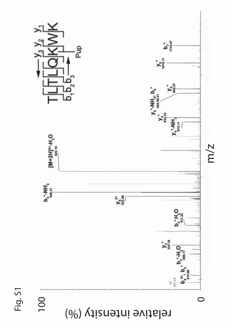
Fig.
S1
m/z
100 0
relative intensity (%)

Fig. S2
A.
B.
PafA
Pup~titin-I27Puptitin-I27
0 20 40 60 0 20 40 60 min
folded unfolded
645139
28
14
19
6
m/z
100
0
rela
tive
inte
nsity
(%)

Eyal GurNaomi Ofer, Nadav Forer, Maayan Korman, Marina Vishkautzan, Isam Khalaila and
Ubiquitin-like Protein (Pup) LigaseAllosteric Transitions Direct Protein Tagging by PafA, the Prokaryotic
doi: 10.1074/jbc.M112.435842 originally published online March 7, 20132013, 288:11287-11293.J. Biol. Chem.
10.1074/jbc.M112.435842Access the most updated version of this article at doi:
Alerts:
When a correction for this article is posted•
When this article is cited•
to choose from all of JBC's e-mail alertsClick here
Supplemental material:
http://www.jbc.org/content/suppl/2013/03/07/M112.435842.DC1.html
http://www.jbc.org/content/288/16/11287.full.html#ref-list-1
This article cites 26 references, 8 of which can be accessed free at
by guest on August 13, 2016
http://ww
w.jbc.org/
Dow
nloaded from


