Minireview
Nosema ceranae (Microsporidia), a controversial 21stcentury honey bee pathogen
Mariano Higes,1* Aránzazu Meana,2
Carolina Bartolomé,3 Cristina Botías1 andRaquel Martín-Hernández1,4
1Centro Apícola Regional (CAR), Dirección General dela Producción Agropecuaria, Consejería de Agricultura,Junta de Castilla-La Mancha, Spain.2Departamento de Sanidad Animal, Facultad deVeterinaria, Universidad Complutense de Madrid, Spain.3Departamento de Anatomía Patolóxica e CienciasForenses, Grupo de Medicina Xenómica, Universidadede Santiago de Compostela – 15782 Santiago deCompostela, Spain.4Instituto de Recursos Humanos para la Ciencia y laTecnología (INCRECYT), Fundación Parque Científico yTecnológico de Albacete, Spain.
Summary
The worldwide beekeeping sector has been facing agrave threat, with losses up to 100–1000 times greaterthan those previously reported. Despite the scale ofthis honey bee mortality, the causes underlying thisphenomenon remain unclear, yet they are thoughtto be multifactorial processes. Nosema ceranae, amicrosporidium recently detected in the Europeanbee all over the world, has been implicated in theglobal phenomenon of colony loss, although its roleremains controversial. A review of the current knowl-edge about this pathogen is presented focussing ondiscussion related with divergent results, trying toanalyse the differences specially based on differentmethodologies applied and divisive aspects onpathology while considering a biological or veterinar-ian point of view. For authors, the disease producedby N. ceranae infection cannot be considered aregional problem but rather a global one, as indicatedby the wide prevalence of this parasite in multiplehosts. Not only does this type of nosemosis causes aclear pathology on honeybees at both the individual
and colony levels, but it also has significant effectson the production of honeybee products.
Introduction
Globally, honeybees play important ecological and eco-nomic roles as pollinators of many crops and wild plants(FAO, 2006; Bradbear, 2009). Current agricultural prac-tices, such as large-scale monoculture, require a sea-sonal abundance of honeybees in specific geographiclocations that lack adequate year-round pollinator popu-lations. Managed colonies also satisfy the human demandfor honeybee products such as honey, wax and royal jelly(Delaplane and Mayer, 2000), especially in temperateareas of the world where most professional beekeepingis practiced. Indeed, about 40% of European honeybeecolonies are located in temperate Mediterranean areas,such as Spain, Italy and Greece. Thus, maintainingadequate sanitary conditions in honeybee colonies iscrucial to ensure the continued pollination and productionof honeybee products.
For several years, the worldwide beekeeping sectorhas been facing a grave threat, with losses up to100–1000 times greater than those previously reported(European Parliament, 2010). Despite the scale of thishoney bee mortality, the causes underlying this phenom-enon remain unclear, yet they are thought to be multi-factorial processes (European Parliament, 2010; Higeset al., 2010; vanEngelsdorp and Meixner, 2010). Since2007, the heightened awareness among the public andresearchers has stimulated the creation of working groupsand increased the funding available for studies address-ing this global problem, particularly in geographical areaswhere professional beekeeping is significantly affected.
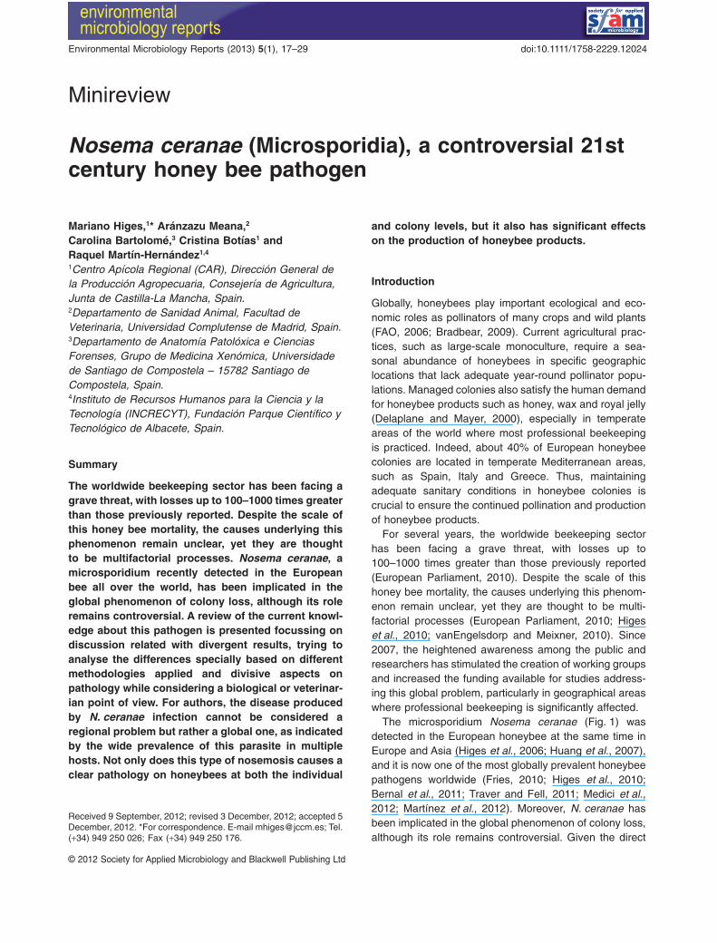
The microsporidium Nosema ceranae (Fig. 1) wasdetected in the European honeybee at the same time inEurope and Asia (Higes et al., 2006; Huang et al., 2007),and it is now one of the most globally prevalent honeybeepathogens worldwide (Fries, 2010; Higes et al., 2010;Bernal et al., 2011; Traver and Fell, 2011; Medici et al.,2012; Martínez et al., 2012). Moreover, N. ceranae hasbeen implicated in the global phenomenon of colony loss,although its role remains controversial. Given the direct
Received 9 September, 2012; revised 3 December, 2012; accepted 5December, 2012. *For correspondence. E-mail [email protected]; Tel.(+34) 949 250 026; Fax (+34) 949 250 176.
bs_bs_banner
Environmental Microbiology Reports (2013) 5(1), 17–29 doi:10.1111/1758-2229.12024
© 2012 Society for Applied Microbiology and Blackwell Publishing Ltd
relationship between N. ceranae and colony losses inSpain (Higes et al., 2009a; 2010), Spanish researchgroups have actively sought to develop strategies to mini-mize the economic losses inflicted upon the professionalsector by this microsporidium. Some studies have sug-gested a link between this pathogen and bee colonydepopulation/loss in other countries with similar climaticconditions (Higes et al., 2005; 2006; 2008; 2009a; Bacan-dritsos et al., 2010; Borneck et al., 2010; Hatjina et al.,2011; Invernizzi et al., 2011; Soroker et al., 2011). By con-trast, in countries from colder climates, the role for thismicrosporidium in colony loss has been ruled out (Gisderet al., 2010; Hedtke et al., 2011; Stevanovic et al., 2011;Dainat et al., 2012a,b), suggesting that specific conditionsare required to promote these pathogenic effects ofN. ceranae.
Disease is the result of complex interactions betweenthe epidemiological triad of pathogen, host and environ-ment. In farmed animals, this interaction is strongly influ-enced by factors related to animal husbandry andmanagement. In this review, we will discuss the factorsthat influence each of the components of this epidemio-logical triad and summarize the current state of N. cera-nae research, incorporating data from distinct fields ofresearch.
Can genetic variants of Nosema ceranae differ intheir pathogenicity?
Several authors have proposed that the pathologicaleffects of N. ceranae in Spain are associated with theincreased virulence of the Spanish strain (Genersch,2010; Huang et al., 2012). To confirm this hypothesis,distinct genetic variants must first be accurately identified,and then they must be analysed to determine whether
they are associated with variation in virulence, geographicdistribution or host specificity.
The molecular classification of microsporidia has beenlargely based on the analysis of ribosomal DNA (rDNA).The organization of the different domains within this genecluster varies according to species (reviewed by Ironside,2007). Nosema ceranae exhibits a unique rDNA unitarrangement consisting of a reversed 5S subunit locatedat the 5′ end, an intergenic spacer region (IGS), a smallsubunit (SSU), an internal transcribed spacer (ITS) and alarge subunit (LSU) at the 3′ end (Huang et al., 2007;2008). While eukaryotes generally contain many copies ofthe rDNA genes organized in tandem repeats (Eickbushand Eickbush, 2007), in Nosema spp. this tandem confor-mation has only been reported for N. apis (Gatehouseand Malone, 1998). Multiple rDNA units have beendescribed in all chromosomes in N. bombycis (Liu et al.,2008) and N. ceranae, in which 46 contigs containingribosomal sequences were identified, although none con-tained a complete ribosomal locus (Cornman et al., 2009).
Homogeneity among the nucleotide sequences of par-alogous rDNA copies is usually maintained throughhomologous recombination and/or gene conversionbetween duplicated regions (Eickbush and Eickbush,2007).Accordingly, rDNAgenes within a species are nearlyidentical, while orthologous rDNA genes may differ consid-erably between species. However, in the case of N. bombi,several authors have demonstrated the existence of two ormore rDNA sequence variants per spore (Tay et al., 2005;O’Mahony et al., 2007), and numerous polymorphismshave been described within single isolates of N. ceranae(Huang et al., 2008; Sagastume et al., 2010).
These findings beg the question as to how such vari-ability is generated. Reports of recombination in N. cera-nae (Sagastume et al., 2010) are surprising in organismsbelieved to have an asexual mode of reproduction. Thus,the only way that two sequences can recombine is if theycome together in the same cell. This implies the existenceof a transient diploid stage after fusion, whereby differentlineages in the same cell undergo homologous recombi-nation to produce a new variant that did not exist in any ofthe parental strains. Recombination in N. ceranae pro-vides better opportunities for evolution and adaptationthan a strict clonal mode of reproduction.
The existence of several non-homologous copies of agene in a genome renders it inadequate for phylogeneticpurposes.Accordingly, several conflicting Nosema phylog-enies based on SSU sequences have been reported(Fries, 2010; Choi et al., 2011). There is no consensusabout the taxonomic relationships between N. ceranaeand its closest relatives: N. apis and N. bombi (Slamovitset al., 2004; Vossbrinck and Debrunner-Vossbrinck, 2005;Wang et al., 2006; Shafer et al., 2009; Chen et al., 2009a;Choi et al., 2011). Similar results have been described for
Fig. 1. Mature spore of Nosema ceranae (electron microscopy).
18 M. Higes et al.
© 2012 Society for Applied Microbiology and Blackwell Publishing Ltd, Environmental Microbiology Reports, 5, 17–29
other Nosema species such as N. vespula and N. oule-mae. While some consider these species as close relativesof N. ceranae (Müller et al., 2000; Chen et al., 2009a),others claim that they evolved separately from N. vespula(Vossbrinck and Debrunner-Vossbrinck, 2005). Similardiscrepancies have arisen for phylogenies based on theLSU (Huang et al., 2007; Shafer et al., 2009; Choi et al.,2011) and on combined data from both the SSU and LSUgenes (Ironside, 2007; Shafer et al., 2009).
Nevertheless, rDNA, and in particular its SSU, hasbeen frequently used to assess the genetic diversity ofN. ceranae isolates from different geographic locations.With very few exceptions, probably biased due to theanalysis of very few sequences or clones (Whitakeret al., 2011; Yoshiyama and Kimura, 2011), most studieshave demonstrated considerable variability among and/orwithin samples (Higes et al., 2006; Huang et al., 2008;Chaimanee et al., 2011; Suwannapong et al., 2011;Medici et al., 2012). Recently, a large panel of clonedrDNA fragments containing the IGS and part of the SSUwas analysed (Sagastume et al., 2010), showing that allisolates produced multiple haplotypes regardless ofwhether DNA was extracted from individual or pooledhoneybees, as described previously (Huang et al., 2008).Furthermore, the haplotypes obtained from a singleisolate could differ by as much as those obtained fromdistinct isolates, with identical haplotypes appearing insamples of very different origin.
Despite these findings, some regional clustering hasbeen reported for Asian, North American (Williams et al.,2008; Chen et al., 2009b) and Argentinean samples(Medici et al., 2012). However, a fraction of the extanthaplotypes may have been overlooked since molecularcloning was not performed in these studies. Indeed, directsequencing is inappropriate to detect any geographiclinkage since the sequence obtained from each isolateprobably represents either the most frequent geneticvariant or a consensus sequence of several differenthaplotypes.
The wide diversity seen in rDNA from N. ceranae iso-lates complicates the determination of taxonomic andgeographic relationships, and highlights the need foralternative genomic markers, ideally single copy genes(Fries, 2010; Sagastume et al., 2010). The draft genomeassembly for N. ceranae (Cornman et al., 2009) offers aunique opportunity to obtain the necessary information,given that rDNA genotyping has proven unreliable forepidemiologic and phylogenetic purposes.
Other N. ceranae loci have been recently used asalternative markers, namely those encoding polar tubeproteins (Chaimanee et al., 2011; Hatjina et al., 2011)that are thought to be highly polymorphic. Such proteinsare major components of the microsporidian polar fila-ment that discharges the sporoplasm into the target cell
and plays an essential role in host invasion (Xu andWeiss, 2005). The phylogenetic relationships betweenN. ceranae isolates from different host species (Apismellifera, A. cerana, A. florea and A. dorsata) werestudied by analysing the sequences of two genes, theribosomal SSU and PTP1 (Chaimanee et al., 2011).While the former revealed no significant differencesbetween isolates, the latter identified three separateclades, one corresponding to the isolates infectingA. mellifera and A. cerana, and the other two to N. cera-nae isolates from A. florea and A. dorsata. Again, theseresults should be interpreted with caution, as the lack ofcloning could give rise to misleading conclusions (seeabove). A second study based on a different polar tubeprotein, PTP3 (Hatjina et al., 2011), demonstrated thatdirect sequencing of PCR products may overlook numer-ous genetic variants, highlighting a significant drawbackof genotyping. Surprisingly, the authors found thatsequence heterogeneity within a sample was notrestricted to rDNA and identified five distinct PTP3sequences in five different clones obtained from a singleisolate. The basis for this high degree of heterogeneityremains unclear, although it may be possible that multi-ple copies of the gene exist in a nucleus, that heterozy-gosis of the two nuclei of a spore occurs or that there isco-infection with several strains. Moreover, the associa-tion of specific haplotypes with different levels of hostdamage remains poorly understood (Tay et al., 2005;Williams et al., 2008). However, it should be noted thatvirulence is a consequence of the trade-off between dif-ferent components of parasite fitness in which bothparasite and host play essential roles (reviewed byFrank, 1996 and Frank and Schmid-Hempel, 2008).These host–parasite interactions may explain the highlevels of polymorphism detected both in the ribosomalSSU (Sagastume et al., 2010) and the PTP3 (Hatjinaet al., 2011) genes of N. ceranae, although furtherresearch is necessary to discern the implications of dif-ferent haplotypes on virulence, distribution and resist-ance to fungicides or host defences.
Variations in host susceptibility and pathological effectsat individual level
The host is a key component of the epidemiological triadin any disease. Accordingly, specific differences betweenlineages have been proposed to underlie the divergentpathological effects of N. ceranae in different laboratoriesor countries. Indeed, a review of the literature availablereveals significant differences in the survival and mortalityof infected bees between studies.
In laboratory experiments, factors including the age ofthe bees, and the origin dose and purification method ofthe infective spores, can have a significant impact on the
N. ceranae an emergent pathogen for beekeeping 19
© 2012 Society for Applied Microbiology and Blackwell Publishing Ltd, Environmental Microbiology Reports, 5, 17–29
survival of infected bees. For example, the risk of manyinfectious diseases varies widely over the animalslifespan due to the physiological changes associated withage. Comparable results have been reported when youngbees (bred in incubators) are used (Higes et al., 2007;Martín-Hernández et al., 2009; 2011; Alaux et al., 2010;Vidau et al., 2011). In these conditions, mortality is greaterin N. ceranae-infected versus uninfected bees, and asimilar trend is observed when young bees are tested(Higes et al., 2007; Alaux et al., 2010; Martín-Hernándezet al., 2011; Aufauvre et al., 2012; Dussaubat et al.,2012), and when worker honeybees from the brood nestare infected (Paxton et al., 2007), with decreased survivalin infected versus uninfected bees. However, very lowmortality was observed when adult worker bees of unde-termined age (average 15 days old) were collected fromcombs and experimentally infected with N. ceranae (For-sgren and Fries, 2010).
A second factor affecting the outcome of infection is thesource and storage conditions of spore inoculum. Theeffect of temperature (frozen, refrigerated or fresh) onspore viability and previous handling (if the spores werepurified or not and the method used) may explain someconflicting findings regarding infectivity and bee mortality.Differences in thermal plasticity have been demonstratedbetween N. apis and N. ceranae (Fenoy et al., 2009;Gisder et al., 2010; reviewed in Martín-Hernández et al.,2009; Fries, 2010) and prior thermal shock may be mis-interpreted as variability in virulence or infectivity betweenstrains. Similarly, the spore dose used may directly influ-ence survival of bees.
These important methodological differences mustbe considered whencomparing different studies. Forexample, mortality at 7 days post infection ranged from11.1% to 93.1% when Percoll-purified spores maintainedat room temperature were used to infect newborn honeybees, given individual doses of 103–105 spores per beeand maintained at 33°C after infection (Martín-Hernándezet al., 2011). In these conditions, the mortality of infectedbees was always greater than that observed in uninfectedcontrols. Conversely, in another study fresh spores (nodata regarding purification or storage temperature areprovided) were used to infect adult worker bees collectedfrom a colony (~ 15 days old) with doses of 101–104
spores per bee, and that were maintained at 30°C post-infection (Forsgren and Fries, 2010). Despite these differ-ences, and even though exhaustive detail on beemortality was not provided in the latter study, increasedmortality (23%) was observed in one cage of beesinfected with N. ceranae, consistent with the findings ofMartín-Hernández and colleagues (2011) with a compa-rable spore dose per bee. However, the conclusions ofboth experiments obviously differ and they cannot becompared due to significant methodological differences.
The aforementioned discrepancies between studiesunderscore the need for standardized protocols tocompare the effects of Nosema infection on honeybeesurvival rates, in which the following parameters are con-trolled: spore source, spore storage conditions, sporepurification process, spore viability and infectivity, beeincubation temperature, the age of bees at infection, thesource of the bees used (colony or laboratory), or evennutritional factors or genetical differences yet to verify.Furthermore, differences in experimental conditionsshould be highlighted to avoid methodological artefactsthat could bias the results and conclusions.
In addition to the effects on honeybee mortality, severalstudies have investigated other aspects of experimentalinfection with N. ceranae in the laboratory, including theeffects on hormone and pheromone production (Dussau-bat et al., 2010; Alaux et al., 2011; Ares et al., 2012),immune suppression (Antúnez et al., 2009; Chaimaneeet al., 2012), energetic stress (Mayack and Naug, 2009;Martín-Hernández et al., 2011), behaviour (Naug andGibbs, 2009; Campbell et al., 2010), anatomopathologicallesions (Higes et al., 2007; 2009b; Paxton et al., 2007;Dussaubat et al., 2012), carbohydrate and aminoacidlevels in hemolymph (Mayack and Naug, 2010; Aliferiset al., 2012), tissue degeneration and cell renewal (Dus-saubat et al., 2012), as well as its synergy with otheragents (Alaux et al., 2010; Vidau et al., 2011; Aufauvreet al., 2012). These studies clearly demonstrate thatN. ceranae exerts pathological effects on A. melliferasimilar to those seen in A. florea (Suwannapong et al.,2010).
In both experimentally and naturally infected bees(A. mellifera), N. ceranae infection significant alters beebehaviour and the physiology of the infected tissue (ven-triculi). In both cases, similar anatomopathological altera-tions have been reported (Higes et al., 2007; 2009b; Chenet al., 2009a; García-Palencia et al., 2010), includingdegeneration of the epithelial ventricular cells, the pre-sence of vacuoles in the cytoplasm, disruption of thecellular membranes and a reduced size of the cell nucleus.
Based on PCR detection of the parasite, it has beensuggested that N. ceranae can parasitize structures otherthan the epithelial cells of the ventriculi in worker bees,such as the Malpighian tubules, hypogharyngeal andsalivary glands (Chen et al., 2009a; Gisder et al., 2010;Copley and Jabaji, 2012), head, thorax, ovaries, sper-matheca and eggs (Traver and Fell, 2012). However,these results have not been confirmed by histopathologi-cal studies and nor have different developmental stagesof Nosema been observed in cell types other in than theepithelial cells of the ventriculum (Higes et al., 2007;García-Palencia et al., 2010).
The numerous negative effects of N. ceranae infectionof individual honeybees are reflected by further effects at
20 M. Higes et al.
© 2012 Society for Applied Microbiology and Blackwell Publishing Ltd, Environmental Microbiology Reports, 5, 17–29
the colony level, directly affecting colony homeostasis. Amathematical model of Nosema and Varroa infections hasbeen generated and demonstrates how forager death rateinfluences colony population, suggesting that chronicallyhigh forager death rates result in rapid population declineand consequent colony failure (Khoury et al., 2011). Somerecent epidemiological studies do not support this model(Fernández et al., 2012; Dainat et al., 2012a,b), althoughthe methodology used in those studies do not let assessadequately the effect of the parasite at the individual andthe colony level.
All the methodological differences here revised makedifficult to establish if the differences in mortality are dueto the methodology or to the different susceptibility of thehost subspecies. Since recently a Danish strain of bees(drones) get after selective breeding has been describedto be more tolerant to N. ceranae infections (Huang et al.,2012) more research should be performed with differentA. mellifera subspecies using standardized methods.
Does Nosema ceranae present differentepidemiological patterns?
Environmental conditions also strongly influence manyparasitic relationships and, regardless of the effects ofaltitude, flora and colony management, in warm countrieslike Spain the influence of temperature on the conse-quences of N. ceranae has been observed. This factormay contribute to the divergent effects of N. ceranae oninfected colonies around the world and indeed, a dif-ferent pattern of climatic preferences has been recentlydescribed for the two Nosema species that infect honey-bees (Martín-Hernández et al., 2012). While both specieswere widely disseminated within the study area, N. cera-nae was the most prevalent microsporidia found in A. mel-lifera in hotter regions (Mediterranean regions), whileN. apis was more prevalent in milder regions. SinceN. ceranae infection appears to be more common inwarmer climates and in specific geographical areas, thisshould be considered when importing bees from suchareas (Fries, 2010), when engaging in professional api-culture in warmer countries (e.g. Spain, Greece, Italy),and when evaluating the impact of Nosema infection inwarm climates.
Environmental effects have also been implicated in theinterspecies competition between N. ceranae and N. apis.Nosema ceranae is currently the most prevalent micro-sporidium and frequently, it is the only species detected inbees (Klee et al., 2007; Chen et al., 2008; Chen andHuang, 2010; Yoshiyama and Kimura, 2011; Martínezet al., 2012). However, this tendency was not observed inregions of Germany or the UK (Budge et al., 2010; Gisderet al., 2010) where N. apis remains more prevalent. Fur-thermore, a recent analysis of the prevalence of Nosema
spp. at different levels (individual, colony, apiary, country,bee castes) revealed that no replacement was observed(Martín-Hernández et al., 2012). Although N. ceranaewas the dominant species throughout the year, the preva-lence of N. apis (much lower) followed a classical epide-miological pattern (peaking in spring and autumn), similarto that described historically (Gómez Pajuelo andFernández Arroyo, 1979; Orantes Bermejo and García-Fernández, 1997). Similar results have been observedtoo in the USA (Runckel et al., 2011; Dr R. Cramer, pers.comm.).
Most classical studies described typical patterns ofprevalence in temperate climates, with a large peak inspring when more colonies have detectable levels ofinfection (Borchert, 1928 reviewed in Bailey, 1955; Fries,2010). Importantly, when these studies were performedN. apis was considered the only aetiological agent ofnosemosis. More recently, a lack of seasonality has beenreported in tropical and subtropical conditions (Fries andRaina, 2003; reviewed in Fries, 2010), while N. ceranaeinfection has always been detected throughout the year atdifferent latitudes (Martín-Hernández et al., 2007; 2012;Calderón et al., 2008; Higes et al., 2008; Hedtke et al.,2011; Runckel et al., 2011; Stevanovic et al., 2011; Traverand Fell, 2011; Whitaker et al., 2011; Medici et al., 2012;Smart and Sheppard, 2012; Botías et al., 2012a,b; Dainatet al., 2012a).
In any discussion of environmental effects, seasonalitymust be defined. In early studies, this term only describedthe detection of the agent, or the observation of clinicalsigns at a particular time point. However, with the devel-opment of new scientific methods, seasonality is nowmeasured on the basis of prevalence (Fries, 2010), thepercentage of infected bees (Higes et al., 2008), the meanspore count (Higes et al., 2008, Traver et al., 2011), the Ctvalue and the average copy numbers (Traver and Fell,2012). The use of one or other parameter has a directimpact on the epidemiological pattern. For instance,although spore count is not a reliable parameter of infec-tion (Meana et al., 2010), comparing colony infection usingthis parameter reveals a higher level in spring (April–June:Gisder et al., 2010; Traver and Fell, 2011; Traver et al.,2012) or in autumn–winter (Higes et al., 2008).
Finally, the fact is that N. ceranae is highly prevalent inwarmer areas and its presence in the colony is very high.Under those conditions there are many bees infected thatcan be directly related to the clinical signs observed underfield conditions.
Clinical signs of N. ceranae infection inhoneybee colonies
Probably the most controversial aspect of N. ceranaeinfection in beekeeping is its ability to depopulate or kill a
N. ceranae an emergent pathogen for beekeeping 21
© 2012 Society for Applied Microbiology and Blackwell Publishing Ltd, Environmental Microbiology Reports, 5, 17–29
colony. After N. ceranae parasitization of honeybees wasfirst detected and linked with colony collapse in Spain(Higes et al., 2006; Martín-Hernández et al., 2007), otherauthors ruled out its role in colony loss (Cox-Foster et al.,2007; Klee et al., 2007). At this time, the little data avail-able on the virulence of N. ceranae at the colony levelwere contradictory, probably due to a failure to properlyidentify the clinical signs of disease, the parameter withwhich to evaluate the impact of the illness and a poorunderstanding of the subclinical effects of parasitism.Moreover, at the time, the only common feature of colonycollapse described all over the world was death.
Traditionally Koch’s postulates have been used ascriteria to determine whether a given microorganismcauses a specific disease. Those postulates were ini-tially developed for bacteria and despite their importancein microbiology, they have severe limitations particularlywhen applied to diseases caused by non-bacterialmicroorganisms. For example, for some microorganismsthat cannot be grown in pure culture in the laboratory,including bee microsporidia, they can be used to infectthe host, mimicking the disease. Additional limitationsarise in cases where the clinical signs of an infectionhave not been accurately described (or widelyaccepted), as is the case of nosemosis type C causedby N. ceranae infection.
Taking these limitations into account, Koch’s postulateswere demonstrated for honeybee colonies infected withN. ceranae (Higes et al., 2008), as previously confirmedin individual bees (see above). Nosema ceranae wasextracted from an affected colony and identified by PCR,and it was then transmitted to healthy colonies where itinduced disease and colony collapse. Finally, the infectiveagent was isolated from these newly infected colonies.These findings were subsequently confirmed in laterstudies (Botías et al., 2010; 2012a) and among otherpathogens, colony loss has been linked with the presenceof N. ceranae in several reports (Higes et al., 2005; 2006;2008; 2009a; Borneck et al., 2010; Hatjina et al., 2011;Nabian et al., 2011). However, studies conducted incolder areas have revealed contradictory findings (Gisderet al., 2010; Hedtke et al., 2011; Stevanovic et al., 2011;Dainat et al., 2012a,b). Thus, it is of great interest todetermine whether these differential effects results aredue to distinct behaviours of N. ceranae at differentlatitudes, or basic criteria are not the same such asthe identificacion of clinical/subclinical signs of diseasebetween researchers. In some works definition of colonyloss or collapsed colonies are the only description of adisease that seems not to present any other clinical sign.An expert is sometimes needed to detect signs as lowerbee population, lower honey production, unexpectedbrood in cold months or younger bees starting to forage.Due to the fact that bees are social insects, a biological
point of view must be properly differentiate from a veteri-narian one, each one complementing the other.
A biological perspective. Honeybee colonies are socialsystems whose performance depends on the equilibriumthat exists between the members of the colony. Accord-ingly, any effects at the individual level due to N. ceranaeinfection will disturb colony homeostasis. The colonypopulation depends largely on the lifespan of the workerbees (Woyke, 1984; Khoury et al., 2011), which may beseverely affected by N. ceranae infection (Higes et al.,2007; Paxton et al., 2007; Martín-Hernández et al., 2011).These findings demonstrate that N. ceranae can activelyreduce the adult bee population (Higes et al., 2008;Botías et al., 2010; 2012a; Soroker et al., 2011; Eischenet al., 2012).
A direct consequence of the high mortality rate of for-agers that is provoked by Nosema infection (Higes et al.,2008; 2010), is that younger bees begin to forage earlierto compensate for the loss of available foragers (Huangand Robinson, 1996; Amdam and Omholt, 2003), therebymodifying the entire work profile of the colony (Wang andMoeller, 1970). However, this compensatory mechanismshortens the overall lifespan of adult bees (Neukirch,1982; Schmid-Hempel and Wolf, 1988; Wolf and Schmid-Hempel, 1989), and their efficacy and resilience as forag-ers (Oskay, 2007), as well as reducing the time each beededicates to colony growth and brood production. Further-more, internal colony activities such as hygiene and broodcaring may be negatively affected due to the decreasedavailability of nurse bees that attend to the brood, in turnincreasing the risk of developing brood diseases suchas chalkbrood (Hedtke et al., 2011). When the colonyreaches the point at which it cannot sustain brood produc-tion at a sufficient rate to replace the adult bee losses, theextent of colony decline accelerates rapidly, resulting indepopulation (Khoury et al., 2011), which represents theonly clear sign of infection described for Nosema ceranae(Higes et al., 2008; 2011; Botías et al., 2010; 2012a;Eischen et al., 2012).
The accelerated behavioural development observedin Nosema-infected bees has been associated withincreased titres of juvenile hormone (JH: Lin et al., 2009;reviewed in Higes et al., 2010; Ares et al., 2012), inhibitionof vitellogenin (Vg) gene expression (Antúnez et al.,2009) and increased levels of the pheromone ethyl oleate(EO: Dussaubat et al., 2010). These factors are impli-cated in regulating the division of labour among workerbees, as well as maturation and the nurse–forager tran-sition. This manipulation of host endocrinology in infectedinsects has previously been described as an adaptivemechanism that serves to increase the reproductivefitness of the parasite (prolonging the host larval state tomaximize spore yield: Down et al., 2008).
22 M. Higes et al.
© 2012 Society for Applied Microbiology and Blackwell Publishing Ltd, Environmental Microbiology Reports, 5, 17–29
Nosema ceranae also significantly alters the feedingbehaviour of infected bees, increasing their responsive-ness to sucrose and decreasing food sharing betweenbees (Naug and Gibbs, 2009). In addition, this microsporid-ium can lower the haemolymph sugar level of individualforagers and uncouple their energetic state from that of thecolony, to which it is normally closely related (Mayack andNaug, 2010). In turn, this altered energetic state candecrease the flying ability of foragers by about two-thirdswhen compared with uninfected foragers, as confirmed bythe impaired orientation skills in Nosema-infected beesand a lower rate of infected bees among the returningversus departing foragers (Kralj and Fuchs, 2010 ). Thisobservation suggests that infected forager bees die whileforaging, as described previously (Higes et al., 2008).Accordingly, it is unusual to find dead or dying bees in thevicinity of the hive in cases of nosemosis type C (Higeset al., 2008; 2009a; 2010; Borneck et al., 2010).
The preference for higher temperatures exhibited byN. ceranae-infected bees (Campbell et al., 2010) may bedue to the pathological stress imposed on the host bythe infection. However, this preference also appears tobenefit the pathogen, as the reproductive potential ofN. ceranae increases at higher temperatures (Martín-Hernández et al., 2009). Energetic stress and a concur-rent increase in appetite are the primary effects ofN. ceranae infection in honeybees (Mayack and Naug,2009). This increased energetic stress may explain someof the changes observed in host behaviour due to star-vation, lack of thermoregulatory capacity or alterations inthe rates of trophallaxis, which may enhance diseasetransmission and increase the risk of death (Martín-Hernández et al., 2011). Indeed, these changes in thehive conditions may also be implicated in outbreaks ofstress-related diseases such as chalkbrood (Hedtkeet al., 2011) or even in the loss of efficacy of contact-dependent treatments to control the parasite Varroadestructor (Botías et al., 2011).
A colony infected with Nosema constantly loses oldbees, which are supplemented by younger bees. Thepercentage of younger bees in the colony population canexplain the large differences in parasite burden (as eitherpercentage of infected bees or parasite burden) observedat the moment of death. Colonies that die in winter gen-erally contain few or no newborns. Accordingly, the sur-viving interior bees are strongly infected and can eventransmit the infection to the queen (Higes et al., 2008;2009b). If the colony collapses in early spring, the increas-ing numbers of newborn bees will reduce the percentageof infected interior bees and the parasitic load, such thatthe intensity of infection will be lower than that seen inwinter (Higes et al., 2008). This scenario differs to thatwhich occurs when colonies collapse due to Varroadestructor infection (Rosenkranz et al., 2010). In this
case, the bees that remain at the time of the collapseexhibit a high infestation rate due to exponential multipli-cation of mites in the previous months. The unawarenessof these factors could explain discrepancies found insome works (Cox-Foster et al., 2007; Dainat et al.,2012a,b; Fernández et al., 2012).
In addition to the effects of infection on colony dynam-ics, several additional factors may increase the severity ofN. ceranae infection in field conditions, including expo-sure to sublethal doses of insecticides (Wu et al., 2012),which can increase mortality and colony susceptibility topathogens and promote the interaction of certain viruses(Bromenshenk et al., 2010; Bacandritsos et al., 2010). Arecent study reported a negative correlation betweendeformed wing virus (DWV) and N. ceranae (Costa et al.,2011), which may result from competition for host cells orspecific cell functions in the honeybee midgut: N. ceranaeinduces lesions and degeneration of the epithelial cells ofthis tissue (Higes et al., 2007), and the digestive tractappears to be critical for DWV pathogenesis (Boncristianiet al., 2009).
A veterinary perspective. In veterinary medicine, a clini-cal sign is an objective indication of a specific medicalevent or characteristic that can be detected by a veteri-narian during an examination or by a clinical scientist bymeans of in vivo or in vitro analysis of the subject ofinterest. Because each group member is differentlyaffected by one or more of the factors of the epidemiologictriad, a disease in a group (apiary) is often manifested asa spectrum ranging from unapparent to subclinical to clini-cal to fatal. In this context, clinical signs are indications ofdisease that can be detected during a normal clinicalexamination, while subclinical signs are indications ofdisease that cannot be detected without performing aspecific test. Decreased productivity is one of the mostcommon subclinical signs in farming animals.
Many analyses of N. ceranae infection measure onlycolony losses in a short time period (as example Williamset al., 2010; Fernández et al., 2012). However, independ-ent of their role as pollinators, honeybees are farmanimals that produce food and non-food products ofhuman interest, although honeybee diseases are notusually discussed in this context. Indeed, the effects ofmicrosporidia infection on hive products remain largelyunknown. Several factors may affect honey production byhoneybee colonies, including climatic conditions (Kauffeldet al., 1976; Giray et al., 2007; vanEngelsdorp andMeixner, 2010), the availability of an adequate foragingarea (Naug, 2009), and colony strength (i.e. brood pro-duction, adult bee population, worker bee lifespan andworker bee productivity: Woyke, 1984; Szabo and Lefko-vitch, 1989; Eckert et al., 1994). Furthermore, honeybeeparasites and pathogens have for long been reported to
N. ceranae an emergent pathogen for beekeeping 23
© 2012 Society for Applied Microbiology and Blackwell Publishing Ltd, Environmental Microbiology Reports, 5, 17–29
negatively influence colony productivity (Kauffeld et al.,1972; Fries, 1988; Anderson and Giacon, 1992; Murilhas,2002).
As negative effects of N. apis infection on colony pro-ductivity have been reported previously (Farrar, 1947;Moeller, 1962; Kauffeld et al., 1972; Fries et al., 1984),similar effects were assumed for N. ceranae, given itsrapid spread and high prevalence (reviewed by Fries,2010 and Higes et al., 2010). Indeed, honey productionhas been directly correlated with the level of colony infec-tion (Botías et al., 2010; 2012a). A negative correlationbetween N. ceranae infection rates (percentage ofinfected forager bees per colony) and colony honey pro-duction (determined by weighing the amount of honeyproduced per colony) was described in field studies ofexperimental colonies in Spain (Botías et al., 2010;2012a). Nevertheless, a recent publication from Spain(Fernández et al., 2012) suggested that N. ceranaeinfected honey bee colonies remained alive with normalproduction during the referenced study. Surprisingly thehoney and pollen production were not recorded during theassay, and other clinical or subclinical sings were nottaken into account or measured. For this reason thisstatement has to be evaluated cautiously. However, addi-tional data collected in different climates with differentbeekeeping management techniques are required to cor-roborate these findings. In a Canadian study, honey andpollen cells were visually evaluated as indicators of colonystrength to compare untreated and fumagillin-treatedcolonies (Williams et al., 2010). No correlation was estab-lished between the presence of N. ceranae and colonystrength or winter mortality, although not all fumagillin-treated and untreated groups studied exhibited differentlevels of infection.
The probability of acquiring an infectious diseaseincreases with age, and novel diseases can be readily
picked up by foraging bees (Schmid-Hempel, 1994).Accordingly, several studies have reported that foragerbees are more likely to be infected with Nosema thanhouse bees (L’Arrivée, 1963; Taber and Lee, 1973;Pickard and El-Shemy, 1989; Higes et al., 2008; Meanaet al., 2010; Botías et al., 2012a; Martín-Hernández et al.,2012; Smart and Sheppard, 2012). By contrast, a recentstudy reported no significant differences in the extent ofinfection between in-hive and forager bees (Traver et al.,2012), although both parameters, the average DNA copynumber and average spore count of N. ceranae, tended tobe higher in the forager bee samples.
Another aspect of N. ceranae infection that is rarelyassessed is the influence of beekeeping practices ondisease evolution at the colony level. A recent study dem-onstrated the central role of the queen in the evolution ofN. ceranae infection of honeybee colonies (Botías et al.,2012a). Removal of the queen and subsequent replace-ment with a younger queen decreased the proportion ofNosema infected forager and house bees, maintaining theoverall infection rate at a level compatible with colonyviability. This effect should be taken into account whenstudying the evolution of Nosema disease in differentgeographical areas where queens are frequently replaced(either naturally or by the beekeeper). Possible repercus-sions of different beekeeping practices such as prophy-lactic measures should also be considered, as theireffects on N. ceranae have not been established, andhandling techniques used in Northern Europe (Hedtkeet al., 2011) differ widely from those of Mediterraneanareas (Higes et al., 2008; 2009a; 2010; Bacandritsoset al., 2010; Hatjina et al., 2011).
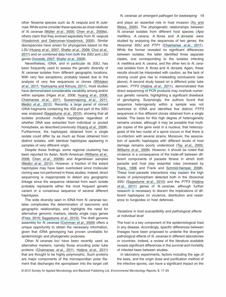
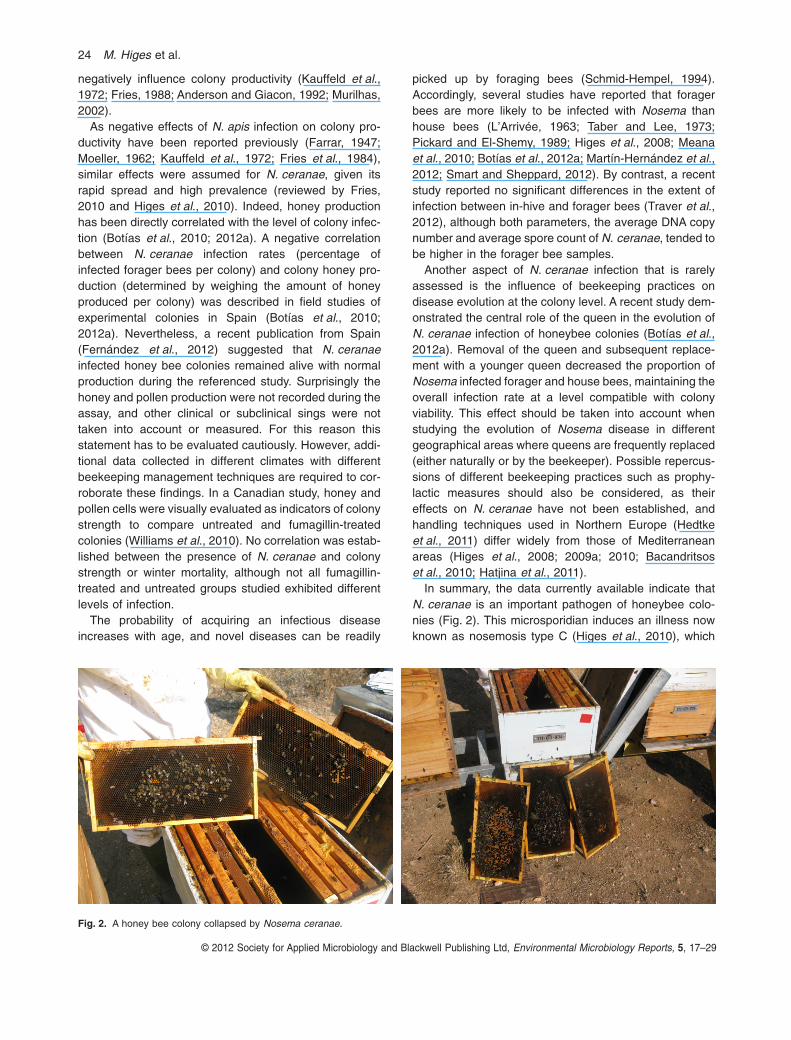
In summary, the data currently available indicate thatN. ceranae is an important pathogen of honeybee colo-nies (Fig. 2). This microsporidian induces an illness nowknown as nosemosis type C (Higes et al., 2010), which
Fig. 2. A honey bee colony collapsed by Nosema ceranae.
24 M. Higes et al.
© 2012 Society for Applied Microbiology and Blackwell Publishing Ltd, Environmental Microbiology Reports, 5, 17–29
presents a variety of manifestations due to severalfactors, some of which are understood and others thatremain to be identified. Indeed, many of the clinical signsproduced by this disease have received little attention,with colony collapse generally taken as the main diseaseindicator by both researchers and beekeepers.
The disease produced by N. ceranae infection cannotbe considered a Spanish regional problem as previouslysuggested, but rather a global one, as indicated by thewide prevalence of this parasite in multiple hosts. Not onlydoes this type of nosemosis affect honeybees at boththe individual and colony levels, but it also has signifi-cant effects on the production of honeybee products.Accordingly, in addition to colony collapse, it is essential tocontinue studying the many effects of N. ceranae infectionin bee colonies in different geographical regions, in orderto provide beekeepers with appropriate control strategies.This is particularly necessary in temperate zones, suchas Mediterranean countries, where N. ceranae is moreprevalent and more damaging. Moreover, this region ishome to a large number of professional beekeepers and itproduces high yields of honey and pollen, highlighting theimportance of minimizing the economic toll of N. ceranaeinfection. For this reason we cannot simply bury our headin the sand but for the sake of these professional bee-keepers, we must search for a practical solution to thisproblem.
Acknowledgements
The authors wish to thank O. Sánchez, A. I. Asensio, V.Albendea, C. Rogerio, T. Corrales and C. Abascal for theirtechnical support. Finantial support was provided by Junta deComunidades de Castilla-La Mancha (Consejería de Agricul-tura) and INIA-FEDER funds (RTA 2008-00020-C02-0,RTA2009- 000105-C02-01 and RTA2009-00057).
References
Alaux, C., Brunet, J.L., Dussaubat, C., Mondet, F., Tch-amitchan, S., Cousin, M., et al. (2010) Interactionsbetween Nosema microspores and a neonicotinoidweaken honeybees (Apis mellifera). Environ Microbiol 12:774–782.
Alaux, C., Folschweiller, M., McDonnell, C., Beslay, D.,Cousin, M., Dussaubat, C., et al. (2011) Pathologicaleffects of the microsporidium Nosema ceranae on honeybee queen physiology (Apis mellifera). J Invertebr Pathol106: 380–385.
Aliferis, K.A., Copley, T., and Jabali, S. (2012) Gaschromatography-mass spectrometry metabolite profiling ofworker honey bee (Apis mellifera L.) hemolymph for thestudy of Nosema ceranae infection. J Insect Physiol 58:1349–1359.
Amdam, G.V., and Omholt, W. (2003) The hive bee to forager
transition in honeybee colonies: the double repressorhypothesis. J Theor Biol 223: 451–464.
Anderson, D.L., and Giacon, H. (1992) Reduced pollen col-lection by honey bee (Hymenoptera, Apidae) coloniesinfected with Nosema apis and sacbrood virus. J EconEntomol 85: 47–51.
Antúnez, K., Martín-Hernández, R., Prieto, L., Meana, A.,Zunino, P., and Higes, M. (2009) Immune-suppression inthe honey bee (Apis mellifera) following infection byNosema ceranae (Microsporidia). Environ Microbiol 11:2284–2290.
Ares, A.M., Nozal, M.J., Bernal, J.L., Martín-Hernández, R.,Higes, M., and Bernal, J. (2012) Liquid chromatographycoupled to ion trap-tandem mass spectrometry to evaluatejuvenile hormone III levels in bee hemolymph fromNosema spp. infected colonies. J Chromatogr B AnalytTechnol Biomed Life Sci 899: 146–153.
Aufauvre, J., Biron, D.G., Vidau, C., Fontbonne, R., Roudel,M., Diogon, M., et al. (2012) Parasite-insecticide interac-tions: a case study of Nosema ceranae and fipronil synergyon honeybee. Sci Rep 2: 326 (1–7).
Bacandritsos, N., Granato, A., Budge, G., Papanastasiou, I.,Roinioti, E., Caldon, M., et al. (2010) Sudden deaths andcolony population decline in Greek honey bee colonies.J Invertebr Pathol 105: 335–340.
Bailey, L. (1955) The epidemiology and control of Nosemadisease of the honeybee. Ann Appl Biol 43: 379–389.
Bernal, J., Martín-Hernandez, R., Diego, J.C., Nozal, M.J.,González-Porto, A.V., Bernal, J.L., and Higes, M. (2011) Anexposure study to assess the potential impact of fipronil intreated sunflower seeds on honey bee colony losses inSpain. Pest Manag Sci 67: 1320–1331.
Boncristiani, H.F., Di Prisco, G., Pettis, J.S., Hamilton, M.,and Chen, Y.P. (2009) Molecular approaches to the analy-sis of deformed wing virus replication and pathogenesis inthe honey bee, Apis mellifera. Virol J 6: 221 (1–9).doi:10.1186/1743-422X-6-221.
Borchert, A. (1928) Beiträge zur Kenntnis der Bienen Para-siten Nosema apis. Archiv Bienenkunde 9: 115–178.
Borneck, R., Viry, A., Martín-Hernández, R., and Higes, M.(2010) Honey bee colony losses in the Jura Region,France and related pathogens. J Apic Res 49: 334–336.
Botías, C., Martín-Hernández, R., Meana, A., and Higes, M.(2010) Negative effects of Nosema infection in honey pro-duction and vitality of honey bee (Apis mellifera) colonies inSpain. EURBEE, 4th European Conference of Apidilogy(Edited by Meral Kence), 7–9 September 2010, Ankara,Turkey.
Botías, C., Martín-Hernández, R., Barrios, L., Garrido-Bailón,E., Nanetti, A., Meana, A., and Higes, M. (2011) Nosemaspp. parasitization decreases the effectiveness of acaricidestrips (Apivar®) in treating varroosis of honey bee (Apismellifera iberiensis) colonies. Environ Microbiol Rep 4:57–65.
Botías, C., Martín-Hernández, R., Días, J., García-Palencia,P., Matabuena, M., Juarranz, A., et al. (2012a) The effect ofinduced queen replacement on Nosema spp. infection inhoney bee (Apis mellifera iberiensis) colonies. EnvironMicrobiol 14: 845–859.
Botías, C., Anderson, D., Meana, A., Garrido-Bailón, E.,Martín-Hernández, R., and Higes, M. (2012b) Further
N. ceranae an emergent pathogen for beekeeping 25
© 2012 Society for Applied Microbiology and Blackwell Publishing Ltd, Environmental Microbiology Reports, 5, 17–29
evidence of an oriental origin for Nosema ceranae (Micro-sporidia: Nosematidae). J Invertebr Pathol 110: 108–113.
Bradbear, N. (2009) Bees and Their Role in Forest Liveli-hoods. A Guide to the Services Provided by Bees and theSustainable Harvesting, Processing and Marketing of TheirProducts. Non-Wood Forest Products 19. Rome, Italy:Food and Agriculture Organization of the United Nations(FAO).
Bromenshenk, J.J., Henderson, C.B., Wick, C.H., Stanford,M.F., Zulich, A.W., Jabbour, R.E., et al. (2010) Iridovirusand microsporidian linked to honey bee colony decline.PLoS ONE 5: e13181.
Budge, G., Powell, M., Roberts, K., Adams, I., Jones, B.,Marris, G., et al. (2010) What has Nosema got to do withlosses? Monitoring both Nosema species in the UK. InProceedings of the 4th European Conference of Apidology(Edited by Meral Kence), 7–9 September 2010, Ankara,Turkey.
Calderón, R.A., Sanchez, L.A., Yáñez, O., and Fallas, N.(2008) Presence of Nosema ceranae in Africanized honeybee colonies in Costa Rica. J Apic Res 47: 328–329.
Campbell, J., Kessler, B., Mayack, C., and Naug, D. (2010)Behavioural fever in infected honeybees: parasitic manipu-lation or coincidental benefit? . Parasitology 137: 1487–1491.
Chaimanee, V., Chen, Y., Pettis, J.S., Scott Cornman, R.,and Chantawannakul, P. (2011) Phylogenetic analysis ofNosema ceranae isolated from European and Asian hon-eybees in Northern Thailand. J Invertebr Pathol 107: 229–233.
Chaimanee, V., Chantawannakul, P., Chen, Y., Evans, J.D.,and Pettis, J.S. (2012) Differential expression of immunegenes of adult honey bee (Apis mellifera) after inoculatedby Nosema ceranae. J Insect Physiol 58: 1090–1095.
Chen, Y., Evans, J.D., Smith, I.B., and Pettis, J.S. (2008)Nosema ceranae is a long-present and wide-spreadmicrosporidian infection of the European honey bee (Apismellifera) in the United States. J Invertebr Pathol 97: 186–188.
Chen, Y., Evans, J.D., Zhou, L., Boncristiani, H., Kimura, K.,Xiao, T., et al. (2009b) Asymmetrical coexistence ofNosema ceranae and Nosema apis in honey bees. J Inver-tebr Pathol 101: 204–209.
Chen, Y.P., and Huang, Z.Y. (2010) Nosema ceranae, a newlyidentified pathogen of Apis mellifera in the USA and Asia.Apidologie 41: 364 – 374.
Chen, Y.P., Evans, J.D., Murphy, C., Gutell, R., Zuker, M.,Gundensen-Rindal, D., and Pettis, J.S. (2009a) Morpho-logical, molecular, and phylogenetic characterization ofNosema ceranae, a microsporidian parasite isolated fromthe European honey bee, Apis mellifera. J Eukaryot Micro-biol 56: 142–147.
Choi, Y., Lee, Y., Cho, K.S., Lee, S., Russell, J., Choi, J., andJeong, G. (2011) Chimerical nature of the ribosomal RNAgene of a Nosema species. J Invertebr Pathol 107: 86–89.
Copley, T.R., and Jabaji, S.H. (2012) Honeybee glands aspossible infection reservoirs of Nosema ceranae andNosema apis in naturally infected forager bees. J ApplMicrobiol 112: 15–24.
Cornman, R.S., Chen, Y.P., Schatz, M.C., Street, C., Zhao, Y.,Desany, B., et al. (2009) Genomic analyses of the micro-
sporidian Nosema ceranae, an emergent pathogen ofhoney bees. PLoS Pathog 5: e1000466.
Costa, C., Tanner, G., Lodesani, M., Maistrello, L., andNeumann, P. (2011) Negative correlation between Nosemaceranae spore loads and deformed wing virus infectionlevels in adult honey bee workers. J Invertebr Pathol 108:224–225.
Cox-Foster, D.L., Conlan, S., Holmes, E.C., Palacios, G.,Evans, J.D., Moran, N.A., et al. (2007) A metagenomicsurvey of microbes in honey bee colony collapse disorder.Science 318: 283–287.
Dainat, B., Evans, J.D., Chen, Y.P., Gauthier, L., andNeumann, P. (2012a) Predictive markers of honey beecolony collapse. PLoS ONE 7: e32151.
Dainat, B., vanEngelsdorp, D., and Neumann, P. (2012b)Colony collapse disorder in Europe. Environ Microbiol Rep4: 123–121.
Delaplane, K.S., and Mayer, D.F. (2000) Crop Pollination byBees. Wallingford, UK and New York, USA: CABI Publish-ing, ISBN 0 85199 448 2 (HB).
Down, R.E., Bell, H.A., Bryning, G., Kirkbride-Smith, A.E.,Edwards, J.P., and Weaver, R.J. (2008) Infection by themicrosporidium Vairimorpha necatrix (Microspora: Micro-sporidia) elevates juvenile hormone titres in larvae of thetomato moth, Lacanobia oleracea (Lepidoptera: Noctui-dae). J Invertebr Pathol 97: 223–229.
Dussaubat, C., Maisonnasse, A., Alaux, C., Tchamitchan, S.,Brunet, J.L., Plettener, E., et al. (2010) Nosema spp. infec-tation alters pheromone production in honey bee (Apismellifera). J Chem Ecol 36: 522–525.
Dussaubat, C., Brunet, J.L., Higes, M., Colbourne, J.K.,López, J., Choi, J.H., et al. (2012) Gut pathology andresponses to the microsporidium Nosema ceranae in thehoney bee Apis mellifera. PLoS ONE 7: e37017.
Eckert, C.D., Winston, M.L., and Ydenberg, R.C. (1994) Therelationship between population size, amount of brood, andindividual foraging behaviour in the honey bee, Apis mel-lifera L. Oecologia 97: 248–255.
Eickbush, T.H., and Eickbush, D.G. (2007) Finely orches-trated movements: evolution of the ribosomal RNA genes.Genetics 175: 477–485.
Eischen, F.A., Graham, R.H., and Rivera, R. (2012) Impact ofNosema ceranae on honey bee colonies: a 14 monthstudy. In Proceedings of the 2012 American Bee ResearchConference: 7–8 February 2012; Greenbelt MD.
vanEngelsdorp, D., and Meixner, M.D. (2010) A historicalreview of managed honey bee populations in Europe andthe United States and the factors that may affect them.J Invertebr Pathol 103: S80–S95.
European Parliament (2010) Resolution of 25 November2010 on the situation in the beekeeping sector [WWWdocument]. URL http://www.europarl.europa.eu/sides/getDoc.do?pubRef=-//EP//TEXT+TA+P7-TA-2010-0440+0+DOC+XML+V0//EN.
Farrar, C.L. (1947) Nosema losses in package bees asrelated to queen supersedure and honey yields. J EconEntomol 40: 333–338.
Fenoy, S., Rueda, C., Higes, M., Martín-Hernández, R., andÁguila, C. (2009) High-level resistance of Nosemaceranae, a parasite of the honeybee, to temperature anddesiccation. Appl Environ Microbiol 75: 6886–6889.
26 M. Higes et al.
© 2012 Society for Applied Microbiology and Blackwell Publishing Ltd, Environmental Microbiology Reports, 5, 17–29
Fernández, J.M., Puerta, F., Cousinou, M., Dios-Palomares,R., Campano, F., and Redondo, L. (2012) Asymptomaticpresence of Nosema spp. in Spanish comercial apiaries.J Invertebr Pathol 111: 106–110.
Food and Agriculture Organization of the United Nations(FAO) (2006) Economic valuation of pollination services:review of methods. In Tools for Conservation and Use ofPollination Services. FAO (ed.). Rome, Italy: FAO Agricul-ture Department, Seed and Plant Genetic Resources Divi-sion (AGPS), p. 43.
Forsgren, E., and Fries, I. (2010) Comparative virulence ofNosema ceranae and Nosema apis in individual Europeanhoney bees. Vet Parasitol 170: 212–217.
Frank, S.A. (1996) Models of parasite virulence. Q Rev Biol71: 37–78.
Frank, S.A., and Schmid-Hempel, P. (2008) Mechanismsof pathogenesis and the evolution of parasite virulence.J Evol Biol 21: 396–404.
Fries, I. (1988) Comb replacement and Nosema disease(Nosema apis Z.) in honey bee colonies. Apidologie 19:343–354.
Fries, I. (2010) Nosema ceranae in European honey bees(Apis mellifera). J Invertebr Pathol 103 (Suppl. 1): S73–S79.
Fries, I., and Raina, S. (2003) American foulbrood (Paeniba-cillus larvae larvae) and African honey bees (Apis melliferascutellata). J Econ Entomol 96: 1641–1646.
Fries, I., Ekbohm, G., and Villumstad, E. (1984) Nosema apis,sampling techniques and honey yield. J Apic Res 23: 102–105.
García-Palencia, P., Martín-Hernández, R., González-Porto,A.V., Marín, P., Meana, A., and Higes, M. (2010) Naturalinfection by Nosema ceranae causes similar lesions as inexperimentally infected caged-workers honey bees (Apismellifera). J Apic Res 49: 278–283.
Gatehouse, H.S., and Malone, L.A. (1998) The ribosomalRNA gene region of Nosema apis (Microspora): DNAsequence for small and large subunit rRNA genes andevidence of a large tandem repeat unit size. J InvertebrPathol 71: 97–105.
Genersch, E. (2010) Honey bee pathology: current threats tohoney bees and beekeeping. Appl Microbiol Biotechnol 87:87–97.
Giray, T., Çakmak, I., Ayding, L., Kandemir, I., Inci, A.,Oskay, D., et al. (2007) Preliminary survey results on2006–2007 colony losses in Turkey. Uluda Bee J 7: 101–107.
Gisder, S., Hedtke, K., Möckel, N., Frielitz, M.C., Linde, A.,and Genersch, E. (2010) Five-year cohort study of Nosemaspp. in Germany: does climate shape virulence and assert-iveness of Nosema ceranae? Appl Environ Microbiol 76:3032–3038.
Gómez Pajuelo, A., and Fernández Arroyo, M.P. (1979)Enfermedades de las abejas en España, Madrid, 1979.
Hatjina, F., Tsoktouridis, G., Bouga, M., Charistos, L.,Evangelou, V., Avtzis, D., et al. (2011) Polar tube proteingene diversity among Nosema ceranae strains derivedfrom a Greek honey bee health study. J Invertebr Pathol108: 131–134.
Hedtke, K., Jensen, P.M., Jensen, A.B., and Genersch, E.(2011) Evidence for emerging parasites and pathogens
influencing outbreaks of stress-related diseases like chalk-brood. J Invertebr Pathol 108: 167–173.
Higes, M., Martín, R., Sanz, A., Álvarez, N., Sanz, A., García-Palencia, P., and Meana, A. (2005) El Síndrome deDespoblamiento de las Colmenas en España. Considera-ciones sobre su origen. Vida Apícola 133: 15–21.
Higes, M., Martín, R., and Meana, A. (2006) Nosemaceranae, a new microsporidian parasite in honeybees inEurope. J Invertebr Pathol 92: 93–95.
Higes, M., García-Palencia, P., Martín-Hernández, R., andMeana, A. (2007) Experimental infection of Apis melliferahoneybees with the Microsporidia Nosema ceranae.J Invertebr Pathol 94: 211–217.
Higes, M., Martín-Hernández, R., Botías, C., Garrido-Bailón,E., González-Porto, A.V., Barrios, L., et al. (2008) Hownatural infection by Nosema ceranae causes honeybeecolony collapse. Environ Microbiol 10: 2659–2669.
Higes, M., Martín-Hernández, R., Garrido-Bailón, E.,González-Porto, A.V., García-Palencia, P., Meana, A.,et al. (2009a) Honeybee colony collapse due to Nosemaceranae in professional apiaries. Environ Microbiol Rep 1:110–113.
Higes, M., Martín-Hernández, R., García-Palencia, P., Marín,P., and Meana, A. (2009b) Horizontal transmission ofNosema ceranae (Microsporidia) from worker honeybeesto queens (Apis mellifera). Environ Microbiol 1: 495–498.
Higes, M., Martín-Hernández, R., and Meana, A. (2010)Nosema ceranae in Europe: an emergent type C nosemo-sis. Apidologie 41: 375–392.
Higes, M., Nozal, M.J., Álvaro, A., Barrios, L., Meana, A.,Martín-Hernández, R., et al. (2011) The stability and effec-tiveness of fumagillin in controlling Nosema ceranae(Microsporidia) infection in honey bees (Apis mellifera)under laboratory and field conditions. Apidologie 42: 364–377.
Huang, Q., Kryger, P., Le Conte, Y., and Moritz, R.F.A. (2012)Survival and immune response of drones of a nosemosistolerant honey bee strain towards N. ceranae infections.J Invertebr Pathol 109: 297–302.
Huang, W.F., Jiang, J.H., Chen, Y.W., and Wang, C.H. (2007)A Nosema ceranae isolate from the honeybee Apis mellif-era. Apidologie 38: 30–37.
Huang, W.F., Bocquet, M., Lee, K.C., Sung, I.H., Jiang, J.H.,Chen, Y.W., and Wang, C.H. (2008) The comparison ofrDNA spacer regions of Nosema ceranae isolates fromdifferent hosts and locations. J Invertebr Pathol 97: 9–13.
Huang, Z.Y., and Robinson, G.E. (1996) Regulation of honeybee division of labor by colony age demography. BehavEcol Sociobiol 39: 147–158.
Invernizzi, C., Santos, E., García, E., Daners, G., Di Landro,R., Saadoun, A., and Cabrera, C. (2011) Sanitaryand nutritional characterization of honeybee colonies inEucalyptus grandis plantations. Arch Zootec 60: 1303–1314.
Ironside, J.E. (2007) Multiple losses of sex within a singlegenus of Microsporidia. BMC Evol Biol 7: 48.
Kauffeld, N.M., Williams, J.L., Lehnert, T., and Moeller, F.E.(1972) Nosema control in package bee production – fumi-gation with ethylene oxide and feeding with fumagillin. AmBee J 112: 297–301.
N. ceranae an emergent pathogen for beekeeping 27
© 2012 Society for Applied Microbiology and Blackwell Publishing Ltd, Environmental Microbiology Reports, 5, 17–29
Kauffeld, N.M., Everitt, J.H., and Taylor, E.A. (1976) Honeybee problems in the Rio Grande Valley of Texas. Am Bee J116: 220–222, 232.
Khoury, D.S., Myerscough, M.R., and Barron, A.B. (2011) Aquantitative model of honey bee colony population dynam-ics. PLoS ONE 6: e18491.
Klee, J., Besana, A.M., Genersch, E., Gisder, S., Nanetti, A.,Tam, D.Q., et al. (2007) Widespread dispersal of the micro-sporidian Nosema ceranae, an emergent pathogen of thewestern honey bee, Apis mellifera. J Invertebr Pathol 96:1–10.
Kralj, J., and Fuchs, S. (2010) Nosema sp. influences flightbehavior of infected honey bee (Apis mellifera) foragers.Apidologie 41: 21–28.
L’Arrivée, J.C.M. (1963) The effect of sampling sites onNosema determination. J Insect Pathol 5: 349–355.
Lin, H., Sullivan, J., and Huang, Z.Y. (2009) Mechanismsthrough which Nosema apis affects onset of foraging inworker honeybees (Apis mellifera L.). In: ProceedingsWorkshop ‘Nosema disease: lack of knowledge and workstandardization’ (COST Action FA0803) Guadalajara,19–22 October 2000 [WWW document]. URL http://www.coloss.org/news/nosema-workshop-proceedings-online.
Liu, H., Pan, G., Song, S., Xu, J., Li, T., Deng, Y., and Zhou,Z. (2008) Multiple rDNA units distributed on all chromo-somes of Nosema bombycis. J Invertebr Pathol 99: 235–238.
Martínez, J., Leal, G., and Conget, P. (2012) Nosemaceranae an emergent pathogen of Apis mellifera in Chile.Parasitol Res 111: 601–607.
Martín-Hernández, R., Meana, A., Prieto, L., Martínez-Salvador, A., Garrido-Bailón, E., and Higes, M. (2007)Outcome of colonization of Apis mellifera by Nosemaceranae. Appl Environ Microbiol 73: 6331–6338.
Martín-Hernández, R., Meana, A., García-Palencia, P.,Marín, P., Botías, C., Garrido-Bailón, E., et al. (2009) Effectof temperature on the biotic potential of honeybee micro-sporidia. Appl Environ Microbiol 75: 2554–2557.
Martín-Hernández, R., Botías, C., Barrios, L., Martínez-Salvador, A., Meana, A., Mayack, C., and Higes, M.(2011) Comparison of the energetic stress associatedwith experimental Nosema ceranae and Nosema apisinfection of honeybees (Apis mellifera). Parasitol Res 3:605–612.
Martín-Hernández, R., Botías, C., Bailón, E.G., Martínez-Salvador, A., Prieto, L., Meana, A., and Higes, M. (2012)Microsporidia infecting Apis mellifera: coexistence or com-petition. Is Nosema ceranae replacing Nosema apis?Environ Microbiol 14: 2127–2138.
Mayack, C., and Naug, D. (2009) Energetic stress in thehoneybee Apis mellifera from Nosema ceranae infection.J Invertebr Pathol 100: 185–188.
Mayack, C., and Naug, D. (2010) Parasitic infection leads todecline in hemolymph sugar levels in honeybee foragers.J Insect Physiol 56: 1572–1575.
Meana, A., Martín-Hernández, R., and Higes, M. (2010) Thereliability of spore counts to diagnose Nosema ceranaeinfections in honey bees. J Apic Res Bee World 49: 212–214.
Medici, S.K., Sarlo, E.G., Porrini, M.P., Braunstein, M., andEguaras, M.J. (2012) Genetic variation and widespread
dispersal of Nosema ceranae in Apis mellifera apiariesfrom Argentina. Parasitol Res 110: 859–864.
Moeller, F.E. (1962) Nosema disease control in packagebees. Am Bee J 90: 390–392.
Müller, A., Trammer, T., Chioralia, G., Seitz, H.M., Diehl, V.,and Franzen, C. (2000) Ribosomal RNA of Nosemaalgerae and phylogenetic relationship to other micro-sporidia. Parasitol Res 86: 18–23.
Murilhas, A.M. (2002) Varroa destructor infestation impact onApis mellifera carnica capped worker brood production,bee population and honey storage in a Mediterraneanclimate. Apidologie 33: 271–281.
Nabian, S., Ahmadi, K., Nazem Shirazi, M.H., andSadeghian, G.A. (2011) First detection of Nosemaceranae, a microsporidian protozoa of European honey-bees (Apis mellifera) In Iran. Iran J Parasitol 6: 89–95.
Naug, D. (2009) Nutritional stress due to habitat loss mayexplain recent honeybee colony collapses. Biol Conserv142: 2369–2372.
Naug, D., and Gibbs, A. (2009) Behavioral changes mediatedby hunger in honeybees infected with Nosema ceranae.Apidologie 40: 595–599.
Neukirch, A. (1982) Dependence of the life span of the hon-eybee (Apis mellifica) upon flight performance and energyconsumption. J Comp Physiol 146: 35–40.
O’Mahony, E.M., Tay, W.T., and Paxton, R.J. (2007)Multiple rRNA variants in a single spore of the micro-sporidian Nosema bombi. J Eukaryot Microbiol 54: 103–109.
Orantes Bermejo, F.J., and García-Fernández, P. (1997)Nosema disease in the honey bee (Apis mellifera L.)infected with Varroa mites in Southern Spain. Apidologie28: 105–112.
Oskay, D. (2007) Plasticity in flight muscle development andhoney bee division of labor. Thesis submitted to theDepartment of Biology, Faculty of Natural Science, Univer-sity of Puerto Rico, Puerto Rico.
Paxton, R.J., Klee, J., Korpela, S., and Fries, I. (2007)Nosema ceranae has infected Apis mellifera in Europesince at least 1998 and may be more virulent than Nosemaapis. Apidologie 38: 558–565.
Pickard, P.S., and El-Shemy, A.A.M. (1989) Seasonal varia-tion in the infection of honeybee colonies with Nosema apisZander. J Apic Res 28: 93–100.
Rosenkranz, P., Aumeier, P., and Ziegelmann, B. (2010)Biology and control of Varroa destructor. J Invertebr Pathol103: S96–S119.
Runckel, C., Flenniken, M.L., Engel, J.C., Ruby, J.G.,Ganem, D., Andino, R., and DeRisi, J.L. (2011) Temporalanalysis of the honey bee microbiome reveals four novelviruses and seasonal prevalence of known viruses,Nosema, and Crithidia. PLoS ONE 6: e20656.
Sagastume, S., del Águila, C., Martín-Hernandez, R., Higes,M., and Henriques-Gil, N. (2010) Polymorphism andrecombination for rDNA in the putatively asexual micro-sporidian Nosema ceranae, a pathogen of honeybees.Environ Microbiol 13: 84–95.
Schmid-Hempel, P. (1994) Infection and colony variability insocial insects. Philos Trans R Soc Lond B Biol Sci 346:313–321.
Schmid-Hempel, P., and Wolf, R.J. (1988) Foraging effort and
28 M. Higes et al.
© 2012 Society for Applied Microbiology and Blackwell Publishing Ltd, Environmental Microbiology Reports, 5, 17–29
life span in workers of social insects. J Anim Ecol 57:509–522.
Shafer, A.B., Williams, G.R., Shutler, D., Rogers, R.E., andStewart, D.T. (2009) Cophylogeny of Nosema (Micro-sporidia: Nosematidae) and bees (Hymenoptera: Apidae)suggests both cospeciation and a host-switch. J Parasitol95: 198–203.
Slamovits, C.H., Williams, B.A., and Keeling, P.J. (2004)Transfer of Nosema locustae (Microsporidia) to Antono-spora locustae n. comb. based on molecular andultrastructural data. J Eukaryot Microbiol 51: 207–213.
Smart, M.D., and Sheppard, W.S. (2012) Nosema ceranaein age cohorts of the western honey bee (Apis mellifera).J Invertebr Pathol 109: 148–151.
Soroker, V., Hertzroni, A., Yakobson, B., David, D., David, A.,Voet, H., et al. (2011) Evaluation of colony losses in Israelin relation to the incidence of pathogens and pests. Apid-ologie 42: 192–199.
Stevanovic, J., Stanimirovic, Z., Genersch, E., Kovacevic,S.R., Ljubenkovic, J., Radakovic, M., and Aleksic, N.(2011) Dominance of Nosema ceranae in honey bees inthe Balkan countries in the absence of symptoms of colonycollapse disorder. Apidologie 42: 49–58.
Suwannapong, G., Maksong, S., Seanbualuang, P., andBenbowd, M.E. (2010) Experimental infection of red dwarfhoneybee, Apis florea, with Nosema ceranae. J Asia PacEntomol 13: 361–364.
Suwannapong, G., Yemor, T., Boonpakdee, C., and Benbow,M.E. (2011) Nosema ceranae, a new parasite in Thai hon-eybees. J Invertebr Pathol 106: 236–241.
Szabo, T.I., and Lefkovitch, L.P. (1989) Effect of broodproduction and population size on honey production ofhoneybee colonies in Alberta, Canada. Apidologie 20:157–163.
Taber, S., and Lee, H. (1973) Seasonal variation in levels ofNosema infection in honey bees in Arizona. Glean Bee Cult101: 281–282, 299.
Tay, W.T., O’Mahony, E.M., and Paxton, R.J. (2005) Com-plete rRNA gene sequences reveal that the microsporidiumNosema bombi infects diverse bumblebee (Bombus spp.)hosts and contains multiple polymorphic sites. J EukaryotMicrobiol 52: 505–513.
Traver, B.E., and Fell, R.D. (2011) Prevalence and infectionintensity of Nosema in honey bee (Apis mellifera L.) colo-nies in Virginia. J Invertebr Pathol 107: 43–49.
Traver, B.E., and Fell, R.D. (2012) Low natural levels ofNosema ceranae in Apis mellifera queens. J InvertebrPathol 110: 408–410.
Traver, B.E., Williams, M.R., and Fell, R.D. (2012) Compari-
son of within hive sampling and seasonal activity ofNosema ceranae in honey bee colonies. J Invertebr Pathol109: 187–193.
Vidau, C., Diogon, M., Aufauvre, J., Fontbonne, R., Viguès,B., Brunet, J.L., et al. (2011) Exposure to sublethal dosesof fipronil and thiacloprid highly increases mortality of hon-eybees previously infected by Nosema ceranae. PLoSONE 6: e21550.
Vossbrinck, C.R., and Debrunner-Vossbrinck, B.A. (2005)Molecular phylogeny of the Microsporidia: ecological,ultrastructural and taxonomic considerations. Folia Parasi-tol (Praha) 52: 131–142. discussion 130.
Wang, D.I., and Moeller, F.E. (1970) The division of labor andqueen attendance behaviour of Nosema infected workerhoneybees. J Econ Entomol 63: 1539–1541.
Wang, L.L., Chen, K.P., Zhang, Z., Yao, Q., Gao, G.T., andZhao, Y. (2006) Phylogenetic analysis of Nosema anther-aeae (Microsporidia) isolated from Chinese oak silkworm,Antheraea pernyi. J Eukaryot Microbiol 53: 310–313.
Whitaker, J., Szalansky, A.L., and Kence, M. (2011) Molecu-lar detection of Nosema ceranae and N. apis from Turkishhoney bees. Apidologie 42: 174–180.
Williams, G.R., Shafer, A.B., Rogers, R.E., Shutler, D., andStewart, D.T. (2008) First detection of Nosema ceranae, amicrosporidian parasite of European honey bees (Apismellifera), in Canada and central USA. J Invertebr Pathol97: 189–192.
Williams, G.R., Shutler, D., Little, C.M., Burgher-Maclellan,K.L., and Rogers, R.E.L. (2010) The microsporidianNosema ceranae, the antibiotic Fumagilin-B®, and westernhoney bee (Apis mellifera) colony strength. Apidologie 42:15–22.
Wolf, T.J., and Schmid-Hempel, P. (1989) Extra loads andforaging life-span in honeybee workers. J Anim Ecol 58:943–954.
Woyke, J. (1984) Correlations and interactions betweenpopulation, length of worker life and honey production byhoneybees in a temperate region. J Apic Res 23: 148–156.
Wu, Y.J., Smart, M.D., Anelli, C.M., and Sheppard, W.S.(2012) Honey bees (Apis mellifera) reared in brood combscontaining high levels of pesticide residues exhibitincreased susceptibility to Nosema (Microsporidia) infec-tion. J Invertebr Pathol 109: 326–329.
Xu, Y., and Weiss, L.M. (2005) The microsporidian polar tube:a highly specialised invasion organelle. Int J Parasitol 35:941–953.
Yoshiyama, M., and Kimura, K. (2011) Distribution of Nosemaceranae in the European honeybee, Apis mellifera inJapan. J Invertebr Pathol 106: 263–267.
N. ceranae an emergent pathogen for beekeeping 29
© 2012 Society for Applied Microbiology and Blackwell Publishing Ltd, Environmental Microbiology Reports, 5, 17–29
Recommended