Embed Size (px)
Citation preview
c© INRA/DIB-AGIB/EDP Sciences, 2010DOI: 10.1051/apido/2010034
Original article
Dominance of Nosema ceranae in honey bees in the Balkancountries in the absence of symptoms of colony collapse
disorder*
Jevrosima Stevanovic1, Zoran Stanimirovic1, Elke Genersch2,Sanja R. Kovacevic1 Jovan Ljubenkovic1, Milena Radakovic1, Nevenka Aleksic3
1 Department of Biology, Faculty of Veterinary Medicine, University of Belgrade, Bul. oslobodjenja 18,11000 Belgrade, Serbia
2 Institute for Bee Research, Friedrich-Engels-Str. 32, 16540 Hohen Neuendorf, Germany3 Department of Parasitic Diseases, Faculty of Veterinary Medicine, University of Belgrade,
Bul. oslobodjenja 18, 11000 Belgrade, Serbia
Received 27 October 2009 – Revised 16 February 2010 – Accepted 26 February 2010
Abstract – Nosema species were determined in honey bees from Balkan countries. A total of 273 Nosema-positive samples were analysed. Duplex PCR and PCR-RFLP with newly designed primers, nos-16S-fw/rv,were used to differentiate between N. apis and N. ceranae. N. apis was detected in only one sample (col-lected in 2008 in Serbia) and N. ceranae in all the others (N = 272) including 35 older samples from Serbiacollected between 2000 and 2005. No co-infection was detected. The results suggest (1) the dominance ofN. ceranae infection in all Balkan countries monitored throughout the last three years; (2) the presence ofN. ceranae in Serbia at least since 2000, which means that N. ceranae has not recently displaced N. apis; (3)the higher efficacy of PCR-RFLP with newly designed primers, nos-16S-fw/rv, in comparison with duplexPCR (100%:82%, respectively). The prevalence of N. ceranae in Balkan countries was not associated withan increase in nosemosis or colony losses resembling Colony Collapse Disorder (CCD).
Nosema ceranae / PCR-RFLP / duplex PCR / Balkan countries
1. INTRODUCTION
The European honey bee Apis mellifera wasformerly considered parasitized with a singlemicrosporidian species, Nosema apis Zander1909. However, many recent reports show thatanother species of the same genus, N. ceranaeFries (Fries et al., 1996), infects A. melliferaalmost worldwide (Huang et al., 2005, 2007;Higes et al., 2006; Fries et al., 2006; Kleeet al., 2007; Paxton et al., 2007; Chauzat et al.,2007; Chen et al., 2008; Williams et al., 2008a;Invernizzi et al., 2009; Tapaszti et al., 2009;Giersch et al., 2009). Those reports also con-firm that N. ceranae is not restricted to A. cer-
Corresponding author: J. Stevanovic,[email protected]* Manuscript editor: Peter Rosenkranz
ana in East Asia as it was originally consid-ered (Fries et al., 1996; Fries, 1997). Recently,the presence of N. ceranae (Microsporidia)was detected even in North African honey bees(A. mellifera intermissa) (Higes et al., 2009a).
According to Klee et al. (2007), N. cer-anae is an emergent pathogen of the Euro-pean honey bee that most likely jumped fromits host, the Asian honey bee A. cerana, to A.mellifera. Although it is still impossible to de-termine the exact time of the jump and trans-mission route of N. ceranae from A. ceranato A. mellifera, Klee et al. (2007) suggestedthat the process occurred most likely withinthe last decade, based on pure N. apis infec-tion revealed in older (pre-2003) samples ofA. mellifera. The same authors proposed thatA. mellifera was formerly parasitized by N.
Apidologie (2011) 42:49–58
apis alone and not N. ceranae and that N. cer-anae, as an exotic parasite of A. mellifera, isdisplacing N. apis in A. mellifera populations.This view is supported by the results of Paxtonet al. (2007) who found that N. ceranae hasbeen present in Europe (Finland) since at least1998, and that all Nosema infections in 2006involved N. ceranae, either alone or with N.apis, meaning that N. ceranae has increasedin frequency across this time period relativeto N. apis. In the USA, Chen et al. (2008)found N. ceranae in A. mellifera as early as in1995 and concluded that N. ceranae was nota new emerging pathogen of A. mellifera, buthad transferred from A. cerana to A. melliferaearlier than Klee et al. (2007) previously rec-ognized. In addition, there are indications ofthe presence of N. ceranae in Uruguay before1990 (Invernizzi et al., 2009).
Several years ago, when incomprehensi-ble colony losses occurred, N. ceranae wasthought to be associated with the honey beedepopulation syndrome, lower honey pro-duction and increased mortality in colonies(symptoms resembling colony collapse disor-der, CCD) detected throughout the year in cen-tral and southern Europe (Higes et al., 2005,2006; Imdorf et al., 2006). It was shown thatN. ceranae was highly virulent when exper-imentally inoculated into A. mellifera (Higeset al., 2007), but this study lacked any con-trols with N. apis. When the virulence of N.apis and N. ceranae was directly compared incage experiment performed in 1994, the latterinduced significantly higher mortality in com-parison to the former (Paxton et al., 2007). Ac-cording to Martín-Hernández et al. (2007), asignificant causative association between thepresence of N. ceranae and hive depopulationindicates that the colonization of A. melliferaby N. ceranae is related to bee losses accompa-nied by symptoms of CCD. Moreover, Higeset al. (2008, 2009b) reported sudden collapsesof bee colonies in field conditions as a result ofN. ceranae infection. On the other hand, stud-ies of Cox-Foster et al. (2007); vanEngelsdorpet al. (2009) and Johnson et al. (2009) didnot confirm N. ceranae as the causal agent ofCCD.
In Serbia and surrounding Balkan coun-tries, N. apis was considered the only Nosema
species that infects A. mellifera colonies, un-til Klee et al. (2007) revealed N. ceranae inhoney bees in south Serbia in 2006. How-ever, the prevalence and distribution of thisspecies throughout Serbia is still obscure, andno data about Nosema species in honey beesare available for surrounding countries, Bosniaand Herzegovina, Montenegro and FormerYugoslav Republic of Macedonia (FYROM).According to the records of Veterinary Di-rectorate, Ministry of Agriculture, Forestryand Water Management, Republic of Ser-bia (MAFWMRS) there were no significantchanges in the incidence of nosemosis in Ser-bia in the last decade. However, lack of season-ality, a characteristic of nosemosis caused byN. ceranae (Martín-Hernández et al., 2007),was reported by beekeepers in the last three-year time.
Today, molecular techniques are availablethat enable accurate differentiation between N.apis and N. ceranae: sequencing of the 16SSSU rRNA gene (Fries et al., 1996), PCR–RFLP of partial SSU rRNA (Klee et al., 2007),and duplex PCR using species-specific primerpairs (Martín-Hernández et al., 2007). The twolatter methods were used in this study to revealthe species of Nosema prevalent in honey beesin Serbia, Bosnia and Herzegovina, Montene-gro and FYROM in last three-year time (2006–2009) and to demonstrate whether or not N.ceranae displaced N. apis in these countriesin the recent past. Furthermore, older sam-ples collected between 2000 and 2005 fromSerbia were analysed to reveal if N. ceranaeis an emerging parasite that became prevalentin A. mellifera recently, or if A. mellifera hasbeen harbouring N. ceranae for some time butwas overlooked in routine microscopical diag-nosis due to its morphological similarity withN. apis.
2. MATERIALS AND METHODS
2.1. Samples, spore detection and DNAextraction
A total of 325 honey bee samples were collectedfrom Apis mellifera colonies located in four Balkancountries: Serbia (N = 215), Bosnia and Herzegov-ina (N = 45), Montenegro (N = 28) and FYROM
J. Stevanovic et al.50
Nosema ceranae
(N = 37) in the years 2006–2009. Samplings weredone in accordance with the guidelines of the OfficeInternational des Epizooties (OIE, 2008) by officialperson (Ivan Pavlovic, details given in Acknowl-edgements). Each sample was comprised of beesfrom the same hive (at least 60 adult forager beescollected from the hive entrance). Three to six hiveswere sampled per apiary/location. Sampling loca-tions were picked depending on their geographicposition with the aim of getting samples throughoutthe chosen Balkan countries. Bees were sampled inspring and summer (March–July). Hives were sam-pled irrespective of nosemosis and/or signs of otherdiseases; for each sampled colony it was recordedif the characteristics of CCD (sudden disappear-ance of adult bees prior to colony death, unat-tended brood, colony weakness, no dead or trem-bling bees around the hives) were observed at thetime of sampling and in the years afterwards. In thefinal re-evaluation, updated CCD characteristics de-fined by vanEngelsdorp et al. (2009) were taken intoconsideration. Among the samples from Serbia 35were older, collected between 2000 and 2005 (pro-vided by veterinary institutes). These were collectedin spring; the colonies from which these samplesoriginated were categorised in the institutes’ pro-tocols as Nosema-positive with medium infection.No CCD symptoms were recognized after review-ing the records of the bee owners.
The samples were examined microscopically at400× magnification for the presence of Nosemaspores according to the guidelines of OIE (OIE,2008). Only Nosema-positive samples (N = 273)were used for DNA extraction (178 from Serbia,38 from Bosnia and Herzegovina, 22 from Mon-tenegro and 35 from FYROM). From each colony,abdomens of 60 adult bees were homogenized in amortar with 60 mL of ddH2O. In case of Nosema-positive samples, homogenates were filtered to re-move coarse bee parts and obtained spore sus-pensions stored at –20 ◦C until used for DNAextraction.
For DNA extraction, 1 mL of spore suspensionwas centrifuged (5 min, 16 100 g) and the super-natant discarded. Pellets were frozen in liquid ni-trogen and crushed using sterile pellet pestles. DNAwas extracted applying DNeasy Plant Mini Extrac-tion Kit (Qiagen, Cat. No. 69104). The extractswere kept at –20 ◦C until needed as DNA templatein PCRs. Reference DNA extracts of N. apis and N.ceranae (kindly provided by Eva Forsgren, SLU-Uppsala, Sweden) confirmed to be positive by se-quencing were used as the template for the posi-
tive controls. Double-distilled water (ddH2O) wasthe template for the negative control.
2.1.1. Identification of Nosema specieswith duplex PCR
In duplex PCR, species-specific primers de-signed by Martín-Hernández et al. (2007) were usedfor differential diagnosis of N. apis and N. ceranae(321APIS-FOR/REV primers for detection of N.apis and 218MITOC-FOR/REV primers for detec-tion of N. ceranae).
PCR conditions given by Martín-Hernándezet al. (2007) were followed, with slight modifi-cations. All PCRs were performed using a Mas-tercycler Personal (Eppendorf) in 25 µL volumescontaining 1× PCR-buffer A (Kapa Biosystems),3 mM MgCl2 (Kapa Biosystems), 0.4 mM of eachdeoxynucleoside triphosphate (Qiagen), 0.4 µMof each primer (321APIS-FOR, 321APIS-REV,218MITOC-FOR, 218MITOC-REV, made by Invit-rogen, Carlsbad, CA), 0.2 mg/mL bovine serum al-bumin (New England Biolabs), 0.5 U of Taq poly-merase (Kapa Biosystems) and 5 µL of templateDNA.
The thermocycler program consisted of 94 ◦Cfor 4 min, followed by 10 cycles of 15 s at 94 ◦C,30 s at 61.8 ◦C, and 45 s at 72 ◦C, 20 cycles of 15 sat 94 ◦C, 30 s at 61.8 ◦C, and 50 s at 72 ◦C plus anadditional 5 s of elongation for each successive cy-cle, and a final extension step at 72 ◦C for 7 min. Foreach PCR, positive controls (reference N. apis andN. ceranae DNA extracts as template) and negativecontrols (ddH2O as template) were run along withDNA extracts of isolates as template. PCR products(4 µL of amplified DNA) were electrophoresed on2% agarose gels (1× TBE), stained with ethidiumbromide, and visualised under UV light. A commer-cial O’RangeRulerTM 50 bp DNA Ladder (Fermen-tas) was used as a size marker.
2.1.2. Identification of Nosema specieswith PCR-RFLP
A region of the 16S rRNA gene which isconserved for N. apis and N. ceranae (Kleeet al., 2007) was selected for primer design us-ing MacVector 6.5 (Oxford Molecular Ltd., Ox-ford, UK). Primers nos-16S-fw (5′- CGTAGACGC-TATTCCCTAAGATT -3′, positions 422 to 444in U97150; Gatehouse and Malone, 1998) and
in the Balkan countries 51
nos-16S-rv (5′− CTCCCAACTATACAGTACAC-CTCATA -3′, positions 884 to 909 in U97150, Gate-house and Malone, 1998) were used to amplify ca.488 bp of partial 16S rRNA gene. All PCRs wereperformed using a Mastercycler Personal (Eppen-dorf) in 25 µL volumes containing 1× PCR-buffer A(Kapa Biosystems), 2.5 mM MgCl2 (Kapa Biosys-tems), 0.2 mM of each deoxynucleoside triphos-phate (Qiagen), 0.5 µM of each primer (nos-16S-fw,nos-16S-rv, made by Operon Biotechnologies, Inc.,Huntsville, Al), 0.5 U of Taq polymerase (KapaBiosystems) and 5 µL of template DNA.
The PCR parameters for amplification were: ini-tial DNA denaturation of 4 min at 95 ◦C followedby 45 cycles of 1 min at 95 ◦C, 1 min at 53 ◦Cand 1 min at 72 ◦C, and terminated with a finalextension step at 72 ◦C for 4 min. For each PCR,positive controls (reference N. apis and N. ceranaeDNA extracts as template) and negative controls(ddH2O as template) were run along with DNA ex-tracts of isolates as template. PCR products (4 µLof amplified DNA) were electrophoresed on 1.4%agarose gels (1× TBE), stained with ethidium bro-mide, and visualised under UV light. A commercialO’RangeRulerTM 50 bp DNA Ladder (Fermentas)was used as a size marker.
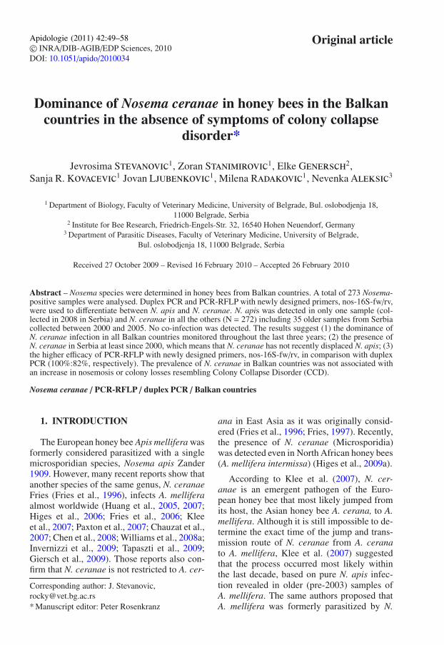
To differentiate between the species N. apis andN. ceranae, discriminating restriction endonucle-ase sites present in the PCR amplicon were used(Klee et al., 2007). The restriction endonucleasePac I provides one unique digestion site for N. cer-anae whilst the enzyme Nde I only digests N. apis.Msp I digests N. apis and N. ceranae and is usedas a control for successful restriction digestion ofPCR products. The predicted restriction fragmentsproduced from digestion of the PCR amplicons are97 bp, 118 bp, and 269 bp for N. ceranae and, 91 b,131 bp and 266 bp for N. apis (Fig. 1). PCR am-plicons were digested with Msp I/Pac I and withMsp I/Nde I (New England Biolabs, NEB) in tworeactions at 37 ◦C for 3 h to analyze and confirmthe presence of each Nosema species in each sam-ple. Digests were performed in 12.5 µL volumewith 10 µL of the amplified DNA and 1.5 Unitsof each enzyme. The 1× NEBuffer 2 (provided byNEB with Msp I) was used as buffer for the reac-tions. Msp I and Nde I show 100% activity with thisbuffer whilst Pac I performs to 75% in this buffer.Fragments were separated in a 3% NuSieve agarosegel (Cambrex Bio Science) in 1× TBE buffer with a20 bp ladder as size marker at 110 V for 1 h 30 min.Gels were stained with ethidium bromide and visu-alised under UV light.
Figure 1. Restriction fragment length polymor-phism (RFLP) of a 16S rRNA gene region con-served for N. apis and N. ceranae. A conservedregion of the 16S rRNA gene of N. apis and N. cer-anae was amplified by PCR. Subsequently, discrim-inating restriction endonuclease sites within this re-gion were used to differentiate between the twoNosema species. Digestion by Msp I/Nde I resultsin three bands (91 bp, 131 bp, 266 bp) for N.apis (lane 1). Digestion by Msp I/Pac I results inthree bands (97 bp, 118 bp, 269 bp) for N. ceranae(lane 2). Weak bands of 222 bp and 215 bp representincomplete digests by Nde I for N. apis and Pac I forN. ceranae, respectively. Mixed infections can alsobe identified using this method (lane 3).
3. RESULTS
By microscopic examination of collectedbee samples (N = 325) 273 were foundpositive for Nosema sp. (84.0%). In sam-ples from Serbia, 178 (including all 35 oldersamples collected between 2000 and 2005)out of 215 examined, were Nosema-positive
J. Stevanovic et al.52
Nosema ceranae
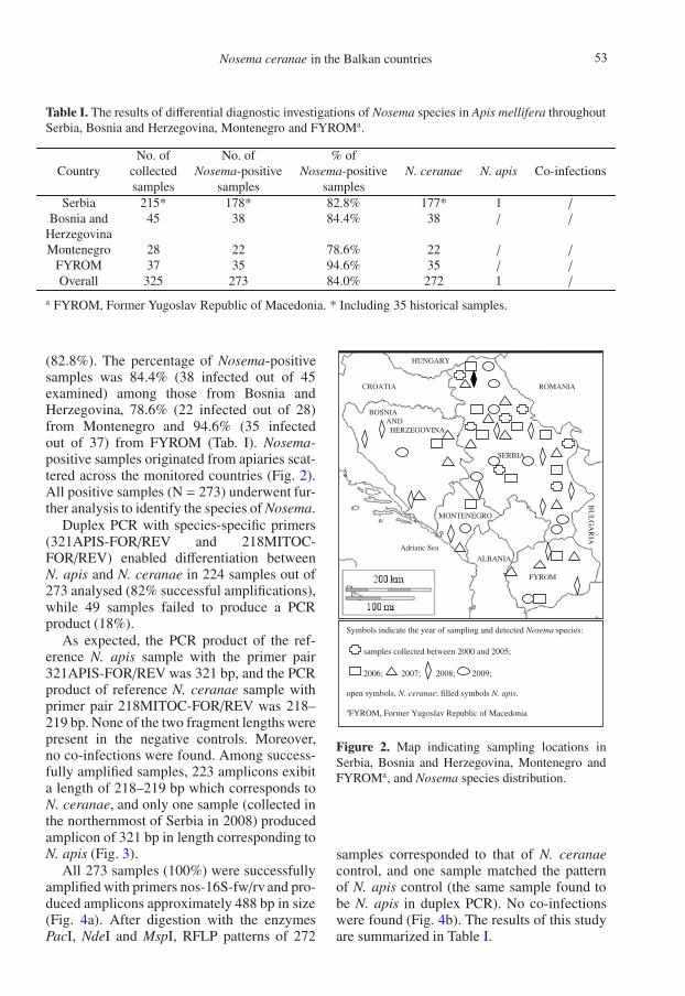
Table I. The results of differential diagnostic investigations of Nosema species in Apis mellifera throughoutSerbia, Bosnia and Herzegovina, Montenegro and FYROMa.
No. of No. of % ofCountry collected Nosema-positive Nosema-positive N. ceranae N. apis Co-infections
samples samples samplesSerbia 215* 178* 82.8% 177* 1 /
Bosnia and 45 38 84.4% 38 / /
HerzegovinaMontenegro 28 22 78.6% 22 / /
FYROM 37 35 94.6% 35 / /
Overall 325 273 84.0% 272 1 /
a FYROM, Former Yugoslav Republic of Macedonia. * Including 35 historical samples.
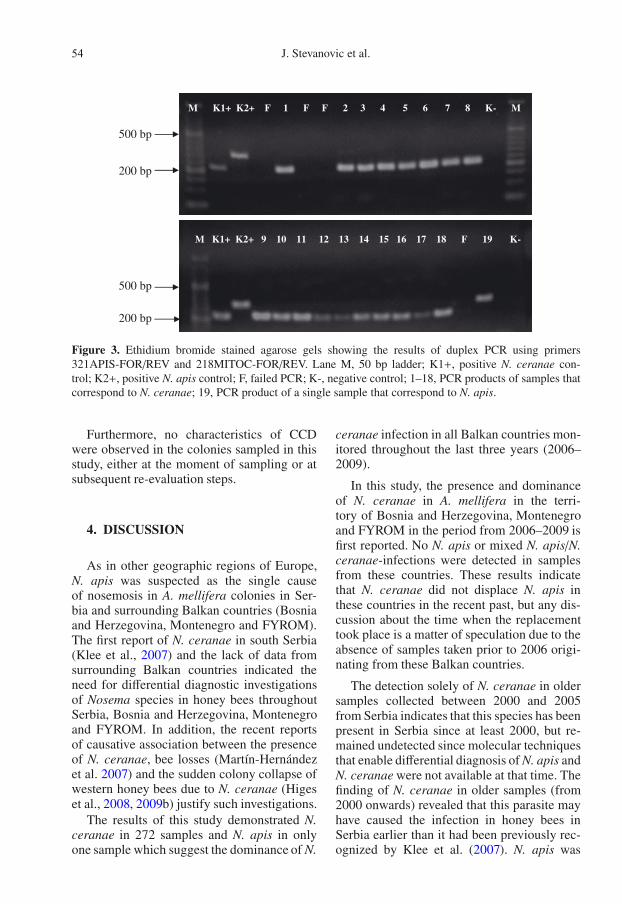
(82.8%). The percentage of Nosema-positivesamples was 84.4% (38 infected out of 45examined) among those from Bosnia andHerzegovina, 78.6% (22 infected out of 28)from Montenegro and 94.6% (35 infectedout of 37) from FYROM (Tab. I). Nosema-positive samples originated from apiaries scat-tered across the monitored countries (Fig. 2).All positive samples (N = 273) underwent fur-ther analysis to identify the species of Nosema.
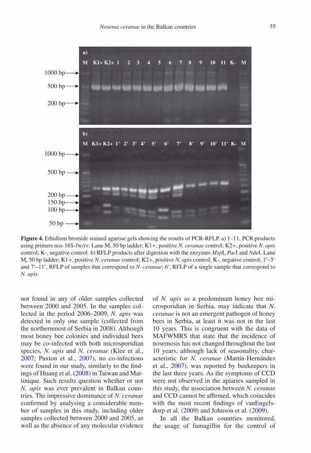
Duplex PCR with species-specific primers(321APIS-FOR/REV and 218MITOC-FOR/REV) enabled differentiation betweenN. apis and N. ceranae in 224 samples out of273 analysed (82% successful amplifications),while 49 samples failed to produce a PCRproduct (18%).
As expected, the PCR product of the ref-erence N. apis sample with the primer pair321APIS-FOR/REV was 321 bp, and the PCRproduct of reference N. ceranae sample withprimer pair 218MITOC-FOR/REV was 218–219 bp. None of the two fragment lengths werepresent in the negative controls. Moreover,no co-infections were found. Among success-fully amplified samples, 223 amplicons exibita length of 218–219 bp which corresponds toN. ceranae, and only one sample (collected inthe northernmost of Serbia in 2008) producedamplicon of 321 bp in length corresponding toN. apis (Fig. 3).
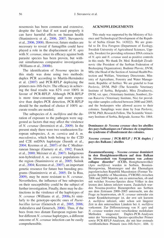
All 273 samples (100%) were successfullyamplified with primers nos-16S-fw/rv and pro-duced amplicons approximately 488 bp in size(Fig. 4a). After digestion with the enzymesPacI, NdeI and MspI, RFLP patterns of 272
SERBIA
BOSNIA AND HERZEGOVINA
HUNGARY
ROMANIACROATIA
MONTENEGRO
ALBANIA
BU
LG
AR
IA
Adriatic Sea
FYROM
Symbols indicate the year of sampling and detected Nosema species:
samples collected between 2000 and 2005;
2006; 2007; 2008; 2009;
open symbols, N. ceranae; filled symbols N. apis.
aFYROM, Former Yugoslav Republic of Macedonia
Figure 2. Map indicating sampling locations inSerbia, Bosnia and Herzegovina, Montenegro andFYROMa, and Nosema species distribution.
samples corresponded to that of N. ceranaecontrol, and one sample matched the patternof N. apis control (the same sample found tobe N. apis in duplex PCR). No co-infectionswere found (Fig. 4b). The results of this studyare summarized in Table I.
in the Balkan countries 53
500 bp
200 bp
500 bp
200 bp
M K1+ K2+ F 1 F F 2 3 4 5 6 7 8 K- M
M K1+ K2+ 9 10 11 12 13 14 15 16 17 18 F 19 K-
Figure 3. Ethidium bromide stained agarose gels showing the results of duplex PCR using primers321APIS-FOR/REV and 218MITOC-FOR/REV. Lane M, 50 bp ladder; K1+, positive N. ceranae con-trol; K2+, positive N. apis control; F, failed PCR; K-, negative control; 1–18, PCR products of samples thatcorrespond to N. ceranae; 19, PCR product of a single sample that correspond to N. apis.
Furthermore, no characteristics of CCDwere observed in the colonies sampled in thisstudy, either at the moment of sampling or atsubsequent re-evaluation steps.
4. DISCUSSION
As in other geographic regions of Europe,N. apis was suspected as the single causeof nosemosis in A. mellifera colonies in Ser-bia and surrounding Balkan countries (Bosniaand Herzegovina, Montenegro and FYROM).The first report of N. ceranae in south Serbia(Klee et al., 2007) and the lack of data fromsurrounding Balkan countries indicated theneed for differential diagnostic investigationsof Nosema species in honey bees throughoutSerbia, Bosnia and Herzegovina, Montenegroand FYROM. In addition, the recent reportsof causative association between the presenceof N. ceranae, bee losses (Martín-Hernándezet al. 2007) and the sudden colony collapse ofwestern honey bees due to N. ceranae (Higeset al., 2008, 2009b) justify such investigations.
The results of this study demonstrated N.ceranae in 272 samples and N. apis in onlyone sample which suggest the dominance of N.
ceranae infection in all Balkan countries mon-itored throughout the last three years (2006–2009).
In this study, the presence and dominanceof N. ceranae in A. mellifera in the terri-tory of Bosnia and Herzegovina, Montenegroand FYROM in the period from 2006–2009 isfirst reported. No N. apis or mixed N. apis/N.ceranae-infections were detected in samplesfrom these countries. These results indicatethat N. ceranae did not displace N. apis inthese countries in the recent past, but any dis-cussion about the time when the replacementtook place is a matter of speculation due to theabsence of samples taken prior to 2006 origi-nating from these Balkan countries.
The detection solely of N. ceranae in oldersamples collected between 2000 and 2005from Serbia indicates that this species has beenpresent in Serbia since at least 2000, but re-mained undetected since molecular techniquesthat enable differential diagnosis of N. apis andN. ceranae were not available at that time. Thefinding of N. ceranae in older samples (from2000 onwards) revealed that this parasite mayhave caused the infection in honey bees inSerbia earlier than it had been previously rec-ognized by Klee et al. (2007). N. apis was
J. Stevanovic et al.54
Nosema ceranae
1000 bp
500 bp
200 bp
1000 bp
500 bp
200 bp 150 bp 100 bp
50 bp
a)
M K1+ K2+ 1 2 3 4 5 6 7 8 9 10 11 K- M
b)
M K1+ K2+ 1’ 2’ 3’ 4’ 5’ 6’ 7’ 8’ 9’ 10’ 11’ K- M
Figure 4. Ethidium bromide stained agarose gels showing the results of PCR-RFLP. a) 1–11, PCR productsusing primers nos-16S-fw/rv; Lane M, 50 bp ladder; K1+, positive N. ceranae control; K2+, positive N. apiscontrol; K-, negative control. b) RFLP products after digestion with the enzymes MspI, PacI and NdeI. LaneM, 50 bp ladder; K1+, positive N. ceranae control; K2+, positive N. apis control; K-, negative control; 1′–5′
and 7′–11′, RFLP of samples that correspond to N. ceranae; 6′, RFLP of a single sample that correspond toN. apis.
not found in any of older samples collectedbetween 2000 and 2005. In the samples col-lected in the period 2006–2009, N. apis wasdetected in only one sample (collected fromthe northernmost of Serbia in 2008). Althoughmost honey bee colonies and individual beesmay be co-infected with both microsporidianspecies, N. apis and N. ceranae (Klee et al.,2007; Paxton et al., 2007), no co-infectionswere found in our study, similarly to the find-ings of Huang et al. (2008) in Taiwan and Mar-tinique. Such results question whether or notN. apis was ever prevalent in Balkan coun-tries. The impressive dominance of N. ceranaeconfirmed by analysing a considerable num-ber of samples in this study, including oldersamples collected between 2000 and 2005, aswell as the absence of any molecular evidence
of N. apis as a predominant honey bee mi-crosporidian in Serbia, may indicate that N.ceranae is not an emergent pathogen of honeybees in Serbia, at least it was not in the last10 years. This is congruent with the data ofMAFWMRS that state that the incidence ofnosemosis has not changed throughout the last10 years, although lack of seasonality, char-acteristic for N. ceranae (Martín-Hernándezet al., 2007), was reported by beekeepers inthe last three years. As the symptoms of CCDwere not observed in the apiaries sampled inthis study, the association between N. ceranaeand CCD cannot be affirmed, which coincideswith the most recent findings of vanEngels-dorp et al. (2009) and Johnson et al. (2009).
In all the Balkan countries monitored,the usage of fumagillin for the control of
in the Balkan countries 55
nosemosis has been common and extensive,despite the fact that if not used properly itcan have harmful effects on human health(Stanimirovic et al., 2006, 2007; Stevanovicet al., 2006, 2008). However, future studies arenecessary to reveal if fumagillin could haveplayed a role in the displacement of N. apiswith N. ceranae, since its efficacy against bothparasite species has been proven, but with-out simultaneous comparative investigations(Williams et al., 2008b).
The identification of Nosema species inthis study was done using two methods:duplex PCR according to Martín-Hernándezet al. (2007) and PCR-RFLP deploying theprimers nos-16S-fw/rv. The efficacy in achiev-ing the final results was 82% over 100% infavour of PCR-RFLP. Although PCR-RFLPis more time-consuming and more expen-sive than duplex-PCR detection, PCR-RFLPshould be the method of choice if 100% ac-curate results are needed.
The subspecies of A. mellifera and the du-ration of exposure to the pathogen were sug-gested as factors that may affect the virulenceof N. ceranae (Invernizzi et al., 2009). In thepresent study there were two southeastern Eu-ropean subspecies, A. m. carnica and A. m.macedonica, which both belong to the C2Dand C2E mtDNA haplotype (Susnik et al.,2004; Kozmus et al., 2007) of the C Mediter-ranean lineage (Garnery et al., 1992; Francket al., 2000; Meixner et al., 2007). Indigenousnon-hybridized A. m. carnica populations inthe region (Stanimirovic et al., 2005; Sušniket al., 2004; Kozmus et al., 2007), an importantgenetic resource for future bee breeding pro-grams (Stanimirovic et al., 2005; De la Rua,2009), may be more resistant to N. ceranae.Nevertheless, the influence of bee subspecieson their susceptibility could be the subject offurther investigation. Finally, there may be dis-tinctions in the virulence of the haplotypes ofN. ceranae (Williams et al., 2008a, b) simi-larly to the genotype-specific ones of Paeni-bacillus larvae (Genersch et al., 2005, 2006;Ashiralieva and Genersch, 2006). Thus, if thebees from two distant European regions har-bor different N. ceranae haplotypes, a differentoutcome of N. ceranae infection can be easilycomprehensible.
ACKNOWLEDGEMENTS
This study was supported by the Ministry of Sci-ence and Technological Development of the Repub-lic of Serbia (Grant No. 143022). We are grate-ful to Dr. Eva Forsgren (Department of Ecology,Swedish University of Agricultural Sciences, Upp-sala, Sweden) for providing reference DNA extractsof N. apis and N. ceranae used as positive controlsin this study. We thank Dr. Med. Rodoljub Zivadi-novic (the President of the Serbian Federation ofBeekeeping Organization), Budimir Plavsic, DVM,MSc (the Head of the Section of Animal Health Pro-tection and Welfare, Veterinary Directorate, Min-istry of Agriculture, Forestry and Water Manage-ment, Republic of Serbia). We are grateful to IvanPavlovic, DVM, PhD (The Scientific VeterinaryInstitute of Serbia, Belgrade), Mira Zivadinovic,DVM, vet. spec. (Veterinary Institute, Sombor), Zo-ran Raicevic (Veterinary Institute, Nis) for provid-ing older samples collected between 2000 and 2005,and the beekeepers who allowed access to theircolonies. Samplings were done by Ivan Pavlovic,DVM, PhD, Research Fellow, The Scientific Veteri-nary Institute of Serbia, Belgrade, license No. 1864.
Dominance de Nosema ceranae chez les abeillesdes pays balkaniques en l’absence de symptômesdu syndrome d’effondrement des colonies.
Nosema ceranae / PCR-RFLP / PCR duplex /pays des Balkans / abeilles
Zusammenfassung – Nosema ceranae dominiertin den Honigbienenvölkern auf dem Balkanin Abwesenheit von Symptomen von ,colonycollapse disorder’ (CCD). Honigbienenvölker(Apis mellifera) wurden in Serbien, Bosnien-Herzegowina, Montenegro und der Ehemaligenjugoslawischen Republik Mazedonien (Former Yu-goslav Republic of Macedonia, FYROM) zwischen2006 und 2009 beprobt, um zu untersuchen, ob undmit welcher Nosema-Spezies diese Völker in denletzten drei Jahren infiziert waren. Zusätzlich wur-den Nosema-positive Bienenproben aus Serbienaus dem Zeitraum 2000–2005 untersucht, um dieFrage zu beantworten, ob N. ceranae entweder einneuer Krankheitserreger ist, der erst seit kurzemA. mellifera infiziert, oder schon seit längererZeit in den untersuchten Ländern bei A. melliferavorkommt. Zur Differenzierung von N. apis undN. ceranae wurden zwei verschiedene molekulareMethoden eingesetzt: Duplex-PCR-Analysenunter der Verwendung Spezies-spezifischer Primersowie PCR-RFLP-Analysen, die mit hier erstmalsveröffentlichten Primern (nos-16S-fw/rv; Abb. 1)
J. Stevanovic et al.56
Nosema ceranae
durchgeführt wurden. Insgesamt wurden 273Nosema-positive Proben (N = 178, 38, 22 und35 jeweils aus Serbien, Bosnien-Herzegowina,Montenegro und Mazedonien) untersucht. In nureiner Probe wurde N. apis nachgewiesen, währendin allen anderen Proben (N = 272) einschließlichder älteren Proben aus Serbien, die aus demZeitraum 2000–2005 stammten, nur N. ceranaenachgewiesen werden konnte (Tab. I, Abb. 2–4).Mischinfektionen wurden nicht gefunden. DieseErgebnisse zeigen, (1) dass N. ceranae-Infektionenin den genannten Balkanstaaten in den letztendrei Jahren klar dominierten, und (2) dass N.ceranae in Serbien mindestens seit dem Jahr2000 vorkommt und hier nicht erst kürzlich N.apis in der Honigbienenpopulation verdrängt hat.Darüber hinaus zeigte der Methodenvergleich, dassdie PCR-RFLP-Analysen mit den neuen Primernnos-16S-fw/rv im Vergleich zur Duplex-PCRgenauere Ergebnisse liefern (100 % gegenüber82% korrekt positive Ergebnisse). Ein wichtigesErgebnis dieser Studie ist auch, dass das Vorkom-men von N. ceranae in den untersuchten Ländernnicht einherging mit einem Anstieg an Nosemosisoder mit Völkerverlusten, die von „colony collapsedisorder“-ähnlichen (CCD-ähnlichen) Symptomenbegleitet waren.
Nosema ceranae / PCR-RFLP / Duplex-PCR /Balkanstaaten
REFERENCES
Ashiralieva A., Genersch E. (2006) Reclassification,genotypes and virulence of Paenibacillus larvae,the etiological agent of American foulbrood inhoneybees – a review, Apidologie 37, 411–420.
Chauzat M.P., Higes M., Martín-Hernández R., MeanaA., Cougoule N., Faucon J.P. (2007) Presence ofNosema ceranae in French honeybee colonies, J.Apic. Res. 46, 127–128.
Chen Y., Evans J.D., Smith I.B., Pettis J.S. (2008)Nosema ceranae is a long-present and wide-spread microsporidean infection of the Europeanhoney bee (Apis mellifera) in the United States, J.Invertebr. Pathol. 97, 186–188.
Cox-Foster D.L., Conlan S., Holmes E.C., PalaciosG., Evans J.D., Moran N.A., Quan P.L., BrieseT., Hornig M., Geiser D.M., Martinson V.,vanEngelsdorp D., Kalkstein A.L., Drysdale A.,Hui J., Zhai J.H., Cui L.W., Hutchison S.K.,Simons J.F., Egholm M., Pettis J.S., Lipkin W.I.(2007) A metagenomic survey of microbes inhoney bee colony collapse disorder, Science 318,283–287.
De la Rúa P., Jaffé R., Dall’Olio R., Muñoz I., SerranoJ. (2009) Biodiversity, conservation and currentthreats to European honeybees, Apidologie 40,263–284.
Franck P., Garnery L., Solignac M., Cornuet J.M.(2000) Molecular confirmation of a fourth lineagein honey bees from the Near East, Apidologie 31,167–180.
Fries I. (1997) Protozoa, in: Morse R.A., Flottum K.(Eds.), Honey bee pests, predators and diseases,3rd ed., A.I. Root Company, Medina, Ohio, USA,pp. 59–76.
Fries I., Feng F., da Silva A., Slemenda S.B., PieniazekJ. (1996) Nosema ceranae n. sp. (Microspora,Nosematidae), morphological and molecular char-acterization of a microsporidian parasite of theAsian honey bee Apis cerana (Hymenoptera,Apidae), Eur. J. Protistol. 32, 356–365.
Fries I., Martín R., Meana A., García-Palencia P.,Higes M. (2006) Natural infections of Nosemaceranae in European honey bees, J. Apic. Res. 45,230–233.
Garnery L., Cornuet J.M., Solignac M. (1992)Evolutionary History of the Honey Bee Apis mel-lifera Inferred from Mitochondrial DNA Analysis,Mol. Ecol. 1, 145–154.
Gatehouse H.S., Malone L.A. (1998) The ribosomalRNA gene region of Nosema apis (Microspora):DNA sequence for small and large subunit rRNAgenes and evidence of a large tandem repeat unitsize, J. Invertebr. Pathol. 71, 97–105.
Genersch E., Ashiralieva A., Fries I. (2005) Strainandgenotype-specific differences in virulence ofPaenibacillus larvae subsp. larvae, a bacterialpathogen causing American foulbrood disease inhoneybees, Appl. Environ. Microbiol. 71, 7551–7555.
Genersch E., Forsgren E., Pentikäinen J., AshiralievaA., Rauch S., Kilwinski J., Fries I. (2006)Reclassification of Paenibacillus larvae subsp.pulvifaciens and Paenibacillus larvae subsp. lar-vae as Paenibacillus larvae without subspeciesclassification, Int. J. Syst. Evol. Microbiol. 56,501–511.
Giersch T., Berg T., Galea F., Hornitzky M. (2009)Nosema ceranae infects honey bees (Apis mel-lifera) and contaminates honey in Australia,Apidologie 40, 117–123.
Higes M., García-Palencia P., Martín-Hernández R.,Meana A. (2007) Experimental infection ofApis mellifera honeybees with Nosema ceranae(Microsporidia), J. Invertebr. Pathol. 94, 211–217.
Higes M., Martín R., Meana A. (2006) Nosema cer-anae, a new microsporidian parasite in honeybeesin Europe, J. Invertebr. Pathol. 92, 93–95.
Higes M., Martín R., Sanz A., Álvarez N., Sanz A.,García-Palencia P., Meana A. (2005) El síndromede despoblamiento de las colmenas en España.Consideraciones sobre su origen, Vida Apícola133, 15–21.
Higes M., Martín-Hernández R., Botías C., Garrido-Bailón E., González-Porto A.V., Barrios L.,Del Nozal M.J., Bernal J.L., Jiménez J.J., García-Palencia P., Meana A. (2008) How natural
in the Balkan countries 57
infection by Nosema ceranae causes honeybeecolony collapse, Environ. Microbiol. 10, 2659–2669.
Higes M., Martín-Hernández R., Garrido-Bailón E.,Botías C., Meana A. (2009a) The presence ofNosema ceranae (Microsporidia) in North Africanhoney bees (Apis mellifera intermissa), J. Apic.Res. 48, 217–219.
Higes M., Martín-Hernández R., Garrido-Bailón E.,González-Porto A.V., García-Palencia P., MeanaA., del Nozal M.J., Mayo R., Bernal J.L. (2009b)Honeybee colony collapse due to Nosema ceranaein professional apiaries, Environ. Microbiol. Rep.1, 110–113.
Huang W.F., Jiang J.H., Chen Y.W., Wang C.H. (2005)Complete rRNA sequence of the Nosema ceranaefrom honeybee (Apis mellifera), https://gra103.aca.ntu.edu.tw/gdoc/F90632004a.pdf.
Huang W.F., Bocquet M., Lee KC, Sung I.H., JiangJ.H., Chen Y.W., Wang C.H. (2008) The com-parison of rDNA spacer regions of Nosema cer-anae isolates from different hosts and locations, J.Invertebr. Pathol. 97, 9–13.
Huang W.F., Jiang J.H., Chen Y.W., Wang C.H. (2007)A Nosema ceranae isolate from the honeybee Apismellifera, Apidologie 38, 30–37.
Imdorf A., Charrière J.D., Gallmann P. (2006)Mögliche Ursachen für die Völkerverluste der let-zten Jahre, Schweiz. Bienenztg. 129, 6–10.
Invernizzi C., Abud C., Tomasco I.H., Harriet J.,Ramallo G., Campá J., Katz H., Gardiol G.,Mendoza Y. (2009) Presence of Nosema cer-anae in honeybees (Apis mellifera) in Uruguay, J.Invertebr. Pathol. 101, 150–153.
Johnson R.M., Evans J.D., Robinson G.E., BerenbaumM.R. (2009) Changes in transcript abundance re-lating to colony collapse disorder in honey bees(Apis mellifera), PNAS 106, 14790–14795.
Klee J., Besana A.M., Genersch E., Gisder S., NanettiA., Tam D.Q., Chinh T.X., Puerta F., Ruz J.M.,Kryger P., Message D., Hatjina F., Korpela S.,Fries I., Paxton R.J. (2007) Widespread dispersalof the microsporidian Nosema ceranae, an emer-gent pathogen of the western honey bee, Apis mel-lifera, J. Invertebr. Pathol. 96, 1–10.
Kozmus P., Stevanovic J., Stanimirovic Z., Stojic V.,Kulisic Z., Meglic V. (2007) Analysis of mito-chondrial DNA in honey bees (Apis mellifera)from Serbia, Acta Vet.-Beograd 57, 465–476.
Martín-Hernández R., Meana A., Prieto L., MartínezSalvador A., Garrido-Bailón E., Higes M. (2007)Outcome of colonization of Apis mellifera byNosema ceranae, Appl. Environ. Microbiol. 73,6331–6338.
Meixner M.D., Worobik M., Wilde J., Fuchs S.,Koeniger N. (2007) Apis mellifera melliferain eastern Europe – morphometric variation and
determination of its range limits, Apidologie 38,191–197.
OIE (2008) Manual of Diagnostic Tests and Vaccinesfor Terrestrial Animals, Chap. 2.2.4., Nosemosisof Honey Bees, http://www.oie.int/fr/normes/mmanual/2008/pdf/2.02.04_NOSEMOSIS.pdf.
Paxton R.J., Klee J., Korpela S., Fries I. (2007)Nosema ceranae has infected Apis mellifera inEurope since at least 1998 and may be more viru-lent than Nosema apis, Apidologie 38, 558–565.
Stanimirovic Z., Stevanovic J., Andjelkovic M. (2005)Chromosomal diversity in Apis mellifera carnicafrom Serbia, Apidologie 36, 31–42.
Stanimirovic Z., Stevanovic J., Bajic V., Radovic I.(2007) Evaluation of genotoxic effects of fumag-illin (dicyclohexylamine) by citogenetic tests invivo, Mutat. Res. 628, 1–10.
Stanimirovic Z., Stevanovic J., Kulic M., Stojic V.(2006) Frequency of chromosomal aberrations inthe evaluation of genotoxic potential of dicy-clohexylamine (fumagillin) in vivo, Acta Vet.-Beograd 56, 353–366.
Stevanovic J., Stanimirovic Z., Pejin I.I., Lazarevic M.(2006) Monitoring of mitotic index and frequencyof micronuclei in evaluation of genotoxic potentialof fumagillin (dicyclohexylamine) in vivo, ActaVet.-Beograd 56, 437–448.
Stevanovic J., Stanimirovic Z., Radakovic M., Stojic V.(2008) In vitro evaluation of the clastogenicity offumagillin, Environ. Mol. Mutagen. 49, 594–601.
Sušnik S., Kozmus P., Poklukar J., Megliè V. (2004)Molecular characterisation of indigenous Apismellifera carnica in Slovenia, Apidologie 35, 623–636.
Tapaszti Z., Forgách P., Kovágó C., Békési L., BakonyiT., Rusvai M. (2009) First detection and domi-nance of Nosema ceranae in Hungarian honeybeecolonies, Acta Vet. Hung. 57, 383–388.
vanEngelsdorp D., Evans J.D., Saegerman C., MullinC., Haubruge E., Nguyen B.K., Frazier M., FrazierJ., Cox-Foster D., Chen Y., Underwood R., TarpyD.R., Pettis J.S. (2009) Colony collapse disorder:a descriptive study, PLoS One 4, e6481.
Williams G.R., Shafer A.B.A., Rogers R.E.L., ShutlerD., Stewart D.T. (2008a) First detection ofNosema ceranae, a microsporidean parasite ofEuropean honey bees (Apis mellifera), in Canadaand central USA, J. Invertebr. Pathol. 97, 189–192.
Williams G.R., Sampson MA, Shutler D., RogersR.E.L. (2008b) Does fumagillin control the re-cently detected invasive parasite Nosema cer-anae in western honey bees (Apis mellifera)? J.Invertebr. Pathol. 99, 342–344.
Zander E. (1909) Tierische Parasiten alsKrankheitserreger bei der Biene, MünchenerBienenzeitung 31, 196–204.
8 J. Stevanovic et al.5


