Review ArticlePropeller Flaps: A Review of Indications, Technique, and Results
Salvatore D’Arpa, Francesca Toia,Roberto Pirrello, Francesco Moschella, and Adriana Cordova
Chirurgia Plastica e Ricostruttiva, Dipartimento di Discipline Chirurgiche, Oncologiche e Stomatologiche,Universita degli Studi di Palermo, Via del Vespro, 129 90127 Palermo, Italy
Correspondence should be addressed to Salvatore D’Arpa; [email protected]
Received 26 February 2014; Accepted 5 May 2014; Published 26 May 2014
Academic Editor: Adrian Dragu
Copyright © 2014 Salvatore D’Arpa et al. This is an open access article distributed under the Creative Commons AttributionLicense, which permits unrestricted use, distribution, and reproduction in any medium, provided the original work is properlycited.
In the last years, propeller flaps have become an appealing option for coverage of a large range of defects. Besides having a morereliable vascular pedicle than traditional flap, propeller flaps allow for great freedom in design and for widemobilization that extendthe possibility of reconstructing difficult wounds with local tissues and minimal donor-site morbidity. They also allow one-stagereconstruction of defects that usually require multiple procedures. Harvesting of a propeller flap requires accurate patient selection,preoperative planning, and dissection technique. Complication rate can be kept low, provided that potential problems are prevented,promptly recognized, and adequately treated. This paper reviews current knowledge on propeller flaps. Definition, classification,and indications in the different body regions are discussed based on a review of the literature and on the authors’ experience. Detailsabout surgical technique are provided, together with tips to avoid and manage complications.
1. Introduction
The term “propeller flap” was first used in 1991 by Hyakusokuet al. [1] to describe an adipocutaneous flap based on a centralsubcutaneous pedicle, with a shape resembling a propellerthat was rotated 90 degrees.
In 2006, combining the concept of propeller flaps andperforator based flaps,Hallock [2] reported a fasciocutaneousflap that was similar in shape to the one described byHyakusoku butwas based on a skeletonized perforating vesseland was rotated 180 degrees on an eccentric pivot point. Teo[3] gave the greatest contribution to the surgical techniqueand the application of the perforator propeller flap.
In the last years, the introduction of the propeller flapsgained great popularity; these flaps have been increasinglyused for reconstruction of soft tissue defects of different partsof the body, and surgical technique has been refined andwell described by several authors [4–9]. Perforator propellerflaps have a reliable vascular pedicle and can undergo widemobilization and rotation; their harvest is fast and easy anddoes not require microsurgery; however, accurate patientselection, preoperative planning, and dissection techniqueare mandatory to prevent complications.
In this paper, recommendations for judicious planning ofperforator propeller flaps in different body areas are provided;technical refinements and tips on how to avoid commonmistakes are discussed.
2. Materials and Methods
Pertinent literature was collected: a pubmed search wasperformed using the keywords propeller flap, perforator flap,and freestyle flap. Forty papers were eventually includedin this review. Definition, classification, and indications ofpropeller flaps in the different body regions are discussed;details about surgical technique are provided, together withtips to avoid and manage complications, based on selectedrelevant articles and on the authors’ experience.
3. Results and Discussion
Propeller flaps can either be based on a known source vesselor can be harvested from any anatomical region as long as aDoppler signal of a perforator is detected (free-style propellerflaps).
Hindawi Publishing CorporationBioMed Research InternationalVolume 2014, Article ID 986829, 7 pageshttp://dx.doi.org/10.1155/2014/986829
2 BioMed Research International
The advantages of perforator propeller flaps can besummarized as follows [3, 4, 6, 8].
(i) They allow for a great freedom in design and choiceof the donor site, based on the quality and volume ofsoft tissue required and on scar orientation.
(ii) They represent a simpler and faster alternative to freeflaps and expand the possibilities of reconstructingdifficult wounds with local tissues.
(iii) Their harvest is easy and fast, provided that appropri-ate dissection technique is applied.
(iv) Donor site morbidity is kept very low, avoiding thesacrifice of any unnecessary tissue [11].
Thanks to these characteristics and to the technicalrefinements achieved in the last years, perforator propellerflaps find increasing indications in the reconstruction ofdifferent parts of the body.
3.1. Classification. A clear definition of propeller flap wasgiven in 2009 by the advisory panel of the first Tokyomeetingon perforator and propeller flaps [12], who defined it as an“island flap that reaches the recipient site through an axialrotation.” The difference between a propeller flap and otherpedicled flaps is that the rotation in the case of a propellerflap is “axial”: this means that the flaps turn around a pivotthat is made of the pedicle and this is similar to a propeller.
According to the Gent consensus on perforator flaps [13]and to the advisory panel of the first Tokyo meeting onperforator and propeller flaps [12], perforator propeller flapsshould be named after their nutrient vessels. They can beclassified according to the type of nourishing pedicle.
(i) Subcutaneous pedicled propeller flap is based on arandom subcutaneous pedicle and allows for rota-tions up to 90∘.
(ii) Perforator pedicled propeller flap is based on askeletonized perforator pedicle. This is the mostcommonly used type of propeller flap and can berotated up to 180∘.
(iii) Supercharged propeller flap is modification of theperforator pedicled propeller flap, in which a super-ficial or perforating vein of the flap is anastomosed toa recipient vein or an extra artery is anastomosed to asecond arterial pedicle of the flap, to increase venousoutflow or arterial inflow.
Recently, we described a novel type of propeller: the“axial propeller flap” [14] that includes propeller flaps basedon known vessels (e.g., suprathrochlear artery and lingualartery) [14–16] and not on a perforator.
3.2. Surgical Technique
3.2.1. Preoperative Planning. A hand held Doppler probe isalways sufficient for preoperative vascular assessment. Thewhole region should be investigated to have a clear preoper-ative picture of the location of all perforators or of the axial
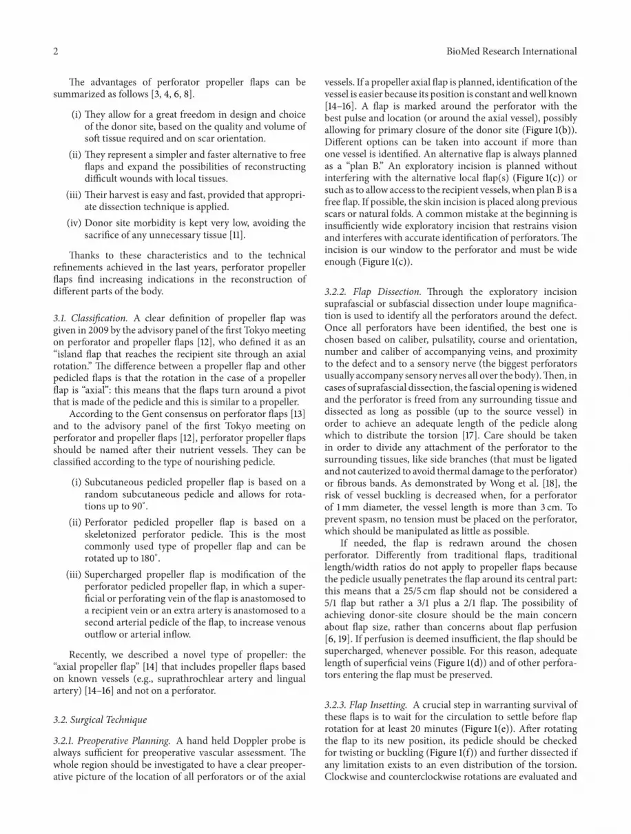
vessels. If a propeller axial flap is planned, identification of thevessel is easier because its position is constant andwell known[14–16]. A flap is marked around the perforator with thebest pulse and location (or around the axial vessel), possiblyallowing for primary closure of the donor site (Figure 1(b)).Different options can be taken into account if more thanone vessel is identified. An alternative flap is always plannedas a “plan B.” An exploratory incision is planned withoutinterfering with the alternative local flap(s) (Figure 1(c)) orsuch as to allow access to the recipient vessels, when planB is afree flap. If possible, the skin incision is placed along previousscars or natural folds. A common mistake at the beginning isinsufficiently wide exploratory incision that restrains visionand interferes with accurate identification of perforators.Theincision is our window to the perforator and must be wideenough (Figure 1(c)).
3.2.2. Flap Dissection. Through the exploratory incisionsuprafascial or subfascial dissection under loupe magnifica-tion is used to identify all the perforators around the defect.Once all perforators have been identified, the best one ischosen based on caliber, pulsatility, course and orientation,number and caliber of accompanying veins, and proximityto the defect and to a sensory nerve (the biggest perforatorsusually accompany sensory nerves all over the body).Then, incases of suprafascial dissection, the fascial opening iswidenedand the perforator is freed from any surrounding tissue anddissected as long as possible (up to the source vessel) inorder to achieve an adequate length of the pedicle alongwhich to distribute the torsion [17]. Care should be takenin order to divide any attachment of the perforator to thesurrounding tissues, like side branches (that must be ligatedandnot cauterized to avoid thermal damage to the perforator)or fibrous bands. As demonstrated by Wong et al. [18], therisk of vessel buckling is decreased when, for a perforatorof 1mm diameter, the vessel length is more than 3 cm. Toprevent spasm, no tension must be placed on the perforator,which should be manipulated as little as possible.
If needed, the flap is redrawn around the chosenperforator. Differently from traditional flaps, traditionallength/width ratios do not apply to propeller flaps becausethe pedicle usually penetrates the flap around its central part:this means that a 25/5 cm flap should not be considered a5/1 flap but rather a 3/1 plus a 2/1 flap. The possibility ofachieving donor-site closure should be the main concernabout flap size, rather than concerns about flap perfusion[6, 19]. If perfusion is deemed insufficient, the flap should besupercharged, whenever possible. For this reason, adequatelength of superficial veins (Figure 1(d)) and of other perfora-tors entering the flap must be preserved.
3.2.3. Flap Insetting. A crucial step in warranting survival ofthese flaps is to wait for the circulation to settle before flaprotation for at least 20 minutes (Figure 1(e)). After rotatingthe flap to its new position, its pedicle should be checkedfor twisting or buckling (Figure 1(f)) and further dissected ifany limitation exists to an even distribution of the torsion.Clockwise and counterclockwise rotations are evaluated and

BioMed Research International 3
(a) (b)
(c) (d)
(e) (f)
(g) (h)
Figure 1: (a) Left Achilles tendon exposure after open repair. (b)The flap has been drawn around the perforator with the best sound. A wideexploratory incision is performed to visualize it. (c) Optimal perforator visualization, such as that shown in the picture, must be possiblethrough the exploratory incision. When exposure is inadequate, the incision should be lengthened. In this case the proximal perforator wasdirected to the skin and the distal to the soleusmuscle; plan B option for this case. (d)Theflap has been islanded and left on the perforator aloneto let the circulation settle. A superficial vein (greater saphenous vein in this case) should be preserved for venous supercharging wheneverpossible. A strip of soleus tendon is harvested for Achilles tendon reconstruction as described by Cavadas and Landin [10]. (e) The mostconvenient sense of rotation is chosen. While the flap’s circulation settles before rotation, donor site closure can be accomplished. (f) Afterrotation, the pedicle is always double checked for torsions, traction, or kinking that must be, if present, immediately eliminated. (g) Closuremust be obtained without any tension. Note that the flap is a little longer than required to compensate postoperative swelling. (h) Six monthspostoperative result shows complete flap survival.

4 BioMed Research International
the best one in terms of vessel rotation is chosen. The senseof rotation should be documented should the flap need to bereexplored.The flap is then secured in position and observedfor color, capillary refilling, and bleeding (Figure 1(g)). Ifinsufficient arterial inflow is observed due to arterial spasmcaused by surgical manipulation, the flap should be broughtback to its original position until spasm resolution (usuallyabout 20 minutes) and its pedicle rinsed with lidocaine orpapaverine. If the spasm persists after derotation, the flapshould not be transferred anyway but rather left in place anddelayed a few days before wound coverage [6].
3.2.4. Postoperative Care. Limbs should be kept in a splint forthe first postoperative days; compression on the flap shouldbe avoided and elevation should be maintained for head andlimbs flaps.
Flaps are checked every second hour during the firstpostoperative days, to allow for prompt identification ofeventual complications.
3.3. Complications: Prevention and Management
3.3.1. Arterial Insufficiency. This complication is extremelyrare: accurate planning of the flap and choice of the perforatorhelp preventing it.When, due to persistence of arterial spasm,the flap remains pale due to insufficient arterial inflow, theflap can be derotated to its original position for a few daysbefore rotating it [4, 6].
3.3.2. Venous Insufficiency. Venous congestion is the mostfrequent complication of propeller flaps, because veins aremore prone to torsion than arteries. Venous insufficiencyshould be distinguished from the temporary congestionthat often characterizes perforator flaps and fades out withstabilization of flow. True venous insufficiency worsens withtime and should be promptly recognized and treated. Whenit is limited to an apical part of the flap, its evolution isobserved. A small number of cases evolve in necrosis, whichis usually superficial, so that deep vital tissue is still present atthe recipient site.
Cases of mild venous congestions in thin flaps can beaddressed with leech therapy.
When venous congestion is significant and worsens overtime, reexploration and venous supercharging are the bestoption, in case a superficial or perforating vein of the flap wasprepared during dissection. Should venous supercharging notbe feasible, an alternative option is to temporarily derotate theflap (a few days) to relieve torsion on the pedicle [4, 6, 12] andlet the circulation settle.
3.3.3. Partial Necrosis. Total flap loss is rare. Partial necrosisseems to occur in about 5% of cases [6] and is often limited tothe skin.After eschar removal, an adequate bed for a skin graftis often present. Healing by secondary intention is anotheralternative for small wounds.
3.4. Propeller Flaps in the Different Body Regions
3.4.1. Head and Neck. The head and neck region is char-acterized by a very rich vascularization, and several localflaps are available for reconstruction. However, propellerperforator flaps allow turning a two-stage operation to a one-stage operation, thus simplifying reconstructions that usuallyrequire two or more procedures, accelerating recovery, andminimizing discomfort for the patient. Their freedom indesign also allows for a better concealing of the scars.
Free-style flaps can be based on perforators of the facialartery and have been successfully used as propeller flaps fornasal ala reconstruction in a single stage [5–9, 11, 13, 20, 21].
One-stage nasal reconstruction with a perforator pro-peller flap from the forehead, named the suprathrochlearartery propeller perforator flap, has been reported as well [14,15]. Other cutaneous, as well as mucosal intraoral, traditionalflaps such as the lingual flaps [16] can be modified into apropeller flap based on their vascular pedicle for increasedreconstructive possibilities.
The head and neck is the ideal donor site to start withwhen approaching propeller flap surgery for the followingreasons:
(i) flaps in this area are more forgiving and have higherchances of survival compared, for example, to thelimbs due to the rich vascular network of the head andneck;
(ii) the head and neck vessels seem to better toleratetorsion and to suffer less from 180∘ rotations even ifbased on short pedicles;
(iii) perforator vessels are usually very small and thusrequire that good skills are developed for their dissec-tion.
3.4.2. Lower Limb. In lower limb reconstruction, defects ofthe lower third of the leg are a challenging problem, due tothe paucity of local tissues available for reconstruction [4].Propeller flaps allow bringing proximal skin distally to coveraverage size defects that would otherwise require a free flap.
Free flaps are still the gold standard for large defects, butpropeller perforator flaps are an appealing option for smallandmedium defects. Bajantri et al. [22] recommend their usefor defects up to 50 cm2 in size; however, we believe that weare very far from establishing a maximum flap size, whichdepends on the patient’s body and leg size, skin laxity, flapvolume, perforator enrolled, adequate donor site closure andmany other factors.
Based on the vascular supply of the lower limb, very longlongitudinal flaps can be raised compared to other anatomicalregions [6, 8]. Tibial posterior perforators seem to have anadvantage over anterior tibial and peroneal artery perforatorsbecause they usually have a larger caliber and better veins [6].
When a deep defect has to be filled, a cuff ofmuscle can betransferred with the distal end of the perforator. The desiredmuscle is harvested around a perforator located in the tip ofthe flap, which is divided on a plane deeper to the muscle.The muscle cuff will be supplied by a reverse flow from theskin island through the divided perforator [4, 12]. This is

BioMed Research International 5
(a) (b)
(c) (d)
Figure 2: (a) Squamous cell carcinoma of the dorsal aspect of the little finger. Excision and reconstruction with a perforator propeller flapwas planned. (b) Dissection view of the flap. (c) Immediate postoperative result. (d) Final result.
an example of the degree of customization provided by theseflaps.While we used to harvestmusculocutaneous flaps in thepast, to warrant vascular supply to the skin, nowwemoved tocutaneous-muscular flaps, where only the portion of muscleneeded for the reconstruction (and not as a vehicle for thevessels) is taken with the flap.This optimizes outcomes at therecipient site and minimizes morbidity.
The donor defect can be closed primarily only in narrowlongitudinal defects. Skin grafting of the donor site is oftenrequired. If a skin grafted donor site in the leg is consideredunappealing, a free flap would avoid further scars in the leg[6]; when reconstructionwith local flaps is planned, however,propeller flaps are often among the few available optionsand provide better aesthetic results compared to graftedadipofascial flaps.
Even when a free flap is needed, pedicled propeller flapscan be a valuable help for reconstruction. Cavadas and Teran-Saavedra [23] described a “razor flap,” inwhich a combinationof the TDAP and the LD (Latissimus dorsi) flaps on thesame thoracodorsal vessels permits a free rotation of theskin island and a great freedom of positioning. The flapwas used for simultaneous release of popliteal retractionand circumferential resurfacing of the leg without increasingdonor-site morbidity.
3.4.3. Upper Limb. Clinical experiences with propeller flapsin upper limb are less numerous than in lower limbs, but withsimilar advantages. They allow reconstructing like-with-like,using a local and simple option, with the additional benefit ofalmost always allowing direct closure of the donor site. Theyhave been used for reconstruction of the elbow and forearm
region, and recently their use has been proposed for fingerand hand defects [6, 9, 24–26] (Figure 2).
As for lower limb, propeller flaps also permit recon-structing defects of different tissue with a single flap. As anexample, Battiston et al. [27] reported a composite teno-fasciocutaneous flap based on a perforator branch of thesecond dorsal intermetacarpal artery, for reconstruction of acomplex defect of the dorsal aspect of the index finger.
A higher rate of venous insufficiency has been reportedin the forearm. This is probably due to predominance ofthe superficial venous circulation, with subsequent venousengorgement of a flap based on the perforating veins alone.We advocate routine venous supercharging in the forearm towarrant sufficient venous drainage [6].
3.4.4. Trunk. Although the potential of propeller flaps hasbeen better documented in extremity surgery, their indica-tions in reconstruction of trunk defects are steadily increas-ing.
Ang et al. [28] andWoo et al. [29] reported the use of pro-peller DIEP (deep inferior epigastric perforator) flaps rotated180∘ for coverage of large abdominal defects, respectively,following resection of colorectal cancer cutaneous metastasisand fibrodermatosarcoma protuberans. A preexpanded pro-peller DIEP flap has been reported by Cheng and Saint-Cyr[30] for reconstruction of an abdominal burn scar.
Other well-known perforator flaps are routinely used aspedicled propeller flaps for trunk reconstruction. Their longpedicle allows for a great range of freedom, allowing for anextensive arc of rotation, and makes these flaps particularlysuitable for being rotated with little concerns on vessels’torsion and blood supply.

6 BioMed Research International
Several authors reported reconstruction of complexabdominal or pelvic defects with pedicled propeller ALT(anterolateral thigh) flaps [23, 31–33].
ALT, SGAP (superior gluteal artery perforator) flap,IGAP (inferior gluteal artery perforator) flap, and TDAP(toracodorsal artery perforator) flap have been described aspedicled propeller flaps for reconstruction of difficult woundsof axillary, gluteal, or inguinal regions following resection ofhidradenitis suppurativa [6, 34–37].
ICAP (intercostal artery perforator) flaps, LTAP (lateralthoracic artery perforator) flaps, and TDAP flaps are a valu-able option for partial breast reconstruction [7, 38–40]; LTAP,ICAP, and IMAP flaps find indication in reconstruction ofother complex thoracic defects [38, 41, 42].
4. Conclusions
Perforator propeller flaps are a valid reconstructive option fordifficult wounds and can be raised from any part of the body.Their harvesting is easy and fast, provided that an accuratedissection technique is applied, and allows for great freedomin design and choice of the donor site.
Propeller perforator flaps represent an alternative to freeflaps when traditional flaps are not an option, allow toreconstruct even complex wounds with local tissues and alowdonor-sitemorbidity, and present several advantages overtraditional pedicled flaps: their freedom in design allows toreconstruct complex defects usually requiring multiple pro-cedures in a single stage, accelerating recovery, minimizingmorbidity and discomfort for the patient, and allowing abetter aesthetic result and concealing of scars.
Conflict of Interests
The authors declare that there is no conflict of interestsregarding the publication of this paper.
References
[1] H. Hyakusoku, T. Yamamoto, and M. Fumiiri, “The propellerflap method,” British Journal of Plastic Surgery, vol. 44, no. 1, pp.53–54, 1991.
[2] G. G. Hallock, “The propeller flap version of the adductormuscle perforator flap for coverage of ischial or trochantericpressure sores,”Annals of Plastic Surgery, vol. 56, no. 5, pp. 540–542, 2006.
[3] T. C. Teo, “Perforator local flaps in lower limb reconstruction,”Cirugia Plastica Ibero-Latinoamericana, vol. 32, no. 4, pp. 15–292, 2006.
[4] M. Pignatti, S. D’Arpa, and T. C. S. Cubison, “Novel fascio-cutaneous flaps for the reconstruction of complicated lowerextremity wounds,” Techniques in Orthopaedics, vol. 24, no. 2,pp. 88–95, 2009.
[5] S. D’Arpa, A. Cordova, R. Pirrello, and F. Moschella, “Freestyle facial artery perforator flap for one stage reconstructionof the nasal ala,” Journal of Plastic, Reconstructive and AestheticSurgery, vol. 62, no. 1, pp. 36–42, 2009.
[6] S. D’Arpa, A. Cordova, M. Pignatti, and F. Moschella, “Freestylepedicled perforator flaps: safety, prevention of complications,
and management based on 85 consecutive cases,” Plastic andReconstructive Surgery, vol. 128, no. 4, pp. 892–906, 2011.
[7] M. Hamdi, K. van Landuyt, S. Monstrey, and P. Blondeel,“Pedicled perforator flaps in breast reconstruction: a newconcept,” British Journal of Plastic Surgery, vol. 57, no. 6, pp. 531–539, 2004.
[8] C. Lecours, M. Saint-Cyr, C. Wong et al., “Freestyle pedicleperforator flaps: clinical results and vascular anatomy,” Plasticand Reconstructive Surgery, vol. 126, no. 5, pp. 1589–1603, 2010.
[9] M. A. Mateev and H. O. M. Kuokkanen, “Reconstruction ofsoft tissue defects in the extremities with a pedicled perforatorflap: series of 25 patients,” Journal of Plastic Surgery and HandSurgery, vol. 46, no. 1, pp. 32–36, 2012.
[10] P. C. Cavadas and L. Landin, “Reconstruction of chronic achillestendon defects with posterior tibial perforator flap and soleustendon graft: clinical series,” Plastic and Reconstructive Surgery,vol. 117, no. 1, pp. 266–271, 2006.
[11] F. G. Bravo and H. P. Schwarze, “Free-style local perforatorflaps: concept and classification system,” Journal of Plastic,Reconstructive and Aesthetic Surgery, vol. 62, no. 5, pp. 602–608,2009.
[12] M. Pignatti, R. Ogawa, G. G. Hallock et al., “The “Tokyo” con-sensus on propeller flaps,” Plastic and Reconstructive Surgery,vol. 127, no. 2, pp. 716–722, 2011.
[13] P.N. Blondeel, K.H. I. van Landuyt, S. J.M.Monstrey et al., “The“Gent” consensus on perforator flap terminology: preliminarydefinitions,” Plastic and Reconstructive Surgery, vol. 112, no. 5,pp. 1378–1382, 2003.
[14] A. Cordova, S. D’Arpa, M. Tripoli, F. Toia, and F. Moschella,“A propeller flap for single stage nose reconstruction: STAAPflap (Supra Trochlear artery axial propeller flap),” Facial PlasticSurgery, vol. 30, pp. 332–341, 2014.
[15] A. Cordova, S. D’Arpa, and F. Moschella, “A new one-stagemethod for nose reconstruction: the supratrochlear arteryperforator propeller flap,” Plastic and Reconstructive Surgery,vol. 129, no. 3, pp. 571e–573e, 2012.
[16] A. Cordova, “Apporto innovativo dei perforanti nella chirurgiadel distretto testa-collo,” in 62mo Congresso SICPRE, p. 28, Bari,Italy, 2012.
[17] G. Selvaggi, S. Anicic, and L. Formaggia, “Mathematical expla-nation of the buckling of the vessels after twisting of themicroanastomosis,” Microsurgery, vol. 26, no. 7, pp. 524–528,2006.
[18] C.-H. Wong, F. Cui, B.-K. Tan et al., “Nonlinear finite elementsimulations to elucidate the determinants of perforator patencyin propeller flaps,” Annals of Plastic Surgery, vol. 59, no. 6, pp.672–678, 2007.
[19] S. D’Arpa, M. Pignatti, A. Cordova, and F. Moschella, “Reply:how large can a pedicled perforator flap be?” Plastic & Recon-structive Surgery, vol. 130, no. 1, pp. 196e–198e, 2012.
[20] S. D’Arpa, A. Cordova, R. Pirrello, and F. Moschella, “One-stage reconstruction of the nasal ala: the free-style nasolabialperforator flap,” Plastic and Reconstructive Surgery, vol. 123, no.2, pp. 66e–67e, 2009.
[21] S. D’Arpa, R. Pirrello, F. Toia, F. Moschella, and A. Cordova,“One stage nasal reconstruction with freestyle facial arteryperforator flaps,” Facial Plastic Surgery, vol. 30, pp. 277–286,2014.
[22] B. Bajantri, R. R. Bharathi, and S. R. Sabapathy, “Woundcoverage considerations for defects of the lower third of the leg,”Indian Journal of Plastic Surgery, vol. 45, no. 2, pp. 283–290,2012.

BioMed Research International 7
[23] P. C. Cavadas and P. P. Teran-Saavedra, “Combined latissimusdorsi-thoracodorsal artery perforator free flap: the ‘razor flap’,”Journal of Reconstructive Microsurgery, vol. 18, no. 1, pp. 29–31,2002.
[24] M. Murakami, S. Ono, N. Ishii, and H. Hyakusoku, “Recon-struction of elbow region defects using radial collateral arteryperforator (RCAP)-based propeller flaps,” Journal of Plastic,Reconstructive & Aesthetic Surgery, vol. 65, no. 10, pp. 1418–1421,2012.
[25] R. Uchida, H.Matsumura, R. Imai, K. Tanaka, andK.Watanabe,“Anatomical study of the perforators from the ulnar palmardigital artery of the little finger and clinical uses of digitalartery perforator flaps,” Scandinavian Journal of Plastic andReconstructive Surgery and Hand Surgery, vol. 43, no. 2, pp. 90–93, 2009.
[26] F. Toia, M. Marchese, B. Boniforti, P. Tos, and L. Delcroix,“The little finger ulnar palmar digital artery perforator flap:anatomical basis,” Surgical and Radiologic Anatomy, vol. 35, no.8, pp. 737–740, 2013.
[27] B. Battiston, S. Artiaco, A. Antonini, V. Camilleri, and P. Tos,“Dorsal metacarpal artery perforator-based propeller flap forcomplex defect of the dorsal aspect in the index finger,” Journalof Hand Surgery: European Volume, vol. 34, no. 6, pp. 807–809,2009.
[28] G. G. Ang, W. M. Rozen, A. Chauhan, and R. Acosta, “Thepedicled “propeller” deep inferior epigastric perforator (DIEP)flap for a large abdominal wall defect,” Journal of Plastic,Reconstructive and Aesthetic Surgery, vol. 64, no. 1, pp. 133–135,2011.
[29] K.-J. Woo, J.-K. Pyon, S.-Y. Lim, G.-H. Mun, S.-I. Bang, and K.-S. Oh, “Deep superior epigastric artery perforator “propeller”flap for abdominal wall reconstruction: a case report,” Journalof Plastic, Reconstructive and Aesthetic Surgery, vol. 63, no. 7, pp.1223–1226, 2010.
[30] A. Cheng andM. Saint-Cyr, “Use of a pre-expanded “propeller”deep inferior epigastric perforator (DIEP) flap for a largeabdominal wall defect,” Journal of Plastic, Reconstructive &Aesthetic Surgery, vol. 66, no. 5, pp. 851–854, 2013.
[31] J. Ting, D. Trotter, and D. Grinsell, “A pedicled anterolateralthigh (ALT) flap for reconstruction of the epigastrium: casereport,” Journal of Plastic, Reconstructive and Aesthetic Surgery,vol. 63, no. 1, pp. e65–e67, 2010.
[32] D. A. Lannon, G. L. Ross, P. D. Addison, C. B. Novak, J. E.Lipa, and P. C. Neligan, “Versatility of the proximally pedicledanterolateral thigh flap and its use in complex abdominal andpelvic reconstruction,” Plastic and Reconstructive Surgery, vol.127, no. 2, pp. 677–688, 2011.
[33] S. Kayano, M. Sakuraba, S. Miyamoto et al., “Comparison ofpedicled and free anterolateral thigh flaps for reconstruction ofcomplex defects of the abdominal wall: review of 20 consecutivecases,” Journal of Plastic, Reconstructive &Aesthetic Surgery, vol.65, no. 11, pp. 1525–1529, 2012.
[34] F. F. Busnardo, P. S. Coltro, M. V. Olivan, A. P. V. Busnardo,and M. C. Ferreira, “The thoracodorsal artery perforator flapin the treatment of axillary hidradenitis suppurativa: effecton preservation of arm abduction,” Plastic and ReconstructiveSurgery, vol. 128, no. 4, pp. 949–953, 2011.
[35] C. Unal, O. A. Yirmibesoglu, J. Ozdemir, and M. Hasdemir,“Superior and inferior gluteal artery perforator flaps in recon-struction of gluteal and perianal/perineal hidradenitis suppura-tiva lesions,”Microsurgery, vol. 31, no. 7, pp. 539–544, 2011.
[36] S. Jandali, M. N. Mirzabeigi, J. Fosnot, and D. W. Low,“Thoracodorsal artery perforator flaps and muscle-sparinglatissimus dorsimyocutaneous flaps for the treatment of axillaryhidradenitis,” Annals of Plastic Surgery, vol. 69, no. 4, pp. 371–375, 2012.
[37] O. Egemen, O. Ozkaya, D. Bingol, C. Orman, and M. Akan,“Islanded perforator flaps in the reconstruction of hidradenitissuppurativa defects,” Journal of Reconstructive Microsurgery,vol. 29, no. 5, pp. 297–302, 2013.
[38] M. Hamdi, K. van Landuyt, B. de Frene, N. Roche, P. Blondeel,and S. Monstrey, “The versatility of the inter-costal arteryperforator (ICAP) flaps,” Journal of Plastic, Reconstructive andAesthetic Surgery, vol. 59, no. 6, pp. 644–652, 2006.
[39] M. Hamdi, A. Spano, K. V. Landuyt, K. D’Herde, P. Blondeel,and S. Monstrey, “The lateral intercostal artery perforators:anatomical study and clinical application in breast surgery,”Plastic and Reconstructive Surgery, vol. 121, no. 2, pp. 389–396,2008.
[40] A.M.Munhoz, E. Montag, E. Arruda et al., “Immediate conser-vative breast surgery reconstruction with perforator flaps: newchallenges in the era of partial mastectomy reconstruction?”Breast, vol. 20, no. 3, pp. 233–240, 2011.
[41] E. M. Ruegg, L. Lantieri, and A. Marchac, “Dual perforatorpropeller internal mammary artery perforator (IMAP) flap forsoft-tissue defect of the contralateral clavicular area,” Journal ofPlastic, Reconstructive & Aesthetic Surgery, vol. 65, no. 10, pp.1414–1417, 2012.
[42] D. Y. Kim, H. Y. Kim, Y. S. Han, and J. H. Park, “Chestwall reconstruction with a lateral thoracic artery perforatorpropeller flap for a radiation ulcer on the anterior chest,” Journalof Plastic, Reconstructive & Aesthetic Surgery, vol. 66, no. 1, pp.134–136, 2013.
Recommended