771 (2002) 303–328Journal of Chromatography B,www.elsevier.com/ locate /chromb
Review
Proteome database of hepatocellular carcinomaa a a aRosa C.M.Y. Liang , Jason C.H. Neo , Siaw Ling Lo , Gek San Tan ,
a a,b ,*Teck Keong Seow , Maxey C.M. ChungaBioprocessing Technology Centre, Singapore, Singapore
bDepartment of Biochemistry, Faculty of Medicine, National University of Singapore, 10 Kent Ridge Crescent,Singapore 119260, Singapore
Abstract
Hepatocellular carcinoma (HCC or hepatoma) is the most common primary cancer of the liver. Persistent viral infectionby the hepatic B or C virus is probably the most important cause of HCC worldwide. It is responsible for approximately onemillion deaths each year, predominantly in the underdeveloped and developing countries, but its incidence is also on the risein the developed countries. For most patients suffering from HCC, long-term survival is rare, as they are presented late andare often unsuitable for curative treatment. Thus there is great interest to identify novel HCC diagnostic markers for earlydetection of the disease, and tumour specific associated proteins as potential therapeutic targets in the treatment of HCC.Proteome analyses of HCC cell lines and liver tumour tissues should facilitate the screening and discovery of these HCCproteins. The creation of a comprehensive HCC proteome database would be an important first step towards achieving thisgoal. This review presents an update of the two-dimensional electrophoresis proteome database of the cell line, HCC-M,which is also now freely accessible through the World Wide Web at http: / /proteome.btc.nus.edu.sg /hccm/. 2002Elsevier Science B.V. All rights reserved.
Keywords: Reviews; Proteomics; Hepatocellular carcinoma
Contents
1. Introduction ............................................................................................................................................................................ 3042. Hepatocellular carcinoma......................................................................................................................................................... 304
2.1. Epidemiology and aetiology ............................................................................................................................................ 3042.2. Treatment....................................................................................................................................................................... 305
3. Proteome analysis of hepatocellular carcinoma .......................................................................................................................... 3053.1. Cell culture and sample preparation.................................................................................................................................. 3053.2. Isoelectric focusing ......................................................................................................................................................... 305
3.2.1. Regular strip holder ............................................................................................................................................. 3063.2.2. Cup-loading strip holder ...................................................................................................................................... 306
3.3. Sodium dodecyl sulphate–polyacrylamide gel electrophoresis ............................................................................................ 306
*Corresponding author.E-mail address: [email protected] (M.C.M. Chung).
1570-0232/02/$ – see front matter 2002 Elsevier Science B.V. All rights reserved.PI I : S1570-0232( 02 )00041-7
771 (2002) 303–328304 R.C.M.Y. Liang et al. / J. Chromatogr. B
3.4. Visualisation................................................................................................................................................................... 3063.4.1. Silver staining ..................................................................................................................................................... 3063.4.2. Fluorescent staining............................................................................................................................................. 306
3.5. Reduction and alkylation ................................................................................................................................................. 3063.6. Enzymatic digestion ........................................................................................................................................................ 3073.7. Matrix-assisted laser desorption / ionisation time-of-flight mass spectrometry....................................................................... 3073.8. Nanoelectrospray ionisation tandem mass spectrometry ..................................................................................................... 3073.9. Two-dimensional electrophoresis maps............................................................................................................................. 3073.10. Cup-loading versus in-gel rehydration sample application ................................................................................................ 3093.11. Fluorescent dyes versus silver staining............................................................................................................................ 3103.12. Two-dimensional electrophoresis proteome database of HCC-M....................................................................................... 310
3.12.1. Protein categorisation ........................................................................................................................................ 3103.12.2. HCC-M two-dimensional electrophoresis proteome web site ................................................................................ 310
4. Conclusions ............................................................................................................................................................................ 3275. Nomenclature ......................................................................................................................................................................... 328Acknowledgements ...................................................................................................................................................................... 328References .................................................................................................................................................................................. 328
1. Introduction 2. Hepatocellular carcinoma
Proteomics refers to the study of the proteome,2.1. Epidemiology and aetiology
which is the total protein complement of a genome.There are two major proteomic approaches: one ofwhich is concerned with the global analysis of the HCC is the most common primary cancer in liver,total cellular proteins in a given cell type or tissue and is responsible for about 1 million deaths per year(protein expression proteomics), while the other [7]. For most patients suffering from HCC, long-termseeks to define protein–protein interactions to under- survival is rare as they usually die within a year ofstand gene function (functional or cell mapping diagnosis. HCC has been a malignancy of theproteomics) [1]. Thus, it has recently been hailed as underdeveloped and developing countries but itsthe next frontier in biology in the postgenomic era. incidence is also on the rise in the developedThere are numerous applications in proteomics, but countries. Depending on geographical location, HCCthe one that is most well established is in the clinical is four to eight times more common in males than inand biomedical fields [2,3]. For example, using females, and its occurrence also increases pro-protein expression proteomics, disease specific /asso- gressively with age [7].ciated proteins can be identified by comparing the Persistent viral infection is probably the mostprotein profiles of normal versus diseased tissues or important cause of HCC. Two viruses, hepatitis Bbiological fluids. Since these proteins are potential virus (HBV) and hepatitis C virus (HCV), causediagnostic tools or leads for drugs, proteomics also almost all these tumours. For example, the risk ofhas great potential in the modern drug discovery HCC in a chronic HBV carrier is increased 100-foldprocess [4]. The disease that has received the great- as compared to a non-infected individual [8]. HBVest attention by proteomics studies is cancer. Several infection leads to chronic liver injury, and thisexcellent reviews have been published on this subject includes inflammation, liver regeneration, liver fi-recently [1,5,6]. In this review, we present an update brosis and cirrhosis. In fact, it has been shown thaton the results of the proteome analysis of the more than 80% of patients with HCC have a cirrhotichepatocellular carcinoma (HCC or hepatoma) cell liver [7]. Other aetiological factors of HCC includeline, HCC-M, that would also now be made available exposure to aflatoxins, excessive alcohol consump-on the world wide web at http: / tion, haematochromatosis, tyrosinaemia, and Wil-/proteome.btc.nus.edu.sg /hccm/. son’s disease [7,8].
771 (2002) 303–328 305R.C.M.Y. Liang et al. / J. Chromatogr. B
2.2. Treatment by Seow et al. [12], Ou et al. [13], and Choong et al.[14]. An integrated approach consisting of 2-DE,
The treatment options available to patients with matrix-assisted laser desorption / ionisation time-of-HCC are surgery, systemic chemotherapy, loco-re- flight mass spectrometry (MALDI-TOF MS),gional treatment, and symptomatic relief [8]. Of nanoelectrospray ionisation tandem MS (nESI-MS–these, only surgery has the potential to cure. How- MS), bioinformatics, and molecular biology tech-ever, at presentation, liver resection is only feasible niques was employed to separate, identify and char-for 10–15% of patients. The reasons for this low acterise the expressed proteins of this cell line. Theseresectability rate include extensive local disease, proteins have now been organised into an interactivepresence of extra-hepatic disease, and poor func- protein database that integrates the spots with thetional liver reserve precluding any form of liver 2-DE map, and will be posted on the world wideresection. In the light of this, there is a need to web.develop better methods to detect HCC at an early We present below the brief experimental protocolsstage to allow the performance of curative surgery. used in this proteomics project, with emphasis onBy analysing the proteome of HCC, one hopes to some of the newer techniques, such as sampleidentify novel diagnostic markers and specific dis- loading and fluorescent staining with SYPRO Ruby,ease associated proteins that are potential therapeutic that were used since our original publication [12] andtargets in the treatment of HCC [8]. an update [13]. The results on these newer experi-
ments and the web database will be presented anddiscussed.
3. Proteome analysis of hepatocellularcarcinoma 3.1. Cell culture and sample preparation
Several hepatoma cell lines [9–11] have been used The HCC-M cell line was cultured as describedfor proteome analyses with the view to better previously [12], in Dulbelcco’s modified Eagleunderstand the underlying process of hepatocarcino- medium (DMEM) supplemented with 10% foetalgenesis. Cell lines were chosen as they were more calf serum (FCS), and harvested once a monolayerhomogeneous in comparison to liver tumour tissues. culture was attained. During harvesting, the cellsMoreover, cell lines derived from human tumours were rinsed with DMEM without FCS, and thehave been used extensively as in vitro models of harvested cells were stored at 280 8C. The harvestedvarious diseases. For example, in an earlier publi- HCC-M cells were disrupted with a cocktail of 7 Mcation, Wirth et al. [9], on the basis of 60 commonly urea, 2 M thiourea, 4% 3-[(3-cholamidop-expressed human liver proteins, reported that the ropyl)dimethylammonio]-1-propanesulphonate (CH-proteins present in the nontransformed cell lines, APS), 40 mM Tris, 1 mM phenylmethylsulphonylChang and WRL-68, were similar to those found in fluoride (PMSF), 50 mg/ml DNase I, and 50 mg/mlnormal human liver. However, proteins expressed in RNase A.the human hepatoma derived cell lines, HepG2,FOCUS, Huh-7 and SK-Hep1 were markedly differ- 3.2. Isoelectric focusingent from those found in normal liver [9]. In a morerecent study, Yu et al. [11] also reported differences The first dimensional isoelectric focusing (IEF)in the proteins expressed between a human hepatoma experiment was carried out on precast 18 cm (or 13derived (BEL-7404) and normal (L-02) liver cell cm) immobilised pH gradient (IPG) strips at 20 8Cline using two-dimensional electrophoresis (2-DE) with a maximum current setting of 50 mA/strip in anand liquid chromatography–ion-trap mass spec- IPGphor electrophoretic unit (Amersham Biosci-trometry. ences, Uppsala, Sweden). Two types of ceramic strip
The most comprehensive proteome analysis of a holders were used for IEF: the regular strip holders,hepatoma cell line, HCC-M, was carried out recently and the newer cup-loading strip holders.
771 (2002) 303–328306 R.C.M.Y. Liang et al. / J. Chromatogr. B
3.2.1. Regular strip holder 3.4. VisualisationThe strips were rehydrated at 30 V for 6 h and 60 V
for a further 6 h in the regular strip holders in 350 ml The protein spots on the 2-DE gels were visualised(250 ml for 13 cm strips) of sample containing 7 M using two different staining methods: silver staining,urea, 2 M thiourea, 4% CHAPS, 20 mM dithio- and fluorescent staining.threitol (DTT), and 0.5% IPG buffer. The amount ofprotein loaded was |120 mg. After rehydration, IEF 3.4.1. Silver stainingwas carried out according to the following con- Silver staining of the gels was performed asditions: (i) 200 V, 200 Vh; (ii) 500 V, 500 Vh; (iii) described previously [12]. Briefly, the gels were1000 V, 500 Vh; (iv) 1000–8000 Vgradient, 2250 Vh; fixed in 50% methanol, 5% acetic acid in water forand (v) 8000 V, 32 000 Vh (24 000 Vh for 13 cm 30 min followed by washing in 50% methanol instrips). Voltage increases for (i–iii) were performed water for 10 min. The gels were then washed againon a step-wise basis, while the increase for (iv) was with water for 60 min and sensitised with 0.02%on a linear gradient. sodium thiosulphate for 2 min. After the gels were
rinsed twice with water for 1 min each, they wereincubated in chilled 0.1% silver nitrate for 40 min at
3.2.2. Cup-loading strip holder 4 8C. After rinsing with two changes of water forThe strips were rehydrated overnight in 340 ml of 1 min each, the gels were developed in 0.04%
7 M urea, 2 M thiourea, 4% CHAPS, 20 mM DTT, formalin in 2% sodium carbonate. When the desiredand 0.5% IPG buffer. After rehydration, 10 ml of intensity was attained, the development was stoppedsample was loaded onto the anodic end of the IPG with 1.5% EDTA for 10 min. The staining procedurestrip using a loading cup. The amount of protein was completed by three rinses with water for 5 minloaded was |120 mg. IEF was performed according each.to the following regiment: (i) 200 V, 100 Vh; (ii) 500 V,250 Vh; (iii) 1000 V, 500 Vh; (iv) 1000–8000 V 3.4.2. Fluorescent staininggradient, 2250 Vh; and (v) 8000 V, 32 000 Vh. Fluorescent staining was carried using the prepre-Again, voltage increases for (i–iii) were performed pared SYPRO Ruby fluorescent dye from Molecularon a step-wise basis, while the increase for (iv) was Probes (Eugene, OR, USA), according to the manu-on a linear gradient. facturer’s instruction. The 2-DE gels were fixed in
10% methanol, 7% acetic acid in water for 30 min,before being incubated in the dark with the SYPRO
3.3. Sodium dodecyl sulphate–polyacrylamide gel Ruby dye for at least 3 h. The gels were rinsed twiceelectrophoresis with water for 5 min each, before being scanned on
the Typhoon 8600 Imager (Amersham Biosciences).Before carrying out the second-dimensional so-
dium dodecyl sulphate–polyacrylamide gel electro- 3.5. Reduction and alkylationphoresis (SDS–PAGE), the strips were subjected to atwo-step equilibration process: the first being reduc- After the protein spots were excised manually astion with DTT, followed by a second alkylation step described previously [12], they were subjected to awith iodoacetamide (IAA), as described previously reduction and alkylation step before proteolysis. In[12]. SDS–PAGE was performed on 1.0 mm thick essence, each excised spot was soaked with 150 ml10% polyacrylamide gels at 10 8C, either at a con- of washing solution consisting of 2.5 mM ammo-stant voltage of 110 V in an ISO-DALT (for both 13- nium bicarbonate in 50% aqueous acetonitrileand 18-cm strips) apparatus (Amersham Biosci- (ACN), and stored at 4 8C for at least 24 h. A freshences), or at a constant current of 30 mA per gel in a aliquot of washing solution was replaced and eachPROTEAN II xi Cell IPG Conversion (for 18-cm spot was incubated for 20 min at 37 8C, followed bystrips) or a PROTEAN II xi Cell (for 13-cm strips) drying in a centrifugal concentrator. The spots wereunit (Bio-Rad, Hercules, CA, USA). then subjected to reduction and alkylation as de-

771 (2002) 303–328 307R.C.M.Y. Liang et al. / J. Chromatogr. B
scribed [15]. Briefly, 20 ml of 10 mM DTT in 100 3.8. Nanoelectrospray ionisation tandem massmM ammonium bicarbonate was added to each gel spectrometryspot and incubated at 56 8C for 1 h. After cooling toroom temperature, each spot was then incubated with Samples that did not return any confident matches20 ml of 55 mM IAA in 100 mM ammonium from the MALDI-TOF MS database searches werebicarbonate in the dark at ambient temperature for 45 subjected to nESI-MS–MS analysis as describedmin. After washing each spot with 100 ml of 100 [13]. Briefly, the remaining tryptic digested proteinmM ammonium bicarbonate for 10 min, the gel spots samples were each passed through a C ZipTip18
were dehydrated with 100 ml of ACN for 10 min. (Millipore, Bedford, MA, USA), and eluted with 2The washing and dehydration steps were repeated, ml of 1% formic acid in 60% methanol. Each elutedbefore the spots were dried in a centrifugal concen- sample was loaded into a spray capillary needle andtrator. the spray was initiated by applying a potential of 850
V. Data acquisition, spectra processing, and databasesearches were performed using the Analyst QS
3.6. Enzymatic digestion software (Applied Biosystems). The searches wereperformed either manually against the SWISS-PROT and
Enzymatic digestion was performed with the NCBI non-redundant databases, or automatically usingaddition of 10 ml of 0.02 mg/ml modified trypsin in the Mascot search engine (Matrix Science, London,25 mM ammonium bicarbonate to each gel spot, and UK) [16].incubated at 37 8C for 16 h with shaking. To enhancepeptide extraction, 10 ml of 0.1% trifluoroacetic acid(TFA) in 50% aqueous ACN was added to each spot 3.9. Two-dimensional electrophoresis mapsafter the tryptic digestion, and sonicated for 20 min.
With the advent of high-resolution and reproduc-ible 2-DE using IPG strips in the first dimension, it is
3.7. Matrix-assisted laser desorption /ionisation now feasible to obtain high quality 2-DE maps oftime-of-flight mass spectrometry tissues and cells with reasonable speed for proteome
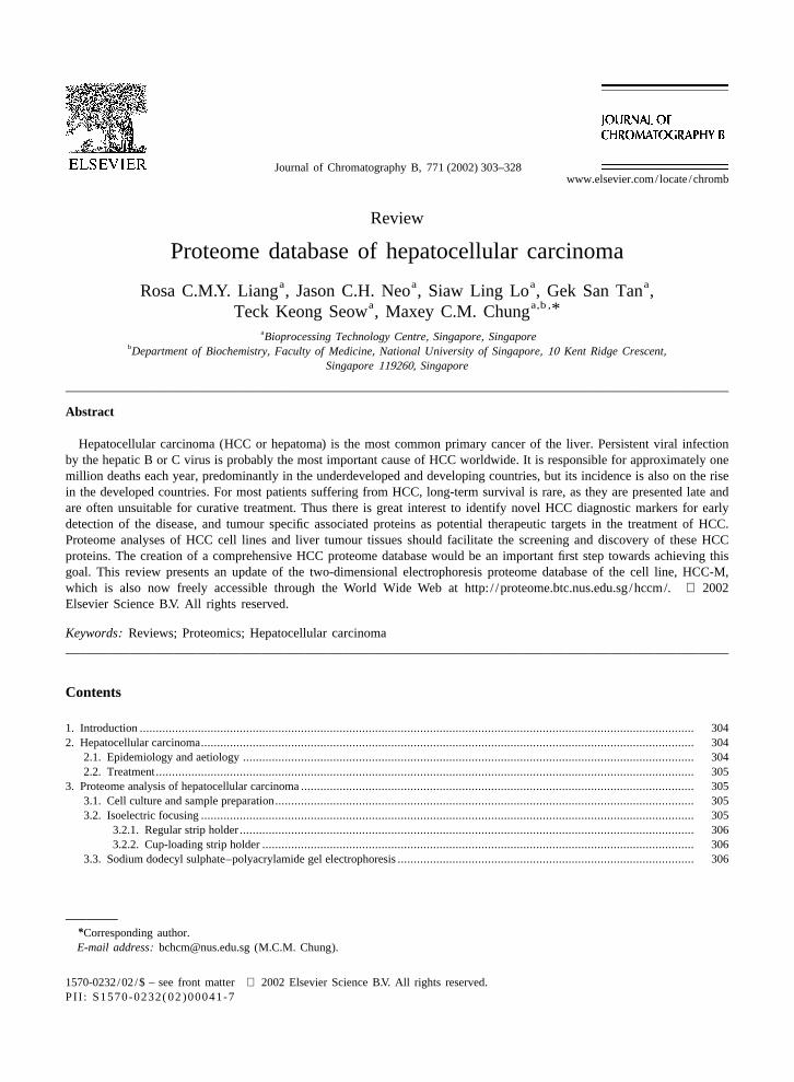
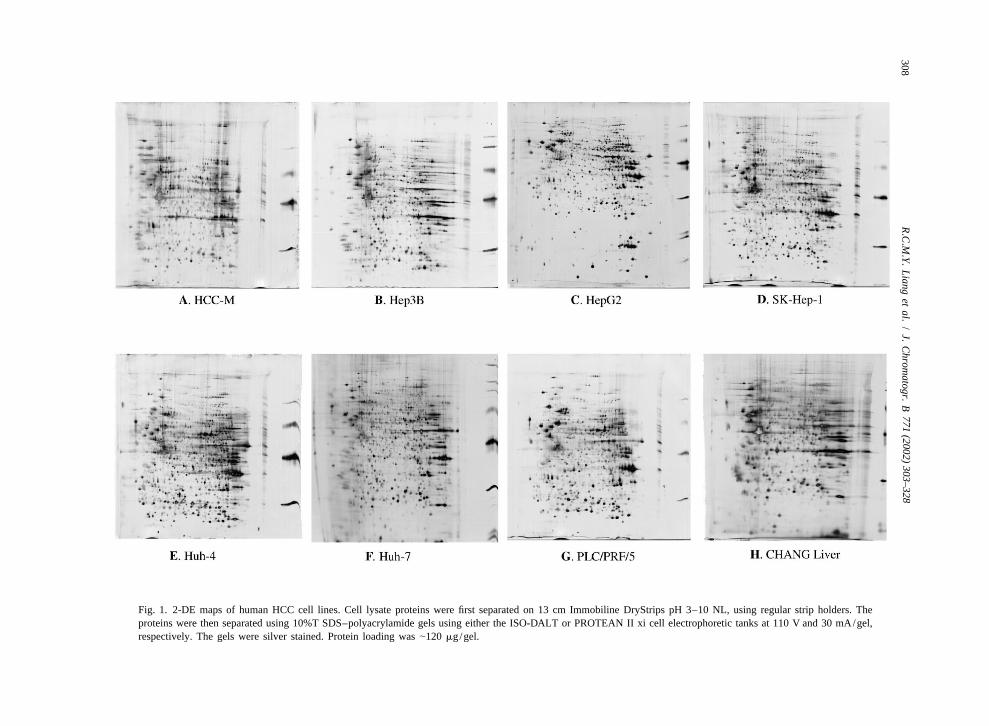
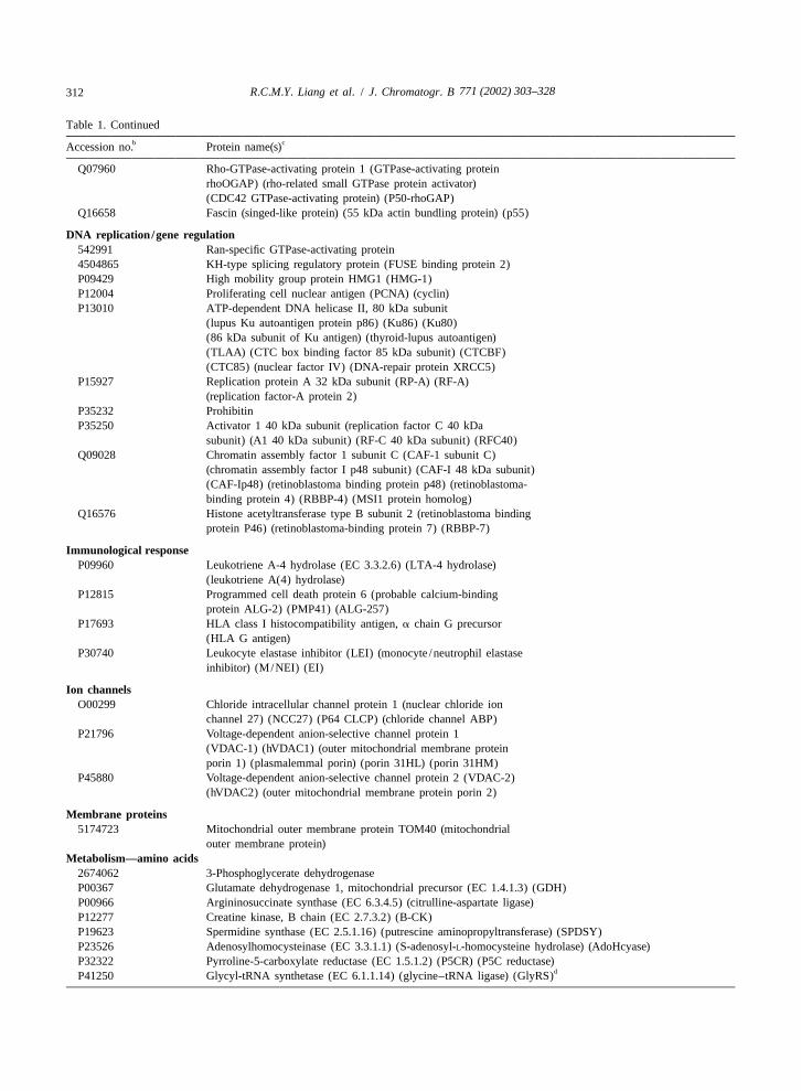
analyses. We present here the 2-DE maps of sevenMALDI-TOF MS analyses were performed as hepatoma derived cell lines, HCC-M, Hep3B,
described previously [12]. Essentially, 1 ml of the HepG2, SK-Hep1, Huh-4, Huh-7, and PLC/PRF/5,extracted sample from each of the gel spots was and a non-transformed cell line, Chang liver (Fig. 1).dispensed onto the MALDI sample plate with 1 ml of It is apparent that these 2-DE maps exhibited differ-matrix solution (10 mg/ml a-cyano-4-hydroxy- ences in the protein profiles when compared withcinnamic acid, 0.1% TFA, 50% ACN), and allowed each other, and hence can be used as a basis toto dry under ambient conditions. The acquisition of classify or differentiate the various hepatoma cellspectra for each sample was performed using the lines. This result is consistent with the recent genedelayed extraction and reflector mode as described expression profile studies of Kawai et al. [17], who[12]. Spectra were automatically calibrated upon showed that the a-fetoprotein producing cell lines,acquisition using a two-point calibration with re- HepG2, Huh-7, Hep3B, PLC/PRF/5 and Huh-6sidual porcine trypsin autolytic fragments (842.51 have common gene-expression profiles when com-
1and 2210.10 [M1H ] ions). Assignment of peaks pared with HLE and SK-Hep1, which are a-fetopro-and protein identification were performed automat- tein negative hepatoma cell lines, and cancer cellically using the AutoMS-Fit software, which is part lines of non-hepatocyte origin (HeLa and KMBC).of the Proteomics Solution 1 system (Applied Bio- In addition, HepG2, Huh-7, and Hep3B which hadsystems, Foster City, CA, USA). Searches were higher expressions of a-fetoprotein shared a commonqueried against the SWISS-PROT and NCBI non-redun- gene expression profile when compared with thedant databases, using parameters described previous- other a-fetoprotein producing cells (Huh-6 and PLC/ly [12]. PRF/5).
771 (2002) 303–328308
R.C
.M.Y.
Liang
etal.
/J.
Chrom
atogr.B
Fig. 1. 2-DE maps of human HCC cell lines. Cell lysate proteins were first separated on 13 cm Immobiline DryStrips pH 3–10 NL, using regular strip holders. Theproteins were then separated using 10%T SDS–polyacrylamide gels using either the ISO-DALT or PROTEAN II xi cell electrophoretic tanks at 110 V and 30 mA/gel,respectively. The gels were silver stained. Protein loading was |120 mg/gel.
771 (2002) 303–328 309R.C.M.Y. Liang et al. / J. Chromatogr. B
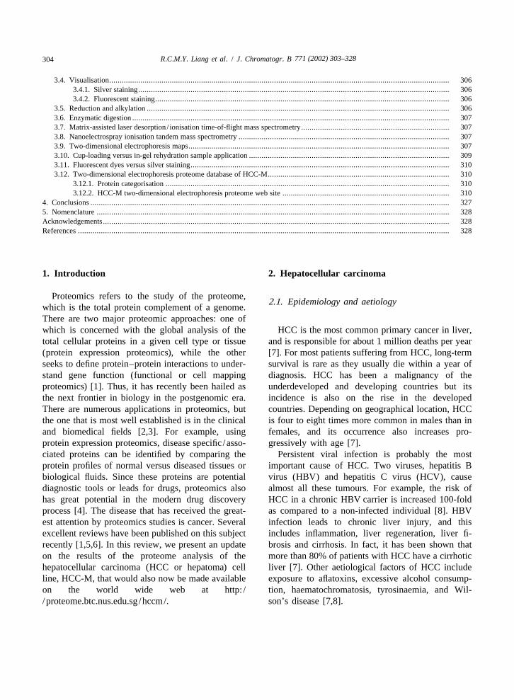
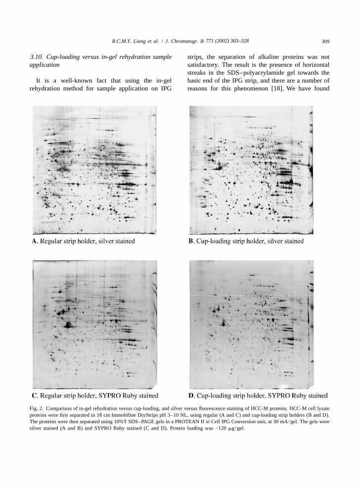
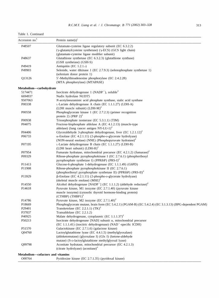
3.10. Cup-loading versus in-gel rehydration sample strips, the separation of alkaline proteins was notapplication satisfactory. The result is the presence of horizontal
streaks in the SDS–polyacrylamide gel towards theIt is a well-known fact that using the in-gel basic end of the IPG strip, and there are a number of
rehydration method for sample application on IPG reasons for this phenomenon [18]. We have found
Fig. 2. Comparison of in-gel rehydration versus cup-loading, and silver versus fluorescence staining of HCC-M proteins. HCC-M cell lysateproteins were first separated in 18 cm Immobiline DryStrips pH 3–10 NL, using regular (A and C) and cup-loading strip holders (B and D).The proteins were then separated using 10%T SDS–PAGE gels in a PROTEAN II xi Cell IPG Conversion unit, at 30 mA/gel. The gels weresilver stained (A and B) and SYPRO Ruby stained (C and D). Protein loading was |120 mg/gel.
771 (2002) 303–328310 R.C.M.Y. Liang et al. / J. Chromatogr. B
that sample application using the cup-loading method cation /gene regulation, (e) immunological response,on the anode end of the IPG strips seemed to reduce (f) ion channels, (g) membrane proteins, (h) metabo-the horizontal streaks to a certain extent (Fig. 2). lism, (i) oncogenes / tumour suppressor genes, (j)This method was greatly facilitated by the recent protection and detoxification, (k) protein synthesisrelease of the Universal strip holder for use on the and degradation, (l) signal transduction, (m) trans-IPGphor electrophoretic unit (Amersham Biosci- port /binding proteins, (n) tumour associated proteins,ences). and (o) unannotated / function inferred (Table 1). In
addition, we have further grouped the proteins that3.11. Fluorescent dyes versus silver staining have been shown to be implicated in HCC and other
types of cancers into a separate list from the differentSilver staining is very sensitive (as low as 0.1 ng categories of HCC-M proteins (see Table 2). We
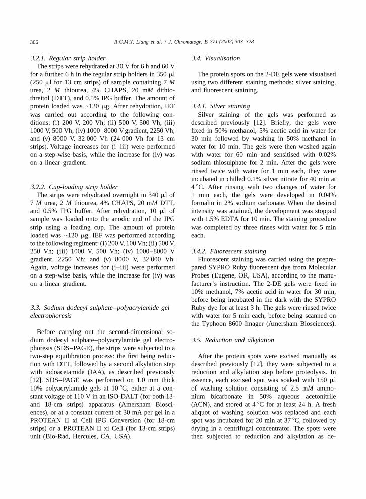
of protein per spot can be detected), but it is a believe that such a categorisation and grouping ofmultistep procedure with a very limited linear dy- HCC-M proteins will simplify the 2DE proteinnamic range. In addition it often leads to the database of HCC proteins, which in turn will facili-formation of hollow spots or result in a doughnut tate the rapid identification and discovery of noveleffect which can complicate image analyses. On the proteins that are involved in hepatocarcinogenesis.other hand, staining with fluorescent dyes such asSYPRO Ruby is relatively sensitive, simple, and 3.12.2. HCC-M two-dimensional electrophoresisreproducible. In addition, it also has a broader linear proteome web sitedynamic range. We had compared the two staining Finally, in line with our wish to allow the sci-methods for the 2-DE maps of HCC-M, and found entific community to access our extensive work onSYPRO Ruby to be considerably less sensitive than the identity of the proteins on the HCC-M 2-DEsilver staining (Fig. 2). Moreover, it was also found map, we have created an on-line database thatthat some protein spots stained better with SYPRO provides an interactive way to query the HCC-MRuby than with silver nitrate, and the reverse was protein database. The data from our earlier publi-true as well. This observation is in full agreement cations [12,13] were first converted to MySQLwith the report by Gorg et al. [18] who showed that database for data manipulation and retrieval. We arethe patterns obtained with silver staining and SYPRO running an Apache web server and using JavaRuby staining were similar, but not identical. Finally, servlets and applets technology to process andto facilitate the excision of the protein spots from display the data.2-DE gels following SYPRO Ruby staining and There are two options to query the HCC-Mimage analysis, we have also developed a protocol to database (Fig. 3).restain the gel with silver nitrate (results not shown). Option 1: protein search by NCBI / SWISS-PROT Ac-
cession Number, Protein Name (full and partial3.12. Two-dimensional electrophoresis proteome name) and Protein ID (ID as published in ourdatabase of HCC-M papers);
Option 2: interactive protein spots query on the3.12.1. Protein categorisation original 2-DE image maps.
We have earlier reported that, from a total of 408 A query using Option 1 will retrieve a row or a listunique spots excised from the 2-DE gel of HCC-M, of proteins (if there are more than one match in the272 and 29 spots were identified by MALDI-TOF database) for selection as shown in Fig. 4. A click onMS and nESI-MS–MS respectively [12,13]. This the Sno column will display the protein identity pageresult represented the most comprehensive 2-DE (Fig. 5) which includes information on the theoret-protein database for any HCC cell line reported thus ical MW and pI, experimental MW and pI, a link tofar. In this review, we have reorganised the database NCBI / SWISS-PROT information page, protein descrip-by grouping the proteins into different functional tion, peptides matched from MALDI, subcellularcategories under (a) cell cycle, (b) chaperone /stress location, method of identification and remarks. Theresponse, (c) cytoskeleton /mobility, (d) DNA repli- location of the protein on the 2-DE map can also be
771 (2002) 303–328 311R.C.M.Y. Liang et al. / J. Chromatogr. B
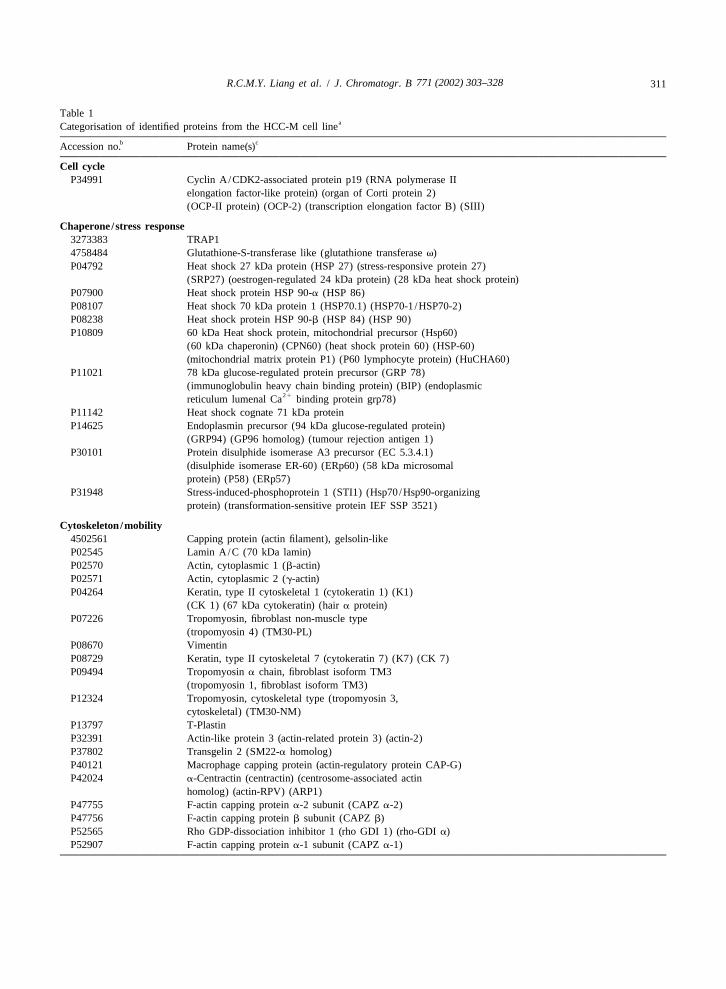
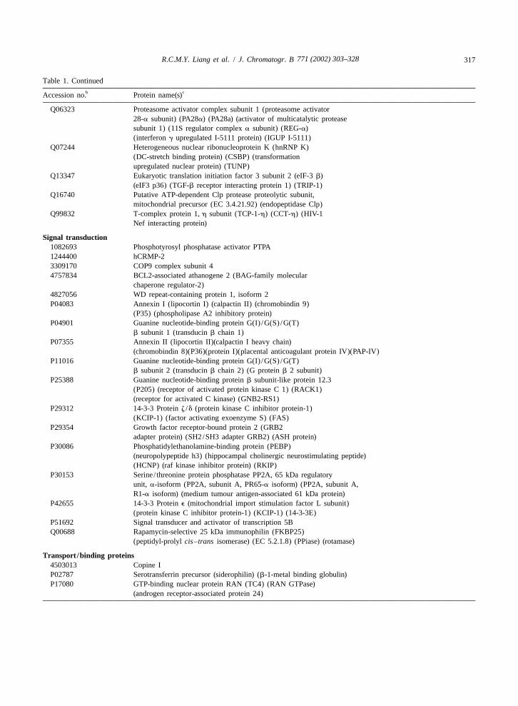
Table 1aCategorisation of identified proteins from the HCC-M cell line
b cAccession no. Protein name(s)
Cell cycleP34991 Cyclin A/CDK2-associated protein p19 (RNA polymerase II
elongation factor-like protein) (organ of Corti protein 2)(OCP-II protein) (OCP-2) (transcription elongation factor B) (SIII)
Chaperone/stress response3273383 TRAP14758484 Glutathione-S-transferase like (glutathione transferase v)P04792 Heat shock 27 kDa protein (HSP 27) (stress-responsive protein 27)
(SRP27) (oestrogen-regulated 24 kDa protein) (28 kDa heat shock protein)P07900 Heat shock protein HSP 90-a (HSP 86)P08107 Heat shock 70 kDa protein 1 (HSP70.1) (HSP70-1 /HSP70-2)P08238 Heat shock protein HSP 90-b (HSP 84) (HSP 90)P10809 60 kDa Heat shock protein, mitochondrial precursor (Hsp60)
(60 kDa chaperonin) (CPN60) (heat shock protein 60) (HSP-60)(mitochondrial matrix protein P1) (P60 lymphocyte protein) (HuCHA60)
P11021 78 kDa glucose-regulated protein precursor (GRP 78)(immunoglobulin heavy chain binding protein) (BIP) (endoplasmic
21reticulum lumenal Ca binding protein grp78)P11142 Heat shock cognate 71 kDa proteinP14625 Endoplasmin precursor (94 kDa glucose-regulated protein)
(GRP94) (GP96 homolog) (tumour rejection antigen 1)P30101 Protein disulphide isomerase A3 precursor (EC 5.3.4.1)
(disulphide isomerase ER-60) (ERp60) (58 kDa microsomalprotein) (P58) (ERp57)
P31948 Stress-induced-phosphoprotein 1 (STI1) (Hsp70/Hsp90-organizingprotein) (transformation-sensitive protein IEF SSP 3521)
Cytoskeleton/mobility4502561 Capping protein (actin filament), gelsolin-likeP02545 Lamin A/C (70 kDa lamin)P02570 Actin, cytoplasmic 1 (b-actin)P02571 Actin, cytoplasmic 2 (g-actin)P04264 Keratin, type II cytoskeletal 1 (cytokeratin 1) (K1)
(CK 1) (67 kDa cytokeratin) (hair a protein)P07226 Tropomyosin, fibroblast non-muscle type
(tropomyosin 4) (TM30-PL)P08670 VimentinP08729 Keratin, type II cytoskeletal 7 (cytokeratin 7) (K7) (CK 7)P09494 Tropomyosin a chain, fibroblast isoform TM3
(tropomyosin 1, fibroblast isoform TM3)P12324 Tropomyosin, cytoskeletal type (tropomyosin 3,
cytoskeletal) (TM30-NM)P13797 T-PlastinP32391 Actin-like protein 3 (actin-related protein 3) (actin-2)P37802 Transgelin 2 (SM22-a homolog)P40121 Macrophage capping protein (actin-regulatory protein CAP-G)P42024 a-Centractin (centractin) (centrosome-associated actin
homolog) (actin-RPV) (ARP1)P47755 F-actin capping protein a-2 subunit (CAPZ a-2)P47756 F-actin capping protein b subunit (CAPZ b)P52565 Rho GDP-dissociation inhibitor 1 (rho GDI 1) (rho-GDI a)P52907 F-actin capping protein a-1 subunit (CAPZ a-1)
771 (2002) 303–328312 R.C.M.Y. Liang et al. / J. Chromatogr. B
Table 1. Continuedb cAccession no. Protein name(s)
Q07960 Rho-GTPase-activating protein 1 (GTPase-activating proteinrhoOGAP) (rho-related small GTPase protein activator)(CDC42 GTPase-activating protein) (P50-rhoGAP)
Q16658 Fascin (singed-like protein) (55 kDa actin bundling protein) (p55)
DNA replication/gene regulation542991 Ran-specific GTPase-activating protein4504865 KH-type splicing regulatory protein (FUSE binding protein 2)P09429 High mobility group protein HMG1 (HMG-1)P12004 Proliferating cell nuclear antigen (PCNA) (cyclin)P13010 ATP-dependent DNA helicase II, 80 kDa subunit
(lupus Ku autoantigen protein p86) (Ku86) (Ku80)(86 kDa subunit of Ku antigen) (thyroid-lupus autoantigen)(TLAA) (CTC box binding factor 85 kDa subunit) (CTCBF)(CTC85) (nuclear factor IV) (DNA-repair protein XRCC5)
P15927 Replication protein A 32 kDa subunit (RP-A) (RF-A)(replication factor-A protein 2)
P35232 ProhibitinP35250 Activator 1 40 kDa subunit (replication factor C 40 kDa
subunit) (A1 40 kDa subunit) (RF-C 40 kDa subunit) (RFC40)Q09028 Chromatin assembly factor 1 subunit C (CAF-1 subunit C)
(chromatin assembly factor I p48 subunit) (CAF-I 48 kDa subunit)(CAF-Ip48) (retinoblastoma binding protein p48) (retinoblastoma-binding protein 4) (RBBP-4) (MSI1 protein homolog)
Q16576 Histone acetyltransferase type B subunit 2 (retinoblastoma bindingprotein P46) (retinoblastoma-binding protein 7) (RBBP-7)
Immunological responseP09960 Leukotriene A-4 hydrolase (EC 3.3.2.6) (LTA-4 hydrolase)
(leukotriene A(4) hydrolase)P12815 Programmed cell death protein 6 (probable calcium-binding
protein ALG-2) (PMP41) (ALG-257)P17693 HLA class I histocompatibility antigen, a chain G precursor
(HLA G antigen)P30740 Leukocyte elastase inhibitor (LEI) (monocyte /neutrophil elastase
inhibitor) (M/NEI) (EI)
Ion channelsO00299 Chloride intracellular channel protein 1 (nuclear chloride ion
channel 27) (NCC27) (P64 CLCP) (chloride channel ABP)P21796 Voltage-dependent anion-selective channel protein 1
(VDAC-1) (hVDAC1) (outer mitochondrial membrane proteinporin 1) (plasmalemmal porin) (porin 31HL) (porin 31HM)
P45880 Voltage-dependent anion-selective channel protein 2 (VDAC-2)(hVDAC2) (outer mitochondrial membrane protein porin 2)
Membrane proteins5174723 Mitochondrial outer membrane protein TOM40 (mitochondrial
outer membrane protein)Metabolism—amino acids
2674062 3-Phosphoglycerate dehydrogenaseP00367 Glutamate dehydrogenase 1, mitochondrial precursor (EC 1.4.1.3) (GDH)P00966 Argininosuccinate synthase (EC 6.3.4.5) (citrulline-aspartate ligase)P12277 Creatine kinase, B chain (EC 2.7.3.2) (B-CK)P19623 Spermidine synthase (EC 2.5.1.16) (putrescine aminopropyltransferase) (SPDSY)P23526 Adenosylhomocysteinase (EC 3.3.1.1) (S-adenosyl-L-homocysteine hydrolase) (AdoHcyase)P32322 Pyrroline-5-carboxylate reductase (EC 1.5.1.2) (P5CR) (P5C reductase)
dP41250 Glycyl-tRNA synthetase (EC 6.1.1.14) (glycine–tRNA ligase) (GlyRS)
771 (2002) 303–328 313R.C.M.Y. Liang et al. / J. Chromatogr. B
Table 1. Continuedb cAccession no. Protein name(s)
P48507 Glutamate-cysteine ligase regulatory subunit (EC 6.3.2.2)(g-glutamylcysteine synthetase) (g-ECS) (GCS light chain)(glutamate-cysteine ligase modifier subunit)
P48637 Glutathione synthetase (EC 6.3.2.3) (glutathione synthase)(GSH synthetase) (GSH-S)
P49419 Antiquitin (EC 1.2.1.-)P49903 Selenide, water dikinase 1 (EC 2.7.9.3) (selenophosphate synthetase 1)
(selenium donor protein 1)Q13126 59-Methylthioadenosine phosphorylase (EC 2.4.2.28)
(MTA phosphorylase) (MTAPASE)
Metabolism—carbohydrate1 d5174471 Isocitrate dehydrogenase 1 (NADP ), soluble
6694937 Nudix hydrolase NUDT59507063 N-acetylneuraminic acid phosphate synthase, sialic acid synthaseP00338 L-Lactate dehydrogenase A chain (EC 1.1.1.27) (LDH-A)
d(LDH muscle subunit) (LDH-M)P00558 Phosphoglycerate kinase 1 (EC 2.7.2.3) (primer recognition
dprotein 2) (PRP 2)P00938 Triosephosphate isomerase (EC 5.3.1.1) (TIM)P04075 Fructose-bisphosphate aldolase A (EC 4.1.2.13) (muscle-type
daldolase) (lung cancer antigen NY-LU-1)dP04406 Glyceraldehyde 3-phosphate dehydrogenase, liver (EC 1.2.1.12)
P06733 a-Enolase (EC 4.2.1.11) (2-phospho-D-glycerate hydrolyase)d(NON-neural enolase) (NNE) (Phosphopyruvate hydratase)
P07195 L-Lactate dehydrogenase B chain (EC 1.1.1.27) (LDH-B)d(LDH heart subunit) (LDH-H)
dP07954 Fumarate hydratase, mitochondrial precursor (EC 4.2.1.2) (fumarase)P09329 Ribose-phosphate pyrophosphokinase I (EC 2.7.6.1) (phosphoribosyl
dpyrophosphate synthetase I) (PPRibP) (PRS-I)P11413 Glucose-6-phosphate 1-dehydrogenase (EC 1.1.1.49) (G6PD)P11908 Ribose-phosphate pyrophosphokinase II (EC 2.7.6.1)
d(phosphoribosyl pyrophosphate synthetase II) (PPRibP) (PRS-II)P13929 b-Enolase (EC 4.2.1.11) (2-phospho-D-glycerate hydrolyase)
d(skeletal muscle enolase) (MSE)1 dP14550 Alcohol dehydrogenase [NADP ] (EC 1.1.1.2) (aldehyde reductase)
P14618 Pyruvate kinase, M1 isozyme (EC 2.7.1.40) (pyruvate kinasemuscle isozyme) (cytosolic thyroid hormone-binding protein)
d(CTHBP) (THBP1)dP14786 Pyruvate kinase, M2 isozyme (EC 2.7.1.40)
P18669 Phosphoglycerate mutase, brain form (EC 5.4.2.1) (PGAM-B) (EC 5.4.2.4) (EC 3.1.3.13) (BPG-dependent PGAM)dP29401 Transketolase (EC 2.2.1.1) (TK)
P37837 Transaldolase (EC 2.2.1.2)dP40925 Malate dehydrogenase, cytoplasmic (EC 1.1.1.37)
P50213 Isocitrate dehydrogenase [NAD] subunit a, mitochondrial precursor1(EC 1.1.1.41) (isocitric dehydrogenase) (NAD -specific ICDH)
P51570 Galactokinase (EC 2.7.1.6) (galactose kinase)Q04760 Lactoylglutathione lyase (EC 4.4.1.5) (methylglyoxalase)
(aldoketomutase) (glyoxalase I) (Glx I) (ketone-aldehydemutase) (S-D-lactoylglutathione methylglyoxal lyase)
Q99798 Aconitate hydratase, mitochondrial precursor (EC 4.2.1.3)d(citrate hydrolyase) (aconitase)
Metabolism—cofactors and vitaminsO00764 Pyridoxine kinase (EC 2.7.1.35) (pyridoxal kinase)

771 (2002) 303–328314 R.C.M.Y. Liang et al. / J. Chromatogr. B
Table 1. Continuedb cAccession no. Protein name(s)
P30043 Flavin reductase (EC 1.6.99.1) (FR) (NADPH-dependentdiaphorase) (NADPH-flavin reductase) (FLR) (biliverdinreductase B) (EC 1.3.1.24) (BVR-B) (biliverdin-IX b-reductase)(green haem binding protein) (GHBP)
Metabolism–energyP06576 ATP synthase b chain, mitochondrial precursor (EC 3.6.3.14)P13804 Electron transfer flavoprotein a-subunit, mitochondrial precursor (a-ETF)P22695 Ubiquinol-cytochrome C reductase complex core protein 2,
mitochondrial precursor (EC 1.10.2.2) (complex III subunit II)Q15181 Inorganic pyrophosphatase (EC 3.6.1.1) (pyrophosphate
phospho-hydrolase) (PPase)Metabolism—lipid
5174389 Acetyl-coenzyme A acetyltransferase 2 (acetoacetyl coenzyme Adthiolase) (acetoacetyl coenzyme A thiolase)
O00154 Cytosolic acyl coenzyme A thioester hydrolase (EC 3.1.2.2)(long chain acyl-CoA thioester hydrolase) (CTE-II)(brain acyl-CoA hydrolase) (BACH)
P02647 Apolipoprotein A-I precursor (Apo-AI)P42126 3,2-trans-enoyl-CoA isomerase, mitochondrial precursor
(EC 5.3.3.8) (dodecenoyl-CoA d-isomerase)P54619 59-AMP-activated protein kinase, g-1 subunit (AMPK g-1 chain) (AMPKg)P55809 Succinyl-CoA:3-ketoacid-coenzyme A transferase, mitochondrial
dprecursor (EC 2.8.3.5) (succinyl CoA:3-oxoacid CoA-transferase)Q99714 3-Hydroxyacyl-CoA dehydrogenase type II (EC 1.1.1.35)
(Type II HADH) (endoplasmic reticulum-associated amyloid b-peptidedbinding protein) (short-chain type dehydrogenase / reductase XH98G2)
Metabolism—nucleotidedP00491 Purine nucleoside phosphorylase (EC 2.4.2.1) (inosine phosphorylase) (PNP)
P00568 Adenylate kinase isoenzyme 1 (EC 2.7.4.3) (ATP–AMPtransphosphorylase) (AK1) (myokinase)
P07741 Adenine phosphoribosyltransferase (EC 2.4.2.7) (APRT)P12268 Inosine-59-monophosphate dehydrogenase 2 (EC 1.1.1.205)
(IMP dehydrogenase 2) (IMPDH-II) (IMPD 2)P15531 Nucleoside diphosphate kinase A (EC 2.7.4.6) (NDK A)
(NDP kinase A) (tumour metastatic process-associated protein)(metastasis inhibition factor nm23) (nm23-H1)
P49915 GMP synthase [glutamine-hydrolyzing] (EC 6.3.5.2)d(glutamine amidotransferase) (GMP synthetase)
P55263 Adenosine kinase (EC 2.7.1.20) (AK) (adenosine 59-phosphotransferase)
Oncogenes / tumour suppressor genes4503801 Far upstream element-binding protein (far upstream element
binding protein) (FUSE-binding protein)6005749 RNA-binding protein regulatory subunit9910460 Nit protein 2P36952 Maspin precursor (protease inhibitor 5)
Protection and detoxification2135069 Probable thioredoxin peroxidase (EC 1.11.1.-)4507149 Superoxide dismutase 1, soluble [amyotrophic lateral sclerosis 1
(adult)] (Cu/Zn superoxide dismutase)P00441 Superoxide dismutase [Cu–Zn]P04179 Superoxide dismutase [Mn], mitochondrial precursor (EC 1.15.1.1)
771 (2002) 303–328 315R.C.M.Y. Liang et al. / J. Chromatogr. B
Table 1. Continuedb cAccession no. Protein name(s)
P08758 Annexin V (lipocortin V) (endonexin II) (calphobindin I) (CBP-I)(placental anticoagulant protein I) (PAP-I) (PP4) (thromboplastininhibitor) (vascular anticoagulant-a) (VAC-a) (anchorin CII)
P09211 Glutathione S-transferase P (EC 2.5.1.18) (GST class-PI) (GSTP1-1)P30041 Antioxidant protein 2 (1-Cys peroxiredoxin) (1-Cys PRX)
(acidic calcium-independent phospholipase A2) (EC 3.1.1.-) (aiPLA2)(non-selenium glutathione peroxidase) (EC 1.11.1.7) (NSGPx)(24 kDa protein) (liver 2D PAGE spot 40) (red blood cells PAGE spot 12)
P30048 Thioredoxin-dependent peroxide reductase, mitochondrial precursor(peroxiredoxin 3) (antioxidant protein 1) (AOP-1) (MER5 proteinhomolog) (HBC189) (PRX III)
P32119 Peroxiredoxin 2 (thioredoxin peroxidase 1) (thioredoxin-dependentperoxide reductase 1) (thiol-specific antioxidant protein) (TSA) (PRP)(natural killer cell enhancing factor B) (NKEF-B)
P38646 Stress-70 protein, mitochondrial precursor (75 kDa glucoseregulated protein) (GRP 75) (peptide-binding protein 74) (PBP74) (mortalin) (MOT)
Q06830 Peroxiredoxin 1 (thioredoxin peroxidase 2) (thioredoxin-dependent peroxidereductase 2) (proliferation-associated protein PAG) (natural killercell enhancing factor A) (NKEF-A)
Protein synthesis and degradation542852 hnRNP protein E13986482 Translation initiation factor eIF3 p40 subunit (eIF3p40)4468218 unr-interacting protein4503519 Eukaryotic translation initiation factor 3, subunit 5 (e, 47 kDa)4506195 Proteasome (prosome, macropain) subunit, b type, 2
(proteasome subunit, b type, 2)4506217 Proteasome (prosome, macropain) 26S subunit, non-ATPase, 104506223 Proteasome (prosome, macropain) 26S subunit, non-ATPase,
13 (hypothetical protein) (26S proteasome subunit p40.5)4506237 Proteasome activator HPA28 subunit b
4506753 TATA binding protein interacting protein 49 kDa5031981 26S proteasome-associated pad1 homolog5031997 Proteasome (prosome, macropain) activator subunit 3
(PA28 g, Ki) (Ki nuclear autoantigen)5174731 Translin-associated factor XP04632 Calcium-dependent protease, small subunit (calpain
regulatory subunit) (calcium-activated neutral proteinase) (CANP)P04720 Elongation factor 1-a 1 (EF-1-a-1) (elongation factor 1 A-1)
(eEF1A-1) (elongation factor Tu) (EF-Tu)P05198 Eukaryotic translation initiation factor 2 subunit 1 (eukaryotic
translation initiation factor 2 a subunit) (eIF-2-a) (EIF-2a) (EIF-2A)P07237 Protein disulphide isomerase precursor (PDI) (EC 5.3.4.1)
(prolyl 4-hydroxylase b subunit) (cellular thyroid hormone binding protein) (P55)P07602 Proactivator polypeptide precursor [Contains: saposin A (protein A); saposin B
(sphingolipid activator protein 1) (SAP-1) (cerebroside sulphate activator) (CSAct)(dispersin) (sulphatide /GM1 activator); saposin C (Co-b-glucosidase) (A1 activator)(glucosylceramidase activator) (sphingolipid activator protein 2) (SAP-2); saposin D(protein C) (component C)]
P08865 40S ribosomal protein SA (P40) (34/67 kDa laminin receptor)(colon carcinoma laminin-binding protein) (NEM/1CHD4)
771 (2002) 303–328316 R.C.M.Y. Liang et al. / J. Chromatogr. B
Table 1. Continuedb cAccession no. Protein name(s)
P09651 Heterogeneous nuclear ribonucleoprotein A1 (helix-destabilizingprotein) (single-strand binding protein) (hnRNP core protein A1)
P12081 Histidyl-tRNA synthetase (EC 6.1.1.21) (histidine–tRNA ligase) (HisRS)P13639 Elongation factor 2 (EF-2)P13798 Acylamino-acid-releasing enzyme (EC 3.4.19.1) (acyl-peptide
hydrolase) (APH) (acylaminoacyl-peptidase) (DNF15S2 protein)P14866 Heterogeneous nuclear ribonucleoprotein L (hnRNP L)P15374 Ubiquitin carboxyl-terminal hydrolase isozyme L3 (EC 3.4.19.12)
(UCH-L3) (ubiquitin thiolesterase L3)P17987 T-complex protein 1, a subunit (TCP-1-a) (CCT-a)P22061 Protein-L-isoaspartate(D-aspartate) O-methyltransferase (EC 2.1.1.77)
(protein-b-aspartate methyltransferase) (PIMT) (protein L-isoaspartyl /D-aspartyl methyltransferase) (L-isoaspartyl protein carboxyl methyltransferase)
P22626 Heterogeneous nuclear ribonucleoproteins A2/B1 (hnRNP A2/hnRNP B1)P25786 Proteasome subunit a type 1 (EC 3.4.25.1) (proteasome component C2)
(macropain subunit C2) (multicatalytic endopeptidase complex subunit C2)(proteasome n chain) (30 kDa prosomal protein) (PROS-30)
P25787 Proteasome subunit alpha type 2 (EC 3.4.25.1) (proteasome component C3)(macropain subunit C3) (multicatalytic endopeptidase complex subunit C3)
P25789 Proteasome subunit alpha type 4 (EC 3.4.25.1)(proteasome component C9)(macropain subunit C9) (multicatalytic endopeptidase complex subunit C9)(proteasome subunit L)
P27924 Ubiquitin-conjugating enzyme E2-25 kDa (EC 6.3.2.19) (ubiquitin–proteinligase) (ubiquitin carrier protein) (Huntington interacting protein) (HIP-2)
P28070 proteasome subunit b type 4 precursor (EC 3.4.25.1) (proteasome b-chain)(macropain b chain) (multicatalytic endopeptidase complex b-chain)(proteasome chain 3) (HSN3) (HsBPROS26)
P28072 Proteasome subunit b type 6 precursor (EC 3.4.25.1)(proteasome d-chain) (macropain d chain) (multicatalytic endopeptidasecomplex d chain) (proteasome subunit Y)
P30040 Endoplasmic reticulum protein ERp29 precursor (ERp31) (ERp28)P31943 Heterogeneous nuclear ribonucleoprotein H (hnRNP H)P33240 Cleavage stimulation factor, 64 kDa subunit (CSTF 64 kDa
subunit) (CF-1 64 kDa subunit)P34062 Proteasome subunit a type 6 (EC 3.4.25.1) (proteasome i chain)
(macropain i chain) (multicatalytic endopeptidase complex i chain)(27 kDa prosomal protein) (PROS-27) (p27K)
P35237 Placental thrombin inhibitor (cytoplasmic antiproteinase) (CAP)(protease inhibitor 6)
P35998 26S protease regulatory subunit 7 (MSS1 protein)P40227 T-complex protein 1, z subunit (TCP-1-z) (CCT-z) (CCT-z-1) (Tcp20) (HTR3)P48643 T-complex protein 1, e subunit (TCP-1-e) (CCT-e)P49368 T-complex protein 1, g subunit (TCP-1-g) (CCT-g)P49411 Elongation factor Tu, mitochondrial precursor (P43)P49720 Proteasome subunit b type 3 (EC 3.4.25.1) (proteasome u chain)
(proteasome chain 13) (proteasome component C10-II)P50990 T-complex protein 1, u subunit (TCP-1-u) (CCT-u)P50991 T-complex protein 1, d subunit (TCP-1-d) (CCT-d)
(stimulator of TAR RNA binding)P55795 Heterogeneous nuclear ribonucleoprotein H9 (hnRNP H9) (FTP-3)
771 (2002) 303–328 317R.C.M.Y. Liang et al. / J. Chromatogr. B
Table 1. Continuedb cAccession no. Protein name(s)
Q06323 Proteasome activator complex subunit 1 (proteasome activator28-a subunit) (PA28a) (PA28a) (activator of multicatalytic proteasesubunit 1) (11S regulator complex a subunit) (REG-a)(interferon g upregulated I-5111 protein) (IGUP I-5111)
Q07244 Heterogeneous nuclear ribonucleoprotein K (hnRNP K)(DC-stretch binding protein) (CSBP) (transformationupregulated nuclear protein) (TUNP)
Q13347 Eukaryotic translation initiation factor 3 subunit 2 (eIF-3 b)(eIF3 p36) (TGF-b receptor interacting protein 1) (TRIP-1)
Q16740 Putative ATP-dependent Clp protease proteolytic subunit,mitochondrial precursor (EC 3.4.21.92) (endopeptidase Clp)
Q99832 T-complex protein 1, h subunit (TCP-1-h) (CCT-h) (HIV-1Nef interacting protein)
Signal transduction1082693 Phosphotyrosyl phosphatase activator PTPA1244400 hCRMP-23309170 COP9 complex subunit 44757834 BCL2-associated athanogene 2 (BAG-family molecular
chaperone regulator-2)4827056 WD repeat-containing protein 1, isoform 2P04083 Annexin I (lipocortin I) (calpactin II) (chromobindin 9)
(P35) (phospholipase A2 inhibitory protein)P04901 Guanine nucleotide-binding protein G(I) /G(S) /G(T)
b subunit 1 (transducin b chain 1)P07355 Annexin II (lipocortin II)(calpactin I heavy chain)
(chromobindin 8)(P36)(protein I)(placental anticoagulant protein IV)(PAP-IV)P11016 Guanine nucleotide-binding protein G(I) /G(S) /G(T)
b subunit 2 (transducin b chain 2) (G protein b 2 subunit)P25388 Guanine nucleotide-binding protein b subunit-like protein 12.3
(P205) (receptor of activated protein kinase C 1) (RACK1)(receptor for activated C kinase) (GNB2-RS1)
P29312 14-3-3 Protein z /d (protein kinase C inhibitor protein-1)(KCIP-1) (factor activating exoenzyme S) (FAS)
P29354 Growth factor receptor-bound protein 2 (GRB2adapter protein) (SH2/SH3 adapter GRB2) (ASH protein)
P30086 Phosphatidylethanolamine-binding protein (PEBP)(neuropolypeptide h3) (hippocampal cholinergic neurostimulating peptide)(HCNP) (raf kinase inhibitor protein) (RKIP)
P30153 Serine / threonine protein phosphatase PP2A, 65 kDa regulatoryunit, a-isoform (PP2A, subunit A, PR65-a isoform) (PP2A, subunit A,R1-a isoform) (medium tumour antigen-associated 61 kDa protein)
P42655 14-3-3 Protein e (mitochondrial import stimulation factor L subunit)(protein kinase C inhibitor protein-1) (KCIP-1) (14-3-3E)
P51692 Signal transducer and activator of transcription 5BQ00688 Rapamycin-selective 25 kDa immunophilin (FKBP25)
(peptidyl-prolyl cis–trans isomerase) (EC 5.2.1.8) (PPiase) (rotamase)
Transport /binding proteins4503013 Copine IP02787 Serotransferrin precursor (siderophilin) (b-1-metal binding globulin)P17080 GTP-binding nuclear protein RAN (TC4) (RAN GTPase)
(androgen receptor-associated protein 24)
771 (2002) 303–328318 R.C.M.Y. Liang et al. / J. Chromatogr. B
Table 1. Continuedb cAccession no. Protein name(s)
P17931 Galectin-3 (galactose-specific lectin 3) (MAC-2 antigen)(IgE-binding protein) (35 kDa lectin) (carbohydrate binding protein 35)(CBP 35) (laminin-binding protein) (lectin L-29) (L-31)(galactoside-binding protein) (GALBP)
P54920 a-Soluble NSF attachment protein (SNAP-a) (N-ethylmaleimide-sensitive factor attachment protein, a)
Q02790 FK506-binding protein 4 (possible peptidyl-prolyl cis–transisomerase FKBP4) (EC 5.2.1.8) (PPiase) (rotamase) (p59protein) (HSP binding immunophilin) (HBI) (FKBP52 protein)(52 kDa FK506 binding protein) (FKBP59)
Tumour associated proteinsP13693 Translationally controlled tumour protein (TCTP) (p23)
(histamine-releasing factor) (HRF)
Unannotated/ function inferred509033 GARS protein2984585 P1.11659 4
]2135068 Enhancer protein3420179 WDR1 protein3646128 Thioredoxin-like protein3882167 KIAA0723 protein4468253 A6 related protein9966764 Lysophospholipase IIP12429 Annexin III (lipocortin III) (placental anticoagulant protein III)
(PAP-III) (35-a calcimedin) (inositol 1,2-cyclic phosphate2-phosphohydrolase)
Q61990 Poly(rC)-binding protein 2 (a-CP2) (putative heterogeneousnuclear ribonucleoprotein X) (hnRNP X) (CTBP) (CBP)
a Based on the list of proteins identified in our earlier papers [12,13]. Note that protein names are according to the latest updates in theNCBI and SWISS-PROT databases, which may differ from the names reported in the previous papers, but the accession numbers remain the same.
b The proteins are sorted according to accession numbers within each category.c Names in brackets are synonyms.d Proteins that are involved in more than one metabolic pathway.
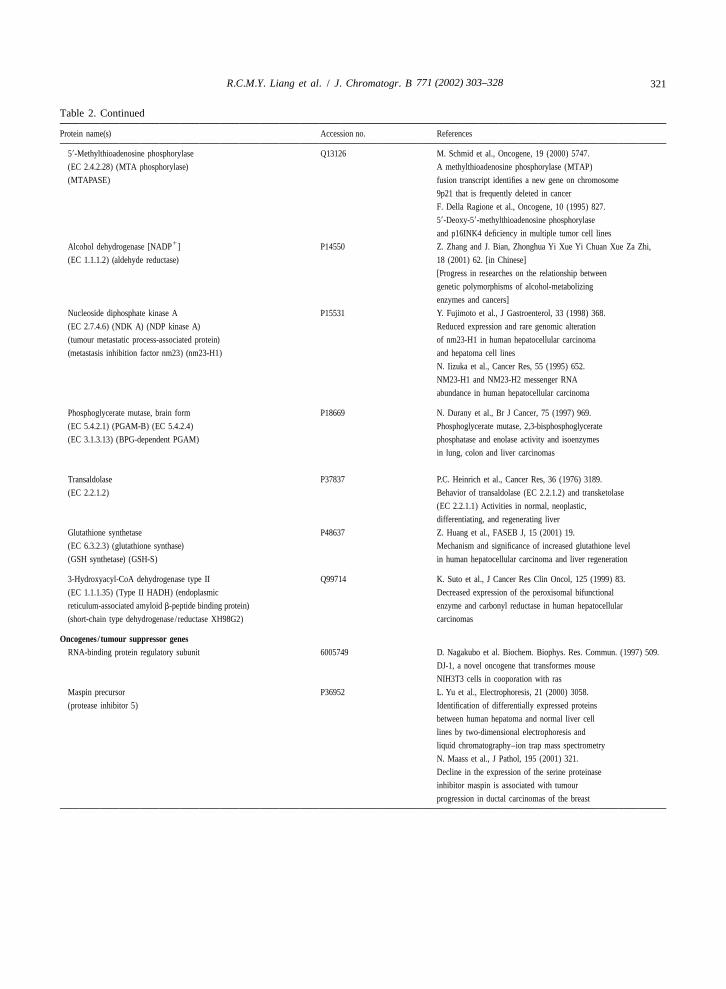
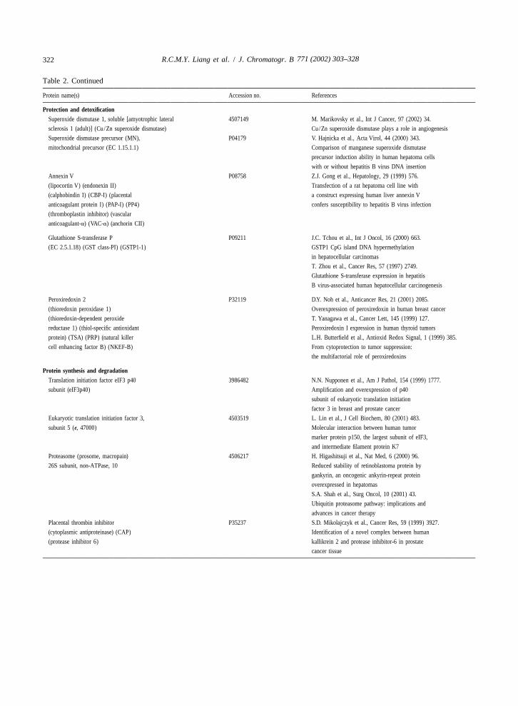
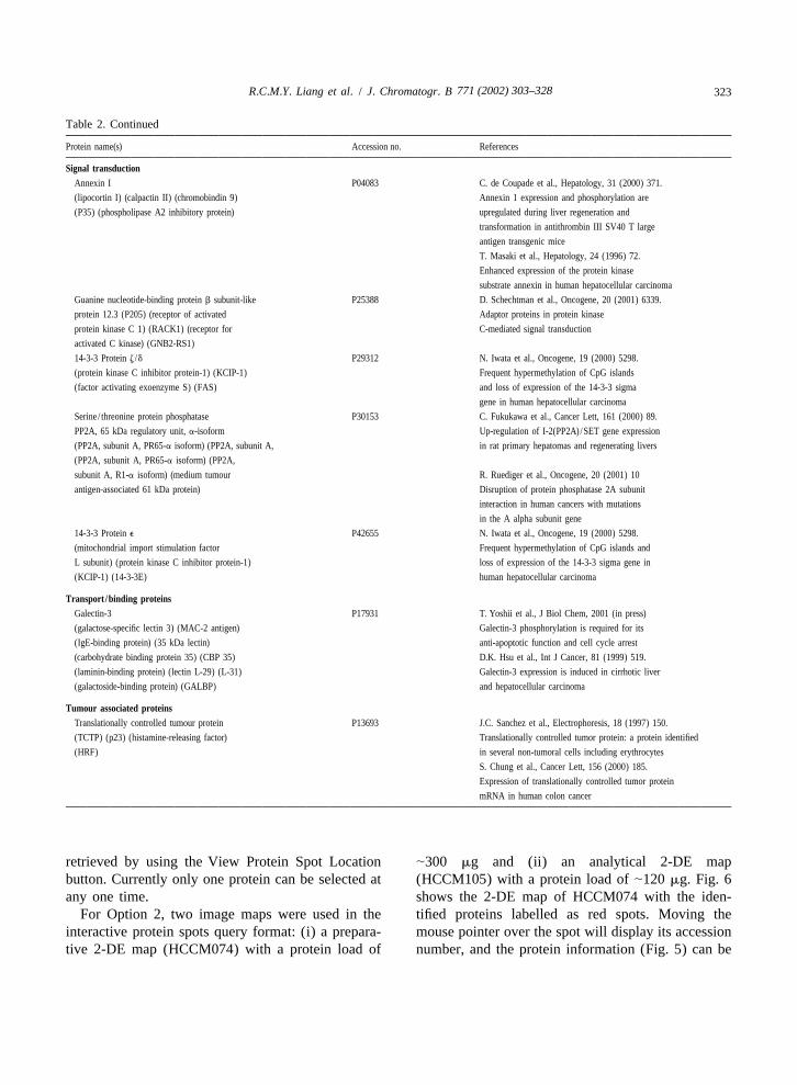
Table 2List of HCC-M proteins implicated in HCC and other cancers
Protein name(s) Accession no. References
Chaperone/stress inducedHeat shock 27 kDa protein P04792 L.Yu et al., Electrophoresis, 21 (2000) 3058.
(HSP 27) (stress-responsive protein 27) Identification of differentially expressed proteins
(SRP27) (oestrogen-regulated 24 kDa protein) between human hepatoma and normal liver cell
(28 kDa heat shock protein) lines by two-dimensional electrophoresis and liquid
chromatography–ion trap mass spectrometry
Heat shock protein HSP 90-a P07900 J. Hu and C. Seeger, Proc Natl Acad Sci USA
HSP 86) 93 (1996) 1060. Hsp90 is required for the
activity of a hepatitis B virus reverse transcriptase
G. Cho et al., Biochem Biophys Res Commun,
269 (2000) 191. Localization of HSP90 binding
in the human hepatitis B virus polymerase
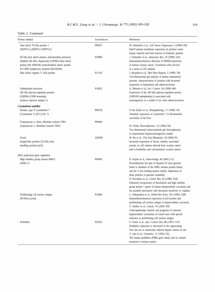
771 (2002) 303–328 319R.C.M.Y. Liang et al. / J. Chromatogr. B
Table 2. Continued
Protein name(s) Accession no. References
Heat shock 70 kDa protein 1 P08107 M. Hantschel et al., Cell Stress Chaperones, 5 (2000) 438.
(HSP70.1) (HSP70-1 /HSP70-2) Hsp70 plasma membrane expression on primary tumor
biopsy material and bone marrow of leukemic patients
60 kDa heat shock protein, mitochondrial precursor P10809 J. Schneider et al., Anticancer Res, 19 (1999) 2141.
(Hsp60) (60 kDa chaperonin) (CPN60) (heat shock Immunohistochemical detection of HSP60-expression
protein 60) (HSP-60) (mitochondrial matrix protein in human ovarian cancer. Correlation with survival
P1) (P60 lymphocyte protein) (HuCHA60) in a series of 247 patients
Heat shock cognate 71 kDa protein P11142 I. Byrjalsen et al., Mol Hum Reprod, 5 (1999) 748.
Two-dimensional gel analysis of human endometrial
proteins: characterization of proteins with increased
expression in hyperplasia and adenocarcinoma
Endoplasmin precursor P14625 A. Menoret et al., Int J Cancer, 56 (1994) 400.
(94 kDa glucose-regulated protein) Expression of the 100 kDa glucose-regulated protein
(GRP94) (GP96 homolog) (GRP100/endoplasmin) is associated with
(tumour rejection antigen 1) tumorigenicity in a model of rat colon adenocarcinoma
Cytoskeleton/mobilityKeratin, type II cytoskeletal 7 P08729 P. Van Eyken et al., Histopathology, 17 (1990) 101.
(Cytokeratin 7) (K7) (CK 7) Abundant expression of cytokeratin 7 in fibrolamellar
carcinoma of the liver
Tropomyosin a chain, fibroblast isoform TM3 P09494
(tropomyosin 1, fibroblast isoform TM3) P.J. Wirth, Electrophoresis. 15 (1994) 358.
Two-dimensional polyacrylamide gel electrophoresis
in experimental hepatocarcinogenesis studies
Fascin Q16658 W. Hu et al., Clin Exp Metastasis, 18 (2000) 83.
(singed-like protein) (55 kDa actin Increased expression of fascin, motility associated
bundling protein) (p55) protein, in cell cultures derived from ovarian cancer
and in borderline and carcinomatous ovarian tumors
DNA replication /gene regulation
High mobility group protein HMG1 P09429 K. Kajino et al., Intervirology 44 (2001) 311.
(HMG-1) Recombination hot spot of hepatitis B virus genome
binds to members of the HMG domain protein family
and the Y box binding protein family; implication of
these proteins in genomic instability
N. Kawahara et al., Cancer Res, 56 (1996) 5330.
Enhanced coexpression of thioredoxin and high mobility
group protein 1 genes in human hepatocellular carcinoma and
the possible association with decreased sensitivity to cisplatin
Proliferating cell nuclear antigen P12004 L. Nakopoulou et al., Pathol Res Pract, 191 (1995) 1208.
(PCNA) (cyclin) Immunohistochemical expression of p53 protein and
proliferating cell nuclear antigen in hepatocellular carcinoma
T. Suehiro et al., Cancer, 76 (1995) 399.
Clinicopathologic features and prognosis of resected
hepatocellular carcinomas of varied sizes with special
reference to proliferating cell nuclear antigen
Prohibitin P35232 S. Tanno et al., Jpn J Cancer Res, 88 (1997) 1155.
Prohibitin expression is decreased in the regenerating
liver but not in chemically induced hepatic tumors in rats
T. Sato et al., Genomics, 17 (1993) 762.
The human prohibitin (PHB) gene family and its somatic
mutations in human tumors
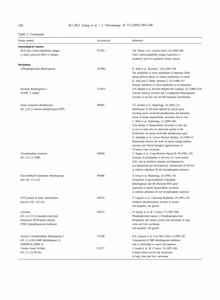
771 (2002) 303–328320 R.C.M.Y. Liang et al. / J. Chromatogr. B
Table 2. Continued
Protein name(s) Accession no. References
Immunological responseHLA class I histocompatibility antigen, P17693 D.H. Moore et al., Gynecol Oncol, 38 (1990) 458.
a chainG precursor (HLA G antigen) Class I histocompatibility antigen expression: a
prognostic factor for aneuploid ovarian cancers
Metabolism3-Phosphoglycerate dehydrogenase 2674062 K. Snell et al., Biochem J. 245 (1987) 609.
The modulation of serine metabolism in hepatoma 3924a
during different phases of cellular proliferation in culture
K. Snell and G. Weber, Biochem J, 233 (1986) 617.
Enzymic imbalance in serine metabolism in rat hepatomas
Isocitrate dehydrogenase 1 5174471 A.N. Murphy et al., Biochem Biophys Res Commun, 157 (1988) 1218.1(NADP ), soluble Calcium sensitive isocitrate and 2-oxoglutarate dehydrogenase
activities in rat liver and AS-30D hepatoma mitochondria
Purine nucleoside phosphorylase P00491 T.U. Krohne et al., Hepatology, 34 (2001) 511.
(EC 2.4.2.1) (inosine phosphorylase) (PNP) Mechanisms of cell death induced by suicide genes
encoding purine nucleoside phosphorylase and thymidine
kinase in human hepatocellular carcinoma cells in vitro
L. Mohr et al., Hepatology, 31 (2000) 606.
Gene therapy of hepatocellular carcinoma in vitro and
in vivo in nude mice by adenoviral transfer of the
Escherichia coli purine nucleoside phosphorylase gene
O. Sanfilippo et al., Cancer Biochem Biophys, 14 (1994) 57.
Relationship between the levels of purine salvage pathway
enzymes and clinical /biological aggressiveness of
of human colon carcinoma
Triosephosphate isomerase P00938 T. Nagase et al., Comp Biochem Physiol B, 99 (1991) 193.
(EC 5.3.1.1) (TIM) Analyses of polypeptides in the liver of a novel mutant
(LEC rats) to hereditary hepatitis and hepatoma by
two-dimensional gel electrophoresis: identification of P29/6.8
as carbonic anhydrase III and triosephosphate isomerase
Glyceraldehyde-3-phosphate dehydrogenase, P04406 Y. Gong et al., Hepatology, 23 (1996) 734.
liver (EC 1.2.1.12) Comparison of glyceraldehyde-3-phosphate
dehydrogenase and 28s-ribosomal RNA gene
expression in human hepatocellular carcinoma
as carbonic anhydrase III and triosephosphate isomerase
ATP synthase b chain, mitochondrial P06576 F. Capuano et al., J Bioenerg Biomembr, 29 (1997) 379.
precursor (EC 3.6.3.14) Oxidative phosphorylation enzymes in normal
and neoplastic cell growth
a-Enolase P06733 N. Durany et al., Br J Cancer, 75 (1997) 969.
(EC 4.2.1.11) (2-phospho-D-glycerate Phosphoglycerate mutase, 2,3-bisphosphoglycerate
hydrolyase) (NON-neural enolase) phosphatase and enolase activity and isoenzymes in lung,
(NNE) (phosphopyruvate hydratase) colon and liver carcinomas
and neoplastic cell growth
Inosine-59-monophosphate dehydrogenase 2 P12268 H.N. Jayaram et al., Curr Med Chem, 6 (1999) 561.
(EC 1.1.1.205) (IMP dehydrogenase 2) Consequences of IMP dehydrogenase inhibition,
(IMPDH-II) (IMPD 2) and its relationship to cancer and apoptosis
Creatine kinase, B-chain P12277 J. Joseph et al., Br J Cancer, 76 (1997) 600.
(EC 2.7.3.2) (B-CK) Creatine kinase activity and isoenzymes
in lung, colon and liver carcinomas
771 (2002) 303–328 321R.C.M.Y. Liang et al. / J. Chromatogr. B
Table 2. Continued
Protein name(s) Accession no. References
59-Methylthioadenosine phosphorylase Q13126 M. Schmid et al., Oncogene, 19 (2000) 5747.
(EC 2.4.2.28) (MTA phosphorylase) A methylthioadenosine phosphorylase (MTAP)
(MTAPASE) fusion transcript identifies a new gene on chromosome
9p21 that is frequently deleted in cancer
F. Della Ragione et al., Oncogene, 10 (1995) 827.
59-Deoxy-59-methylthioadenosine phosphorylase
and p16INK4 deficiency in multiple tumor cell lines1Alcohol dehydrogenase [NADP ] P14550 Z. Zhang and J. Bian, Zhonghua Yi Xue Yi Chuan Xue Za Zhi,
(EC 1.1.1.2) (aldehyde reductase) 18 (2001) 62. [in Chinese]
[Progress in researches on the relationship between
genetic polymorphisms of alcohol-metabolizing
enzymes and cancers]
Nucleoside diphosphate kinase A P15531 Y. Fujimoto et al., J Gastroenterol, 33 (1998) 368.
(EC 2.7.4.6) (NDK A) (NDP kinase A) Reduced expression and rare genomic alteration
(tumour metastatic process-associated protein) of nm23-H1 in human hepatocellular carcinoma
(metastasis inhibition factor nm23) (nm23-H1) and hepatoma cell lines
N. Iizuka et al., Cancer Res, 55 (1995) 652.
NM23-H1 and NM23-H2 messenger RNA
abundance in human hepatocellular carcinoma
Phosphoglycerate mutase, brain form P18669 N. Durany et al., Br J Cancer, 75 (1997) 969.
(EC 5.4.2.1) (PGAM-B) (EC 5.4.2.4) Phosphoglycerate mutase, 2,3-bisphosphoglycerate
(EC 3.1.3.13) (BPG-dependent PGAM) phosphatase and enolase activity and isoenzymes
in lung, colon and liver carcinomas
Transaldolase P37837 P.C. Heinrich et al., Cancer Res, 36 (1976) 3189.
(EC 2.2.1.2) Behavior of transaldolase (EC 2.2.1.2) and transketolase
(EC 2.2.1.1) Activities in normal, neoplastic,
differentiating, and regenerating liver
Glutathione synthetase P48637 Z. Huang et al., FASEB J, 15 (2001) 19.
(EC 6.3.2.3) (glutathione synthase) Mechanism and significance of increased glutathione level
(GSH synthetase) (GSH-S) in human hepatocellular carcinoma and liver regeneration
3-Hydroxyacyl-CoA dehydrogenase type II Q99714 K. Suto et al., J Cancer Res Clin Oncol, 125 (1999) 83.
(EC 1.1.1.35) (Type II HADH) (endoplasmic Decreased expression of the peroxisomal bifunctional
reticulum-associated amyloid b-peptide binding protein) enzyme and carbonyl reductase in human hepatocellular
(short-chain type dehydrogenase / reductase XH98G2) carcinomas
Oncogenes / tumour suppressor genesRNA-binding protein regulatory subunit 6005749 D. Nagakubo et al. Biochem. Biophys. Res. Commun. (1997) 509.
DJ-1, a novel oncogene that transformes mouse
NIH3T3 cells in cooporation with ras
Maspin precursor P36952 L. Yu et al., Electrophoresis, 21 (2000) 3058.
(protease inhibitor 5) Identification of differentially expressed proteins
between human hepatoma and normal liver cell
lines by two-dimensional electrophoresis and
liquid chromatography–ion trap mass spectrometry
N. Maass et al., J Pathol, 195 (2001) 321.
Decline in the expression of the serine proteinase
inhibitor maspin is associated with tumour
progression in ductal carcinomas of the breast
771 (2002) 303–328322 R.C.M.Y. Liang et al. / J. Chromatogr. B
Table 2. Continued
Protein name(s) Accession no. References
Protection and detoxificationSuperoxide dismutase 1, soluble [amyotrophic lateral 4507149 M. Marikovsky et al., Int J Cancer, 97 (2002) 34.
sclerosis 1 (adult)] (Cu/Zn superoxide dismutase) Cu/Zn superoxide dismutase plays a role in angiogenesis
Superoxide dismutase precursor (MN), P04179 V. Hajnicka et al., Acta Virol, 44 (2000) 343.
mitochondrial precursor (EC 1.15.1.1) Comparison of manganese superoxide dismutase
precursor induction ability in human hepatoma cells
with or without hepatitis B virus DNA insertion
Annexin V P08758 Z.J. Gong et al., Hepatology, 29 (1999) 576.
(lipocortin V) (endonexin II) Transfection of a rat hepatoma cell line with
(calphobindin I) (CBP-I) (placental a construct expressing human liver annexin V
anticoagulant protein I) (PAP-I) (PP4) confers susceptibility to hepatitis B virus infection
(thromboplastin inhibitor) (vascular
anticoagulant-a) (VAC-a) (anchorin CII)
Glutathione S-transferase P P09211 J.C. Tchou et al., Int J Oncol, 16 (2000) 663.
(EC 2.5.1.18) (GST class-PI) (GSTP1-1) GSTP1 CpG island DNA hypermethylation
in hepatocellular carcinomas
T. Zhou et al., Cancer Res, 57 (1997) 2749.
Glutathione S-transferase expression in hepatitis
B virus-associated human hepatocellular carcinogenesis
Peroxiredoxin 2 P32119 D.Y. Noh et al., Anticancer Res, 21 (2001) 2085.
(thioredoxin peroxidase 1) Overexpression of peroxiredoxin in human breast cancer
(thioredoxin-dependent peroxide T. Yanagawa et al., Cancer Lett, 145 (1999) 127.
reductase 1) (thiol-specific antioxidant Peroxiredoxin I expression in human thyroid tumors
protein) (TSA) (PRP) (natural killer L.H. Butterfield et al., Antioxid Redox Signal, 1 (1999) 385.
cell enhancing factor B) (NKEF-B) From cytoprotection to tumor suppression:
the multifactorial role of peroxiredoxins
Protein synthesis and degradationTranslation initiation factor eIF3 p40 3986482 N.N. Nupponen et al., Am J Pathol, 154 (1999) 1777.
subunit (eIF3p40) Amplification and overexpression of p40
subunit of eukaryotic translation initiation
factor 3 in breast and prostate cancer
Eukaryotic translation initiation factor 3, 4503519 L. Lin et al., J Cell Biochem, 80 (2001) 483.
subunit 5 (e, 47000) Molecular interaction between human tumor
marker protein p150, the largest subunit of eIF3,
and intermediate filament protein K7
Proteasome (prosome, macropain) 4506217 H. Higashitsuji et al., Nat Med, 6 (2000) 96.
26S subunit, non-ATPase, 10 Reduced stability of retinoblastoma protein by
gankyrin, an oncogenic ankyrin-repeat protein
overexpressed in hepatomas
S.A. Shah et al., Surg Oncol, 10 (2001) 43.
Ubiquitin proteasome pathway: implications and
advances in cancer therapy
Placental thrombin inhibitor P35237 S.D. Mikolajczyk et al., Cancer Res, 59 (1999) 3927.
(cytoplasmic antiproteinase) (CAP) Identification of a novel complex between human
(protease inhibitor 6) kallikrein 2 and protease inhibitor-6 in prostate
cancer tissue
771 (2002) 303–328 323R.C.M.Y. Liang et al. / J. Chromatogr. B
Table 2. Continued
Protein name(s) Accession no. References
Signal transductionAnnexin I P04083 C. de Coupade et al., Hepatology, 31 (2000) 371.
(lipocortin I) (calpactin II) (chromobindin 9) Annexin 1 expression and phosphorylation are
(P35) (phospholipase A2 inhibitory protein) upregulated during liver regeneration and
transformation in antithrombin III SV40 T large
antigen transgenic mice
T. Masaki et al., Hepatology, 24 (1996) 72.
Enhanced expression of the protein kinase
substrate annexin in human hepatocellular carcinoma
Guanine nucleotide-binding protein b subunit-like P25388 D. Schechtman et al., Oncogene, 20 (2001) 6339.
protein 12.3 (P205) (receptor of activated Adaptor proteins in protein kinase
protein kinase C 1) (RACK1) (receptor for C-mediated signal transduction
activated C kinase) (GNB2-RS1)
14-3-3 Protein z /d P29312 N. Iwata et al., Oncogene, 19 (2000) 5298.
(protein kinase C inhibitor protein-1) (KCIP-1) Frequent hypermethylation of CpG islands
(factor activating exoenzyme S) (FAS) and loss of expression of the 14-3-3 sigma
gene in human hepatocellular carcinoma
Serine / threonine protein phosphatase P30153 C. Fukukawa et al., Cancer Lett, 161 (2000) 89.
PP2A, 65 kDa regulatory unit, a-isoform Up-regulation of I-2(PP2A)/SET gene expression
(PP2A, subunit A, PR65-a isoform) (PP2A, subunit A, in rat primary hepatomas and regenerating livers
(PP2A, subunit A, PR65-a isoform) (PP2A,
subunit A, R1-a isoform) (medium tumour R. Ruediger et al., Oncogene, 20 (2001) 10
antigen-associated 61 kDa protein) Disruption of protein phosphatase 2A subunit
interaction in human cancers with mutations
in the A alpha subunit gene
14-3-3 Protein e P42655 N. Iwata et al., Oncogene, 19 (2000) 5298.
(mitochondrial import stimulation factor Frequent hypermethylation of CpG islands and
L subunit) (protein kinase C inhibitor protein-1) loss of expression of the 14-3-3 sigma gene in
(KCIP-1) (14-3-3E) human hepatocellular carcinoma
Transport /binding proteinsGalectin-3 P17931 T. Yoshii et al., J Biol Chem, 2001 (in press)
(galactose-specific lectin 3) (MAC-2 antigen) Galectin-3 phosphorylation is required for its
(IgE-binding protein) (35 kDa lectin) anti-apoptotic function and cell cycle arrest
(carbohydrate binding protein 35) (CBP 35) D.K. Hsu et al., Int J Cancer, 81 (1999) 519.
(laminin-binding protein) (lectin L-29) (L-31) Galectin-3 expression is induced in cirrhotic liver
(galactoside-binding protein) (GALBP) and hepatocellular carcinoma
Tumour associated proteinsTranslationally controlled tumour protein P13693 J.C. Sanchez et al., Electrophoresis, 18 (1997) 150.
(TCTP) (p23) (histamine-releasing factor) Translationally controlled tumor protein: a protein identified
(HRF) in several non-tumoral cells including erythrocytes
S. Chung et al., Cancer Lett, 156 (2000) 185.
Expression of translationally controlled tumor protein
mRNA in human colon cancer
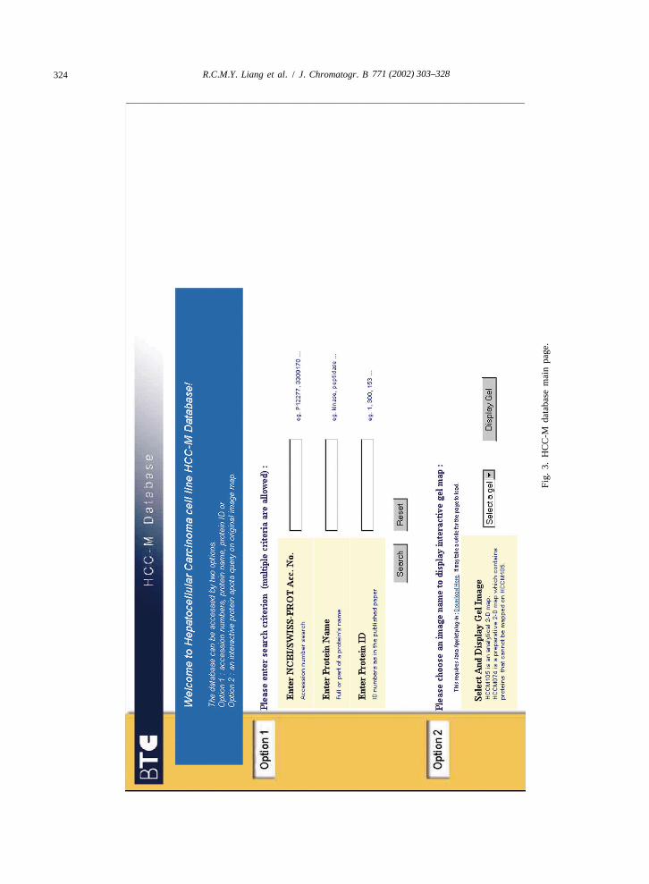
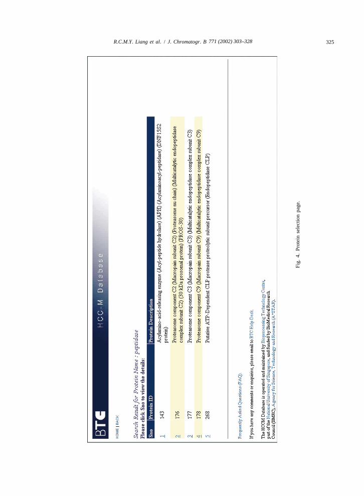
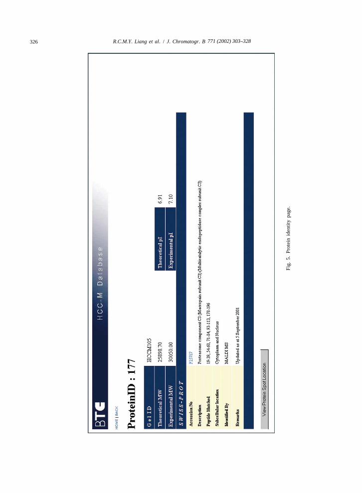
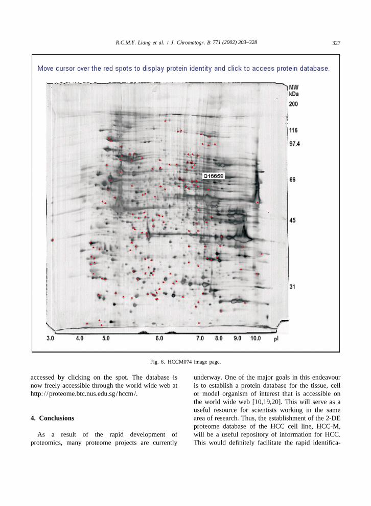
retrieved by using the View Protein Spot Location |300 mg and (ii) an analytical 2-DE mapbutton. Currently only one protein can be selected at (HCCM105) with a protein load of |120 mg. Fig. 6any one time. shows the 2-DE map of HCCM074 with the iden-
For Option 2, two image maps were used in the tified proteins labelled as red spots. Moving theinteractive protein spots query format: (i) a prepara- mouse pointer over the spot will display its accessiontive 2-DE map (HCCM074) with a protein load of number, and the protein information (Fig. 5) can be
771 (2002) 303–328324 R.C.M.Y. Liang et al. / J. Chromatogr. B
Fig.
3.H
CC
-Mda
taba
sem
ain
page
.
771 (2002) 303–328 325R.C.M.Y. Liang et al. / J. Chromatogr. B
Fig.
4.Pr
otei
nse
lect
ion
page
.
771 (2002) 303–328326 R.C.M.Y. Liang et al. / J. Chromatogr. B
Fig.
5.Pr
otei
nid
entit
ypa
ge.
771 (2002) 303–328 327R.C.M.Y. Liang et al. / J. Chromatogr. B
Fig. 6. HCCM074 image page.
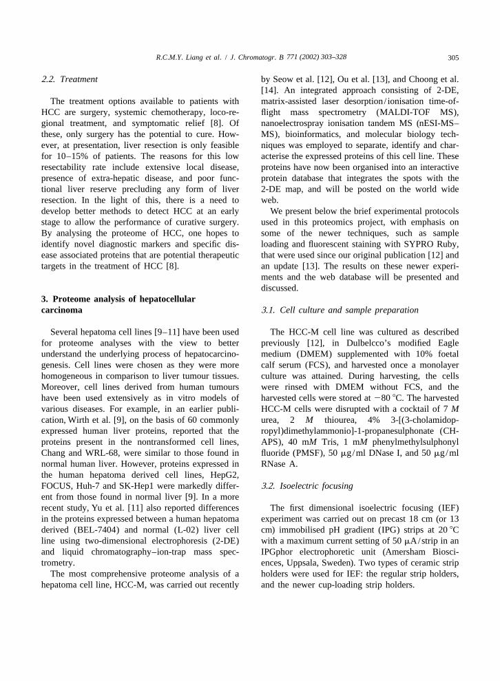
accessed by clicking on the spot. The database is underway. One of the major goals in this endeavournow freely accessible through the world wide web at is to establish a protein database for the tissue, cellhttp: / /proteome.btc.nus.edu.sg /hccm/. or model organism of interest that is accessible on
the world wide web [10,19,20]. This will serve as auseful resource for scientists working in the same
4. Conclusions area of research. Thus, the establishment of the 2-DEproteome database of the HCC cell line, HCC-M,
As a result of the rapid development of will be a useful repository of information for HCC.proteomics, many proteome projects are currently This would definitely facilitate the rapid identifica-
771 (2002) 303–328328 R.C.M.Y. Liang et al. / J. Chromatogr. B
tion of novel diagnostic and therapeutic markers for ReferencesHCC, which is an important first step towards theearly diagnosis and treatment of this cancer. [1] R.J. Simpson, D.S. Dorow, in: W. Blackstock, M. Mann
(Eds.), Proteomics: A Trends Guide, Elsevier, 2001, p. S40.[2] R.E. Banks, M.J. Dunn, D.F. Hochstrasser, J.-C. Sanchez, W.
Blackstock, D.J. Pappin, P.J. Selby, Lancet 356 (2000) 1749.5. Nomenclature [3] G. Chambers, L. Lawrie, P. Cash, G.I. Murray, J. Pathol. 192
(2000) 280.HCC or hepatoma hepatocellular carcinoma [4] M.J. Page, B. Amess, C. Rohlff, C. Stubberfield, R. Parekh,
Drug Discov. Today 4 (1999) 55.HBV hepatitis B virus[5] A.A. Alaiya, B. Franzen, G. Auer, S. Linder, ElectrophoresisHCV hepatitis C virus
21 (2000) 1210.2-DE two-dimensional electropho- [6] Cancer Proteomics in: S. Hanash (ed.) Proteomics 1 (2001)
resis 1191.MALDI-TOF MS matrix-assisted laser desorp- [7] D.F. Schafer, M.F. Sorrell, Lancet 353 (1999) 1253.
[8] T.K. Seow, R.C.M.Y. Liang, C.K. Leow, M.C.M. Chung,tion / ionisation time-of-flightProteomics 1 (2001) 1249.mass spectrometry
[9] P.J. Wirth, T.N. Hoang, T. Benjamin, Electrophoresis 16nESI-MS–MS nanoelectrospray ionisation tan- (1995) 1946.
dem MS [10] J.-C. Sanchez, R.D. Appel, O. Golaz, C. Pasquali, F. Ravier,DMEM Dulbelcco’s modified Eagle A. Bairoch, D.F. Hochstrasser, Electrophoresis 16 (1995)
1131.medium[11] L.-R. Yu, R. Zeng, X.-X. Shao, N. Wang, Y.-H. Xu, Q.-C.FCS foetal calf serum
Xia, Electrophoresis 21 (2000) 3058.CHAPS 3-[(3-cholamidop- [12] T.K. Seow, S.-E. Ong, R.C.M.Y. Liang, E.-C. Ren, L. Chan,
ropyl)dimethylammonio]-1-pro- K. Ou, M.C.M. Chung, Electrophoresis 21 (2000) 1787.panesulphonate [13] K. Ou, T.K. Seow, R.C.M.Y. Liang, M.C.M. Chung, Electro-
phoresis 22 (2001) 2804.PMSF phenylmethylsulphonyl fluoride[14] M.L. Choong, L.K. Tan, S.L. Lo, E.-C. Ren, K. Ou, S.-E.IEF isoelectric focusing
Ong, R.C.M.Y. Liang, T.K. Seow, M.C.M. Chung, FEBSIPG immobilised pH gradient Lett. 496 (2001) 109.DTT dithiothreitol [15] T.K. Seow, R. Korke, R.C.M.Y. Liang, S.-E. Ong, K. Ou, K.SDS–PAGE sodium dodecyl sulphate–poly- Wong, W.-S. Hu, M.C.M. Chung, Biotechnol. Prog. 17
(2001) 1137.acrylamide gel electrophoresis[16] D.N. Perkins, D.J. Pappin, D.M. Creasy, J.S. Cottrell,IAA iodoacetamide
Electrophoresis 20 (1999) 3551.ACN acetonitrile [17] H.F. Kawai, S. Kaneko, M. Honda, Y. Shirota, K. Kobayashi,TFA trifluoroacetic acid Hepatology 33 (2001) 676.
[18] A. Gorg, C. Obermaier, G. Boguth, A. Harder, B. Scheibe,R. Wildgruber, W. Weiss, Electrophoresis 21 (2000) 1037.
[19] C. Hoogland, J.-C. Sanchez, D. Walter, V. Baujard, O.AcknowledgementsBaujard, L. Tonella, D.F. Hochstrasser, R.F. Appel, Electro-phoresis 20 (1999) 3568.
We gratefully acknowledge the assistance of all [20] J.E. Celis, M. Ostergaard, N.A. Jensen, I. Gromova, H.H.the proteomics staff in the preparation of this review. Rasmussen, P. Gromov, FEBS Lett. 430 (1998) 64.
Recommended


