of November 28, 2013.This information is current as
Dendritic Cell FunctionsRegulatory Activity of Autocrine IL-10 on
Pastore and Giampiero GirolomoniSilvia Corinti, Cristina Albanesi, Andrea la Sala, Saveria
http://www.jimmunol.org/content/166/7/43122001; 166:4312-4318; ;J Immunol
Referenceshttp://www.jimmunol.org/content/166/7/4312.full#ref-list-1
, 39 of which you can access for free at: cites 55 articlesThis article
Subscriptionshttp://jimmunol.org/subscriptions
is online at: The Journal of ImmunologyInformation about subscribing to
Permissionshttp://www.aai.org/ji/copyright.htmlSubmit copyright permission requests at:
Email Alertshttp://jimmunol.org/cgi/alerts/etocReceive free email-alerts when new articles cite this article. Sign up at:
Print ISSN: 0022-1767 Online ISSN: 1550-6606. Immunologists All rights reserved.Copyright © 2001 by The American Association of9650 Rockville Pike, Bethesda, MD 20814-3994.The American Association of Immunologists, Inc.,
is published twice each month byThe Journal of Immunology
by guest on Novem
ber 28, 2013http://w
ww
.jimm
unol.org/D
ownloaded from
by guest on N
ovember 28, 2013
http://ww
w.jim
munol.org/
Dow
nloaded from
Regulatory Activity of Autocrine IL-10 on Dendritic CellFunctions1
Silvia Corinti, 2 Cristina Albanesi, Andrea la Sala, Saveria Pastore, and Giampiero Girolomoni
IL-10 is a critical cytokine that blocks the maturation of dendritic cells (DCs), but the relevance of autocrine IL-10 on DC functionshas not been investigated. In this study, we found that immature monocyte-derived DCs released low but sizeable amounts ofIL-10. After stimulation with bacteria, LPS, lipoteichoic acid, or soluble CD40 ligand, DCs secreted high levels of IL-10. Additionof an anti-IL-10-neutralizing Ab to immature DCs as well as to soluble CD40 ligand- or LPS-maturing DCs led to enhancedexpression of surface CD83, CD80, CD86, and MHC molecules and markedly augmented release of TNF-a and IL-12, butdiminished IL-10 mRNA expression. Moreover, DCs treated with anti-IL-10 Ab showed an increased capacity to activate allo-geneic T cells and primed naive T cells to a more prominent Th1 polarization. DC maturation and IL-10 neutralization wereassociated with enhanced accumulation of the IL-10 receptor binding chain (IL-10R1) mRNA and intracellular IL-10R1 protein.In contrast, surface IL-10R1 and IL-10 binding activity diminished in mature DCs. These results indicate that autocrine IL-10prevents spontaneous maturation of DCs in vitro, limits LPS- and CD40-mediated maturation, and increases IL-10 production byDCs. Moreover, IL-10R expression appears to be regulated by both transcriptional and posttranscriptional mechanisms. Endog-enous IL-10 and IL-10R can be relevant targets for the manipulation of DC functions. The Journal of Immunology,2001, 166:4312–4318.
D endritic cells (DCs)3 are essential elements in the initi-ation of immune responses (1). DCs reside in unper-turbed tissues in an immature form, where they are
adapted for capturing and accumulating Ags. Indeed, DCs possessa wide spectrum of recognition systems for an efficient screeningof the tissue environment. A variety of danger signals, includingmicroorganisms, dying cells, or proinflammatory cytokines, in-duce the terminal differentiation, also known as maturation, of DCs(1–5). Mature DCs migrate to lymph nodes, acquire potent Ag-presenting capacity, and stimulate T cell responses vigorously.Moreover, maturation of DCs is strengthened during interactionswith T cells by signals such CD40 ligand (CD40L) provided by Tcells themselves (6–8). Mature DCs express high levels of Ag-presenting and costimulatory molecules, and release large amountsof IL-12, thereby stimulating preferentially Th1 responses. Thus,DC maturation is a key checkpoint in the initiation of immunityand has important consequences also on the quality of the immuneresponse.
To prevent exaggerated and unwanted immune responses, DCmaturation and functions are tightly regulated. IL-10 has beenidentified as a major factor that can prevent the differentiation ofDCs from monocytes and strongly inhibit DC maturation induced
by different stimuli (9–14). In particular, IL-10 blocks the up-regulation of costimulatory molecules and IL-12 production andthus impairs the ability of DCs to generate Th1 responses (15).IL-10-treated DCs are not only less efficient at stimulating T cellresponses but can induce a state of Ag-specific tolerance (9, 16,17). IL-10 can be secreted by different cell types, including mono-cytes, mast cells, T regulatory cells, and tumor cells, and has animportant role in limiting allergic and autoimmune reactions or inmediating tumor escape from immune surveillance (17, 18). Incontrast, several reports have shown that mature DCs are no longersensitive to the inhibitory effects of IL-10 (14, 15, 17, 19–21), butthe molecular bases of this phenomenon are as yet unknown. IL-10exerts its actions through a heterodimeric membrane receptorformed by a binding chain (IL-10R1) and a transducing chain (IL-10R2, also known as CFR2–4), whose mutual interaction activatesa series of intracellular signaling molecules, including STAT pro-teins (22–29).
Although some DC subsets can produce IL-10 (3, 13, 30–32),the impact of endogenous IL-10 on DC biology has been onlymarginally investigated (13, 33). In this work, we provide evidencethat autocrine IL-10 serves as a potent mechanism for limiting thematuration of monocyte-derived DCs and their capacity to initiateTh1 responses. Moreover, we show that mature DCs accumulatehigher amounts of IL-10R1 mRNA and intracellular IL-10R1 pro-tein but reduce surface IL-10R1 expression and IL-10 binding ac-tivity. Thus, endogenous IL-10 and IL-10R appear to be importantregulators of DC biology and can represent relevant targets for themanipulation of DC functions.
Materials and MethodsReagents and Abs
Streptococcus gordoniistrain GP1221 was a gift from Dr. D. Medaglini(University of Siena, Siena, Italy), andSalmonella typhi(Neotyf) was pro-vided by Chiron Italia (Siena, Italy). LPS (fromSalmonella typhimurium),lipoteichoic acid (LTA; fromStaphylococcus aureus), and poly I:C werepurchased from Sigma-Aldrich (Milan, Italy). Soluble CD40L (sCD40L)was obtained from Alexis (San Diego, CA). It is composed of the extra-cellular domain of human CD40L fused to the N terminus of a linker
Laboratory of Immunology, Istituto Dermopatico dell’Immacolata, Istituto di Ricov-ero e Cura a Carattere Scientifico, Rome, Italy
Received for publication November 2, 2000. Accepted for publication January 19,2001.
The costs of publication of this article were defrayed in part by the payment of pagecharges. This article must therefore be hereby markedadvertisementin accordancewith 18 U.S.C. Section 1734 solely to indicate this fact.1 This work was supported by the Associazione Italiana per la Ricerca sul Cancro, theIstituto Superiore di Sanita (AIDS project, Grant 40B/1.18), the Ministero dellaSanita, and the European Community (Biomed 2 program, Grant BMH4-CT98-3713).2 Address correspondence and reprint requests to Dr. Silvia Corinti, Laboratory ofImmunology, Istituto Dermopatico dell’Immacolata, Via Monti di Creta 104, 00167Roma, Italy. E-mail address: [email protected] Abbreviations used in this paper: DC, dendritic cell; CD40L, CD40 ligand; sCD40L,soluble CD40L; LTA, lipoteichoic acid.
Copyright © 2001 by The American Association of Immunologists 0022-1767/01/$02.00
by guest on Novem
ber 28, 2013http://w
ww
.jimm
unol.org/D
ownloaded from
peptide and a FLAG-tag and was used together with an enhancer thatincreases the biological activity of sCD40L. The mAbs FITC-conjugatedand pure anti-HLA-DR (L243), FITC-conjugated anti-CD14 (MFP9), anti-CD3 (SK7), anti-CD4 (SK3), and anti-CD45RA (L48) were obtained fromBecton Dickinson (San Jose, CA). FITC-conjugated anti-CD1a (HI149),anti-CD86 (2331), anti-CD40 (5C3), anti-IFN-g (4S.B3), pure anti-CD28(CD28.2), PE-conjugated anti-IL-4 (MP4–25D2, rat), and pure anti-CD45RO (UCHL1) were obtained from BD PharMingen (San Diego, CA).FITC-conjugated anti-CD54 (84H10) and anti-CD80 (MAB104), and pureanti-CD83 (HB15A) and anti-CD3 (UCHT1) came from Immunotech(Marseille, France). Anti-MHC class I (W6/32) was obtained from Dako(Glostrup, Denmark). Control mouse or rat Ig were obtained from BectonDickinson or BD PharMingen. The mouse mAb anti-human IL-10(23738.11) came from R&D Systems (Minneapolis, MN), and the rat anti-human IL-10R1 mAb 3F9 was obtained from BD PharMingen. Anti-IL-10mAb and sCD40L had undetectable endotoxin levels (,10 pg/mg) by theLimulusamebocyte lysate assay (BioWhittaker, Walkersville, MD).
DC preparation and stimulation
DCs were prepared from PBMC of healthy individuals as described pre-viously (3). Briefly, PBMC were separated on multistep Percoll gradients(Pharmacia, Uppsala, Sweden), and cells from the light density fraction(42.5–50%;.90% CD141) were cultured at 13 106 cells/ml in RPMI1640 (Life Technologies, Gaithersburg, MD) containing 1 mM sodiumpyruvate, 0.1 mM nonessential amino acids, 2 mML-glutamine, 25 mMHEPES, 100 U/ml penicillin, 100mg/ml streptomycin (all obtained fromLife Technologies), and 0.05 mM 2-ME (Merck, Darmstadt, Germany)(complete RPMI), and supplemented with 10% FBS (HyClone, Logan,UT), 100 ng/ml human rGM-CSF (Mielogen, Schering-Plough, Milan, It-aly), and 200 U/ml human rIL-4 (R&D Systems) at 37°C with 5% CO2.Medium was changed after 3 days, and at day 6 of culture, cells wererecovered and depleted of CD21 and CD191 cells by means of immuno-magnetic beads coated with specific mAbs (Dynal, Oslo, Norway). Thisprocedure gave.97% pure CD1a1 and CD142 DC preparations. DCswere incubated in 6-well plates (13 106 cells/ml) at 37°C withS. gordonii(bacteria-to-DCs ratio, 50:1),S. typhi(bacteria-to-DCs ratio, 1:1), LPS (10mg/ml), LTA (10 mg/ml), poly I:C (100 ng/ml), or sCD40L (1mg/ml).Where indicated, DCs were treated with anti-HLA-DR mAb (10mg/ml) for1 h at 4°C, washed, and cultured at 37°C in the presence or absence ofsCD40L. To block endogenous IL-10, the neutralizing anti-IL-10 mAb ormouse Ig were added to unstimulated DC cultures or at the time of stim-ulation with LPS or sCD40L, and cells were analyzed after 18 h. Prelim-inary dose-response experiments indicated that the optimal neutralizingdose of anti-IL-10 mAb was 10mg/ml.
ELISA
Measurement of IL-12 (p70) and TNF-a in DC supernatants was per-formed by using OptEIA kits from BD PharMingen, as per the manufac-turer’s protocol. IL-10R1 was determined in DC supernatants and cell ly-sates by using the Ab pair mouse mAb 370607.11 for coating and goatpolyclonal biotinylated AF-274-NA for detection (R&D Systems).
Flow cytometry analysis of DCs
DCs either untreated or stimulated for 18 h with LPS or sCD40L in thepresence of anti-IL-10 mAb or control IgG were washed and then incu-bated in PBS containing 2% FBS and 0.01% NaN3 with FITC-conjugatedmAbs for 40 min at 4°C. When pure mAbs were used, a secondincubation with a FITC-coupled goat (Fab9)2 anti-mouse IgG (SouthernBiotechnology Associates, Birmingham, AL) was performed. Matchedisotype mouse Ig were used in control samples. Binding of biotinylatedIL-10 was performed on DCs fixed with 2% paraformaldehyde by usingthe Fluorokine kit (R&D Systems), as per the manufacturers’ protocol.To detect intracellular IL-10R and IL-10 binding activity, DCs werefixed and permeabilized with 0.5% saponin prior the staining. Cellswere analyzed in a FACScan equipped with CellQuest software (BectonDickinson, Mountain View, CA).
DC-T cell cocultures
For the primary MLR assay, T lymphocytes were purified (.95% CD31)from the heavy-density fraction (50–60%) of Percoll gradients followed byimmunomagnetic depletion with a mixture of anti-MHC class II- and anti-CD19 mAb-conjugated beads (Dynal). DCs were washed and then culturedin 96-well microculture plates in serial dilutions (78 to 53 103 cells/well)together with purified allogeneic T lymphocytes (1.53 105 cells/well) incomplete RPMI supplemented with 5% human serum (Sigma). Cocultureswere pulsed at day 3 with 1mCi/well [3H]thymidine (Amersham, Little
Chalfont, U.K.) for;16 h at 37°C and then harvested onto fiber-coated96-well plates (Packard Instruments, Groningen, The Netherlands). Radio-activity was measured in ab-counter (Topcount; Packard Instruments).Results are given as mean cpm6 SD of triplicate cultures. Where indi-cated, allogeneic naive T cells were purified (.95% CD45RA1) by incu-bation of CD31 T cells with anti-CD45RO mAb followed by a goat anti-mouse Ig coupled to immunomagnetic beads (Dynal), and then cocultured(106 cells/well) with DCs (53 104 cells/well) in 24-well plates in completeRPMI plus 5% human serum. After 6 days, T cells were restimulated withplate-coated anti-CD3 and soluble anti-CD28 mAbs (both at 1mg/ml) andexamined for IFN-g and IL-4 production. Two-color intracellular stainingfor IFN-g and IL-4 was performed 6 h after stimulation. Monensin (10mM;Sigma) and brefeldin A (10mg/ml; Sigma) were added into the culturesbefore the staining to prevent cytokine secretion. T cells then were fixedwith 2% paraformaldehyde, permeabilized with 0.5% saponin, stained withFITC-conjugated mouse anti-IFN-g and PE-conjugated rat anti-IL-4, andfinally analyzed with a FACScan. In control samples, staining was per-formed by using isotype-matched control Ig. In parallel samples, cytokineswere measured in the T cell supernatants 48 h after activation by ELISA byusing matched pairs of mAbs (for IL-4 and IFN-g) and OptEIA kit (forIL-5) from BD PharMingen. T cells that were not restimulated at day 6 didnot show any lymphokine production (data not shown).
Northern blot analysis
The probes specific for human IL-10, IL-10R1, and IL-10R2 were obtainedby RT-PCR on RNA isolated from purified DCs. IL-10-specific syntheticoligonucleotides were 59-GAA GGA TCA GCT GGA CAA CTTGTTG-39 and 59-CTC ATG GCT TTG TAG ATG CCT TTC TC-39.IL-10R1- and IL-10R2-specific primer pairs were 59-CCG TCT GTG TGGTTT GAA GCA GAA-39 and 59-GAT GAT GAC GTT GGT CAC GGTGAA-39 (22), and 59-GAG TCA CCT GCT TTT GCC AAA GGG-39and59-CAT GGG TTG TTT GCT CAC AGA CAG-39(24), respectively.Amplificates were cloned into pCR-TOPO vector (Invitrogen, Carlsbad,CA), and then subjected to an automated sequence analysis with a Perkin-Elmer sequencer (model ABI Prism 377 XL; Perkin-Elmer, Norwalk, CT).Total RNA was extracted from DCs by using the TRIzol solution (LifeTechnologies), fractionated on denaturing-agarose gels, and blotted tonylon membranes (Amersham Pharmacia Biotech, Milan, Italy). After UVfixation, the membranes were hybridized with32P-radiolabeled probes for1 h at 68°C in Quickhyb solution (Stratagene, La Jolla, CA). The filterswere washed under high stringency conditions and exposed at280°C toKodak Biomax MS-1 films (Kodak, Rochester, NY). Before blotting, 28Sand 18S rRNA were stained on gels with ethidium bromide and photo-graphed with a UV transilluminator using Polaroid positive/negative films(Polaroid, Cambridge, MA). Films were subjected to densitometry by anImaging Densitometer model GS-670 (Bio-Rad, Richmond, CA) supportedby the Quantity One software, and densitometric values were calculated bydividing the values of specific bands by the values of 28S rRNA.
Western blot analysis
Western blot analysis of tyrosine-phosphorylated STAT-1 and STAT-3was determined in DC whole-cell lysates. In brief, DCs (23 107 cells/ml)were resuspended in RIPA buffer containing 1% Nonidet P-40, 0.5% so-dium deoxycolate, 0.1% SDS, 1 mM sodium orthovanadate, and proteaseinhibitors and sheared through a 28-gauge needle. After a 30-min incuba-tion on ice, lysates were centrifuged at 13,5003 g for 15 min and super-natants were collected. A total of 5–10mg of proteins were subjected to6.5% SDS-PAGE under reducing conditions and then transferred to poly-vinylidene difluoride membranes (Hybond-P; Amersham). Membraneswere probed with anti-phosphotyrosine STAT-1 and STAT-3 rabbit poly-clonal Abs (New England Biolabs, Beverly, MA), and developed withHRP-conjugated anti-rabbit Ig (Santa Cruz Biotechnology, Santa Cruz,CA) by using the ECL-plus detection system (Amersham).
Statistics
The unpaired two-tailed Student’st test was used to compare differences inDC membrane markers expression, cytokines release, and T cell prolifer-ation, andp # 0.05 were considered significant.
ResultsDCs release IL-10 in response to different maturation signals
In the first series of experiments, we tested the capacity of DCs torelease IL-10 in response to different maturation signals. Fig. 1shows that immature DCs secreted low but sizeable amounts ofIL-10 (20–50 pg/ml/106 cells). DC stimulated with Gram-negative
4313The Journal of Immunology
by guest on Novem
ber 28, 2013http://w
ww
.jimm
unol.org/D
ownloaded from
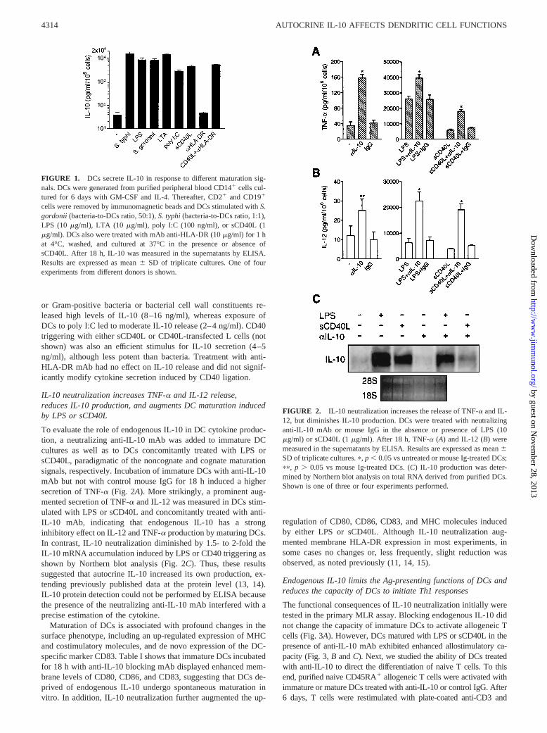
or Gram-positive bacteria or bacterial cell wall constituents re-leased high levels of IL-10 (8–16 ng/ml), whereas exposure ofDCs to poly I:C led to moderate IL-10 release (2–4 ng/ml). CD40triggering with either sCD40L or CD40L-transfected L cells (notshown) was also an efficient stimulus for IL-10 secretion (4–5ng/ml), although less potent than bacteria. Treatment with anti-HLA-DR mAb had no effect on IL-10 release and did not signif-icantly modify cytokine secretion induced by CD40 ligation.
IL-10 neutralization increases TNF-a and IL-12 release,reduces IL-10 production, and augments DC maturation inducedby LPS or sCD40L
To evaluate the role of endogenous IL-10 in DC cytokine produc-tion, a neutralizing anti-IL-10 mAb was added to immature DCcultures as well as to DCs concomitantly treated with LPS orsCD40L, paradigmatic of the noncognate and cognate maturationsignals, respectively. Incubation of immature DCs with anti-IL-10mAb but not with control mouse IgG for 18 h induced a highersecretion of TNF-a (Fig. 2A). More strikingly, a prominent aug-mented secretion of TNF-a and IL-12 was measured in DCs stim-ulated with LPS or sCD40L and concomitantly treated with anti-IL-10 mAb, indicating that endogenous IL-10 has a stronginhibitory effect on IL-12 and TNF-a production by maturing DCs.In contrast, IL-10 neutralization diminished by 1.5- to 2-fold theIL-10 mRNA accumulation induced by LPS or CD40 triggering asshown by Northern blot analysis (Fig. 2C). Thus, these resultssuggested that autocrine IL-10 increased its own production, ex-tending previously published data at the protein level (13, 14).IL-10 protein detection could not be performed by ELISA becausethe presence of the neutralizing anti-IL-10 mAb interfered with aprecise estimation of the cytokine.
Maturation of DCs is associated with profound changes in thesurface phenotype, including an up-regulated expression of MHCand costimulatory molecules, and de novo expression of the DC-specific marker CD83. Table I shows that immature DCs incubatedfor 18 h with anti-IL-10 blocking mAb displayed enhanced mem-brane levels of CD80, CD86, and CD83, suggesting that DCs de-prived of endogenous IL-10 undergo spontaneous maturation invitro. In addition, IL-10 neutralization further augmented the up-
regulation of CD80, CD86, CD83, and MHC molecules inducedby either LPS or sCD40L. Although IL-10 neutralization aug-mented membrane HLA-DR expression in most experiments, insome cases no changes or, less frequently, slight reduction wasobserved, as noted previously (11, 14, 15).
Endogenous IL-10 limits the Ag-presenting functions of DCs andreduces the capacity of DCs to initiate Th1 responses
The functional consequences of IL-10 neutralization initially weretested in the primary MLR assay. Blocking endogenous IL-10 didnot change the capacity of immature DCs to activate allogeneic Tcells (Fig. 3A). However, DCsmatured with LPS or sCD40L in thepresence of anti-IL-10 mAb exhibited enhanced allostimulatory ca-pacity (Fig. 3,B andC). Next, we studied the ability of DCs treatedwith anti-IL-10 to direct the differentiation of naive T cells. To thisend, purified naive CD45RA1 allogeneic T cells were activated withimmature or mature DCs treated with anti-IL-10 or control IgG. After6 days, T cells were restimulated with plate-coated anti-CD3 and
FIGURE 1. DCs secrete IL-10 in response to different maturation sig-nals. DCs were generated from purified peripheral blood CD141 cells cul-tured for 6 days with GM-CSF and IL-4. Thereafter, CD21 and CD191
cells were removed by immunomagnetic beads and DCs stimulated withS.gordonii (bacteria-to-DCs ratio, 50:1),S. typhi(bacteria-to-DCs ratio, 1:1),LPS (10mg/ml), LTA (10 mg/ml), poly I:C (100 ng/ml), or sCD40L (1mg/ml). DCs also were treated with mAb anti-HLA-DR (10mg/ml) for 1 hat 4°C, washed, and cultured at 37°C in the presence or absence ofsCD40L. After 18 h, IL-10 was measured in the supernatants by ELISA.Results are expressed as mean6 SD of triplicate cultures. One of fourexperiments from different donors is shown.
FIGURE 2. IL-10 neutralization increases the release of TNF-a and IL-12, but diminishes IL-10 production. DCs were treated with neutralizinganti-IL-10 mAb or mouse IgG in the absence or presence of LPS (10mg/ml) or sCD40L (1mg/ml). After 18 h, TNF-a (A) and IL-12 (B) weremeasured in the supernatants by ELISA. Results are expressed as mean6SD of triplicate cultures.p, p , 0.05 vs untreated or mouse Ig-treated DCs;pp, p . 0.05 vs mouse Ig-treated DCs. (C) IL-10 production was deter-mined by Northern blot analysis on total RNA derived from purified DCs.Shown is one of three or four experiments performed.
4314 AUTOCRINE IL-10 AFFECTS DENDRITIC CELL FUNCTIONS
by guest on Novem
ber 28, 2013http://w
ww
.jimm
unol.org/D
ownloaded from
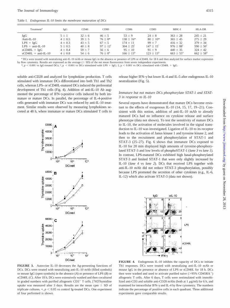
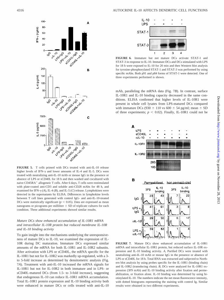
soluble anti-CD28 and analyzed for lymphokine production. T cellsstimulated with immature DCs differentiated into both Th1 and Th2cells, whereas LPS- or sCD40L-matured DCs induced the preferentialdevelopment of Th1 cells (Fig. 4). Addition of anti-IL-10 Ab aug-mented the percentage of IFN-g-positive cells induced by both im-mature or mature DCs. In parallel, the percentage of IL-4-positivecells generated with immature DCs was reduced by anti-IL-10 treat-ment. Similar results were observed by measuring lymphokines se-creted at 48 h, where immature or mature DCs stimulated T cells to
release higher IFN-g but lower IL-4 and IL-5 after endogenous IL-10neutralization (Fig. 5).
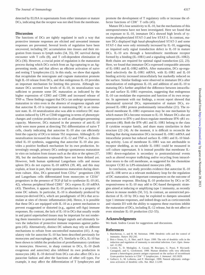
Immature but not mature DCs phosphorylate STAT-1 and STAT-3 in response to IL-10
Several reports have demonstrated that mature DCs become resis-tant to the effects of exogenous IL-10 (14, 15, 17, 19–21). Con-sistent with this notion, addition of anti-IL-10 mAb to alreadymatured DCs had no influence on cytokine release and surfacephenotype (data not shown). To test the sensitivity of mature DCsto IL-10, the activation of molecules involved in the signal trans-duction to IL-10 was investigated. Ligation of IL-10 to its receptorleads to the activation of Janus kinase 1 and tyrosine kinase 2, andthen to the recruitment and phosphorylation of STAT-1 andSTAT-3 (25–27). Fig. 6 shows that immature DCs exposed toIL-10 for 20 min displayed high amounts of tyrosine-phosphory-lated STAT-3 and low levels ofphosphoSTAT-1 (lane 3vs lane 1).In contrast, LPS-matured DCs exhibited high basal-phosphorylatedSTAT-3 and limited STAT-1 that were only slightly increased byIL-10 (lane 4 vs lane 2). DCs that received LPS together withanti-IL-10 mAb did not reduce STAT-3 phosphorylation, possiblybecause LPS promoted the secretion of other cytokines (e.g., IL-6,IL-12) which also activate STAT-3 (data not shown).
FIGURE 3. Autocrine IL-10 decreases the Ag-presenting functions ofDCs. DCs were treated with neutralizing anti-IL-10 mAb (filled symbols)or mouse IgG (open symbols) in the absence (A) or presence of LPS (B) orsCD40L (C). After 18 h, DCs were extensively washed and then coculturedin graded numbers with purified allogeneic CD31 T cells. [3H]Thymidineuptake was measured after 3 days. Results are the mean cpm6 SD oftriplicate cultures.p, p , 0.05 vs control Ig-treated DCs. One experimentof four performed is shown.
FIGURE 4. Endogenous IL-10 inhibits the capacity of DCs to initiateTh1 responses. DCs were treated with neutralizing anti-IL-10 mAb ormouse IgG in the presence or absence of LPS or sCD40L for 18 h. DCsthen were washed and used to activate purified naive (.95% CD45RA1)allogeneic T cells. After 6 days, T cells were restimulated with immobi-lized anti-CD3 and soluble anti-CD28 mAbs (both at 1mg/ml) for 6 h, andexamined for intracellular IFN-g and IL-4 by flow cytometry. The numbersindicate the percentage of positive cells in each quadrant. Three additionalexperiments gave comparable results.
Table I. Endogenous IL-10 limits the membrane maturation of DCs
Treatmenta IgG CD40 CD80 CD86 CD83 MHC-I HLA-DR
IgG 5 6 1 326 6 466 5 536 9 246 8 3636 28 2456 21Anti-IL-10 4 6 0.5 396 5 766 8* 1386 16* 806 10* 3656 45 2756 29LPS 1 IgG 46 0.3 436 5 676 5 1746 11 996 7 4166 32 3796 34LPS 1 anti-IL-10 56 0.5 406 8 976 12† 3046 25† 1476 15† 9766 88† 5906 50†
sCD40L1 IgG 4 6 0.4 596 7 566 6 956 10 916 9 4406 35 3246 42sCD40L1 anti-IL-10 46 0.8 546 6 766 8‡ 1666 13‡ 1236 13‡ 6636 55‡ 6626 58‡
a DCs were treated with neutralizing anti-IL-10 mAb or mouse IgG in the absence or presence of LPS or sCD40L for 18 h and then analyzed for surface marker expressionby flow cytometry. Results are expressed as the average (6SD) of the net mean fluorescence from seven independent experiments.
*, p , 0.001 vs IgG-treated DCs; †, p, 0.001 vs DCs stimulated with LPS1 IgG; ‡, p, 0.001 vs DCs stimulated with CD40L1 IgG.
4315The Journal of Immunology
by guest on Novem
ber 28, 2013http://w
ww
.jimm
unol.org/D
ownloaded from
Mature DCs show enhanced accumulation of IL-10R1 mRNAand intracellular IL-10R protein but reduced membrane IL-10Rand IL-10 binding activity
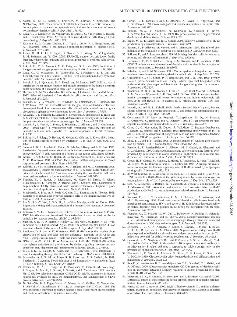
To gain insight into the mechanisms underlying the unresponsive-ness of mature DCs to IL-10, we examined the expression of IL-10R during DC maturation. Immature DCs expressed similaramounts of the mRNA for both IL-10R1 and IL-10R2 subunits.After activation with LPS or sCD40L, the mRNA specific for theIL-10R1 but not for IL-10R2 was markedly up-regulated, with a 3-to 5-fold increase as determined by densitometric analysis (Fig.7A). Treatment with anti-IL-10 increased the mRNA signals forIL-10R1 but not for IL-10R2 in both immature and in LPS- orsCD40L-matured DCs (from 1.5- to 3-fold increase), suggestingthat endogenous IL-10 can reduce IL-10R1 mRNA accumulation.Total IL-10R1 protein expression and IL-10 binding activity bothwere enhanced in mature DCs or cells treated with anti-IL-10
mAb, paralleling the mRNA data (Fig. 7B). In contrast, surfaceIL-10R1 and IL-10 binding capacity decreased in the same con-ditions. ELISA confirmed that higher levels of IL-10R1 werepresent in whole cell lysates from LPS-matured DCs comparedwith immature DCs (9306 110 vs 6006 54 pg/ml; mean6 SDof three experiments;p , 0.02). Finally, IL-10R1 could not be
FIGURE 5. T cells primed with DCs treated with anti-IL-10 releasehigher levels of IFN-g and lower amounts of IL-4 and IL-5. DCs weretreated with neutralizing anti-IL-10 mAb or mouse IgG in the presence orabsence of LPS or sCD40L for 18 h and then washed and cocultured withnaive CD45RA1 allogeneic T cells. After 6 days, T cells were restimulatedwith plate-coated anti-CD3 and soluble anti-CD28 mAbs for 48 h, andexamined for IFN-g (A), IL-4 (B), and IL-5 (C) release. Lymphokines weredetected in the supernatants by ELISA. Differences in lymphokine levelsbetween T cell lines generated with control IgG- and anti-IL-10-treatedDCs were statistically significant (p, 0.05). Data are expressed as meannanograms or picograms per milliliter6 SD of triplicate cultures for eachcondition. Three additional experiments showed similar results.
FIGURE 6. Immature but not mature DCs activate STAT-1 andSTAT-3 in response to IL-10. Immature DCs and DCs stimulated with LPSfor 18 h were exposed to IL-10 for 20 min and then Western blot analysisfor tyrosine-phosphorylated STAT-1 and STAT-3 was performed by usingspecific mAbs. Both p91 and p84 forms of STAT-1 were detected. One ofthree experiments performed is shown.
FIGURE 7. Mature DCs show enhanced accumulation of IL-10R1mRNA and intracellular IL-10R1 protein, but reduced surface IL-10R ex-pression and IL-10 binding activity.A, Purified DCs were treated withneutralizing anti-IL-10 mAb or mouse IgG in the presence or absence ofLPS or sCD40L for 18 h. Total RNA was extracted and subjected to North-ern blot analysis by using probes specific for the IL-10R1 (binding chain)and IL-10R2 (transducing chain).B, DCs were analyzed for IL-10R1 ex-pression (3F9 mAb) and IL-10 binding activity after fixation and perme-abilization, or fixation alone. IL-10 binding was determined by using bi-otinylated IL-10. The numbers indicate the net mean fluorescence intensity,with dotted histograms representing the staining with control Ig. Similarresults were obtained in two different experiments.
4316 AUTOCRINE IL-10 AFFECTS DENDRITIC CELL FUNCTIONS
by guest on Novem
ber 28, 2013http://w
ww
.jimm
unol.org/D
ownloaded from
detected by ELISA in supernatants from either immature or matureDCs, indicating that the receptor was not shed from the membrane.
DiscussionThe functions of DCs are tightly regulated in such a way thatprotective immune responses are elicited and unwanted immuneresponses are prevented. Several levels of regulation have beenuncovered, including DC accumulation into tissues and their mi-gration from tissues to lymph nodes (34, 35), DC survival duringencounter with T cells (8), and T cell-dependent elimination ofDCs (36). However, a crucial point of regulation is the maturationprocess during which DCs switch from an Ag-capturing to an Ag-presenting mode, and that allows an efficient activation of naiveand resting T lymphocytes (1). In this study, we show that signalsthat recapitulate the noncognate and cognate maturation promotehigh IL-10 release from DCs, and that endogenous IL-10 providesan important mechanism for limiting this process. Although im-mature DCs secreted low levels of IL-10, its neutralization wassufficient to promote some DC maturation as indicated by thehigher expression of CD83 and CD86 and increased release ofTNF-a. These findings suggest that DCs can undergo spontaneousmaturation in vitro even in the absence of exogenous signals andthat autocrine IL-10 is important in maintaining DC in an imma-ture state. IL-10 neutralization also was able to reinforce DC mat-uration induced by LPS or CD40 triggering in terms of phenotypicchanges and cytokine production as well as alloantigen-presentingcapacity. Moreover, DCs matured in the presence of anti-IL-10mAb were more potent at directing Th1 differentiation of naive Tcells, clearly indicating that autocrine IL-10 also can efficientlylimit the capacity of DCs to initiate Th1 responses. Although IL-10neutralization increased the release of IL-12 and TNF-a, it dimin-ished IL-10 synthesis, suggesting that autocrine IL-10 also pro-vides a positive feedback mechanism for its own production. In-terestingly enough, primary DCs undergo spontaneous maturationin vitro on isolation from tissues such as the skin or the spleen (37,38), but the mechanisms responsible have not been defined yet.However, both human epidermal Langerhans cells and mousespleen DCs do not express IL-10 mRNA (39, 40), and this mayexplain, at least in part, their spontaneous maturation during short-term culture. Also, DCs generated from CD1a1 progenitors (30)and Langerhans cells differentiated from monocytes or CD341
progenitors in the presence of TGF-b fail to synthesize IL-10 (41,42), whereas peripheral blood CD831 DCs express IL-10 mRNA(43). Therefore, it appears that IL-10 production is a property ofsome DC subsets. In particular, IL-10 is abundantly produced bymonocyte-derived DCs, which are paradigmatic of DCs that accu-mulate at sites of chronic inflammation (44). Hence, it is possiblethat these DCs are equipped with IL-10 as a potent mechanism toprevent exaggerated or distorted (e.g., against self-Ags) immuneresponses. By contrast, the lack of IL-10 in DCs that usually residein and patrol unperturbed tissues may be important for not render-ing them insensitive to potential danger signals and ultimately fa-vors the induction of protective immune responses against patho-gens (45). Alternatively, distinct DC subsets may rely on differentmechanisms to refrain from uncontrolled maturation (41). A reg-ulatory role for autocrine IL-10 has been described previously formonocytes and macrophages (46, 47). Similarly to DCs, IL-10 hasbeen shown to inhibit the production of proinflammatory cytokinesin monocytes. However, in sharp contrast to DCs, IL-10 (bothexogenous and autocrine) also inhibited its own production inmonocytes (46). IL-10 derived from DCs can also affect DCs in aparacrine fashion and alter the functions of other cell types. Forexample, it may affect the differentiation of T lymphocytes and
promote the development of T regulatory cells or increase the ef-fector functions of CD81 T cells (45).
Mature DCs lose sensitivity to IL-10, but the mechanisms of thisunresponsiveness have not been investigated. Here we found thaton exposure to IL-10, immature DCs showed high levels of ty-rosine-phosphorylated STAT-3 and low STAT-1. In contrast, ma-ture DCs displayed high basal-phosphorylated STAT-3 and someSTAT-1 that were only minimally increased by IL-10, suggestingan impaired early signal transduction defect to IL-10 in matureDCs. IL-10 acts through a heterodimeric membrane receptorformed by a binding (IL-10R1) and a signaling subunit (IL-10R2).Both chains are required for optimal signal transduction (22, 23).Here, we found that immature DCs expressed comparable amountsof IL-10R1 and IL-10R2 mRNA. After maturation, DCs up-regu-lated selectively the IL-10R1 mRNA, with IL-10R1 and IL-10binding activity increased intracellularly but markedly reduced onthe surface. Similar findings were observed in immature DCs afterneutralization of endogenous IL-10, and addition of anti-IL-10 tomaturing DCs further amplified the difference between intracellu-lar and surface IL-10R1 expression, suggesting that endogenousIL-10 can modulate the expression and function of its own recep-tor. In agreement with our findings, a recent report showed thatrheumatoid synovial DCs, representative of mature DCs, ex-pressed IL-10R1 protein predominantly intracellular (21). The re-duced membrane IL-10R1 expression may be one mechanism bywhich mature DCs become resistant to IL-10. Mature DCs also areunresponsive to IFN-g and down-regulate membrane IFN-gR1 ex-pression (48). Both the IFN-gR1 and IL-10R1 belong to the classII cytokine receptor family, and share many similarities in theirstructure (22–24). At the moment, it is difficult to reconcile thefinding that during maturation DCs increased IL-10R1 mRNA andintracellular protein but reduced surface IL-10R1 and IL-10 bind-ing activity. Loss of membrane IL-10R1 was not attributable toreceptor shedding, as no soluble IL-10R1 could be measured incell culture supernatant. It is instead possible that membrane IL-10R1 down-regulation is secondary to posttranslational eventssuch as altered receptor trafficking and/or recycling from intracel-lular stores to the cell membrane, as suggested for the chemokinereceptor CCR5 in LPS-stimulated monocytes (49).
In conclusion, our study provides evidence that autocrine IL-10and IL-10R serve as a relevant modulatory loop for the regulationof DC maturation, with important consequences on the outcome ofthe immune response. Blocking IL-10 production by DCs or DCresponsiveness to IL-10 may add to DC-based therapeutic strate-gies aimed at inducing or amplifying type 1 immunity, as recentlyshown in mouse models (50, 51). In contrast, an unrestricted pro-duction of IL-10 by DCs may be exploited to dampen unwantedtype 1 immune responses, and indeed drugs such as corticosteroidsand vitamin D3 with the ability to suppress these reactions inhibitthe maturation of DCs, including IL-12 release, but do not affect oreven stimulate IL-10 production (52–55).
AcknowledgmentsWe thank Andrea Cavani for suggestions and discussions.
References1. Banchereau, J., and R. M. Steinman. 1998. Dendritic cells and the control of
immunity. Nature 392:245.2. Reis e Sousa, G., A. Sher, and P. Kaye. 1999. The role of dendritic cells in the
induction and regulation of immunity to microbial infection.Curr. Opin. Immu-nol. 11:392.
3. Corinti, S., D. Medaglini, A. Cavani, M. Rescigno, G. Pozzi, P. Ricciardi-Castagnoli, and G. Girolomoni. 1999. Human dendritic cells very efficientlypresent a heterologous protein antigen expressed on the surface of recombinantGram-positive bacteria to CD41 T lymphocytes.J. Immunol. 163:3029.
4. Gallucci, S., M. Lolkema, and P. Matzinger. 1999. Natural adjuvants: endoge-nous activators of dendritic cells. Nat. Med. 5:1249.
4317The Journal of Immunology
by guest on Novem
ber 28, 2013http://w
ww
.jimm
unol.org/D
ownloaded from
5. Sauter, B., M. L. Albert, L. Francisco, M. Larsson, S. Somersan, andN. Bhardwaj. 2000. Consequences of cell death: exposure to necrotic tumor cells,but not primary tissue cells or apoptotic cells, induces the maturation of immu-nostimulatory dendritic cells. J. Exp. Med. 191:423.
6. Caux, C., C. Massacrier, B. Vanbervliet, B. Dubois, C. Van Kooten, I. Durand,and J. Banchereau. 1994. Activation of human dendritic cells through CD40cross-linking. J. Exp. Med. 180:1263.
7. Kitajima, T., G. Caceres-Dittmar, F. J. Tapia, J. Jester, P. R. Bergstresser, andA. Takashima. 1996. T cell-mediated terminal maturation of dendritic cells.J. Immunol. 157:2340.
8. Josien, R., H. L. Li, E. Ingulli, S. Sarma, B. R. Wong, M. Vologodskaia,R. M. Steinman, and Y. Choi. 2000. TRANCE, a tumor necrosis factor familymember, enhances the longevity and adjuvant properties of dendritic cells in vivo.J. Exp. Med. 191:495.
9. Enk, A. H., V. L. Angeloni, M. C. Udey, and S. I. Katz. 1993. Inhibition ofLangerhans cell antigen-presenting function by IL-10.J. Immunol. 151:2390.
10. Caux, C., C. Massacrier, B. Vanbervliet, C. Barthelemy, Y. J. Liu, andJ. Banchereau. 1994. Interleukin 10 inhibits T cell alloreaction induced by humandendritic cells.Int. Immunol. 6:1177.
11. Morel, A. S., S. Quaratino, D. C. Douek, and M. Londei. 1997. Split activity ofinterleukin-10 on antigen capture and antigen presentation by human dendriticcells: definition of a maturative step.Eur. J. Immunol. 27:26.
12. De Smedt, T., M. Van Mechelen, G. De Becker, J. Urbain, O. Leo, and M. Moser.1997. Effect of interleukin-10 on dendritic cell maturation and function.Eur.J. Immunol. 27:1229.
13. Buelens, C., V. Verhasselt, D. De Groote, K. Thielemans, M. Goldman, andF. Willems. 1997. Interleukin-10 prevents the generation of dendritic cells fromhuman peripheral blood mononuclear cells cultured with interleukin-4 and gran-ulocyte/macrophage-colony-stimulating factor.Eur. J. Immunol. 27:756.
14. Allavena, P., L.Piemonti, D. Longoni, S. Bernasconi, A. Stoppacciaro, L. Ruco, andA. Mantovani. 1998. IL-10 prevents the differentiation of monocytes to dendritic cellsbut promotes their maturation to macrophages.Eur. J. Immunol. 28:359.
15. Cavani, A., F. Nasorri, C. Prezzi, S. Sebastiani, C. Albanesi, and G. Girolomoni.2000. Human CD41 T lymphocytes with remarkable regulatory functions ondendritic cells and nickel-specific Th1 immune responses. J. Invest. Dermatol.114:295.
16. Enk, A. H., J. Saloga, D. Becker, M. Mohamadzadeh, and J. Knop. 1994. Induc-tion of hapten-specific tolerance by interleukin-10 in vivo.J. Exp. Med. 179:1397.
17. Steinbrink, K., H. Jonuleit, G. Muller, G. Schuler, J. Knop, and A. H. Enk. 1999.Interleukin-10-treated human dendritic cells induce a melanoma-antigen-specificanergy in CD81 T cells resulting in a failure to lyse tumor cells.Blood 93:1634.
18. Groux, H., A. O’Garra, M. Bigler, M. Rouleau, S. Antonenko, J. E. de Vries, andM. G. Roncarolo. 1997. A CD41 T-cell subset inhibits antigen-specific T-cellresponses and prevents colitis.Nature 389:737.
19. Kalinski, P., J. H. N.Schuitmaker, C. M. U. Hilkens, and M. L. Kapsenberg. 1998.Prostaglandin E2 induces the final maturation of IL-12-deficient CD1a1CD831 den-dritic cells: the levels of IL-12 are determined during the final dendritic cell matu-ration and are resistant to further modulation.J. Immunol. 161:2804.
20. Thurner, B., C. Roder, D. Dieckmann, M. Heuer, M. Kruse, A. Glaser,P. Keikavoussi, E. Kampgen, A. Bender, and G. Schuler. 1999. Generation oflarge numbers of fully mature and stable dendritic cells from leukopheresis prod-ucts for clinical application.J. Immunol. Methods 223:1.
21. MacDonald, K. P. A., A. R. Pettit, C. Quinn, G. J. Thomas, and R. Thomas. 1999.Resistance of rheumatoid synovial dendritic cells to the immunosuppressive ef-fects of IL-10.J. Immunol. 163:5599.
22. Liu, Y., S. H.-Y. Wei, A. S.-Y. Ho, R. de Waal Malefyt, and K. W. Moore. 1994.Expression cloning and characterization of a human IL-10 receptor. J. Immunol.152:1821.
23. Kotenko, S. V., C. D. Krause, L. S. Izotova, B. P. Pollack, W. Wu, and S. Pestka.1997. Identification and functional characterization of a second chain of the in-terleukin-10 receptor complex.EMBO J. 16:5894.
24. Spencer, S. D., F. Di Marco, J. Hooley, S. Pitts-Meek, M. Bauer, A. M. Ryan,B. Sordat, V. C. Gibbs, and M. Aguet. 1998. The orphan receptor CRF2–4 is anessential subunit of the interleukin 10 receptor.J. Exp. Med. 187:571.
25. Finbloom, D. S., and K. D. Winestock. 1995. IL-10 induces the tyrosine phos-phorylation of tyk2 and Jak1 and the differential assembly of STAT1a andSTAT3 complexes in human T cells and monocytes. J. Immunol. 155:1079.
26. O’Farrell, A.-M., Y. Liu, K. W. Moore, and A. L.-F. Mui. 1998. IL-10 inhibitsmacrophage activation and proliferation by distinct signaling mechanisms: evi-dence for Stat3-dependent and -independent pathways. EMBO J. 17:1006.
27. Riley, J. K., K. Takeda, S. Akira, and R. D. Schreiber. 1999. Interleukin-10receptor signaling through the JAK-STAT pathway.J. Biol. Chem. 274:16513.
28. Schottelius, A. J. G., M. W. Mayo, R. B. Sartor, and A. S. Baldwin, Jr. 1999.Interleukin-10 signaling blocks inhibitor ofkB kinase activity and nuclear factorkB DNA binding. J. Biol. Chem. 274:31868.
29. Cassatella, M. A., S. Gasperini, C. Bovolenta, F. Calzetti, M. Vollebregt,P. Scapini, M. Marchi, R. Suzuki, A. Suzuki, and A. Yoshimura. 1999. Interleu-kin-10 (IL-10) selectively enhances CIS3/SOCS3 mRNA expression in humanneutrophils: evidence for an IL-10-induced pathway that is independent of STATprotein activation.Blood 94:2880.
30. De Saint-Vis, B., I. Fugier-Vivier, C. Massacrier, C. Gaillard, B. Vanbervliet,S. Ait-Yahia, J. Banchereau, Y. J. Liu, S. Lebecque, and C. Caux. 1998. Thecytokine profile expressed by human dendritic cells is dependent on cell subtypeand mode of activation.J. Immunol. 160:1666.
31. Corinti, S., E. Fanales-Belasio, C. Albanesi, A. Cavani, P. Angelisova, andG. Girolomoni. 1999. Crosslinking of CD43 induces maturation of dendritic cells.J. Immunol. 162:633.
32. Rissoan, M.-C., V. Soumelis, N. Kadowaki, G. Grouard, F. Briere,R. de Waal Malefyt, and Y. J. Liu. 1999. Reciprocal control of T helper cell anddendritic cell differentiation.Science 283:1183.
33. Braun, M. C., E. Lahey, and B. L. Kelsall. 2000. Selective suppression of IL-12production by chemoattractants.J. Immunol. 164:3009.
34. Sozzani, S., P. Allavena, A. Vecchi, and A. Mantovani. 1999. The role of che-mokines in the regulation of dendritic cell trafficking.J. Leukocyte Biol. 66:1.
35. Sallusto, F., and A. Lanzavecchia. 1999. Mobilizing dendritic cells for tolerance,priming, and chronic inflammation.J. Exp. Med. 189:611.
36. Hermans, I. F., D. S. Ritchie, J. Yang, J. M. Roberts, and F. Ronchese. 2000.CD81 T cell-dependent elimination of dendritic cells in vivo limits induction ofantitumor immunity. J. Immunol. 164:3095.
37. Schuler, G., and R. M. Steinman. 1985. Murine epidermal Langerhans cells ma-ture into potent immunostimulatory dendritic cells in vitro. J. Exp. Med. 161:526.
38. Girolomoni, G., J. C. Simon, P. R. Bergstresser, and P. D. Cruz. 1990. Freshlyisolated spleen dendritic cells and freshly isolated epidermal Langerhans cellsundergo similar phenotypic and functional changes during short term culture.J. Immunol. 145:2820.
39. Teunissen, M. B., C. W. Koomen, J. Jansen., R. de Waal Malefyt, E. Schmitt,R. M. Van den Wijngaard, P. K. Das, and J. D. Bos. 1997. In contrast to theirmurine counterparts, normal human keratinocytes and human epidermoid celllines A431 and HaCaT fail to express IL-10 mRNA and protein.Clin. Exp.Immunol. 107:213.
40. Iwasaki, A., and B. L. Kelsall. 1999. Freshly isolated Peyer’s patch, but notspleen, dendritic cells produce interleukin 10 and induce the differentiation of Thelper 2 cells.J. Exp. Med. 190:229.
41. Geissmann, F., P. Revy, A. Regnault, Y. Lepelletier, M. Dy, N. Brousse,S. Amigorena, O. Hermine, and A. Durandy. 1999. TGF-b1 prevents the non-cognate maturation of human dendritic cells.J. Immunol. 162:4567.
42. Caux, C., C. Massacrier, B. Dubois, J. Valladeau, C. Dezutter-Dambuyant,I. Durand, D. Schmitt, and S. Saeland. 1999. Respective involvement of TGF-band IL-4 in the development of Langerhans cells and non-Langerhans dendriticcells from CD341 progenitors.J. Leukocyte Biol. 66:781.
43. Zhou, L.-J., and T. F. Tedder. 1995. A distinct pattern of cytokine gene expres-sion by human CD831 blood dendritic cells.Blood 86:3295.
44. Pastore, S., E. Fanales-Belasio, C. Albanesi, M. L. Chinni, A. Giannetti, andG. Girolomoni. 1997. Granulocyte/macrophage colony-stimulating factor is over-produced by keratinocytes in atopic dermatitis: implications for sustained den-dritic cell activation in the skin.J. Clin. Invest. 99:3009.
45. Groux, H., F. Cottrez, M. Rouleau, S. Mauze, S. Antonenko, S. Hurst, T. McNeil,M. Bigler, M. G. Roncarolo, and R. L. Coffmanet. 1999. A transgenic mousemodel to analyze the immunoregulatory role of IL-10 secreted by antigen-pre-senting cells.J. Immunol. 162:1723.
46. de Waal Malefyt, R., J. Abrams, B. Bennett, C. G. Figdor, and J. E. de Vries.1991. Interleukin 10 (IL-10) inhibits cytokine synthesis by human monocytes: anautoregulatory role of IL-10 produced by monocytes.J. Exp. Med. 174:1209.
47. Sica, A., A. Saccani, B. Bottazzi, N. Polentarutti, A. Vecchi, J. Van Damme, andA. Mantovani. 2000. Autocrine production of IL-10 mediates defective IL-12production and NF-kB activation in tumor-associated macrophages.J. Immunol.164:762.
48. Kalinski, P., J. H. N. Schuitmaker, C. M. U. Hilkens, E. A. Wierenga, andM. L. Kapsenberg. 1999. Final maturation of dendritic cells is associated withimpaired responsiveness to IFN-g and bacterial IL-12 inducers: decreased abilityof mature dendritic cells to produce IL-12 during the interaction with Th cells.J. Immunol. 162:3231.
49. Franchin, G., G. Zybarth, W. W. Dai, L. Dubrovsky, N. Reiling, H. Schmidt-mayerova, M. Bukrinsky, and B. Sherry. 2000. Lipopolysaccharide inhibitsHIV-1 infection of monocyte-derived macrophages through direct and sustaineddown-regulation of CC chemokine receptor 5.J. Immunol. 164:2592.
50. Igietseme, J. U., G. A. Ananaba, J. Bolier, S. Bowers, T. Moore, T. Belay,F. O. Eko, D. Lyn, and C. M. Black. 2000. Suppression of endogenous IL-10gene expression in dendritic cells enhances antigen presentation for specific Th1induction: potential for cellular vaccine development.J. Immunol. 164:4212.
51. Castro, A. G., M. Neighbors, S. D. Hurst, F. Zonin, R. A. Silva, E. Murphy, Y.-J.Liu, and A. O’Garra. 2000. Anti-interleukin 10 receptor monoclonal antibody isan adjuvant for T helper cell type 1 responses to soluble antigen only in thepresence of lipopolysaccharide.J. Exp. Med. 192:1529.
52. Piemonti, L., P. Monti, P. Allavena, M. Sironi, B. E. Leone, C. Socci, andV. Di Carlo. 1999. Glucocorticoids affect human dendritic cell differentiation andmaturation.J. Immunol. 162:6473.
53. Rea, D., C. van Kooten, K. E. van Meijgaarden, T. H. Ottenhoff, C. J. Melief, andR. Offringa. 2000. Glucocorticoids transform CD40-triggering of dendritic cellsinto an alternative activation pathway resulting in antigen-presenting cells thatsecrete IL-10.Blood 95:3162.
54. Matyszak, M. K., S. Citterio, M. Rescigno, and P. Ricciardi-Castagnoli. 2000.Differential effects of corticosteroids during different stages of dendritic cell mat-uration.Eur. J. Immunol. 30:1233.
55. Penna, G., and L. Adorini. 2000. 1a,25-Dihydroxyvitamin D3 inhibits differen-tiation, maturation, activation, and survival of dendritic cells leading to impairedalloreactive T cell activation.J. Immunol. 164:2405.
4318 AUTOCRINE IL-10 AFFECTS DENDRITIC CELL FUNCTIONS
by guest on Novem
ber 28, 2013http://w
ww
.jimm
unol.org/D
ownloaded from
Recommended


