Duquesne UniversityDuquesne Scholarship Collection
Electronic Theses and Dissertations
2009
Role of Thyroid Hormone in the IntestinalDevelopment of Eleutherodactylus coquiSrikanth Singamsetty
Follow this and additional works at: https://dsc.duq.edu/etd
This Immediate Access is brought to you for free and open access by Duquesne Scholarship Collection. It has been accepted for inclusion in ElectronicTheses and Dissertations by an authorized administrator of Duquesne Scholarship Collection. For more information, please [email protected].
Recommended CitationSingamsetty, S. (2009). Role of Thyroid Hormone in the Intestinal Development of Eleutherodactylus coqui (Doctoral dissertation,Duquesne University). Retrieved from https://dsc.duq.edu/etd/1200
ROLE OF THYROID HORMONE IN THE INTESTINAL DEVELOPMENT OF
ELEUTHERODACTYLUS COQUI
A Dissertation
Submitted to the Bayer School of Natural and Environmental Sciences
Duquesne University
In partial fulfillment of the requirements for
the degree of Doctor of Philosophy
By
Srikanth Singamsetty
December 2009

Copyright by
Srikanth Singamsetty
2009
iii
ROLE OF THYROID HORMONE IN THE INTESTINAL DEVELOPMENT OF
ELEUTHERODACTYLUS COQUI
By
Srikanth Singamsetty
Approved October 21, 2009 ________________________________ Richard P. Elinson Professor of Biological Sciences (Committee Chair)
________________________________ Rita Mihailescu Assistant Professor of Chemistry and Biochemistry (Committee Member)
________________________________ John A. Pollock Associate Professor of Biological Sciences (Committee Member)
________________________________ John F. Stolz Professor of Biological Sciences (Committee Member)
________________________________ David W. Seybert Dean, Bayer School of Natural & Environmental Sciences Professor of Chemistry & Biochemistry
________________________________ Philip E. Auron Chair, Department of Biological Sciences Professor of Biological Sciences
iv
ABSTRACT
ROLE OF THYROID HORMONE IN THE INTESTINAL DEVELOPMENT OF
ELEUTHERODACTYLUS COQUI
By
Srikanth Singamsetty
December 2009 Dissertation supervised by Richard P. Elinson Thyroid hormone (TH) is required for the metamorphosis of the long, coiled
tadpole gut in Xenopus laevis into a shorter mature adult gut. Eleutherodactylus coqui, a
direct developing frog, lacks a tadpole. Its embryonic gut is a miniature adult form with a
mass of yolky endodermal cells attached to the small intestine to provide nutrition. The
requirement of TH for the gut development in E. coqui was tested in this study. Inhibition
of TH synthesis with methimazole arrested gut development in its embryonic form. T3,
the active form of TH, induced gut development. Embryos treated with methimazole
failed to utilize the yolk in their nutritional endoderm, and survived for weeks without
any further development. Acidification of the yolk platelet is an initial step in the
breakdown of yolk in X. laevis. E. coqui embryos in methimazole failed to acidify their
yolk platelets, but acidification was stimulated by TH indicating its role in yolk
v
utilization. In X. laevis, TRβ is upregulated in response to TH and induces differentiation
of the adult gut. Similarly, EcTRβ, the E. coqui orthologue, was upregulated by TH in the
gut. EcTRβ expression was high in the nutritional endodermal cells indicating a direct
role for TH in yolk utilization. The low expression level of EcSox17, an endodermal
transcription factor, in these nutritional endodermal cells was consistent with the fact that
these cells did not contribute to the definitive gut. This study indicated a novel role for
TH in yolk utilization in addition to its conserved role in gut development and
differentiation.
vi
DEDICATION
This dissertation is dedicated to Dr. Edward Weisberg, who passed away on March 2,
2005. He was a great advisor and a wonderful teacher. His help during my initial years at
grad school and life in the US was amazing. He was very friendly, approachable and a
kind person, I will always remember him. Thanks for all the help and your words of
encouragement.
vii
ACKNOWLEDGEMENT
Guru Brahma Gurur Vishnu
Guru Devo Maheshwaraha
Guru Saakshat Para Brahma
Tasmai Sree Gurave Namaha
Essence: He creates, sustains knowledge and destroys the weeds of ignorance. I salute
such a Guru. - Adi Shankaracharya, Indian Philosopher
This Sanskrit verse that I was taught as a 3 year-old epitomizes the meaning of a
teacher. I had the opportunity to learn from many such “Gurus” over the last three
decades and would like to thank them all for what I am today. I would like to express my
deepest gratitude to Dr. Elinson for accepting me into his lab and for providing
extraordinary guidance. I am grateful to Dr. Elinson for his patience, encouragement and
for cultivating scientific thinking in me. Thanks to Dr. Pollock for his words of
encouragement and for his insightful comments during committee meetings. Thanks to
Dr. Stolz for serving on my committee and for help with the microscope. Thanks to Dr.
Mihailescu from chemistry for agreeing to serve on my dissertation committee. Thank
you all for your helpful comments and observations that helped a lot in orienting my
research in the right direction.
Special thanks to Kim Nath, our lab technician and my lab guru, for teaching me the
applied aspect of research and answering my questions including the nonsensical stuff on
a range of subjects. Thanks to Dr. Auron and Dr. Sneddon for their initial help with real-
time PCR. I would like to specially thank Jeanne Workman for her guidance during my
time in A&P. Thanks to Phil Hoschar and Lalitha Rajakumar who taught me organization
during initial days and being my role models in that aspect. Thanks to the undergraduate
viii
lab gurus Sean Williamson and Michelle Sabo for teaching me the tricks of RNA
extractions. Thanks to my lab fellows Steven Sandelich, Zach Walton, Seung Yun Lee,
Kelly Sopko, Cara Fischer, Kourtney Krohn and Dom Beer for the fun times and
suggestions during lab meetings. Special thanks to Pam and Judy for making my
wonderful stay at Duquesne even better. Thanks to Dr. Seybert, Mary Ann Quinn,
Carolina Martin and Heather Costello from the Dean’s office for their help. All my
interaction with the biology faculty and friends has left me richer in knowledge and
experience, thank you for everything.
Thanks to my special friends from biology Jen Gabel, Shalini Singh, Matt Henkel,
Puneet Anand, Bree Zeyzus, Beth Weisenberger and Juraj Adamik. On a personal note, I
would like to thank my parents, sisters and especially my brother for their continued
support. Thanks to my friends, Satti, Kanthi, Afru, Rahul, Honey, Raj, Vamsi and
Kalyani for their support. Finally, thanks to Uma for her continued support and love
throughout.
ix
TABLE OF CONTENTS
Page
Abstract .......................................................................................................................... iv
Dedication ...................................................................................................................... vi
Acknowledgement ......................................................................................................... vii
List of Tables ............................................................................................................... xiii
List of Figures .............................................................................................................. xiv
List of Abbreviations .................................................................................................... xvi
Chapter One: Introduction ........................................................................................... 1
I. Frog development .................................................................................................... 1
A. Indirect development ......................................................................................... 1
B. Direct development ........................................................................................... 2
C. Metamorphosis .................................................................................................. 3
II. Thyroid hormone control of metamorphosis ............................................................ 5
A. Thyroid hormone .............................................................................................. 5
B. Thyroid hormone receptors................................................................................ 6
C. Thyroid hormone role in E. coqui development ................................................. 9
III. Gut development .................................................................................................. 11
A. Development of the tadpole and frog gut ......................................................... 11
B. Molecular development of the frog gut ............................................................ 14
C. Development of the E. coqui gut ..................................................................... 18
D. Molecular development of E. coqui gut ........................................................... 21
x
IV. Yolk and embryonic development ......................................................................... 22
A. Biogenesis of yolk........................................................................................... 22
B. Composition of yolk platelets .......................................................................... 24
C. Cell biology of yolk......................................................................................... 26
D. Yolk utilization in the embryo ......................................................................... 27
V. Objectives of the study .......................................................................................... 33
Chapter Two: Materials and Methods ....................................................................... 34
I. Embryo collection and care ................................................................................... 34
II. Embryo culture and treatment ............................................................................... 34
III. Dissection of embryonic gut ................................................................................. 35
IV. Dissociation of yolky cells .................................................................................... 35
V. RNA extraction ..................................................................................................... 36
VI. Reverse transcription ............................................................................................ 38
VII. Genomic EcSox17 Sequencing ............................................................................. 39
VIII. Primer design ....................................................................................................... 41
IX. PCR ..................................................................................................................... 42
X. Ethidium bromide gels and gel photography ....................................................... 43
XI. Real-time PCR.................................................................................................... 44
XII. Histology ........................................................................................................... 45
XIII. Whole mount in situ hybridization ..................................................................... 48
XIV. In situ hybridization on sections ......................................................................... 53
XV. Acridine orange staining of yolk platelets .......................................................... 53
xi
XVI. Photography and image processing ................................................................... 54
Chapter Three: Results ............................................................................................... 55
I. Expression of gut patterning genes ........................................................................ 55
A. Gene expression in the developing gut ............................................................ 55
B. Expression of TH receptors in the differentiating gut ....................................... 60
C. EcSox17 and EcTRβ expression dynamics in the gut ....................................... 60
II. Thyroid hormone requirement for gut development................................................... 68
A. Morphological development of the gut is arrested by methimazole treatment .. 68
B. Abnormal gut histology upon TH inhibition .................................................... 76
C. TH rescue of gut morphology .......................................................................... 77
D. TH stimulation of gut differentiation ............................................................... 86
III. Thyroid hormone induction of gut gene expression .................................................. 87
A. Down regulation of gut gene expression on thyroid hormone inhibition .......... 87
B. Thyroid hormone dependency of EcTRβ expression ........................................ 96
C. T3 induction of EcTRβ and EcSox17 expression in the gut ............................... 97
IV. Direct action of thyroid hormone on nutritional endoderm ..................................... 102
A. EcTRβ expression in nutritional endoderm .................................................... 102
B. Inhibition of TH prevents yolk utilization ...................................................... 108
C. Effect of TH on utilization of yolk ................................................................. 108
Chapter Four: Discussion ......................................................................................... 115
I. Novel contributions of this study ......................................................................... 115
II. Thyroid hormone role in gut development ........................................................... 116
xii
A. Morphology of the gut .................................................................................. 116
B. Transformation from embryonic to an adult gut ............................................. 118
C. Remodeling nutritional endoderm ................................................................. 119
III. Gene expression in the gut .................................................................................. 120
A. Definitive gut development ........................................................................... 120
B. TH regulation of gut development ................................................................. 121
IV. Origin of adult gut epithelium ............................................................................. 122
V. Yolk utilization .................................................................................................. 124
A. Vacuolation in the intestine ........................................................................... 124
B. Acidification of yolk platelets........................................................................ 125
C. TH role in yolk utilization ............................................................................. 126
C. 1. TH is directly required for yolk utilization in E. coqui .......................... 126
C. 2. TH role in yolk utilization before thyroid gland development ............... 128
D. Proteolysis of yolk ........................................................................................ 129
VI. Biology of nutritional endoderm ........................................................................ 131
VII. Analogues of nutritional endoderm .................................................................... 135
VIII. Evolutionary significance of this study .............................................................. 137
References .................................................................................................................. 141
xiii
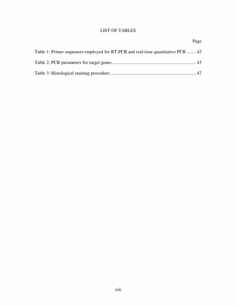
LIST OF TABLES
Page
Table 1: Primer sequences employed for RT-PCR and real-time quantitative PCR ........ 42
Table 2: PCR parameters for target genes ...................................................................... 43
Table 3: Histological staining procedure ........................................................................ 47
xiv
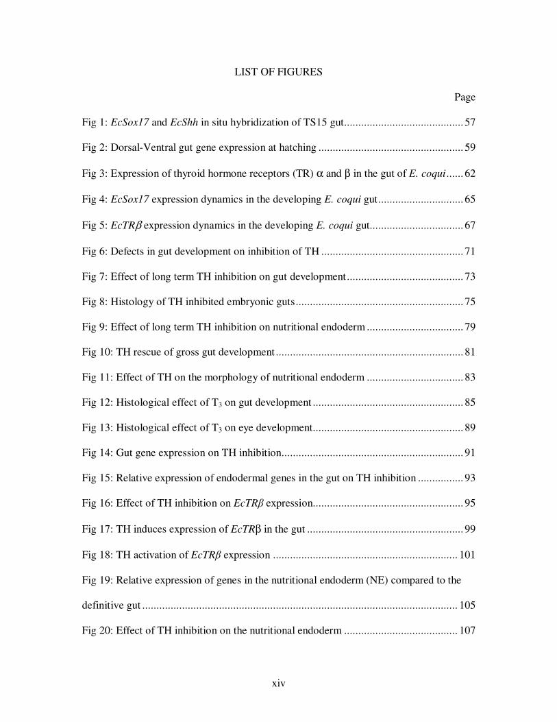
LIST OF FIGURES
Page
Fig 1: EcSox17 and EcShh in situ hybridization of TS15 gut .......................................... 57
Fig 2: Dorsal-Ventral gut gene expression at hatching ................................................... 59
Fig 3: Expression of thyroid hormone receptors (TR) α and β in the gut of E. coqui ...... 62
Fig 4: EcSox17 expression dynamics in the developing E. coqui gut .............................. 65
Fig 5: EcTRβ expression dynamics in the developing E. coqui gut ................................. 67
Fig 6: Defects in gut development on inhibition of TH .................................................. 71
Fig 7: Effect of long term TH inhibition on gut development ......................................... 73
Fig 8: Histology of TH inhibited embryonic guts ........................................................... 75
Fig 9: Effect of long term TH inhibition on nutritional endoderm .................................. 79
Fig 10: TH rescue of gross gut development .................................................................. 81
Fig 11: Effect of TH on the morphology of nutritional endoderm .................................. 83
Fig 12: Histological effect of T3 on gut development ..................................................... 85
Fig 13: Histological effect of T3 on eye development..................................................... 89
Fig 14: Gut gene expression on TH inhibition................................................................ 91
Fig 15: Relative expression of endodermal genes in the gut on TH inhibition ................ 93
Fig 16: Effect of TH inhibition on EcTRβ expression..................................................... 95
Fig 17: TH induces expression of EcTRβ in the gut ....................................................... 99
Fig 18: TH activation of EcTRβ expression ................................................................. 101
Fig 19: Relative expression of genes in the nutritional endoderm (NE) compared to the
definitive gut ............................................................................................................... 105
Fig 20: Effect of TH inhibition on the nutritional endoderm ........................................ 107
xv
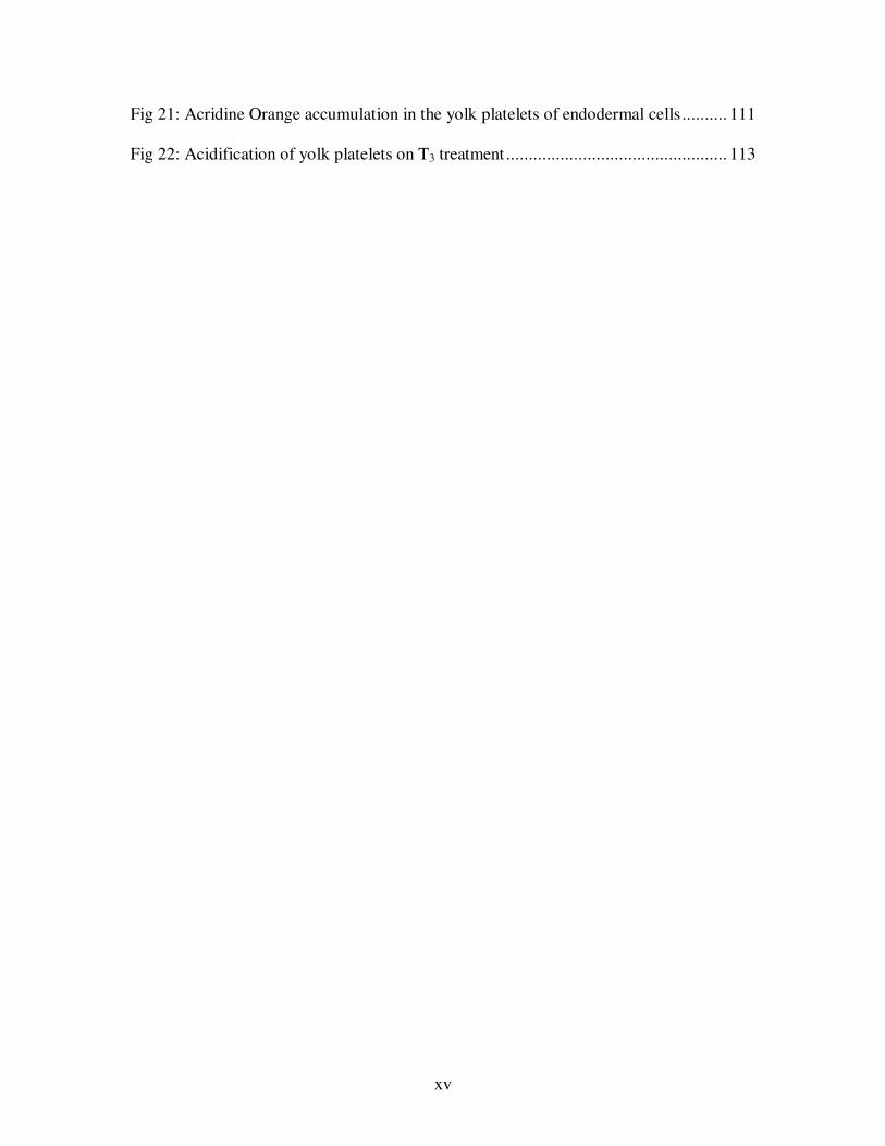
Fig 21: Acridine Orange accumulation in the yolk platelets of endodermal cells .......... 111
Fig 22: Acidification of yolk platelets on T3 treatment ................................................. 113
xvi
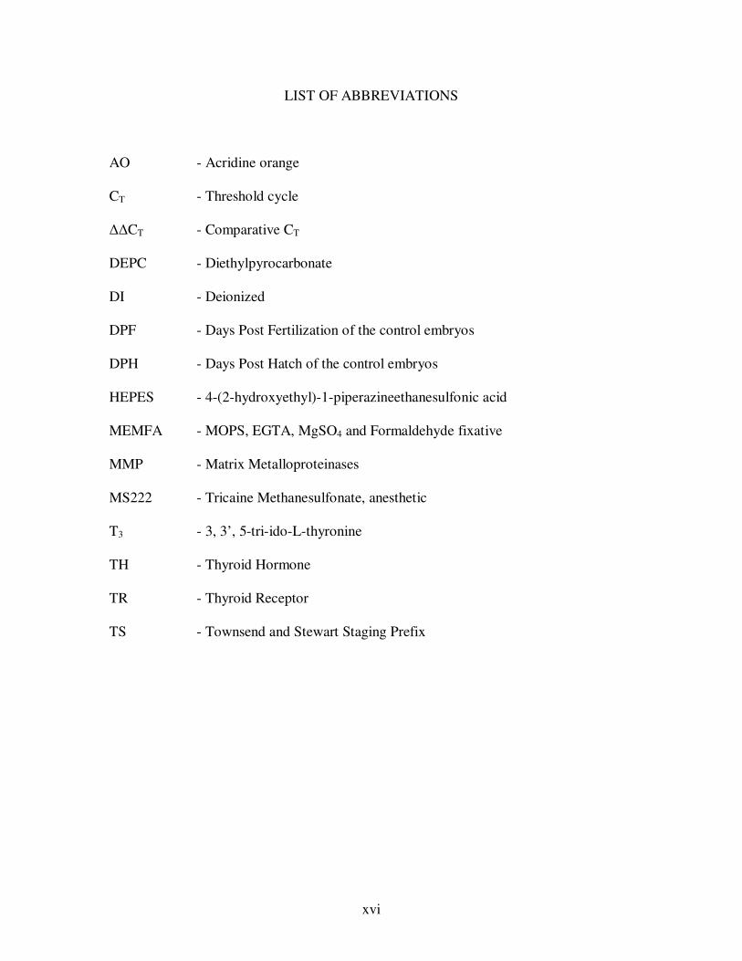
LIST OF ABBREVIATIONS
AO - Acridine orange
CT - Threshold cycle
∆∆CT - Comparative CT
DEPC - Diethylpyrocarbonate
DI - Deionized
DPF - Days Post Fertilization of the control embryos
DPH - Days Post Hatch of the control embryos
HEPES - 4-(2-hydroxyethyl)-1-piperazineethanesulfonic acid
MEMFA - MOPS, EGTA, MgSO4 and Formaldehyde fixative
MMP - Matrix Metalloproteinases
MS222 - Tricaine Methanesulfonate, anesthetic
T3 - 3, 3’, 5-tri-ido-L-thyronine
TH - Thyroid Hormone
TR - Thyroid Receptor
TS - Townsend and Stewart Staging Prefix
1
CHAPTER ONE: INTRODUCTION
Development is the progress of a single-celled zygote to a multicellular organism with
all the functional organ systems. A functioning digestive system consists of organs
required for ingestion, digestion and absorption of nutrients for the survival of an
organism. One of the important factors for the survival of a young embryo is the
availability of nutrition until it can feed and digest food. These nutrients are supplied to a
varying degree maternally via yolk, placenta, colostrum or a combination of these during
embryonic development. In addition to the nutrients, cells depend on molecular cues for
the proper development of a functional gut. Thyroid hormone (TH) plays a major role in
the development and differentiation of the gut in amphibians (Shi and Ishizuya-Oka,
1996; Shi and Ishizuya-Oka, 2001). This thesis deals with TH dependent gut
development and embryonic nutrition in the direct developing frog, Eleutherodactylus
coqui.
I. FROG DEVELOPMENT
A. Indirect development
Most frogs and toads, the anuran amphibians, have an intermediate larval stage in their
life-cycle, and this pattern is an indirect or biphasic type of development. Early
embryonic life is represented by larva or tadpole that later transforms into an adult.
Tadpoles are drastically different from their adults in various aspects. The tadpole swims
with a tail, respires through gills, excretes through pronephros, and digests plant material
in their long coiled gut. In their adults, limbs help in movement, lungs aid in respiration,
2
excretion is through the mesonephros, and animal material is digested in their short gut
(Brown and Cai, 2007). Adult frogs and toads are usually terrestrial whereas the tadpole
is aquatic. The period of rapid change in morphology from tadpole to adult is referred to
as metamorphosis and is dependent on TH. A detailed review of a TH role in
development is discussed later.
B. Direct development
There is an alternative life-history in some anurans where the adult develops directly
without a feeding larval stage, and this life-history is referred to as direct development.
Some anurans evolved large eggs with lots of yolk to cover for the lack of a feeding
young (Townsend and Stewart, 1985; Elinson, 1987a, b). A few examples for direct
development include frogs in the genera Gastrotheca, Flectonotus, Chiromantis,
Rheobatrachus, and Eleutherodactylus (Lynn and Peadon, 1955; Duellman and Trueb,
1986; Elinson et al., 1990). Direct development is a derived character and evolved many
times in different amphibian orders independently (Duellman and Trueb, 1986; Elinson,
1990; Callery et al., 2001; Elinson, 2001).
Eleutherodactylus coqui, the Puerto Rican tree frog, develops directly from a large,
yolky egg (Townsend and Stewart, 1985; Elinson, 1987a; Elinson et al., 1990). The frogs
breed on land, and following internal fertilization, the embryos are brooded by the male
(Townsend et al., 1981; Townsend et al., 1985; Elinson, 1987a). The large 3.5 mm egg of
E. coqui has 20 times the volume compared to the 1.3 mm Xenopus laevis egg (Elinson
and Beckham, 2002). The large quantities of yolk provide access to nutrients and
3
minerals like calcium, magnesium and phosphorous to build its body until the embryo
hatches out as a froglet (Packard et al., 1996).
The dry mass of the yolk decreased rapidly in the later development as the embryonic
carcass gained a proportional increase in its weight, demonstrating the role of the yolk in
these direct developing frogs (Packard et al., 1996). Embryos in E. coqui undergo
holoblastic cleavage and the yolk is partitioned into individual cells (Elinson, 1987;
Elinson, 1990; Buchholz et al., 2007; Elinson, 2009). The yolky cells are attached to the
gut within a vascularized intestinal sac during development (Lynn, 1942; Valett and
Jameson, 1961; Buchholz et al., 2007). These nutrient yolk reserves last for a while, well
past the hatching of the E. coqui froglets.
Many larval characters like cement gland, long coiled gut, lateral line organs, larval
mouth parts, and some cranial cartilages are lost in these animals with the most extreme
form of direct development (Elinson, 1990). Although E. coqui lacks the larval stage,
metamorphosis does occur in these frogs (Callery and Elinson, 2000).
C. Metamorphosis
Metamorphosis is the process during which the larva transforms into an adult. “Meta”
means “change” and “Morph” means “form” in Greek. Metamorphosis is the
postembryonic period of profound morphological, physiological, biochemical,
behavioral, and ecological changes which alter the animal’s mode of living .
Metamorphosis is known to occur in all extant chordates except amniotes (Dent, 1968;
Just et al., 1981) and has been especially well characterized in amphibians. Insect larva-
to-adult (Drosophila, butterflies and beetles) and amphibian tadpole-to-frog (Xenopus,
4
Rana and Bufo) transitions are some of the extreme examples of metamorphosis (Gilbert
et al., 1996; Ishizuya-Oka and Shi, 2007; Yoshizato, 2007). Many species of fish also
undergo TH-dependent metamorphosis, transitioning from a larva to a juvenile (Youson,
1980).
Amphibians have a variety of strategies in metamorphosis, varying from direct
development (Callery et al., 2001) to paedomorphic salamanders (Wakahara, 1996).
Some of the organs completely disappear and newer ones assume their functions. The
tadpole pronephric kidney regresses and disappears completely at the end of
metamorphosis and the mesonephros takes its place (Fox, 1970). Remodeling also occurs
in the tadpole skin (Kobayasi et al., 1996), respiratory organs (Dodd and Dodd, 1976),
liver (Atkinson et al., 1998), pancreas (Mukhi., et al., 2005) the immune system (Rollins-
Smith, 1998), the brain and spinal cord (Kollros, 1981), the eye (Hoskin, 1986; Mann and
Holt, 2001), the nose (Higgs and Burd, 2001), the pituitary (Kikuyama et al., 1993;
Buckbinder and Brown, 1993; Huang et al., 2001), the haematopoietic system (Weber,
1996) and much of the skeleton (Trueb and Hanken, 1992).
Some degree of remodeling occurs in the cranial cartilages, limbs, and tail during the
later part of embryonic development in E. coqui hinting at metamorphosis in these direct
developers (Townsend and Stewart, 1985; Hanken et al., 1992; Elinson, 1994; Hanken et
al., 1997). Later studies provided proof for the metamorphic events in E. coqui embryo
that required TH (Callery and Elinson, 2000). TH dependent remodeling was
demonstrated in the skin, limbs, jaw musculature, cartilage, tail, and trunk musculature of
E. coqui.
5
II. THYROID HORMONE CONTROL OF METAMORPHOSIS
A. Thyroid hormone
Development and differentiation early on in life depends on thyroid hormone (TH),
while later on, TH influences metabolism in almost all tissues. Physiological processes
affected by TH include metabolism, thermogenesis, vascular resistance, arterial blood
pressure, renal sodium reabsorption, blood volume, and heart rate. TH plays a significant
role in the expression of candidate genes that are required for the maturation of brain in
newborn mammals (Thompson and Bottcher, 1997; Bernal, 2005). Deficiency of TH
results in cretinism in children and goiter in adults. Hyperthyroidism causes
hypolipidemia and tachycardia leading to cardiac arrhythmia. Graves disease is the result
of hyperactive thyroid and is an autoimmune disease. TH is required for the completion
of life cycle in many higher vertebrates from birds to humans.
In 1912, J. F. Gudernatsch discovered that a substance in the thyroid gland could
induce precocious metamorphosis in frog tadpoles. Soon E. C. Kendall showed that the
active ingredient was thyroid hormone. The thyroid gland has an endodermal origin and
is located around the endostyle or pharyngeal region in all chordates (Braverman and
Utiger, 1996; reviewed in Paris and Laudet, 2008). In mammals and amphibians, the
follicles of thyroid gland produce the precursor T4 (thyroxine) that is subsequently
transformed into T3 (triiodothyronine), the active form of TH (Harrington, 1926; Gross
and Pitt-Rivers, 1952). A major portion of the TH is transported through the blood bound
to protein-carriers like thyroxine binding globulin (TBG), transthyretin (TTR), and
albumin. In Xenopus, TH uptake into the cell is facilitated by the organic anion
transporters (OATPs), and L-type amino acid transporters (LATs) of the
6
monocarboxylate transporter family (MCT8) (Jansen et al., 2005, reviewed in Visser et
al., 2008). Availability of T3 in the cell is controlled by Cytosolic Thyroid Hormone
Binding Protein (CTHBP) during Xenopus metamorphosis (Shi, 1994).
Metamorphosis in insects and vertebrates is attributed to hormonal changes that occur
during their development (Gilbert et al., 1996). Ecdysone induces major developmental
changes at metamorphosis in insects and crustaceans (Thummel 1996; Gilbert et al.,
2002; Truman and Riddiford 2002; Bonneton et al 2003). Similarly, thyroid hormone (T3)
role in metamorphosis is highly conserved across amphibians. Circulating concentrations
of plasma TH increase markedly in anurans (Leloup and Buscaglia, 1977; Brown and
Cai, 2007) and urodeles (Larras-Regard et al., 1981; Alberch et al., 1986) and correlates
with the metamorphic climax (reviewed by Tata, 2006). TH regulates metamorphosis in
fish and other amphibians as well (Power et al., 2001; Youson and Sower 2001; Crespi
and Denver 2005; Heyland et al, 2005; Page et al., 2009). In anurans, exogenous TH
induces premature metamorphosis (Gudernatsch, 1912), and TH inhibition blocks
metamorphosis resulting in giant tadpoles (Allen, 1916; Buchholz et al, 2006).
Environmental conditions, such as water volume and food availability, also modulate TH
levels inducing precocious metamorphosis (Denver, 1998; Newman, 1998; Boorse and
Denver, 2003; Ito et al., 2004; Boelen et al., 2008).
B. Thyroid hormone receptors
Thyroid hormone brings about the morphological changes during metamorphosis by
inducing hormone-activated transcription factors and thereby modulating gene expression
(Shi, 1999). TH functions by interacting with thyroid hormone receptors (TRs) and
7
induces transcription of target genes. TR belongs to a family of nuclear receptors that
include the steroid hormone receptors and 9-cis retinoic acid receptors (RXRs), and that
mediates T3 dependent effects of metamorphosis (Evans, 1988; Yen and Chin, 1994; Tsai
and O’Malley, 1994). The TR has a 100-amino acid DNA binding domain that
recognizes and binds the thyroid hormone response element (TRE) in the target genes.
The TR/RXR heterodimers, bound to the TREs, activate or repress the expression of T3
response genes in a ligand dependent manner (Tsai and O’Malley, 1994; Yen, 2001).
In the absence of TH, thyroid receptor binds to the DNA leading to transcriptional
repression (Tsai and O’Malley, 1994; Yen, 2001). TR forms corepressor complexes with
silencing mediator for retinoid and thyroid receptor (SMRT) and nuclear receptor
corerepressor (N-CoR) in the absence of the ligand (Zhang and Lazar, 2000; Sachs et al.,
2003; Jones and Shi, 2003; Tomita et al., 2004; Sato et al., 2007). In the presence of TH,
the TR interacts with it forming coactivator complexes with histone acetyltransferase
(Onate et al., 1995; Huang et al., 2003), CREB (cAMP-response element-binding
protein)-binding protein (CBP) and protein p300 (Chen et al., 1997; Demarest et al.,
2002; Paul and Shi, 2003; Paul et al., 2005; Paul., et al 2007). Hormone binding leads to
a conformational change in the TRs that causes it to function as a transcriptional activator
(Tsai and O’Malley, 1994; Yen, 2001).
TH induced genes are classified based on their response during metamorphosis
(reviewed by Shi, 1999; Ishizuya-Oka and Shi; 2008). Genes that are regulated in the
premetamorphic tadpoles within 24 hours of T3 treatment are early response genes (Wang
and Brown, 1991; Buckbinder and Brown, 1992; Shi and Brown, 1993; Denver et al.,
1997) and those that are regulated after 24 hours are late response genes (Shi and Brown,
8
1990; Shi and Hayes, 1994; Amano, 1998). Genes regulated by T3 at the transcriptional
level are direct response genes and the ones that require synthesis of another protein are
indirect response genes.
Microarrays indicated a wider and larger range of TH induced gene expression in
tadpoles (Yen et al., 2003; Das et al., 2006; Buchholz et al., 2007; Cai et al., 2007)
compared to the PCR-based subtractive screens (Shi and Brown, 1993; Shi, 1996; Denver
et al., 1997). As many as 21,807 genes representing over 98% of the X. laevis genome,
were visualized on a microarray. Of which, 1997 genes were differentially regulated in
the intestine by 1.5-fold or more on T3 treatment (Buchholz et al., 2007).
Many of the early T3 response genes were identified by expression screens and coded
for transcription factors (Buckbinder and Brown, 1992; Shi and Brown, 1993; Remo and
Pinder, 1996; Kanamori and Brown, 1993; Denver et al., 1997; Das et al., 2006). TRβ is
one of the early response genes and is directly upregulated by TH by autoinduction
(Yaoita and Brown, 1990; Kanamori and Brown, 1992; Machuca et al., 1995; Tata,
2000). Transcripts of the two TH receptors TRα and TRβ are detected at low levels in
eggs and early embryos. TH forms a complex with TRs inducing the expression of TRβ.
TRβ binds its own promoter resulting in its upregulation, and this is referred to as
autoinduction (Machuca and Tata, 1992; Machuca et al., 1995; reviewed by Tata, 2006).
The upregulation of TRβ in X. laevis and Rana catesbeiana temporally correlates with the
onset of metamorphosis, implicating its role in metamorphosis (Yaoita and Brown, 1990;
Kawahara et al., 1991; Helbing et al., 1992).
Human thyroid hormone receptors were first cloned and identified as members of
nuclear receptor transcription factors (Weinberger et al., 1986; Sap et al., 1986). The X.
9
laevis orthologues of TRα and TRβ have since been cloned (Brooks et al., 1989; Yaoita et
al., 1990; Shi et al., 1992). A dominant negative TR construct driven by ubiquitous
promoter represses gene expression and inhibits metamorphosis of the tadpoles
(Schreiber et al., 2001; Buchholz et al 2003). The dominant positive construct leads to
precocious activation of genes inducing metamorphosis in the premetamorphic tadpoles
even in the absence of TH (Buchholz et al., 2004). Experiments involving dominant
negative and dominant positive constructs have shown that the TRs are sufficient to
directly mediate the developmental effects during metamorphosis (reviewed by Ishizuya-
Oka and Shi, 2008). One of the most studied aspects of TH dependent metamorphosis is
the remodeling of the tadpole gut, and this is discussed in detail in later sections.
C. Thyroid hormone role in E. coqui development
Direct development is a derived state in E. coqui, based on the other members of its
family, Leptodactylidae. The biphasic development in anurans, with a tadpole or larval
stage, is the primitive condition (Townsend and Stewart, 1985; Elinson, 1990; Callery et
al., 2001). Evolution of direct development occurred independently as many as 10 times
in the anurans and Eleutherodactylus is an extreme case (Fang and Elinson, 1996).
In the direct developing frogs of the genus Eleutherodactylus, Lynn (1948) reported
that the resorption of the tail and differentiation of hind limbs were dependent on TH.
Later, the lack of limb differentiation was attributed to drug toxicity and not a result of
TH inhibition (Lynn and Peadon, 1955). Only the features affected by thyroid inhibition
that responded to thyroxine treatment were attributed to TH. These features included
degeneration of the pronephros, loss of the egg tooth and resorption of the tail (Lynn and
10
Peadon, 1955). Other experiments done to understand the effect of TH dependence in the
direct developers involved endocrine inhibition or exogenous application only (Hughes,
1966; Huges and Reier, 1972; Elinson, 1994; Callery and Elinson, 1996).
The thyroid gland first appears in E. coqui at stage TS10 as mass of tissue around the
embryonic hyobranchial skeleton (Jennings and Hanken, 1998). Follicle organization
becomes apparent at TS11, and colloid is seen in the well-differentiated follicles at TS12
suggesting the start of its function.
There was no molecular data available to support the dependence of direct developers
on TH. The first attempt to look at a TH dependent molecular event was an examination
of the regulation of the urea-cycle enzyme arginase (Callery and Elinson, 1996). Arginase
is an enzyme required in amphibians during the switch from the ammonia excreting
aquatic tadpole to the urea excreting terrestrial frog (Munro, 1939; Brown and Cohen,
1960). Treatment of embryos with T3 resulted in precocious induction of arginase protein
and activity suggesting the role of TH in E. coqui development.
The temporal expression pattern of the TRs, correlated with the metamorphic
remodeling in E. coqui, was demonstrated by a series of experiments (Callery and
Elinson, 2000). An E. coqui TR orthologue, EcTRα is expressed throughout development
and shows very modest increase during the embryonic development. EcTRβ expression
was barely detectable at TS5 and was not detected at TS7. Once the thyroid gland was
active at TS10, EcTRβ expression reached peak levels during the last third of
embryogenesis and remained high 1 – 2 weeks after hatching. Inhibition of TH resulted
in the arrest of embryonic development at TS12 and affected the remodeling of various
organ systems. The inhibited embryos treated with exogenous T3 were morphologically
11
indistinguishable from their corresponding controls, providing proof of TH requirement
and the occurrence of metamorphosis in these direct developers. These remodeling events
correlated temporally with the increased levels of EcTRβ expression in the embryos
(Callery and Elinson, 2000).
Significantly high levels of EcTRα and EcTRβ transcripts in the oocyte indicated a
maternal expression (Callery and Elinson, 2000). TH was detected in X. laevis oocyte
(Morvon et al., 2006), and the presence of maternal TRs in these direct developers poses
an interesting question about their possible role in early development (Callery and
Elinson, 2000).
III. GUT DEVELOPMENT
A. Development of the tadpole and frog gut
The amphibian embryo develops from the three germ layers. In X. laevis, the
pigmented animal hemisphere forms ectoderm, the yolky vegetal hemisphere forms the
endoderm, and the mesoderm forms as a ring at the equator (Clements et al., 1999; Horb
and Slack, 2001). By the end of gastrulation, the characteristic spatial arrangement of the
embryo is achieved with ectoderm on the outside, endoderm on the inside, and mesoderm
in between these two layers (Winklbauer and Schürfeld, 1999). The endodermal cells are
fated to form the lining of gut that includes the pharynx, the esophagus, the stomach and
the intestines. Epithelium associated with the organs of digestive system like liver, gall
bladder, pancreas and respiratory system also originate from endoderm. Smooth muscle
and connective tissue surrounding the epithelium are mesodermal in origin (Chalmers
and Slack, 1998).
12
During the formation of these internal organs, there is an extensive change in shape of
the developing gut along with the occurrence of a wide range of cell differentiation. The
tadpole gut is a simple tube with rudimentary stomach and long coiled intestine (Shi and
Ishizuya-Oka, 1996). The rudimentary stomach in tadpole is referred to as the manicotto
glandulare (larval stomach). It has branched tubular glands with single layers of
longitudinal and circular muscle cells; however, the manicotto lacks submucosa and
muscularis mucosae (Ueck, 1967; Viertel and Richter, 1999). The main function of the
manicotto in tadpole is considered to be storage, and not digestion of food. The adult
stomach is formed during metamorphosis from the foregut of the tadpole as a result of
extensive remodeling (Ishizuya-Oka et al., 2003; Ikuzawa et al., 2003; Ikuzawa et al.,
2004). Proliferating cells are localized in the neck of the gastric glands in the adult frog
stomach (Oinuma et al., 1992). The gastric epithelium later differentiates to give rise to
the surface mucosal epithelium and the glandular epithelium. The gastric epithelium
consists of cells producing pepsinogen, mucous and endocrine secretions (Inokuchi et al.,
1995; Ishizuya-Oka et al., 1998; Holmberg et al., 2001). The adult progenitor cells of the
stomach and intestinal epithelium express musashi-1 at metamorphosis, and this gene
serves as a stem cell marker (Ishizuya-Oka et al., 2003).
The anterior part of larval intestine has a single epithelial fold with a lot of connective
tissue and is known as the typhlosole (Ueck, 1967; Marshall and Dixon, 1978;
Kordylewski, 1983; Rovira et al., 1993; Sesama et al., 1995). The X. laevis larval
intestine consists of a single layer of primary epithelium and a thin layer of immature
connective tissue and muscle (McAvoy and Dixon, 1977; Kordylewski, 1983; Ishizuya-
Oka and Shimozawa, 1987a; Shi and Ishizuya-Oka, 1996). The epithelium lining the
13
larval gut undergoes very little proliferation and has no detectable undifferentiated cells
(Marshall and Dixon, 1978a; Marshall and Dixon, 1978b).
Metamorphosis in amphibians is triggered by TH (Dodd and Dodd, 1976; Yoshizato,
1989; Kikuyama et al, 1993; Yoshizato, 2007). The highly coiled herbivorous tadpole gut
undergoes remodeling into a relatively short gut of an adult carnivorous frog under the
influence of TH (Shi, 1999). At the metamorphic climax of X. laevis (stage 60), all of the
larval gut epithelium is subjected to apoptosis (Nieuwkoop and Faber, 1967; Ishizuya-
Oka and Ueda, 1996). A few undifferentiated stem cells are detected in between the
connective tissue and the degenerating larval gut epithelium (Hourdry and Dauca, 1977;
McAvoy and Dixon, 1977; Ishizuya-Oka et al., 2003). The transformation to the frog
intestine is a result of the apoptosis of larval epithelium, and concurrent proliferation and
differentiation of adult gut epithelium (Ishizuya-Oka and Ueda, 1996; Shi, 1999; Shi and
Ishizuya-Oka, 2001). Stem cells originated by dedifferentiation from the larval
epithelium in response to TH and generate the adult intestinal epithelium (Ishizuya-Oka
et al., 2009). The stem cells in the epithelium proliferate actively and invaginate into the
underlying connective tissue layer. These interactions are important for the
morphogenesis of folds in the intestine (Hourdry and Dauca, 1977; McAvoy and Dixon,
1977; Ishizuya-Oka and Shimozawa, 1992; Shi, 1999; reviewed by Shi et al., 2007).
During this larval-to-adult transformation of the epithelium, the underlying connective
tissue and muscle also undergoes proliferation and increases in thickness (Marshall and
Dixon, 1978a). Once metamorphosis reaches a climax, the gut shortens drastically and
forms secretory glands. The small intestine assumes the adult structure with the
development of intestinal folds (Ishizuya-Oka and Shi, 2005).
14
The intestine of an adult frog and that of a higher vertebrate resemble each other in
their luminal morphology and histological construction (Glass, 1968; McAvoy and
Dixon, 1977; Ishizuya-Oka and Shimozawa, 1987a; Shi and Ishizuya-Oka, 1996).
Mammals and birds achieve large luminal surfaces in the intestine by forming many folds
that provide a large surface area for efficient processing and absorption of food. The
circular folds possess fingerlike projections and valleys referred to as villi and crypts
respectively. Additionally, each villus is lined by a densely packed single layer of
columnar cells that have brush border on their apical surface referred to as microvilli. In
the adult X. laevis, the intestine has folds but lacks the villi or microvilli seen in the
mammalian gut.
B. Molecular development of the frog gut
In X. laevis, vegetal cells are committed to an endodermal fate autonomously as a
result of the localized maternal determinant VegT (Clements et al, 1999; Xanthos et al.,
2001). Maternally expressed VegT directly activates Xnr, Bix1, and Bix4 expression in X.
laevis (Kofron et al., 1999; Casey et al., 1999). Xnr is Xenopus nodal-related, and nodal
signaling leads to Gata5 and mixer expression, which in turn regulates Sox17 expression
(Xanthos et al., 2001). Bix1 and 4 also are involved in the regulation of Sox17 expression,
independent of nodal signaling (Engleka et al., 2001).
XSox17 is a HMG-box (High Mobility Group) transcription factor and is expressed
throughout the presumptive endoderm of the gastrula. The two isoforms in X. laevis,
XSox17α and XSox17β, are necessary and sufficient for endodermal development
(Hudson et al., 1997; Clements and Woodland, 2000; Clements et al., 2003). The
15
Xenopus tropicalis orthologue, XtSox17α, is expressed similarly in the presumptive
endoderm (D’Souza et al., 2003). In X. laevis, endoderm differentiation occurs between
stages NF 25-30 as indicated by dorsal pancreatic insulin expression. Late endodermal
differentiation occurs at stage NF 30-35, when liver and intestinal markers, LFABP and
IFABP, are expressed (Horb and Slack, 2001).
In zebrafish, nodal signaling turns on Casanova (Cas), a Sox17-related transcription
factor, during endoderm development (Tam et al., 2003). The Sox17-null mutation in
mouse results in the reduction of the size of the foregut. The midgut and hindgut
degenerate into a cord-like structure. The Sox17-null embryonic stem cells fail to
colonize the gut endoderm of chimeric mice (Kanai-Azuma et al., 2002). These findings
indicate the important role Sox17 plays in the endodermal development of mouse.
Hedgehogs form a family of signaling molecule associated with crucial developmental
events in both invertebrates and vertebrates (Ekker et al., 1995; Wells and Melton, 1999;
reviewed by Stainier, 2005). A key member of this family, sonic hedgehog (Shh), plays a
major role in the development of neural tube, dorso-ventral neural patterning, and limb
bud formation (Riddle et al., 1993; Echelard et al., 1993; Laufer et al., 1994; Ericson et
al., 1995; Stern et al., 1995). In mouse, Shh is required for the normal intestinal
development (Zhang et al., 2001) and organogenesis of the digestive system (Litingtung
et al., 1998; Ramalho-Santos et al., 2000; reviewed by Ishizuya-Oka, 2007).
In X. laevis, Shh is expressed in the remodeling intestinal epithelial stem cells
(Ishizuya-Oka et al., 2001b) and is a direct TH response gene (Stolow and Shi, 1995).
The epithelium-specific Shh expression then induces expression of BMP-4, a signaling
molecule, in the fibroblasts of the underlying connective tissue (Ishizuya-Oka et al.,
16
2006). Shh expression in higher vertebrates along with bone morphogenic protein-4
(BMP-4) signaling is known to play an important role during gut organogenesis
(Litingtung et al., 1998; Ramalho-Santos et al., 2000). Excessive Shh protein prevents
differentiation of the intestinal epithelium and induces BMP-4 dependent proliferation of
connective tissue leading to the abnormal closure of the lumen (Ishizuya-Oka et al.,
2001b).
Caudal, a member of caudal-related (Cdx) family of homeodomain transcription
factors is expressed in posterior region of vertebrate gut epithelium and regulates the
expression of differentiation factors of a mature gut (Troelsen et al., 1997; Drummond et
al., 1998; Park et al., 2000). X. laevis expresses the caudal-related genes Xcad1, Xcad2,
and Xcad3, during its gut development. X. tropicalis also expresses XtCad1, XtCad2 and
XtCad3 (Reece-Hoyes et al., 2002). In mouse, represented as Cdx, and X. laevis,
functional studies indicate that Cad genes are involved in patterning of the gut along its
A-P axis (Epstein et al., 1997; Isaacs et al., 1998). The expression pattern of the
orthologues genes is highly conserved between X. laevis and X. tropicalis as shown by in
situ hybridization (Chalmers and Slack, 2000; Reece-Hoyes et al., 2002). Of particular
interest is the expression of orthologues XCad1 and XtCad1 in the whole gut endoderm
except in anterior region fated to form the stomach (Chalmers and Slack, 2000; Reece-
Hoyes et al., 2002).
Matrix metalloproteinases (MMPs) are Zn2+ dependent extracellular or membrane-
bound proteases including collagenases, gelatinases, and stromelysins. MMPs are capable
of cleaving extracellular matrix (ECM) and other proteins (Nagase and Woessner, 1999;
Pei, 1999; Murphy et al., 2002; Visse and Nagase, 2003; Mott and Werb, 2004; Fu et al.,
17
2009). In X. laevis, stromelysin 3 (ST3) and MMP9 are TH direct response genes, and
they have a thyroid response element in their promoters (Fujimoto et al., 2006; Fu et al.,
2006). ST3 is required for TH dependent ECM remodeling and apoptosis of the larval gut
epithelium (Fu et al., 2005). MMPs identified and studied so far were all regulated by TH
and, recently many more MMP candidate genes and collagenases have been discovered
(Hasebe et al., 2007; Fu et al., 2009).
TH also induces a variety of proteolytic MMPs including ST3, collagenase-3 (Wang
and Brown, 1993; Brown et al., 1996), gelatinase A (Jung et al., 2002), and collagenase 9
(Fujimoto et al., 2006). A set of intracellular lysosomal hydrolases and serine proteases
are upregulated by TH in the tail during tail resorption in the tadpole (Berry et al., 1998;
Das et al., 2006).
Mutual interactions between the cells in the gut epithelium and mesenchyme are
regulated by TH, and result in the transformation of the epithelium and remodeling at
metamorphosis (Ishizuya-Oka and Shimozawa, 1994). A dominant negative TR inhibits
these metamorphic changes (Schreiber et al., 2001). Shh and BMP4 are implicated in
these TH-induced changes (Ishizuya-Oka et al., 2006). Involvement of the same
candidate genes in the TH dependent remodeling and resorption of the tail and intestine
suggest conserved molecular pathways (Buchholz et al., 2006).
In X. laevis, TRα expression begins at tail bud stage (NF35) and mediates early
metamorphic events that are growth related (Yaoita and Brown, 1990; Eliceiri and
Brown, 1994). The less active T4 is released into circulation (Huang et al., 2001).
Conversion of T4 to an active T3 occurs locally in cells expressing the outer ring
18
deiodinase (D2), especially in the limbs and brain and is indicated in their growth
(Becker et al., 1997; Cai and Brown, 2004).
TR coactivators and corepressors in X. laevis have been cloned and have been
implicated in the tadpole metamorphosis (Furlow and Neff, 2006). One of the earliest
regulation events at the onset of metamorphosis is the upregulation of TRβ gene
expression by the high levels of T3 (Yaoita and Brown, 1990). TRβ is a direct TH
response gene (Kanamori and Brown, 1992). The TR binds to thyroid hormone response
element (TRE) of the TRβ in the presence of the ligand resulting in its autoinduction
(Yaoita and Brown, 1990; Tata, 1994). This autoinduction results in a 20 to 50-fold
increase in TRβ expression and correlates with metamorphosis (Shi and Ishizuya-Oka,
1997). TRα is involved in developmental mechanisms like cellular proliferation where as
TRβ functions include cell death, remodeling and differentiation (Shi and Ishizuya-Oka,
1997).
TH induces pancreatic (Leone at al., 1976; Shi and Brown, 1990) and liver (Moskaitis
et al., 1989) remodeling at metamorphosis. Tadpoles of Rana catesbeiana switch from
ammonia to urea excretion as they metamorphose into land dwelling frogs (Paik and
Cohen, 1960). Urea synthesis occurs in the liver, and the transcription of enzymes
required for the urea cycle is upregulated at metamorphosis in R. catesbeiana (Helbing.,
et al., 1996).
C. Development of the E. coqui gut
E. coqui has a large egg of about 3.5 mm diameter compared to a smaller 1.3 mm X.
laevis egg (Elinson, 1987). As the feeding tadpole has been eliminated, the direct
19
developers are provided with a large amount of yolk for nutrition (Elinson and Beckham,
2002). The increase of egg size in frogs and other amphibians has led to alterations in the
development, but the basic amphibian pattern of early development has been retained. In
E. coqui, the first horizontal division separates the eight animal cells that represent 1% of
the embryo’s volume. These 8-animal cells contribute to most of the mesoderm and
ectoderm (Ninomiya et al., 2001). The presence of large amount of yolk is considered to
have caused the events of embryogenesis to occur more towards the animal pole.
In E. coqui, the yolk-rich vegetal region is internalized at gastrulation and forms the
gut of the free-living froglets. Unlike other amphibians, the internalized yolk in E. coqui
is surrounded secondarily by the ectoderm and mesoderm of the body wall (Elinson and
Fang, 1998; Elinson and Beckham, 2002). The yolk-rich vegetal cells provide nutrition to
the developing embryo and are referred to as “nutritional endoderm” as they do not
contribute to the embryonic tissue (Buchholz et al., 2007). Significance of the nutritional
endodermal tissue will be described in detail later on in this section.
Information on E. coqui gut development is limited, but its development is
significantly different from the development of X. laevis. At the time of hatching, the
developing gut consists of narrow anterior and posterior tubes attached to a large, yolk-
rich central region with a lumen (Buchholz et al., 2007). The anterior and posterior tubes,
as well as the dorsal part of the nutritional endoderm, look like definitive gut tissue.
There is very little coiling in the nutritional endoderm of the gut at TS 10-13 of the
embryonic development. The E. coqui lacks an herbivorous larval stage and its gut never
has the complexity of the highly coiled gut of X. laevis tadpole (Chalmers and Slack,
1998).
20
Histologically, the TS14 anterior and posterior tubes of the gut are undifferentiated
and have a single cell thick epithelium, little connective tissue and a single layer thick
muscle layer (Buchholz et al., 2007). The yolky nutritional endoderm is still considerably
large at this stage with different sized cells filled with yolk platelets and held by the thin
layer of mesenteric connective tissue. The typhlosole, a distinct characteristic of tadpole
intestine is never seen in E. coqui gut development. The gross external folds in yolky
tissue are lost by the hatching stage (TS15), and nutritional endoderm appears as an out-
pocketing of the future small intestine. The anterior stomach region, the posterior
hindgut, and the dorsal roof of the yolky nutritional endoderm appear translucent and
differentiated. As the yolky tissue is completely used up over the next two weeks, the
out-pocketing is completely replaced by the differentiated gut. Remodeling begins at
hatching (TS15) and the gut starts to assume the more complex structure of an adult. The
epithelium has multiple folds in the lumen with distinct muscle and connective tissue in
the anterior gut. The intestinal epithelium from the dorsal endoderm starts to expand
ventrally replacing the nutritional endoderm.
The large yolky nutritional endodermal cells are different from other cells of the
embryo in several aspects. By TS15, the epithelial cells surrounding the yolky cells are
completely devoid of yolk platelets, whereas the yolky cells are large, have lots of yolk
platelets, and their irregularly shaped nuclei stain differently. The cells of the yolky tissue
utilize all of their yolk platelets, and the empty cells slough-off into the lumen. The fate
of these large yolky cells was determined by injecting FDA, a fluorescent tracer dye, into
the large yolky cells at the 40-60 cell morula stage. The dye was seen in the yolky tissue
attached to the gut but not the differentiated gut at hatching. Once all the yolk is used up,
21
FDA is not seen in the embryo indicating that the yolky cells do not contribute to the
embryonic tissue (Buchholz et al., 2007).
A proliferation assay recognized comparatively heavy activity in the anterior and
posterior regions of the gut compared to the yolky nutritional endoderm during the last
third of embryonic development (Langer, 2003). The cells of the nutritional endoderm
seem to undergo cleavage divisions and become multinucleated even as the cells in other
tissues have differentiated (U. Karadge, personal communication). These observations
provide hints that suggest that the nutritional endodermal cells might be responding to
molecular cues differently compared to the gut epithelium.
D. Molecular development of E. coqui gut
E. coqui orthologues of genes expressed in X. laevis endoderm at development
including Vg1, Shh, VegT, Sox17, BMP4 and Cadl, have been cloned (Hanken et al.,
2001; Beckham et al., 2003; Buchholz et al., 2007; Sandelich, unpublished). EcVegT
RNA is present in the animal third of the oocyte at a concentration over 200 times that in
the vegetal region (Beckham et al., 2003). The animal location of this important
transcription factor in E. coqui suggests that the mesoderm and endoderm might be
derived from the more animal region of the embryo.
The blastopore lip in E. coqui forms more towards the animal pole compared to X.
laevis embryo. In situ hybridization experiments indicate that EcSox17, the E. coqui
orthologue of XSox17, is expressed near the blastopore lip on the surface of the embryo
and not in the yolky vegetal cells (Buchholz et al., 2007). RT-PCR however, detected
EcSox17 in the vegetal region. It is possible that the EcSox17 RNA detected in the
22
vegetal cells is maternal. Maternal expression of Sox17 in E.coqui is a novel finding, as
maternal expression of Sox17 was not reported in other vertebrate (Singamsetty, 2005).
Significance of the maternal EcSox17 expression is not known. EcSox17 is expressed
throughout the development in E. coqui embryos from the cleavage (TS1) through
hatching stages (TS15) (Singamsetty, 2005).
As described previously, Shh is involved in the differentiation of the gut at later stages
of development in X. laevis. The EcShh orthologue is expressed in the foregut and in the
zone of polarizing activity of the limbs at TS5 (Hanken et al., 2001). EcShh is expressed
during the later part of embryonic development from stage TS12 to TS15 (Singamsetty,
2005). This later expression is hypothesized to be from the differentiating gut epithelium
occurring around this point of development (Buchholz et al., 2007)
A 689 bp EcCad1 sequence was cloned and sequenced by Sandelich and Williamson
(unpublished). EcCad1 is expressed throughout the embryonic development in its gut.
EcCad1 RNA was localized to the differentiated dorsal region of the intestine of the
hatched embryo. Expression was absent in the stomach and low in the yolky tissue.
IV. YOLK AND EMBRYONIC DEVELOPMENT
A. Biogenesis of yolk
Many vertebrates pack their eggs with yolk that will provide nutrition for the
development of embryo or larvae during the early phase of its life. Vitellogenin is the
precursor of yolk proteins (Wallace and Jared, 1976), and it is synthesized in the liver of
a sexually active female under the influence of estrogen (reviewed in Wahli et al., 1981,
Jalabert, 2005). In frogs, the hormone activates up to four vitellogenin genes that give
23
rise to multiple forms of the protein (Wiley and Wallace, 1978; Wahli et al., 1980;
Gremond et al., 1983). Vitellogenins are post-translationally glycosylated and
phosphorylated in the endoplasmic reticulum and Golgi before being secreted into the
plasma. Vitellogenin enters the blood stream and is transported to the ovarian follicles.
Oocytes in a wide range of animals, including insects, frogs, fish, and birds, have low-
density lipoprotein receptors on their membranes (Schneider, 1996; Sappington and
Raikhel, 1998).
Once vitellogenin reaches the ovarian follicle, it is selectively incorporated into the
oocytes by micropinocytosis, a receptor-mediated endocytosis (Opresko and Wiley,
1987; Romano and Limatola, 2000; Conner and Schmid, 2003). In X. laevis, most of the
vitellogenin accumulation in the oocytes occurs during stages III-IV of oogenesis
(Wallace and Jared, 1968; Wallace et al., 1983). The internalization occurs in clathrin-
coated pits, pinching off from the oocyte plasma membrane to form vesicles. In teleosts
and amphibians, the vesicles coalesce to form the primordial yolk globules (Ghiara et al.,
1968; Wallace, 1985; Limatola and Filosa, 1989) where vitellogenins undergo primary
proteolysis to lipovitellins, phosvitins, phosvettes and β' component (Wiley and Wallace,
1981; Wallace, 1985; Tyler and Sumpter, 1996). Lipovitellins are higher molecular-
weight constituents, and the phosvitins are smaller and highly phosphorylated
constituents of the yolk platelets (Yoshizaki and Yonezawa, 2004; Carnevali et al., 1999;
Hiramatsu et al., 2002). A secondary degradation of the stored yolk proteins occurs later
on during development and provides nutrition to the embryo.
In chick, following the receptor-mediated endocytosis of the vitellogenin (Perry and
Gilbert, 1979; Nimpf and Schneider, 1991; Ito et al., 2003), the proteinaceous
24
components are cleaved by cathepsin D as in amphibians (Retzek et al., 1992; Elkin et
al., 1995; Ito et al., 2003). Unlike amphibians, the cleaved yolk proteins and lipid
components are stored as yolk spheres for the usage of embryos (Perry and Gilbert, 1985;
Ito et al., 2003).
B. Composition of yolk platelets
The yolk platelets provide a nutritional supply for the development of embryos.
Vitellogenin is a glycolipophosphoprotein and is proteolysed into phosvitin and
lipovitellin and stored in the platelets (Wallace, 1970; Ohlendorf et al., 1978).
Lipovitellin (LV) is composed of two subunits, LV1 and LV2. LV2 has three polypeptide
chains α, β and δ (Wallace, 1985; Wallace et al., 1990a). Phosvitin is proteolytically split
to into phosvettes named phosvette1 and phosvette2 (Wallace et al., 1990b). In X. laevis
yolk platelets, 22% of lipovitellin is lipid of which 75% is phospholipid (Ohlendorf et al.,
1977).
The yolk platelet has three structural components: a crystalline main body, a granular
superficial layer, and a single membrane coat (Karasaki, 1963; Romano et al., 2004). The
innermost crystalline core is made of lipovitellin and phosvitin arranged in a highly
conserved orthorhombic structure in X. laevis and some teleosts (Wallace, 1963;
Ohlendorf et al., 1977; Redshaw and Follett, 1971; Lange et al., 1983; Selman et al.,
1993). In other teleosts, reptiles and birds, the yolk platelets have a homogeneous
noncrystalline structure (Wallace, 1985; Carnevali, et al., 1993; Romano and Limatola,
2000). Surrounding the core is a superficial layer made of electron dense material that
contains RNA and polysaccharides. The outermost limiting membrane encloses the inner
25
two components in these large lysosome-like organelles (Ohno et al., 1964; Kelly et al.,
1971; Wallace, 1985).
The yolk proteins lipovitellins, phosvitin and phosvettes are in the stoichiometric ratio
of 1: 0.69: 0.25 (Wiley and Wallace, 1981; Jeong et al., 2001). The platelets also contain
nucleic acids (Kelley et al., 1971), acid polysaccharides (Ohno et al., 1964; Tander and
La Torre, 1967; Favard and Favard-Sereno, 1969), follistatin (Uchiyama et al., 1994),
lectins (Roberson and Barondes, 1983; Yoshizaki, 1990; Uchiyama et al., 1997), and
activin (Uchiyama et al., 1994). Biliverdin is associated with the lipovitellin of the yolk
platelet (Redshaw et al., 1971; Marinetti and Bagnara, 1983; Anderson et al., 1998;
Montorzi et al., 2002), and claimed to be involved in the dorsal axis formation of X.
laevis (Falchuk et al., 2002; Montorzi et al., 2002).
Yolk platelets also possess cell-adhesion molecules (Komazaki, 1987; Aybar et al.,
1996) and G-protein on their membranes (Gallo et al., 1995). Heterotrimeric G-proteins
are implicated in endosome fusion (Colombo et al.. 1994), regulation of endocytosis
(Haraguchi and Rodbell, 1990), and polarized vesicular transport (Bomsel and Mostov,
1993; Pimplikar and Simons, 1993), suggesting their role in oogenesis (Danilchik and
Gerhart, 1987). G-proteins might have a role in the regulation of ion transport across yolk
platelet membranes (Fagotto and Maxfield, 1994) or the proteolysis of yolk during
embryogenesis (Mallya et al., 1992). During the acidification of yolk platelets, Ca++
bound to the phosvitin is released and participates in the SNARE mediated fusion of
endosomal membranes (Komazaki, 1992; Sutton et al., 1998; Reese et al., 2005; Leabu,
2006).
26
C. Cell biology of yolk
The largest cell in the life of any organism is the oocyte. Oocytes have all the
organelles typical of any eukaryotic cell including endoplasmic reticulum, mitochondria
and Golgi apparatus (Lash and Whittaker, 1974; Wessel et al., 2001). In X. laevis, yolk
platelets occupy 50% volume of oocyte but contribute to 90% of its weight as the stored
vitellogenin derivatives are of very high density (Gurdon and Wakefield, 1986). Small
younger platelets fuse to form larger and denser platelets and are transported to the
vegetal region along the intermediate filaments during oogenesis in X. laevis (Danilchik
and Gerhart, 1987). In the Japanese newt, smaller yolk platelets were present more
towards the animal pole and the larger yolk platelets were located in the vegetal region
(Komazaki et al., 2002).
Following fertilization, cleavage divisions result in formation of a blastula. The
vegetal most cells are large with lots of yolk in the amphibians (Uchiyama et al., 1994;
Gamer and Wright, 1995). The archenteron of the neurula stage X. laevis is lined by a
single layered dorsal endoderm while the ventral endoderm consists of several layers of
large yolky cells. Safranin and fast green staining and light microscopy indicated the
utilization of yolk in different tissues of X. laevis during its embryogenesis (Selman and
Pawsey, 1965; Kielbowna, 1975). Yolk platelets were completely depleted from all
tissues, including the endoderm, by stage 45 of X. laevis. Yolk completely disappears
from the alimentary canal in X. laevis by stage 48, when the tadpole is around 7.5 days
old (Nieuwkoop and Faber, 1994).
The maximum size of egg in amphibians is about 1 cm. Amniotes including turtles,
lizards, snakes and birds have a much larger eggs. Presence of large amounts of yolk in
27
the vegetal region presents considerable difficulties during the early cleavage divisions in
amphibians with large eggs and their development (Elinson, 1987, Buchholz et al., 2007;
Elinson, 2009). E. coqui egg has a 3.5 mm egg and the presence of large amounts of yolk
causes alteration in its early development, although it gastrulates similar to X. laevis that
has smaller 1.3 mm egg (Elinson, 1987; Elinson and Fang, 1998). Some of the
developmental differences include slow cleavage divisions (Fang et al., 2000; Ninomiya
et al., 2001), formation of blastopore lip closer to the animal pole (Elinson and Fang,
1998; Ninomiya et al., 2001), localization of germ layer inducing activity more animally
(Ninomiya et al., 2001), and the presence of novel nutritional endoderm (Buchholz et al.,
2007).
D. Yolk utilization in the embryo
The large yolky ventral cells in the gut of tadpole were previously considered to
disintegrate and undergo extracellular digestion in the lumen during development
(Nieuwkoop and Faber, 1967; Gearhart, 1980; Hausen and Riebesell, 1991; Mathews and
Schoenwolf, 1998). Those cells were considered to be nutritive endoderm (Keller, 1975;
Arendt and Nübler-Jung, 1999), similar to the yolk in birds (Eyal-Giladi and Kochav,
1976; Bachvarova et al., 1998). When the yolky ventral cells were labeled with DiI, a
lipophylic tracer dye, they were found in definitive epithelium of the gut in X. laevis,
indicating that they contribute to the tissue and could not have been digested (Chalmers
and Slack, 2000; Jorgensenet al., 2009).
There had to be a mechanism for yolk to be digested within the cell to provide
nutrition to the developing tadpole before it starts to feed. Breakdown of yolk platelets
28
was reported at different stages in X laevis (Karasaki, 1963; Robertson, 1978; Fagotto
and Maxfield, 1994; Komazaki et al., 2002; Jorgensen et al., 2009). There is very limited
information on the mechanism of yolk utilization during the embryogenesis especially in
amphibians.
One of the mechanisms proposed for the breakdown of yolk involves activation of
enzymes stored during oogenesis along with the platelets. This mechanism appears in
mollusk (Pasteels, 1973), sea urchin (Schuel et al., 1975), trout (Busson-Mabillot, 1984),
Xenopus (Wall and Meleka, 1985), Drosophila (Medina and Vallejo, 1980) and tick
(Fagotto, 1990). The enzymes are synthesized as inactive preproenzymes, and a change
in the pH of lysosomes cleaves the propeptide and activates the catalytic sites that then
digest the yolk (reviewed by Turk et al., 2001). Acidification of these platelets during
embryogenesis activates enzymes that then degrade the yolk (Fagotto, 1991; Nordin et
al., 1991; Mallaya et al., 1992; Fagotto et al., 1994b; Fagotto, 1995). A second
mechanism involves fusion of enzyme-loaded lysosomes with the yolk platelets resulting
in the degradation of the yolk (Lemanski and Aldoroty, 1977; Komazaki and Hiruma,
1999). Experiments in newt embryos indicated that drugs inhibiting endocytic pathways
prevented yolk degradation, providing evidence for the second mechanism (Komazaki
and Hiruma, 1999).
In X. laevis, primary proteolysis of vitellogenin occurs during oogenesis and is
mediated by cathepsin D, an aspartic proteinase of the cathepsin family (Retzek et al.,
1992; Komazaki and Hiruma, 1999). Absence of any further proteolysis in the oocyte
suggests the lack of suitable conditions for the digestion of yolk platelets. Following
fertilization, lipovitellins and phosphovitins in the yolk platelets are subjected to
29
secondary degradation for the nourishment of the embryo (Yoshizaki and Yonezawa,
1998).
In the developing X. laevis tadpole, intracellular consumption of yolk platelets is
correlated with morphological differentiation and is thus, developmentally regulated
(Selman and Pawsey, 1965; Fagotto and Maxfield, 1994; Jeong et al., 2001; Komazaki et
al., 2002; Jorgensen et al., 2009). Yolk degradation in the somites is initiated at tailbud
(Stage 20) and coincides with the start of myotome differentiation, whereas degradation
of yolk platelets in the endoderm is delayed until they are young tadpoles (stage 35)
(Selman and Pawsey, 1965). Fagotto and Maxfield (1994) observed a correlation between
yolk degradation and morphological differentiation in the X. laevis embryo. Furthermore,
inhibition of yolk platelet acidification results in the blockage of neuron and muscle
differentiation (Fagotto and Maxfield, 1994). The fraction of yolk platelets undergoing
degradation increases abruptly in the differentiating ectodermal and mesodermal cells at
neurulation in X. laevis (Jorgensen et al., 2009). Increased yolk platelet consumption
correlates with the differentiation and morphogenesis of various tissues including
forebrain, eye, cement gland, lens, pharyngeal arch and, the oral endoderm. Yolk
consumption is not increased in the deep endodermal cells that are morphologically static
at the tailbud stage (Jorgensen et al., 2009). Similarly, in newts, yolk platelets are most
actively degraded in the animal and dorsal regions that play a lead role in formation of
amphibian body structure compared to the ventral and endodermal regions (Komazaki et
al., 2002). These regions are also actively involved morphogenetic cellular movements
including gastrulation and neurulation.
30
The yolk platelets in X. laevis oocytes are constantly at pH 5.6 (Fagotto and Maxfield,
1994a, b). Yolk platelets are progressively acidified during embryogenesis. The pH
below 5.0 is considered to trigger the breakdown of yolk platelets (Fagotto and Maxfield,
1994; Jeong et al., 2001). Acidification occurs via the vacuolar proton-ATPase pump and
Na+/H+ exchanger, and the inhibition of these channels prevent the change in pH (Fagotto
and Maxfield, 1994a, b; Fagotto, 1995). The pH gradient across the yolk platelets is
involved in accumulation of Na+ through Na+/H+ antiporters (Fagotto and Maxfield,
1994a; Fagotto, 1995). The yolk digesting enzymes do not function at physiological salt
concentrations (Yoshizaki and Yonezawa, 1996). The large amounts of Na+ accumulation
in the yolk platelets leads to high salt concentrations, resulting in solublization of the
yolk, a prerequisite for enzymatic digestion (Yoshizaki and Yonezawa, 1996; Yoshizaki
et al., 1998; Yoshizaki, 1999).
In vitro enzymatic digestion of yolk protein occurs optimally at pH 5.5 and a salt
concentration of more than 0.2 M NaCl (Yoshizaki and Yonezawa, 1996; Yoshizaki and
Yonezawa, 1998; Yoshizaki et al., 1998). Some enzymes required for yolk degradation
are maternally deposited in the yolk platelets and are inactive until the acidification
occurs in X. laevis at the start of embryogenesis (Fagotto, 1995). Maternal cathepsin D
associated with platelets completely digested lipovitellins, but not phosvitin (Yashizaki
and Yonezawa, 1994; Nakamura et al., 1996; Yoshizaki and Yonezawa, 1996). The
activity of cathepsin D is optimal at pH 3.0 and is relatively low around pH 5.0
(Nakamura et al., 1996). The embryonic cysteine proteinase, a chicken cathepsin B-like
enzyme, synthesized during embryogenesis, digests the yolk completely including
31
phosvitin (Yoshizaki et al., 1998). The enzymatic activity of cysteine proteinase is
optimal at pH 5.5 was first detected at the late gastrula stage in X. laevis.
Lysosomal enzymes are also involved in the degradation of yolk during amphibian
embryogenesis (Lemanski and Aldoroty, 1977; Decroly et al., 1979; Komazaki and
Hiruma, 1999; Jorgensen et al., 2009). Yolk degradation in newt embryos occurs as a
consequence of the fusion of newly formed endocytic vesicle delivering lysosomal
enzyme to the yolk platelets (Komazaki and Hiruma, 1999). The same study also
demonstrated the presence of acid phosphatase activity in the degrading yolk platelets.
Recently, Jorgensen et al (2009) showed that once the yolk platelets in X. laevis are
activated by acidification, they are targeted for terminal degradation by fusion with
endocytic vesicles.
Disappearance of the superficial layer is the first sign of yolk platelet degradation and
is followed by erosion and fragmentation of the yolk crystal (Karasaki, 1963b; Jurand and
Selman, 1964; Fagotto and Maxfield, 1994). The activated enzymes digest the superficial
layer to access the protein-rich crystalline core. In a recent study, SERYP (Serpin in the
yolk platelet) protein, localized to the superficial layer of the yolk platelet, is eliminated
prior to the proteolysis of the crystalline core (Jorgensen et al., 2009). SERYP is the
member of an antitrypsin-like protein of the serpin superfamily (Irving et al., 2000). Yolk
platelets that lack SERYP are targeted for terminal degradation by induction into the
endocytic system (Jorgensen et al., 2009).
Although very little is known regarding the regulation of yolk utilization, studies
carried out until now suggests that the mechanism is highly regulated during embryonic
development. Initiation of yolk utilization is correlated with the morphogenesis and
32
differentiation of the embryonic tissue (Selman and Pawsey, 1965; Kielbówna, 1975;
Fagotto and Maxfield, 1994; Komazaki et al., 2002; Jorgensen et al., 2009). Acidification
of the yolk platelets (Fagotto and Maxfield, 1994a, b; Yoshizaki et al., 1998; Yoshizaki
and Yonezawa, 1998), fusion of yolk platelets with endosomes (Fagotto, 1995; Komazaki
and Hiruma, 1999; Komazaki et al., 2002; Jorgensen et al., 2009) and intracellular
signaling (Fagotto and Maxfield, 1994; Jorgensen et al., 2009) play important regulatory
roles in the proteolysis of yolk in the embryo. The presence cell signaling components
like the G-protein subunit on the yolk platelet membrane (Gallo et al., 1995) and
acidification of yolk platelet in response to cAMP (Fagotto and Maxfield, 1994) suggests
the probability of intracellular regulation of yolk breakdown via signaling. Selective
acidification and utilization of yolk platelets suggests a highly regulated yolk utilization
mechanism (Fagotto and Maxfield, 1994; Jorgensen et al., 2009).
In teleosts, the proteases cathepsin L and cathepsin B are activated by H+-ATPase-
mediated acidification of yolk platelets, leading to proteolysis at oocyte maturation
(Matsubara et al., 2003; Carnevali et al., 2006). Cathepsin L, cathepsin B and acid
phosphatases are also involved in yolk proteolysis of fish embryos (Murakami et al.,
1990; Sire et al., 1994; Kwon et al., 2001).
In quail, during the first week of incubation, the endodermal cells of the yolk sac
surround the yolk and sequester the yolk spheres for degradation by cathepsin B and D
(Gerhartz et al., 1997; 1999). After the first week, the limiting membranes of the yolk
spheres are broken down, and the yolk granules are solubilized in the surrounding
medium and digested by cathepsin D (Ito et al., 2003). The partially digested products of
yolk granules directly face the endodermal cells of the yolk sac and are endocytosed
33
along with lipids and circulated via blood throughout the embryo (Yoshizaki et al., 2004).
The yolk granules endocytosed by the endodermal cells of the yolk sac (Lambson, 1970)
are then digested by the lysosomes (Juurlink and Gibson, 1973; Mobbs and McMillan,
1979, 1981).
The lipid portion of yolk is transported to liver of chick during the rapid embryonic
growth occurring over the last week of incubation period (Noble and Moore, 1964;
Romanoff, 1960). The epithelial cells of the chick yolk sac transfer the lipoproteins from
the yolk droplet to the liver via vitelline circulation (Kanai et al., 1996). The high
lipolysomal activity in the chicken liver during embryogenesis is thought to provide
energy to the embryo (Kanai et al., 1997).
V. OBJECTIVES OF THIS STUDY
The adult gut in E. coqui develops without the long coiled morphology of a tadpole
gut. The main objective of this investigation was to determine the requirement for thyroid
hormone in the E. coqui gut development. The specific objective was to determine the
requirement of TH during the development and differentiation of the definitive gut. The
study was also aimed at determining the effect of TH on the novel nutritional endoderm
during the embryonic development.
34
CHAPTER TWO: MATERIALS AND METHODS
I. EMBRYO COLLECTION AND CARE
Adult E. coqui were collected in Puerto Rico under permits issued by the Departmento
de Recursos Naturales and in Hawaii under Injurious Wildlife Exports Permits issued by
the Department of Land and Natural Resources. Embryos were obtained from natural
mating in our laboratory colony as described previously (Elinson et al., 1990). Embryos
were raised in Petri dishes on a filter paper moistened with 20% Steinberg’s. To make a
20% Steinberg’s solution, 10 ml Stock A and 10 ml Stock B were mixed and the final
volume adjusted to 1000 ml with de-ionized (DI) water. Stock A is 1.16 M NaCl, 13 mM
KCl, 17 mM MgSO4:7H2O, and 6.7 mM Ca(NO)3. Stock B is 10 mM Tris, adjusted to
pH 7.4 with 1 N HCl. The developing embryos were staged according to the table
provided by Townsend and Stewart (1985). Townsend and Stewart classified embryonic
development of E. coqui into fifteen stages, from cleavage (TS1) to hatching (TS15).
II. EMBRYO CULTURE AND TREATMENT
Embryos at stage TS8 were transferred into a fresh Petri dish and flooded with 20%
Steinberg’s solution. This caused the jelly layers to swell and the pre-vitelline space to
enlarge. The jelly layer and the fertilization membrane were carefully removed with fine
forceps under the dissecting microscope. Control embryos were cultured in 20%
Steinberg’s solution. Methimazole (Sigma, St. Louis, MO) was used to inhibit thyroid
hormone (TH) synthesis and 3, 3’,5-tri- ido-L-thyronine (T3, Sigma, St. Louis, MO) was
used to provide exogenous TH. A stock solution of 1 M methimazole was made and
35
stored at 4OC. A working solution was made by adding 50 µl to 50 ml of 20% Steinberg’s
for a final concentration of 1 mM as previously described (Callery and Elinson, 2000). A
2.5 mM T3 stock solution was prepared in 1 N NaOH. Working solution of 50 nM was
made by diluting 1 µl of the T3 stock in 50 ml of 20% Steinberg’s solution. Methimazole
and T3 solutions were changed every 2 days as long as the treatments were carried out.
These experimental treatments were the same as previously described (Callery and
Elinson, 2000).
III. DISSECTION OF EMBRYONIC GUT
Embryonic guts were dissected at different stages for either fixation or RNA
extraction. Embryos were anesthetized in 0.05% Tricaine methanesulfonate (MS222),
buffered to pH 7.4 by adding solid Na2PO4 (Buchholz et al., 2007). The anesthetized
embryos were transferred into 200% Steinberg’s solution for dissection. Skin and muscle
layers were removed with fine dissecting forceps, exposing the embryonic gut. The
mysentery holding the gut tissue was gently dissected, the anterior and the posterior
attachments of the gut to the embryo were clipped. The separated gut included the
stomach, liver, small intestine, large intestine, and cloaca.
IV. DISSOCIATION OF YOLKY CELLS
Yolky tissue was separated from the rest of the gut and dissociated in modified Ca++
free 2X Steinberg’s solution with 1.0 mM EGTA (Shibuya and Masui, 1988) and 0.05%
Trypsin. Stock dissociation medium had 116.4 mM NaCl, 1.342 mM KCl, 812 µM
MgSO4:7H2O, 9.25 mM Tris. The pH was set to 7.4 with 1 N HCl. To one liter of the
36
solution 2 ml of 0.5 M EGTA stock was added. Working dissociation medium was made
by diluting the stock 1:1 with DI water and then adding 1 ml of 10X Trypsin (Invitrogen,
Carlsbad, CA) for every 49 ml of the solution. Bovine serum albumin (Sigma, St. Louis,
MO) was added at 0.1% to prevent the yolky cells from sticking to the Petri dish. The
dish was rocked gently for 5-6 hours at room temperature. The dissociated yolky cells
were collected using a glass Pasteur pipette. The glass pipettes were pre-coated with
Sigmacote (Sigma, St. Louis, MO) and allowed to air dry before being used to collect the
yolky cells. The dissociated yolky cells were pooled together and transferred into a 1.5 ml
microfuge tube for RNA extractions. The yolky cells remaining attached to the
connective tissue were pooled with definitive gut for RNA extractions.
V. RNA EXTRACTION
Total RNA was usually extracted from four guts dissected from either control,
methimazole or T3 treated embryos at the desired stages. For dissociation experiments,
10-12 guts were used. The gut or the tissue was transferred into a 1.5 ml microfuge tube,
and excess solution was removed as much as possible with a clean glass Pasteur pipette.
Five hundred microliters of cold Trizol reagent (Invitrogen, Carlsbad, CA) were added to
the microfuge tube, and the embryos or tissue were homogenized with an RNAse free
pestle attached to an electrical homogenizer. The microfuge pestle was sprayed with
RNAse away (Molecular Bio Products, San Diego, CA) and rinsed twice with
diethylpyrocarbonate (DEPC, Sigma, St. Louis, MO) treated water. To prepare DEPC-
treated water, 1 ml of DEPC was added to 1000 ml of nanopure water and autoclaved
after overnight incubation at room temperature.
37
The sample was homogenized thoroughly for 3-4 minutes, after which an additional
500 µl Trizol was added and mixed thoroughly. The homogenate was incubated at room
temperature for 5 minutes. Chloroform (200 µl) was added to the homogenate and mixed
by shaking for 15-20 seconds. The mix was incubated at room temperature for 3 minutes
and centrifuged at 16,100g for 10 minutes. The RNA remained in the supernatant, which
was transferred into a fresh 1.5 ml microfuge tube, taking care not to disturb the
interphase. About 500 µl isopropanol was added, swirled gently, and incubated at room
temperature for 10 minutes. The mix was centrifuged for 10 minutes, and the supernatant
was discarded carefully without disturbing the pellet at the bottom of the microfuge tube.
About 1 ml of 70% ethanol was added, vortexed for about 30 seconds, and spun for 4
minutes.
The ethanol was discarded and the pellet was allowed to dry at room temperature for
15-20 minutes. The dried pellet was dissolved in 30 µl DEPC water and incubated at
60OC for 5 min. The solution was vortexed gently and centrifuged briefly. To this, 10 µl
React buffer-2 (Invitrogen, Carlsbed, CA), 2 µl RNAse free DNAse (Promega, Madison,
WI) and 0.5 µl RNAse inhibitor (Takara Biomedicals, Shiga, Japan) were added, mixed
gently and incubated at 37OC for 45 minutes. Following incubation, 150 µl DEPC-treated
water and 200 µl PCI (phenol: chloroform: isoamyl alcohol:: 25: 24: 1) were added and
vortexed for 30 seconds.
The mix was centrifuged for 5 minutes, and the upper aqueous layer was transferred
into a fresh centrifuge tube. Eighteen microliters of 3 M sodium acetate, pH 6.0 and 500
µl ice cold ethanol were added and incubated overnight at –20OC. The mix was
centrifuged for 15 minutes; the supernatant was discarded and the pellet was washed with
38
70 % ethanol. The pellet was dried for 15-20 minutes and dissolved in about 25 µl of
DEPC by thoroughly mixing with a pipette. The microfuge tubes were transferred onto
ice or stored at –20OC until further use. The amount of RNA was quantified at a
wavelength of 260 nm using a spectrophotometer by dissolving 1 µl of RNA sample in
99 µl of DEPC water. The absorption at 260 nm was multiplied by a correction factor
(40) and a dilution factor (100) to give the concentration of RNA in µg/ml.
VI. REVERSE TRANSCRIPTION
Complementary DNA (cDNA) was made from RNA by a reverse transcription (RT)
reaction. The reaction mixture was assembled on ice in a 1.5 ml microfuge tube.
Reagents were added to the microfuge tubes in the following order: 10 µl 5X RT buffer
(Promega, Madison, WI), 2.5 µl 100 mM dNTPs (USB, Cleveland, OH), 5 µl 500 nM/µl
OligodT (Promega, Madison, WI), mRNA sample of a particular stage up to 10 µl, and 2
µl RNAse inhibitor (Takara, Shiga, Japan). The final volume was adjusted to 47.5 µl with
DEPC-water.
The amount of RNA was quantified, and 4 µg of total RNA of each sample was used
per reaction. To the above mixture, 2.5 µl M-MLV RT (Promega, Madison, WI) was
added and incubated at 37OC for 1 hour. Following the completion of the reaction, the
reaction mixture was applied to Probequant-50 (Amersham/GE health care, Piscataway,
NJ) and allowed to stand for 1 minute. The cDNA was collected in a fresh 1.5 ml
microfuge tube by centrifuging the Probequant for 1 minute. Each reaction gave a 50 µl
cDNA sample and was immediately placed on ice or stored at –20OC until further use.
39
VII. GENOMIC EcSox17 SEQUENCING
The genomic EcSox17 was cloned and sequenced for use as control in the PCR
expression studies. The rationale was that if genomic DNA contamination occurs PCR
will yield a higher molecular weight band due to the presence of an intronic sequence.
Genomic sequences of Sox17 from X. tropicalis and Homo sapiens were aligned with
EcSox17 cDNA. The alignment indicated the location of the putative intronic site. Exact
primers designed were located on either side of the putative intronic site.
Tissue was dissected from E. coqui embryo and minced into smaller pieces with a
razor blade. The tissue was transferred into a 1.5 ml microfuge tube and 500 µl of ABI
lysis buffer (100 mM Tris, 4.0 M Urea, 200 mM NaCl, 100 mM CDTA and 0.5% n-
Laurylsarcosine) and 3 µl proteinase-K (10 µg/µl stock) and vortexed for a few seconds.
The reaction was incubated overnight at 55OC or until the tissue was completely digested.
Equilibrated PCI (Phenol: Chloroform: Isoamyl alcohol:: 25: 24: 1) was added in equal
amounts and mixed by repeatedly inverting 7 times.
The mixture was then centrifuged for 10 minutes and the supernatant was transferred
into a fresh microcentrifuge tube. Five hundred microliters of chloroform were added to
the mixed by inverting the tube 7 times. The mixture was centrifuged for 2 minutes and
the supernatant transferred into a fresh microcentrifuge tube. One thousand microliters of
ice cold 95% ethanol was added to the supernatant and mixed by inverting the tube, a
cottony precipitate of DNA appeared. DNA pellet was obtained by centrifugation for 20
minutes at 4OC. The supernatant was discarded without disturbing the pellet. The pellet
was then washed with cold 70% ethanol and ethanol discarded by pouring off the ethanol.
The DNA pellet was allowed to dry overnight with the lid of the microcentrifuge tube
40
open. The dry pellet was dissolved in 100 µl of nuclease free water and quantified using a
spectrophotometer.
PCR was performed using the forward primer 5'-TATGAACGCGTTTATGGTTTG-3'
and, reverse primer 5'-TTGGTCGGTACTTGTAATTA-3' that amplify a 206 bp cDNA
sequence. Reactions were carried out in PCR tubes and assembled on ice. The reagents
used per reaction were: 2 µl 10X PCR buffer without MgCl2 (Invitrogen, Carlsbad, CA),
0.4 µl 10 mM dNTPs (USB, Cleveland, OH), 0.6 µl 50 mM MgCl2 (Invitrogen, Carlsbad,
CA), 0.1 µl Taq polymerase (Invitrogen, Carlsbad, CA), 11.9 µl DEPC-treated water, 1
µl genomic DNA and 2 µl of each specific primer set. The samples with the primers
were run for 29 cycles. The initial denaturation was done at 94OC for 5 minutes. Each of
the cycles had 50 second denaturation at 94OC, a 50 second annealing at 55OC and, a 50
second extension at 72OC. The final extension was allowed to occur at 72OC for 10
minutes. At the end of PCR cycles the samples were run on a gel or stored at 4OC until
further use.
The primers amplified a larger genomic DNA sequence that was cloned. The PCR
reaction was run an ethidium bromide gel and the higher molecular band was cut out. The
band was extracted and purified using a gel extraction kit as per manufacturer’s
instructions (Quiagen, Valencia, CA). The purified band was cloned into a pGEM-T easy
vector system according to the manufacturer’s instructions (Promega Madison, WI). The
clone was sequenced at the DNA sequencing facility, University of Pittsburgh,
Pittsburgh, PA using ABI 3100 sequencer (Applied Biosystems Inc, Foster City, CA).
The genomic sequence cloned was 1152 nucleotides and includes a 948 bp intron.
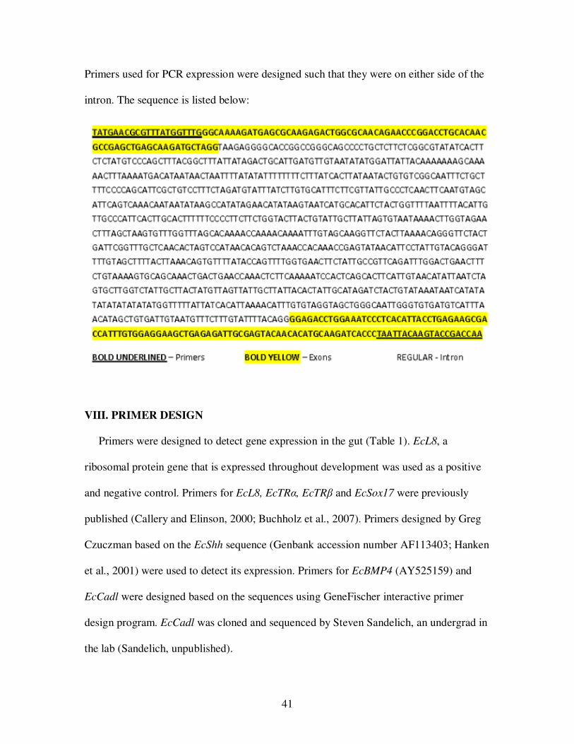
41
Primers used for PCR expression were designed such that they were on either side of the
intron. The sequence is listed below:
VIII. PRIMER DESIGN
Primers were designed to detect gene expression in the gut (Table 1). EcL8, a
ribosomal protein gene that is expressed throughout development was used as a positive
and negative control. Primers for EcL8, EcTRα, EcTRβ and EcSox17 were previously
published (Callery and Elinson, 2000; Buchholz et al., 2007). Primers designed by Greg
Czuczman based on the EcShh sequence (Genbank accession number AF113403; Hanken
et al., 2001) were used to detect its expression. Primers for EcBMP4 (AY525159) and
EcCadl were designed based on the sequences using GeneFischer interactive primer
design program. EcCadl was cloned and sequenced by Steven Sandelich, an undergrad in
the lab (Sandelich, unpublished).
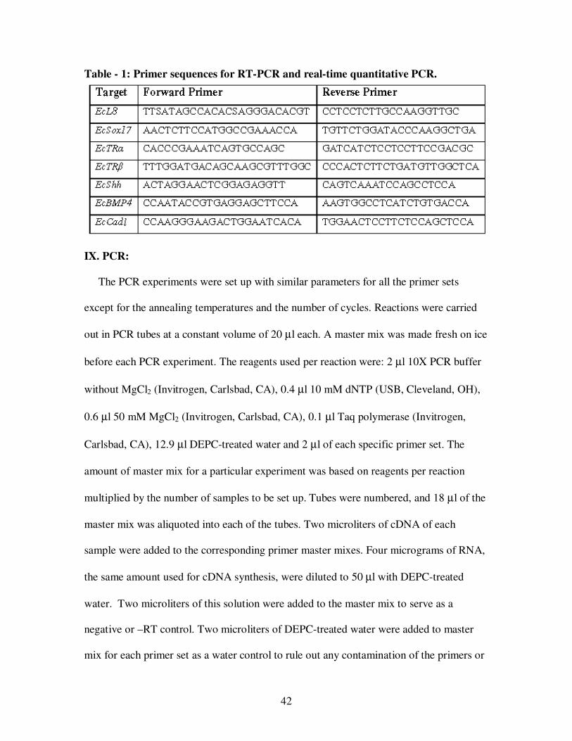
42
Table - 1: Primer sequences for RT-PCR and real-time quantitative PCR.
IX. PCR:
The PCR experiments were set up with similar parameters for all the primer sets
except for the annealing temperatures and the number of cycles. Reactions were carried
out in PCR tubes at a constant volume of 20 µl each. A master mix was made fresh on ice
before each PCR experiment. The reagents used per reaction were: 2 µl 10X PCR buffer
without MgCl2 (Invitrogen, Carlsbad, CA), 0.4 µl 10 mM dNTP (USB, Cleveland, OH),
0.6 µl 50 mM MgCl2 (Invitrogen, Carlsbad, CA), 0.1 µl Taq polymerase (Invitrogen,
Carlsbad, CA), 12.9 µl DEPC-treated water and 2 µl of each specific primer set. The
amount of master mix for a particular experiment was based on reagents per reaction
multiplied by the number of samples to be set up. Tubes were numbered, and 18 µl of the
master mix was aliquoted into each of the tubes. Two microliters of cDNA of each
sample were added to the corresponding primer master mixes. Four micrograms of RNA,
the same amount used for cDNA synthesis, were diluted to 50 µl with DEPC-treated
water. Two microliters of this solution were added to the master mix to serve as a
negative or –RT control. Two microliters of DEPC-treated water were added to master
mix for each primer set as a water control to rule out any contamination of the primers or
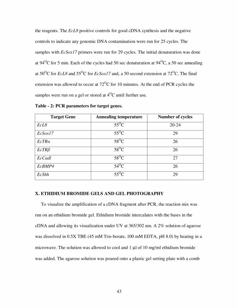
43
the reagents. The EcL8 positive controls for good cDNA synthesis and the negative
controls to indicate any genomic DNA contamination were run for 25 cycles. The
samples with EcSox17 primers were run for 29 cycles. The initial denaturation was done
at 94OC for 5 min. Each of the cycles had 50 sec denaturation at 94OC, a 50 sec annealing
at 50OC for EcL8 and 55OC for EcSox17 and, a 50 second extension at 72OC. The final
extension was allowed to occur at 72OC for 10 minutes. At the end of PCR cycles the
samples were run on a gel or stored at 4OC until further use.
Table - 2: PCR parameters for target genes.
Target Gene Annealing temperature Number of cycles
EcL8 55OC 20-24
EcSox17 55OC 29
EcTRα 58OC 26
EcTRβ 58OC 26
EcCadl 58OC 27
EcBMP4 54OC 26
EcShh 55OC 29
X. ETHIDIUM BROMIDE GELS AND GEL PHOTOGRAPHY
To visualize the amplification of a cDNA fragment after PCR, the reaction mix was
run on an ethidium bromide gel. Ethidium bromide intercalates with the bases in the
cDNA and allowing its visualization under UV at 365/302 nm. A 2% solution of agarose
was dissolved in 0.5X TBE (45 mM Tris-borate, 100 mM EDTA, pH 8.0) by heating in a
microwave. The solution was allowed to cool and 1 µl of 10 mg/ml ethidium bromide
was added. The agarose solution was poured onto a plastic gel setting plate with a comb
44
and allowed to cool at room temperature. After the gel was set, it was transferred into a
gel running chamber with 0.5X TBE solutions.
To each PCR sample in the tube, 1 µl 10X tracking dye (Promega, Madison, WI) was
added to visualize the progression of the dye front of the samples. The samples were
carefully loaded into the wells on the gel. A 1 kb DNA ladder (Promega, Madison WI)
was run for size reference. The gels were run at 100-135 V for 20-30 minutes or until the
dye front was almost to the end of the gel. The gels were photographed on a Kodak
imaging station and saved as tiff files with Adobe Photoshop software. The visualization
of a bright band of expected size was considered as evidence of expression of that
particular RNA. For the data to be considered reliable, it was required that the EcL8
primers amplified a 480bp band in the positive but not in the negative control.
XI. REAL-TIME PCR
Quantitative real-time PCR was done to determine differential gene expression. The
experiments were setup in MicroAmp fast optical 48-well reaction plates (Applied
Biosystems, Foster City, CA). The reaction mix was assembled in 1.5 ml microfuge tubes
on ice. Complimentary DNA (cDNA) was used along with primers and 2X power SYBR
green master mix (Applied Biosystems, Foster City, CA). Each reaction had 40 ng of
cDNA, forward and reverse primers at a final concentration of 500 nM, and 10 µl 2X
power SYBR green master mix in a 20 µl reaction. Every sample-primer pair was
represented in triplicate on each plate. The plates were sealed using a MicroAmp 48-well
optical adhesive film (Applied Biosystems, Foster City, CA) and set for run on StepOne
Real-Time PCR System (Applied Biosystems, Foster City, CA).
45
The instrument was set to run a Quantitation-comparative CT (∆∆CT) type of
experiment. The instrument was directed to detect SYBR green in the target sequence and
perform a melt curve at the end of each experimental run. The initial holding was at 94OC
for 10 minutes. Each of the following 40 cycles of amplification had a 50 second
denaturation at 94OC, a 50 second annealing at 58.5OC and a 60 second extension at
72OC. The annealing temperature and primer efficiencies were empirically determined.
Differences between efficiencies of the target primers were within 10% of that of the
endogenous control.
EcL8, a ribosomal protein gene, was used as the endogenous control. The experiments
were analyzed by the StepOne v2.0 and the output was in the form of relative quantity
(Applied Biosystems, Foster City, CA). The software determined the relative quantity of
target in each sample by comparing normalized target quantity in each sample to
normalized target quantity in the reference sample. Experiments that had multiple
products, as evidenced by a melt curve indicated genomic contamination, were thrown
out. The log phase amplification was checked for an even slope indicating an exponential
amplification. Experiments with uneven amplification during log phase were discarded.
XII. HISTOLOGY
Embryos or guts were fixed in Smith’s fixative for histology. Smith’s fixative was
prepared right before use by mixing equal quantities of two stock solutions. Smith’s stock
A is 1% potassium dichromate (K2Cr2O7), an orange colored solution. Smith’s stock B is
5% acetic acid with 7.4% formaldehyde (Rugh, 1965). The embryos were anesthetized in
0.05% MS222 (Sigma, St. Louis, MO) and put in glass vials with Smith’s fixative. The
46
vials were rocked on a nutator in the dark for 1-2 days for fixation. Fixed embryos were
rinsed twice with DI water for 30 minutes each. Following the washes, the embryos were
stored in 1.5% formaldehyde solution at room temperature.
Embryos were taken through a series of dehydration steps before being embedded in
paraplast. The embryos in 1.5% formaldehyde were washed twice with DI water for 10
minute, one 30 minute wash in 50% ethanol, followed by two 30 minute washes in 70%
ethanol, one 20 minute wash in 90% ethanol and two 20 minute washes in 100% ethanol.
The embryos were left in amyl acetate overnight to allow for further removal of any
water remaining in the tissues. The embryos were taken through three 30 min changes in
toluene in the fume hood. Following the final change in toluene, the embryos were
transferred into molten paraffin at 600C in an incubator. The paraffin was replaced three
times at intervals of 30 min. After the last change, the embryos were left in paraffin
overnight for better infiltration.
Paper boats were made out of index cards and filled with freshly molten paraffin.
Embryos in paraffin were quickly transferred into the boats and oriented in the desired
direction with heated dissecting needles. The embryos in paraffin were rapidly cooled on
a cold surface of Tissue-tec II apparatus (Miles Laboratories, Westmont, IL). Embedded
embryos were allowed to solidify overnight at room temperature. Paraffin blocks with the
embryos were taken out of the boats and mounted on wooden stubs for sectioning. The
blocks were cut into a trapezoid shape with the embryo at the center. Sections were taken
at 10 µm on a Spencer No. 820 microtome (American Optical Company, Buffalo, NY).
The long ribbon of sections was cut into smaller pieces to fit on Silane-Prep slides
(Sigma, St. Louis, MO). The slides were placed on a slide warmer with DI water on its
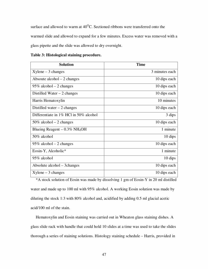
47
surface and allowed to warm at 40OC. Sectioned ribbons were transferred onto the
warmed slide and allowed to expand for a few minutes. Excess water was removed with a
glass pipette and the slide was allowed to dry overnight.
Table 3: Histological staining procedure.
Solution Time
Xylene – 3 changes 3 minutes each
Absoute alcohol – 2 changes 10 dips each
95% alcohol – 2 changes 10 dips each
Distilled Water – 2 changes 10 dips each
Harris Hematoxylin 10 minutes
Distilled water – 2 changes 10 dips each
Differentiate in 1% HCl in 50% alcohol 3 dips
50% alcohol – 2 changes 10 dips each
Blueing Reagent – 0.3% NH4OH 1 minute
50% alcohol 10 dips
95% alcohol – 2 changes 10 dips each
Eosin-Y, Alcoholic* 1 minute
95% alcohol 10 dips
Absolute alcohol – 3changes 10 dips each
Xylene – 3 changes 10 dips each
*A stock solution of Eosin was made by dissolving 1 gm of Eosin-Y in 20 ml distilled
water and made up to 100 ml with 95% alcohol. A working Eosin solution was made by
diluting the stock 1:3 with 80% alcohol and, acidified by adding 0.5 ml glacial acetic
acid/100 ml of the stain.
Hematoxylin and Eosin staining was carried out in Wheaton glass staining dishes. A
glass slide rack with handle that could hold 10 slides at a time was used to take the slides
thorough a series of staining solutions. Histology staining schedule – Harris, provided in
48
the manufacturer’s instruction manual, was followed (Lerner laboratories, Pittsburgh,
PA) during the procedure.
After the last wash, slides were completely drained and allowed to air-dry for a few
seconds. The slides were mounted by adding a few drops of Permount (Fisher Scientific,
Hanover Park, IL), and a coverslip was placed over the mounting medium gently. Care
was taken to prevent any air bubbles from forming on the slide. The slides were placed
on slide warmer for a few hours to allow proper drying, and stored in boxes until
examined and photographed.
XIII. WHOLE MOUNT IN SITU HYBRIDIZATION
The embryos were dejellied and anesthetized with MS222. The guts were dissected as
described earlier and transferred into a 10 ml glass vial with MEMFA. MEMFA is made
fresh before each use at a concentration of 100 mM MOPS pH 7.0, 2 mM EGTA, 1 mM
MgSO4 and 3.7 % formaldehyde. The vial was rocked vertically on a nutator for three
hours at room temperature or overnight at 4OC. The guts were then washed twice in
100% ethanol, and stored at –20OC in 100% ethanol until further use.
To transcription of the antisense and sense probes for in situ hybridization the
EcSox17 plasmid was linearized by digestion with restriction enzymes. Nco-I (Fisher
Scientific, Hanover park, IL) cuts the plasmid past its 5' end with SP6 promoter at the 3'
end and provides a template for antisense EcSox17 transcript. Spe-I (Fisher Scientific,
Hanover Park, IL) cuts the insert past its 3' end with T7 promoter at it 5' end and provides
a template for transcription of sense strand for EcSox17. The EcShh plasmid was
linearized by digesting with Sal-I (Fisher Scientific, Hanover Park, IL) that cuts the
49
plasmid past its 5' end with T7 promoter at the 3' end and provides a template for
antisense transcript. Xba-I (Fisher Scientific, Hanover Park, IL) cuts the insert past its 3'
end with T3 promoter at it 5' end and provides a template for transcription of sense strand
for EcShh. The reaction was set with 10 µg plasmid, 20 µl restriction buffer (Fisher
Scientific, Hanover Park, IL), 10 µl restriction enzyme (Fisher Scientific, Hanover Park,
IL), and the final volume made up to 200 µl with DEPC-water. The reaction mix was
incubated at 37OC for 3-4 hrs in a water bath. At the end of the digest, 3 µl of 1 mg/ml
proteinase K along with 10 µl 10% SDS was added and incubated at 55OC for 45
minutes. An equal volume of phenol: chloroform was added, the mix was vortexed for
about 1 minute, and centrifuged for 10 minutes. The top layer was transferred to a new
microfuge tube and vortexed with an equal volume of chloroform for about 1 minute. To
the top layer in a new microfuge tube, 20 µl 3 M sodium acetate, pH 5.2, and 500 µl of
ice cold ethanol were added and left at –20OC overnight. The mix was centrifuged for 10
minutes, and the pellet was washed twice with 70 % ethanol, and dried for 10-15 minutes.
The dried pellet was resuspended in DEPC-water, quantified with a spectrophotometer,
and stored at –20OC until further use.
The linearized plasmid was used as a template to transcribe labeled sense and
antisense probes for in situ. The reagents were assembled at room temperature in a 1.5 ml
microfuge tube in the order specified below, but the reagents themselves were always
kept on ice. Each reaction has 4 µl 5X transcription buffer, 2 µl 2X DIG RNA labeling
mix (Roche, Indianapolis, IN), 1 µl 100mM DTT (Fisher Scientific, Hanover Park, IL), 1
µl RNAse inhibitor (Takara, Madison, WI), 2 µl RNA polymerase SP6 (Invitrogen,
Carlsbad, CA), T7 (Fisher Scientific, Hanover Park, IL) or T3 (Fisher Scientific, Hanover
50
Park, IL), 2 µg template and the final volume was made up to 20 µl with DEPC-water.
The reaction mix was incubated at 37OC for 3 hours in the hybridization oven.
Transcription was verified by running 1 µl of the reaction mix on an ethidium bromide
gel. An equal volume of 8 M LiCl was added to the remaining probe and left at –20OC
overnight. The probe mix was centrifuged for 10 minutes and the supernatant was
discarded. The pellet was washed twice with 70% ethanol and air-dried. The pellet was
dissolved in 100 µl DEPC-water and was quantified by spectrophotometry. The probe
was hydrolyzed by adding 100 µl of 80 mM sodium bicarbonate and 120 mM sodium
carbonate mixture and incubating at 60OC for 30 min. To this, 12.5 µl 8 M LiCl and 500
µl 100% ethanol were added and precipitated overnight at –20OC. The mix was
centrifuged for 5 minutes, the supernatant discarded, and the pellet was washed twice
with 70% ethanol and air-dried. The pellet was dissolved in hybridization buffer, and the
amount of probe was estimated on the basis of spectrophotometry reading taken just
before hydrolysis.
The guts fixed in MEMFA were transferred into a 5 ml labeled baked glass vials, one
each for sense and antisense. Guts were always transferred with a plastic pipette, and
baked glass Pasteur was used for changing solutions. All the glassware, solutions and
equipment used for this experiment were RNAse free. For each experiment, at least three
guts for each antisense and sense experiment were used. The guts were rehydrated in a
series of solutions starting with 100 % ethanol, a mix of 75 % ethanol and 25 % DEPC-
water, 50% ethanol and 50 % DEPC-water, 25 % ethanol and 75 % PTw (1X PBS and
0.1 % Tween-20, Fisher Scientific, Hanover Park, IL) and 100 % PTw each for about 5
minutes. The guts were washed thrice with 100 % PTw and treated with 1 ml 10 µg/ml
51
proteinase K for 20 min. The guts were washed twice for 5 minutes each with 5 ml 0.1 M
triethanolamine pH 7.5, with gentle rocking. To the rinsed guts in 5 ml triethanolamine,
12.5 µl acetic anhydride (Sigma, St. Louis, MO) were added and rocked for 5 minutes, an
additional 12.5 µl acetic anhydride was added and rocked for another 5 minutes.
The guts were washed thrice with 100 % PTw 5 minutes each on a nutator and re-
fixed in 4% paraformaldehyde for 20 minutes. To make 20 % paraformaldehyde in water,
the cloudy solution was neutralized with NaOH and heated at 65OC while shaking gently
until the solution was clear. The 20% paraformadehyde was covered in foil and stored in
cold until use. The guts were washed thrice with 100 % PTw, 5 minutes each. All but 1
ml of PTw was removed, and 250 µl hybridization buffer (50 % formamide, 5X SSC, 1
mg/ml Torula RNA (Sigma, St. Louis, MO), 100 µg/ml Heparin (Sigma, St. Louis, MO),
1X Denhart’s, 0.1 % Tween-20, 0.1 % CHAPS (Sigma, St. Louis, MO), 10 mM EDTA
was added and the guts were allowed to settle for 5 minutes. The solution was replaced
with 500 µl fresh hybridization buffer and incubated in a hybridization oven at 60OC for
ten min. The solution was replaced with fresh hybridization buffer and incubated
overnight at 60OC for pre-hybridization.
The solution was replaced with fresh 500 µl hybridization solution with the sense or
antisense probe at a concentration of 1 µg/ml and allowed to hybridize overnight at 60OC.
The probe was replaced with fresh hybridization buffer and incubated at 60OC for 10
minutes. The embryos were washed with 2X SSC (Sambrook et al., 1989) thrice for 20
min each at 60OC and then washed in 2X SSC containing 20 µg/ml RNAse A (Sigma, St.
Louis, MO) and ten units/ml RNAse T1 (Sigma, St. Louis, MO) for 30 min at 37OC. This
solution was replaced with 2X SSC and rocked gently for ten min at room temperature on
52
a nutator. The embryos were washed twice with 0.2X SSC for 30 min each at 60OC. The
embryos were further washed, twice in maleic acid buffer (MAB; 100 mM Maleic acid,
150 mM NaCl; pH 7.5) for 15 min each at room temperature. The solution was replaced
with a solution of MAB and 2% Boehringer Blocking Reagent (BMB) (Roche,
Indianapolis, IN) and rocked gently on a nutator for 1 hour at room temperature. The
embryos were then washed in a solution 500 µl MAB + 2% BMB + 20 % heat-treated
lamb serum (Invitrogen, Carlsbad, CA) for 1 hour on a nutator. The solution was replaced
with 500 µl fresh MAB + 2%BMB solution and 0.25 µl antidigoxigening antibody
coupled to alkaline phosphatase (Roche, Indianapolis, IN) was added. The vials were
rocked gently overnight at 4OC. The unbound antibody was removed and the embryos
washed 5 times for 1 hour each at room temperature with maleic acid buffer, one of the
washes was overnight at 4OC.
For the chromogenic reaction, the embryos were washed twice in 5 ml freshly made
alkaline phosphatase buffer (100 mM Tris, pH 9.5, 50mM MgCl2, 100 mM NaCl) for 5
minutes each on a nutator. The solution was removed as much as possible and 0.5 ml BM
purple staining reagent (Roche, Indianapolis, IN) was added. These vials were rocked
gently on a nutator in dark at room temperature. Progress of staining was checked every
hour by transferring the guts into alkaline phosphatase buffer. They were placed back in
BM purple for darker staining as necessary. Staining times ranged from 2-18 hrs in these
experiments. Embryos that showed satisfactory staining were transferred into MEMFA,
fixed for 3 hrs, and washed twice in 100 % ethanol. The embryos were then transferred to
100 % ethanol for photography or were stored until further use.
53
XIV. IN SITU HYBRIDIZATION ON SECTIONS
The embryos used for sectioned in situ were fixed in MEMFA as previously described
for whole mount in situ hybridization. Dehydration, embedding and sectioning were done
as described for histology. DEPC water was used instead of DI water for mounting the
ribbons on the slides. The dried slides were stored at 4OC until used for staining. The
protocol for rehydration, hybridization and staining was done as described (Wessely and
De Robertis, 2000).
XV. ACRIDINE ORANGE STAINING OF YOLK PLATELETS
Acridine Orange was used to assay acidification of yolk platelets in X. laevis (Fagotto
and Maxfield, 1994). To identify the effect of TH on yolk utilization, their protocol was
followed in E. coqui. The yolky part of the gut from embryos was dissected in 200%
Steinberg’s solution. Each dissected piece of yolky tissue was dissociated in 150 µl
dissociation medium by repeatedly sucking it through the narrow tip of a pipette. The
dissociation medium contained 100 mM KCl, 20 mM HEPES-KOH, 10 mM NaCl, 2 mM
MgSO4, 1 mM sodium EDTA in DI water pH 7.4. Dissociation of the tissue caused the
release of the platelets from the yolky nutritional endoderm. The dissociated tissue was
incubated in 3 ml of a Na+/K+ free, low Cl- buffer with Acridine Orange (AO). The
incubation buffer had 10 µM AO in 10 mM HCl, 100 mM gluconic acid, 20 mM HEPES,
2 mM MgSO4, titrated to pH 7.4 with N-methyl glucosamine. The yolk platelets were
incubated in AO for 45 – 60 minutes in the dark. Following the incubation, 75 µl of the
solution along with the dissociated platelets was placed on Silane-Prep slides (Sigma, St.
Louis, MO). Slides were immediately observed under a Nikon Eclipse E600 microscope
54
(Nikon Corporation, Tokyo, Japan) and pictures were taken after exciting the platelets
with UV light. Acidified platelets appeared orange/red, and the neutral platelets were
green. A total of hundred platelets were counted from each sample, and the proportion of
acidified platelets was used for statistical analysis.
Dissociation of nutritional endodermal cells was done as described earlier. The
dissociated cells were stained with AO and pictures taken as described for yolk platelets.
XVI. PHOTOGRAPHY AND IMAGE PROCESSING
Gross morphological and histological pictures of the gut were taken with a QImaging
Retiga 1300 digital camera (Q-Imaging Corporation, Surrey, BC, Canada). The guts were
manipulated gently with a hair loop to prevent damage and were oriented the Petri dish
with 200% Steinberg solution for fresh specimen or in 100% ethanol in case of in situ
hybridization specimen. Pictures of gross morphology were taken under a Leica MZ6
dissecting microscope (Leica Microsystems Inc., Bannockburn, IL). Histological pictures
were taken using a Leitz compound microscope (Walter A. Carveth Ltd, Toronto,
Canada). Pictures were formatted using Adobe Photoshop, at density of 100 pixel/sq.
inch.

55
CHAPTER THREE: RESULTS
I. EXPRESSION OF GUT PATTERNING GENES
A. Gene expression in the developing gut
The large yolky nutritional endodermal cells do not contribute to the definitive gut
tissue (Buchholz et al., 2007). EcSox17, an endodermal transcription factor, is expected
to be expressed in the differentiating gut epithelium. EcShh, a signaling molecule, is
expected to be expressed in the gut epithelium during development. Whole mount and
section in situ hybridization was done to check the localization of the transcripts in the
TS15 gut. The whole mount in situ hybridization was performed on TS15 gut with a
dorsal piece of endodermal tissue and the large yolky nutritional endoderm located
ventrally. The staining indicates EcSox17 and EcShh localization in the dorsal region of
the gut but not in the yolky tissue (Fig 1A, 1B). The sections show staining in the
epithelium of the endoderm and not in the large nutritional cells (Fig 1C).
The lack of staining in the yolky cells could be due to the failure of probe to penetrate
the tissue. A more sensitive RT-PCR was done to elucidate the expression of the
transcript in the large yolky endodermal cells. The dorsal and ventral pieces of the gut
representing the definitive and nutritional endoderm respectively were dissected (Figure
2A) and assayed for differential gene expression (Figure 2B). Dissections were done on
the guts from a stage TS14 embryo. The definitive gut included the stomach, intestine
and liver. The nutritional endoderm included a large mass of yolky cells held together by
a thin layer of connective tissue. The cDNAs made from the two pools of tissue were
tested for the expression of various endodermal and gut patterning genes by RT-PCR.
56
Figure 1: EcSox17 and EcShh in situ hybridization of TS15 gut. (A) EcSox17
expression in the dorsal (D) definitive gut in the antisense (top) and not in the sense
(bottom). The yolky nutritional endoderm is free of any staining (Y). (B) EcShh
expression in the dorsal (D) definitive gut in the antisense (top) and not in the sense
(bottom). The yolky endoderm is free of any staining in the antisense. The yolky
nutritional endoderm is free of any staining (Y). (C) Sectioned gut in situ hybridization
showing EcSox17 expression in the definitive gut epithelium (*) in the antisense.
EcSox17expression is not seen in the epithelium of sense gut.
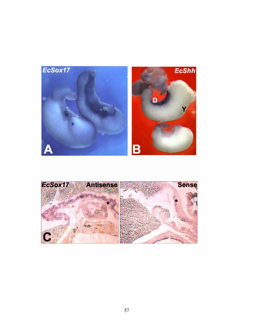
57
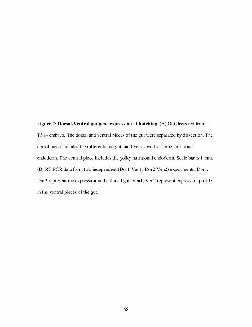
58
Figure 2: Dorsal-Ventral gut gene expression at hatching. (A) Gut dissected from a
TS14 embryo. The dorsal and ventral pieces of the gut were separated by dissection. The
dorsal piece includes the differentiated gut and liver as well as some nutritional
endoderm. The ventral piece includes the yolky nutritional endoderm. Scale bar is 1 mm.
(B) RT-PCR data from two independent (Dor1-Ven1; Dor2-Ven2) experiments. Dor1,
Dor2 represent the expression in the dorsal gut. Ven1, Ven2 represent expression profile
in the ventral pieces of the gut.
59
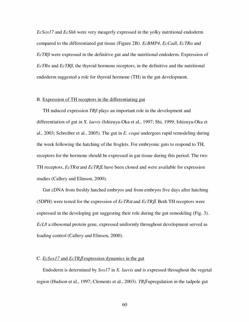
60
EcSox17 and EcShh were very meagerly expressed in the yolky nutritional endoderm
compared to the differentiated gut tissue (Figure 2B). EcBMP4, EcCadl, EcTRα and
EcTRβ were expressed in the definitive gut and the nutritional endoderm. Expression of
EcTRα and EcTRβ, the thyroid hormone receptors, in the definitive and the nutritional
endoderm suggested a role for thyroid hormone (TH) in the gut development.
B. Expression of TH receptors in the differentiating gut
TH induced expression TRβ plays an important role in the development and
differentiation of gut in X. laevis (Ishizuya-Oka et al., 1997; Shi, 1999; Ishizuya-Oka et
al., 2003; Schreiber et al., 2005). The gut in E. coqui undergoes rapid remodeling during
the week following the hatching of the froglets. For embryonic guts to respond to TH,
receptors for the hormone should be expressed in gut tissue during this period. The two
TH receptors, EcTRα and EcTRβ, have been cloned and were available for expression
studies (Callery and Elinson, 2000).
Gut cDNA from freshly hatched embryos and from embryos five days after hatching
(5DPH) were tested for the expression of EcTRα and EcTRβ. Both TH receptors were
expressed in the developing gut suggesting their role during the gut remodeling (Fig. 3).
EcL8 a ribosomal protein gene, expressed uniformly throughout development served as
loading control (Callery and Elinson, 2000).
C. EcSox17 and EcTRβ expression dynamics in the gut
Endoderm is determined by Sox17 in X. laevis and is expressed throughout the vegetal
region (Hudson et al., 1997; Clements et al., 2003). TRβ upregulation in the tadpole gut
61
Figure 3: Expression of thyroid hormone receptors (TR) αααα and ββββ in the gut of E.
coqui. RNA was extracted from hatched and 5 day post-hatch embryos (5DPH) for
cDNA synthesis. EcL8, a ribosomal protein gene, was a loading control. No band
appeared in the –RT control with RNA instead of cDNA. Three independent experiments
were done, and the PCR was repeated at least twice.
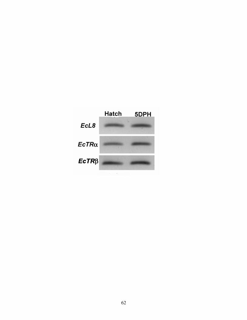
62
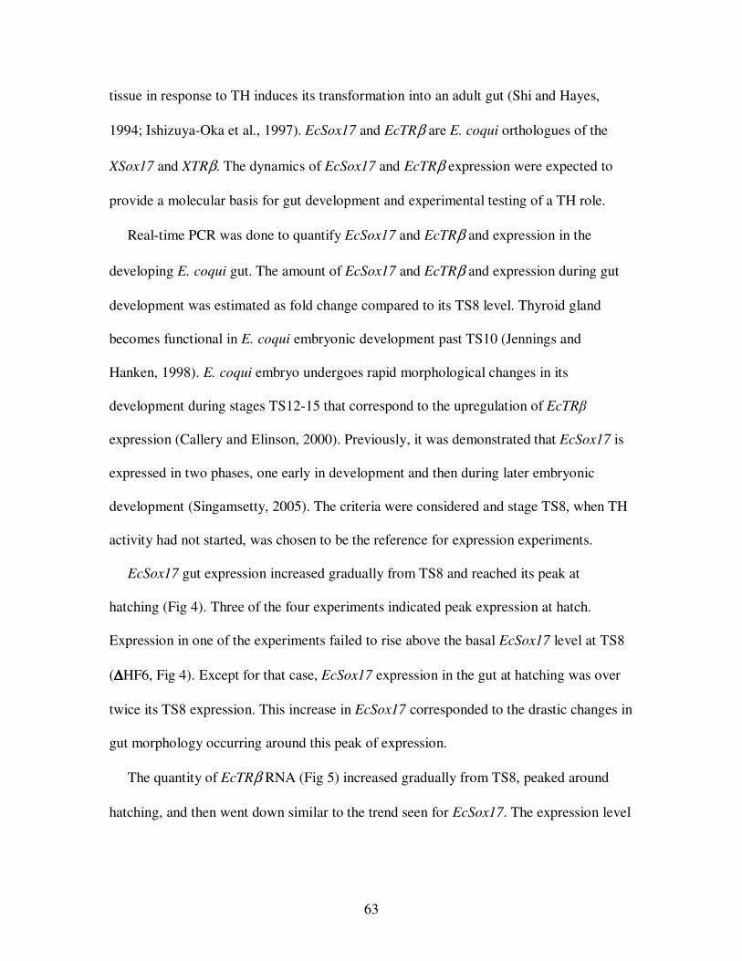
63
tissue in response to TH induces its transformation into an adult gut (Shi and Hayes,
1994; Ishizuya-Oka et al., 1997). EcSox17 and EcTRβ are E. coqui orthologues of the
XSox17 and XTRβ. The dynamics of EcSox17 and EcTRβ expression were expected to
provide a molecular basis for gut development and experimental testing of a TH role.
Real-time PCR was done to quantify EcSox17 and EcTRβ and expression in the
developing E. coqui gut. The amount of EcSox17 and EcTRβ and expression during gut
development was estimated as fold change compared to its TS8 level. Thyroid gland
becomes functional in E. coqui embryonic development past TS10 (Jennings and
Hanken, 1998). E. coqui embryo undergoes rapid morphological changes in its
development during stages TS12-15 that correspond to the upregulation of EcTRβ
expression (Callery and Elinson, 2000). Previously, it was demonstrated that EcSox17 is
expressed in two phases, one early in development and then during later embryonic
development (Singamsetty, 2005). The criteria were considered and stage TS8, when TH
activity had not started, was chosen to be the reference for expression experiments.
EcSox17 gut expression increased gradually from TS8 and reached its peak at
hatching (Fig 4). Three of the four experiments indicated peak expression at hatch.
Expression in one of the experiments failed to rise above the basal EcSox17 level at TS8
(∆∆∆∆HF6, Fig 4). Except for that case, EcSox17 expression in the gut at hatching was over
twice its TS8 expression. This increase in EcSox17 corresponded to the drastic changes in
gut morphology occurring around this peak of expression.
The quantity of EcTRβ RNA (Fig 5) increased gradually from TS8, peaked around
hatching, and then went down similar to the trend seen for EcSox17. The expression level
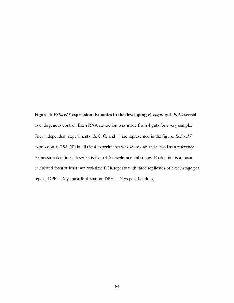
64
Figure 4: EcSox17 expression dynamics in the developing E. coqui gut. EcL8 served
as endogenous control. Each RNA extraction was made from 4 guts for every sample.
Four independent experiments (∆, ◊, Ο, and � ) are represented in the figure. EcSox17
expression at TS8 (Ж) in all the 4 experiments was set to one and served as a reference.
Expression data in each series is from 4-6 developmental stages. Each point is a mean
calculated from at least two real-time PCR repeats with three replicates of every stage per
repeat. DPF – Days post-fertilization; DPH – Days post-hatching.
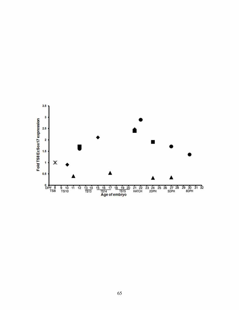
65
66
Figure 5: EcTRββββ expression dynamics in the developing E. coqui gut. EcL8 served as
endogenous control. Each RNA extraction was made from 4 guts for every sample. Four
independent experiments (∆, ◊, Ο, and � ) are represented in the figure. EcTRβ expression
at TS8 (Ж) in all the 4 experiments was set to one and served as a reference. Expression
data in each series is from 4-6 developmental stages. Each point is a mean calculated
from at least two real-time PCR repeats with three replicates of every stage per repeat.
DPF – Days post-fertilization; DPH – Days post-hatching.
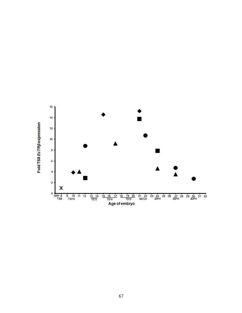
67
68
from stage TS12 until two days after hatching was consistently more than 8-fold or
higher compared to expression at TS8. Highest level of expression was seen at hatching,
a 14-fold increase. Decline was gradual from a 14-fold at hatching to 3-fold by eight days
after hatching (8DPH). All four experimental series showed the trend for EcTRβ gut
expression. However, EcTRβ expression levels were not as high in one of the
experimental series (∆∆∆∆HF6, Fig 5), although it indicated a 9-fold increase. EcSox17
expression levels in the same experimental series failed to rise above its basal TS8
expression suggesting some aberration in these samples (∆∆∆∆HF6, Fig 4). EcSox17 and
EcTRβ expression peaking at hatch suggest the play an important role during the gut
development.
II. TH REQUIREMENT FOR GUT DEVELOPMENT
A. Morphological development of the gut is arrested by methimazole treatment
Embryonic development is arrested at TS12-13 by methimazole, an inhibitor of TH
synthesis (Callery and Elinson, 2000). In repeating the methimazole treatment, gross
developmental differences between the controls and treated embryos became very
obvious once the controls hatched. The control froglets hopped out of the solution and
suspended themselves from the lid of the Petri dish. Embryos in methimazole did not
complete their development and remained aquatic.
The effect of TH inhibition on gut development was not reported previously. To
address this question, guts were dissected from both control and methimazole-treated
embryos five days after the controls hatched to observe the effects of TH inhibition. TH
inhibition led to the arrest of gross gut development with obvious differences compared
69
to the control (Fig 6). The anterior of the control gut, representing the stomach, was
funnel shaped, widest at the oral region and tapering towards its attachment to the yolky
tissue (Fig 6a). Posterior to the yolky tissue the intestine was spindle shaped. The
intestine was shaped like a vase bulged in the middle and narrow at both ends. One end of
the intestine was attached to the yolky tissue and the other end to the cloaca. The stomach
and intestine of the gut dissected from methimazole-treated embryo were narrower in
diameter as compared to the control gut (Fig 6b). The stomach looked more like a horn
than a funnel. The anterior part of the stomach was only a little wider than the other end
attached to the yolky tissue. The intestine was more slender compared to the control
embryos.
Control embryos had relatively little yolk remaining attached to the gut (Fig 6a). The
cloaca was filled with debris from the breakdown of the yolky nutritional endoderm. The
yolk completely disappeared from the control gut about 2-3 weeks after hatching. With
longer times post-hatch, embryos in methimazole failed to change the slender gut
morphology, induced by lack of TH (Fig 7). The definitive gut, represented by anterior
stomach and posterior intestine, remained arrested as long as the embryos were in
methimazole solution. Methimazole treated embryos retained a large amounts of yolk
attached to the slender gut. Some yolky cells got sloughed from the walls of the gut and
were lodged in the lumen. The debris consisted of yolky cells, ranging from those with
greenish tinge to those that were white. Some regions of the gut were devoid of yolky
cells and a clear transparent membrane remained in its place. The observations indicate
that the gut development is arrested by the lack of TH.
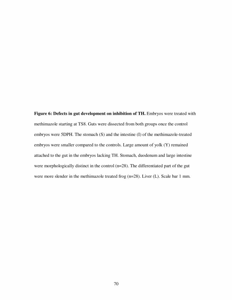
70
Figure 6: Defects in gut development on inhibition of TH. Embryos were treated with
methimazole starting at TS8. Guts were dissected from both groups once the control
embryos were 5DPH. The stomach (S) and the intestine (I) of the methimazole-treated
embryos were smaller compared to the controls. Large amount of yolk (Y) remained
attached to the gut in the embryos lacking TH. Stomach, duodenum and large intestine
were morphologically distinct in the control (n=28). The differentiated part of the gut
were more slender in the methimazole treated frog (n=28). Liver (L). Scale bar 1 mm.
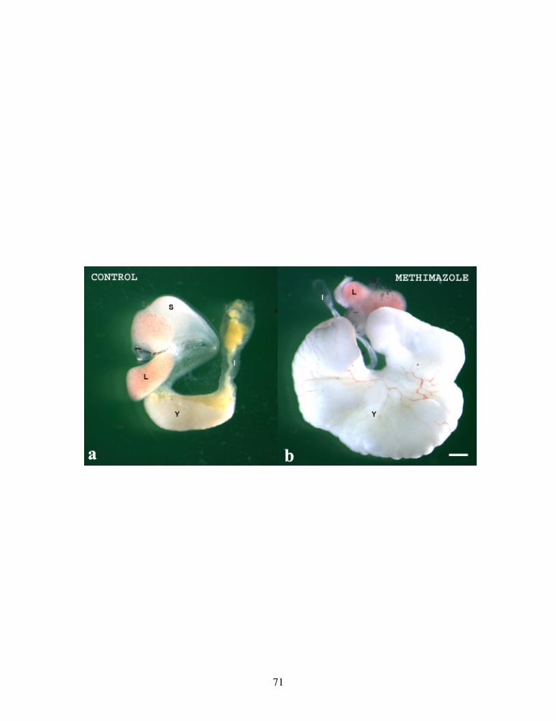
71
72
Figure 7: Effect of long term TH inhibition on gut development. The guts were
dissected from 29DPH (left), 32DPH (center) and 22DPH (right) methimazole-treated
embryos. Control embryos did not survive past 15-22 days of hatching as the yolk
reserves were completely gone. Transparent tissue without yolk was seen in the 29DPH
and 32DPH guts. The opened 22DPH gut contents included intact yolky cells and other
greenish debris in its lumen. Scale bar 1 mm.
73
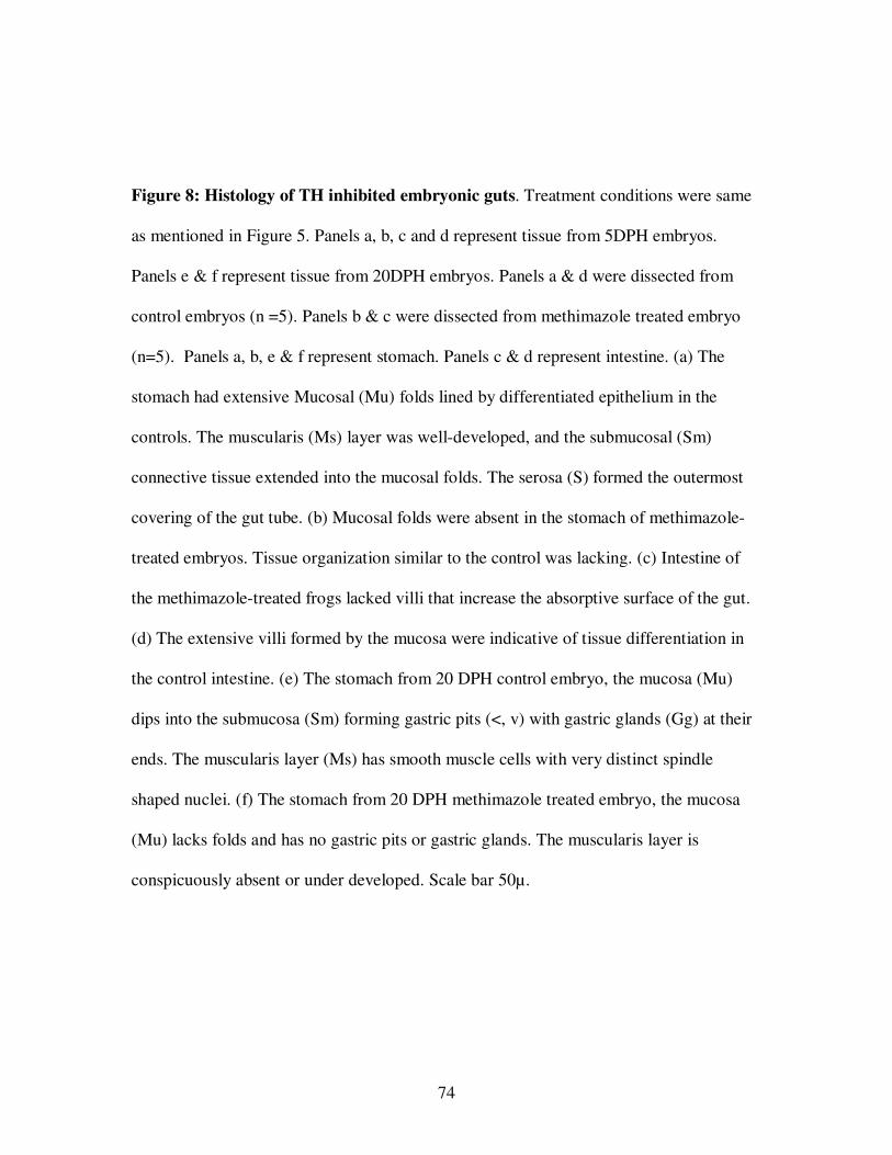
74
Figure 8: Histology of TH inhibited embryonic guts. Treatment conditions were same
as mentioned in Figure 5. Panels a, b, c and d represent tissue from 5DPH embryos.
Panels e & f represent tissue from 20DPH embryos. Panels a & d were dissected from
control embryos (n =5). Panels b & c were dissected from methimazole treated embryo
(n=5). Panels a, b, e & f represent stomach. Panels c & d represent intestine. (a) The
stomach had extensive Mucosal (Mu) folds lined by differentiated epithelium in the
controls. The muscularis (Ms) layer was well-developed, and the submucosal (Sm)
connective tissue extended into the mucosal folds. The serosa (S) formed the outermost
covering of the gut tube. (b) Mucosal folds were absent in the stomach of methimazole-
treated embryos. Tissue organization similar to the control was lacking. (c) Intestine of
the methimazole-treated frogs lacked villi that increase the absorptive surface of the gut.
(d) The extensive villi formed by the mucosa were indicative of tissue differentiation in
the control intestine. (e) The stomach from 20 DPH control embryo, the mucosa (Mu)
dips into the submucosa (Sm) forming gastric pits (<, v) with gastric glands (Gg) at their
ends. The muscularis layer (Ms) has smooth muscle cells with very distinct spindle
shaped nuclei. (f) The stomach from 20 DPH methimazole treated embryo, the mucosa
(Mu) lacks folds and has no gastric pits or gastric glands. The muscularis layer is
conspicuously absent or under developed. Scale bar 50µ.
75
76
B. Abnormal gut histology upon TH inhibition
Histologically, the control gut was highly differentiated (Fig 8a, d, e). The lumen of
the stomach consisted of a single layer of columnar epithelial cells forming the mucosal
layer (Fig 8a, e). The mucosal epithelium extended into the submucosa regularly forming
the gastric pits. The submucosa had gastric glands extending from the mucosal
epithelium. These undulations in the mucosal epithelium gave a folded appearance to the
lumen of the gut. These folds were an indication of gut differentiation providing a larger
surface area for digestion and absorption of food material. Well-differentiated, spindle-
shaped, smooth muscle cells formed a thick layer of muscle tissue around the submucosa.
The outermost layer was serosa, a thin membranous connective tissue that suspends the
gut in the abdomen.
The most obvious tissue defect in the stomach of the TH inhibited embryo was the
lack of mucosal folds (Fig 7b, f). There was no gastric pit or gastric gland development as
in the differentiated gut of controls. The smooth muscle layer was barely visible in the
methimazole treated embryonic guts compared to that of control. The submucosal
connective tissue had loosely packed cells and did not extend into the mucosal
epithelium.
The small intestine of control embryos consisted of many finger-like projections
extending into the lumen referred to as villi (Figure 7d). A layer of tall columnar cells
lined the mucosal epithelium of intestinal villi. The connective tissue extended into each
of these villi. The muscular layer in the intestine was thinner compared to the stomach
region. Methimazole-treated embryos lacked villi in their small intestine (Figure 7c). The
submucosal connective tissue did not extend into the villi. The smooth muscle layer was

77
not distinctly developed in the methimazole treated embryos. These histological findings
corresponded to the gross morphological defects in the gut size of embryos lacking TH
(Fig 6).
The gut of embryos treated for longer times with methimazole had denuded some of
the nutritional endodermal cells and flattened epithelium remained in its place (Fig 8, 9).
Embryos in methimazole at 22, 29 and 32DPH had lots of yolk attached to their guts. The
corresponding controls did not have any nutritional endoderm or died at this stage.
Embryos in methimazole were fixed for histology at 20, 32 and 48DPH. The definitive
gut was still undifferentiated as evidenced by the lack of mucosal folds in a 48DPH
embryo (Fig 9A). The yolky cells became more flattened along their apical-basal axis.
The undifferentiated yolky region of the gut was highly distended occupying most of the
abdomen and filled with intact yolky cells along with other debris. There were a few yolk
platelets in the flat cells of the nutritional endoderm (Fig 9B).
C. TH rescue of gut morphology
The ability of TH to rescue the defects induced by methimazole treatment was
experimentally determined. Embryos were treated with methimazole or methimazole and
T3 starting at TS8. Embryonic guts were dissected seven days after the treatment control
embryos were at stage TS13. The gut dissected from embryos treated with methimazole
containing T3 was morphologically similar to the control gut whereas the gut dissected
from methimazole treated embryos was more slender (Fig 10). The size of gut dissected
from embryos with T3 was relatively larger than either the control or the methimazole
inhibited embryos. The sizes of both the stomach and intestine were increased by T3.
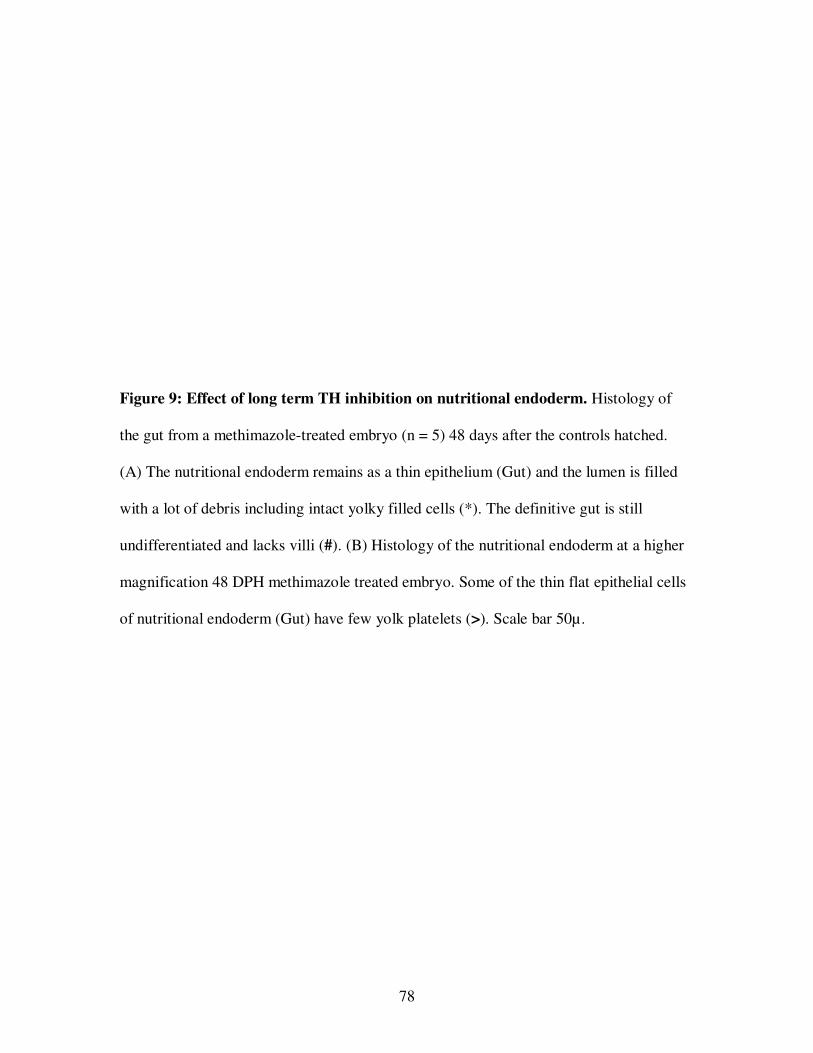
78
Figure 9: Effect of long term TH inhibition on nutritional endoderm. Histology of
the gut from a methimazole-treated embryo (n = 5) 48 days after the controls hatched.
(A) The nutritional endoderm remains as a thin epithelium (Gut) and the lumen is filled
with a lot of debris including intact yolky filled cells (*). The definitive gut is still
undifferentiated and lacks villi (#). (B) Histology of the nutritional endoderm at a higher
magnification 48 DPH methimazole treated embryo. Some of the thin flat epithelial cells
of nutritional endoderm (Gut) have few yolk platelets (>). Scale bar 50µ.
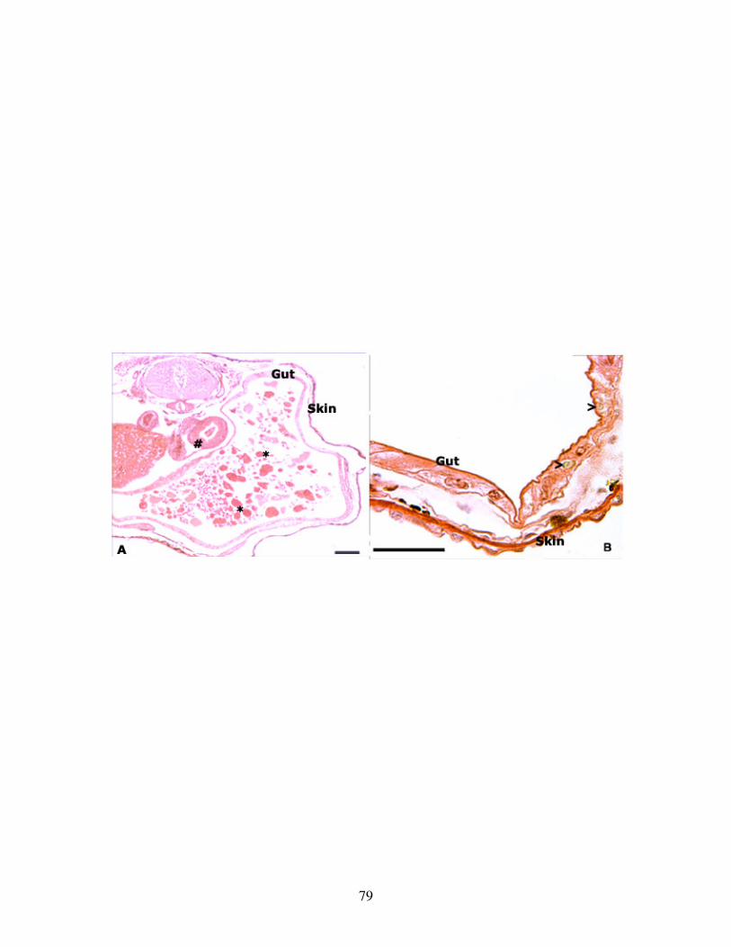
79
80
Figure 10: TH rescue of gross gut development. Three groups of embryos were set up
for the experiments at TS8. Controls were raised in 20% Steinberg’s. Methimazole was
added to attain a final concentration of 1mM to inhibit TH. The third group had
methimazole along with 50 nM T3 added to the 20% Steinberg’s. Guts were dissected by
snipping the most anterior and posterior attachments after 7 days (TS13). Each gut was
dissected into three pieces representing the anterior, posterior and yolky regions. (n=4 per
treatment group)
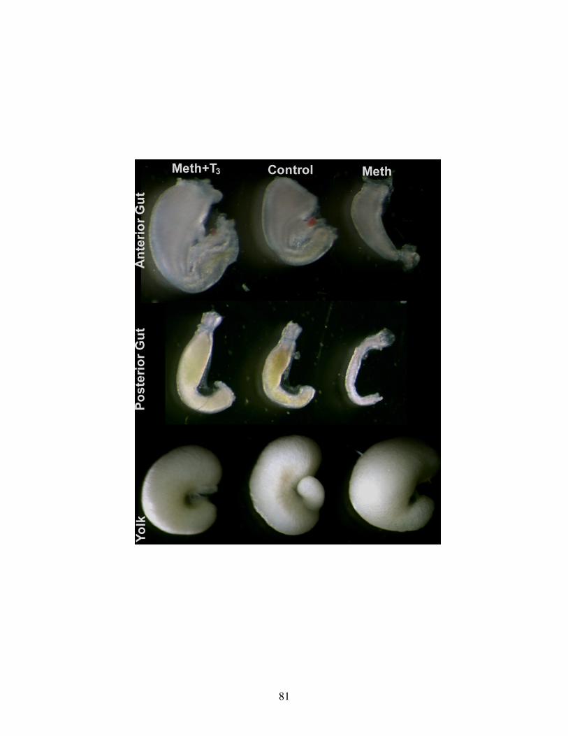
81
82
Figure 11: Effect of TH on the morphology of nutritional endoderm. Embryos were
treated with methimazole (right) or methimazole +T3 (left) starting at TS8. The guts were
dissected after 7 days of treatment; the controls (center) were at TS13 at the time (n=4 per
treatment group).Scale bar 1mm
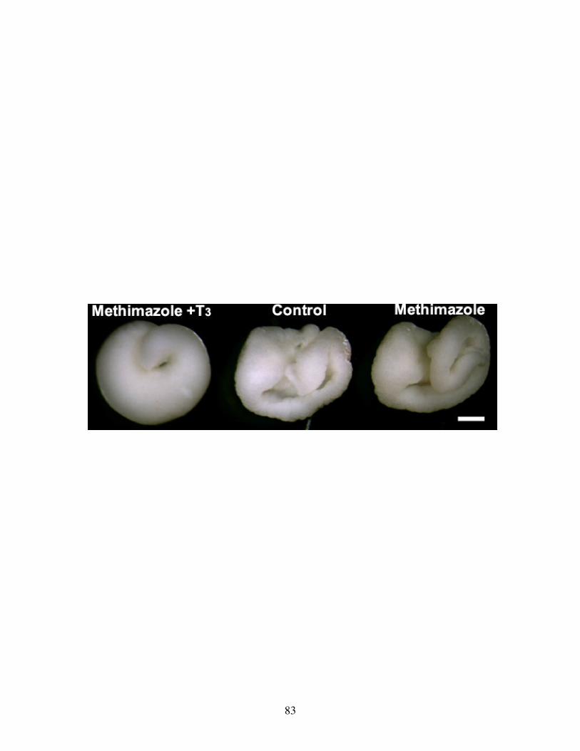
83
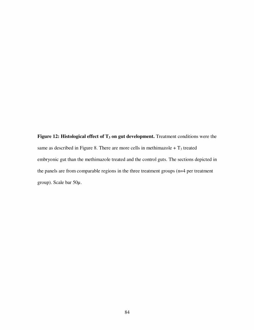
84
Figure 12: Histological effect of T3 on gut development. Treatment conditions were the
same as described in Figure 8. There are more cells in methimazole + T3 treated
embryonic gut than the methimazole treated and the control guts. The sections depicted in
the panels are from comparable regions in the three treatment groups (n=4 per treatment
group). Scale bar 50µ.
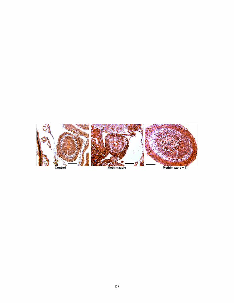
85
86
Dissociated yolky cells filled the lumen of the T3 treated gut while greenish brown debris
was found in the controls. The intestine was slender in methimazole-treated embryo and
was devoid of any debris.
The nutritional endoderm of the gut in methimazole-treated and control embryos had
more folds compared to T3 (Fig 11). The nutritional endoderm in the T3 treated gut had a
rounded and smooth appearance suggesting precocious remodeling of the gut by TH. The
folds in yolky nutritional endoderm of the control gut are a characteristic of the
developing embryonic gut at TS13.
D. TH stimulation of gut differentiation
The stomach and intestinal region of the embryonic guts treated with T3 along with
methimazole were larger (Fig 11) than those in methimazole alone. Histological
sectioning and staining of embryos from the experiments was done to look at the tissue
and cellular effects of T3. Guts were dissected and fixed for histology when the control
embryos were at TS13. Guts from embryos treated with methimazole + T3 had a large
diameter and a lot of cells (Figure 12). The lumen of the large intestine was filled with
intact yolk platelets. There was formation and differentiation of a thick muscularis layer
in the presence of T3. The stomach epithelium in the presence of T3 developed gastric
glands and folds an indication of differentiation. The diameter of the gut was smallest in
methimazole treated embryos and the submucosa and muscularis layers were not
distinctly formed. The control gut had a relatively thin muscularis layer and distinct
submucosal connective tissue. These results suggest an induction of proliferation by the
TH. Similar histological pattern with lots of cells were observed in the eye, brain and
87
other tissues of the T3 treated embryos (Fig 13). These results indicate the role of TH in
the development and differentiation of the embryonic gut.
III. TH INDUCTION OF GUT GENE EXPRESSION
A. Down regulation of gut gene expression on thyroid hormone inhibition
Morphological and histological data suggest that TH is required for proper gut
development in E. coqui. To determine the molecular effect of TH on gut development,
RT-PCR was performed on dissected guts. Genes tested for their response to TH
inhibition included transcription factors and signaling molecules that have a likely
function in endoderm development (Fig 2). Expression levels in the guts dissected from
methimazole-treated embryos (Fig 6B) were compared to expression in their
corresponding 5DPH control guts (Fig 6A). The amount of RNA used for cDNA
synthesis was kept constant for each sample. Serial dilutions were used to determine
uniform loading and to maintain the cycle number in log phase.
The arrest of gut development on methimazole treatment might cause defects in gut
gene expression. The RT-PCR data however, indicated no difference in control and
methimazole expression of EcSox17, EcShh, EcBMP4 and EcCadl genes (Fig 14).
Failure to demonstrate a TH influence on endodermal candidate genes in contrast to the
drastic defects in gut differentiation and morphology (Fig 6, 7) was puzzling. This
discordance between molecular and morphological observations suggested that a more
quantitative analysis was required.
Quantitative real-time PCR assay was developed to quantify the expression of the
endodermal candidate genes in guts lacking TH. EcL8, a housekeeping gene, was used as
88
Figure 13: Histological effect of T3 on eye development. Treatment conditions were the
same as described in Figure 8 and 12. There are more cells in methimazole + T3 treated
embryonic retina (R) than the methimazole treated and the control guts. The sections
depicted in the panels are from comparable regions in the three treatment groups (n=4 per
treatment group). Lens of the eye is seen in methimazole + T3 treated embryo.Scale bar
50µ.
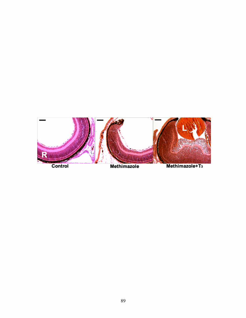
89
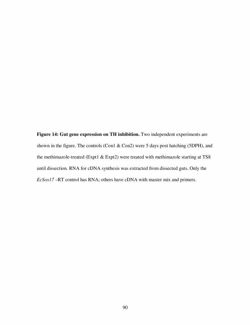
90
Figure 14: Gut gene expression on TH inhibition. Two independent experiments are
shown in the figure. The controls (Con1 & Con2) were 5 days post hatching (5DPH), and
the methimazole-treated (Expt1 & Expt2) were treated with methimazole starting at TS8
until dissection. RNA for cDNA synthesis was extracted from dissected guts. Only the
EcSox17 –RT control has RNA; others have cDNA with master mix and primers.
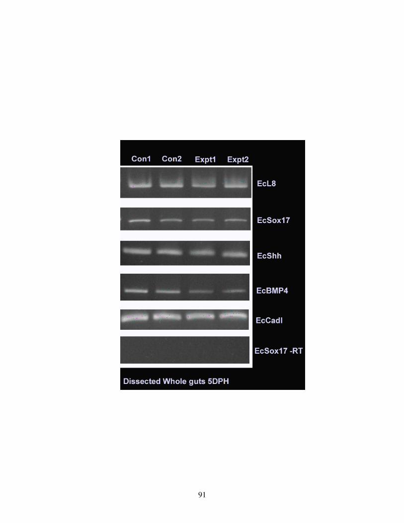
91
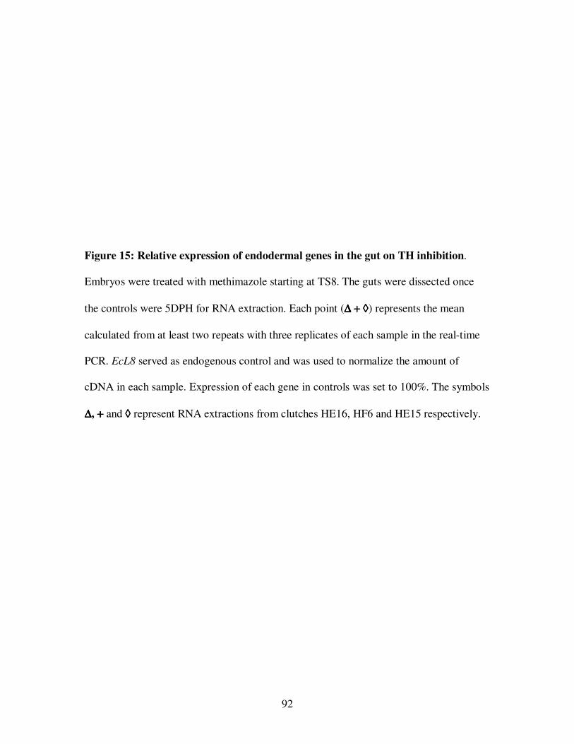
92
Figure 15: Relative expression of endodermal genes in the gut on TH inhibition.
Embryos were treated with methimazole starting at TS8. The guts were dissected once
the controls were 5DPH for RNA extraction. Each point (∆∆∆∆ ++++ ◊◊◊◊) represents the mean
calculated from at least two repeats with three replicates of each sample in the real-time
PCR. EcL8 served as endogenous control and was used to normalize the amount of
cDNA in each sample. Expression of each gene in controls was set to 100%. The symbols
∆∆∆∆, ++++ and ◊◊◊◊ represent RNA extractions from clutches HE16, HF6 and HE15 respectively.
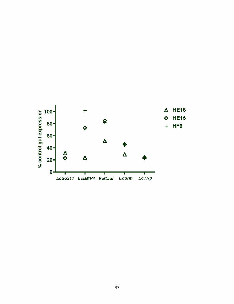
93
94
Figure 16: Effect of TH inhibition on EcTRβ expression. Three independent
experiments are represented in the gel. Embryos were treated with methimazole (Meth)
starting at TS8 and their guts dissected when the controls (Con) were at TS15.
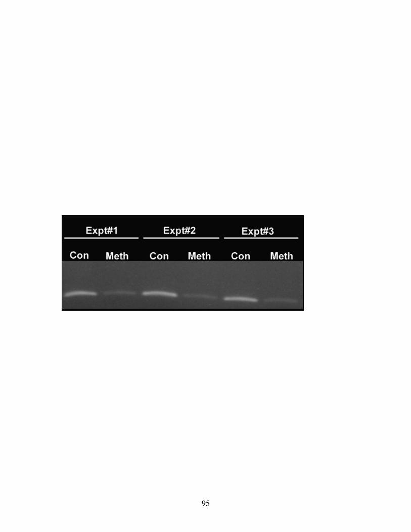
95
96
an endogenous control for cDNA loading. Expression of the candidate genes in the
controls was set as a reference and the relative expression in TH inhibited guts was
determined. TH inhibition resulted in the reduction of EcSox17 expression to about 30%
that of the control gut (Fig 15). The reduction in EcSox17 expression is consistent with
the lack of gut differentiation in embryos lacking TH (Figure 6b and 8b).
BMP4, a signaling molecule, is expressed in the gut submucosa in response to Shh
expression in the gut epithelium of X. laevis (Ishizuya-Oka et al., 2006). EcShh
expression was down-regulated to 40% of that in control on methimazole inhibition (Fig
15). EcCadl and EcBMP4 expressions in TH inhibited guts were lower than in the
controls, but consistent expression levels were not obtained in repeated experiments (Fig
15).
B. Thyroid hormone dependency of EcTRβ expression
Thyroid hormone receptor, TRβ, is a direct TH response gene in X. laevis (Yaoita and
Brown, 1990; Kanamori and Brown, 1992). XTRβ expression was upregulated in
response to TH in X. laevis gut and played a major role in its remodeling and
differentiation (Ishizuya-Oka et al., 2001). RT-PCR was used to check for the effect of
TH inhibition on the expression of EcTRβ, the orthologue of TRβ in E. coqui. EcTRβ
levels were high around hatching in the gut (Fig 3, 5). Gut RNA from control and
methimazole treated embryos was used to synthesize cDNA when the controls were at
stage, TS15. EcTRβ expression was down-regulated in the absence of TH compared to
control guts (Fig 16).
97
For a quantitative analysis, real-time PCR was done on guts dissected from the
methimazole-treated and control embryos at 5DPH for a quantitative analysis. Expression
of EcTRβ in the methimazole treated embryonic guts was 24% that of the corresponding
controls (Fig 15). The low levels of EcTRβ expression on methimazole-treatment
suggested the role of TH during gut development.
C. T3 induction of EcTRβ and EcSox17 expression in the gut
Gut expression of EcTRβ was downregulated in the absence of TH (Fig 15, 16). To
test the requirement of TH for EcTRβ expression, embryos were inhibited by
methimazole. Some of the methimazole inhibited embryos were supplied with T3
exogenously. Two different regimens were followed for the experiments. In the first
experiment, embryos were treated with methimazole or methimazole + T3 at TS8, and
RNA was extracted from their guts when the controls were at TS13. EcTRβ expression
was lower in the guts at TS13 on methimazole treatment and was increased in embryonic
guts on treatment with methimazole + T3 (Fig 17).
In the second experiment, embryos were treated with methimazole starting at TS8.
Once the controls were at TS13, half the embryos in methimazole were treated with
methimazole + T3. The guts from the three groups were dissected once the controls
reached TS15 and assayed for gene expression. The effect of TH inhibition was more
obvious in the guts of embryos at TS15, where EcTRβ signal was barely detectable. The
guts from the embryos in T3 had significant induction of EcTRβ expression (Fig 17).
These results corroborate the requirement of TH for induction of EcTRβ during gut
development.
98
Figure 17: TH induces expression of EcTRββββ in the gut. Two experiments were
conducted. In the first embryos were treated with methimazole or methimazole + T3
starting at TS8, and RNA was extracted from the gut at TS13. Methimazole treatment
repressed EcTRβ expression in the gut and T3 was able to induce its expression. TH
influence on EcTRβ repression and induction was more apparent at TS15.
For TS15 expression studies, embryos in methimazole until TS13 were supplied with T3
and dissected at TS15 for RNA extraction.
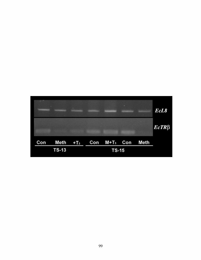
99
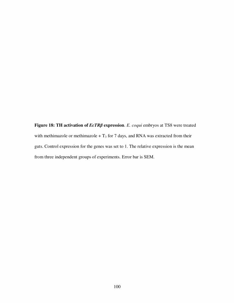
100
Figure 18: TH activation of EcTRβ expression. E. coqui embryos at TS8 were treated
with methimazole or methimazole + T3 for 7 days, and RNA was extracted from their
guts. Control expression for the genes was set to 1. The relative expression is the mean
from three independent groups of experiments. Error bar is SEM.

101
102
While RT-PCR provided qualitative evidence for TH induction, a more sensitive
quantification of the molecular TH induction was done by real-time PCR. Embryos were
treated with methimazole or methimazole + T3 starting at TS8. The guts were dissected
following seven days of treatment and the extracted RNA was assayed for quantitative
expression. Relative expression of EcSox17 and EcTRβ in the two experimental groups
was compared to the control expression.
TH inhibition consistently downregulated EcSox17 and EcTRβ to one-fourth the
expression in the control guts (Fig 18). When methimazole was fortified with T3, EcTRβ
was induced by 4-fold and EcSox17 to three-fourths of the control gut expression. These
results suggested a requirement for TH induction of EcSox17 and EcTRβ expression
during the gut development.
IV. DIRECT ACTION OF TH ON NUTRITIONAL ENDODERM
A. EcTRβ expression in nutritional endoderm
In the embryos treated with methimazole, a lot of yolky nutritional endodermal tissue
was retained attached to the undifferentiated gut (Fig 6b). Their respective controls had
very little yolk remaining attached to the differentiated gut (Fig 6a). Precocious T3
treatment of embryos induced differentiation of the definitive gut and remodeling of the
nutritional endoderm (Fig 10, 11, 12). These observations suggest the role of TH in
differentiation of the gut and utilization of the yolky nutritional endoderm. There are two
possible mechanisms of TH action on the nutritional endoderm. In the first scenario, it
can be hypothesized that TH is required for the utilization of yolk platelets in the
definitive gut and the nutritional endoderm. In the second scenario, TH acts directly on
103
the nutritional endodermal cells resulting in the utilization of yolk platelets. For the tissue
to respond, TRs should be expressed in the cells.
To test these hypotheses, the large yolky cells of the nutritional endoderm were
assayed for EcTRβ expression. The guts dissected from embryos at TS15 were used for
the assay. The stomach and intestinal regions were discarded and the remaining
nutritional endoderm along with a piece of dorsal differentiated gut was used for the
experiment (Fig 19a). The dorsal differentiated gut was carefully cut open to expose the
large yolky nutritional endodermal cells lining the lumen. A Ca++ free dissociation
medium with trypsin was used to separate the large yolky cells from the differentiated
gut. The dissociated cells were pooled and represented the nutritional endoderm. The
remaining tissue consisting of dorsal differentiated gut and the ventral region with
undissociated yolky cells represented the definitive gut. RNA pooled from the dissociated
yolky cells of the nutritional endoderm and the definitive guts were assayed for gene
expression.
Differential expression of EcSox17, EcTRβ and EcBMP4 between the two samples
was quantified using real-time PCR. Dissociated yolky cells are endodermal in origin and
were expected to express the endodermal transcription factor, EcSox17, but not the
mesodermal signaling molecule, EcBMP4. The mesodermal connective tissue of the
definitive gut was expected to express EcBMP4. Relatively low expression of EcBMP4 in
the yolky cells suggested that the pool of yolky cells was not contaminated by
mesodermal tissue (Fig 19b). High expression of EcTRβ in the nutritional endoderm
suggested that they respond to TH. Three independent experiments were performed and
all three experiments confirmed this expression pattern.
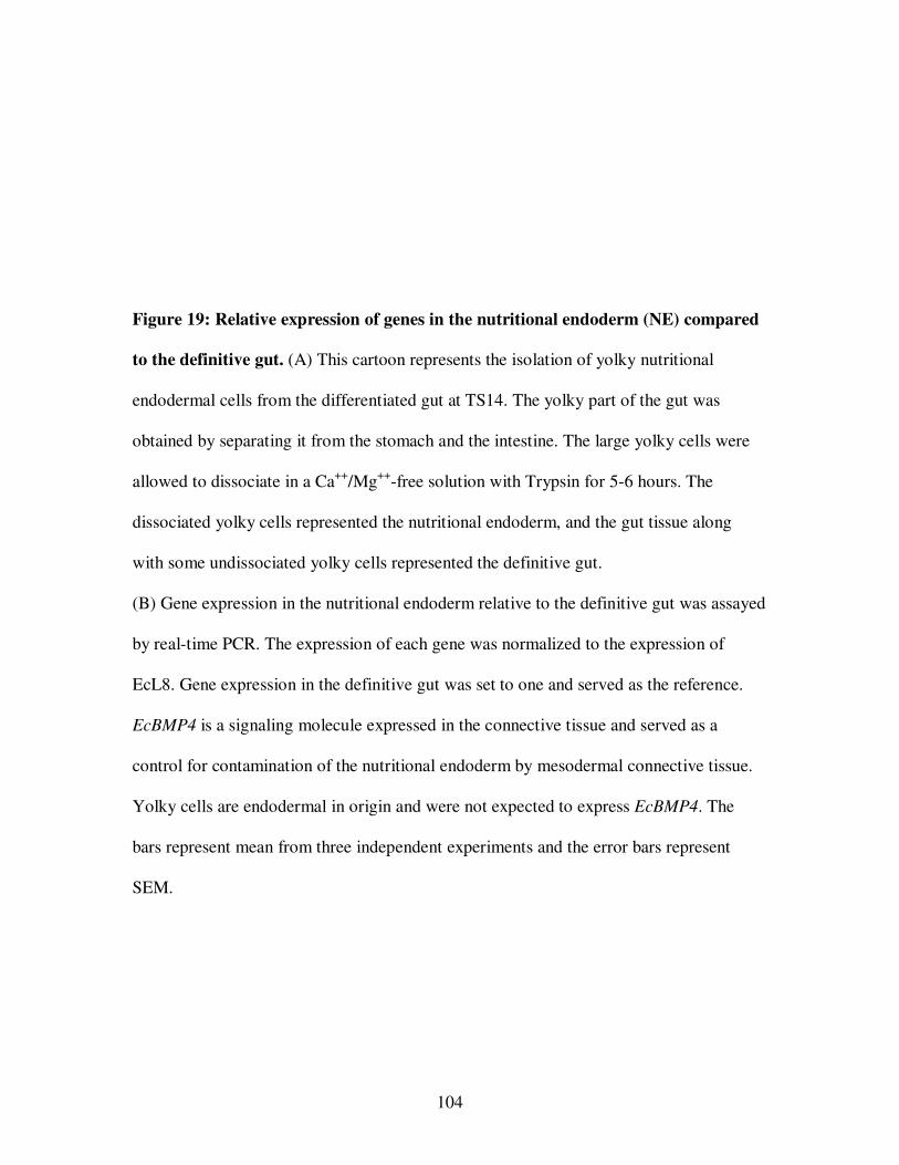
104
Figure 19: Relative expression of genes in the nutritional endoderm (NE) compared
to the definitive gut. (A) This cartoon represents the isolation of yolky nutritional
endodermal cells from the differentiated gut at TS14. The yolky part of the gut was
obtained by separating it from the stomach and the intestine. The large yolky cells were
allowed to dissociate in a Ca++/Mg++-free solution with Trypsin for 5-6 hours. The
dissociated yolky cells represented the nutritional endoderm, and the gut tissue along
with some undissociated yolky cells represented the definitive gut.
(B) Gene expression in the nutritional endoderm relative to the definitive gut was assayed
by real-time PCR. The expression of each gene was normalized to the expression of
EcL8. Gene expression in the definitive gut was set to one and served as the reference.
EcBMP4 is a signaling molecule expressed in the connective tissue and served as a
control for contamination of the nutritional endoderm by mesodermal connective tissue.
Yolky cells are endodermal in origin and were not expected to express EcBMP4. The
bars represent mean from three independent experiments and the error bars represent
SEM.
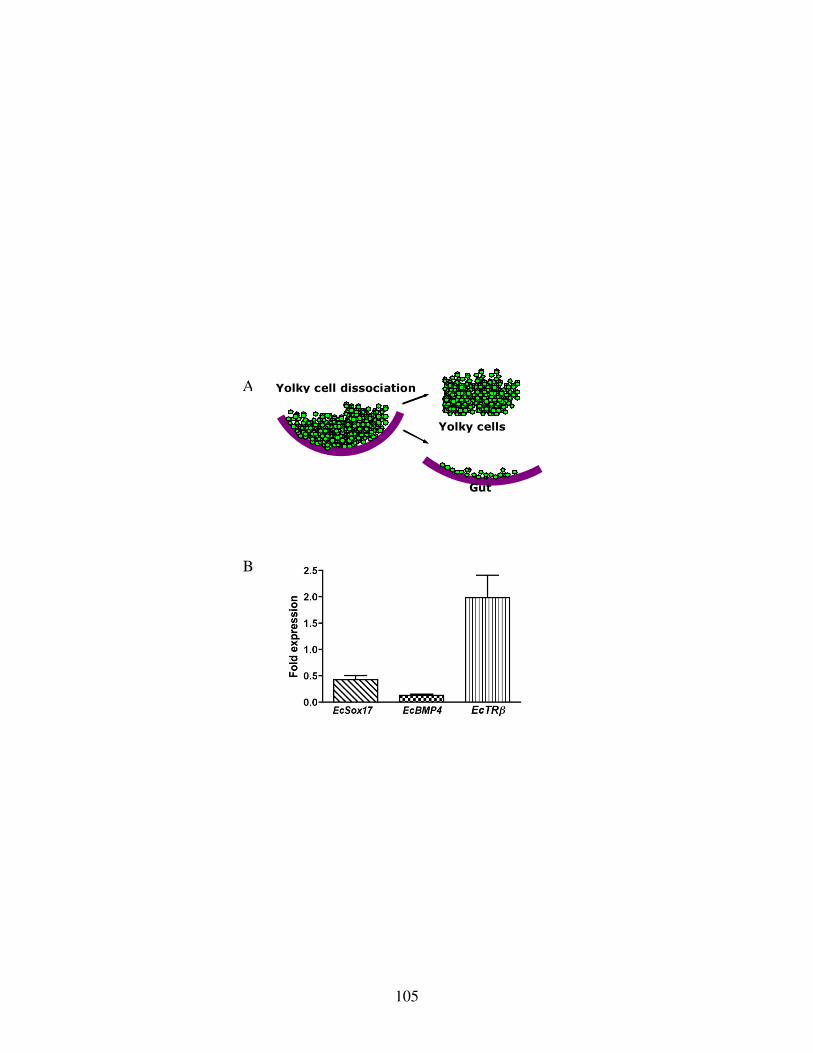
105
Yolky cell dissociation
Gut
Yolky cells
A
B
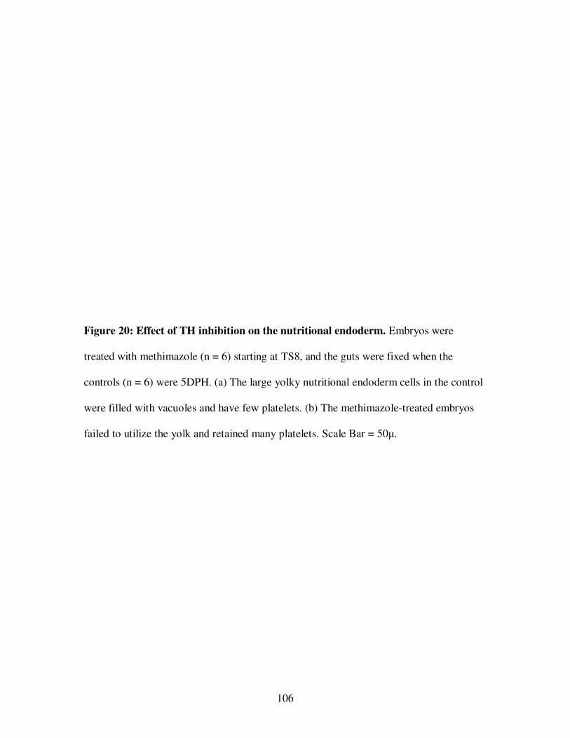
106
Figure 20: Effect of TH inhibition on the nutritional endoderm. Embryos were
treated with methimazole (n = 6) starting at TS8, and the guts were fixed when the
controls (n = 6) were 5DPH. (a) The large yolky nutritional endoderm cells in the control
were filled with vacuoles and have few platelets. (b) The methimazole-treated embryos
failed to utilize the yolk and retained many platelets. Scale Bar = 50µ.
107
108
B. Inhibition of TH prevents yolk utilization
Methimazole treated E. coqui embryos retained their yolk reserves and were
developmentally arrested (Fig 6B). The high expression levels of EcTRβ in the yolky
cells suggested an important role for TH in the activities of the nutritional endoderm (Fig
19b). The effect of TH inhibition on the histology of the nutritional endoderm was
observed on 5DPH embryonic gut. Control embryos had cells of different sizes with one
or more nuclei (Fig 20a). The cells of the nutritional endoderm appeared highly
vacuolated, and some had a few yolk platelets. The corresponding nutritional endodermal
tissue in the methimazole-inhibited embryos had relatively larger cells (Fig 19b). The
yolky cells had a large number of yolk platelets and did not have a vacuolated
appearance. These observations show that the nutritional endodermal cells retain yolk
platelets in the absence of TH.
C. Effect of TH on utilization of yolk
A biochemical approach was used to analyze the utilization of yolk platelets on TH
inhibition. One of the steps in initiation of yolk utilization in X. laevis is the acidification
of the yolk platelets (Fagotto and Maxfield, 1994). Acridine orange is a pH sensitive dye
and accumulates in vesicles based on their internal pH. Organelles with acidic pH appear
orange when excited with UV and the more basic organelles appear green. Yolk platelets
are endosomal in origin and acridine orange can be used to differentiate acidic platelets
from basic ones.
The large yolky nutritional endodermal cells are vacuolated and are devoid of yolk
platelets at 5DPH (Fig 20b). The observation suggests that the yolk platelets are being
109
utilized during this period of development. It was hypothesized that the breakdown of
yolk platelets might be triggered a little earlier than this stage. The nutritional endodermal
cells were dissociated from the guts dissected from embryos 2DPH and incubated with
AO for 5-6 hrs. Many nutritional endodermal cells had acidified and un-acidified
platelets indicated by the presence of red and green platelets respectively (Fig 21). The
degree of acidification varied within the cells from the same gut as indicated by
variations in the ratios of red to green platelets. Due to the variation the counting
acidified cells could not be employed to determine the utilization of yolk and its
requirement of TH.
A different experimental approach was employed to test the effects of TH on yolk
platelet utilization. The embryos were treated starting at TS8 with methimazole. Some of
the embryos in methimazole were provided with T3 at TS13, and acidification of platelets
was assayed two days after the controls hatched. The yolky tissues of the control,
methimazole and methimazole + T3 were dissociated and incubated with acridine orange.
Pictures were taken of the platelets following UV excitation. Acidic platelets were orange
and the rest were green. A total of one hundred were counted from every specimen. The
experiment was repeated on three separate clutches, with two embryos per treatment.
The mean, calculated from the three experiments, indicated that 70% of the platelets
were acidified in the control embryos (Fig 22). About 21% of the yolk platelets in
methimazole-treated embryos were acidified. When provided with T3 the inhibited
embryos were able to induce a lowering of pH in about 46% of the platelets. The amount
of yolky tissue remaining in the controls at the time of experiment was a lot less
110
Figure 21: Acridine Orange (AO) accumulation in the yolk platelets of endodermal
cells. The nutritional endodermal cells from a 5DPH embryo were dissociated and
incubated with 10 µM AO for 5 hours. The pictures were taken after exciting the cells
with UV. The acidified yolk platelets accumulate high concentration of AO and appear
red. Yolk platelet that are not acidified bind accumulate little AO and appear green.
Scale bar 100 µm.
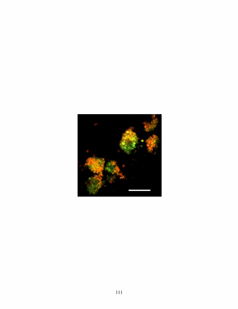
111
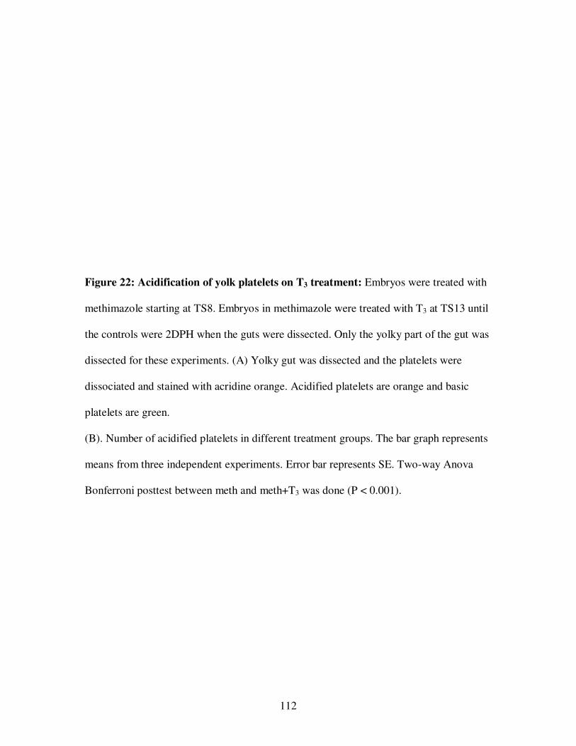
112
Figure 22: Acidification of yolk platelets on T3 treatment: Embryos were treated with
methimazole starting at TS8. Embryos in methimazole were treated with T3 at TS13 until
the controls were 2DPH when the guts were dissected. Only the yolky part of the gut was
dissected for these experiments. (A) Yolky gut was dissected and the platelets were
dissociated and stained with acridine orange. Acidified platelets are orange and basic
platelets are green.
(B). Number of acidified platelets in different treatment groups. The bar graph represents
means from three independent experiments. Error bar represents SE. Two-way Anova
Bonferroni posttest between meth and meth+T3 was done (P < 0.001).
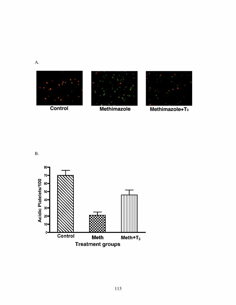
113
A.
B.

114
compared to the methimazole treated embryos. These results suggested a direct
involvement of TH in utilization of the yolk platelets in the nutritional endoderm.
115
CHAPTER FOUR: DISCUSSION
In X. laevis, TH is required for the replacement of the coiled tadpole gut by an adult
one during metamorphosis (McAvoy and Dixon, 1978; Ishizuya-Oka and Shi, 2005). The
embryonic gut in the E. coqui embryo is a miniature version of its adult form with yolk-
rich nutritional endodermal cells attached to intestine (Buchholz et al., 2007). In light of
the fact that E. coqui is a direct developer and lacks a coiled tadpole gut, the major
findings of this study will be discussed in contrast to the TH dependent gut development
in X. laevis and other vertebrates.
I. NOVEL CONTRIBUTIONS OF THIS STUDY
In E. coqui, EcSox17 and EcShh were expressed in the definitive gut and were
relatively undetectable in the nutritional endoderm. Transcripts of EcSox17 and EcShh
were localized to the epithelium of the definitive gut. Both EcSox17 and the thyroid
hormone receptor, EcTRβ, were upregulated in the gut at the time of hatch and remained
high post-hatch. These gene expressions suggest that TH dependent gut remodeling
occurs around hatching of the froglet.
The role of TH was tested by examining the effects of methimazole, an inhibitor of
TH synthesis. Methimazole arrested gut development at TS12 and did not allow any
further development as long as TH synthesis was blocked. The stomach and intestine of
the arrested gut were slender and did not assume the adult morphology. The definitive gut
failed to differentiate from an embryonic to an adult structure; it lacked muscle and
mucosal development. Treatment of the embryos with TH resulted in the increase in size
116
of the gut compared to control and methimazole-treated embryos. The increase in size
appeared to be due to cell proliferation in the definitive gut. TH induced precocious
muscle development and differentiation of the gut epithelium in the E. coqui.
EcSox17, EcShh and EcTRβ were downregulated in guts developmentally arrested by
methimazole, and treatment of embryos with TH resulted in the upregulation of EcTRβ
and EcSox17. EcTRβ was expressed at higher levels in the yolky nutritional endodermal
cells relative to the definitive gut tissue in hatching stage embryos, but EcSox17
expression was low in these nutritional endodermal cells relative to the definitive gut at
the peak of TH induced remodeling. Nutritional endodermal tissue was retained in the
absence of TH, and the cells remained filled with yolk platelets. An initial step in yolk
utilization was the acidification of yolk platelets. Yolk platelet acidification and
utilization was minimal when TH synthesis was prevented.
Previous experiments showed that TH played a vital role in inducing a cryptic
metamorphosis in E. coqui (Callery and Elinson, 2000). In this study, it was found that
methimazole treatment in E. coqui arrested gut development in its embryonic form.
Further, the breakdown of yolk platelets for embryonic nutrition is directly dependent
upon TH. While the role of TH in definitive gut development is conserved in these frogs,
E. coqui has evolved a new function for TH in the utilization of yolk.
II. TH ROLE IN GUT DEVELOPMENT
A. Morphology of the gut
The gut of the X. laevis tadpole is characterized by a rudimentary stomach referred to
as manicotto, a fold in the intestine called the typhlosole, and a long coiled gut (Ueck,
117
1967; Viertel and Richter, 1999). The histological structure of the tadpole gut consists of
a single layer of columnar epithelium, thin musculature, and immature connective tissue
(McAvoy and Dixon, 1977; Kordylewski, 1983; Shi and Ishizuya-Oka, 1996). The
manicotto and typhlosole are completely remodeled at metamorphosis in response to TH,
as an adult gut forms resembling that of higher vertebrates (McAvoy and Dixon, 1977;
Shi and Ishizuya-Oka, 1996; Ikuzawa et al., 2003, 2004). Histologically, the adult
vertebrate gut has multiple circular folds of the epithelium and lots of connective tissue
and muscle.
The definitive gut in the E. coqui embryo is represented by the anterior tube, the
posterior tube, and the dorsal piece of the gut attached to the nutritional endoderm
(Buchholz et al., 2007). The embryonic gut in E. coqui is the miniature form of an adult
gut. Methimazole arrested the development of the definitive gut at TS12 as long as the
embryos were kept in that solution. On co-treatment with T3, the stomach and intestine
developed adult morphologies indicating the role of TH in gut development.
The arrested gut consists of a single layer of columnar epithelial cells, little connective
tissue, and an indistinct muscle layer similar to the X. laevis tadpole gut. It lacks the
epithelial folds, gastric glands, and a thick muscle layer that are the characteristics of an
adult gut. TH induced EcTRβ upregulation correlates with the development adult gut
morphology, including mucosal folds, thick musculature, gastric glands, and gastric pits.
The gut thus formed, resembles that of an adult vertebrate and indicates the retention of a
conserved role of TH in these frogs.
The absence of structures analogous to the manicotto and typhlosole in E. coqui
indicate that these frogs do not form tadpole like structures that are replaced at
118
metamorphosis. It appears that in E. coqui, the function of tadpole gut in providing
nutrition has been replaced by the nutritional endoderm.
B. Transformation from embryonic to an adult gut
The long, coiled tadpole gut in X. laevis shortens by about 75% of its length at
metamorphic climax and forms the stomach and intestine of a typical vertebrate
(Marshall and Dixon, 1978; McAvoy and Dixon, 1978; Ishizuya-Oka and Shimozawa,
1987a; Ishizuya-Oka and Shi, 2005). The shortening is accompanied by heaping of the
epithelium into a temporary multicellular lining. The larval epithelium undergoes rapid
apoptosis at metamorphic climax and is replaced by proliferating adult epithelium in
response to TH (Ishizuya-Oka and Ueda, 1996; Shi and Ishizuya-Oka, 1997). In
zebrafish, TH treatment induces over-proliferation of the gut epithelium and constriction
of yolk sac band (Liu and Chan, 2002).
There is no apparent shortening of the gut in E. coqui during its development
(Buchholz et al., 2007). The nutritional endodermal cells do not contribute to the
definitive gut and are replaced by the proliferating epithelium in the post-hatch gut. The
epithelial lining does not replace the nutritional endoderm in the absence of TH.
Treatment with T3 leads to over-proliferation of the definitive gut epithelium, giving the
epithelium a multilayered appearance as well as a thick muscle layer. High rates of
proliferation were detected by antiphospho-H3 staining in the definitive gut of E. coqui
but not in the nutritional endoderm (Langer, 2003). These observations suggest that the
definitive gut formed by TS12 is embryonic and requires activation by the TH to be
transformed into an adult gut in E. coqui.
119
C. Remodeling nutritional endoderm
All the cells in the yolky vegetal region in Xenopus tropicalis express endodermal
marker genes XtSox17α, XtSox17β, and XtFoxA1 (D’Souza et al., 2003). Similarly, all the
vegetal cells contribute to the definitive gut in X. laevis tadpole (Chalmers and Slack,
2000). Differentiated primary epithelium of the tadpole gives rise to the primordial stem
cells that proliferate and replace the larval epithelium under the influence of TH
(Marshall and Dixon, 1978; Shi and Ishizuya-Oka, 1996; Schreiber et al., 2005; Ishizuya-
Oka et al., 2009). Matrix metalloproteinases (MMPs) are upregulated by TH in
fibroblasts of the connective tissue layer of the intestine (Ishizuya-Oka et al., 1996;
Hasebe et al., 2006; 2007). They degrade proteins in the extracellular matrix, resulting in
the degradation and removal of the degenerating larval epithelium.
Once all the yolk platelets are utilized in E. coqui embryos, the empty nutritional
endodermal cells are sloughed into the lumen of the gut (Buchholz et al., 2007). In this
investigation, the nutritional endoderm was retained in E. coqui embryos treated with
methimazole and was not replaced by the definitive gut. These large, yolky nutritional
endoderm cells are like the larval epithelium of X. laevis tadpole intestine in that they are
lost in response to TH. However, unlike X. laevis larval epithelium (Ishizuya-Oka and
Ueda, 1996; Ishizuya-Oka et al., 1998), TUNEL staining did not indicate apoptosis in the
E. coqui nutritional endoderm (Buchholz et al., 2007). The process by which the
nutritional endodermal cells die is unknown. It is also not known if all the epithelial cells
lining the embryonic gut or only few stem cells proliferate in E. coqui.
The nutritional endodermal cells are rapidly sloughed into the lumen following
exposure to exogenous TH. The expression of extracellular remodeling proteins in the
120
fibroblast on treatment with TH can be tested in E. coqui. The upregulation of MMPs in
the connective tissue in the gut following TH treatment would indicate its role in the
remodeling of nutritional endoderm.
III. GENE EXPRESSION IN THE GUT
A. Definitive gut development
Sox17, expressed in the definitive endoderm, is both necessary and sufficient for gut
formation in X. laevis and zebrafish (Hudson et al., 1997; Alexander and Stainier, 1999;
Clements and Woodland, 2000; Clements et al., 2003). In mice, Sox17-/- mutants have
defects in gut development (Kanai-Azuma et al., 2002). Sox17 is an endodermal
transcription factor, required for the expression of Edd, HNF1β, IFABP, Foxa1, and
Foxa2 that are involved in organ differentiation and tissue specific gene expression
(Demartis et al., 1994; Shi and Hayes, 1994; Sasai et al., 1996; Hudson et al., 1997;
Clements et al., 2003; Sinner et al., 2004). In X. laevis, Shh expression is specifically
upregulated in the adult stomach and intestinal epithelium at metamorphosis (Stolow et
al., 1997; Ishizuya-Oka et al., 2001; Ishizuya-Oka et al., 2003). Shh was shown to be vital
for the intestinal organogenesis employing mutant mice (Litingtung et al., 1998;
Ramalho-Santos et al., 2000).
In this study, EcSox17 was used as a marker for endodermal differentiation. EcShh,
the XShh orthologue, was used as a marker for the postembryonic epithelium. The
expression of EcSox17 and EcShh is lower in the nutritional endoderm compared to their
levels in the definitive gut tissue at hatching. The expression deficiency of these
endodermal genes is correlated with the lack of contribution of the nutritional endoderm
121
to the adult gut epithelium. The upregulation of EcSox17 at hatching indicates a likely
role in the transcription of downstream endodermal genes in the newly forming
epithelium. Localization of EcShh to the cells lining the gut epithelium at hatching
indicates that these cells represent the adult gut epithelium. The expression of
endodermal genes in the definitive gut and their deficiency in the nutritional endoderm
supports the presence of two different sets of cells with different fates.
B. TH regulation of gut development
Peak expression of TRβ in the tadpole is a characteristic of metamorphic climax (Tata
et al., 1993). In X. laevis, the TH induced upregulation of TRβ mediates the remodeling
events leading to the formation of adult gut epithelium (Shi and Brown, 1993). Shh is a
direct TH response gene and is upregulated in the postembryonic epithelium of X. laevis
(Shi and Brown, 1993; Stolow and Shi 1995; Ishizuya-Oka et al., 2001b). Some genes are
down-regulated at metamorphic climax in the intestinal epithelium and the stomach and
are expressed again at the end of metamorphosis (Shi and Hayes, 1994; Ishizuya-Oka et
al., 1997; Ikuzawa et al., 2004). One such target, Ifabp, the late TH response gene, is
downregulated in the metamorphic gut and is upregulated in the post embryonic,
differentiated adult gut (Shi and Hayes, 1994).
EcTRβ peaks at hatching in the E. coqui gut, and this peak is correlated with drastic
changes that occur in the definitive gut and the nutritional endoderm. The upregulation of
EcTRβ in the gut is concurrent with the TH dependent metamorphic events in other
tissues (Callery and Elinson, 2000). Methimazole treatment downregulates the expression
of EcTRβ in the developing gut blocking TH induced autoregulation. EcTRβ levels in the
122
gut reach their peak levels at hatching and remain high post-hatch suggesting the role of
TH during the remodeling event replacing the nutritional endoderm with epithelium.
Treatment with T3 causes the nutritional endodermal cells to be sloughed from the
underlying epithelium.
The formation of new gut epithelium replacing the sloughed nutritional endodermal
cells in E. coqui correlates with the increased EcSox17 expression. The upregulation of
EcTRβ and EcSox17 also correlate with the appearance of active thyroid gland at TS12,
representing the last third of embryonic development (Jennings and Hanken, 1998;
Callery and Elinson, 2000). Failure of the definitive gut epithelium to replace the
nutritional endoderm in methimazole-treated gut implies the requirement of TH
dependent regulation of the remodeling events in E. coqui.
The effect of TH on remodeling of the embryonic gut and its replacement with an
adult gut can be tested. A polyclonal anti-IFABP antibody is available in X. laevis
(Ishizuya-Oka et al., 1997) and can be used in E. coqui. The detection of IFABP protein
in the gut epithelium before the onset on TH activity in E. coqui will indicate the
presence of differentiated tadpole-like epithelium. Lack of IFABP protein in the
embryonic gut epithelium before the onset of metamorphosis and its detection following
the onset of TH activity will indicate that an adult gut forms directly.
IV. ORIGIN OF ADULT GUT EPITHELIUM
There are two models proposed for the origin of stem cells that proliferate to form the
adult gut epithelium in X. laevis. The first is that there are nests of adult stem cells that
proliferate to form the adult gut epithelium. These cells appear as small islets at
123
metamorphic climax (McAvoy and Dixon, 1977; Houdry and Dauca, 1977; Shi and
Ishizuya-Oka, 1996; Ishizuya-Oka et al., 2003). A second train of thought is that adult
stem cells form from differentiated larval epithelium (Marshall and Dixon, 1978;
Ishizuya-Oka and Shimozawa, 1991; Amano et al., 1998; Schreiber et al., 2005). In a
recent study, Ishizuya-Oka (2009) indicated that TH induces dedifferentiation of larval
epithelial cells to become stem cells (Ishizuya-Oka et al., 2009). A similar phenomenon
was observed in the mammalian intestine. Partially differentiated epithelium, referred to
as “epithelial transit cells”, dedifferentiate into stem cells that then regenerate the
absorptive epithelium of the intestine (Potten et al., 1997; Potten, 1998).
Shh is expressed in the adult stem cell primordia in X. laevis tadpoles (Ishizuya-Oka et
al., 2001). Similarly, Shh is expressed in mammalian stem cell primordia and their
descendents (van den Brink et al., 2002). Other markers of stem cells, expressed in the
metamorphosing intestine of X. laevis, include Musashi-1 (Ishizuya-Oka et al., 2003), a
phosphorylated form of phosphatase and tensin (PTEN), and Akt (Ishizuya-Oka and Shi,
2007). Their homologues are candidates for adult mammalian stem cell markers in the
intestine (De Santa Barbara et al., 2003; Kayahara et al., 2003; Potten et al., 2003; He et
al., 2004).
Results of this study show that EcShh is localized to all cells lining the definitive gut
at hatching. Previously, EcShh expression was shown to be biphasic during
embryogenesis (Singamsetty, 2005). The early expression phase is present in the floor
plate of neural tube and in the foregut from late TS3 to TS5 (Hanken et al., 2001). It was
hypothesized that the later phase of EcShh expression from TS12 to TS15 is a TH
induced expression in the differentiating adult stem cells of the gut. EcShh is
124
downregulated in the methimazole-treated embryonic gut. The downregulation is
hypothesized to have inhibited the development of stem cells and thus preventing
formation of an adult gut. These observations suggest that the cells lining the gut are
larval at TS12 and require activation by TH to become adult stem cells. To test this, the
homologues of genes expressed in X. laevis intestinal stem cells, such as, Masashi-1
(Ishizuya-Oka et al., 2003), PTEN and Akt (Ishizuya-Oka et al., 2009), should be cloned
in E. coqui and assayed for their expression in the developing gut.
V. YOLK UTILIZATION
A. Vacuolation in the intestine
Vacuolation is observed in the cells undergoing histolysis in the tail of X. laevis at
metamorphosis (Kerr et al., 1974; Kinoshita et al., 1985; Yoshizato., 1989). The
epithelium of the distal small intestine in suckling rats and piglets is highly vacuolated
and appears foamy in histology (Baintner, 1994). Large acidic vacuoles are seen in the
avian yolk sac (Moxon and Wild, 1976) and ileum of cat, rabbit and suckling ruminants
(reviewed by Baintner, 2007). The vacuoles are lysosomal in origin and function as
intracellular digestive compartments. These vacuoles disappear with the onset of luminal
digestion (reviewed by Baintner, 2007).
Highly vacuolated nutritional endodermal cells, devoid of yolk platelets, were
sloughed into the lumen of the gut during remodeling in E. coqui. In contrast,
methimazole-treated guts had many yolk platelets in their cells and lacked vacuoles. The
appearance of vacuoles in the nutritional endodermal cells was concurrent with the
disappearance of yolk platelets. These observations indicated high levels of endosomal
125
activity related to the intracellular digestion of yolk platelets in E. coqui, similar to that
reported in the Japanese newt (Komazaki and Hiruma, 1999) and X. laevis (Jorgensen et
al., 2009).
B. Acidification of yolk platelets
Acidification of the yolk platelets is an important mechanism that activates yolk
utilization (Fagotto, 1991; Nordin et al., 1991; Mallaya et al., 1992; Fagotto and
Maxfield, 1994). Acridine orange (AO) accumulation in the yolk platelets was used to
assay the change of pH in X. laevis (Fagotto and Maxfield, 1994). Similarly, acidic pH in
the digestive vacuoles of the intestinal epithelial cells in the suckling rat and piglet was
also demonstrated using AO (Baintner, 1994). Acidified organelles appear red on
excitation with UV, whereas unacidified platelets appear green.
Acidified yolk platelets were present togeather with unacidified platelets in a cell
during early embryonic development in X. laevis (Fagotto and Maxfield, 1994). These
observations suggested that acidification of yolk platelets is a highly regulated
intracellular process in the embryo. Jorgensen et al (2009) showed that degrading and
intact yolk platelets were present in the same cell based on the presence or absence of a
marker localized to the superficial layer of the yolk platelet. These findings indicate that
the proteolysis of yolk platelets occurs on a per platelet basis in X. laevis (Fagotto and
Maxfield, 1994; Jorgensen et al., 2009).
The same strategy was usedto assay yolk utilization in E. coqui nutritional endoderm.
Increase in acidification of yolk platelets was assayed using AO and acidification
occurred a few days before vacuolation appeared in the nutritional endodermal cells.
126
Green and red yolk platelets representing intact and degrading platelets respectively were
detected in the same cell indicating that the yolk platelets were being acidified on per
platelet basis. These observations suggest that yolk proteolysis is a conserved and a
highly regulated intracellular process initiated by change in pH, a mechanism similar to
that in X. laevis.
C. TH role in yolk utilization
Novel findings in this study indicate a TH requirement for yolk utilization in the
nutritional endoderm of E. coqui. However, yolk utilization occurs in the other
embryonic tissues of E. coqui and throughout the embryo in X. laevis, before the
development of thyroid gland. These observations will be discussed under two different
sections. The first will deal with the role of TH in yolk utilization in the nutritional
endoderm. The second will discuss the utilization of yolk before thyroid gland develops
in E. coqui and X. laevis.
C. 1. TH is directly required for yolk utilization in E. coqui
Previously, it was shown that the nutritional endodermal cells are rapidly depleted of
yolk and sloughed into the lumen after hatching in E. coqui (Buchholz et al., 2007). In the
methimazole-treated embryos EcTRβ expression was inhibited, and there was a parallel
lack of yolk platelet utilization implying the role of TH via TRs. The high levels of
EcTRβ RNA in the nutritional endodermal cells observed at hatching suggested its direct
involvement in yolk utilization. Acidification of yolk platelet is an early step in yolk
proteolysis in X. laevis (Fagotto and Maxfield, 1994). This step of yolk platelet
127
acidification was inhibited by methimazole treatment and, was induced by co-treatment
with TH in E. coqui. The experimental results indicate TH directly controls utilization of
yolk, a novel evolutionary role for the hormone in E. coqui that was not reported in any
other organism.
The tail of X. laevis undergoes histolysis towards the end of metamorphosis directly in
response to TH (Weber, 1967; Tata et al., 1991). The earliest changes include formation
of vacuoles with acid phosphatase activity (Kerr et al., 1974; Kinoshita et al., 1985;
Yoshizato, 1989). The cathepsin activity in the tail of X. laevis increases following TH
treatment (Weber, 1967). Intracellular hydrolases and serine proteases are upregulated by
TH in the tail fibroblasts (Berry et al., 1998). Microarray analysis indicated that 40% of
the lysosomal genes were induced by TH including the lysosome specific cathepsins and
hyaluronidase (Das et al., 2006).
TH in E. coqui could be involved in the upregulation of lysosomal enzymes as
observed in the tadpole tail of X. laevis. The TH induced expression of EcTRβ, a
transcription factor, could be involved in the transcription of the genes coding for the
lysosomal enzymes. The vacuolation observed in the nutritional endodermal cells
observed a few days after the hatching suggests increased lysosomal activity. Expression
of the enzymes can be tested by cloning the lysosomal genes in E. coqui and examining
their expression in the nutritional endodermal cells. Antibodies against the lysosomal
enzyme can be used to examine their upregulation the nutritional endoderm in response
to TH. Antibodies can also be used to measure the activity of lysosomal enzymes in the
nutritional endoderm in response to TH.
128
C. 2. TH role in yolk utilization before thyroid gland development
In X. laevis, the yolk is completely utilized before the formation of a functional
thyroid gland. The thyroid gland in X. laevis matures at stage 54 (Dodd and Dodd, 1976),
while yolk platelets are depleted from all the tissues by stage 48 (Nieuwkoop and Faber,
1994). Yolk utilization in X. laevis appears to be independent of TH production. On the
other hand, high levels of TH (Morvan Dubois et al., 2006) and TR (Eliceiri and Brown;
1994; Havis et al., 2006) have been detected during embryogenesis in X. laevis. This
raises the possibility that yolk utilization in X. laevis embryos depends on maternal TH
and TR.
Maternal TH is associated with the yolk in chicken and Japanese quail (Hilfer and
Searls, 1980; Sechman and Bobeck, 1988; Prati et al., 1992; Wilson and McNabb, 1997).
High levels of maternal TH accelerated the differentiation of the cartilage to bone in quail
embryos (Wilson and McNabb, 1997). Embryos raised from hypothyroid female birds
had retarded growth, delayed hatching and higher mortality at hatch suggesting the role
of maternal TH (Chen et al., 2008). However, there are no reports of TH controlling the
utilization of yolk in any of the organisms.
Functional thyroid follicles with colloid are formed at TS12 in E. coqui (Jennings and
Hanken, 1998). The results indicate TH to be directly involved in inducing acidification
of yolk platelets in the nutritional endoderm. A possible explanation for yolk utilization
in E. coqui before TS12 is the presence of maternal stores of TH. Maternal TH is
deposited in the eggs of E. coqui (Jennings, 1994, 1997) and is probably associated with
yolk similar to X. laevis (Morvan Dubois et al., 2006) and birds (Hilfer and Searls, 1980;
Sechman and Bobeck, 1988; Prati et al., 1992; McNabb and Wilson, 1997). Maternal
129
deposited TH cannot be inhibited by methimazole treatment of the embryos, since
methimazole inhibits new TH synthesis. High levels of maternal EcTRβ were also
reported in the developing embryos (Callery and Elinson, 2000). These findings raise an
interesting possibility that maternal TH and EcTRβ are involved in yolk utilization during
early embryogenesis, well before the development of an active thyroid gland.
Maternal TH, associated with yolk platelets, might be involved in the initiation of
acidification before thyroid gland development in the E. coqui and X. laevis embryo. The
role of TH in yolk utilization in early development the embryos can be tested by injecting
TH exogenously into the embryos. The expectation is that the exogenous TH will induce
premature acidification of yolk platelets. A TRβ specific agonist (GC-1) and antagonist
(NH-3) were recently synthesized by Scanlan and his colleagues (Lim et al., 2002). The
role of EcTRβ in the utilization of yolk platelets during early development can be assayed
by using these chemicals. The agonist would be expected to accelerate the acidification of
yolk platelets, and the antagonist would prevent acidification in early embryos. The rates
of acidification can be assayed using AO staining.
D. Proteolysis of yolk
SERYP (serpin in the yolk platelet), an antitrypsin-like protein, is produced maternally
and endocytosed into the oocyte during embryogenesis along with vitellogenin (Holland
et al., 1992; Irving et al., 2000; Jorgensen et al., 2009). Recently, Jorgensen and his
colleagues (2009) identified that SERYP is localized to the superficial layer of the yolk
platelet. The protein provided the first molecular tool to assay yolk utilization in X.
laevis. The yolk platelets, undergoing vitellogenin proteolysis, lacked SERYP indicating
130
that the superficial layer is degraded prior to the digestion of vitellogenin rich central
core. The study also indicated that only those yolk platelets that lacked SERYP fused
with endosomes leading to the terminal degradation of yolk. This finding supported the
report indicating the role of endosome in yolk platelet degradation in newt embryos
(Komazaki and Hiruma, 1999).
In X. laevis, yolk platelet consumption correlated with the morphological
differentiation of the tissue but not to the size of the cell (Jorgensen et al., 2009). Cellular
differentiation was previously claimed to initiate the degradation of yolk platelets in the
Japanese newt (Komazaki and Asashima, 1987; Komazaki and Hiruma, 1999; Komazaki
et al., 2002) and X. laevis (Selman and Pawsey, 1965; Fagotto and Maxfield, 1994). Most
yolk platelets present in the morphologically static, deep endodermal cells in the early
embryo were temporally the last to be proteolysed in X. laevis (Jorgensen et al., 2009).
The nutrients obtained by digesting the yolk proteins intracellularly are hypothesized to
be circulated in the blood to the growing tissues.
Antibodies to SERYP can be used to provide molecular evidence for utilization of
yolk, provided they cross react with the E. coqui orthologue. The lack of TH prevents
yolk platelet acidification in E. coqui; however, the step that TH regulates in the initiation
of yolk utilization is unknown. The presence or absence of SERYP in methimazole-
treated embryos in E. coqui would suggest whether TH regulates an event prior to or after
the degradation of the superficial layer respectively. The absence of SERYP in
methimazole-treated nutritional endodermal cells would suggest that upregulation of
lysosomal enzymatic activity requires TH.
131
The nutritional endoderm in E. coqui is analogous to the deep endoderm of X. laevis in
the sense that it is the last tissue to utilize the yolk in the embryo. At stage TS12 in E.
coqui, the only cells containing yolk platelets is the nutritional endoderm, the other
tissues have completely utilized their yolk. The onset of TH activity and the ensuing
rapid remodeling of various organs put a high demand for nutrients. The nutritional
endoderm is highly vascularized by this point and is responsible for providing nutrients to
the developing embryo. The vacuolation observed in the nutritional endoderm of control
embryos could be due to the high endosomal production in response to TH.
VI. BIOLOGY OF NUTRITIONAL ENDODERM
The lineage tracer FDA, injected into the large yolk cells at the 30-60 cell stage, labels
the nutritional endoderm (Buchholz et al., 2007). Some of the label appears in the debris
of the hind gut suggesting yolky cell breakdown, and some of the label is trapped in the
mesonephros. The presence of label in the mesonephros indicates that nutrients are
entering the blood stream from the nutritional endodermal cells. It is however not clear if
FDA is picked up directly from the nutritional endoderm or from the yolk cell debris in
the lumen of the gut. It is likely that the blood vessels in contact with the nutritional
endoderm pick up the nutrients and circulate them to other tissues in the embryo that are
devoid of yolk platelets at hatching.
The nutritional endoderm is hypothesized to be a transitional state between holoblastic
and meroblastic cleavage (Elinson, 2009). Evolution of meroblastic cleavage is
associated with increase in egg size except in teleosts (Collazo, 1994, 1996). Cleavage
divisions in all amphibians are holoblastic (Elinson, 1987a; Buchholz et al., 2007). In
132
amphibians with large eggs, this poses a problem for the cleavage divisions and provides
clues to the evolution of holoblastic-meroblastic divisions. During early development,
cleavage divisions in the animal region are relatively quick and give rise to the embryonic
tissues. Divisions in the vegetal region forming the nutritional endoderm are rather slow.
The E. coqui embryo at around 40 cells has a cap of small animal cells sitting on top of
giant vegetal cells. The vegetal cells continue to divide, so that a large mass of vegetal
cells is produced. Cells of the nutritional endoderm are multinucleated in hatched froglets
post TS15. Binucleate cells were detected by DAPI staining of the dissociated yolky cells
from TS5 to TS10 (Uma Karadge, personal communication). The cells appear to be
cleaving into smaller cells and giving rise to multinucleated cells until TH becomes
available. TH via an undetermined process initiates yolk breakdown in the large mass of
nutritional endodermal cells.
The continued division of the nutritional endodermal cells, even as the other cells are
undergoing differentiation, suggests that these cells are not responding to developmental
cues. One reason could be that the presence of maternal EcSox17 RNA has already
determined these cells to an endodermal fate. Once TH signaling becomes active at TS12,
the cells respond by initiating yolk utilization. Is there a molecular event that TH activity
triggers in these cells that leads to the initiation of yolk platelet breakdown? Some studies
suggest cellular differentiation as the trigger that initiates yolk platelet utilization in other
amphibian species, such as X. laevis and the Japanese newt (Selman and Pawsey, 1965;
Fagotto and Maxfield, 1994; Jeong et al., 2001; Komazaki et al., 2002; Jorgensen et al.,
2009). This hypothesis might be true for the cells that contribute to the embryo proper in
E. coqui. The nutritional endodermal cells however, are poor in EcSox17 and EcShh
133
expressions, comparable to the cells forming definitive gut epithelium. The fact that the
nutritional endodermal cells do not contribute to the embryonic tissues suggests that they
are not differentiating. These findings demonstrate that the nutritional endoderm in E.
coqui is responding to TH in a novel manner by proteolysing the yolk to provide nutrients
to the developing embryo.
Previously, it was thought that the some extracellular digestion of yolk platelets was
occurring in X. laevis (Gerhart, 1980; Hausen and Riebesell, 1991); however, digestion of
yolk platelets even in the deep endodermal cells is intracellular (Jorgensen et al., 2009).
The major components of yolk platelets, lipovitellin and phosvitin, were completely
digested by intracellular cathepsins (Yoshizaki and Yonezawa, 1998).
In many vertebrates, biliverdin, a major pigment in bile, gives it a green color (Lin et
al., 1974; Collearn and O’Carra, 1977; McDonagh and Palma, 1982). Biliverdin is
associated with yolk in X. laevis (Redshaw and Follett, 1971; Redshaw et al., 1971,
Falchuk et al., 2002; Montorzi et al., 2002). The green color observed in the intestine at
the peak of nutritional endoderm degradation in E. coqui is likely due to biliverdin. If so,
there are two possible sources of biliverdin. The first source would be the breakdown
product from the heme portion of the hemoglobin of the embryonic red blood cells. Heme
is converted to biliverdin and is stored in the gall bladder. The second source could be the
biliverdin associated with yolk. Biliverdin is probably released from the degrading yolk
platelets. The released biliverdin would be picked up by the blood along with the
nutrients from the nutritional endodermal cells and transported into the gall bladder.
The gall bladder in control embryos is filled with green colored bile and was observed
to be flowing into the intestine at hatching. The bile pigment in the liver likely originated
134
from the biliverdin released from the rapidly degrading yolk platelets. E. coqui embryo
also releases biliverdin from its embryonic hemoglobin as the adult version takes over.
The switch from embryonic to adult hemoglobin is a TH dependent event in E. coqui
(Tan, 1996). This is the time when lots of yolk platelet-depleted, nutritional endodermal
cells are sloughing off (Buchholz et al., 2007). Most of the protein-rich yolk appears to be
digested intracellularly in E. coqui, similar to X. laevis. The sloughed nutritional
endodermal cells most likely consist of cellular membranes, intracellular organelles, and
their associated protein. The presence of lipids in the intestine most probably triggered
the release of bile observed in the control embryos. These observations suggest
extracellular digestion of the sloughed cells in E. coqui.
Biochemical assays can be performed to detect the presence of enzymatic activity. In
E. coqui, it is expected that the activity of any protein digesting enzyme, like pancreatic
trypsin, will be minimal until the frog starts to feed, as the protein rich yolk is completely
digested intracellularly. Detection of high levels of lipase activity would support this
hypothesis of extracellular lipid digestion. The cellular membranes of the depleted
sloughed nutritional endoderm might be the substrates for a lipase activity as the froglet
had not started feeding at this time.
Methimazole-treated embryos do not have any bile in their intestine compared to
controls. This observation suggests lack of yolk platelet utilization and the associated
release of biliverdin. The methimazole-treated specimens remain embryonic and might
not switch to adult type hemoglobin. However, greenish debris in the gut and green
staining of some nutritional endodermal cells was observed in methimazole-treated
embryos 28 – 32 days after the controls hatched. The stained cells were sloughed into the
135
lumen and were still filled with yolk platelets. The greenish color is most probably from
biliverdin released from the yolk platelets undergoing spontaneous degradation. It is
unlikely that the biliverdin is from embryo as hemoglobin switch would not occur in the
arrested specimen.
VII. ANALOGUES OF NUTRITIONAL ENDODERM
The nutritional endoderm in E. coqui is a transitional tissue that is not a part of the gut
of the free-living, eating froglet. The yolky nutritional endoderm is located in the small
intestine attached to the ileum, posterior to the duodenal loop and anterior to the
distended cloaca along the A-P axis of the gut. EcCadl expression in the nutritional
endodermal tissue establishes its intestinal fate in E. coqui (Sandelich, unpublished)
similar to X. laevis (Reece-Hoyes et al., 2002). Methimazole in E. coqui results in the
persistence of the nutritional endodermal pouch and thinned musculature compared to the
control embryos.
Extraembryonic structures are associated with the small intestine in amniotes and are
only present in the embryonic stages. One of the structures is the yolk sac in birds,
reptiles and mammals (Jollie, 1990; Ellis, 1997; reviewed in Thompson and Speake,
2006). The small intestine consists of duodenum, jejunum and ileum along its A-P axis
followed by the large intestine. In birds, the internalized yolk is transported from the yolk
sac into the ileum through the yolk stalk (Esteban et al., 1991; Noy and Sklan, 2001;
Sakiyama et al., 2001). Similarly, the yolk sac placenta in lizards and snakes especially
with large eggs absorb nutrients from the yolk during development (Stewart and
Thompson, 2000). The yolk sac contributes to the placenta in mammals, including
136
monotremes, marsupials and eutherians. It transfers nutrients from the uterine glands or
the maternal blood (reviewed in Freyer and Renfree, 2009). In humans, the secondary
yolk sac placenta connects with the ileum of the small intestine during early embryonic
life and disappears during fifth to sixth week of intrauterine life (St-Vil et al., 1991; Jones
and Jaunaix, 1995).
The visceral yolk sac in the mouse embryo degrades protein, providing amino acids
and peptides for de novo protein synthesis. This function is referred to as histiotrophic
nutrition (Balkan et al., 1988). It is analogous to the function of the nutritional endoderm
with the breakdown of yolk platelets to provide nutrition to the developing E. coqui
embryo. The presence of lysosomal cathepsin B activity in the yolk sac epithelium of rats
suggests conservation of the proteolysis mechanisms (Kugler, 1982). The anatomical
location of the yolk sac is similar to the nutritional endodermal tissue in that it is attached
to the ileum of the small intestine.
The yolk sac structures in amniotes are lost after birth or hatching, and their
persistence is an abnormality. A remnant of the omphalomesenteric (vitellointestinal)
duct is referred to as Meckel’s or ileal diverticulum and it is a congenital abnormality in
humans, occurring at a frequency of around 2% (Okazaki et al., 2003; Özel et al., 2005).
Meckel’s diverticulum is also observed in other mammalian species including dogs,
horses, rats and mice (Bates and Deutsch, 2003). It is located opposite to the mesenteric
surface of the ileum about 100 cm proximal to the caecum in humans (Bates et al., 2003;
Özel et al., 2005). Meckel’s diverticulum is lined by ileal mucosa and is considered a true
diverticulum.
137
Treatment of maternal hyperthyroidism in pregnant women led to the persistence of
Meckel’s diverticulum in the newborns (Milham, 1985; Kannan et al., 2008). In a
different report, the ileum of the newborn from a hyperthyroid pregnant woman on
methimazole had persistent Meckel’s diverticulum with a thinner muscular layer
(Okazaki et al., 2003). An abnormal embryonic bud analogous to Meckel’s diverticulum
was reported during the study of mesodermal interactions on the development of gut in
mouse, at frequency equivalent to its occurrence in humans (Bossard and Zaret, 2000).
Similar to E. coqui, where the nutritional endoderm persists in the absence of TH, the
yolk sac rudiment in the form of Meckel’s diverticulum is observed in human babies
(Milham et al., 1985; Kamagata et al., 1998; Okazaki et al., 2003; Kannan et al., 2008).
These observations suggest role of TH in development of embryonic gut in humans. The
yolk sac in birds, mammals and reptiles is analogous to the nutritional endoderm in E.
coqui based on its anatomical position, function in providing nutrients, and the fact that
they degenerate at birth or hatching. These observations pose an interesting question, as
to whether TH is required for yolk sac loss in amniotes, similar to its requirement in the
nutritional endoderm of E. coqui. However, there is no scientific data on the role of TH in
yolk sac degeneration, and this would be an interesting path to pursue. The discovery of a
TH role in yolk sac degeneration in amniotes would suggest that nutritional endoderm is
a yolk sac homologue in E. coqui anda basal character in the tetrapod phylogeny.
VIII. EVOLUTIONARY SIGNIFICANCE OF THIS STUDY
In birds, the cellularized yolk spheres lose their cell membranes and are present as
yolk-granules in the intestinal lumen (Ito et al., 2003; Yoshizaki et al., 2004). The
138
partially digested yolk granules are further digested in cells of the yolk sac, and the
nutrients are circulated through the blood to different tissues of the embryo during its
development. Unlike the definitive placenta formed by the allantois, the yolk sac is
smaller, degenerates early and has no connection with the uterus in humans (Exalto,
1993; reviewed by Freyer and Renfree, 2009). However, the yolk sac is important for
blood vessel formation, nutrient and gas transfer during early embryonic development
(reviewed by Freyer and Renfree, 2009). These findings suggest the role of yolk sac-like
structures for the provision of nutrition in vertebrates.
In frogs with small eggs, yolk is partitioned into all the cells (Selman and Pawsey,
1965; Fagotto and Maxfield, 1994; Jorgensen et al., 2009). The platelets are digested
intracellularly to provide nutrients until the tadpole starts to feed. In E. coqui and likely
other frogs with large eggs, most of the yolk is present in specialized nutritional
endodermal cells (Buchholz et al., 2007; Elinson, 2009). The presence of nutritional
endodermal cells in frogs with tadpoles has not been determined (Elinson, 2009). The
results of this study suggest that the nutritional endodermal cells provide nutrients to the
embryo throughout its embryonic development. In X. laevis, large numbers of yolk
platelets are present in the cells of deep endoderm that are the last to utilize their yolk
(Jorgensen et al., 2009). These cells might have increased in numbers and evolved the
capability to retain the yolk platelets for longer times during evolution of E. coqui. The
empty nutritional endodermal cells as food in E. coqui might have altered their gut
phenotype from a long, coiled one to a shorter one to suit their carnivorous life-style
Many organisms have the potential to alter their phenotype in response to external
stimulus through a process referred to as phenotypic plasticity (reviewed in West-
139
Eberhard, 2003; Ledon-Rettig et al., 2008). Environmental influences can lead
genetically identical organisms to display different morphologies. The northwestern
Atlantic snail develops a thicker shell when exposed to predatory crabs and can revert
back to its original phenotype in their absence (Trussel and Smith, 2000). Similarly,
dietary signals can induce morphological changes adapting the gut for optimum
assimilation of nutrients (Barton and Houston, 1993; Hume, 2005). In amphibians,
feeding on small plant material induces long coiled gut (Altig et al., 2007), and feeding
on material of animal origin induces a shortened gut (Yung, 1904; Babak, 1905)
demonstrating phenotypic plasticity. The detritus feeding tadpoles of spadefoot toad
normally form a long coiled gut. When the tadpoles were fed high-nutrient shrimp diet,
they formed a shorter gut (Lendon-Rettig et al., 2008). E. coqui during its development
does not feed on plant material or detritus like the tadpoles of X. laevis and thus do not
require a long coiled gut. They most likely start digesting the membranes of vacuolated
yolky cells at hatching, preparing its gut for a carnivorous life-style.
E. coqui do not form larval mouthparts, cement glands, lateral line organs or the long
coiled tadpole gut. The typhlosole, a typical character of the X. laevis tadpole gut, is not
seen in E. coqui. The lack of a long coiled gut was hypothesized to have relieved these
direct developers from their dependence on TH for gut development (Lynn, 1942). On
the contrary, this study indicated that the direct developing E. coqui retained its
dependence on TH for gut development. In addition, it has co-opted the mechanism of
TH dependent remodeling of larval tissue to include the breakdown of yolk and the
provision of nutrients to the developing embryo.
140
Tissues can gain or lose their dependence on TH during evolution. Birds and
mammals independently evolved TH control on lipogenesis, lipolysis and thermogenic
process that are absent in cold-blooded vertebrates (Oppenheimer et al., 1995). In zebra
fish, pectoral fin morphology and intestinal development are dependent on TH (Brown,
1997; Liu and Chan, 2002), but skin development is not. Similar to X. laevis, the role of
TH in skin development and Meckel’s cartilage proliferation has been conserved in E.
coqui (Callery and Elinson, 2000). TH is required for limb development in X. laevis
(Brown et al., 2005), while limb development in E. coqui is considered to be highly
derived as TH has a limited effect (Elinson, 1994; Callery and Elinson, 2000). This study
demonstrates that the TH role in gut development has also been conserved in E. coqui
and that TH has evolved a novel role in yolk utilization.
141
REFERENCES
Alberch, P., Gale, E. A. and Larsen, P. R. (1986). Plasma T4 and T3 levels in naturally
metamorphosing Eurycea bislineata (Amphibia; Plethodontidae). Gen Comp Endocrinol
61, 153-63.
Alexander, J. and Stainier, D. Y. (1999). A molecular pathway leading to endoderm
formation in zebrafish. Curr Biol 9, 1147-57.
Allen, K. E. (1916). Extirpation experiments in Rana pipiens larva Science 44, 755-757.
Altig, R., Whiles, M. R. and Taylor, C. L. (2007). What do tadpoles really eat?
Assessing the trophic status of an understudied and imperiled group of consumers in
freshwater habitats. Freshwater Biology 52, 386 - 395.
Amano, T., Noro, N., Kawabata, H., Kobayashi, Y. and Yoshizato, K. (1998).
Metamorphosis-associated and region-specific expression of calbindin gene in the
posterior intestinal epithelium of Xenopus laevis larva. Dev Growth Differ 40, 177-88.
Anderson, T. A., Levitt, D. G. and Banaszak, L. J. (1998). The structural basis of lipid
interactions in lipovitellin, a soluble lipoprotein. Structure 6, 895-909.
Arendt, D. and Nubler-Jung, K. (1999). Rearranging gastrulation in the name of yolk:
evolution of gastrulation in yolk-rich amniote eggs. Mech Dev 81, 3-22.
Atkinson, B. G., Warkman, A. S. and Chen, Y. (1998). Thyroid hormone induces a
reprogramming of gene expression in the liver of premetamorphic Rana catesbeiana
tadpoles. Wound Repair Regen 6, 323-37.
Aybar, M. J., Genta, S. B., Villecco, E. I., Riera, A. N. and Sanchez, S. S. (1996).
Amphibian Bufo arenarum vitronectin-like protein: its localization during oogenesis. Int
J Dev Biol 40, 997-1008.
142
Baba´k, E. (1905). Uber die morphogenetische reaktion des darmkanals der
froschlarve auf muskelprotein berschiedner tierklassen. Beitr. Chem.
Physiol. Path. 7, 323–330.
Baintner, K. (1994). Demonstration of acidity in intestinal vacuoles of the suckling rat
and pig. J Histochem Cytochem 42, 231-8.
Baintner, K. (2007). Transmission of antibodies from mother to young: Evolutionary
strategies in a proteolytic environment. Vet Immunol Immunopathol 117, 153-61.
Balkan, W., Phillips, L. S., Goldstein, S. and Sadler, T. W. (1988). Potential role of
somatomedin inhibitors in the production of diabetic embryopathies. Teratology 37, 271-
82.
Barton, N. W. H., and Houston, D. C. (1993). A comparison of digestive
efficiency in birds of prey. Ibis 135, 363–371.
Bates, M. D. (2003). Utilizing the SPICES model to evaluate the continuous
improvement of a clinical development programme facilitated by Clinical Squadron, 22
Field Hospital. Med Teach 25, 551.
Bates, M. D. and Deutsch, G. H. (2003). Molecular insights into congenital disorders of
the digestive system. Pediatr Dev Pathol 6, 284-98.
Becker, K. B., Stephens, K. C., Davey, J. C., Schneider, M. J. and Galton, V. A.
(1997). The type 2 and type 3 iodothyronine deiodinases play important roles in
coordinating development in Rana catesbeiana tadpoles. Endocrinology 138, 2989-97.
Beckham, Y. M., Nath, K. and Elinson, R. P. (2003). Localization of RNAs in oocytes
of Eleutherodactylus coqui, a direct developing frog, differs from Xenopus laevis. Evol
Dev 5, 562-71.
143
Bernal, J. (2005). Thyroid hormones and brain development. Vitam Horm 71, 95-122.
Berry, D. L., Rose, C. S., Remo, B. F. and Brown, D. D. (1998). The expression pattern
of thyroid hormone response genes in remodeling tadpole tissues defines distinct growth
and resorption gene expression programs. Dev Biol 203, 24-35.
Boelen, A., Wiersinga, W. M. and Fliers, E. (2008). Fasting-induced changes in the
hypothalamus-pituitary-thyroid axis. Thyroid 18, 123-9.
Bomsel, M. and Mostov, K. E. (1993). Possible role of both the alpha and beta gamma
subunits of the heterotrimeric G protein, Gs, in transcytosis of the polymeric
immunoglobulin receptor. J Biol Chem 268, 25824-35.
Bonneton, F., Zelus, D., Iwema, T., Robinson-Rechavi, M. and Laudet, V. (2003).
Rapid divergence of the ecdysone receptor in Diptera and Lepidoptera suggests
coevolution between ECR and USP-RXR. Mol Biol Evol 20, 541-53.
Boorse, G. C., and Denver, R. J. (2003). Endocrine mechanisms underlying
plasticity in metamorphic timing in spadefoot toads. Integr. Comp. Biol. 43, 646–657.
Bossard, P. and Zaret, K. S. (2000). Repressive and restrictive mesodermal interactions
with gut endoderm: possible relation to Meckel's Diverticulum. Development 127, 4915-
23.
Braverman LE, U. R. (1996). The thyroid. A fundamental and clinical
text., (ed. Lippincott-Raven.). Philadelphia, NY:
Brooks, A. R., Sweeney, G. and Old, R. W. (1989). Structure and functional expression
of a cloned Xenopus thyroid hormone receptor. Nucleic Acids Res 17, 9395-405.
Brown, D. D. (1997). The role of thyroid hormone in zebrafish and axolotl development.
Proc Natl Acad Sci U S A 94, 13011-6.
144
Brown, D. D. and Cai, L. (2007). Amphibian metamorphosis. Dev Biol 306, 20-33.
Brown, D. D., Cai, L., Das, B., Marsh-Armstrong, N., Schreiber, A. M. and Juste, R.
(2005). Thyroid hormone controls multiple independent programs required for limb
development in Xenopus laevis metamorphosis. Proc Natl Acad Sci U S A 102, 12455-8.
Brown, D. D., Wang, Z., Furlow, J. D., Kanamori, A., Schwartzman, R. A., Remo, B.
F. and Pinder, A. (1996). The thyroid hormone-induced tail resorption program during
Xenopus laevis metamorphosis. Proc Natl Acad Sci U S A 93, 1924-9.
Brown, G. J., Jr. and Cohen, P. P. (1960). Comparative biochemistry of urea synthesis.
3. Activities of urea-cycle enzymes in various higher and lower vertebrates. Biochem J
75, 82-91.
Buchholz, D. R., Paul, B. D., Fu, L. and Shi, Y. B. (2006). Molecular and
developmental analyses of thyroid hormone receptor function in Xenopus laevis, the
African clawed frog. Gen Comp Endocrinol 145, 1-19.
Buchholz, D. R., Singamsetty, S., Karadge, U., Williamson, S., Langer, C. E. and
Elinson, R. P. (2007). Nutritional endoderm in a direct developing frog: a potential
parallel to the evolution of the amniote egg. Dev Dyn 236, 1259-72.
Buchholz, D. R., Tomita, A., Fu, L., Paul, B. D. and Shi, Y. B. (2004). Transgenic
analysis reveals that thyroid hormone receptor is sufficient to mediate the thyroid
hormone signal in frog metamorphosis. Mol Cell Biol 24, 9026-37.
Buckbinder, L. and Brown, D. D. (1992). Thyroid hormone-induced gene expression
changes in the developing frog limb. J Biol Chem 267, 25786-91.
Buckbinder, L. and Brown, D. D. (1993). Expression of the Xenopus laevis prolactin
and thyrotropin genes during metamorphosis. Proc Natl Acad Sci U S A 90, 3820-4.
145
Busson-Mabillot, S. (1984). Endosomes transfer yolk proteins to lysosomes in the
vitellogenic oocyte of the trout. Biol Cell 51, 53-66.
Cai, L. and Brown, D. D. (2004). Expression of type II iodothyronine deiodinase marks
the time that a tissue responds to thyroid hormone-induced metamorphosis in Xenopus
laevis. Dev Biol 266, 87-95.
Callery, E. M. and Elinson, R. P. (1996). Developmental regulation of the urea-cycle
enzyme arginase in the direct developing frog Eleutherodactylus coqui. J Exp Zool 275,
61-6.
Callery, E. M. and Elinson, R. P. (2000). Thyroid hormone-dependent metamorphosis
in a direct developing frog. Proc Natl Acad Sci U S A 97, 2615-20.
Callery, E. M., Fang, H. and Elinson, R. P. (2001). Frogs without polliwogs: evolution
of anuran direct development. Bioessays 23, 233-41.
Carnevali, O., Carletta, R., Cambi, A., Vita, A. and Bromage, N. (1999). Yolk
formation and degradation during oocyte maturation in seabream Sparus aurata:
involvement of two lysosomal proteinases. Biol Reprod 60, 140-6.
Carnevali, O., Cionna, C., Tosti, L., Lubzens, E. and Maradonna, F. (2006). Role of
cathepsins in ovarian follicle growth and maturation. Gen Comp Endocrinol 146, 195-
203.
Carnevali, O., Mosconi, G., Yamamoto, K., Kobayashi, T., Kikuyama, S. and
Polzonetti-Magni, A. M. (1993). In-vitro effects of mammalian and amphibian
prolactins on hepatic vitellogenin synthesis in Rana esculenta. J Endocrinol 137, 383-9.
Casey, E. S., Tada, M., Fairclough, L., Wylie, C. C., Heasman, J. and Smith, J. C.
(1999). Bix4 is activated directly by VegT and mediates endoderm formation in Xenopus
146
development. Development 126, 4193-200.
Chalmers, A. D. and Slack, J. M. (1998). Development of the gut in Xenopus laevis.
Dev Dyn 212, 509-21.
Chalmers, A. D. and Slack, J. M. (2000). The Xenopus tadpole gut: fate maps and
morphogenetic movements. Development 127, 381-92.
Chen, H., Guo, Z., Yu, F., Qi, J., Zhang, L., Zhou, Y., Yang, Z., Xu, Y., Wu, J. and
Xu, G. (2008). Influence of La3+ ions on the egg-yolk phosphatidylcholine and sodium
taurocholate self-assemblies in aqueous suspension. J Colloid Interface Sci 328, 158-65.
Chen, H., Lin, R. J., Schiltz, R. L., Chakravarti, D., Nash, A., Nagy, L., Privalsky,
M. L., Nakatani, Y. and Evans, R. M. (1997). Nuclear receptor coactivator ACTR is a
novel histone acetyltransferase and forms a multimeric activation complex with P/CAF
and CBP/p300. Cell 90, 569-80.
Chen, X., Xu, H., Yuan, P., Fang, F., Huss, M., Vega, V. B., Wong, E., Orlov, Y. L.,
Zhang, W., Jiang, J. et al. (2008). Integration of external signaling pathways with the
core transcriptional network in embryonic stem cells. Cell 133, 1106-17.
Clements, D., Friday, R. V. and Woodland, H. R. (1999). Mode of action of VegT in
mesoderm and endoderm formation. Development 126, 4903-11.
Clements, D. and Woodland, H. R. (2000). Changes in embryonic cell fate produced by
expression of an endodermal transcription factor, Xsox17. Mech Dev 99, 65-70.
Collazo, A. (1996). Evolutionary Correlations Between Early Development and Life
History in Plethodontid Salamanders and Teleost Fishes1. American Zoologist 36, 116-
131.
Collazo, A., Bolker, J. A. and Keller, R. (1994). A phylogenetic perspective on teleost
147
gastrulation
American Naturalist 144, 133-142.
Colombo, C., Crosignani, A., Apostolo, M. G., Marzano, M. T., Bettinardi, N. and
Giunta, A. (1994). Oral bile acids in cystic fibrosis-associated liver disease. J R Soc Med
87 Suppl 21, 20-4.
Conner, S. D. and Schmid, S. L. (2003). Regulated portals of entry into the cell. Nature
422, 37-44.
Crespi, E. J. and Denver, R. J. (2005). Roles of stress hormones in food intake
regulation in anuran amphibians throughout the life cycle. Comp Biochem Physiol A Mol
Integr Physiol 141, 381-90.
Danilchik, M. V. and Gerhart, J. C. (1987). Differentiation of the animal-vegetal axis
in Xenopus laevis oocytes. I. Polarized intracellular translocation of platelets establishes
the yolk gradient. Dev Biol 122, 101-12.
Das, B., Cai, L., Carter, M. G., Piao, Y. L., Sharov, A. A., Ko, M. S. and Brown, D.
D. (2006). Gene expression changes at metamorphosis induced by thyroid hormone in
Xenopus laevis tadpoles. Dev Biol 291, 342-55.
de Santa Barbara, P., van den Brink, G. R. and Roberts, D. J. (2003). Development
and differentiation of the intestinal epithelium. Cell Mol Life Sci 60, 1322-32.
Demarest, S. J., Martinez-Yamout, M., Chung, J., Chen, H., Xu, W., Dyson, H. J.,
Evans, R. M. and Wright, P. E. (2002). Mutual synergistic folding in recruitment of
CBP/p300 by p160 nuclear receptor coactivators. Nature 415, 549-53.
Demartis, A., Maffei, M., Vignali, R., Barsacchi, G. and De Simone, V. (1994).
Cloning and developmental expression of LFB3/HNF1 beta transcription factor in
148
Xenopus laevis. Mech Dev 47, 19-28.
Dent, J. N. (1968). Survey of amphibian metamorphosis. New York: Appleton-Crofts.
Denver, R. J. (1998). Hormonal correlates of environmentally induced metamorphosis in
the Western spadefoot toad, Scaphiopus hammondii. Gen Comp Endocrinol 110, 326-36.
Denver, R. J., Pavgi, S. and Shi, Y. B. (1997). Thyroid hormone-dependent gene
expression program for Xenopus neural development. J Biol Chem 272, 8179-88.
Dodd, M. H. I. and Dodd, J. M. (1976). The biology of metamorphosis. New York:
Academic Press.
Drummond, F. J., Sowden, J., Morrison, K. and Edwards, Y. H. (1998). Colon
carbonic anhydrase 1: transactivation of gene expression by the homeodomain protein
Cdx2. FEBS Lett 423, 218-22.
D'Souza, A., Lee, M., Taverner, N., Mason, J., Carruthers, S., Smith, J. C., Amaya,
E., Papalopulu, N. and Zorn, A. M. (2003). Molecular components of the endoderm
specification pathway in Xenopus tropicalis. Dev Dyn 226, 118-27.
Duellman, W. E. and Trueb, L. (1986). Biology of amphibians. New York: McGraw
Hill.
Echelard, Y., Epstein, D. J., St-Jacques, B., Shen, L., Mohler, J., McMahon, J. A.
and McMahon, A. P. (1993). Sonic hedgehog, a member of a family of putative
signaling molecules, is implicated in the regulation of CNS polarity. Cell 75, 1417-30.
Ekker, S. C., McGrew, L. L., Lai, C. J., Lee, J. J., von Kessler, D. P., Moon, R. T.
and Beachy, P. A. (1995). Distinct expression and shared activities of members of the
hedgehog gene family of Xenopus laevis. Development 121, 2337-47.
Eliceiri, B. P. and Brown, D. D. (1994). Quantitation of endogenous thyroid hormone
149
receptors alpha and beta during embryogenesis and metamorphosis in Xenopus laevis. J
Biol Chem 269, 24459-65.
Elinson, R. P. (1987). Change in developmental patterns: embryos of amphibians with
large eggs. New York: Alan R. Liss, Inc.
Elinson, R. P. (1987). Fertilization and aqueous development of the puerto rican
terrestrial-breeding frog, Eleutherodactylus coqui. Journal of Morphology 193, 217 - 224.
Elinson, R. P. (1990). Cortical and cytoplasmic phases in amphibian eggs. Ann N Y Acad
Sci 582, 31-9.
Elinson, R. P. (1994). Leg development in a frog without a tadpole (Eleutherodactylus
coqui). J Exp Zool 270, 202-10.
Elinson, R. P. (2001). Direct development: an alternative way to make a frog. Genesis
29, 91-5.
Elinson, R. P. (2009). Nutritional endoderm: a way to breach the holoblastic-meroblastic
barrier in tetrapods. J Exp Zoolog B Mol Dev Evol 312, 526-32.
Elinson, R. P. and Beckham, Y. (2002). Development in frogs with large eggs and the
origin of amniotes. Zoology (Jena) 105, 105-17.
Elinson, R. P., Del Pino, E. M., Townsend, D. S., Cuesta, F. C. and Eichhorn, P.
(1990). A Practical Guide to the Developmental Biology of Terrestrial-Breeding Frogs:
The Biological Bulletin.
Elinson, R. P. and Fang, H. (1998). Secondary coverage of the yolk by the body wall in
the direct developing frog, Eleutherodactylus coqui: an unusual process for amphibian
embryos. Dev Genes Evol 208, 457-66.
Elkin, R. G., Freed, M. B., Danetz, S. A. and Bidwell, C. A. (1995). Proteolysis of
150
Japanese quail and chicken plasma apolipoprotein B and vitellogenin by cathepsin D:
similarity of the resulting protein fragments with egg yolk polypeptides. Comp Biochem
Physiol B Biochem Mol Biol 112, 191-6.
Engleka, M. J., Craig, E. J. and Kessler, D. S. (2001). VegT activation of Sox17 at the
midblastula transition alters the response to nodal signals in the vegetal endoderm
domain. Dev Biol 237, 159-72.
Epstein, M., Pillemer, G., Yelin, R., Yisraeli, J. K. and Fainsod, A. (1997). Patterning
of the embryo along the anterior-posterior axis: the role of the caudal genes. Development
124, 3805-14.
Esteban, C., Geuskens, M. and Uriel, J. (1991). Activation of an alpha-fetoprotein
(AFP)/receptor autocrine loop in HT-29 human colon carcinoma cells. Int J Cancer 49,
425-30.
Exalto, N. (1993). Yolk sac abnormalities: a clinical review. Berlin, Heidelberg:
Springer.
Eyal-Giladi, H. and Kochav, S. (1976). From cleavage to primitive streak formation: a
complementary normal table and a new look at the first stages of the development of the
chick. I. General morphology. Dev Biol 49, 321-37.
Fagotto, F. (1990). Yolk degradation in tick eggs: II. Evidence that cathepsin L-like
proteinase is stored as a latent, acid-activable proenzyme. Arch Insect Biochem Physiol
14, 237-52.
Fagotto, F. (1991). Yolk degradation in tick eggs: III. Developmentally regulated
acidification of the yolk spheres. Dev. Growth Differ 33, 57-66.
Fagotto, F. (1995). Regulation of yolk degradation, or how to make sleepy lysosomes. J
151
Cell Sci 108 ( Pt 12), 3645-7.
Fagotto, F. and Maxfield, F. R. (1994). Changes in yolk platelet pH during Xenopus
laevis development correlate with yolk utilization. A quantitative confocal microscopy
study. J Cell Sci 107 ( Pt 12), 3325-37.
Fagotto, F. and Maxfield, F. R. (1994). Yolk platelets in Xenopus oocytes maintain an
acidic internal pH which may be essential for sodium accumulation. J Cell Biol 125,
1047-56.
Falchuk, K. H., Contin, J. M., Dziedzic, T. S., Feng, Z., French, T. C., Heffron, G. J.
and Montorzi, M. (2002). A role for biliverdin IXalpha in dorsal axis development of
Xenopus laevis embryos. Proc Natl Acad Sci U S A 99, 251-6.
Fang, H. and Elinson, R. P. (1996). Patterns of distal-less gene expression and inductive
interactions in the head of the direct developing frog Eleutherodactylus coqui. Dev Biol
179, 160-72.
Fang, H., Marikawa, Y. and Elinson, R. P. (2000). Ectopic expression of Xenopus
noggin RNA induces complete secondary body axes in embryos of the direct developing
frog Eleutherodactylus coqui. Dev Genes Evol 210, 21-7.
Favard, P. and Favard-Sereno, C. (1969). Electron microscopic study of
polysaccharides in amphibian oocytes. J. Submicrosc. Cytol 1, 91-111.
Fox, H. (1970). Tissue degeneration: an electron microscopic study of the pronephros of
Rana temporaria. J Embryol Exp Morphol 24, 139-57.
Freyer, C. and Renfree, M. B. (2009). The mammalian yolk sac placenta. J Exp Zoolog
B Mol Dev Evol 312, 545-54.
Fu, L., Das, B., Mathew, S. and Shi, Y. B. (2009). Genome-wide identification of
152
Xenopus matrix metalloproteinases: conservation and unique duplications in amphibians.
BMC Genomics 10, 81.
Fujimoto, K., Nakajima, K. and Yaoita, Y. (2006). One of the duplicated matrix
metalloproteinase-9 genes is expressed in regressing tail during anuran metamorphosis.
Dev Growth Differ 48, 223-41.
Furlow, J. D. and Neff, E. S. (2006). A developmental switch induced by thyroid
hormone: Xenopus laevis metamorphosis. Trends Endocrinol Metab 17, 40-7.
Gallo, C. J., Hand, A. R., Jones, T. L. and Jaffe, L. A. (1995). Stimulation of Xenopus
oocyte maturation by inhibition of the G-protein alpha S subunit, a component of the
plasma membrane and yolk platelet membranes. J Cell Biol 130, 275-84.
Gamer, L. W. and Wright, C. V. (1995). Autonomous endodermal determination in
Xenopus: regulation of expression of the pancreatic gene XlHbox 8. Dev Biol 171, 240-
51.
Gerhart, J. C. (1980). Mechanisms regulating pattern formation in the amphibian egg
and early embryo. In biological regulation and development. New York: Plenum press.
Gerhartz, B., Auerswald, E. A., Mentele, R., Fritz, H., Machleidt, W., Kolb, H. J.
and Wittmann, J. (1997). Proteolytic enzymes in yolk-sac membrane of quail egg.
Purification and enzymatic characterisation. Comp Biochem Physiol B Biochem Mol Biol
118, 159-66.
Gerhartz, B., Kolb, H. J. and Wittmann, J. (1999). Proteolytic activity in the yolk sac
membrane of quail eggs. Comp Biochem Physiol A Mol Integr Physiol 123, 1-8.
Germond, J. E., ten Heggeler, B., Schubiger, J. L., Walker, P., Westley, B. and
Wahli, W. (1983). Vitellogenin B2 gene in Xenopus laevis: isolation, in vitro
153
transcription and relation to other vitellogenin genes. Nucleic Acids Res 11, 2979-97.
Ghiara, G., Limatola, E. and Filosa, S. (1968). Ultrastructural aspects of nutritive
process in growing oocytes of lizards. Rome.
Gilbert, L. I., Tata, J.R. and Atkinson, B.G. (1996). Metamorphosis: Postembryonic
reprogramming of gene expression in amphibian and insect cells. San Diego, CA:
Academic Press.
Gilbert, L. I., Rybczynski, R. and Warren, J. T. (2002). Control and biochemical
nature of the ecdysteroidogenic pathway. Annu Rev Entomol 47, 883-916.
Glass, G. B. J. (1968). Introduction to Gastrointestinal Physiology. New Jersey:
Prentice-Hall.
Gross, J. and Pitt-Rivers, R. (1952). The identification of 3:5:3'-L-triiodothyronine in
human plasma. Lancet 1, 439-41.
Gross, J. and Pitt-Rivers, R. (1952). Physiological activity of 3:5:3'-L-triiodothyronine.
Lancet 1, 593-4.
Gudernatsch, J. F. (1912). Feeding experiments on tadpoles: I. The influence of specific
organs given as food on growth and differentiation. A contribution to the knowledge of
organs with internal secretion.: Wilhelm Roux Arch Entwicklungsmech Organismen.
Gurdon, J. B. and WakeField, L. (1986). Microinjection of amphibian oocytes and eggs
for the analysis of transcription.
. London: Academic Press.
Hanken, J., Carl, T. F., Richardson, M. K., Olsson, L., Schlosser, G., Osabutey, C.
K. and Klymkowsky, M. W. (2001). Limb development in a "nonmodel" vertebrate, the
direct-developing frog Eleutherodactylus coqui. J Exp Zool 291, 375-88.
154
Hanken, J., Klymkowsky, M. W., Alley, K. E. and Jennings, D. H. (1997). Jaw
muscle development as evidence for embryonic repatterning in direct-developing frogs.
Proc Biol Sci 264, 1349-54.
Hanken, J., Klymkowsky, M. W., Summers, C. H., Seufert, D. W. and Ingebrigtsen,
N. (1992). Cranial ontogeny in the direct-developing frog, Eleutherodactylus coqui
(Anura: Leptodactylidae), analyzed using whole-mount immunohistochemistry. J
Morphol 211, 95-118.
Haraguchi, K. and Rodbell, M. (1990). Isoproterenol stimulates shift of G proteins
from plasma membrane to pinocytotic vesicles in rat adipocytes: a possible means of
signal dissemination. Proc Natl Acad Sci U S A 87, 1208-12.
Harington, C. R. (1926). Chemistry of Thyroxine: Isolation of Thyroxine from the
Thyroid Gland. Biochem J 20, 293-9.
Hasebe, T., Hartman, R., Matsuda, H. and Shi, Y. B. (2006). Spatial and temporal
expression profiles suggest the involvement of gelatinase A and membrane type 1 matrix
metalloproteinase in amphibian metamorphosis. Cell Tissue Res 324, 105-16.
Hasebe, T., Kajita, M., Fujimoto, K., Yaoita, Y. and Ishizuya-Oka, A. (2007).
Expression profiles of the duplicated matrix metalloproteinase-9 genes suggest their
different roles in apoptosis of larval intestinal epithelial cells during Xenopus laevis
metamorphosis. Dev Dyn 236, 2338-45.
Hausen, P., and Riebesell, M. (1991). The early development of Xenopus laevis: An
atlas of histology. Berlin: Springer-Verlag
Havis, E., Le Mevel, S., Morvan Dubois, G., Shi, D. L., Scanlan, T. S., Demeneix, B.
A. and Sachs, L. M. (2006). Unliganded thyroid hormone receptor is essential for
155
Xenopus laevis eye development. EMBO J 25, 4943-51.
He, X. C., Zhang, J., Tong, W. G., Tawfik, O., Ross, J., Scoville, D. H., Tian, Q.,
Zeng, X., He, X., Wiedemann, L. M. et al. (2004). BMP signaling inhibits intestinal
stem cell self-renewal through suppression of Wnt-beta-catenin signaling. Nat Genet 36,
1117-21.
Helbing, C., Gallimore, C. and Atkinson, B. G. (1996). Characterization of a Rana
catesbeiana hsp30 gene and its expression in the liver of this amphibian during both
spontaneous and thyroid hormone-induced metamorphosis. Dev Genet 18, 223-33.
Helbing, C., Gergely, G. and Atkinson, B. G. (1992). Sequential up-regulation of
thyroid hormone beta receptor, ornithine transcarbamylase, and carbamyl phosphate
synthetase mRNAs in the liver of Rana catesbeiana tadpoles during spontaneous and
thyroid hormone-induced metamorphosis. Dev Genet 13, 289-301.
Heyland, A., Hodin, J. and Reitzel, A. M. (2005). Hormone signaling in evolution and
development: a non-model system approach. Bioessays 27, 64-75.
Higgs, D. M. and Burd, G. D. (2001). Neuronal turnover in the Xenopus laevis olfactory
epithelium during metamorphosis. J Comp Neurol 433, 124-30.
Hilfer, S. R. and Searls, R. L. (1980). Differentiation of the thyroid in the
hypophysectomized chick embryo. Dev Biol 79, 107-18.
Hiramatsu, N., Ichikawa, N., Fukada, H., Fujita, T., Sullivan, C. V. and Hara, A.
(2002). Identification and characterization of proteases involved in specific proteolysis of
vitellogenin and yolk proteins in salmonids. J Exp Zool 292, 11-25.
Holland, L. J., Suksang, C., Wall, A. A., Roberts, L. R., Moser, D. R. and
Bhattacharya, A. (1992). A major estrogen-regulated protein secreted from the liver of
156
Xenopus laevis is a member of the serpin superfamily. Nucleotide sequence of cDNA and
hormonal induction of mRNA. J Biol Chem 267, 7053-9.
Horb, M. E. and Slack, J. M. (2001). Endoderm specification and differentiation in
Xenopus embryos. Dev Biol 236, 330-43.
Hoskins, S. G. (1986). Control of the development of the ipsilateral retinothalamic
projection in Xenopus laevis by thyroxine: results and speculation. J Neurobiol 17, 203-
29.
Hourdry, J. and Dauca, M. (1977). Cytological and cytochemical changes in the
intestinal epithelium during anuran metamorphosis. Int. Rev. Cytol. 5, 337-385.
Huang, H., Cai, L., Remo, B. F. and Brown, D. D. (2001). Timing of metamorphosis
and the onset of the negative feedback loop between the thyroid gland and the pituitary is
controlled by type II iodothyronine deiodinase in Xenopus laevis. Proc Natl Acad Sci U S
A 98, 7348-53.
Huang, Z. Q., Li, J., Sachs, L. M., Cole, P. A. and Wong, J. (2003). A role for
cofactor-cofactor and cofactor-histone interactions in targeting p300, SWI/SNF and
Mediator for transcription. EMBO J 22, 2146-55.
Hudson, C., Clements, D., Friday, R. V., Stott, D. and Woodland, H. R. (1997).
Xsox17alpha and -beta mediate endoderm formation in Xenopus. Cell 91, 397-405.
Hughes, A. (1966). The thyroid and the development of the nervous system in
Eleutherodactylus martinicensis: an experimental study. J Embryol Exp Morphol 16, 401-
30.
Hughes, A. and Reier, P. (1972). A preliminary study on the effects of bovine prolactin
on embryos of Eleutherodactylus ricordii. Gen Comp Endocrinol 19, 304-12.
157
Hume, I. D. (2005). Concepts of digestive efficiency. Enfield: Science Publishers.
Ikuzawa, M., Yasumasu, S., Inokuchi, T., Kobayashi, K., Nomura, K. and Iuchi, I.
(2003). Differential expression of two cathepsin Es during metamorphosis-associated
remodeling of the larval to adult type epithelium in Xenopus stomach. J Biochem 134,
385-94.
Ikuzawa, M., Yasumasu, S., Kobayashi, K., Inokuchi, T. and Iuchi, I. (2004).
Stomach remodeling-associated changes of H+/K+-ATPase beta subunit expression in
Xenopus laevis and H+/K+-ATPase-dependent acid secretion in tadpole stomach. J Exp
Zoolog A Comp Exp Biol 301, 992-1002.
Irving, J. A., Pike, R. N., Lesk, A. M. and Whisstock, J. C. (2000). Phylogeny of the
serpin superfamily: implications of patterns of amino acid conservation for structure and
function. Genome Res 10, 1845-64.
Isaacs, H. V., Pownall, M. E. and Slack, J. M. (1998). Regulation of Hox gene
expression and posterior development by the Xenopus caudal homologue Xcad3. EMBO
J 17, 3413-27.
Ishizuya-Oka, A. (2007). Regeneration of the amphibian intestinal epithelium under the
control of stem cell niche. Dev Growth Differ 49, 99-107.
Ishizuya-Oka, A., Hasebe, T., Buchholz, D. R., Kajita, M., Fu, L. and Shi, Y. B.
(2009). Origin of the adult intestinal stem cells induced by thyroid hormone in Xenopus
laevis. FASEB J 23, 2568-75.
Ishizuya-Oka, A., Hasebe, T., Shimizu, K., Suzuki, K. and Ueda, S. (2006).
Shh/BMP-4 signaling pathway is essential for intestinal epithelial development during
Xenopus larval-to-adult remodeling. Dev Dyn 235, 3240-9.
158
Ishizuya-Oka, A., Inokuchi, T. and Ueda, S. (1998). Thyroid hormone-induced
apoptosis of larval cells and differentiation of pepsinogen-producing cells in the stomach
of Xenopus laevis in vitro. Differentiation 63, 59-68.
Ishizuya-Oka, A. and Shi, Y. B. (2005). Molecular mechanisms for thyroid hormone-
induced remodeling in the amphibian digestive tract: a model for studying organ
regeneration. Dev Growth Differ 47, 601-7.
Ishizuya-Oka, A. and Shi, Y. B. (2007). Regulation of adult intestinal epithelial stem
cell development by thyroid hormone during Xenopus laevis metamorphosis. Dev Dyn
236, 3358-68.
Ishizuya-Oka, A. and Shi, Y. B. (2008). Thyroid hormone regulation of stem cell
development during intestinal remodeling. Mol Cell Endocrinol 288, 71-8.
Ishizuya-Oka, A., Shimizu, K., Sakakibara, S., Okano, H. and Ueda, S. (2003).
Thyroid hormone-upregulated expression of Musashi-1 is specific for progenitor cells of
the adult epithelium during amphibian gastrointestinal remodeling. J Cell Sci 116, 3157-
64.
Ishizuya-Oka, A. and Shimozawa, A. (1987). Ultrastructural changes in the intestinal
connective tissue of Xenopus laevis during metamorphosis. J Morphol 193, 13-22.
Ishizuya-Oka, A. and Shimozawa, A. (1991). Induction of metamorphosis by thyroid
hormone in anuran small intestine cultured organotypically in vitro. In Vitro Cell Dev
Biol 27A, 853-7.
Ishizuya-Oka, A. and Shimozawa, A. (1992). Programmed cell death and heterolysis of
larval epithelial cells by macrophage-like cells in the anuran small intestine in vivo and in
vitro. J Morphol 213, 185-95.
159
Ishizuya-Oka, A. and Shimozawa, A. (1994). Inductive action of epithelium on
differentiation of intestinal connective tissue of Xenopus laevis tadpoles during
metamorphosis in vitro. Cell Tissue Res 277, 427-36.
Ishizuya-Oka, A. and Ueda, S. (1996). Apoptosis and cell proliferation in the Xenopus
small intestine during metamorphosis. Cell Tissue Res 286, 467-76.
Ishizuya-Oka, A., Ueda, S., Amano, T., Shimizu, K., Suzuki, K., Ueno, N. and
Yoshizato, K. (2001). Thyroid-hormone-dependent and fibroblast-specific expression of
BMP-4 correlates with adult epithelial development during amphibian intestinal
remodeling. Cell Tissue Res 303, 187-95.
Ishizuya-Oka, A., Ueda, S., Inokuchi, T., Amano, T., Damjanovski, S., Stolow, M.
and Shi, Y. B. (2001). Thyroid hormone-induced expression of sonic hedgehog
correlates with adult epithelial development during remodeling of the Xenopus stomach
and intestine. Differentiation 69, 27-37.
Ishizuya-Oka, A., Ueda, S. and Shi, Y. B. (1996). Transient expression of stromelysin-3
mRNA in the amphibian small intestine during metamorphosis. Cell Tissue Res 283, 325-
9.
Ishizuya-Oka, A., Ueda, S. and Shi, Y. B. (1997). Temporal and spatial regulation of a
putative transcriptional repressor implicates it as playing a role in thyroid hormone-
dependent organ transformation. Dev Genet 20, 329-37.
Ito, Y., Kihara, M., Nakamura, E., Yonezawa, S. and Yoshizaki, N. (2003).
Vitellogenin transport and yolk formation in the quail ovary. Zoolog Sci 20, 717-26.
Ito, Y., Yoshida, H., Tomoda, C., Uruno, T., Takamura, Y., Miya, A., Kobayashi,
K., Matsuzuka, F., Matsuura, N., Kuma, K. et al. (2004). Maspin expression is
160
directly associated with biological aggressiveness of thyroid carcinoma. Thyroid 14, 13-
8.
Jalabert, B. (2005). Particularities of reproduction and oogenesis in teleost fish
compared to mammals. Reprod Nutr Dev 45, 261-79.
Jansen, J., Friesema, E. C., Milici, C. and Visser, T. J. (2005). Thyroid hormone
transporters in health and disease. Thyroid 15, 757-68.
Jennings, D. H. (1994). Thyroid hormone meditation of embryonic development in non-
metamorphosing frog, Eleutherodactylus coqui. J. Morphology. 220, 359 (Abstr.).
Jennings, D. H. (1997). Evolution of endocrine control of development in direct-
developing amphibians. Doctoral Dissertation, Univ of Colorado.
Jennings, D. H. and Hanken, J. (1998). Mechanistic basis of life history evolution in
anuran amphibians: thyroid gland development in the direct-developing frog,
Eleutherodactylus coqui. Gen Comp Endocrinol 111, 225-32.
Jeong, Y. E., Chung, H. M. and Ahn, T. I. (2001). Utilization of a Storage Protein in
the Embryonic Development of Drosophila and Xenopus. Korean Journal of Biological
Sciences 5, 85.
Jollie, W. P. (1990). Development, morphology, and function of the yolk-sac placenta of
laboratory rodents. Teratology 41, 361-81.
Jones, C. J. and Jauniaux, E. (1995). Ultrastructure of the materno-embryonic interface
in the first trimester of pregnancy. Micron 26, 145-73.
Jones, P. L. and Shi, Y. B. (2003). N-CoR-HDAC corepressor complexes: roles in
transcriptional regulation by nuclear hormone receptors. Curr Top Microbiol Immunol
274, 237-68.
161
Jorgensen, P., Steen, J. A., Steen, H. and Kirschner, M. W. (2009). The mechanism
and pattern of yolk consumption provide insight into embryonic nutrition in Xenopus.
Development 136, 1539-48.
Jung, J. C., Leco, K. J., Edwards, D. R. and Fini, M. E. (2002). Matrix
metalloproteinases mediate the dismantling of mesenchymal structures in the tadpole tail
during thyroid hormone-induced tail resorption. Dev Dyn 223, 402-13.
Jurand, A. and Selman, G. G. (1964). Yolk Utilization in the Notochord of Newt as
Studied by Electron Microscopy. J Embryol Exp Morphol 12, 43-50.
Just, A., Jorgensen, H. and Fernandez, J. A. (1981). The digestive capacity of the
caecum-colon and the value of the nitrogen absorbed from the hind gut for protein
synthesis in pigs. Br J Nutr 46, 209-19.
Juurlink, B. H. and Gibson, M. A. (1973). Histogenesis of the yolk sac in the chick.
Can J Zool 51, 509-19.
Kamagata, S., Rin, K., Hirobe, S., Mukai, H., Masuyama, H., Oka, Y., Tanaka, H.,
Ishida, H., Yokoyama, T. and Hasegawa, Y. (1998). a patent omphalomesenteric duct
and hernia of the umbilical cord in a neonate delivered to a mother who was treated with
anti-hyperthyroid drug for graves disease. Nippon Shinseiji Gakkai Zassahi
34, 262.
Kanai, M., Soji, T. and Herbert, D. C. (1997). Biogenesis and function of
lipolysosomes in developing chick hepatocytes. Microsc Res Tech 39, 444-52.
Kanai, Y., Kanai-Azuma, M., Noce, T., Saido, T. C., Shiroishi, T., Hayashi, Y. and
Yazaki, K. (1996). Identification of two Sox17 messenger RNA isoforms, with and
without the high mobility group box region, and their differential expression in mouse
162
spermatogenesis. J Cell Biol 133, 667-81.
Kanai-Azuma, M., Kanai, Y., Gad, J. M., Tajima, Y., Taya, C., Kurohmaru, M.,
Sanai, Y., Yonekawa, H., Yazaki, K., Tam, P. P. et al. (2002). Depletion of definitive
gut endoderm in Sox17-null mutant mice. Development 129, 2367-79.
Kanamori, A. and Brown, D. D. (1992). The regulation of thyroid hormone receptor
beta genes by thyroid hormone in Xenopus laevis. J Biol Chem 267, 739-45.
Kannan, L., Mishra, S., Agarwal, R., Kartikeyan, V., Gupta, N. and Kabra, M.
(2008). Carbimazole embryopathy-bilateral choanal atresia and patent vitello-intestinal
duct: a case report and review of literature. Birth Defects Res A Clin Mol Teratol 82, 649-
51.
Karasaki, S. (1963). Studies on amphibian yolk 1. The ultrastructure of the yolk platelet.
J Cell Biol 18, 135-51.
Karasaki, S. (1963). Studies on Amphibian Yolk. 5. Electron Microscopic Observations
on the Utilization of Yolk Platelets during Embryogenesis. J Ultrastruct Res 59, 225-47.
Kawahara, A., Baker, B. S. and Tata, J. R. (1991). Developmental and regional
expression of thyroid hormone receptor genes during Xenopus metamorphosis.
Development 112, 933-43.
Kawahara, A., Baker, B. S. and Tata, J. R. (1991). Developmental and regional
expression of thyroid hormone receptor genes during Xenopus metamorphosis.
Development 112, 933-43.
Kayahara, T., Sawada, M., Takaishi, S., Fukui, H., Seno, H., Fukuzawa, H., Suzuki,
K., Hiai, H., Kageyama, R., Okano, H. et al. (2003). Candidate markers for stem and
early progenitor cells, Musashi-1 and Hes1, are expressed in crypt base columnar cells of

163
mouse small intestine. FEBS Lett 535, 131-5.
Keller, R. E. (1975). Vital dye mapping of the gastrula and neurula of Xenopus laevis. I.
Prospective areas and morphogenetic movements of the superficial layer. Dev Biol 42,
222-41.
Kelley, R. O., Nakai, G. S. and Guganig, M. E. (1971). A biochemical and
ultrastructural study of RNA in yolk platelets of Xenopus gastrulae. J Embryol Exp
Morphol 26, 181-93.
Kerr, J. F., Harmon, B. and Searle, J. (1974). An electron-microscope study of cell
deletion in the anuran tadpole tail during spontaneous metamorphosis with special
reference to apoptosis of striated muscle fibers. J Cell Sci 14, 571-85.
Kielbowna, L. (1975). Utilization of yolk platelets and lipid bodies during the
myogenesis of Xenopus laevis (Daudin). Cell Tissue Res 159, 279-86.
Kikuyama, S., Kawamura, K., Tanaka, S. and Yamamoto, K. (1993). Aspects of
amphibian metamorphosis: hormonal control. Int Rev Cytol 145, 105-48.
Kinoshita, T., Sasaki, F. and Watanabe, K. (1985). Autolysis and heterolysis of the
epidermal cells in anuran tadpole tail regression. Journal of Morphology 185, 269 - 275.
Kobayashi, H., Sato, H. and Yoshizato, K. (1996). Regionally regulated conversion of
protein expression in the skin during anuran metamorphosis. J Exp Zool 274, 187-92.
Kofron, M., Demel, T., Xanthos, J., Lohr, J., Sun, B., Sive, H., Osada, S., Wright, C.,
Wylie, C. and Heasman, J. (1999). Mesoderm induction in Xenopus is a zygotic event
regulated by maternal VegT via TGFbeta growth factors. Development 126, 5759-70.
Kollros, J. J. (1981). Transitions in the nervous system during embryonic
metamorphosis. . New York: Plenum Press.
164
Komazaki, S., and Asashima, M. (1987). Structural changes of yolk platelets and
related organelles during development of the newt embryo. Development Growth and
Differentiation. 29, 323-331.
Komazaki, S. (1987). A Yolk-granule Component Acts as an Adhesive Material for
Dissociated Gastrula Cells of the Newt, Cynops pyrrhogaster. Development, Growth &
Differentiation 29, 517 - 526.
Komazaki, S. (1992). Epibolic extension of the presumptive ectodermal layer of
embryos of the newt Cynops pyrrhogaster before and during gastrulation. J Exp Zool 263,
414-22.
Komazaki, S. and Hiruma, T. (1999). Degradation of yolk platelets in the early
amphibian embryo is regulated by fusion with late endosomes. Dev Growth Differ 41,
173-81.
Komazaki, S., Tanaka, N. and Nakamura, H. (2002). Regional differences in yolk
platelet degradation activity and in types of yolk platelets degraded during early
amphibian embryogenesis. Cells Tissues Organs 172, 13-20.
Kordylewski, L. (1983). Light and electron microscopic observations of the
development of intestinal musculature in Xenopus. Z Mikrosk Anat Forsch 97, 719-34.
Kugler, P. (1982). Fluorescent histochemical demonstration of cathepsin B in the rat
yolk sac. Histochemistry 75, 215-8.
Kwon, J. Y., Prat, F., Randall, C. and Tyler, C. R. (2001). Molecular characterization
of putative yolk processing enzymes and their expression during oogenesis and
embryogenesis in rainbow trout (Oncorhynchus mykiss). Biol Reprod 65, 1701-9.
Lambson, R. O. (1970). An electron microscopic study of the entodermal cells of the
165
yolk sac of the chick during incubation and after hatching. Am J Anat 129, 1-19.
Lange, R. H., Richter, H. P., Riehl, R., Zierold, K., Trandaburu, T. and Magdowski,
G. (1983). Lipovitellin-phosvitin crystals with orthorhombic features: thin-section
electron microscopy, gel electrophoresis, and microanalysis in teleost and amphibian yolk
platelets and a comparison with other vertebrates. J Ultrastruct Res 83, 122-40.
Langer, C. E. (2003). Uncoiling the gut of Eleutherodactylus coqui: Characterization of
the Anatomical Development and Proliferation Pattern. MS Thesis. Duquesne University.
Larras-Regard, E., Taurog, A. and Dorris, M. (1981). Plasma T4 and T3 levels in
Ambystoma tigrinum at various stages of metamorphosis. Gen Comp Endocrinol 43,
443-50.
Leabu, M. (2006). Membrane fusion in cells: molecular machinery and mechanisms. J
Cell Mol Med 10, 423-7.
Ledon-Rettig, C. C., Pfennig, D. W. and Nascone-Yoder, N. (2008). Ancestral
variation and the potential for genetic accommodation in larval amphibians: implications
for the evolution of novel feeding strategies. Evol Dev 10, 316-25.
Leloup, J., and Buscaglia, M. (1977). La triiodothyronine: hormone de la
metamorphose des amphibians. C R Acad Sci 284, 2261-2263.
Lemanski, L. F. and Aldoroty, R. (1977). Role of acid phosphatase in the breakdown of
yolk platelets in developing amphibian embryos. J Morphol 153, 419-25.
Leone, F., Lambert-Gardini, S., Sartori, C. and Scapin, S. (1976). Ultrastructural
analysis of some functional aspects of Xenopus laevis pancreas during development and
metamorphosis. J Embryol Exp Morphol 36, 711-24.
Lim, W., Nguyen, N. H., Yang, H. Y., Scanlan, T. S. and Furlow, J. D. (2002). A
166
thyroid hormone antagonist that inhibits thyroid hormone action in vivo. J Biol Chem
277, 35664-70.
Limatola, E. and Filosa, S. (1989). Exogenous vitellogenesis and micropinocytosis in
the lizard, Podarcis sicula, treated with follicle-stimulating hormone. Gen Comp
Endocrinol 75, 165-76.
Lin, G. L., Himes, J. A. and Cornelius, C. E. (1974). Bilirubin and biliverdin excretion
by the chicken. Am J Physiol 226, 881-5.
Litingtung, Y., Lei, L., Westphal, H. and Chiang, C. (1998). Sonic hedgehog is
essential to foregut development. Nat Genet 20, 58-61.
Liu, Y. W. and Chan, W. K. (2002). Thyroid hormones are important for embryonic to
larval transitory phase in zebrafish. Differentiation 70, 36-45.
Lynn, W. G. (1942). A study of thyroid in Eleutherodactylus nubicola. Anat. Rec 64,
525-539.
Lynn, W. G. (1948). The effects of thiourea and phenylthiourea upon the development of
eleutherodactylus ricordii. Biol Bull 94, 1-15.
Lynn, W. G. and Peadon, A. M. (1955). The role of the thyroid gland in direct
development in the anuran, Eleutherodactylus martinicensis. Growth 19, 263-85.
Mallya, S. K., Partin, J. S., Valdizan, M. C. and Lennarz, W. J. (1992). Proteolysis of
the major yolk glycoproteins is regulated by acidification of the yolk platelets in sea
urchin embryos. J Cell Biol 117, 1211-21.
Mann, F. and Holt, C. E. (2001). Control of retinal growth and axon divergence at the
chiasm: lessons from Xenopus. Bioessays 23, 319-26.
Marinetti, G. V. and Bagnara, J. T. (1983). Yolk pigments of the Mexican leaf frog.
167
Science 219, 985-7.
Marshall, J. A. and Dixon, K. E. (1978). Cell specialization in the epithelium of the
small intestine of feeding Xenopus laevis tadpoles. J Anat 126, 133-44.
Matsubara, T., Nagae, M., Ohkubo, N., Andoh, T., Sawaguchi, S., Hiramatsu, N.,
Sullivan, S.V., Hara, A. (2003). Multiple vitellogenins and their unique roles in marine
teleosts. Fish Physiology and Biochemistry 28, 295-299.
McAvoy, J. W. and Dixon, K. E. (1977). Cell proliferation and renewal in the small
intestine epithelium of metamorphosing and adult Xenopus laevis. J. Expt. Zool. 202,
129-138.
McAvoy, J. W. and Dixon, K. E. (1978). Cell specialization in the small intestinal
epithelium of adult Xenopus laevis: structural aspects. J Anat 125, 155-69.
McDonagh, A. F. and Palma, L. A. (1982). Heme catabolism in fish. Bile pigments in
gallbladder bile of the electric torpedo, Torpedo californicus. Comp Biochem Physiol B
73, 501-7.
Medina, M. and Vallejo, C. G. (1989). An aspartic proteinase in Drosophila: maternal
origin and yolk localization. Int J Dev Biol 33, 313-5.
Milham, S., Jr. (1985). Scalp defects in infants of mothers treated for hyperthyroidism
with methimazole or carbimazole during pregnancy. Teratology 32, 321.
Mobbs, I. G. and McMillan, D. B. (1979). Structure of the endodermal epithelium of
the chick yolk sac during early stages of development. Am J Anat 155, 287-309.
Mobbs, I. G. and McMillan, D. B. (1981). Transport across endodermal cells of the
chick yolk sac during early stages of development. Am J Anat 160, 285-308.
Montorzi, M., Dziedzic, T. S. and Falchuk, K. H. (2002). Biliverdin during Xenopus
168
laevis oogenesis and early embryogenesis. Biochemistry 41, 10115-22.
Morvan Dubois, G., Sebillot, A,. Kuiper, G.G., Verhoelst, C.H., Darras, V.M.,
Visser, T.J., Demeneix, B.A. (2006). Deiodinase activity is present in Xenopus laevis
during early embryogenesis. Endocrinology 147, 4941-4949.
Mott, J. D. and Werb, Z. (2004). Regulation of matrix biology by matrix
metalloproteinases. Curr Opin Cell Biol 16, 558-64.
Moxon, L. A. and Wild, A. E. (1976). Localisation of proteins in coated
micropinocytotic vesicles during transport across rabbit yolk sac endoderm. Cell Tissue
Res 171, 175-93.
Mukhi, S., Mao, J. and Brown, D. D. (2008). Remodeling the exocrine pancreas at
metamorphosis in Xenopus laevis. Proc Natl Acad Sci U S A 105, 8962-7.
Munro, A. F. (1939). Nitrogen excretion and arginase activity during amphibian
development. Biochem J 33, 1957-65.
Murakami, M., Iuchi, L., Yamagami, K. . (1990). Yolk Phosphoprotein Metabolism
during Early Development of the Fish, Oryzias latipes. Development, Growth &
Differentiation 32, 619 - 627.
Murphy, F. R., Issa, R., Zhou, X., Ratnarajah, S., Nagase, H., Arthur, M. J.,
Benyon, C. and Iredale, J. P. (2002). Inhibition of apoptosis of activated hepatic stellate
cells by tissue inhibitor of metalloproteinase-1 is mediated via effects on matrix
metalloproteinase inhibition: implications for reversibility of liver fibrosis. J Biol Chem
277, 11069-76.
Nagase, H. and Woessner, J. F., Jr. (1999). Matrix metalloproteinases. J Biol Chem
274, 21491-4.
169
Nakamura, K., Yonezawa, S. and Yoshizaki, N. (1996). Vitellogenesis-related ovary
cathepsin D from Xenopus laevis: purification and properties in comparison with liver
cathepsin D. Comp Biochem Physiol B Biochem Mol Biol 113, 835-40.
Newman, R. A. (1998). Ecological constraints on amphibian metamorphosis:
Interactions of temperature and larval density with responses to changing food level.
Oecologia. 115, 9-16.
Nieuwkoop, P. D. and Faber, J. (1967). Normal table of Xenopus laevis. Amstradam:
North-Holland Publishing.
Nieuwkoop, P. D. and Faber, J. (1994). Normal table of Xenopus laevis. New York:
Garland Publishing.
Nimpf, J. and Schneider, W. J. (1991). Receptor-mediated lipoprotein transport in
laying hens. J Nutr 121, 1471-4.
Ninomiya, H., Zhang, Q. and Elinson, R. P. (2001). Mesoderm formation in
Eleutherodactylus coqui: body patterning in a frog with a large egg. Dev Biol 236, 109-
23.
Noble, R. C. and Moore, J. H. (1964). Studies on the Lipid Metabolism of the Chick
Embryo. Can J Biochem Physiol 42, 1729-41.
Nordin, J. H., Beaudoin, E. L. and Liu, X. (1991). Acidification of yolk granules in
Blattella germanica eggs coincident with proteolytic processing of vitellin. Archives of
Insect Biochemistry and Physiology 18, 177-192.
Noy, Y. and Sklan, D. (2001). Yolk and exogenous feed utilization in the posthatch
chick. Poult Sci 80, 1490-5.
Ohlendorf, D. H., Barbarash, G. R., Trout, A., Kent, C. and Banaszak, L. J. (1977).
170
Lipid and polypeptide components of the crystalline yolk system from Xenopus laevis. J
Biol Chem 252, 7992-8001.
Ohlendorf, D. H., Wrenn, R. F. and Banaszak, L. J. (1978). Three-dimensional
structure of the lipovitellin-phosvitin complex from amphibian oocytes. Nature 272, 28-
32.
Ohno, S., Karasaki, S. and Takata, K. (1964). Histo- and Cytochemical Studies on the
Superficial Layer of Yolk Platelets in the Triturus Embryo. Exp Cell Res 33, 310-8.
Okazaki, S., Higuchi, A., Yamamoto, H., Haruhiko, M., Kondo, Y., Matsuda, M.,
Baba, K., Nishimura, R., Yamashita, H., Fukuda, M. et al. (2003). Perforation of
Meckel's diverticulum in a neonate delivered to a mother with hyperthyroidism.
International Congress Series 1255, 149-156
Onate, S. A., Tsai, S. Y., Tsai, M. J. and O'Malley, B. W. (1995). Sequence and
characterization of a coactivator for the steroid hormone receptor superfamily. Science
270, 1354-7.
Oppenheimer, J. H., Braverman, L. E., Toft, A., Jackson, I. M. and Ladenson, P. W.
(1995). A therapeutic controversy. Thyroid hormone treatment: when and what? J Clin
Endocrinol Metab 80, 2873-83.
Opresko, L. K. and Wiley, H. S. (1987). Receptor-mediated endocytosis in Xenopus
oocytes. I. Characterization of the vitellogenin receptor system. J Biol Chem 262, 4109-
15.
Ozel, L. Z., Talu, M., User, Y., Aydin, N. and Marur, T. (2005). Coexistence of a
Meckel's diverticulum and a urachal remnant. Clin Anat 18, 609-12.
Page, R. B., Monaghan, J. R., Walker, J. A. and Voss, S. R. (2009). A model of
171
transcriptional and morphological changes during thyroid hormone-induced
metamorphosis of the axolotl. Gen Comp Endocrinol. 162, 219-232.
Paik, W. K. and Cohen, P. P. (1960). Biochemical studies on amphibian
metamorphosis. I. The effect of thyroxine on protein synthesis in the tadpole. J Gen
Physiol 43, 683-96.
Paris, M. and Laudet, V. (2008). The history of a developmental stage: metamorphosis
in chordates. Genesis 46, 657-72.
Park, J., Schulz, S. and Waldman, S. A. (2000). Intestine-specific activity of the human
guanylyl cyclase C promoter is regulated by Cdx2. Gastroenterology 119, 89-96.
Pasteels, J. J. (1973). Yolk and Lysosome. Amsterdam: North-Holland Publishing.
Perry, M. M. and Gilbert, A. B. (1979). Yolk transport in the ovarian follicle of the hen
(Gallus domesticus): lipoprotein-like particles at the periphery of the oocyte in the rapid
growth phase. J Cell Sci 39, 257-72.
Pie, D. (1999). Leukolysin/MMP25/MT6-MMP: a novel matrix metalloproteinase
specifically expressed in the leukocyte lineage. Cell Research 9, 291-303.
Pimplikar, S. W. and Simons, K. (1993). Role of heterotrimeric G proteins in polarized
membrane transport. J Cell Sci Suppl 17, 27-32.
Potten, C. S. (1998). Stem cells in gastrointestinal epithelium: numbers, characteristics
and death. Philos Trans R Soc Lond B Biol Sci 353, 821-30.
Potten, C. S., Booth, C. and Pritchard, D. M. (1997). The intestinal epithelial stem cell:
the mucosal governor. Int J Exp Pathol 78, 219-43.
Potten, C. S., Booth, C., Tudor, G. L., Booth, D., Brady, G., Hurley, P., Ashton, G.,
Clarke, R., Sakakibara, S. and Okano, H. (2003). Identification of a putative intestinal
172
stem cell and early lineage marker; musashi-1. Differentiation 71, 28-41.
Power, D. M., Llewellyn, L., Faustino, M., Nowell, M. A., Bjornsson, B. T.,
Einarsdottir, I. E., Canario, A. V. and Sweeney, G. E. (2001). Thyroid hormones in
growth and development of fish. Comp Biochem Physiol C Toxicol Pharmacol 130, 447-
59.
Prati, M., Calvo, R., Morreale, G. and Morreale de Escobar, G. (1992). L-thyroxine
and 3,5,3'-triiodothyronine concentrations in the chicken egg and in the embryo before
and after the onset of thyroid function. Endocrinology 130, 2651-9.
Ramalho-Santos, M., Melton, D. A. and McMahon, A. P. (2000). Hedgehog signals
regulate multiple aspects of gastrointestinal development. Development 127, 2763-72.
Redshaw, M. R. and Follett, B. K. (1971). The crystalline yolk-platelet proteins and
their soluble plasma precursor in an amphibian, Xenopus laevis. Biochem J 124, 759-66.
Redshaw, N. R. (1971). The Australian scrub wallaby (Wallabia rufogrisea) as a
laboratory animal. Lab Anim 5, 15-23.
Reece-Hoyes, J. S., Keenan, I. D. and Isaacs, H. V. (2002). Cloning and expression of
the Cdx family from the frog Xenopus tropicalis. Dev Dyn 223, 134-40.
Reese, C., Heise, F. and Mayer, A. (2005). Trans-SNARE pairing can precede a
hemifusion intermediate in intracellular membrane fusion. Nature 436, 410-4.
Retzek, H., Steyrer, E., Sanders, E. J., Nimpf, J. and Schneider, W. J. (1992).
Molecular cloning and functional characterization of chicken cathepsin D, a key enzyme
for yolk formation. DNA Cell Biol 11, 661-72.
Roberson, M. M. and Barondes, S. H. (1983). Xenopus laevis lectin is localized at
several sites in Xenopus oocytes, eggs, and embryos. J Cell Biol 97, 1875-81.
173
Roberson, N. (1978). Labilization of superficial layer and reduction in size of yolk
platelets during early development of Xenopus laevis. Cell Differentiation 7, 185-192.
Rollins-Smith, L. A. (1998). Metamorphosis and the amphibian immune system.
Immunol Rev 166, 221-30.
Romano, M. and Limatola, E. (2000). Oocyte plasma membrane proteins and the
appearance of vitellogenin binding protein during oocyte growth in the lizard Podarcis
sicula. Gen Comp Endocrinol 118, 383-92.
Romano, M., Rosanova, P., Anteo, C. and Limatola, E. (2004). Vertebrate yolk
proteins: a review. Mol Reprod Dev 69, 109-16.
Sakiyama, J., Yokouchi, Y. and Kuroiwa, A. (2001). HoxA and HoxB cluster genes
subdivide the digestive tract into morphological domains during chick development.
Mech Dev 101, 233-6.
Sap, J., Munoz, A., Damm, K., Goldberg, Y., Ghysdael, J., Leutz, A., Beug, H. and
Vennstrom, B. (1986). The c-erb-A protein is a high-affinity receptor for thyroid
hormone. Nature 324, 635-40.
Sappington, T. W. and Raikhel, A. S. (1998). Ligand-binding domains in vitellogenin
receptors and other LDL-receptor family members share a common ancestral ordering of
cysteine-rich repeats. J Mol Evol 46, 476-87.
Sasai, Y., Lu, B., Piccolo, S. and De Robertis, E. M. (1996). Endoderm induction by
the organizer-secreted factors chordin and noggin in Xenopus animal caps. EMBO J 15,
4547-55.
Schneider, W. J. (1996). Vitellogenin receptors: oocyte-specific members of the low-
density lipoprotein receptor supergene family. Int Rev Cytol 166, 103-37.
174
Schreiber, A. M., Cai, L. and Brown, D. D. (2005). Remodeling of the intestine during
metamorphosis of Xenopus laevis. Proc Natl Acad Sci U S A 102, 3720-5.
Schreiber, A. M., Das, B., Huang, H., Marsh-Armstrong, N. and Brown, D. D.
(2001). Diverse developmental programs of Xenopus laevis metamorphosis are inhibited
by a dominant negative thyroid hormone receptor. Proc Natl Acad Sci U S A 98, 10739-
44.
Schuel, H., Wilson, W. L., Wilson, J. R. and Bressler, R. S. (1975). Heterogeneous
distribution of "lysosomal" hydrolases in yolk platelets isolated from unfertilized sea
urchin eggs by zonal centrifugation. Dev Biol 46, 404-12.
Sechman, A. and Bobek, S. (1988). Presence of iodothyronines in the yolk of the hen's
egg. Gen Comp Endocrinol 69, 99-105.
Selman, G. G. and Pawsey, G. J. (1965). The utilization of yolk platelets by tissues of
Xenopus embryos studied by a safranin staining method. J Embryol Exp Morphol 14,
191-212.
Shi, Y. B. (1994). Molecular biology of amphibian metamorphosis A new approach to an
old problem. Trends Endocrinol Metab 5, 14-20.
Shi, Y. B. (1999). Amphibian Metamorphosis: from morphology to molecular biology.
New York: Wiely-Liss.
Shi, Y. B. and Brown, D. D. (1990). Developmental and thyroid hormone-dependent
regulation of pancreatic genes in Xenopus laevis. Genes Dev 4, 1107-13.
Shi, Y. B. and Brown, D. D. (1993). The earliest changes in gene expression in tadpole
intestine induced by thyroid hormone. J Biol Chem 268, 20312-7.
Shi, Y. B., Fu, L., Hasebe, T. and Ishizuya-Oka, A. (2007). Regulation of extracellular
175
matrix remodeling and cell fate determination by matrix metalloproteinase stromelysin-3
during thyroid hormone-dependent post-embryonic development. Pharmacol Ther 116,
391-400.
Shi, Y. B. and Hayes, W. P. (1994). Thyroid hormone-dependent regulation of the
intestinal fatty acid-binding protein gene during amphibian metamorphosis. Dev Biol 161,
48-58.
Shi, Y. B. and Ishizuya-Oka, A. (1996). Biphasic intestinal development in amphibians:
embryogenesis and remodeling during metamorphosis. Curr Top Dev Biol 32, 205-35.
Shi, Y. B. and Ishizuya-Oka, A. (1997). Autoactivation of Xenopus Thyroid Hormone
Receptor beta Genes Correlates with Larval Epithelial Apoptosis and Adult Cell
Proliferation. J Biomed Sci 4, 9-18.
Shi, Y. B. and Ishizuya-Oka, A. (2001). Thyroid hormone regulation of apoptotic tissue
remodeling: implications from molecular analysis of amphibian metamorphosis. Prog
Nucleic Acid Res Mol Biol 65, 53-100.
Shi, Y. B., Yaoita, Y. and Brown, D. D. (1992). Genomic organization and alternative
promoter usage of the two thyroid hormone receptor beta genes in Xenopus laevis. J Biol
Chem 267, 733-8.
Shibuya, E. K. and Masui, Y. (1988). Stabilization and enhancement of primary
cytostatic factor (CSF) by ATP and NaF in amphibian egg cytosols. Dev Biol 129, 253-
64.
Singamsetty, S. (2005). Expression of the endodermal marker, EcSox17, during
embryogenesis of the direct developing frog, Eleutherodactylus coqui. MS Thesis.
Duquesne University.
176
Sinner, D., Rankin, S., Lee, M. and Zorn, A. M. (2004). Sox17 and beta-catenin
cooperate to regulate the transcription of endodermal genes. Development 131, 3069-80.
Sire, M. F., Babin, P. J. and Vernier, J. M. (1994). Involvement of the lysosomal
system in yolk protein deposit and degradation during vitellogenesis and embryonic
development in trout. The Journal of experimental zoology 269, 69-83.
Stainier, D. Y. (2005). No organ left behind: tales of gut development and evolution.
Science 307, 1902-4.
Stewart, J. R. and Florian, J. D., Jr. (2000). Ontogeny of the extraembryonic
membranes of the oviparous lizard, Eumeces fasciatus (Squamata: scincidae). J Morphol
244, 81-107.
STOLOW, M. A., ISMZUYA-OKA, A., SU, Y., Shi, Y. B. and (1997). Gene
Regulation by Thyroid Hormone During Amphibian Metamorphosis: Implications on the
Role of Cell-Cell and Cell-Extracellular Matrix Interactions. American Zoologist. 37,
195-207.
Stolow, M. A. and Shi, Y. B. (1995). Xenopus sonic hedgehog as a potential morphogen
during embryogenesis and thyroid hormone-dependent metamorphosis. Nucleic Acids
Res 23, 2555-62.
St-Vil, D., Brandt, M. L., Panic, S., Bensoussan, A. L. and Blanchard, H. (1991).
Meckel's diverticulum in children: a 20-year review. J Pediatr Surg 26, 1289-92.
Sutton, R. B., Fasshauer, D., Jahn, R. and Brunger, A. T. (1998). Crystal structure of
a SNARE complex involved in synaptic exocytosis at 2.4 A resolution. Nature 395, 347-
53.
Tam, P. P., Kanai-Azuma, M. and Kanai, Y. (2003). Early endoderm development in
177
vertebrates: lineage differentiation and morphogenetic function. Curr Opin Genet Dev 13,
393-400.
Tan, F. E. (1996). Globin expression in the direct developing frog, Eleutherodactulus
coqui. MS Thesis. University of Toronto.
Tandler, C. J. and La Torre, J. L. (1967). An acid polysaccharide in the yolk platelets
of Bufo arenarum oocytes. Exp Cell Res 45, 491-4.
Tata, J. R. (1994). Autoregulation and crossregulation of nuclear receptor genes. Trends
Endocrinol Metab 5, 283-90.
Tata, J. R. (2000). Autoinduction of nuclear hormone receptors during metamorphosis
and its significance. Insect Biochem Mol Biol 30, 645-51.
Tata, J. R. (2006). Amphibian metamorphosis as a model for the developmental actions
of thyroid hormone. Mol Cell Endocrinol 246, 10-20.
Tata, J. R., Baker, B. S., Machuca, I., Rabelo, E. M. and Yamauchi, K. (1993).
Autoinduction of nuclear receptor genes and its significance. J Steroid Biochem Mol Biol
46, 105-19.
Thompson, C. C. and Bottcher, M. C. (1997). The product of a thyroid hormone-
responsive gene interacts with thyroid hormone receptors. Proc Natl Acad Sci U S A 94,
8527-32.
Thompson, M. B. and Speake, B. K. (2006). A review of the evolution of viviparity in
lizards: structure, function and physiology of the placenta. J Comp Physiol B 176, 179-
89.
Thummel, C. S. (1996). Files on steroids--Drosophila metamorphosis and the
mechanisms of steroid hormone action. Trends Genet 12, 306-10.
178
Townsend, D. S. and Stewart, M. M. (1985). Direct development in Eleutherodactylus
coqui (Anura: Leptodactylidea); a staging table: Copeia.
Townsend, D. S., Stewart, M. M., Pough, F. H. and Brussard, P. F. (1981). Internal
fertilization in an oviparous frog. Science 212, 469-71.
Troelsen, J. T., Mitchelmore, C., Spodsberg, N., Jensen, A. M., Noren, O. and
Sjostrom, H. (1997). Regulation of lactase-phlorizin hydrolase gene expression by the
caudal-related homoeodomain protein Cdx-2. Biochem J 322 ( Pt 3), 833-8.
Trueb, L. and Hanken, J. (1992). Skeletal development in Xenopus laevis (Anura:
Pipidae). J Morphol 214, 1-41.
Truman, J. W. and Riddiford, L. M. (2002). Endocrine insights into the evolution of
metamorphosis in insects. Annu Rev Entomol 47, 467-500.
Trussell, G. C. and Smith, L. D. (2000). Induced defenses in response to an invading
crab predator: an explanation of historical and geographic phenotypic change. Proc Natl
Acad Sci U S A 97, 2123-7.
Tsai, M. J. and O'Malley, B. W. (1994). Molecular mechanisms of action of
steroid/thyroid receptor superfamily members. Annu Rev Biochem 63, 451-86.
Turk, D., Janjic, V., Stern, I., Podobnik, M., Lamba, D., Dahl, S. W., Lauritzen, C.,
Pedersen, J., Turk, V. and Turk, B. (2001). Structure of human dipeptidyl peptidase I
(cathepsin C): exclusion domain added to an endopeptidase framework creates the
machine for activation of granular serine proteases. EMBO J 20, 6570-82.
Tyler, C. R. and Sumpter, J. P. (1996). Oocyte growth and development in teleosts.
Rev. Fish Biol. Fish. 6, 287-318.
Uchiyama, H., Komazaki, S., Oyama, M., Matsui, T. and Ozeki, Y. (1997).
179
Distribution and localization of galectin purified from Rana catesbeiana oocytes.
Glycobiology 7, 1159-65.
Uchiyama, H., Nakamura, T., Komazaki, S., Takio, K., Asashima, M. and Sugino,
H. (1994). Localization of activin and follistatin proteins in the Xenopus oocyte. Biochem
Biophys Res Commun 202, 484-9.
Ueck, M. (1967). Der Mannicotto glandulare ("Drusenmagen") der Anurren larve in Bau,
Funkiton und Beziehung zur Gesamtlange des Darmes. Eine mikroskopisch-anatomische,
histchemische, und mikrophagen larve von Xenopus laevis und der carnivoren und
microphagen larve von Hymenochirus boettgeri (Anura: Pipidae). Zett. Wiss. Zool. 176,
173-270.
Valett, B. B. and Jameson, D. L. (1961). The embryology of Eleutherodactylus augusti
latrans. Copeia 1961, 103-109.
van den Brink, G. R., Hardwick, J. C., Nielsen, C., Xu, C., ten Kate, F. J., Glickman,
J., van Deventer, S. J., Roberts, D. J. and Peppelenbosch, M. P. (2002). Sonic
hedgehog expression correlates with fundic gland differentiation in the adult
gastrointestinal tract. Gut 51, 628-33.
Viertel, B. and Richter, S. (1999). Anatomy: Viscera and Endocrines. In Tadpoles: The
Biology of Anuran Larvae, (ed. R. W. McDiarmid and R. Altig), pp. 92-148. Chicago.:
The University of Chicago Press.
Visse, R. and Nagase, H. (2003). Matrix metalloproteinases and tissue inhibitors of
metalloproteinases: structure, function, and biochemistry. Circ Res 92, 827-39.
Visser, W. E., Friesema, E. C., Jansen, J. and Visser, T. J. (2008). Thyroid hormone
transport in and out of cells. Trends Endocrinol Metab 19, 50-6.
180
Wahli, W., Dawid, I. B., Ryffel, G. U. and Weber, R. (1981). Vitellogenesis and the
vitellogenin gene family. Science 212, 298-304.
Wahli, W., Dawid, I. B., Wyler, T., Weber, R. and Ryffel, G. U. (1980). Comparative
analysis of the structural organization of two closely related vitellogenin genes in X.
laevis. Cell 20, 107-17.
Wakahara, M. (1996). Heterochrony and neotenic salamanders: possible clues for
understanding the animal development and evolution. Zoolog Sci 13, 765-76.
Wall, D. A. and Meleka, I. (1985). An unusual lysosome compartment involved in
vitellogenin endocytosis by Xenopus oocytes. J Cell Biol 101, 1651-64.
Wallace, R. A. (1963). Studies on Amphibian Yolk. Iii. A Resolution of Yolk Platelet
Components. Biochim Biophys Acta 74, 495-504.
Wallace, R. A. (1970). Studies on amphibian yolk. IX. Xenopus vitellogenin. Biochim
Biophys Acta 215, 176-83.
Wallace, R. A. (1985). Vitellogenesis and oocyte growth in nonmammalian vertebrates.
Dev Biol (N Y 1985) 1, 127-77.
Wallace, R. A., Carnevali, O. and Hollinger, T. G. (1990). Preparation and rapid
resolution of Xenopus phosvitins and phosvettes by high-performance liquid
chromatography. J Chromatogr 519, 75-86.
Wallace, R. A., Hoch, K. L. and Carnevali, O. (1990). Placement of small lipovitellin
subunits within the vitellogenin precursor in Xenopus laevis. J Mol Biol 213, 407-9.
Wallace, R. A. and Jared, D. W. (1968). Studies on amphibian yolk. VII. Serum
phosphoprotein synthesis by vitellogenic females and estrogen-treated males of Xenopus
laevis. Can J Biochem 46, 953-9.
181
Wallace, R. A. and Jared, D. W. (1976). Protein incorporation by isolated amphibian
oocytes. V. Specificity for vitellogenin incorporation. J Cell Biol 69, 345-51.
Wallace, R. A., Opresko, L., Wiley, H. S. and Selman, K. (1983). The oocyte as an
endocytic cell. Ciba Found Symp 98, 228-48.
Wang, Z. and Brown, D. D. (1991). A gene expression screen. Proc Natl Acad Sci U S
A 88, 11505-9.
Wang, Z. and Brown, D. D. (1993). Thyroid hormone-induced gene expression program
for amphibian tail resorption. J Biol Chem 268, 16270-8.
Weber, R. (1967). Biochemistry of amphibian metamorphosis. New York: Academis
Press.
Weber, R. (1996). Switching of global genes during anuran metamorphosis. New York:
Academic Press.
Weinberger, C., Giguere, V., Hollenberg, S., Rosenfeld, M. G. and Evans, R. M.
(1986). Human steroid receptors and erbA proto-oncogene products: members of a new
superfamily of enhancer binding proteins. Cold Spring Harb Symp Quant Biol 51 Pt 2,
759-72.
Wells, J. M. and Melton, D. A. (1999). Vertebrate endoderm development. Annu Rev
Cell Dev Biol 15, 393-410.
Wessel, G. M., Brooks, J. M., Green, E., Haley, S., Voronina, E., Wong, J.,
Zaydfudim, V. and Conner, S. (2001). The biology of cortical granules. Int Rev Cytol
209, 117-206.
Wessely, O. and De Robertis, E. M. (2000). The Xenopus homologue of Bicaudal-C is
a localized maternal mRNA that can induce endoderm formation. Development 127,
182
2053-62.
West-Eberhard, M. J. (2003). Developmental Plasticity and Evolution. New York:
Oxford University Press.
Wiley, H. S. and Wallace, R. A. (1978). Three different molecular weight forms of the
vitellogenin peptide from Xenopus laevis. Biochem Biophys Res Commun 85, 153-9.
Wiley, H. S. and Wallace, R. A. (1981). The structure of vitellogenin. Multiple
vitellogenins in Xenopus laevis give rise to multiple forms of the yolk proteins. J Biol
Chem 256, 8626-34.
Wilson, C. M. and McNabb, F. M. (1997). Maternal thyroid hormones in Japanese quail
eggs and their influence on embryonic development. Gen Comp Endocrinol 107, 153-65.
Winklbauer, R. and Schurfeld, M. (1999). Vegetal rotation, a new gastrulation
movement involved in the internalization of the mesoderm and endoderm in Xenopus.
Development 126, 3703-13.
Xanthos, J. B., Kofron, M., Wylie, C. and Heasman, J. (2001). Maternal VegT is the
initiator of a molecular network specifying endoderm in Xenopus laevis. Development
128, 167-80.
Yaoita, Y. and Brown, D. D. (1990). A correlation of thyroid hormone receptor gene
expression with amphibian metamorphosis. Genes Dev 4, 1917-24.
Yen, P. M. (2001). Physiological and molecular basis of thyroid hormone action. Physiol
Rev 81, 1097-142.
Yen, P. M., Feng, X., Flamant, F., Chen, Y., Walker, R. L., Weiss, R. E., Chassande,
O., Samarut, J., Refetoff, S. and Meltzer, P. S. (2003). Effects of ligand and thyroid
hormone receptor isoforms on hepatic gene expression profiles of thyroid hormone
183
receptor knockout mice. EMBO Rep 4, 581-7.
Yoshizaki, N. (1990). Localization and characterization of lectins in yolk platelets of
Xenopus laevis. Development, Growth & Differentiation 32, 342-352.
Yoshizaki, N. (1999). Sodium accumulation in decomposing yolk platelets during the
development of Xenopus laevis. Zool. Sci. 16, 131-137.
Yoshizaki, N., Moriyama, A. and Yonezawa, S. (1998). Purification and properties of
embryonic cysteine proteinase which participates in yolk-lysis of Xenopus laevis. Comp
Biochem Physiol B Biochem Mol Biol 119, 571-6.
Yoshizaki, N., Soga, M., Ito, Y., Mao, K. M., Sultana, F. and Yonezawa, S. (2004).
Two-step consumption of yolk granules during the development of quail embryos. Dev
Growth Differ 46, 229-38.
Yoshizaki, N. and Yonezawa, S. (1994). Cathepsin D Activity in the Vitellogenesis of
Xenopus laevis. 36, 299-306.
Yoshizaki, N. and Yonezawa, S. (1996). Salt concentration-dependency of vitellogenin
processing by cathepsin D. Development, Growth & Differentiation 38, 549-556.
Yoshizaki, N. and Yonezawa, S. (1998). Cysteine proteinase plays a key role for the
initiation of yolk digestion during development of Xenopus laevis. Dev Growth Differ 40,
659-67.
Yoshizato, K. (1989). Biochemistry and cell biology of amphibian metamorphosis with a
special emphasis on the mechanism of removal of larval organs. Int Rev Cytol 119, 97-
149.
Yoshizato, K. (2007). Molecular mechanism and evolutional significance of epithelial-
mesenchymal interactions in the body- and tail-dependent metamorphic transformation of
184
anuran larval skin. Int Rev Cytol 260, 213-60.
Youson, J. H. and Sower, S. A. (2001). Theory on the evolutionary history of lamprey
metamorphosis: role of reproductive and thyroid axes. Comp Biochem Physiol B Biochem
Mol Biol 129, 337-45.
Yung, E. (1904). De l’influence de l’alimentation sur la longuer de l’intestin.
Experinces sur les larves de Rana esulenta. Comples Rendus Congr. Int.
Zool. 6, 297–314.
Zhang, J. and Lazar, M. A. (2000). The mechanism of action of thyroid hormones.
Annu Rev Physiol 62, 439-66.
Zhang, J., Rosenthal, A., de Sauvage, F. J. and Shivdasani, R. A. (2001).
Downregulation of Hedgehog signaling is required for organogenesis of the small
intestine in Xenopus. Dev Biol 229, 188-202.
Recommended


