Tumour Necrosis Factor-a Regulates Human EosinophilApoptosis via Ligation of TNF-Receptor 1 and Balancebetween NF-kB and AP-1Hannu Kankaanranta1,2*, Pinja Ilmarinen1, Xianzhi Zhang1, Ian M. Adcock3, Aleksi Lahti1, Peter J. Barnes3,
Mark A. Giembycz4, Mark A. Lindsay5, Eeva Moilanen1
1 The Immunopharmacology Research Group, School of Medicine, University of Tampere and Tampere University Hospital, Tampere, Finland, 2 Department of Respiratory
Medicine, Seinajoki Central Hospital, Seinajoki, Finland, and University of Tampere, Tampere, Finland, 3 Airway Disease Section, Imperial College School of Medicine at the
National Heart and Lung Institute, London, United Kingdom, 4 Department of Physiology & Pharmacology, Snyder Institute of Chronic Diseases, Faculty of Medicine,
University of Calgary, Calgary, Alberta, Canada, 5 Department of Pharmacy and Pharmacology, University of Bath, Bath, United Kingdom
Abstract
Eosinophils play a central role in asthma. The present study was performed to investigate the effect of tumour necrosisfactor-a (TNF-a) on longevity of isolated human eosinophils. In contrast to Fas, TNF-a inhibited eosinophil apoptosis asevidenced by a combination of flow cytometry, DNA fragmentation assay and morphological analyses. The effect of TNF-aon eosinophil apoptosis was reversed by a TNF-a neutralising antibody. The anti-apoptotic effect of TNF-a was not due toautocrine release of known survival-prolonging cytokines interleukins 3 and 5 or granulocyte-macrophage-colony-stimulating factor as their neutralisation did not affect the effect of TNF-a. The anti-apoptotic signal was mediated mainly bythe TNF-receptor 1. TNF-a induced phosphorylation and degradation of IkB and an increase in NF-kB DNA-binding activity.The survival-prolonging effect of TNF-a was reversed by inhibitors of NF-kB pyrrolidinedithiocarbamate and gliotoxin andby an inhibitor of IkB kinase, BMS-345541. TNF-a induced also an increase in AP-1 DNA-binding activity and theantiapoptotic effect of TNF-a was potentiated by inhibitors of AP-1, SR 11302 and tanshinone IIA and by an inhibitor of c-jun-N-terminal kinase, SP600125, which is an upstream kinase activating AP-1. Our results thus suggest that TNF-a delayshuman eosinophil apoptosis via TNF-receptor 1 and the resulting changes in longevity depend on yin-yang balancebetween activation of NF-kB and AP-1.
Citation: Kankaanranta H, Ilmarinen P, Zhang X, Adcock IM, Lahti A, et al. (2014) Tumour Necrosis Factor-a Regulates Human Eosinophil Apoptosis via Ligation ofTNF-Receptor 1 and Balance between NF-kB and AP-1. PLoS ONE 9(2): e90298. doi:10.1371/journal.pone.0090298
Editor: Rajesh Mohanraj, UAE University, Faculty of Medicine & Health Sciences, United Arab Emirates
Received June 20, 2013; Accepted February 2, 2014; Published February 28, 2014
Copyright: � 2014 Kankaanranta et al. This is an open-access article distributed under the terms of the Creative Commons Attribution License, which permitsunrestricted use, distribution, and reproduction in any medium, provided the original author and source are credited.
Funding: This study was supported by grants obtained from Tampere Tuberculosis Foundation (Tampere, Finland), the Finnish Anti-Tuberculosis AssociationFoundation (Helsinki, Finland), and the Competitive Research Funding of Seinajoki Central Hospital and Tampere University Hospital (Grants VTR15, EVO1115,9N023, 9M059, 9K048 and 9H031). The funders had no role in study design, data collection and analysis, decision to publish, or preparation of manuscript.
Competing Interests: The authors have declared that no competing interests exist.
* E-mail: [email protected]
Introduction
Eosinophils have been implicated in exacerbations of asthma [1]
and chronic obstructive pulmonary disease (COPD) [2]. The
balance between cell maturation and death is of great importance
in determining the number of eosinophils in the blood and tissues
[2]. Following in vitro culture in the absence of cytokines,
eosinophils undergo apoptosis or programmed cell death, a
process that can be inhibited by a number of cytokines principally
interleukin (IL) -3, IL-5, and granulocyte macrophage-colony
stimulating factor (GM-CSF) [3,4]. Apoptosis is characterised by
specific biochemical and morphological changes including cell
shrinkage, surface blebbing, chromatin condensation and endo-
nuclease-catalysed DNA break down. This is then followed by
fragmentation of the eosinophil into discrete apoptotic bodies that
are recognised and engulfed by phagocytic cells without inducing
inflammatory reaction [3–6]. This process is distinct from cell
necrosis which is characterised by cell lysis and uncontrolled
release of cellular contents that may be harmful to surrounding
tissues [3].
Tumour necrosis factor (TNF)-a is a pleiotropic cytokine
exerting growth promotion, cytotoxicity, inflammation and
immunomodulation [7,8]. TNF-a has been suggested to play a
significant role in many inflammatory diseases [7]. TNF-a has
been shown to activate several inflammatory cells, including
eosinophils [7,8]. There is significant literature to support a
pathologic role for TNF-a in asthma, especially in severe
refractory asthma and COPD [8]. Even though two recent studies
with TNF-a inhibitors failed to demonstrate a favourable risk-
benefit profile in severe asthma [9] or COPD [10], TNF-ainhibitors are still regarded as potential new medications in asthma
and COPD management [8].
The effects of TNF-a at a cellular level are mediated via TNF-areceptors 1 (TNF-R1; Tnfrsf1a) and 2 (TNF-R2; Tnfrsf1b) [7,11].
The TNF superfamily consists of more than 35 specific ligand-
receptor pairs including e.g. Fas, which is a cell surface receptor
for Fas Ligand (FasL) [7]. FasL, after binding to its receptor,
induces apoptosis in Fas-bearing cells [3,7]. Whereas dozens of
factors are known to promote growth, differentiation or survival,
only a few cytokines, including FasL and TNF-a have been found
PLOS ONE | www.plosone.org 1 February 2014 | Volume 9 | Issue 2 | e90298
to induce apoptosis. Fas and the TNF-R1 share a cytoplasmic
death domain [12] suggesting that the effects transduced by means
of one or the other of these surface receptors would have similar
characteristics. Human eosinophils have been reported to express
Fas receptor and incubation of eosinophils with the agonistic
monoclonal anti-Fas antibody results in apoptotic cell death [3]. In
contrast, TNF-a has been proposed to prolong human eosinophil
survival, possibly via a mechanism including GM-CSF and p38
mitogen-activated protein kinase activation [13,14]. In several
other cell types, TNF-a has been shown to activate nuclear factor-
kB (NF-kB), which has been proposed to mediate cell survival
[7,11]. In fact, there is evidence to suggest the activation and
involvement of this pathway in eosinophil survival [15–17].
Delayed eosinophil apoptosis is considered to be a pathogenic
mechanism in eosinophilic diseases [3–6]. In fact, eosinophil
apoptosis has been shown to be delayed in asthma and upper
airways allergic disease [18,19]. Given the finding that TNF-alevels are up-regulated in severe refractory asthma and COPD
[8,20], it is tempting to speculate that TNF-a might regulate the
longevity of eosinophils as a possible pathogenic mechanism.
Thus, we have assessed the extent to which TNF-a regulates
human eosinophil apoptosis and the mechanism behind its actions.
Materials and Methods
MaterialsBMS-345541 (N-(1,8-Dimethylimidazo[1,2-a]quinoxalin-4-yl)-
1,2-ethanediamine hydrochloride), gliotoxin ((3R,5aS,6S,10aR)-
2,3,5a,6-Tetrahydro-6-hydroxy-3-(hydroxymethyl)-2-methyl-10H-
3,10a-epidithiopyrazino[1,2-a]indole-1,4-dione), methylgliotoxin
and Igepal CA-630 were purchased from the Sigma-Aldrich Co.
(St. Louis, MO, USA). Recombinant human TNF-a and IL-5,
anti-human TNF R1 (Tnfrsf1a) and R2 (Tnfrsf1b) neutralising
antibodies, recombinant human IL-5, anti-human IL-3 and IL-5
neutralising antibodies and mouse IgG1 isotype control were
obtained from R&D Systems Europe (Abingdon, UK). Other
reagents were obtained as follows: ammonium pyrrolidinedithio-
carbamate (PDTC) and SR 11302 ((E,E,Z,E)-3-Methyl-7-(4-
methylphenyl)-9-(2,6,6-trimethyl-1-cyclohexen-1-yl)-2,4,6,8-nona-
tetraenoic acid) (Tocris Cookson Ltd., Bristol, UK), monoclonal
anti-human Fas [CD95] antibody, clone ZB4 (Kamiya Biomedical
Co., Tukwila, WA, USA), monoclonal anti-human TNF-aantibody (Genzyme, Cambridge, MA, USA), monoclonal anti-
human CD95 Fas antibody, clone CH-11 and IgM isotype control
(Immunotech, Marseille, France), monoclonal anti-human GM-
CSF monoclonal antibody (Pharmingen, San Diego, CA, USA),
IkBa antibody, actin antibody and horseradish-peroxidase conju-
gated secondary anti-rabbit antibody (Santa-Cruz Biotechnology,
Inc., London, UK), IkBa (phospho S32+S36) antibody (Abcam,
Cambridge, UK), horseradish peroxidase conjugated goat anti-
mouse antibody (Pierce Biotechnology Inc., Rockford, IL, USA),
AP-1 and NF-kB consensus oligonucleotides (Promega Corp.,
Madison, WI, USA), PD98059 (2-(2-Amino-3-methoxyphenyl)-
4H-1-benzopyran-4-one) (Calbiochem Novabiochem, Notting-
ham, UK), SP600125 (Anthra[1-9-cd]pyrazol-6(2H)-one) (Merck
Biosciences, Darmstadt, Germany), soluble recombinant human
APO-1/Fas ligand (RhFasL) (Alexis Corp., Laufelfingen, Switzer-
land) and tanshinone IIA (1,6,6-Trimethyl-6,7,8,9-tetrahydrophe-
nanthro[1,2-b]furan-10,11-dione Dan Shen ketone) (Enzo Life
Sciences AG, Lausen, Switzerland). Other reagents were obtained
as described elsewhere [18,21–26].
Eosinophil purificationBlood (100 ml) was obtained from eosinophilic donors. Donors
were either healthy, atopic or asthmatic. They were allowed to use
their normal medication (i.e. antihistamines, nasal steroid, beta2-
agonist or inhaled glucocorticoid as prescribed). This may affect
the basal apoptotic rate of eosinophils as shown previously [18,27]
However, patients on high dose inhaled glucocorticoids
($1000 mg beclomethasone equivalent/d) or on oral steroids were
excluded. Also patients with hypereosinophilic syndrome were
excluded. Subjects gave a written informed consent to a study
protocol approved by the Ethical Committee of Tampere
University Hospital (Tampere, Finland). Eosinophils were isolated
to .99% purity under sterile conditions as previously reported
[18,21–23]. The cells were resuspended at 106 cells/ml and
cultured in Dutch modification of RPMI 1640, 10% fetal calf
serum, antibiotics and L-glutamine at 37uC with 5% CO2.
Cell cultureEosinophils were cultured in the absence or presence of TNF-a
and the indicated inhibitors. Gliotoxin and methylgliotoxin were
dissolved in ethanol. SB203580 (4-[5-(4-Fluorophenyl)-2-[4-
(methylsulfonyl)phenyl]-1H-imidazol-4-yl]pyridine), PD98059, SP
600125, SR11302 and Tanshinone were dissolved in DMSO.
Gliotoxin, methylgliotoxin, BMS-345541, SR11302 and tanshi-
none IIA were added 60 min before and all other inhibitors
30 min before TNF-a. The pre-incubation times were chosen
based on the manufacturer’s recommendation or existing litera-
ture. The neutralizing antibodies were added 15 min before TNF-
a. The cells were incubated under sterile conditions for 40 h for
apoptosis assays unless otherwise stated. The final concentrations
of ethanol and DMSO were 0.2% and 0.5%, respectively, and this
had no effect on the rate of apoptosis in eosinophils (data not
shown). However, DMSO may affect the extent of the inhibitory
effect of TNF-a on eosinophil apoptosis (in the presence of DMSO
(0.5%) the inhibitory effect of TNF-a (10 ng/ml) was 12.7+22.4%
(n = 31; p,0.001), whereas in the absence of DMSO the
inhibitory effect of TNF-a (10 ng/ml) on eosinophil apoptosis
was 26.2+22.4% (n = 31, p,0.001). However, use of DMSO
could not be avoided in the experiments containing pharmaco-
logical inhibitors. A similar concentration of ethanol or DMSO
was added to the control cultures.
Determination of eosinophil apoptosis and viabilityEosinophil apoptosis was determined by using the relative DNA
fragmentation assay in propidium iodide (PI)-stained cells and flow
cytometry (FACScan, Becton Dickinson, San Jose, CA) as
previously described [18,22]. The cells showing decreased relative
DNA content were considered to be apoptotic. The results were
confirmed by morphological analysis of cells spun onto cytospin
slides and stained with May-Grunwald-Giemsa [22,23]. Eosino-
phil apoptosis is expressed as percentage of apoptotic cells (number
of apoptotic cells/total number of cells x100). Oligonucleosomal
DNA fragmentation, a characteristic feature of eosinophil
apoptosis, was analysed by agarose gel DNA electrophoresis as
previously described [22]. After electrophoresis, gels were visual-
ised by ultraviolet light and photographed. Eosinophil viability was
assessed by PI exclusion in isotonic buffer and analysed by flow
cytometry as previously described [22]. Cells impermeable to PI
were considered viable. Annexin-V binding assay was performed
as previously described [23,28]. The cells displaying positive
Annexin-V FITC labelling [FITC+/PI- and FITC+/PI+] were
regarded as apoptotic.
TNF-a and Eosinophil Apoptosis
PLOS ONE | www.plosone.org 2 February 2014 | Volume 9 | Issue 2 | e90298
Extraction of cytosolic proteins and Western blot analysisEosinophils (16106/ml) were incubated in RPMI/FCS in the
absence and presence of TNF-a (100 ng/ml). The incubations
were stopped at the appropriate times (indicated in the text and
figure legends) by centrifugation and cytosolic proteins were
extracted as previously described [22,24]. IkB was then identified
and quantified by Western blot analysis using specific antibody as
previously described [24]. In another set of experiments, the
phosphorylation of IkB was analysed by using a phospho-specific
IkB antibody. The quantification of the chemiluminescent signal
was carried out with the use of FluorChem software version 3.1
(Alpha Innotech Corporation, San Leandro, CA).
Preparation of nuclear extracts and electrophoreticmobility shift assay
Eosinophils were incubated for the indicated times with TNF-a(100 ng/ml) in the presence or absence of test compounds.
Thereafter the cells were rapidly washed with ice-cold PBS and
solubilised in 800 ml of hypotonic buffer A [24]. After incubation
for 30 min in ice, 80 ml of 10% Igepal CA-630 was added, the cells
were vortexed for 30 s and the nuclei were separated by
centrifugation at 21.000 g for 10 s. Nuclei were then resuspended
in buffer C and nuclear extracts were obtained as previously
described [24]. Protein content of the nuclear extracts was
measured by Coomassie blue method as previously described [24].
Consensus NF-kB probe containing the decameric NF-kB site
(underlined) was 59-AGT TGA GGG GAC TTT CCC AGGC-39
(sense strand). For AP-1 consensus oligonucleotide 59-d(CGC
TTG ATG AGT CAG CCG GAA)-39 was used. Specificity was
determined by the prior addition of a 50-fold excess of unlabeled
competitor consensus or oligonucleotide. Binding reactions (3 mg
of nuclear extract) and separation of protein/DNA complexes
from DNA probe by electrophoresis were carried out as previously
described [24]. The quantification of densities of specific bands
was carried out with the use of FluorChem software version 3.1.
StatisticsResults are expressed as the mean 6 standard error of the mean
(SEM). Statistical significance was calculated by paired t-test or
analysis of variance for repeated measures supported by the
Student-Newman-Keuls or Dunnett’s test by using Instat software
(GraphPad Software, San Diego, CA, USA). Differences were
considered significant if p,0.05.
Results
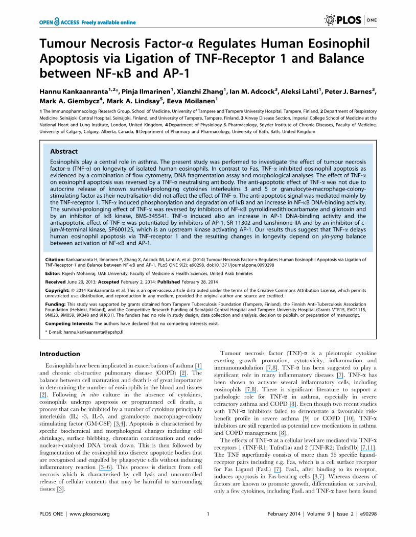
Effect of TNF-a on human eosinophil apoptosisTNF-a (0.1–10 ng/ml) inhibited apoptosis in a concentration
dependent-manner as determined by flow cytometric analysis
measuring the relative DNA fragmentation of PI-stained eosino-
phils (Fig. 1A–C). The inhibition of apoptosis by TNF-a was also
confirmed by the decrease in DNA fragmentation (Fig. 1D) and
morphological analysis of May-Grunwald-Giemsa-stained eosino-
phils (Fig. 1E–F). Culture of eosinophils with TNF-a resulted in a
decrease in the number cells showing typical apoptotic morphol-
ogy in a concentration-dependent manner (Table 1). Furthermore,
the inhibition of apoptosis was confirmed by reduced phosphati-
dylserine expression on the surface of eosinophil as analysed by
Annexin V/propidium iodide-counterstaining (Fig. 1 G–H). In the
absence of TNF-a 55.369.2% of the eosinophils were apoptotic,
whereas in the presence of TNF-a (10 ng/ml) 47.368.1% of the
eosinophils were apoptotic (n = 8, p,0.01). For comparison, in a
separate set of experiments, the effect of IL-5 on apoptosis of
human eosinophils was analysed by Annexin V-binding assay.
TNF-a is clearly less potent inhibitor of apoptosis than IL-5 as
there were 10.861.8% apoptotic cells in the presence of IL-5
(10 pM) and 62.8610.0% in its absence (n = 7, p,0.01). To
confirm the specificity of the effect of TNF-a, the effects of a
neutralising TNF-a antibody were studied. Neutralisation of TNF-
a completely reversed the inhibitory action of TNF-a on human
eosinophil apoptosis (Fig. 1I). To exclude the possibility that TNF-
a would induce primary necrosis (which would lead to a decrease
in the number of apoptotic cells), the numbers of primary necrotic
cells were determined by PI exclusion. The percentage of primary
necrotic cells in control culture after 40 h was 2.861.2 (n = 3) and
was not affected by the highest concentration (10 ng/ml) of TNF-
a (1.960.2%, n = 3). Also, to exclude the possibility that the single
time point studied (40 h) would explain the result the time-
dependency of the effect of TNF-a was studied. TNF-a (100 ng/
ml) demonstrated significant suppression of eosinophil apoptosis
by 17 h which was maintained and reached a maximum at 40 h
(Table 2). Eosinophils have been shown to be able to synthesise
TNF-a [29]. To determine whether production of TNF-a by
eosinophils themselves would be a trophic factor in non-stimulated
eosinophil culture, the effects of the TNF-a neutralising antibody
(5 mg/ml) were studied on the rate of spontaneous eosinophil
apoptosis. However, neutralisation of TNF-a did not change the
rate of spontaneous eosinophil apoptosis (38610 versus 38611%
apoptotic cells in the absence and presence of TNF-a neutralising
antibody after 40 h culture, respectively, n = 4; P.0.05).
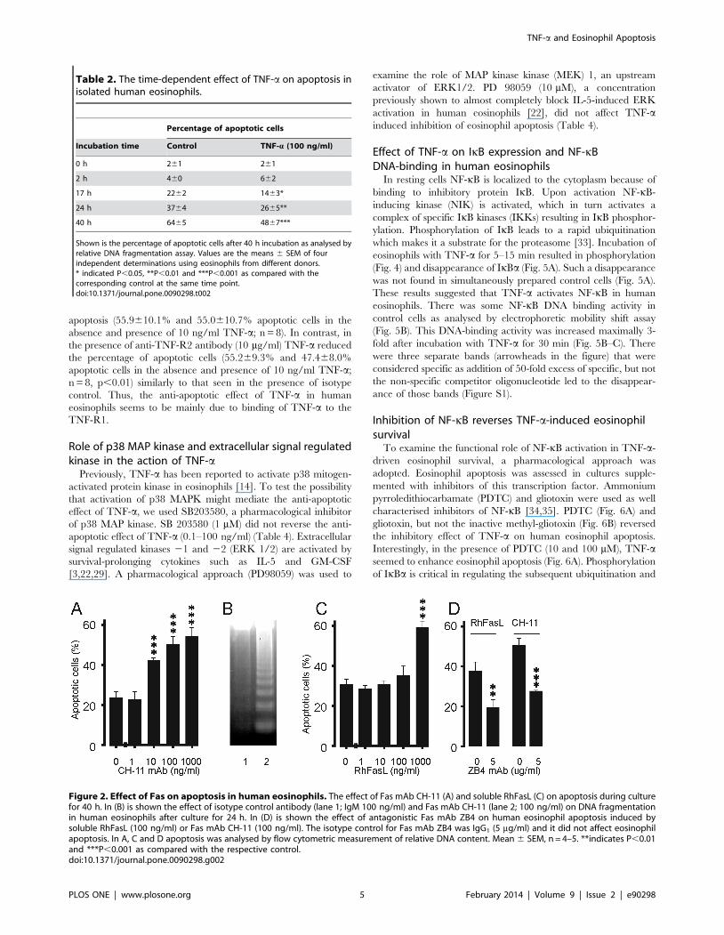
Effect of Fas on human eosinophil apoptosisActivation of another member of TNF receptor superfamily, Fas
has been previously described to induce apoptosis in human
eosinophils [30–32]. To confirm that the experimental conditions
and methodologies were adequate, the effects of activation of Fas
were studied under similar conditions. Similarly to that described
previously [30–32], cross-linking of CD95 by mAb CH-11
increased apoptosis in human eosinophils (Fig. 2A). Increase in
eosinophil apoptosis was also confirmed by increased DNA
fragmentation in the agarose gel electrophoresis assay (Fig. 2B).
Also the natural ligand of CD95, soluble recombinant human
FasLigand (RhFasL) increased apoptotic cell death in human
eosinophils (Fig. 2C). The increase in eosinophil apoptosis by mAb
CH-11 or soluble FasLigand was also confirmed by morphological
analysis of May-Grunwald-Giemsa-stained eosinophils (n = 3, data
not shown). The apoptosis-inducing effects of RhFasL and mAb
CH-11 were prevented by an antagonistic Fas mAb ZB4,
indicating that the effects were mediated by CD95 (Fig. 2D).
The anti-apoptotic effect of TNF-a is not mediated byproduction of IL-3, IL-5 or GM-CSF
Enhancement of eosinophil survival in a co-culture with mast-
cells has been attributed to the production of TNF-a and GM-
CSF in the culture [13]. To determine whether TNF-a-induced
eosinophil survival was due to production of the known eosinophil-
survival-prolonging cytokines IL-3, IL-5 or GM-CSF, TNF-a-
stimulated eosinophils were cultured in the presence of relevant
neutralising antibodies. These neutralising antibodies have been
shown to reverse the prolonged eosinophil survival induced by
addition of exogenous IL-3, IL-5 or GM-CSF in similar conditions
[18]. However, neutralisation of IL-3, IL-5 or GM-CSF in the
culture did not reverse TNF-a-induced inhibition of eosinophil
apoptosis (Table 3).
TNF-a and Eosinophil Apoptosis
PLOS ONE | www.plosone.org 3 February 2014 | Volume 9 | Issue 2 | e90298
Inhibition of eosinophil apoptosis by TNF-a is mainlymediated by the TNF- receptor 1
Human eosinophils express both TNF receptors 1 (TNF-R1;
Tnfrsf1a) and 2 (TNF-R2; Tnfrsf1b) [29]. To study which receptor
subtype mediates the anti-apoptotic effect of TNF-a, eosinophils
were cultured in the presence and absence of antibodies to soluble
and cell surface TNF-R1 and TNF-R2. Both antibodies were used
at a concentration of 10 mg/ml at which concentration there is no
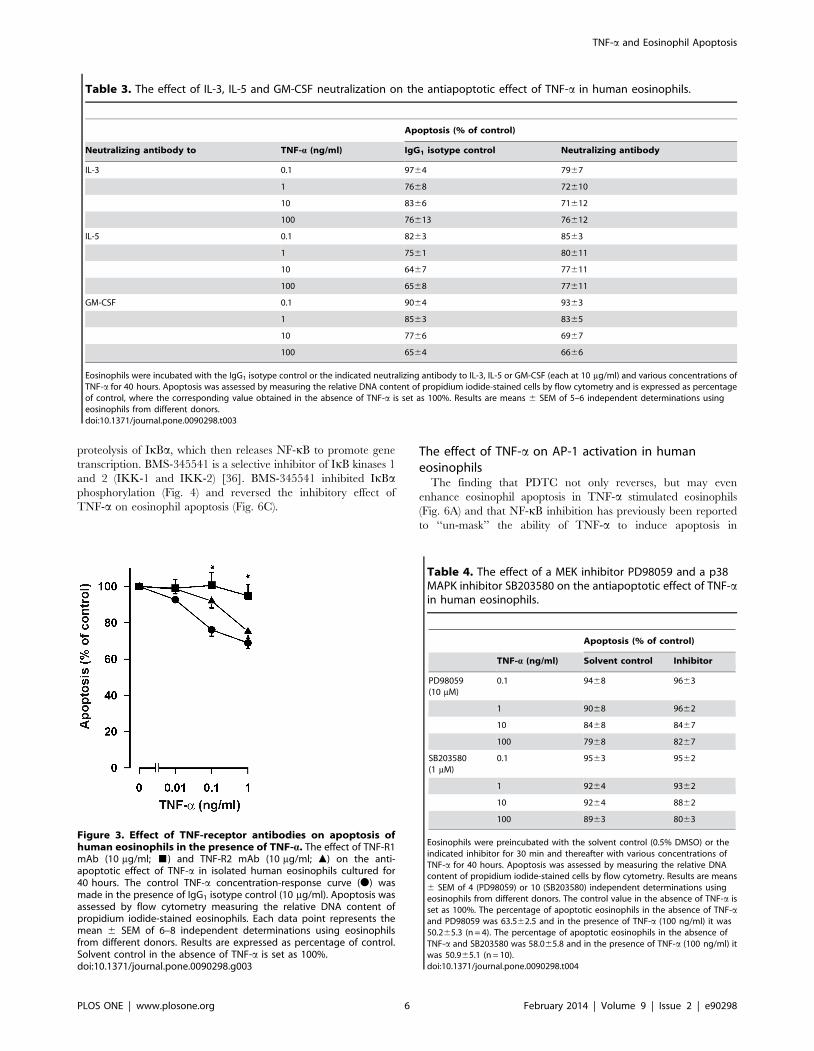
cross-reaction. The TNF-R1 antibody completely reversed the
anti-apoptotic effect of TNF-a at concentrations of 0.1–1 ng/ml
whereas the TNF-R2 antibody did not have a statistically
significant effect (Fig. 3). To confirm this finding, apoptosis was
analysed by Annexin-V binding assay. In the presence of IgG1
isotype control (10 mg/ml), TNF-a inhibited apoptosis
(55.369.2% and 47.368.1% apoptotic cells in the absence and
presence of 10 ng/ml TNF-a; n = 8, p,0.01). In the presence of
anti-TNF-R1 antibody (10 mg/ml) TNF-a did not anymore inhibit
Figure 1. Effect of TNF-a on apoptosis in human eosinophils. Effect of TNF-a on apoptosis in culture for 40 h (A). Representative graphs fromrelative DNA fragmentation assay of propidium iodide-stained eosinophils are shown in (B and C). In (B and C) figures in top right corner representpercentage of cells showing decreased DNA content. In (D) is shown the DNA fragmentation in eosinophils cultured without (lane 1) and with 10 ng/ml TNF-a (lane 2). The typical apoptotic morphology (cell shrinkage and condensation of nuclear chromatin) of May-Grunwald-Giemsa-stainedcytokine-deprived eosinophils is shown in (E) and the reduction in the number of cells showing apoptotic morphology when cultured with TNF-a(10 ng/ml) for 40 h is shown in (F). Scale bar (E and F) is 10 mm. In (G and H) representative graphs from Annexin-V FITC (FL1-H) and uptake ofpropidium iodide (FL2-H) analysis of eosinophils are shown. In (G and H) figures in top right corner represent percentages of Annexin-V positive cells(FITC+/PI- and FITC+/PI+). In (I) is shown the effect of neutralising TNF-a (A-TNF-a) antibody (5 mg/ml) on the inhibition of apoptosis induced by TNF-a.The isotype control was IgG1 (5 mg/ml) and had no effect on eosinophil apoptosis during the 40 h culture. In A and I apoptosis was assessed by flowcytometric measurement of relative DNA content. Each datapoint represents the mean 6 SEM of 4–6 independent determinations using eosinophilsfrom different donors. * indicates P,0.05, **P,0.01 and ***P,0.001 as compared with the respective control without TNF-a and# P,0.05 as compared with the respective control without TNF-a neutralising antibody.doi:10.1371/journal.pone.0090298.g001
Table 1. The effect of TNF-a on apoptosis in isolated humaneosinophils.
TNF-a (ng/ml) Percentage of apoptotic cells
0 69616
0.01 59620
0.1 44618
1 32615*
10 3265*
Shown is the percentage of apoptotic cells after 40 h incubation as analyzed bymorphological criteria. Values are the means 6 SEM of four independentdeterminations using eosinophils from different donors. * indicates P,0.05 ascompared with the respective solvent control.doi:10.1371/journal.pone.0090298.t001
TNF-a and Eosinophil Apoptosis
PLOS ONE | www.plosone.org 4 February 2014 | Volume 9 | Issue 2 | e90298
apoptosis (55.9610.1% and 55.0610.7% apoptotic cells in the
absence and presence of 10 ng/ml TNF-a; n = 8). In contrast, in
the presence of anti-TNF-R2 antibody (10 mg/ml) TNF-a reduced
the percentage of apoptotic cells (55.269.3% and 47.468.0%
apoptotic cells in the absence and presence of 10 ng/ml TNF-a;
n = 8, p,0.01) similarly to that seen in the presence of isotype
control. Thus, the anti-apoptotic effect of TNF-a in human
eosinophils seems to be mainly due to binding of TNF-a to the
TNF-R1.
Role of p38 MAP kinase and extracellular signal regulatedkinase in the action of TNF-a
Previously, TNF-a has been reported to activate p38 mitogen-
activated protein kinase in eosinophils [14]. To test the possibility
that activation of p38 MAPK might mediate the anti-apoptotic
effect of TNF-a, we used SB203580, a pharmacological inhibitor
of p38 MAP kinase. SB 203580 (1 mM) did not reverse the anti-
apoptotic effect of TNF-a (0.1–100 ng/ml) (Table 4). Extracellular
signal regulated kinases 21 and 22 (ERK 1/2) are activated by
survival-prolonging cytokines such as IL-5 and GM-CSF
[3,22,29]. A pharmacological approach (PD98059) was used to
examine the role of MAP kinase kinase (MEK) 1, an upstream
activator of ERK1/2. PD 98059 (10 mM), a concentration
previously shown to almost completely block IL-5-induced ERK
activation in human eosinophils [22], did not affect TNF-ainduced inhibition of eosinophil apoptosis (Table 4).
Effect of TNF-a on IkB expression and NF-kBDNA-binding in human eosinophils
In resting cells NF-kB is localized to the cytoplasm because of
binding to inhibitory protein IkB. Upon activation NF-kB-
inducing kinase (NIK) is activated, which in turn activates a
complex of specific IkB kinases (IKKs) resulting in IkB phosphor-
ylation. Phosphorylation of IkB leads to a rapid ubiquitination
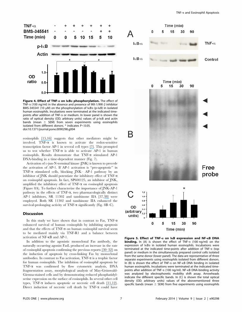
which makes it a substrate for the proteasome [33]. Incubation of
eosinophils with TNF-a for 5–15 min resulted in phosphorylation
(Fig. 4) and disappearance of IkBa (Fig. 5A). Such a disappearance
was not found in simultaneously prepared control cells (Fig. 5A).
These results suggested that TNF-a activates NF-kB in human
eosinophils. There was some NF-kB DNA binding activity in
control cells as analysed by electrophoretic mobility shift assay
(Fig. 5B). This DNA-binding activity was increased maximally 3-
fold after incubation with TNF-a for 30 min (Fig. 5B–C). There
were three separate bands (arrowheads in the figure) that were
considered specific as addition of 50-fold excess of specific, but not
the non-specific competitor oligonucleotide led to the disappear-
ance of those bands (Figure S1).
Inhibition of NF-kB reverses TNF-a-induced eosinophilsurvival
To examine the functional role of NF-kB activation in TNF-a-
driven eosinophil survival, a pharmacological approach was
adopted. Eosinophil apoptosis was assessed in cultures supple-
mented with inhibitors of this transcription factor. Ammonium
pyrroledithiocarbamate (PDTC) and gliotoxin were used as well
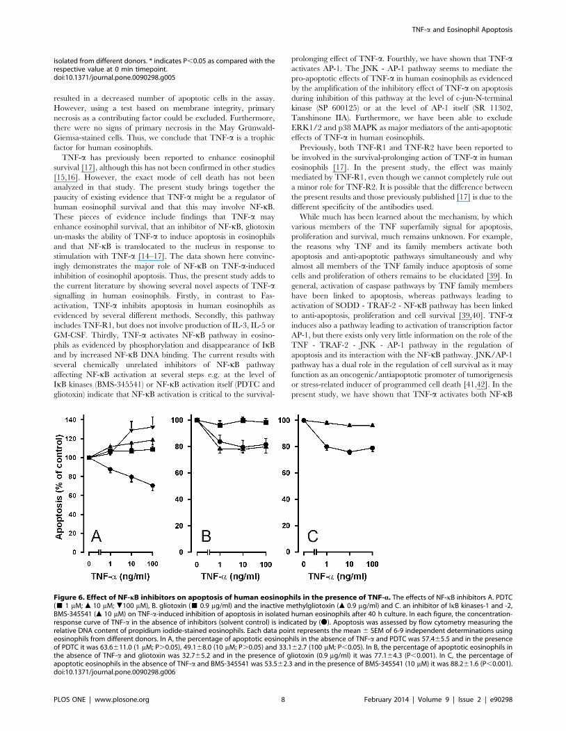
characterised inhibitors of NF-kB [34,35]. PDTC (Fig. 6A) and
gliotoxin, but not the inactive methyl-gliotoxin (Fig. 6B) reversed
the inhibitory effect of TNF-a on human eosinophil apoptosis.
Interestingly, in the presence of PDTC (10 and 100 mM), TNF-aseemed to enhance eosinophil apoptosis (Fig. 6A). Phosphorylation
of IkBa is critical in regulating the subsequent ubiquitination and
Table 2. The time-dependent effect of TNF-a on apoptosis inisolated human eosinophils.
Percentage of apoptotic cells
Incubation time Control TNF-a (100 ng/ml)
0 h 261 261
2 h 460 662
17 h 2262 1463*
24 h 3764 2665**
40 h 6465 4867***
Shown is the percentage of apoptotic cells after 40 h incubation as analysed byrelative DNA fragmentation assay. Values are the means 6 SEM of fourindependent determinations using eosinophils from different donors.* indicated P,0.05, **P,0.01 and ***P,0.001 as compared with thecorresponding control at the same time point.doi:10.1371/journal.pone.0090298.t002
Figure 2. Effect of Fas on apoptosis in human eosinophils. The effect of Fas mAb CH-11 (A) and soluble RhFasL (C) on apoptosis during culturefor 40 h. In (B) is shown the effect of isotype control antibody (lane 1; IgM 100 ng/ml) and Fas mAb CH-11 (lane 2; 100 ng/ml) on DNA fragmentationin human eosinophils after culture for 24 h. In (D) is shown the effect of antagonistic Fas mAb ZB4 on human eosinophil apoptosis induced bysoluble RhFasL (100 ng/ml) or Fas mAb CH-11 (100 ng/ml). The isotype control for Fas mAb ZB4 was IgG1 (5 mg/ml) and it did not affect eosinophilapoptosis. In A, C and D apoptosis was analysed by flow cytometric measurement of relative DNA content. Mean 6 SEM, n = 4–5. **indicates P,0.01and ***P,0.001 as compared with the respective control.doi:10.1371/journal.pone.0090298.g002
TNF-a and Eosinophil Apoptosis
PLOS ONE | www.plosone.org 5 February 2014 | Volume 9 | Issue 2 | e90298
proteolysis of IkBa, which then releases NF-kB to promote gene
transcription. BMS-345541 is a selective inhibitor of IkB kinases 1
and 2 (IKK-1 and IKK-2) [36]. BMS-345541 inhibited IkBaphosphorylation (Fig. 4) and reversed the inhibitory effect of
TNF-a on eosinophil apoptosis (Fig. 6C).
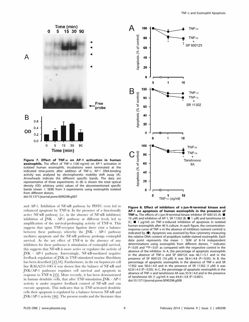
The effect of TNF-a on AP-1 activation in humaneosinophils
The finding that PDTC not only reverses, but may even
enhance eosinophil apoptosis in TNF-a stimulated eosinophils
(Fig. 6A) and that NF-kB inhibition has previously been reported
to ‘‘un-mask’’ the ability of TNF-a to induce apoptosis in
Table 3. The effect of IL-3, IL-5 and GM-CSF neutralization on the antiapoptotic effect of TNF-a in human eosinophils.
Apoptosis (% of control)
Neutralizing antibody to TNF-a (ng/ml) IgG1 isotype control Neutralizing antibody
IL-3 0.1 9764 7967
1 7668 72610
10 8366 71612
100 76613 76612
IL-5 0.1 8263 8563
1 7561 80611
10 6467 77611
100 6568 77611
GM-CSF 0.1 9064 9363
1 8563 8365
10 7766 6967
100 6564 6666
Eosinophils were incubated with the IgG1 isotype control or the indicated neutralizing antibody to IL-3, IL-5 or GM-CSF (each at 10 mg/ml) and various concentrations ofTNF-a for 40 hours. Apoptosis was assessed by measuring the relative DNA content of propidium iodide-stained cells by flow cytometry and is expressed as percentageof control, where the corresponding value obtained in the absence of TNF-a is set as 100%. Results are means 6 SEM of 5–6 independent determinations usingeosinophils from different donors.doi:10.1371/journal.pone.0090298.t003
Figure 3. Effect of TNF-receptor antibodies on apoptosis ofhuman eosinophils in the presence of TNF-a. The effect of TNF-R1mAb (10 mg/ml; &) and TNF-R2 mAb (10 mg/ml; m) on the anti-apoptotic effect of TNF-a in isolated human eosinophils cultured for40 hours. The control TNF-a concentration-response curve (N) wasmade in the presence of IgG1 isotype control (10 mg/ml). Apoptosis wasassessed by flow cytometry measuring the relative DNA content ofpropidium iodide-stained eosinophils. Each data point represents themean 6 SEM of 6–8 independent determinations using eosinophilsfrom different donors. Results are expressed as percentage of control.Solvent control in the absence of TNF-a is set as 100%.doi:10.1371/journal.pone.0090298.g003
Table 4. The effect of a MEK inhibitor PD98059 and a p38MAPK inhibitor SB203580 on the antiapoptotic effect of TNF-ain human eosinophils.
Apoptosis (% of control)
TNF-a (ng/ml) Solvent control Inhibitor
PD98059(10 mM)
0.1 9468 9663
1 9068 9662
10 8468 8467
100 7968 8267
SB203580(1 mM)
0.1 9563 9562
1 9264 9362
10 9264 8862
100 8963 8063
Eosinophils were preincubated with the solvent control (0.5% DMSO) or theindicated inhibitor for 30 min and thereafter with various concentrations ofTNF-a for 40 hours. Apoptosis was assessed by measuring the relative DNAcontent of propidium iodide-stained cells by flow cytometry. Results are means6 SEM of 4 (PD98059) or 10 (SB203580) independent determinations usingeosinophils from different donors. The control value in the absence of TNF-a isset as 100%. The percentage of apoptotic eosinophils in the absence of TNF-aand PD98059 was 63.562.5 and in the presence of TNF-a (100 ng/ml) it was50.265.3 (n = 4). The percentage of apoptotic eosinophils in the absence ofTNF-a and SB203580 was 58.065.8 and in the presence of TNF-a (100 ng/ml) itwas 50.965.1 (n = 10).doi:10.1371/journal.pone.0090298.t004
TNF-a and Eosinophil Apoptosis
PLOS ONE | www.plosone.org 6 February 2014 | Volume 9 | Issue 2 | e90298
eosinophils [15,16] suggests that other mediators might be
involved. TNF-a is known to activate the redox-sensitive
transcription factor AP-1 in several cell types [7]. This prompted
us to test whether TNF-a is able to activate AP-1 in human
eosinophils. Results demonstrate that TNF-a stimulated AP-1
DNA-binding in a time-dependent manner (Fig. 7).
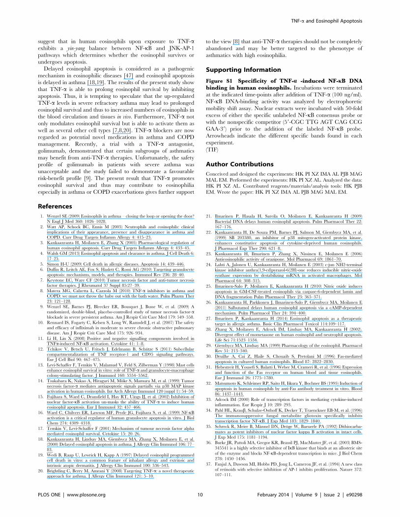
Activation of c-jun-N-terminal kinase (JNK) is known to precede
the activation of AP-1. If AP-1 activation is ‘‘pro-apoptotic’’ in
TNF-a stimulated cells, blocking JNK– AP-1 pathway by an
inhibitor of JNK should potentiate the inhibitory effect of TNF-aon eosinophil apoptosis. In fact, SP600125, an inhibitor of JNK,
amplified the inhibitory effect of TNF-a on eosinophil apoptosis
(Figure 8A). To further characterize the importance of JNK-AP-1
pathway in the effects of TNF-a, two pharmacologically distinct
AP-1 inhibitors, SR 11302 and tanshinone IIA [37,38] were
employed. Both SR 11302 and tanshinone IIA enhanced the
survival-prolonging activity of TNF-a significantly (Fig. 8B–C).
Discussion
In this study we have shown that in contrast to Fas, TNF-aenhanced survival of human eosinophils by inhibiting apoptosis
and that the effects of TNF-a on human eosinophil survival seem
to be mediated mainly via TNF-R1 and a balance between
activation of NF-kB and AP-1.
In addition to the agonistic monoclonal Fas antibody, the
naturally occurring agonist FasL produced an increase in the rate
of eosinophil apoptosis confirming the previous reports [30–32] on
the induction of apoptosis by cross-linking Fas by monoclonal
antibodies. In contrast to Fas activation, TNF-a is a trophic factor
for human eosinophils. The inhibition of eosinophil apoptosis by
TNF-a was confirmed by flow cytometric analysis, DNA
fragmentation assay, morphological analysis of May-Grunwald-
Giemsa-stained cells and by demonstrating reduced phosphatidyl-
serine expression on the surface of eosinophils. In several other cell
types, TNF-a induces apoptotic or necrotic cell death [11,12].
Direct induction of necrotic cell death by TNF-a could have
Figure 4. Effect of TNF-a on IkBa phosphorylation. The effect ofTNF-a (100 ng/ml) in the absence and presence of IKK-1/IKK-2 inhibitorBMS-345541 (10 mM) on the phosphorylation of IkBa (p-IkB) in isolatedhuman eosinophils. Incubations were terminated at the indicated time-points after addition of TNF-a or medium. In lower panel is shown theratio of optical density (OD; arbitrary units) values of p-IkB and actinbands (mean 6 SEM) from seven experiments using eosinophilsisolated from different donors. * indicates P,0.05.doi:10.1371/journal.pone.0090298.g004
Figure 5. Effect of TNF-a on IkB expression and NF-kB DNAbinding. In (A) is shown the effect of TNF-a (100 ng/ml) on theexpression of IkBa in isolated human eosinophils. Incubations wereterminated at the indicated time-points after addition of TNF-a (toppanel) or medium in the simultaneously prepared control cells isolatedfrom the same donor (lower panel). The data are representative of threeseparate experiments using eosinophils isolated from different donors.In (B) is shown the effect of TNF-a on NF-kB DNA binding in isolatedhuman eosinophils. Incubations were terminated at the indicated time-points after addition of TNF-a (100 ng/ml). NF-kB DNA-binding activitywas analyzed by electrophoretic mobility shift assay. Arrowheadsindicate the different specific bands. In (C) is shown the total opticaldensity (OD; arbitrary units) values of the abovementioned threespecific bands (mean 6 SEM) from five experiments using eosinophils
TNF-a and Eosinophil Apoptosis
PLOS ONE | www.plosone.org 7 February 2014 | Volume 9 | Issue 2 | e90298
resulted in a decreased number of apoptotic cells in the assay.
However, using a test based on membrane integrity, primary
necrosis as a contributing factor could be excluded. Furthermore,
there were no signs of primary necrosis in the May Grunwald-
Giemsa-stained cells. Thus, we conclude that TNF-a is a trophic
factor for human eosinophils.
TNF-a has previously been reported to enhance eosinophil
survival [17], although this has not been confirmed in other studies
[15,16]. However, the exact mode of cell death has not been
analyzed in that study. The present study brings together the
paucity of existing evidence that TNF-a might be a regulator of
human eosinophil survival and that this may involve NF-kB.
These pieces of evidence include findings that TNF-a may
enhance eosinophil survival, that an inhibitor of NF-kB, gliotoxin
un-masks the ability of TNF-a to induce apoptosis in eosinophils
and that NF-kB is translocated to the nucleus in response to
stimulation with TNF-a [14–17]. The data shown here convinc-
ingly demonstrates the major role of NF-kB on TNF-a-induced
inhibition of eosinophil apoptosis. Thus, the present study adds to
the current literature by showing several novel aspects of TNF-asignalling in human eosinophils. Firstly, in contrast to Fas-
activation, TNF-a inhibits apoptosis in human eosinophils as
evidenced by several different methods. Secondly, this pathway
includes TNF-R1, but does not involve production of IL-3, IL-5 or
GM-CSF. Thirdly, TNF-a activates NF-kB pathway in eosino-
phils as evidenced by phosphorylation and disappearance of IkB
and by increased NF-kB DNA binding. The current results with
several chemically unrelated inhibitors of NF-kB pathway
affecting NF-kB activation at several steps e.g. at the level of
IkB kinases (BMS-345541) or NF-kB activation itself (PDTC and
gliotoxin) indicate that NF-kB activation is critical to the survival-
prolonging effect of TNF-a. Fourthly, we have shown that TNF-aactivates AP-1. The JNK - AP-1 pathway seems to mediate the
pro-apoptotic effects of TNF-a in human eosinophils as evidenced
by the amplification of the inhibitory effect of TNF-a on apoptosis
during inhibition of this pathway at the level of c-jun-N-terminal
kinase (SP 600125) or at the level of AP-1 itself (SR 11302,
Tanshinone IIA). Furthermore, we have been able to exclude
ERK1/2 and p38 MAPK as major mediators of the anti-apoptotic
effects of TNF-a in human eosinophils.
Previously, both TNF-R1 and TNF-R2 have been reported to
be involved in the survival-prolonging action of TNF-a in human
eosinophils [17]. In the present study, the effect was mainly
mediated by TNF-R1, even though we cannot completely rule out
a minor role for TNF-R2. It is possible that the difference between
the present results and those previously published [17] is due to the
different specificity of the antibodies used.
While much has been learned about the mechanism, by which
various members of the TNF superfamily signal for apoptosis,
proliferation and survival, much remains unknown. For example,
the reasons why TNF and its family members activate both
apoptosis and anti-apoptotic pathways simultaneously and why
almost all members of the TNF family induce apoptosis of some
cells and proliferation of others remains to be elucidated [39]. In
general, activation of caspase pathways by TNF family members
have been linked to apoptosis, whereas pathways leading to
activation of SODD - TRAF-2 - NF-kB pathway has been linked
to anti-apoptosis, proliferation and cell survival [39,40]. TNF-ainduces also a pathway leading to activation of transcription factor
AP-1, but there exists only very little information on the role of the
TNF - TRAF-2 - JNK - AP-1 pathway in the regulation of
apoptosis and its interaction with the NF-kB pathway. JNK/AP-1
pathway has a dual role in the regulation of cell survival as it may
function as an oncogenic/antiapoptotic promoter of tumorigenesis
or stress-related inducer of programmed cell death [41,42]. In the
present study, we have shown that TNF-a activates both NF-kB
isolated from different donors. * indicates P,0.05 as compared with therespective value at 0 min timepoint.doi:10.1371/journal.pone.0090298.g005
Figure 6. Effect of NF-kB inhibitors on apoptosis of human eosinophils in the presence of TNF-a. The effects of NF-kB inhibitors A. PDTC(& 1 mM; m 10 mM; .100 mM), B. gliotoxin (& 0.9 mg/ml) and the inactive methylgliotoxin (m 0.9 mg/ml) and C. an inhibitor of IkB kinases-1 and -2,BMS-345541 (m 10 mM) on TNF-a-induced inhibition of apoptosis in isolated human eosinophils after 40 h culture. In each figure, the concentration-response curve of TNF-a in the absence of inhibitors (solvent control) is indicated by (N). Apoptosis was assessed by flow cytometry measuring therelative DNA content of propidium iodide-stained eosinophils. Each data point represents the mean 6 SEM of 6-9 independent determinations usingeosinophils from different donors. In A, the percentage of apoptotic eosinophils in the absence of TNF-a and PDTC was 57.465.5 and in the presenceof PDTC it was 63.6611.0 (1 mM; P.0.05), 49.168.0 (10 mM; P.0.05) and 33.162.7 (100 mM; P,0.05). In B, the percentage of apoptotic eosinophils inthe absence of TNF-a and gliotoxin was 32.765.2 and in the presence of gliotoxin (0.9 mg/ml) it was 77.164.3 (P,0.001). In C, the percentage ofapoptotic eosinophils in the absence of TNF-a and BMS-345541 was 53.562.3 and in the presence of BMS-345541 (10 mM) it was 88.261.6 (P,0.001).doi:10.1371/journal.pone.0090298.g006
TNF-a and Eosinophil Apoptosis
PLOS ONE | www.plosone.org 8 February 2014 | Volume 9 | Issue 2 | e90298
and AP-1. Inhibition of NF-kB pathway by PDTC even led to
enhanced apoptosis by TNF-a. In the presence of a functionally
active NF-kB pathway (i.e. in the absence of NF-kB inhibitors)
inhibition of JNK - AP-1 pathway at different levels led to
amplification of the survival-prolonging activity of TNF-a. This
suggests that upon TNF-receptor ligation there exist a balance
between these pathways whereby the JNK - AP-1 pathway
mediates apoptosis and the NF-kB pathway prolongs eosinophil
survival. As the net effect of TNF-a in the absence of any
inhibitors for these pathways is stimulation of eosinophil survival,
this suggests that NF-kB is more active or regulates the activity of
JNK - AP-1 pathway. Interestingly, NF-kB-mediated negative
feedback regulation of JNK in TNF-stimulated murine fibroblasts
has been described [43,44]. Furthermore, in the rat hepatocyte cell
line RALA255-10G it was shown that the balance of NF-kB and
JNK/AP-1 pathways regulates cell survival and apoptosis in
response to TNF-a [45]. More recently, it has been demonstrated
in human dendritic cells, that after TNF-stimulation JNK - AP-1
activity is under negative feedback control of NF-kB and can
execute apoptosis. This indicates that in TNF-activated dendritic
cells their apoptosis is regulated by a balance between NF-kB and
JNK/AP-1 activity [46]. The present results and the literature thus
Figure 7. Effect of TNF-a on AP-1 activation in humaneosinophils. The effect of TNF-a (100 ng/ml) on AP-1 activation inisolated human eosinophils. Incubations were terminated at theindicated time-points after addition of TNF-a. AP-1 DNA-bindingactivity was analyzed by electrophoretic mobility shift assay (A).Arrowheads indicate the different specific bands. The data arerepresentative of three experiments. In (B) is shown the total opticaldensity (OD; arbitrary units) values of the abovementioned specificbands (mean 6 SEM) from 3 experiments using eosinophils isolatedfrom different donors.doi:10.1371/journal.pone.0090298.g007
Figure 8. Effect of inhibitors of c-jun-N-terminal kinase andAP-1 on apoptosis of human eosinophils in the presence ofTNF-a. The effects of c-jun-N-terminal kinase inhibitor SP 600125 (A: &10 mM) and inhibitors of AP-1, SR 11302 (B; & 1 mM) and tanshinone IIA(C; & 1 mg/ml) on TNF-a-induced inhibition of apoptosis in isolatedhuman eosinophils after 40 h culture. In each figure, the concentration-response curve of TNF-a in the absence of inhibitors (solvent control) isindicated by (N). Apoptosis was assessed by flow cytometry measuringthe relative DNA content of propidium iodide-stained eosinophils. Eachdata point represents the mean 6 SEM of 8-14 independentdeterminations using eosinophils from different donors. * indicatesP,0.05 and **P,0.01 as compared with the respective control in theabsence of the inhibitor. In A, the percentage of apoptotic eosinophilsin the absence of TNF-a and SP 600125 was 46.165.1 and in thepresence of SP 600125 (10 mM) it was 38.464.8 (P,0.05). In B, thepercentage of apoptotic eosinophils in the absence of TNF-a and SR11302 was 58.664.9 and in the presence of SR 11302 (1 mM) it was62.864.3 (P,0.05). In C, the percentage of apoptotic eosinophils in theabsence of TNF-a and tanshinone IIA was 55.964.0 and in the presenceof tanshinone IIA (1 mg/ml) it was 64.863.8 (P,0.001).doi:10.1371/journal.pone.0090298.g008
TNF-a and Eosinophil Apoptosis
PLOS ONE | www.plosone.org 9 February 2014 | Volume 9 | Issue 2 | e90298
suggest that in human eosinophils upon exposure to TNF-aexhibits a yin-yang balance between NF-kB and JNK-AP-1
pathways which determines whether the eosinophil survives or
undergoes apoptosis.
Delayed eosinophil apoptosis is considered as a pathogenic
mechanism in eosinophilic diseases [47] and eosinophil apoptosis
is delayed in asthma [18,19]. The results of the present study show
that TNF-a is able to prolong eosinophil survival by inhibiting
apoptosis. Thus, it is tempting to speculate that the up-regulated
TNF-a levels in severe refractory asthma may lead to prolonged
eosinophil survival and thus to increased numbers of eosinophils in
the blood circulation and tissues in vivo. Furthermore, TNF-a not
only modulates eosinophil survival but is able to activate them as
well as several other cell types [7,8,20]. TNF-a blockers are now
regarded as potential novel medications in asthma and COPD
management. Recently, a trial with a TNF-a antagonist,
golimumab, demonstrated that certain subgroups of asthmatics
may benefit from anti-TNF-a therapies. Unfortunately, the safety
profile of golimumab in patients with severe asthma was
unacceptable and the study failed to demonstrate a favourable
risk-benefit profile [9]. The present result that TNF-a promotes
eosinophil survival and thus may contribute to eosinophilia
especially in asthma or COPD exacerbations gives further support
to the view [8] that anti-TNF-a therapies should not be completely
abandoned and may be better targeted to the phenotype of
asthmatics with high eosinophilia.
Supporting Information
Figure S1 Specificity of TNF-a -induced NF-kB DNAbinding in human eosinophils. Incubations were terminated
at the indicated time-points after addition of TNF-a (100 ng/ml).
NF-kB DNA-binding activity was analyzed by electrophoretic
mobility shift assay. Nuclear extracts were incubated with 50-fold
excess of either the specific unlabeled NF-kB consensus probe or
with the nonspecific competitor (59-CGC TTG AGT CAG CCG
GAA-39) prior to the addition of the labeled NF-kB probe.
Arrowheads indicate the different specific bands found in each
experiment.
(TIF)
Author Contributions
Conceived and designed the experiments: HK PI XZ IMA AL PJB MAG
MAL EM. Performed the experiments: HK PI XZ AL. Analyzed the data:
HK PI XZ AL. Contributed reagents/materials/analysis tools: HK PJB
EM. Wrote the paper: HK PI XZ IMA AL PJB MAG MAL EM.
References
1. Wenzel SE (2009) Eosinophils in asthma – closing the loop or opening the door?
N Engl J Med 360: 1026–1028.
2. Watt AP, Schock BC, Ennis M (2005) Neutrophils and eosinophils: clinicalimplications of their appearance, presence and disappearance in asthma and
COPD. Curr Drug Targets Inflamm Allergy 4: 415–23.
3. Kankaanranta H, Moilanen E, Zhang X (2005) Pharmacological regulation ofhuman eosinophil apoptosis. Curr Drug Targets Inflamm Allergy 4: 433–45.
4. Walsh GM (2013) Eosinophil apoptosis and clearance in asthma. J Cell Death 6:
17–25.
5. Simon H-U (2009) Cell death in allergic diseases. Apoptosis 14: 439–446.
6. Duffin R, Leitch AE, Fox S, Haslett C, Rossi AG (2010) Targeting granulocyte
apoptosis: mechanisms, models, and therapies. Immunol Rev 236: 28–40.
7. Keystone EC, Ware CF (2010) Tumor necrosis factor and anti-tumor necrosisfactor therapies. J Rheumatol 37 Suppl 85:27–39.
8. Matera MG, Calzetta L, Cazzola M (2010) TNF-a inhibitors in asthma and
COPD: we must not throw the baby out with the bath water. Pulm Pharm Ther
23: 121–128.
9. Wenzel SE, Barnes PJ, Bleecker ER, Bousquet J, Busse W, et al. (2009) A
randomized, double-blind, placebo-controlled study of tumor necrosis factor-ablockade in severe persistent asthma. Am J Respir Crit Care Med 179: 549–558.
10. Rennard IS, Fogarty C, Kelsen S, Long W, Ramsdell J, et al. (2007) The safety
and efficacy of infliximab in moderate to severe chronic obstructive pulmonary
disease. Am J Respir Crit Care Med 175: 926–934.
11. Li H, Lin X (2008) Positive and negative signalling components involved inTNFa-induced NF-kB activation. Cytokine 41: 1–8.
12. Tchikov V, Betsch U, Fritsch J, Edelmann B, Schutze S (2011) Subcellular
compartmentalization of TNF receptor-1 and CD95 signaling pathways.Eur J Cell Biol 90: 467–475.
13. Levi-Schaffer F, Temkin V, Malamud V, Feld S, Zilberman Y (1998) Mast cells
enhance eosinophil survival in vitro: role of TNF-a and granulocyte-macrophagecolony-stimulating factor. J Immunol 160: 5554–5562.
14. Tsukahara K, Nakao A, Hiraguri M, Miike S, Mamura M, et al. (1999) Tumor
necrosis factor-a mediates antiapoptotic signals partially via p38 MAP kinaseactivation in human eosinophils. Int Arch Allergy Immunol 120 (suppl 1): 54–59.
15. Fujihara S, Ward C, Dransfield I, Hay RT, Uings IJ, et al. (2002) Inhibition of
nuclear factor-kB activation un-masks the ability of TNF-a to induce humaneosinophil apoptosis. Eur J Immunol 32: 457–466.
16. Ward C, Chilvers ER, Lawson MF, Pryde JG, Fujihara S, et al. (1999) NF-kB
activation is a critical regulator of human granulocyte apoptosis in vitro. J BiolChem 274: 4309–4318.
17. Temkin V, Levi-Schaffer F (2001) Mechanism of tumour necrosis factor alpha
mediated eosinophil survival. Cytokine 15: 20–26.
18. Kankaanranta H, Lindsay MA, Giembycz MA, Zhang X, Moilanen E, et al.(2000) Delayed eosinophil apoptosis in asthma. J Allergy Clin Immunol 106: 77–
83.
19. Wedi B, Raap U, Lewrick H, Kapp A (1997) Delayed eosinophil programmedcell death in vitro: a common feature of inhalant allergy and extrinsic and
intrinsic atopic dermatitis. J Allergy Clin Immunol 100: 536–543.
20. Brightling C, Berry M, Amrani Y (2008) Targeting TNF-a: a novel therapeuticapproach for asthma. J Allergy Clin Immunol 121: 5–10.
21. Ilmarinen P, Hasala H, Sareila O, Moilanen E, Kankaanranta H (2009)
Bacterial DNA delays human eosinophil apoptosis. Pulm Pharmacol Ther 22:
167–176.
22. Kankaanranta H, De Souza PM, Barnes PJ, Salmon M, Giembycz MA, et al.
(1999) SB 203580, an inhibitor of p38 mitogen-activated protein kinase,
enhances constitutive apoptosis of cytokine-deprived human eosinophils.
J Pharmacol Exp Ther 290: 621–8.
23. Kankaanranta H, Ilmarinen P, Zhang X, Nissinen E, Moilanen E (2006)
Antieosinophilic activity of orazipone. Mol Pharmacol 69: 1861–70.
24. Lahti A, Jalonen U, Kankaanranta H, Moilanen E (2003) c-jun NH2-terminal
kinase inhibitor anthra(1,9-cd)pyrazol-6(2H)-one reduces inducible nitric-oxide
synthase expression by destabilizing mRNA in activated macrophages. Mol
Pharmacol 64: 308–315.
25. Ilmarinen-Salo P, Moilanen E, Kankaanranta H (2010) Nitric oxide induces
apoptosis in GM-CSF-treated eosinophils via caspase-6-dependent lamin and
DNA fragmentation Pulm Pharmacol Ther 23: 365–371.
26. Kankaanranta H, Parkkonen J, Ilmarinen-Salo P, Giembycz MA, Moilanen E
(2011) Salbutamol delays human eosinophil apoptosis via a cAMP-dependent
mechanism. Pulm Pharmacol Ther 24: 394–400.
27. Ilmarinen P, Kankaanranta H (2014) Eosinophil apoptosis as a therapeutic
target in allergic asthma. Basic Clin Pharmacol Toxicol 114:109–117.
28. Zhang X, Moilanen E, Adcock IM, Lindsay MA, Kankaanranta H (2002).
Divergent effect of mometasone on human eosinophil and neutrophil apoptosis.
Life Sci 71:1523–1534.
29. Giembycz MA, Lindsay MA (1999) Pharmacology of the eosinophil. Pharmacol
Rev 51: 213–340.
30. Druilhe A, Cai Z, Haile S, Chouaib S, Pretolani M (1996) Fas-mediated
apoptosis in cultured human eosinophils. Blood 87: 2822–2830.
31. Hebestreit H, Yousefi S, Balatti I, Weber M, Crameri R, et al. (1996) Expression
and function of the Fas receptor on human blood and tissue eosinophils.
Eur J Immunol 26: 1775–1780.
32. Matsumoto K, Schleimer RP, Saito H, Iikura Y, Bochner BS (1995) Induction of
apoptosis in human eosinophils by anti-Fas antibody treatment in vitro. Blood
86: 1437–1443.
33. Adcock IM (2000) Role of transcription factors in mediating cytokine-induced
inflammation. Eur Respir J 10: 289–293.
34. Pahl HL, Kraub, Schulze-Osthoff K, Decker T, Traenckner EB-M, et al. (1996)
The immunosuppressive fungal metabolite gliotoxin specifically inhibits
transcription factor NF-kB. J Exp Med 183: 1829–1840.
35. Schreck R, Meier B, Mannel DN, Droge W, Baeuerle PA (1992) Dithiocarba-
mates as potent inhibitors of nuclear factor kappa B activation in intact cells.
J Exp Med 175: 1181–1194.
36. Burke JR, Pattoli MA, Gregor KR, Brassil PJ, MacMaster JF, et al. (2003) BMS-
345541 is a highly selective inhibitor of IkB kinase that binds at an allosteric site
of the enzyme and blocks NF-kB-dependent transcription in mice. J Biol Chem
278: 1450–1456.
37. Fanjul A, Dawson MI, Hobbs PD, Jong L, Cameron JF, et al. (1994) A new class
of retinoids with selective inhibition of AP-1 inhibits proliferation. Nature 372:
107–111.
TNF-a and Eosinophil Apoptosis
PLOS ONE | www.plosone.org 10 February 2014 | Volume 9 | Issue 2 | e90298
38. Sung HJ, Choi SM, Yoon Y, An KS (1999) Tanshinone IIA, an ingredient of
Salvia mitiorrhiza BUNGE, induces apoptosis in human leukaemia cell lines
through the activation of caspase-3. Exp Mol Med 31: 174–178.
39. Gaur U, Aggarwal BB (2003) Regulation of proliferation, survival and apoptosis
by members of the TNF superfamily. Biochem Pharmacol 66: 1403–1408.
40. Wajant H, Pfizenmaier K, Scheurich P (2003). Tumor necrosis factor signaling.
Cell Death Differentation 10: 45–65.
41. Eferl R, Wagner EF (2003) AP-1: A double-edged sword in tumorigenesis.
Nature Rev Cancer 3: 859–868.
42. Shaulian E, Karin M (2002) AP-1 as a regulator of cell life and death. Nature
Cell Biol 4: E131–E136.
43. De Smaele E, Zazzeroni F, Papa S, Nguyen DU, Jin R, et al. (2001) Induction of
gadd45b by NF-kB downregulates pro-apoptotic JNK signalling. Nature 414:308–313.
44. Tang G, Minemoto Y, Dibling B, Purcell NH, Li Z, et al. (2001) Inhibition of
JNK activation through NF-kB target genes. Nature 414: 313–317.45. Liu H, Lo CR, Czaja MJ (2002) NF-kB inhibition sensitizes hepatocytes to TNF-
induced apoptosis through a sustained activation of JNK and c-jun. Hepatology35: 772–778.
46. Kriehuber E, Bauer W, Charbonnier A-S, Winter D, Amatschek S, et al. (2005)
Balance between NF-kB and JNK/AP-1 activity controls dendritic cell life anddeath. Blood 106: 175–183.
47. Park YM, Bochner BS (2010) Eosinophil survival and apoptosis in health anddisease. Allergy Asthma Immunol Res 2:87–101.
TNF-a and Eosinophil Apoptosis
PLOS ONE | www.plosone.org 11 February 2014 | Volume 9 | Issue 2 | e90298
Recommended


