
Ultra-Efficient PrPSc Amplification HighlightsPotentialities and Pitfalls of PMCA TechnologyGian Mario Cosseddu1,2*, Romolo Nonno1, Gabriele Vaccari1, Cecilia Bucalossi1, Natalia Fernandez-
Borges2,3, Michele Angelo Di Bari1, Joaquin Castilla2,3,4, Umberto Agrimi1
1 Department of Veterinary Public Health and Food Safety, Istituto Superiore di Sanita, Rome, Italy, 2 Department of Infectology, The SCRIPPS Research Institute, Jupiter,
Florida, United States of America, 3 33458CIC bioGUNE, Parque Tecnologico de Bizkaia, Derio, Spain, 4 IKERBASQUE, Basque Foundation for Science, Bilbao, Spain
Abstract
In order to investigate the potential of voles to reproduce in vitro the efficiency of prion replication previously observed invivo, we seeded protein misfolding cyclic amplification (PMCA) reactions with either rodent-adapted TransmissibleSpongiform Encephalopathy (TSE) strains or natural TSE isolates. Vole brain homogenates were shown to be a powerfulsubstrate for both homologous or heterologous PMCA, sustaining the efficient amplification of prions from all the prionsources tested. However, after a few serial automated PMCA (saPMCA) rounds, we also observed the appearance of PK-resistant PrPSc in samples containing exclusively unseeded substrate (negative controls), suggesting the possiblespontaneous generation of infectious prions during PMCA reactions. As we could not definitively rule out cross-contamination through a posteriori biochemical and biological analyses of de novo generated prions, we decided toreplicate the experiments in a different laboratory. Under rigorous prion-free conditions, we did not observe de novoappearance of PrPSc in unseeded samples of M109M and I109I vole substrates, even after many consecutive rounds ofsaPMCA and working in different PMCA settings. Furthermore, when positive and negative samples were processedtogether, the appearance of spurious PrPSc in unseeded negative controls suggested that the most likely explanation for theappearance of de novo PrPSc was the occurrence of cross-contamination during saPMCA. Careful analysis of the PMCAprocess allowed us to identify critical points which are potentially responsible for contamination events. Appropriatetechnical improvements made it possible to overcome PMCA pitfalls, allowing PrPSc to be reliably amplified up to extremelylow dilutions of infected brain homogenate without any false positive results even after many consecutive rounds. Ourfindings underline the potential drawback of ultrasensitive in vitro prion replication and warn on cautious interpretationwhen assessing the spontaneous appearance of prions in vitro.
Citation: Cosseddu GM, Nonno R, Vaccari G, Bucalossi C, Fernandez-Borges N, et al. (2011) Ultra-Efficient PrPSc Amplification Highlights Potentialities and Pitfallsof PMCA Technology. PLoS Pathog 7(11): e1002370. doi:10.1371/journal.ppat.1002370
Editor: Neil A. Mabbott, University of Edinburgh, United Kingdom
Received June 28, 2011; Accepted September 23, 2011; Published November 17, 2011
Copyright: � 2011 Cosseddu et al. This is an open-access article distributed under the terms of the Creative Commons Attribution License, which permitsunrestricted use, distribution, and reproduction in any medium, provided the original author and source are credited.
Funding: This work was funded by the ‘‘Vole PMCA’’ research grant from the Alliance Biosecure Foundation, by the EC Project ‘‘NeuroPrion" (FOOD-CT-2 004-506579), by a national grant from Spain (AGL2009-11553-C02-01) and by a Basque government grant (PI2010-18). The funders had no role in study design, datacollection and analysis, decision to publish, or preparation of the manuscript.
Competing Interests: The authors have declared that no competing interests exist.
* E-mail: [email protected]
Introduction
Transmissible Spongiform Encephalopathies (TSEs) are pro-
gressive and fatal neurodegenerative disorders that include scrapie
of sheep, bovine spongiform encephalopathy (BSE) of cattle and
Creutzfeldt-Jakob disease (CJD) of humans [1]. The nature of the
causal agent of TSEs has long been a matter of intense scientific
debate. The prion hypothesis postulates that the causal agent, the
prion, consists only of proteins without nucleic acid genome [1].
Alternative hypotheses postulate the presence of a small nucleic
acids genome [2], although evidences for this are still lacking. The
virino hypothesis proposes that the causal agent is an informa-
tional hybrid between the agent genome and host conformation-
ally altered PrP [3]. Recently, new evidences were brought in
support of the prion hypothesis, although a fundamental role of
non proteinaceous cofactors could not be definitively excluded
[4,5,6,7,8]. The accumulation in the central nervous system of a
post-translationally altered isoform (PrPSc) of the cellular prion
protein (PrPC) is the key event in TSE pathogenesis [1],
Nevertheless the relationships between PrPSc and infectivity are
not definitively clear and evidences for high titers of TSE
infectivity associated with extremely low levels of PrPSc have been
reported [9]. The modification of PrPC involves mostly unknown
conformational changes during which an increase in the amount
of b-sheet of the normal protein and a decrease in its a-helical
content is observed [10,11]. According to the prion theory, PrPSc
thus acquires, via a template-based mechanism, the ability to
trigger the conversion of PrPC into new PrPSc. The process
proceeds thereafter in an autocatalytic manner, leading PrPSc
aggregates to grow by including new PrPC monomers [1,12].
The severe outbreak of BSE, first detected in the UK in 1986,
and the announcement in 1996 that the BSE agent was
responsible for a newly recognised form of TSE in humans,
named variant CJD, created enormous concern among European
consumers and prompted health authorities to promote the
development of reliable diagnostic tools for BSE [13,14,15].
Recently, four cases of transfusion-related transmission of vCJD
[16] further strengthened the need for reliable preclinical and in
vivo screening tests for TSEs. The protein misfolding cyclic
amplification (PMCA) technology developed in 2001 by Claudio
PLoS Pathogens | www.plospathogens.org 1 November 2011 | Volume 7 | Issue 11 | e1002370

Soto’s group [17], seems one of the most promising approaches.
PMCA is used to amplify minute amounts of PrPSc existing in the
test material to levels which can be readily detected using
conventional assays such as Western blotting. The reaction is
initiated by diluting TSE-infected material in normal brain
homogenate, with the former providing PrPSc seeds and the latter
providing PrPC and other potential cofactors for PrPSc amplifica-
tion. The product is diluted in additional normal brain
homogenates for subsequent serial amplification cycles that allow
theoretically indefinite PrPSc amplification [18]. PMCA has
usefully demonstrated that prion infectivity can be replicated in
vitro [18] and that in vitro-generated prions maintain apparently
unaltered strain properties [19,20]. Moreover, PMCA-induced
replication of prion seems to reproduce in vitro several aspects of
the in vivo replication and is valuable for investigating the
molecular requirements for PrP trans-conformation [4] and to
mimic the intra- and inter-species replication and adaptation of
prions [21]. Finally, PMCA has demonstrated its ability to achieve
ultrasensitive detection of PrPSc in tissues and body fluids of TSE-
affected rodent models [22,23,24].
In spite of its usefulness for investigating basic aspects of TSEs,
very few data have been published proving the robustness of
PMCA as a diagnostic tool for natural TSEs and the appearance
of spurious results has been highlighted in some papers [25,26].
PMCA is hampered by technical difficulties that make improve-
ments necessary in terms of both practicability and control. For
diagnostic purposes the sensitivity and specificity of PMCA need to
be close to 100%. However, the appearance of protease-resistant
bands identical to PrPSc, in PrPSc inoculum-free samples reported
by Soto’s group when more than 10 rounds of standard PMCA
were performed [25] and the recent papers claiming de novo
generation of infectious prions in unseeded PMCA reactions
[4,27] raise fundamental questions about the diagnostic reliability
of PMCA. If the latter findings will be corroborated by replication
in different laboratories, they would assume critical importance in
the interpretation of PMCA results and in its possible clinical-
diagnostic use.
A practical limitation of PMCA is the reported need to use a
substrate that is compatible with the target to be amplified. In an
attempt to transpose to an in vitro system the plasticity shown by
the bank vole model in the transmission of a variety of human and
animal TSEs [28,29,30], we explored the suitability of vole brain
homogenate as a substrate for PMCA. Together with the
observation of a highly efficient replication of a variety of prion
sources from different species, the earliest experiments produced
apparently conflicting results: after serial PMCA rounds using
healthy vole brain homogenates without infectious seed, the
appearance of PK-resistant PrPSc was repeatedly observed. This
product proved to be very infectious when inoculated into voles,
thus suggesting that prions can be generated de novo from healthy
brains. In the present study we illustrate, as supplementary on-line
material, our early evidence of the, de novo generation of prions and
describe in detail the procedures we performed to explore further
and validate those results. When working in rigorously prion-free
conditions, we found no evidence of de novo generation of prions.
Through careful analysis of the PMCA process we identified the
critical points of this procedure that are potentially responsible for
contamination events and false positive results when ultrasensitive
detection is desired. Here we show that technical improvements
can be adopted to overcome the drawbacks of PMCA, and that
PrPSc can be reliably amplified from very high dilutions of infected
brain homogenate in a single PMCA round.
Results
1. Vole PMCA: early resultsWe previously reported the susceptibility of bank voles to
different prion species, including human, rodent, cattle, sheep and
elk TSEs [28, 29, 30 and manuscript in preparation]. Voles have a
polymorphism at codon 109 of PrP, coding for either methionine
or isoleucine, and this amino acid variation influences the
susceptibility of voles to different prion strains [31 and Agrimi et
al., unpublished data]. In order to investigate the potential of voles
to reproduce in vitro the efficiency of prion replication previously
observed in vivo, we seeded PMCA reactions either with rodent-
adapted prion strains, including mouse- and vole-adapted scrapie
and BSE strains and hamster-adapted scrapie 263K, and with
natural isolates of sheep scrapie, chronic wasting disease of elk,
cattle BSE, MM1 sCJD, to a final seed homogenate/vole substrate
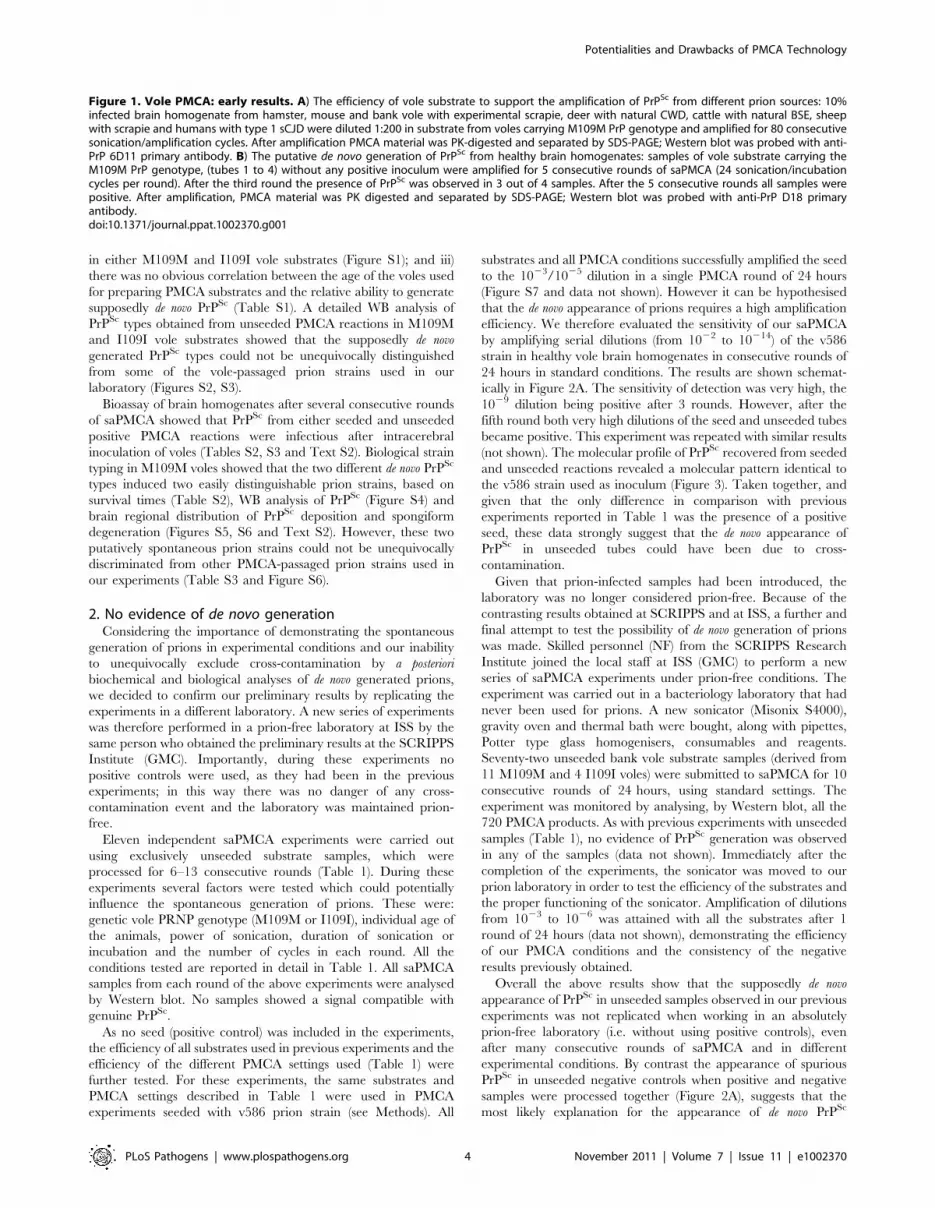
dilution of 1/200. Vole brain homogenates were indeed a
powerful substrate for PMCA, supporting the efficient amplifica-
tion of PrPSc from all the prion sources tested, either derived from
voles or other species, after a single PMCA round of 80
amplification cycles (Figure 1A). However, after a few serial
automated PMCA (saPMCA) rounds, we also observed the
appearance of PK-resistant PrPSc in samples containing exclu-
sively unseeded substrate (negative controls) (Figure 1B). This
finding suggested the possible occurrence of spontaneous gener-
ation of PrPSc during PMCA reactions (see Supplemental Text
S1).
Additional PMCA experiments performed in similar conditions
confirmed the putatively de novo appearance of PrPSc in unseeded
brain homogenates from M109M and I109I voles (Table S1). In
order to exclude the possibility that animals from the facility of
Istituto Superiore di Sanita (ISS) were contaminated, PMCA was
also carried out using substrates from wild voles, trapped in the
countryside, with similar results (see footnotes in Table S1).
Overall, the above experiments showed that: i) the supposedly
spontaneous appearance of PrPSc was a stochastic event, with each
individual sample acting independently (Table S1); ii) two different
PrPSc types could be generated during unseeded PMCA reactions
Author Summary
In an attempt to transpose to an in vitro system theparticular sensitivity of the vole model to human andanimal Transmissible Spongiform Encephalopathies (TSEs),we first explored the suitability of vole brain homogenateas a substrate for PMCA. As well as observing the highlyefficient replication of a variety of prion sources, we alsofound preliminary evidence of de novo prion generation inunseeded reactions. Careful analysis of the PMCA proce-dure, undertaken further to investigate these findings,showed, however, that they were the result of cross-contamination with seeded samples. We next identifiedand investigated the critical points of this procedure thatare potentially responsible for cross-contamination. Ourresults suggest that in vitro systems for prion amplificationcould be more prone to cross-contamination thanpreviously thought, particularly when harsh procedures,such as sonication, are involved. Experimental conditionsable to reproduce spontaneous prion formation in asimple and easily reproducible in vitro system would be ofcrucial interest for understanding TSEs and other impor-tant neurodegenerative disorders. Given that in vitromethods are increasingly used in this field, our resultsemphasise the possible drawbacks of such approaches andcall for the use of rigorously controlled conditions andcautious interpretation of data.
Potentialities and Drawbacks of PMCA Technology
PLoS Pathogens | www.plospathogens.org 2 November 2011 | Volume 7 | Issue 11 | e1002370

Potentialities and Drawbacks of PMCA Technology
PLoS Pathogens | www.plospathogens.org 3 November 2011 | Volume 7 | Issue 11 | e1002370
in either M109M and I109I vole substrates (Figure S1); and iii)
there was no obvious correlation between the age of the voles used
for preparing PMCA substrates and the relative ability to generate
supposedly de novo PrPSc (Table S1). A detailed WB analysis of
PrPSc types obtained from unseeded PMCA reactions in M109M
and I109I vole substrates showed that the supposedly de novo
generated PrPSc types could not be unequivocally distinguished
from some of the vole-passaged prion strains used in our
laboratory (Figures S2, S3).
Bioassay of brain homogenates after several consecutive rounds
of saPMCA showed that PrPSc from either seeded and unseeded
positive PMCA reactions were infectious after intracerebral
inoculation of voles (Tables S2, S3 and Text S2). Biological strain
typing in M109M voles showed that the two different de novo PrPSc
types induced two easily distinguishable prion strains, based on
survival times (Table S2), WB analysis of PrPSc (Figure S4) and
brain regional distribution of PrPSc deposition and spongiform
degeneration (Figures S5, S6 and Text S2). However, these two
putatively spontaneous prion strains could not be unequivocally
discriminated from other PMCA-passaged prion strains used in
our experiments (Table S3 and Figure S6).
2. No evidence of de novo generationConsidering the importance of demonstrating the spontaneous
generation of prions in experimental conditions and our inability
to unequivocally exclude cross-contamination by a posteriori
biochemical and biological analyses of de novo generated prions,
we decided to confirm our preliminary results by replicating the
experiments in a different laboratory. A new series of experiments
was therefore performed in a prion-free laboratory at ISS by the
same person who obtained the preliminary results at the SCRIPPS
Institute (GMC). Importantly, during these experiments no
positive controls were used, as they had been in the previous
experiments; in this way there was no danger of any cross-
contamination event and the laboratory was maintained prion-
free.
Eleven independent saPMCA experiments were carried out
using exclusively unseeded substrate samples, which were
processed for 6–13 consecutive rounds (Table 1). During these
experiments several factors were tested which could potentially
influence the spontaneous generation of prions. These were:
genetic vole PRNP genotype (M109M or I109I), individual age of
the animals, power of sonication, duration of sonication or
incubation and the number of cycles in each round. All the
conditions tested are reported in detail in Table 1. All saPMCA
samples from each round of the above experiments were analysed
by Western blot. No samples showed a signal compatible with
genuine PrPSc.
As no seed (positive control) was included in the experiments,
the efficiency of all substrates used in previous experiments and the
efficiency of the different PMCA settings used (Table 1) were
further tested. For these experiments, the same substrates and
PMCA settings described in Table 1 were used in PMCA
experiments seeded with v586 prion strain (see Methods). All
substrates and all PMCA conditions successfully amplified the seed
to the 1023/1025 dilution in a single PMCA round of 24 hours
(Figure S7 and data not shown). However it can be hypothesised
that the de novo appearance of prions requires a high amplification
efficiency. We therefore evaluated the sensitivity of our saPMCA
by amplifying serial dilutions (from 1022 to 10214) of the v586
strain in healthy vole brain homogenates in consecutive rounds of
24 hours in standard conditions. The results are shown schemat-
ically in Figure 2A. The sensitivity of detection was very high, the
1029 dilution being positive after 3 rounds. However, after the
fifth round both very high dilutions of the seed and unseeded tubes
became positive. This experiment was repeated with similar results
(not shown). The molecular profile of PrPSc recovered from seeded
and unseeded reactions revealed a molecular pattern identical to
the v586 strain used as inoculum (Figure 3). Taken together, and
given that the only difference in comparison with previous
experiments reported in Table 1 was the presence of a positive
seed, these data strongly suggest that the de novo appearance of
PrPSc in unseeded tubes could have been due to cross-
contamination.
Given that prion-infected samples had been introduced, the
laboratory was no longer considered prion-free. Because of the
contrasting results obtained at SCRIPPS and at ISS, a further and
final attempt to test the possibility of de novo generation of prions
was made. Skilled personnel (NF) from the SCRIPPS Research
Institute joined the local staff at ISS (GMC) to perform a new
series of saPMCA experiments under prion-free conditions. The
experiment was carried out in a bacteriology laboratory that had
never been used for prions. A new sonicator (Misonix S4000),
gravity oven and thermal bath were bought, along with pipettes,
Potter type glass homogenisers, consumables and reagents.
Seventy-two unseeded bank vole substrate samples (derived from
11 M109M and 4 I109I voles) were submitted to saPMCA for 10
consecutive rounds of 24 hours, using standard settings. The
experiment was monitored by analysing, by Western blot, all the
720 PMCA products. As with previous experiments with unseeded
samples (Table 1), no evidence of PrPSc generation was observed
in any of the samples (data not shown). Immediately after the
completion of the experiments, the sonicator was moved to our
prion laboratory in order to test the efficiency of the substrates and
the proper functioning of the sonicator. Amplification of dilutions
from 1023 to 1026 was attained with all the substrates after 1
round of 24 hours (data not shown), demonstrating the efficiency
of our PMCA conditions and the consistency of the negative
results previously obtained.
Overall the above results show that the supposedly de novo
appearance of PrPSc in unseeded samples observed in our previous
experiments was not replicated when working in an absolutely
prion-free laboratory (i.e. without using positive controls), even
after many consecutive rounds of saPMCA and in different
experimental conditions. By contrast the appearance of spurious
PrPSc in unseeded negative controls when positive and negative
samples were processed together (Figure 2A), suggests that the
most likely explanation for the appearance of de novo PrPSc
Figure 1. Vole PMCA: early results. A) The efficiency of vole substrate to support the amplification of PrPSc from different prion sources: 10%infected brain homogenate from hamster, mouse and bank vole with experimental scrapie, deer with natural CWD, cattle with natural BSE, sheepwith scrapie and humans with type 1 sCJD were diluted 1:200 in substrate from voles carrying M109M PrP genotype and amplified for 80 consecutivesonication/amplification cycles. After amplification PMCA material was PK-digested and separated by SDS-PAGE; Western blot was probed with anti-PrP 6D11 primary antibody. B) The putative de novo generation of PrPSc from healthy brain homogenates: samples of vole substrate carrying theM109M PrP genotype, (tubes 1 to 4) without any positive inoculum were amplified for 5 consecutive rounds of saPMCA (24 sonication/incubationcycles per round). After the third round the presence of PrPSc was observed in 3 out of 4 samples. After the 5 consecutive rounds all samples werepositive. After amplification, PMCA material was PK digested and separated by SDS-PAGE; Western blot was probed with anti-PrP D18 primaryantibody.doi:10.1371/journal.ppat.1002370.g001
Potentialities and Drawbacks of PMCA Technology
PLoS Pathogens | www.plospathogens.org 4 November 2011 | Volume 7 | Issue 11 | e1002370

previously observed is the occurrence of cross-contamination
during saPMCA.
3. Focusing on contaminationThe occurrence of cross-contamination during saPMCA raises
serious doubts about the advisability of using PMCA for the
detection of prions, particularly when ultrasensitive amplification is
desired. We therefore focused on identifying the critical points
where cross-contamination could occur and the best conditions for
sensitive and specific amplification. In principle, two critical points
at which contamination could have occurred were identified: 1) in
the sonicator, during repeated cycles of sonication/incubation; 2)
during manipulation of samples between PMCA rounds. To assess
which of these two steps was responsible for cross contamination,
serial dilutions of the v586 prion strain and two distinct groups of
unseeded negative controls (A and B) were amplified for 7
consecutive rounds (Figure 2B). All tubes were amplified and
processed together, except that the tubes of control group A were
handled separately after each round in order to avoid cross-
contamination during manipulation of samples. Passages of control
group A were carried out in a new laminar flow hood using a new
pipette, while passages of control group B were carried out together
with seeded samples. The results were comparable to those obtained
in previous experiments (compare Figures 2A and 2B), with control
tubes of both group A and B showing positive signals after 4 and 3
PMCA rounds, respectively. A single PrPSc molecular signature,
identical to the v586 inoculum, was observed in all positive samples
(data not shown). As the tubes used for control group A were never
exposed to possible sources of contamination outside the sonicator,
these data imply that cross-contamination occurs in the sonicator
during the amplification cycles, although they do not allow us to
exclude that tubes may also become contaminated during passages.
To check if the sonicator was able to retain PrPSc molecules and to
release them later as a source of contamination in subsequent
experiments, we then serially amplified unseeded samples in the
sonicator just used for the previous experiments. Passages were
carried out under the flow hood and with the pipette used for
previous seeded experiments. Following 10 consecutive rounds
under standard PMCA conditions, no positive sample was obtained
(data not shown), showing that the sonicator, the flow hood and the
pipette previously used for seeded PMCA reactions are not a strong
source of contamination per se.
Overall the above results indicate that the simultaneous
manipulation and processing of seeded and un-seeded samples is
critical and may easily give rise to cross-contamination events, with
the most likely critical factor being the simultaneous presence of
seeded and unseeded tubes in the sonicator during the amplification
cycles. Moreover, they also indicate that indirect contamination of
the environment (benches, laminar flow hood) and instruments
(pipettes, sonicator, thermal bath) from previous seeded experiments
is not critical, provided that general measures to limit it are taken.
4. Pursuing cross-contamination controlDuring PMCA, the sonication of substrates produces mechan-
ical shearing inside the reaction tube that might force opening of
the vial, thus exposing unseeded samples to cross-contamination. It
is also possible that small prion particles could penetrates through
microscopic cavities between the tube wall and cap contaminating
water in the horn and non-seeded samples. The 0.2 mL PCR
tubes used for PMCA do not lock and are intended to be used in a
thermal cycler. In effect, the caps of the tubes in the thermal cycler
are normally tightened by the pressure of a heated lid. In order to
improve the tightness of the 0.2 mL reaction tubes, we first sealed
the tubes with a small Parafilm M strip positioned on the external
junction between the tube and its cap, and then developed a
‘‘sealer’’ with a moving arm equipped with a sonicator probe.
When the probe was applied to the cap of the vial, it was fastened
to the tube by the heat generated by short sonication pulses. Both
procedures certainly improved the tightness of the tubes, although
when serial PMCA was performed they proved inadequate to
prevent cross-contamination completely (data not shown).
Table 1. Unseeded saPMCA experiments carried out in prion free laboratory.
ID PMCA conditions
Tubes perround
Individual substrates (PrPgenotype and age class)
Sonic/Incub.cycles
Power of sonic.Watts (potency)
Serialrounds
hours/cyclesper round
Met109Met Ile109Ile
1 30 3(a) 2(a) 20 sec./30 min 200 (7) 9 24/48
2 12 1(b) 1(b) 20 sec./30 min 200 (7) 10 24/48
3 12 1(c) 1(c) 20 sec./30 min 200 (7) 13 24/48
4 12 1(d) 1(d) 20 sec./30 min 200 (7) 12 48/96
5 16 1(a), 1(b) 1(a), 1(b) 20 sec./30 min 200 (7) 9 48/96
6 16 1(b), 1(c) 1(b), 1(c) 20 sec./30 min 290 (10) 9 12/24
7 30 3(b) 2(b) 20 sec./30 min 260 (9) 10 12/24
8 75 3(a), 3(b) 3(a), 3(b) 20 sec./30 min 240 (8) 9 8/16
9 70 3(a), 4(b), 3(c) 1(a), 1(b), 2(c) 20 sec./30 min 200 (7) 8 48/96
10 72 3(a), 3(b), 3(c), 3(d) 0 20 sec./30 min 200 (7) 6 24/24
11 20 3(b), 2(c) 0 2 sec./30 Ssec 200 (7) 6 24/48
Footnote to Table 1. Eleven independent experiments were carried out in the prion free laboratory using 4 to 6 replicates of unseeded substrate, in absence of anypositive inoculum. This table provides details on each experiment reporting the number substrates samples, the PrP genotype and the age of the individual voles.Animals belong to four classes of age: a = 4–8 weeks old; b = 2–4 months old; c = 5–12 months old; d = older than 1 year. The settings of saPMCA (the length ofincubation and sonication cycles, the power of sonication, number of consecutive rounds and the length of rounds) are also given.doi:10.1371/journal.ppat.1002370.t001
Potentialities and Drawbacks of PMCA Technology
PLoS Pathogens | www.plospathogens.org 5 November 2011 | Volume 7 | Issue 11 | e1002370

Subsequently we evaluated new vials bought on the market. We
selected Multiply-Safecup Biosphere, handy 0,5 mL tubes with
screw cap and 100 ml volume limitation that works as a double
closure system (Figure S8). The ability of the new 0,5 mL screw-
cap vials to support high amplification levels was evaluated by
amplifying serial dilutions of v586 inoculum. No difference was
observed when we compared the efficiency of the 0,2 ml vial and
the 0,5 mL screw-cap tubes (data not shown). Importantly, the
screw-cap tubes were able completely to prevent cross-contami-
nation, even after many consecutive rounds (see below).
5. Achieving ultrasensitive amplification and control ofcross-contamination during saPMCA
In order to achieve the best conditions for high amplificationefficiency, which is useful in sensitive diagnostic tests, weinvestigated the conditions that could lead to the highest sensitivityin a limited number of rounds using 0,5 mL screw-cap vials. Thiswas done by amplifying dilution curves of v586 inoculum andevaluating the level of amplification obtained after 12, 24, 48 and72 hours of PMCA using standard settings (Figure 4). By increasing
the duration of PMCA from 24 to 48 hours, we observed a 3-log
Figure 2. Sensitivity of detection of saPMCA using vole substrate and v586 inoculum. For each round and seed dilution is reported thenumber of positive samples over the number of replicates tested. Green boxes indicate that all replicates were positive; yellow boxes indicate thatless than 100% of replicates were positive, while boxes are white when no positive replicates occurred. A) Logarithmic dilutions of the seed wereprepared in duplicate from 1023 to 10214 using vole M109M substrate. Eight negative controls were included. Samples were amplified for 7consecutive rounds of 24 hours. All the samples were analysed by Western blot. Results showed that after the first round, amplifications wereobserved in dilutions 1023, 1024 and in one of two duplicates of 1025. After the second round, positive PrPSc signals were detected also in 1025, 1026,and 1027 dilutions and in one of the 1028 duplicates. After three consecutive rounds, 1029 reached positive amplification. Lower dilutions wereamplified between the 5th and 7th round but, at the same time, also negative controls became positive. B) logarithmic dilutions of v586 in duplicatefrom 1023 to 10214 were amplified for 7 consecutive 24-hour rounds using vole M109M substrate. Two groups of 8 unseeded negative controls wereincluded.(groups A and B). All the seeded and unseeded samples were amplified together, but passages of control group A were carried out in aprion-free environment (new laminar flow hood and pipette), while samples from control group B were processed together with the seeded tubes.Results showed similar sensitivity compared to previous experiments, with amplification of 1025 dilution after the first round and up to 1029 after thesecond round. After the third round the 10210, 10211 and 10214 dilutions along with six out of eight negative samples from control group B becomepositive. One round later, three out of eight control group A samples also turned positive.doi:10.1371/journal.ppat.1002370.g002
Potentialities and Drawbacks of PMCA Technology
PLoS Pathogens | www.plospathogens.org 6 November 2011 | Volume 7 | Issue 11 | e1002370

improvement in PrPSc amplification, making it possible to increase
considerably the overall sensitivity attainable in a single round.
Further increasing the duration to 72 hours did not, however, seem
to be useful, since the amplification level did not substantially
increase. This was further investigated by testing the ability of 48 h-
old substrates (previously treated with 48 hours of incubation/
sonication cycles) to support PrPSc amplification. The failure of
48 h-old substrates to amplify PrPSc (Figure S9) confirmed that the
ability of substrates to support amplification dramatically decreases
after 48 hours of PMCA. Single-round 48-hour PMCA was thus set
up and a limit of detection ranging from 1029 to 10210 was
observed when the experiments were repeated several times using
different v586 seeds preparations and different substrates. We then
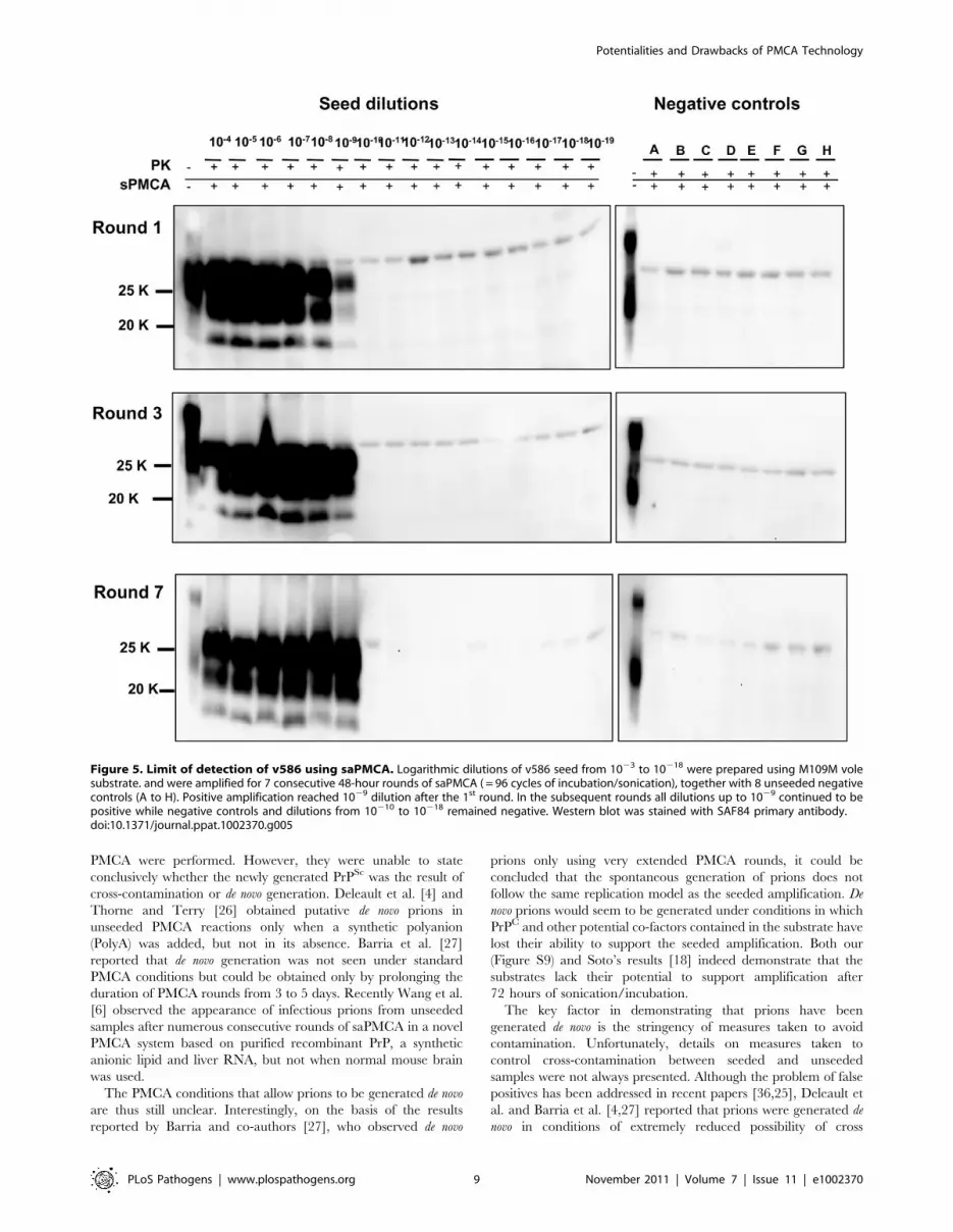
attempted the serial 48-hour PMCA in order to investigate the limit
of detection of v586 under these new conditions. As shown in
Figure 5, the level of amplification reached after the first round did
not increase further in successive PMCA rounds. Importantly,
negative controls and dilutions exceeding the limit of 1029 remained
negative up to the seventh PMCA round. Therefore, by introducing
0,5 mL screw-cap vials and 48-hour PMCA rounds, cross-
contamination was efficiently controlled while ultra-high sensitivity
was maintained for many consecutive rounds.
Since then we have performed numerous saPMCA experiments
using the same working conditions. The appearance of spurious
PrPSc signals was observed in only two out of more than 500
samples that were submitted to saPMCA. One of the two samples
was an unseeded negative control, the other was a10215 dilution of
v586. The PrPSc of these samples preserved the same biochemical
properties as the v586 seed used in the experiments. This clearly
indicates that although cross-contamination in the sonicator can
be effectively prevented by screw-cap vials, contamination during
manipulation of samples can still lead to rare cross-contamination
events, as often happens during ultrasensitive diagnostic tests.
Discussion
In the present study we describe the results of a large series of
PMCA experiments under strictly controlled conditions which
highlight the drawbacks and potential of PMCA when ultrasen-
sitive detection is desired and propose technical improvements to
increase the robustness of the technique.
This work was stimulated by observing how the extraordinary
potential of PMCA is accompanied by a disturbing production of
infectious PrPSc in unseeded reaction tubes, which initially led us
to hypothesise the occurrence of ‘‘spontaneous’’ or de novo
generation of prions.
The de novo generation of prions agrees with the prion theory,
which postulates that sporadic TSEs may originate from the
stochastic occurrence of spontaneous PrPC conversion into PrPSc.
Several lines of evidence support this hypothesis. Infectious prions
have been generated from b-rich recombinant mouse PrP(89–230)
[5]. Aguzzi and colleagues reported the spontaneous generation of
prions in transgenic mice carrying a mutated prion gene (S170N,
N174T) [32]. They also observed prion infectivity in mice over-
expressing wild type PrP (tga20 line) after inoculation of brain
homogenates from extremely aged, uninoculated mice of the same
line. The spontaneous occurrence of prions in cell cultures has
recently been reported by Edgeworth and colleagues [33].
As PMCA is able to reproduce in vitro several aspects of prion
biology, it appears theoretically to be an appropriate technique for
modelling the de novo generation of prions, making it this field of
investigation very attractive.
However, the hypothesis that prions can be generated de novo is
not easy to refute experimentally and - given that de novo prions are
postulated to have the same characteristics and properties as other
prions – there is no way to distinguish prions generated de novo
from potential contaminants. Nonetheless, it was recently reported
that full-length recombinant prion protein, that had been
Figure 3. Comparison between PrPSc from seeded and unseeded PMCA reactions. Characterization of PrPSc from positive samples of theexperiment described in Figure 2A by Western blot, stained with SAF84 primary antibody. The figure shows that samples A (PrPSc from 10% brainhomogenate used as positive inoculum), B (1025 dilution of the inoculum after a single 24-hour round of PMCA) and C (PrPSc from an unseedednegative control which was positive after 6 consecutive rounds of saPMCA) share the same apparent molecular weight before or afterdeglycosylation with PNGase.doi:10.1371/journal.ppat.1002370.g003
Potentialities and Drawbacks of PMCA Technology
PLoS Pathogens | www.plospathogens.org 7 November 2011 | Volume 7 | Issue 11 | e1002370

converted into the cross-beta-sheet amyloid form and subjected to
annealing, gave rise to a disease with clearly distinctive phenotype
from archetypal 263K, upon serial transmission in hamster [7].
However, drawing an a posteriori distinction between prions
putatively generated de novo and prions from other sources
recognised as putative contaminants is hampered by the potential
of prions to mutate, both in vivo [34] and in vitro [35]. In our
experiments, supposedly de novo prions generated in vole PMCA
were not easily distinguishable from other conventional prion
strains (Figures S2, S3). The only way to obtain direct evidence of
the de novo generation of prions is by establishing experimental
conditions in which the occurrence of contamination can be
excluded categorically. Since prions are not thought to be
ubiquitous contaminants, the use of rigorously prion-free proce-
dures appears adequate to achieve these conditions.
We performed several unseeded PMCA experiments with
hundreds of samples processed and analysed over more than 6
months in absolutely prion-free conditions. Different PMCA
settings and several substrates from many voles of different ages
were used. No evidence of de novo generation of prions was
obtained, despite the high levels of sensitivity reached.
Our results appear to be in contrast with those obtained by
other groups. The de novo generation of prions during PMCA
experiments has been reported by several authors. Saa et al. [25]
reported the spontaneous generation of PrPSc in unseeded PMCA
reactions, especially when more than 10 rounds of standard
Figure 4. Kinetics of PrPSc amplification during a single round of PMCA. Logarithmic dilutions of v586 seed from 1023 to 10210, wereprepared in quadruplicate using M109M vole substrate. Samples were amplified for a single round of PMCA together with unseeded negativecontrols. The level of amplification was evaluated by western blot in seeded and unseeded samples after 12 hours (panels A and E), 24 hours (panelsB and F), 48 hours (panels C and G) and 72 hours (panels D and H). Western blot was probed with SAF84 primary antibody.doi:10.1371/journal.ppat.1002370.g004
Potentialities and Drawbacks of PMCA Technology
PLoS Pathogens | www.plospathogens.org 8 November 2011 | Volume 7 | Issue 11 | e1002370
PMCA were performed. However, they were unable to state
conclusively whether the newly generated PrPSc was the result of
cross-contamination or de novo generation. Deleault et al. [4] and
Thorne and Terry [26] obtained putative de novo prions in
unseeded PMCA reactions only when a synthetic polyanion
(PolyA) was added, but not in its absence. Barria et al. [27]
reported that de novo generation was not seen under standard
PMCA conditions but could be obtained only by prolonging the
duration of PMCA rounds from 3 to 5 days. Recently Wang et al.
[6] observed the appearance of infectious prions from unseeded
samples after numerous consecutive rounds of saPMCA in a novel
PMCA system based on purified recombinant PrP, a synthetic
anionic lipid and liver RNA, but not when normal mouse brain
was used.
The PMCA conditions that allow prions to be generated de novo
are thus still unclear. Interestingly, on the basis of the results
reported by Barria and co-authors [27], who observed de novo
prions only using very extended PMCA rounds, it could be
concluded that the spontaneous generation of prions does not
follow the same replication model as the seeded amplification. De
novo prions would seem to be generated under conditions in which
PrPC and other potential co-factors contained in the substrate have
lost their ability to support the seeded amplification. Both our
(Figure S9) and Soto’s results [18] indeed demonstrate that the
substrates lack their potential to support amplification after
72 hours of sonication/incubation.
The key factor in demonstrating that prions have been
generated de novo is the stringency of measures taken to avoid
contamination. Unfortunately, details on measures taken to
control cross-contamination between seeded and unseeded
samples were not always presented. Although the problem of false
positives has been addressed in recent papers [36,25], Deleault et
al. and Barria et al. [4,27] reported that prions were generated de
novo in conditions of extremely reduced possibility of cross
Figure 5. Limit of detection of v586 using saPMCA. Logarithmic dilutions of v586 seed from 1023 to 10218 were prepared using M109M volesubstrate. and were amplified for 7 consecutive 48-hour rounds of saPMCA ( = 96 cycles of incubation/sonication), together with 8 unseeded negativecontrols (A to H). Positive amplification reached 1029 dilution after the 1st round. In the subsequent rounds all dilutions up to 1029 continued to bepositive while negative controls and dilutions from 10210 to 10218 remained negative. Western blot was stained with SAF84 primary antibody.doi:10.1371/journal.ppat.1002370.g005
Potentialities and Drawbacks of PMCA Technology
PLoS Pathogens | www.plospathogens.org 9 November 2011 | Volume 7 | Issue 11 | e1002370

contamination. Nonetheless, Barria et al [27] and Wang et al [6]
concluded that cross-contamination could not be entirely ruled
out.
The efficiency of PMCA is variable, depending on the
combination of seed and substrate. Sonicator settings are often
adapted to reach maximum efficiency [18]. Considering that one
of the main objectives of this study was to investigate the spread of
contamination during saPMCA, we decided to adopt v586 as
infected inoculum for seeded experiments because it was a
contaminant in origin (see Methods). After seeding vole substrate
with v586 strain amplification proved extremely efficient. The
limit of detection, observed in several independent experiments,
ranged between 1029 and 10210 of the infected brain homogenate
in a single 48-hour round of PMCA. Additional rounds did not
improve the overall sensitivity. These data are in agreement with
the efficiency reported for mouse-adapted scrapie and BSE, which
is the highest reported in the literature [37]. Results of the limiting
dilution experiments showed that 10210 is the highest dilution of
the inoculum able to trigger a positive amplification, correspond-
ing approximately to 6 femtoliters of infected brain homogenate.
Similar levels of contamination are not easy to control, especially
for a procedure that is performed using equipment originally
developed for other purposes. We have also observed cross-
contamination when working with PMCA substrates from other
species, such as mice (data not shown), although it was observed at
a lower level and after an higher number of PMCA rounds. This
might be due to the lower amplification efficiency we have
observed with mouse-derived compared to vole-derived substrates.
When ultrasensitive amplification levels are reached, such as those
attained with vole brains as substrates, the risk that minimal
inadvertent contamination of reactions may artificially initiate
amplification is extremely high. These findings suggest the need to
critically reassess previous reports of supposedly de novo prion
generation obtained by standard PMCA setting.
In experiments using only unseeded samples, we determined
that the equipment used (pipettes, sonicator, thermal bath) and the
working environment (laminar flow hood, bench) are not direct
sources of contamination, provided that general safety measures
are adopted. It is unlikely that they can release prions potentially
acquired during previous experiments at levels able to trigger
amplification in subsequent PMCA reactions. In contrast, direct or
indirect cross-contamination easily occurs between seeded and
unseeded samples when these are manipulated and processed at
the same time. The presence of high numbers of seeded tubes in
the sonicator and the progressive and dramatic increase in
amplified products during serial rounds further increase the risk.
Sonication pulses violently shake the samples. The generation of
aerosol and micro-particles within the reaction tubes during
sonication probably represents a key critical factor by enormously
increasing the risk of contamination of the rim of the tube and of
material leakage at the opening of the tubes. The use of Multiply-
Safecup Biosphere tubes radically improved the handling of
samples and, thanks to both the screw cap and the volume
limitation system radically reduced the risk of leakage. The
preservation of the same amplification efficiency using larger tubes
with thicker walls and other significant differences compared with
PCR tubes, without the need to modify the sonication parameters,
demonstrates that PMCA is capable of coping well with significant
changes in the operating environment.
In the course of the study we observed a dramatic decrease in
false positive results from the moment we adopted the screw-cap
tubes. This provides strong evidence that false positive results
actually derive from cross-contamination events which can be
efficiently prevented by tighter closure of the sample tubes.
Spurious results were observed even with the highest level of safety
measures, but these were very rare and comparable to those
observed with other techniques. We cannot exclude the possibility
that some of these events actually concealed the de novo generation
of prions, but while we cannot dismiss alternative explanations, the
occurrence of inadvertent contamination still appears the most
appropriate and scientifically sound interpretation. This is further
strengthened by the unique molecular signature observed
throughout our study, similar to the original v586 seed. The few
spurious results that we observed after the adoption of screw-cap
tubes occurred after several rounds of serial PMCA, and could be
tentatively explained by inadvertent contamination during ma-
nipulation of the tubes for seeding successive rounds of serial
PMCA. In this respect, methods able to increase the sensitivity of
PMCA, as recently reported by Gonzalez-Montalban and
colleagues [38], would reduce the number of serial passages
needed to achieve ultrasensitive detection of PrPSc and, conceiv-
ably, would also be valuable for preventing cross-contamination
events.
The results of the present study certainly do not exclude the
possibility that, in different experimental conditions, prions can be
generated de novo. They provide a serious warning that
contamination can very easily occur when ultrasensitive levels of
detection are reached and that extreme caution is needed to avoid
the over- or misinterpretation of results when delicate issues such
as the de novo generation of prions are explored.
Methods
Ethics statementThe research protocol was approved by the Service for
Biotechnology and Animal Welfare of the Istituto Superiore di
Sanita and authorized by the Italian Ministry of Health, according
to Legislative Decree 116/92, which implemented the European
Directive 86/609/EEC on laboratory animal protection in Italy.
Animal welfare was routinely checked by veterinarians from the
Service for Biotechnology and Animal Welfare.
Preparation of substratesSubstrates were prepared from male and female bank voles of
different ages, carrying either M109M or I109I PrP genotypes.
Unless indicated otherwise (see Tables 1 and S1), the animals were
2–4 months old. Voles were sacrificed using carbon dioxide and
immediately perfused with phosphate-buffered saline (PBS) plus
5 mM ethylendiaminetetraacetic acid (EDTA). Immediately after
perfusion, the brains were dissected and frozen at 280uC. 10%
brain homogenates (weight/volume) were prepared in conversion
buffer (PBS 1x, pH 7,4; 0,15 M NaCl; 1% Triton X-100) with the
Roche Complete Protease inhibitor cocktail (1 tablet in 50 mL
conversion buffer), using new and dedicated glass potters.
Substrates were divided into aliquots and either used or stored
at –80uC immediately after homogenisation. De novo generation
experiments (Tables 1 and S1) were carried out using substrates
from individual voles while for seeded experiments (see Results),
requiring larger amounts of substrate, we prepared homogeneous
pools of substrates by mixing together 5 individual substrates from
voles of 2–4 months of age. Preparation and storage of substrates
were carried out in a laboratory never previously used for prion
research, using equipment specifically dedicated and maintaining
rigorously prion-free conditions.
Preparation of the inoculum for PMCAThe vole-passaged spontaneous M109M strain B (see results),
indicated in the main text as v586, was used as prion inoculum for
Potentialities and Drawbacks of PMCA Technology
PLoS Pathogens | www.plospathogens.org 10 November 2011 | Volume 7 | Issue 11 | e1002370

seeded PMCA experiments. Brain tissue from terminally affected
M109M voles after second passage of M109M strain B (see Table
S2) was homogenised in PBS (10% w/v) containing Complete
Protease inhibitor cocktail (Roche) using disposable Teflon pestles
directly in 1.5 mL Eppendorf tubes. Following preparation the
brain homogenate was divided into aliquots and stored at 220uC.Preparation of dilution curves of the inoculum. v586
brain homogenate was serially diluted in vole substrate. Curves
were prepared as follow: 20 ml of inoculum were added in a
1,5 mL Eppendorf tube containing 180 ml of substrate, followed
by 10-fold serial dilutions in 1,5 mL Eppendorf tubes containing
180 ml of substrate. At each dilution, inoculum and substrate were
mixed by gently pipetting 50 times paying careful attention to
change the tip of the pipette at each dilution step. Finally, 60 ml of
each dilution were introduced into PMCA tubes for the first round
of amplification. Samples were handled under a laminar flow
hood, using dedicated pipettes and double filter tips, carefully
avoiding leakages of material during the manipulation of tubes.
The use of a vortex instead of pipetting is not recommended as, in
our experience it has occasionally been associated with the
appearance of spurious positive PrPSc signals after amplification.
Serial automated PMCA (saPMCA)PMCA was performed according to the protocol of Castilla
et al.[18]. Amplification was performed in a total volume of 60 ml.
Tubes containing the reaction mix were incubated at 37uC, placed
in a disk-shaped rack on the microplate horn of the sonicator
(Misonix S3000). The horn was placed in a gravity oven. Water in
the horn was circulated in a thermal bath at a constant
temperature of 37uC . The standard sonication programme
consisted of 30 second sonication pulses every 30 minutes for 24-
or 48-hour periods. In particular cases single parameters of PMCA
were modified (see Table 1) in order to investigate different
conditions of amplification. At the end of each round, the PMCA
material was gently span and diluted 1:10 in fresh substrate ready
for a new amplification step. Passages of seeded experiments were
carried out in a laminar flow hood, using double filter pipette tips
(Eppendorf Dualfilter T.I.P.S.). During this study two kinds of tube
were used to contain PMCA reactions: we began working with
0.2 mL tubes for PCR (NUNC, cat. 248161), then continued with
0,5 mL screw cap Multiply- Safecup (Sarstedt, cat. 72.733.200)
(see Results).
Additional procedures to minimise contaminationAccess to the laboratory was restricted to authorised personnel
and standard operating procedures were adopted for the use of
laboratory equipment and sample manipulation. All personnel
wore disposable coats, shoe covers and gloves. The working
surfaces and equipment were cleaned with NaOH or NaClO after
each experiment. These measures were further strengthened by
changing the water of the sonicator circuit and decontaminating
the plastic holder and horn cap by immersion in bleach after each
round.
Electrophoresis and immunoblotting20 mL of PMCA material were digested with 100 mg/mL
proteinase K (pK) (Sigma-Aldrich). Samples were shaken at 900
rounds per minute (rpm) for one hour at 37uC. Digestion was then
blocked by adding 2 mL of 10 mM phenylmethylsulfonyl fluoride
(PMSF) (Pierce Biotechnology - Thermo Fisher Scientific); samples
were then incubated for 5 minutes at 4uC. Ten micro-litres of
NuPAGELDS Sample Buffer (4X) and 3 mL NuPAGE Sample
Reducing Agent (10X) were added to each tube and samples were
denatured by incubation at 90uC for 10 minutes. Western blot
analysis: 12 mL of pK-digested PMCA samples were resolved by
SDS-PAGE on a 10% Tris-glycine gel and transferred to PVDF.
The membrane was blocked with 1% powdered skim milk in PBS
for one hour at room temperature and incubated with mouse anti-
PrP monoclonal antibody SAF84 (Cayman Chemical). After
washing, the membrane was incubated with secondary HRP-
conjugated antibody (ImmunoPure Peroxidase Conjugated Goat
Anti-Mouse IgG (H+L) Pierce Biotechnology - Thermo Fisher
Scientific) and signals were detected using SuperSignal West
Femto Maximum Sensitivity Substrate (Pierce Biotechnology -
Thermo Fisher Scientific). Chemiluminescence was detected with
the VersaDoc imaging system (Bio-Rad). All measurements were
performed with QuantityOne software (Bio-Rad).
Supporting Information
Figure S1 Putative de novo vole PrPSc types obtained byvole sa-PMCA. Samples from 23 different unseeded experiments
of vole saPMCA (see Table S1) were studied by western blot.
Based on the differential electrophoretic mobilities, two distinct
PrPSc types (named A and B for the high and low MW types,
respectively) were recovered from both vole genotypes. Putatively
de novo strains A and B were further passaged for up to 10 serial
round of PMCA maintaining their distinctive electrophoretic
mobility. Western blot was stained with D18 primary antibody.
(TIF)
Figure S2 Biochemical comparison between the puta-tive de novo PrPSc types and vole-adapted prions.Prototypical vole TSEs (SSBP1 and SS8, derived from sheep
scrapie, sCJDMV1 and sCJDMV2 derived from human, cattle-
derived BSE and mouse-derived 301C) obtained after serial in vivo
passages in voles, were compared with the four putative de novo
PrPSc types obtained in vitro. PrPSc from all samples was digested
with PK and analysed by WB using two different antibodies,
SAF84 and 12B2. These two antibodies are used for discriminat-
ing PrPres types according to the N-terminal cleavage by PK.
Indeed, SAF84 recognizes a C-terminal epitope and binds all kinds
of vole prion strains, while 12B2 binds an epitope in the region
differentially cleaved by PK. The figure shows that putative de novo
PrPSc types M109MA and I109IA are similar to scrapie-like vole
prions. The same types are also similar to the sCJD MV1-derived
vole prion. On the other hand the de novo PrPSc types M109MB
and I109IB have a molecular weight similar to BSE and sCJD-
MM2-derived vole prions, and are not recognized by 12B2.
(TIF)
Figure S3 Biochemical comparison between putative denovo PrPSc types and PMCA-amplified vole prions. Vole-
adapted prion strains derived from human (sCJDMM1), sheep
(SS8 and SSBP1), cattle (BSE) and mice (301C and 301V) were
amplified in vitro by 15 rounds of saPMCA using vole substrate and
were compared with the four putative de novo PrPSc types obtained
in unseeded PMCA reactions. Replica blots were stained with
mAbs SAF84 and 12B2 (see figure S2). The de novo PrPSc types A
from M109M and I109I show biochemical features similar to
PMCA-passaged scrapie strains and sCJD-MM1, while de novo
PrPSc types B are similar to PMCA-passaged BSE and BSE-
derived strains (301C and 301V).
(TIF)
Figure S4 Biochemical comparison between putative denovo prion strains after in vivo passage in voles.Representative discriminatory WB (see figure S2) from PK
digested brain homogenates of vole-passaged putative de novo
prion strains (M109MA and M109MB). PrPSc from scrapie-like
Potentialities and Drawbacks of PMCA Technology
PLoS Pathogens | www.plospathogens.org 11 November 2011 | Volume 7 | Issue 11 | e1002370

vole-adapted ME7 was used as control. The figure shows that
scrapie-like and BSE-like biochemical properties were preserved
after in vivo passage of strains A and B, respectively.
(TIF)
Figure S5 PrPSc deposition patterns in vole-passagedputative de novo prion strains. A) PET-blot analysis with
SAF84. In M109MA PrPSc deposition was predominant in the
cortex, caudate-putamen and gyrus dentate compared to
M109MB infected voles. Moreover, the hypothalamus was much
more involved in M109MA than in M109MB. B) Immunohisto-
chemistry performed with polyclonal rabbit Ab R486 shows
distinct PrPSc deposition patterns in M109MA and M109MB.
Punctuate PrPSc deposition associated with conspicuous vacuol-
ization was found in the medial geniculate nucleus of M109MA,
whereas in M109MB an astrocytic pattern was observed. In the
parietal cortex of M109MA perivascular and punctuate patterns
were evident, while M109MB showed an astrocytic pattern.
(TIF)
Figure S6 Lesion profiles in voles infected with seededand unseeded PMCA products. A) Lesion profiles in voles
after transmission of M109MA and M109MB show distinct
patterns of spongiform degeneration. B) Comparison of the lesion
profiles in M109MA and SS8 show partial overlapping. C)
Comparison of the lesion profiles in M109MB, 301C and 301V
show near complete overlapping, particularly between M109MB
and 301C. Brain-scoring positions are medulla (1), cerebellum (2),
superior colliculus (3), hypothalamus (4), thalamus (5), hippocam-
pus (6), septum (7), retrosplenial and adjacent motor cortex (8),
and cingulate and adjacent motor cortex (9).
(TIF)
Figure S7 A test of the efficiency of PMCA. Two-fold serial
dilution was prepared from 1:100 to 1:12800 using v586 inculum
and vole M109M substrate. Samples were amplified for a single
round of 24 hours (48 cycles of sonication/incubation). The first
lane shows the unamplified ‘‘frozen’’ 1:200 dilution and the second
lane shows the dilution curve after PMCA. Western blots were
probed with SAF84 primary antibody.
(TIF)
Figure S8 Screw-cap tube for PMCA. Containment of PrPSc
during saPMCA was improved by using Sarstedt 0.5 mL Multiply-
Safecup with 100 ml volume limitation. This tube is easy to handle,
the volume limiter confines reaction mix to the bottom of the vial,
where it is better exposed to the effect of the ultrasound, keeping
the substrate away from the rim of the tube. Reaction sensitivity is
comparable with 0,2 mL PCR tubes, while control of cross-
contamination is much more efficient.
(TIF)
Figure S9 Evaluating the ability of substrate to amplifyPrPSc after 48 h of PMCA. Logarithmic dilutions of v586 seed
were prepared from 1024 to 10210 using either: freshly prepared
M109M vole substrate (A) or a substrate carrying the same PrP
genotype previously submitted to repeated cycles of incubation/
sonication for 48 hours (B). Dilutions were submitted to a single
round of 48 hours of PMCA. Western blots were probed with
SAF84 primary antibody.
(TIF)
Table S1 Supposedly de novo PrPSc appearance inunseeded PMCA reactions using bank vole substrates.(DOC)
Table S2 Transmission of putative de novo vole prionstrains by intra-cerebral inoculation in voles.(DOC)
Table S3 Comparison of seeded and unseeded saPMCAproducts by intra-cerebral inoculation in voles.(DOC)
Text S1 Supporting results. Early experimental evidences
indicated that the appearance of PrPSc in unseeded substrate
samples was frequent after a variable number of saPMCA rounds.
This finding was repeatedly observed in several different indepen-
dent experiments and using numerous vole substrates (Table S1).
PrPSc isolates from unseeded experiments were studied by western
blot. Based on the differential electrophoretic mobilities, two distinct
PrPSc types (named A and B) were recovered from M109M and
I109I vole genotypes. Putatively de novo strains A and B were further
passaged for up to 10 serial round of PMCA maintaining their
distinctive electrophoretic mobility. (Figure S1). The PrPSc types,
that were supposed to be of spontaneous origin, have been
intensively studied using methods available for TSEs strain typing
(Figures S2, S3, S4, S5, S6 and Tables S2 and S3).
(DOC)
Text S2 Method section for supporting Figures andTables.(DOC)
Acknowledgments
The authors would like to thank Dr. Alfredo Caprioli for allowing them to
work in his laboratory at the SPVSA Department of the Istituto Superiore
di Sanita.
Author Contributions
Conceived and designed the experiments: GMC RN GV JC UA.
Performed the experiments: GMC CB NFB MADB. Analyzed the data:
GMC RN GV JC UA. Wrote the paper: GMC RN UA.
References
1. Prusiner SB (1998) Prions. Proc Natl Acad Sci U S A 95: 13363–83.
2. Weissmann C (1991) A ’unified theory’ of prion propagation. Nature 352:
679–83.
3. Dickinson AG, Outram GW (1988) Genetic aspects of unconventional virusinfections: the basis of the virino hypothesis. Ciba Found Symp 135: 63–83.
4. Deleault NR, Harris BT, Rees JR, Supattapone S (2007) Formation of native
prions from minimal components in vitro. Proc Natl Acad Sci U S A 104: 9741–6.
5. Legname G, Baskakov IV, Nguyen HO, Riesner D, Cohen FE, et al. (2004)Synthetic mammalian prions. Science 305: 673–6.
6. Wang F, Wang X, Yuan CG, Ma J (2010) Generating a prion with bacteriallyexpressed recombinant prion protein. Science 327: 1132–1135.
7. Makarava N, Kovacs GG, Bocharova O, Savtchenko R, Alexeeva I, et al. (2010)
Recombinant prion protein induces a new transmissible prion disease in wild-type animals. Acta Neuropathol 119: 177–87.
8. Kim JI, Cali I, Surewicz K, Kong Q, Raymond GJ, et al. (2010) Mammalian
prions generated from bacterially expressed prion protein in the absence of any
mammalian cofactors. J Biol Chem 285: 14083–7.
9. Barron RM, Campbell SL, King D, Bellon A, Chapman KE, et al. (2007) High
titers of transmissible spongiform encephalopathy infectivity associated withextremely low levels of PrPSc in vivo. J Biol Chem 282: 35878–86.
10. Caughey B, Raymond GJ, Ernst D, Race RE (1991) N-terminal truncation of the
scrapie-associated form of PrP by lysosomal protease(s): implications regarding the
site of conversion of PrP to the protease-resistant state. J Virol 65: 6597–603.
11. Pan KM, Baldwin M, Nguyen J, Gasset M, Serban A, et al. (1993) Conversion ofalpha-helices into beta-sheets features in the formation of the scrapie prion
proteins. Proc Natl Acad Sci U S A 90: 10962–6.
12. Gajdusek DC (1988) Transmissible and non-transmissible amyloidoses: auto-
catalytic post-translational conversion of host precursor proteins to beta-pleatedsheet configurations. J Neuroimmunol 20: 95–110.
13. Collinge J, Sidle KC, Meads J, Ironside J, Hill AF (1996) Molecular analysis ofprion strain variation and the aetiology of ’new variant’ CJD. Nature 383: 685–90.
14. Bruce ME, Will RG, Ironside JW, McConnell I, Drummond D, et al. (1997)
Transmissions to mice indicate that ’new variant’ CJD is caused by the BSE
agent. Nature 389: 498–501.
Potentialities and Drawbacks of PMCA Technology
PLoS Pathogens | www.plospathogens.org 12 November 2011 | Volume 7 | Issue 11 | e1002370

15. Lasmezas CI, Deslys JP, Demaimay R, Adjou KT, Lamoury F, et al. (1996) BSE
transmission to macaques. Nature 381: 743–4.
16. Gillies M, Chohan G, Llewelyn CA, MacKenzie J, Ward HJ, et al. (2009) A
retrospective case note review of deceased recipients of vCJD-implicated blood
transfusions. Vox Sang 97: 211–8.
17. Saborio GP, Permanne B, Soto C (2001) Sensitive detection of pathological
prion protein by cyclic amplification of protein misfolding. Nature 411: 810–3.
18. Castilla J, Saa P, Hetz C, Soto C (2005) In vitro generation of infectious scrapie
prions. Cell 121: 195–206.
19. Castilla J, Morales R, Saa P, Barria M, Gambetti P, et al. (2008) Cell-free
propagation of prion strains. EMBO J 27: 2557–66.
20. Green KM, Castilla J, Seward TS, Napier DL, Jewell JE, et al. (2008)
Accelerated high fidelity prion amplification within and across prion species
barriers. PLoS Pathog 4: e1000139.
21. Castilla J, Gonzalez-Romero D, Saa P, Morales R, De Castro J, et al. (2008)
Crossing the species barrier by PrP(Sc) replication in vitro generates unique
infectious prions. Cell 134: 757–68.
22. Saa P, Castilla J, Soto C (2006) Presymptomatic detection of prions in blood.
Science 313: 92–4.
23. Castilla J, Saa P, Soto C (2005) Detection of prions in blood. Nat Med 11:
982–5.
24. Gonzalez-Romero D, Barria MA, Leon P, Morales R, Soto C (2008) Detection
of infectious prions in urine. FEBS Lett 582: 3161–6.
25. Saa P, Castilla J, Soto C (2006) Ultra-efficient replication of infectious prions by
automated protein misfolding cyclic amplification. J Biol Chem 281: 35245–52.
26. Thorne L, Terry LA (2008) In vitro amplification of PrPSc derived from the brain
and blood of sheep infected with scrapie. J Gen Virol 89: 3177–84.
27. Barria MA, Mukherjee A, Gonzalez-Romero D, Morales R, Soto C (2009) De
novo generation of infectious prions in vitro produces a new disease phenotype.
PLoS Pathog 5: e1000421.
28. Nonno R, Di Bari MA, Cardone F, Vaccari G, Fazzi P, et al. (2006) Efficient
transmission and characterization of Creutzfeldt-Jakob disease strains in bankvoles. PLoS Pathog 2: e12.
29. Agrimi U, Nonno R, Dell’Omo G, Di Bari MA, Conte M, et al. (2008) Prion
protein amino acid determinants of differential susceptibility and molecularfeature of prion strains in mice and voles. PLoS Pathog 4: e1000113.
30. Di Bari MA, Chianini F, Vaccari G, Esposito E, Conte M, et al. (2008) The bankvole (Myodes glareolus) as a sensitive bioassay for sheep scrapie. J Gen Virol 89:
2975–85.
31. Cartoni C, Schinina ME, Maras B, Nonno R, Vaccari G, et al. (2005)Identification of the pathological prion protein allotypes in scrapie-infected
heterozygous bank voles (Clethrionomys glareolus) by high-performance liquidchromatography-mass spectrometry. J Chromatogr 1081: 122–6.
32. Sigurdson CJ, Nilsson KP, Hornemann S, Heikenwalder M, Manco G, et al.(2009) De novo generation of a transmissible spongiform encephalopathy by
mouse transgenesis. Proc Natl Acad Sci U S A 106: 304–9.
33. Edgeworth JA, Gros N, Alden J, Joiner S, Wadsworth JD, et al. (2010)Spontaneous generation of mammalian prions. Proc Natl Acad Sci U S A 107:
14402–6.34. Bruce ME (1993) Scrapie strain variation and mutation. Br Med Bull 49:
822–38.
35. Castilla J, Gonzalez-Romero D, Saa P, Morales R, De Castro J, et al. (2008)Crossing the species barrier by PrP(Sc) replication in vitro generates unique
infectious prions. Cell 134: 757–68.36. Colby DW, Zhang Q, Wang S, Groth D, Legname G, et al. (2007) Prion
detection by an amyloid seeding assay. Proc Natl Acad Sci U S A 104: 20914–9.37. Murayama, Y, Yoshioka M, Yokoyama T, Iwamaru Y, Imamura M, et al.
(2007) Efficient in vitro amplification of a mouse-adapted scrapie prion protein.
Neurosci Lett 413: 270–3.38. Gonzalez-Montalban N, Makarava N, Ostapchenko VG, Savtchenk R,
Alexeeva I, et al. (2011) Highly efficient protein misfolding cyclic amplification.PLoS Pathog 7: e1001277.
Potentialities and Drawbacks of PMCA Technology
PLoS Pathogens | www.plospathogens.org 13 November 2011 | Volume 7 | Issue 11 | e1002370
Recommended


