
JOURNAL OF ULTRASTRUCTURE AND MOLECULAR STRUCTURE RESEARCH 96, 114-124 (1986)
Ultrarapid Freezing Reveals That Skeletal Muscle Caveolae Are Semipermanent Structures’
ANN C. POULOS, JOHN E. RASH, AND JULIE K. ELMUND
Department of Anatomy, Colorado State University, Fort Collins, Colorado 80523
Received February 5, I987
Rat myofibers in the resting state and under imposed conditions of moderate to severe phys- iological, pharmacological, and mechanical stress were prepared by ultrarapid freezing and ex- amined by freeze fracture. In rapidly frozen rat myofibers, caveolae morphology and distribution were found to be unchanged by brief or prolonged rest, brief direct electrical stimulation and concomitant contractile activity, prolonged direct electrical stimulation (to fatigue), myofiber stretch (within normal myofiber limits), or careful compressive scission. However, caveolae were greatly reduced or eliminated in number and size by severe mechanical disruption (shredding and/or tearing) of myofibers. Thus, we conclude that unlike apparently similar surface specialization in other cell types, skeletal muscle caveolae are not transient stages in a caveolae - vesicle endocytotic- exocytotic cycle, nor are they a membrane reservoir for normal stretch/contractile activity. Rather, they are (semi)permanent structures in the muscle plasma membrane with as yet undetermined function and kinetics. 0 1986 Academic Press. Inc.
In thin-section images of muscle plasma membranes, caveolae are observed as O-shaped (omega-shaped) indentations or as shallow depressions of the plasma mem- brane (Bundgaard, 1983; Franzini-Arm- strong et al., 1975). In freeze-fracture re- plicas of aldehyde-fixed muscle, caveolae are seen either as cup-like or saucer-like P-face indentations having distincitive borders, or as truncated conical E-face projections of the plasma membrane (Bonilla et al., 198 1; Dulhunty and Franzini-Armstrong, 1975; Shotton, 1982). Several models have been proposed concerning the role of caveolae in a variety of tissues (Bonilla et al., 1981; Bundgaard, 1983; Dulhunty and Franzini- Armstrong, 1975; Franzini-Armstrong et al., 1975; McGuire and Twietmeyer, 1983; Prescott and Brightmann, 1976; Steinman et al., 1983). Their function in skeletal mus- cle has been suggested to be a reservoir of membrane that becomes incorporated into
the sarcolemma during myofiber stretch (Dulhunty and Franzini-Armstrong, 1975). In other tissues, however, caveolae-like structures have been proposed as reversible sites for endocytosis or exocytosis (Stein- man et al., 1983) or as equivalent to the vesicle fusion sites for water and/or ion ex- change (Linder and Staehelin, 1979) but there is no direct evidence to favor any of these concepts for muscle caveolae.
Although the function of caveolae is not yet established, several recent freeze-frac- ture studies of caveolae in glutaraldehyde- fixed skeletal muscle have focused on cave- olae alterations as a possible means of char- acterizing neuromuscular diseases (Bonilla et al., 198 1; Costello and Shafiq, 1979; Lee et al., 1986; Shotton, 1982). Contradictory data in these reports concerning perceived changes in the number, size, and distribu- tion of caveolae in normal and diseased muscle led members of this group to ex- amine the relationship between method of
’ Supported in part by grants from the Muscular Dys- chemical fixation and differences in caveo-
trophy Association of America (to J.E.R. and R. E. lae preservation (Lee et al., 1986). We re- Lee), NIH No. NS 1599 1, and a contract from DOD ported that caveolae are predictably altered (No. DAMDl7-84-C-4010) (to J.E.R. and J.K.E.). based on the mode of glutaraldehyde ex-
114 0889-1605/86 $3.00 Copyright 0 1986 by Academic Press, Inc. All rights of reproduction in any form reserved.

CAVEOLAE IN ULTRARAPIDLY FROZEN MUSCLE 115
posure (immersion vs perfusion). More- over, as glutaraldehyde fixation techniques were progressively improved, caveolar di- ameter (as the degree to which caveolae were distributed in bands) also progressively de- creased according to the following sequence: immersion-fixed central myofibers > im- mersion-fixed surface myofibers > perfu- sion-fixed myofibers. Thus, we concluded (Lee et al., 1985) that the more rapidly a myofiber is exposed to a high concentration of fixative, the smaller the diameter and the more irregularly distributed the caveolae.
Since the primary variables of that study (and of all previous studies of muscle caveo- lae) were time, diffusion distance, and meth- od of aldehyde exposure, the observed vari- ability in caveolae preservation was attributed to temporal, spatial, or chemical artifacts of aldehyde fixation. Moreover, those data seemed to support suggestions that fixation-dependent changes in caveolae size reflected inadequate or delayed pres- ervation of an extremely transient physio- logical process (i.e., that caveolae represent transient states in the sequence: free cortical vesicles +, attached vesicles c-f caveolae). According to that model, different modes of chemical fixation preserve the caveolae at different states of fusion and/or detach- ment from the plasma membrane. Conse- quently, ultrarapid freezing was considered as a method to study caveolae dynamics because this method of ultrastructural pres- ervation provides the necessary time reso- lution and alternative to chemical fixation to contribute to a resolution of this contro- versy.
Recent studies of caveolae in rapidly fro- zen endothelia and fibroblasts (Bretscher and Whytock, 1977; McGuire and Twietmeyer, 1983; Robinson et al., 1984) indicate that there is a significant decrease in the number of caveolae as compared with the number in chemically fixed cells of the same type. In addition, a significant decrease in the di- ameter of caveolae was reported in rapidly frozen aortic endothelial cells as compared with that in chemically fixed cells (McGuire
and Twietmeyer, 1983). Likewise, an initial examination of rapidly frozen rat skeletal muscle sarcolemma by members of this group (Lee et al., 1983) gave preliminary data indicating that rapid freezing drasti- cally reduced the number and size of caveo- lae in muscle plasma membrane. Conse- quently, this detailed investigation was conducted to determine whether caveolae were, in fact, extremely transient structures. Improved techniques (Boyne, 1979) that al- low rapid freezing of surface fibers in intact whole muscles rather than in muscle frag- ments (Lee et al., 1983) allowed us to ex- amine the presumed sequence and time course of caveolae formation and turnover under a wide variety of physiological and nonphysiological conditions. We system- atically investigate the effects of instanta- neous tissue preservation on caveolae mor- phology and distribution and investigate the presumed sequence and time course of the proposed cyclic changes in caveolae mor- phology. We rapidly freeze intact and cut myofibers under a wide variety of condi- tions (rest, normal electrical and contractile activity, extreme conditions of altered mus- cle physiology and pharmacology, and ex- treme mechanical stress) to determine which factors (if any) maximize, minimize, or alter the presumed high rate of caveolae turn- over. Thus, we presumed that this system- atic examination would allow us to deter- mine whether the caveolae variabilities noted by us and by others were related to normal muscle acivity, to artifacts of spec- imen preparation, or to other factors.
MATERIALS AND METHODS
Sarcolemmal caveolae ofrat extensor digitorum lon- gus (EDL) muscles were prepared by rapid freezing under a wide variety of conditions: (I) following brief rest (normal control), (2) following prolonged (drug- enforced) rest, (3) immediately following brief stimu- lation, (4) following prolonged stimulation (to fatigue), (5) during moderate muscle stretch, (6) following myofi- ber scission and stretch, and (7) following lateral shear of the myofibers. For each condition, three whole EDL muscles were surgically removed (one each from three normal male Wistar rats, 29-44 days old, I 12- 170 g). Prior to excision of the EDL muscles from one group

POULOS. RASH. AND ELMUND
FIG. 1. Diagram illustrating the technique used to maintain muscle fiber length during the rapid freezing process.
of rats, animals were anesthetized by intraperitoneal injection with sodium pentobarbital(64 mg/kg) [a cen- trally acting “sedative hypnotic” with general depres- sant activity, including action on peripheral nerves and muscles (Price, 1975)]. To ensure that possible changes in caveolae diameter were not due solely to the method of anesthesia used, a second group of rats was anes- thetized with ketamine (87 mgkgtxylazine (13 mg/ kg), both of which are nonnarcotic centrally acting muscle relaxants with little or no effect on peripheral nerve or muscle activity (Price, 1975; Haver-Lockhart, 1983). A heat lamp was used during surgery to main- tain the normal body temperature ofthe rats. To main- tain tissue homeostasis, surgically exposed or excised muscles were kept moist with 37°C oxygenated rat Ringers buffer (pH 7.2). All excised tissues were mount- ed on Polaron aluminum rapid freeze disks and im- mediately frozen to liquid nitrogen temperature using the Boyne (1979) “Gentleman Jim” rapid freeze de- vice. In all cases (except for the experiments to test the effects of cell scission; see below), less than 1 min ex- pired between excision and rapid freezing of the tissue.
(I) Brief Rest (Control)
Unstimulated EDL muscles were exposed, excised, and rapidly frozen, as described above. Since nerve scission during tissue removal produces a brief burst of nerve depolarizations and since the resulting stim- ulation might result in caveolae alteration and an in- accurate determination of sarcolemmal caveolae in resting myofibers, additional muscles were prepared under conditions in which nerve scission could not induce muscle contraction (i.e., during pancuronium- induced neuromuscular blockade; see below).
(2) Prolonged/Enforced Rest (Neuromuscular Blocking Agent) To ensure that the muscles sampled had been in the
resting state (i.e., not neuronally stimulated) for at least 30 min before rapid freezing, pancuronium [a “stabi- lizing” neuromuscular blocking agent (Koelle, 1975)] was administered 30 min prior to the excision of EDL muscles. After each rat was anesthetized, a tracheot-
omy was performed for mechanical respiration during neuromuscular blockade. The peroneal nerve to the left EDL muscle was exposed and stimulus pulses (0.5 msec pulse duration, 2 V) were delivered at 10 Hz in 2-msec trains via a bipolar stimulating electrode, re- sulting in a normal twitch response. The femoral vein was exposed and pancuronium (2 mg/kg) was admin- istered by intravenous injection. At the onset of res- piratory blockade, each rat was respirated. Indirect neural stimulation ofthe left EDL resulted in no twitch response, but direct electrical stimulation of the same muscle elicited a normal twitch response, thereby dem- onstrating a complete and maintained whole-body neuromuscular blockade. This test stimulation regi- men was repeated every 10 min during the 30-min enforced rest period to ensure continued maintenance of complete neuromuscular blockade. After 30 min, the unstimulated contralateral (right) EDL muscle was carefully exposed, excised, and rapidly frozen, as above.
(3) Brief Stimulation
Intact exposed EDL muscles were directly stimulat- ed via bipolar stimulating electrode using a supramax- imal stimulation consisting of four 6-V pulses, 2 msec in duration. The muscles then were excised and rapidly frozen (15-30 set after stimulation), as above.
(4) Prolonged Stimulation (to Fatigue)
To ascertain whether prolonged stimulation resulted in accumulation of or disappearance of caveolae, intact exposed EDL muscles were fatigued by direct moderate frequency stimulation (20-Hz, 6-V repeated pulses, 2 msec in duration) until there were no visible signs of muscle twitch (l-2 min to muscle fatigue, total of 1000 to 2000 contractions). Muscles then were excised and rapidly frozen, as above.
(5) Maintained Stretch Whole EDL muscles were removed, stretched to
about 150% of rest length, and immobilized at that length on aluminum rapid freeze disks. The ends of each muscle were secured in slots at the end of each disk (Fig. 1) to maintain muscle stretch during the rapid freezing step, which followed immediately. Samples were rapidly frozen, as above.
(6) Cut Fibers
(a) Five-millimeter segments. Whole EDL muscles were removed and immersed in 37°C oxygenated rat Ringers buffer (pH 7.2). The muscles were immediately halved longitudinally, quartered horizontally using a sharp razor blade, and left in rat Ringers buffer for 5 min. The pieces of quartered muscle then were rapidly frozen, as above.
(b) One-millimeter segments (compression-cut ji- bers). To ensure that the only mechanical damage in-

CAVEOLAE IN ULTRARAPIDLY FROZEN MUSCLE 117
TABLE I NUMBER OF CAVEOLAE AND MEAN CAVEOLAR DIAMETER PER 100 pm2 OF MEMBRANE AREA ANALYZED FOR
EACH MYOFIBER CONDITION
Mean diameter Condition No./ 100 ,um* No./pm’ (nm) i SD
(1) Brief rest-control 1190 11.9 62.7 31 22 (2) Prolonged/enforced rest 1057 10.6 70.7 + 25 (3) Brief stimulation 1051 10.5 58.1 + 16 (4) Prolonged stimulation 1271 12.7 64.8 f 24 (5) Maintained stretch 1177 11.8 61.4 ? 26 (6) Cut fibers
(a) 5-mm segments 1101 11.0 63.4 -c 25 (b) 1 -mm segments (compres-
sion cut) 1057 10.6 65.9 fc 28
.Vote. At least one 25-Fm2 area well frozen myofiber from each rat is included in the 100 pm2 analyzed.
curred by myofibers was that produced by a clean/ sharp cut and not by myofiber shredding and/or tear- ing, the following method was used. EDL muscles were removed and immersed in rat Ringers buffer. To ensure that sarcolemmal caveolae in close proximity to the cut ends of fibers were examined, two sharp razor blades were taped together with a spacer in between them so that their cutting edges were separated by 1.0 mm. While immersed in the buffer, several 1 -mm slices were obtained from each muscle using a single guillotine- like cut. The cut segments were left in the buffer for 5 min before being rapidly frozen, as above.
ic) One-millimetersegments (laterallyshearedfibers). EDL muscles were removed and immersed in rat Ring- ers buffer. While immersed in the buffer, several l-mm slices were obtained from each muscle using a sharp razor blade and a shearing or sawing motion. The l-mm segments remained in the buffer for 5 min and were rapidly frozen, as above.
(d) Ripped and/or torn myojibers. To duplicate the method of muscle sampling used in our preliminary investigation (Lee et al., 1983) small bundles of myofi- bers were stripped or tom from intact exposed EDL muscles using a pair of Dumont No. 5 forceps and rapidly frozen, as above.
Freeze Fracture and Electron Microscopy
Rat muscle fibers were fractured and replicated in a Balzers 30 1 freeze-etch device (Balzers, Hudson, NH) and prepared for electron microscopy according to our published methods (Hudson et al., 1981; Rash et al., 198 1). To maintain the integrity of each replica during the required cleaning stages, the frozen replicated sam- ples were coated with Lexan according to the method of Steere and Erbe (1983). After cleaning and mounting of replicas, the Lexan was removed by rinsing the grids in three changes of ethylene dichloride. Replicas were examined in a Philips 400T transmission electron mi- croscope (Philips Electronic Instruments, Mahwah, NJ)
operated at 80 kV. The freeze-etch nomenclature of Branton et al. (1975) is used in this report.
Quantitative Analysis
Caveolae were counted and their diameters mea- sured on x 10 000 micrographs using a x 10 Bausch & Lomb scale lupe. A total area of 100 pm2 of well- frozen sarcolemma (25 pm2 from each of four x 10 000 micrographs, one micrograph from each of four dif- ferent myofibers) was analyzed for each muscle con- dition. No more than two fibers from the same animal were analyzed. Well-frozen sarcolemma was identified by smooth, ripple-free membrane surfaces (indicating the absence of large ice crystals) and by the presence of distinct, “nonstringy” intramembranous particles [indicating the lack of membrane compression (Om- berg and Reese, 1979)]. We note, however, that in addition to caveolae, a few of the openings in the sar- colemma represent openings of T-tubules (Dulhunty and Franzini-Armstrong, 1975; Franzini-Armstrong et al., 1975) but it is not possible to differentiate between these two distinct structures using current freeze-frac- ture techniques. However, previous freeze-fracture in- vestigations examining caveolae morphology and number reported their data as the sum of these two structures. To facilitate comparison, we use the same convention.
RESULTS
Freeze-fracture replicas of plasma mem- branes (sarcolemmas) of rapidly frozen EDL muscles are shown in Figs. 2-4. Caveolae morphology and distribution were com- pared in sarcolemmas of muscle fibers ob- tained from rats anesthetized with the two different anesthetics and treated according to each of the six protocols. Within the four

118 POULOS, RASH, AND ELMUND

9 Brlef rest - Control
CAVEOLAE IN ULTRARAPIDLY FROZEN MUSCLE 119 a Prolonged Stimulation
300 x = 65 i 24 nm 300 - K = 67 f 22 “m
2 Enforced, Prolonged Rest
300 1 R = 71 f 25 nm
200 1
1°~~ 0 50 100 150
3 Brief Stimulation - Brief Rest @XI&~ Cut Fibers
0 0 50 100 150
I Maintained Maximal Stretch
300 x = 61 t 26 nm
300 - x=%3i16 nm 300
200
loo
wt5mm=a; j7=63+25nm art 1 mm = b; R = 66 f 28 nm
0 0 50 loo
Diameter of Caveolae (nm) CHARTS l-6. Histograms of the diameter of caveolae in 100 pm2 of muscle plasma membrane following
different muscle pretreatments. A comparison of means, %, and standard deviations (SD) is shown in Chart 7.
25-pm2 membrane areas analyzed for each brane area) for each of the experimental condition, the number of caveolae ranged conditions (except for the ripped and torn from 10 to 1 3/pm2 while the mean caveolar fibers) revealed that there were 10 to 13 diameters ranged from 52 to 72 nm. The caveolae/pm2 and that their mean diame- pooled data (100 pm2 of total pooled mem- ters ranged from 58 to 73 nm (see Table 1).
F+Gs. 2-4. Sarcolemmal P-faces of rapidly frozen EDL muscles after brief rest (Fig. 2), brief stimulation (Fig. 3), and prolonged/enforced rest (Fig. 4). In each condition, the sarcolemmas contain irregularly arranged caveolae ranging from 10 to 190 nm in diameter. Bar = 0.1 pm/100 nm.

POULOS, RASH, AND ELMUND
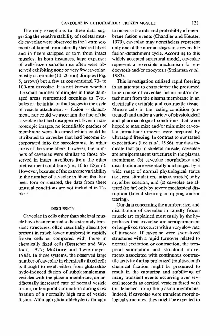
FIG. 5. Sarcolemmal P-face of a rapidly frozen, mechanically damaged EDL myofiber (1 -mm cut segments with laterally sheared ends). Note the presence of only a few minute (10-20 nm) caveolae. Bar = 0.1 Frn/ 100 nm.
These values were consistent within each of the four samples from each condition, as well as between all fibers from all experi- mental conditions (except for torn fibers).
Caveolae were found to be distributed randomly under almost all conditions.
-1
I I 2
t 4 3
I I 4
t q I 5
- 6a
t I 6b
I I I I 0 50 100 150
Mean Diameter of Caveolae (nm) q = mean; I------I = f 1 standard deviation
CHART 7. Direct comparison of means and stan- dard deviations from Charts 1-6. Traces numbered 6a and 6b are from Chart 6. This side-by-side comparison reveals graphically that there is no statistically signif- icant difference between the diameters of caveolae in muscle rapidly frozen after each of these pretreatment regimens.
(Representative micrographs from only three of the six conditions are shown be- cause the results were nearly identical for all conditions, with the exception of ripped and/or torn myofibers; see below.) On rare occasions small areas of sarcolemma were observed where caveolae were present in bands. However, the appearance of banding was neither definite, distinctive, nor con- sistent. This is in contrast to that seen in immersion-fixed myofibers (Lee et al., 1986; Bonilla et al., 1979; Shotton, 1982).
Charts l-6 graphically illustrate the num- ber of caveolae present at each diameter in the 100 pm2 of sarcolemma analyzed for each condition. Similar caveolae size ranges of 1 O-l 90 nm were observed in each group. Note that the curves in each graph are sim- ilar, indicating that caveolar diameter and number of caveolae present at each diam- eter are consistent under all normal phys- iological conditions and under several non- physiological conditions. A comparison of mean diameters (x> and standard devia- tions (SD) in Chart 7 graphically reveals that there were no statistically significant differ- ences in these parameters following any of the pretreatment conditions (except tear- ing).
CAVEOLAE IN ULTRARAPIDLY FROZEN MUSCLE 121
The only exceptions to these data sug- gesting the relative stability of skeletal mus- cle caveolae were observed in the 1 -mm seg- ments obtained from laterally sheared fibers and in fibers stripped or torn from intact muscles. In both instances, large expanses of well-frozen sarcolemma often were ob- served exhibiting none or very few caveolae, mostly as minute (1 O-20 nm) dimples (Fig. 5, arrows) but a few as conventional 70- to 100~nm caveolae. It is not known whether the small number of dimples in these dam- aged areas represented openings to T-tu- bules or the initial or final stages in the cycle of vesicle attachment * fusion t-1 detach- ment, nor could we ascertain the fate of the caveolae that had disappeared. Even in ste- reoscopic images, no identifiable patches of membrane were discerned which could be attributed to caveolae that had become in- corporated into the sarcolemma. In other areas of the same fibers, however, the num- bers of caveolae were similar to those ob- served in intact myofibers from the other pretreatment conditions (i.e., 10 to 1 2/pm2). However, because of the extreme variability in the number of caveolae in fibers that had been torn or sheared, the data from these unusual conditions are not included in Ta- ble 1.
DISCUSSION
Caveolae in cells other than skeletal mus- cle have been reported to be extremely tran- sient structures, often essentially absent (or present in much lower numbers) in rapidly frozen cells as compared with those in chemically fixed cells (Bretscher and Wy- tack, 1977; McGuire and Twietmeyer, 1983). In those systems, the observed large number of caveolae in chemically fixed cells is thought to result either from glutaralde- hyde-induced fusion of subplasmalemmal vesicles with the plasma membrane, an ar- tifactually increased rate of normal vesicle fusion, or temporal summation during slow fixation of a normally high rate of vesicle fusion. Although glutaraldehyde is thought
to increase the rate and probability of mem- brane fusion events (Chandler and Heuser, 1979), caveolae may nonetheless represent only one of the normal stages in a reversible fusion-detachment cycle. According to this widely accepted structural model, caveolae represent a reversible mechanism for en- docytosis and/or exocytosis (Steinman et al., 1983).
This investigation utilized rapid freezing in an attempt to characterize the presumed time course of caveolae fusion and/or de- tachment from the plasma membrane in an electrically excitable and contractile tissue. Muscle cells in the resting condition (un- treated) and under a variety of physiological and pharmacological conditions that were hoped to maximize and/or minimize caveo- lae formation/turnover were prepared by ultrarapid freezing. In contrast to our stated expectations (Lee et al., 1986) our data in- dicate that (a) in skeletal muscle, caveolae are semipermanent structures in the plasma membrane, (b) caveolae morphology and distribution are essentially unchanged by a wide range of normal physiological states (i.e., rest, stimulation, fatigue, stretch) or by myofiber scission, and (c) caveolae are al- tered (so far) only by severe mechanical dis- ruption (lateral shearing or ripping and/or tearing).
Our data concerning the number, size, and distribution of caveolae in rapidly frozen muscle are explained most easily by the hy- pothesis that caveolae are semipermanent or long-lived structures with a very slow rate of turnover. If caveolae were short-lived structures with a rapid turnover related to normal excitation or contraction, the tem- poral summation and structural move- ments associated with continuous contrac- tile activity during prolonged (multisecond) chemical fixation might be presumed to result in the capturing and stabilizing of many transient events occurring over sev- eral seconds as cortical vesicles fused with (or detached from) the plasma membrane. Indeed, if caveolae were transient morpho- logical structures, they might be expected to

122 POULOS. RASH. AND ELMUND
vary according to the state of excitation, contraction, or degree of muscle stretching. According to that model, elimination of temporal summation by the use of the in- stantaneous preservation technique of rapid freezing would, therefore, be expected to re- sult in fewer sarcolemmal caveolae. More- over, the ratio of the different morphologies would then be presumed to reflect the rel- ative duration of each stage of the fusion- detachment cycle. A lower ratio of the num- ber of caveolae found in rapidly frozen my- ofibers to the number found in chemically fixed myofibers would indicate that caveo- lae were short-lived structures. However, our findings from rapidly frozen muscle un- der a variety of conditions demonstrate that this ratio is near unity (i.e., the number of caveolae in rapidly frozen myofibers is vir- tually identical to the number in chemically fixed myofibers). Thus, these data indicate that caveolae are actually relatively long- lived structures with slow (or no) turnover rates. Under all tested conditions (except in torn myofibers, and at the ends of fibers damaged by lateral shear), we found cave- olae in a variety of sizes ranging from 10 to 190 nm (2 = 60 to 70 nm), suggesting that these long-lived structures either (a) cycle very slowly (i.e., many tens of seconds) or(b) are semipermanent structures that are normally present in variable sizes. There- fore, future studies of caveolae function should address other mechanisms of cave- olae formation and turnover (i.e., nutri- tional state, hydration, myofiber growth, and aging).
These same findings indicate that the dis- tinctive and reproducible variabilities in caveolae morphology and distribution re- ported in sarcolemmas fixed by several dif- ferent chemical fixation protocols result from artifacts induced by an as yet unquan- tified aspect(s) of fixation chemistry. One important variable was shown to be the mode of exposure to the chemical fixative. Significant variability in caveolae was re- ported (Lee et al., 1986) based on how rap- idly a myofiber was exposed to the fixative
(immersion vs perfusion). In central fibers of immersion-fixed muscle, the caveolae were of much greater diameter than those in rapidly frozen samples. On the other hand, the consistently small diameter caveolae seen in perfusion-fixed muscle (which we presumed to be the most rapid means of exposing myofibers to chemical fixatives) were not observed in rapidly frozen muscle. Rather, the diameters in caveolae in rapidly frozen muscle were significantly larger than those in perfusion-fixed myofibers, and most nearly resembled those in surface fibers of immersion-fixed muscle. (This may mean that surface fibers are exposed to high con- centrations of glutaraldehyde more quickly than are the fibers in perfusion-fixed mus- cle, where diffusion from the restricted vol- ume of the capillary bed may delay fixation for several additional seconds and expose the fiber to slowly increasing concentration of fixative.) There also appear to be other aspects of fixation chemistry influencing these reported changes (i.e., hypoxia, acid production, and/or loss of positive charges on membranes (Johnson, 1985, 1986). Moreover, the current and previous data (Lee et al., 1986) raise additional concerns (Chandler and Heuser, 1979) as to the ex- tent different chemical fixation methods in- duce artifactual changes in the morphology of other (membranous and cytoplasmic) structures.
Our findings also provide evidence that contradict and, at the same time, explain our preliminary observations that rapid freezing of rapidly dissected myofibers sig- nificantly reduced the size and number of sarcolemmal caveolae (Lee et al., 1983). The results of the mechanical trauma experi- ments indicate that the following factors led to those initial observations. In that prelim- inary study, minute muscle fragments were quickly dissected out (ripped and/or torn from whole muscles) and placed into the small freezing cavities of the propane jet device and rapidly frozen within 30 sec. For comparison, equally small pieces were quickly dissected out and rapidly frozen with

CAVEOLAE IN ULTRARAPIDLY FROZEN MUSCLE 123
the Polaron “Slammer” freezing device. By duplicating those disruptive tissue sampling methods in one portion of this study, as well as by examining l-mm muscle segments with laterally sheared ends, we determined that the methods used in the original sam- pling procedures resulted in the damaging of some surface fibers, thereby creating patches of sarcolemma that contained fewer and smaller caveolae. Thus, it appears that only the sarcolemmas of the most severely damaged myofibers were observed in that earlier study. Similar artifactual alterations of caveolae are considered likely using qual- itatively similar “punch biopsy” and “needle biopsy” methods.
In another of our previous reports (Lee et al., 1986), we suggested rapid freezing as an alternative method ofcaveolae preservation in an attempt to characterize human neu- romuscular diseases. Consequently, it be- came necessary to determine the morphol- ogy of caveolae in the natural (i.e., rapidly frozen) state and to identify preparative conditions that might alter caveolae mor- phology. Our findings in this investigation of rapidly frozen skeletal muscle indicate that several common physiological states (rest, stimulation, fatigue, etc.) have very little effect on caveolae morphology. More- over, since we showed previously that glu- taraldehyde fixation induces artifactual al- terations in caveolae size and distribution but does not affect caveolae number, and since caveolae are now shown to be essen- tially unchanged by a variety of other pre- fixation conditions, the current data pro- vide further support for the conclusion that the number of caveolae is consistently and characteristically increased in Duchenne muscular dystrophy (Bonilla et al., 1981; Shotton, 1982). Nevertheless, because of the substantial and unpredictable alterations of caveolae morphology that occur during rel- atively slower chemical fixation (Lee et al., 1986) the use of rapid freezing techniques may be required to determine whether the observed changes in caveolae morphology and distribution represent reliable patho-
logical alterations for characterizing neu- romuscular disease.
Finally, the findings of this study increase our concerns as to whether chemical fixa- tion, in fact, preserves the morphologic “reality” of cellular structures or causes misinterpretable artifact. Thus, it becomes increasingly important to determine the fac- tor(s) of chemical fixation that may be in- ducing alterations in the morphology of these and other cellular structures.
REFERENCES
BONILLA, E., FISHBECK, K., AND SCHOTLAND, D. L. (198 1) Amer. .I. Puthol. 104, 167-173.
BOYNE, A. F. (1979) J. Neurosci. Methods 1, 353-364. BRANTON, D., BULLIVANT, S., GILULA, N. B.,
KARNOVSKY, M. J., MOOR, H., MUHLETHALER, K., NORTHCOTE, D. H., PACKER, L., SATIR, B., SATIR, P., SPETH, V., STAEHELIN, L. A., STEERE, R. L., AND WEINSTEIN, R. S. (1975) Science 190, 54-56.
BRETSCHER, M. S., AND WHYTOCK, S. (1977) J. Ultra- struct. Res. 61, 215-217.
BUNDGAARD, M. (1983) Fed. Proc. 42, 2425-2430. CHANDLER, D. E., AND HEUSER, J. (1979) J. Cell Biol.
83, 91-108. COSTELLO, B. R., AND SHAFIQ, S. A. (1979) Muscle
Nerve 2, 191. DULHUNTY, A. F., AND FRANZINI-ARMSTRONG, C.
(1975) J. Ph.vsiol. 250, 5 13-539. FRANZINI-ARMSTRONG, C., LANDMESSER, L., AND PI-
LAR, G. (1975) J. Cell Biol. 64, 493-497. HAVER-LOCKHART (Drug Information Sheet), Bayvet
Division, Miles Laboratory, Shawnee, KS. HEUSER, J. E., REESE, T. S., DENIS, M. J., JAN, Y., JAN,
L., AND EVANS, L. (1979) J. Cell Biol. 81, 275-300. HUDSON, C. S., RASH, J. E., AND SHINOWARA, N. L.
(1981) Curr. Trends Morphol. Tech. 2, 183-217. JOHNSON, T. J. A. (1985) J. Electron Microsc. Tech. 2,
129-138. JOHNSON, T. J. A. (1986) in MULLER, M., BECKER, R.
P., BOYD, A., AND WOLOSEWICK, J. J. (Eds.), Science of Biological Specimen Preparation, pp. 5 l-62, SEM, Inc. AMF O’Hare, Chicago.
KOELLE, G. (1975) in GOODMAN, L. S., GILMAN, A., GILMAN, A. G., AND KOELLE, G. B. (Eds.), The Phar- macological Basis of Therapeutics (5th ed.), pp. 575- 588, Macmillan Co., New York.
LEE, R. E., POULOS, A. C., MAYER, R. F., AND RASH, J. E. (1986) MuscleNerve9, 127-137.
LEE, R. E., RASH, J. E., AND POULOS, A. C. (1983) J. Cell Biol. 97, 46 1 a.
LINDER, J. C., AND STAEHELIN, L. A. (1979) J. CellBiol. 83, 37 l-382.
MCGUIRE, P. G., AND TWIETMEYER, T. A. (1983) Circ. Res. 53, 424-429.

124 POULOS, RASH, AND ELMUND
ORNBERG, R. L., AND REESE, T. S. (1979) in RASH, J. E., AND HUDSON, C. S. (Eds.), Freeze Fracture: Meth- ods, Artifacts, and Interpretations, pp. 89-97, Raven Press, New York.
PRESCOTT, L., AND BRIGHTMANN, N. W. (1976) Tissue Cell 8, 241-258.
PRICE, H. L. (1975) in GOODMAN, L. S., GILMAN, A., GII.MAN, A. G., AND KOELLE, G. B. (Eds.), The Phar- macological Basis of Therapeutics (5th ed.), pp. 97- 10 1, Macmillan Co., New York.
R-ZSH, J. E., HUDSON, C. S., GRAHAM, W. F., MAYER,
R. F., WARNICK, J. E., AND ALBUQUERQUE, E. X. (198 1) Lab. Invest. 44, 5 19-530.
ROBINSON, J. M., HOOVER, R. L., AND KARNOVSKY, M. J. (1984) J. Cell Biol. 99, 287a.
SHOTTON, D. M. (1982) J. Neurol. Sci. 57, 161-190. STEERE, R. L., AND ERBE, E. F. (1983) in BAILEY, G.
W. (Ed.), Proceedings of the 4 1 st Annual Meeting of the Electron Microscopy Society ofAmerica, pp. 6 1 S- 619, San Francisco, CA.
STEINMAN, R. M., MELLMAN, I. S., MIILLER, W. A., AND COHN, Z. A. (1983) J. Cell Biol. 96, l-27.
Recommended


