Embed Size (px)
Citation preview

ANTIGEN PROCESSINGand
PRESENTATION OF CELLS
Alric V. Mondragon, MDSection of Allergy and Immunology
University of the Philippines – Philippine General Hospital

OutlineI. Properties of Antigens Recognized by T
LymphocytesII. Antigen Capture and the Functions of Antigen-
Presenting CellsIII. Processing of Protein AntigensIV. Presentation of Non-protein Antigens to Subsets
of T Cells

PROPERTIES OF ANTIGENS RECOGNIZED BY T LYMPHOCYTES

T Lymphocytes• Principal functions of T lymphocytes
a. to eradicate infections by intracellular microbes b. to activate other cells, such as macrophages and
B lymphocytes.
Cellular and Molecular Immunology 8th Ed. (2015) by Abbas et al.

T Lymphocytes• Several challenges to T cells:
1. Very few naive T cells specific for any one antigen• APCs
2. Most T cell functions require that they interact with other cells. • MHC
3. Different T cells have to be able to respond to microbial antigens in different cellular compartments.
Cellular and Molecular Immunology 8th Ed. (2015) by Abbas et al.

PROPERTIES OF ANTIGENS RECOGNIZED BY T LYMPHOCYTES
1. Most T Lymphocytes recognize only short peptides– Induced by foreign protein antigens or small chemical
substances
2. Antigen Receptors of CD4+ and CD8+ T cells are specific for peptide antigens displayed by MHC molecules– TCRs have evolved to be specific for MHC molecules– Majority of T cells recognize only peptides
Cellular and Molecular Immunology 8th Ed. (2015) by Abbas et al.

ANTIGEN CAPTURE AND THE FUNCTIONS OF ANTIGEN-PRESENTING CELLS

ANTIGEN CAPTURE AND THE FUNCTIONS OF ANTIGEN-PRESENTING CELLS
Cellular and Molecular Immunology 8th Ed. (2015) by Abbas et al.

ANTIGEN CAPTURE AND THE FUNCTIONS OF ANTIGEN-PRESENTING CELLS
• APC function is enhanced by exposure to microbial products– Toll-like receptors and other microbial sensors in dendritic cells
and macrophages– Improved antigen presentation efficiency and APC cytokine
production Increase expression of MHC and costimulators– Adjuvants: products of microbes or mimic microbes
• Enhance expression of costimulators and cytokines• Enhance functions of APC’s
Cellular and Molecular Immunology 8th Ed. (2015) by Abbas et al.

ANTIGEN CAPTURE AND THE FUNCTIONS OF ANTIGEN-PRESENTING CELLS
• APCs that present antigen to T cells also receive signals from these Lymphocytes, enhancing their antigen-presenting function– Activated CD4+ express CD40L --- CD40 on dendritic
cells and macrophages IFN-γ secretion, activates APC’s• Leads to increased ability to process and present antigens, • Increased expression of costimulators • Secretion of cytokines
Cellular and Molecular Immunology 8th Ed. (2015) by Abbas et al.

Role of Dendritic Cells in Antigen Capture and Display
Cellular and Molecular Immunology 8th Ed. (2015) by Abbas et al.

Role of Dendritic Cells in Antigen Capture and Display
Cellular and Molecular Immunology 8th Ed. (2015) by Abbas et al.

Role of Dendritic Cells in Antigen Capture and Display
Cellular and Molecular Immunology 8th Ed. (2015) by Abbas et al.

2 Sets of Dendritic CellsClassical DC • Most numerous subset of dendritic cells in the lymphoid
organs• Mostly derived from myeloid precursors
• Constantly sample the environment• May also present self antigens for regulation/self-tolerance.• Upon encountering microbes/cytokines:
• Upregulate costimulatory molecules• Produce inflammatory cytokines• Migrate from peripheral tissue to draining lymph node
Cellular and Molecular Immunology 8th Ed. (2015) by Abbas et al.

2 Sets of Dendritic CellsClassical DC • 2 subsets:
1. High expression of BDCA-1/CD1c – most potent at driving CD4+ responses
2. Expression of BDCA-3 – efficient in process of cross-presentation
Cellular and Molecular Immunology 8th Ed. (2015) by Abbas et al.

2 Sets of Dendritic CellsPlasmacytoidDC
• Resemble plasma cells• Develop in Bone Marrow from same precursor as Classical
DC.• Found in blood and in small numbers in lymphoid organs
• Poorly phagocytic and do NOT sample environmental antigens
• Major function: Secretion of Type I IFN in response to viral infections
• May also differentiate into cells similar to Classical DC and present antigen to Virus-specific T-cells
Cellular and Molecular Immunology 8th Ed. (2015) by Abbas et al.

Antigen Capture and Transport
Cellular and Molecular Immunology 8th Ed. (2015) by Abbas et al.
Membrane Receptors(C-type lectins)
Capture and Endocytose microbes or microbial
products
Process ingested proteins into peptides capable of
binding to MHC

Antigen Capture and Transport
Cellular and Molecular Immunology 8th Ed. (2015) by Abbas et al.
Membrane Receptors(C-type lectins)
Capture and Endocytose microbes or microbial
products
Process ingested proteins into peptides capable of
binding to MHC
Microbial products recognized by TLR
Signals and Cytokines activate DC
(TNF)
Activated DC lose adhesiveness and migrate
to lymph nodes
Antigen Capture and Transport
Cellular and Molecular Immunology 8th Ed. (2015) by Abbas et al.
DC
CCR7 Lymphatic VesselsT cell zones of Lymph Nodes
CCL 19
CCL 21
NaïveT cell
CCR7
“Colocalization”

Antigen Capture and Transport
Cellular and Molecular Immunology 8th Ed. (2015) by Abbas et al.
DC Capture Antigen
Present Antigen to Naïve T cells
Activate Lymphocytes
Express high levels of MHC
Activated DC developInto potent APCs

Cellular and Molecular Immunology 8th Ed. (2015) by Abbas et al.

Antigen Capture and Transport
• Antigens can be transported to lymphoid organs in soluble form
• Afferent Lymphatic Vessel Subcapsular sinus FRC conduits Cortex
• Antigen can be extracted at the conduits, some in the sinuses
Cellular and Molecular Immunology 8th Ed. (2015) by Abbas et al.

Antigen Capture and Transport
Properties that make DC the most efficient APCs for initiating T cell responses1. Strategically located at common sites of entry2. Express receptors that enable capture and response3. Migrate from epithelia and tissues via lymphatics to T cell
zones of LN4. Mature DC express high levels of peptide-MHC complexes,
costimulators, and cytokines
Cellular and Molecular Immunology 8th Ed. (2015) by Abbas et al.

Antigen Capture and Transport
• Dendritic cells can also ingest infected cells and present antigens to CD8+ T lymphocytes– Peptide antigens must be derived from proteins in
the cytosol of DC– Specialized DC: able to ingest virus-infected cells
and deliver viral proteins into their cytosol– “Cross-presentation or Cross-priming”
Cellular and Molecular Immunology 8th Ed. (2015) by Abbas et al.

Function of Other APCs
Cell-mediated Immune Responses Macrophages present Ag of phagocytosed microbes to effector T cells
Humoral Immune Responses B lymphocytes internalize protein Ag and present peptides from these proteins to helper T cells.
Nucleated cells Can present peptides, derived from cytosolic protein antigens CD8+ CTLs
Other cell types that express MHC class II (endothelial and some epithelial cells)
May present Ag to T cells
Cellular and Molecular Immunology 8th Ed. (2015) by Abbas et al.

PROCESSING OF PROTEIN ANTIGENS

PROCESSING OF PROTEIN ANTIGENS
Cellular and Molecular Immunology 8th Ed. (2015) by Abbas et al.

PROCESSING OF PROTEIN ANTIGENS
Cellular and Molecular Immunology 8th Ed. (2015) by Abbas et al.

PROCESSING OF PROTEIN ANTIGENS
Cellular and Molecular Immunology 8th Ed. (2015) by Abbas et al.

Class I MHC Pathway
Cellular and Molecular Immunology 8th Ed. (2015) by Abbas et al.
Injected via Bacterial secretory mechanisms
Phagocytosed
Escape Mechanism

Class I MHC Pathway
Cellular and Molecular Immunology 8th Ed. (2015) by Abbas et al.

Class I MHC Pathway
Cellular and Molecular Immunology 8th Ed. (2015) by Abbas et al.

Class I MHC Pathway
Cellular and Molecular Immunology 8th Ed. (2015) by Abbas et al.

Class I MHC Pathway
Cellular and Molecular Immunology 8th Ed. (2015) by Abbas et al.
Membrane Chaperone:Calnexin Luminal Chaperone:
Calreticulin

Class I MHC Pathway
Cellular and Molecular Immunology 8th Ed. (2015) by Abbas et al.

Class I MHC Pathway
Cellular and Molecular Immunology 8th Ed. (2015) by Abbas et al.
• Peptides transported into ER preferentially bind to Class I MHC but NOT Class II MHC:1. Class I attached to TAP complex2. Class II molecules are blocked by a protein called
the invariant chain

Class I MHC Pathway
Cellular and Molecular Immunology 8th Ed. (2015) by Abbas et al.

Class I MHC Pathway
Cellular and Molecular Immunology 8th Ed. (2015) by Abbas et al.

MHC PROCESSING
Cellular and Molecular Immunology 8th Ed. (2015) by Abbas et al.

Class II MHC Pathway
Cellular and Molecular Immunology 8th Ed. (2015) by Abbas et al.
Endosome-Lysosome
Phagolysosomes
Autophagosomes

Class II MHC Pathway
Cellular and Molecular Immunology 8th Ed. (2015) by Abbas et al.
CATHEPSINS

Class II MHC Pathway
Cellular and Molecular Immunology 8th Ed. (2015) by Abbas et al.

Class II MHC Pathway
Cellular and Molecular Immunology 8th Ed. (2015) by Abbas et al.
Membrane Chaperone:Calnexin

Class II MHC Pathway
Cellular and Molecular Immunology 8th Ed. (2015) by Abbas et al.

Class II MHC Pathway
Cellular and Molecular Immunology 8th Ed. (2015) by Abbas et al.
STEP 1:Cathepsins degrade
Invariant Chain
CLIP STEP 2:HLA-DM removes CLIP

Class II MHC Pathway
Cellular and Molecular Immunology 8th Ed. (2015) by Abbas et al.

Class II MHC Pathway
Cellular and Molecular Immunology 8th Ed. (2015) by Abbas et al.

MHC PROCESSING
Cellular and Molecular Immunology 8th Ed. (2015) by Abbas et al.

Cross-Presentation
Cellular and Molecular Immunology 8th Ed. (2015) by Abbas et al.

Physiologic Significance of MHC-associated Antigen Presentation
Cellular and Molecular Immunology 8th Ed. (2015) by Abbas et al.

Physiologic Significance of MHC-associated Antigen Presentation
Cellular and Molecular Immunology 8th Ed. (2015) by Abbas et al.
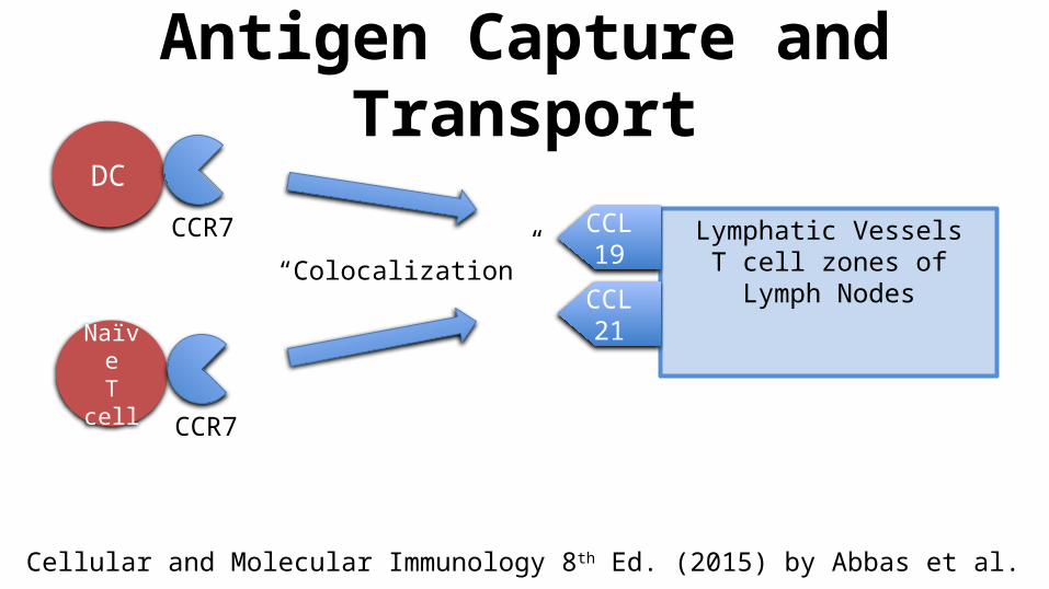
PRESENTATION OF NON-PROTEIN ANTIGENS TO SUBSETS OF T CELLS

Presentation of Non-Protein Antigens• Small populations of T cells can recognize non-
protein antigens without Class I or II MHC: NKT cells and γδ T cells.– NKT: recognize lipids and glycolipids displayed by
CD1– γδ T cells: recognize proteins, lipids,
phosphorylated molecules and alkyl aminesCellular and Molecular Immunology 8th Ed. (2015) by Abbas et al.

OutlineI. Properties of Antigens Recognized by T
LymphocytesII. Antigen Capture and the Functions of Antigen-
Presenting CellsIII. Processing of Protein AntigensIV. Presentation of Non-protein Antigens to Subsets
of T Cells

Summary1. Most T cells recognize antigens only as peptides displayed
by the products of self MHC genes on the surface of APCs.2. MHC is a large genetic region coding for highly
polymorphic, co-dominantly expressed class I and class II MHC molecules
3. The expression of MHC gene products is enhanced by inflammatory and immune stimuli, particularly cytokines like IFN-γ, which stimulate the transcription of MHC genes.
Cellular and Molecular Immunology 8th Ed. (2015) by Abbas et al.

SummaryMHC I MHC II
Composed of an α (or heavy) chain in a non-covalent complex with a β2- microglobulin
Contain two MHC-encoded polymorphic chains, an α chain and a β chain.
Recognized by CD8+ T cells Recognized by CD4+ T cells
Accommodate peptides that are 6 to 16 amino acid residues in length
Allows larger peptides (up to 30 amino acid residues in length or more) to bind
Expressed on all nucleated cells Expressed mainly on specialized APCs
Cytosolic proteins are proteolytically degraded in the proteasome
Extracellular proteins are internalized into endosomes
Cellular and Molecular Immunology 8th Ed. (2015) by Abbas et al.

THANK YOU

References• Cellular and Molecular
Immunology 8th Ed. (2015) by Abbas et al.– Chapter 6: MHC Molecules
and Antigen Presentation to T Lymphocytes