Embed Size (px)
Citation preview
Breast MRI:Updates to BI-RADS Lexicon
T Pousse
JFIM 2013 HONG KONG
Aims
• Recall and illustrate new synthetic items of BI-RADS MRI
• Insist on changes to the BI-RADS MRI lexicon in this third edition
Technical parameters
• Dedicated breast coil, pulse sequence, (T2/T2 fat sat), IVC
• Diffusion weighted or spectroscopy optional
• T2 weigthed recommanded before IVC

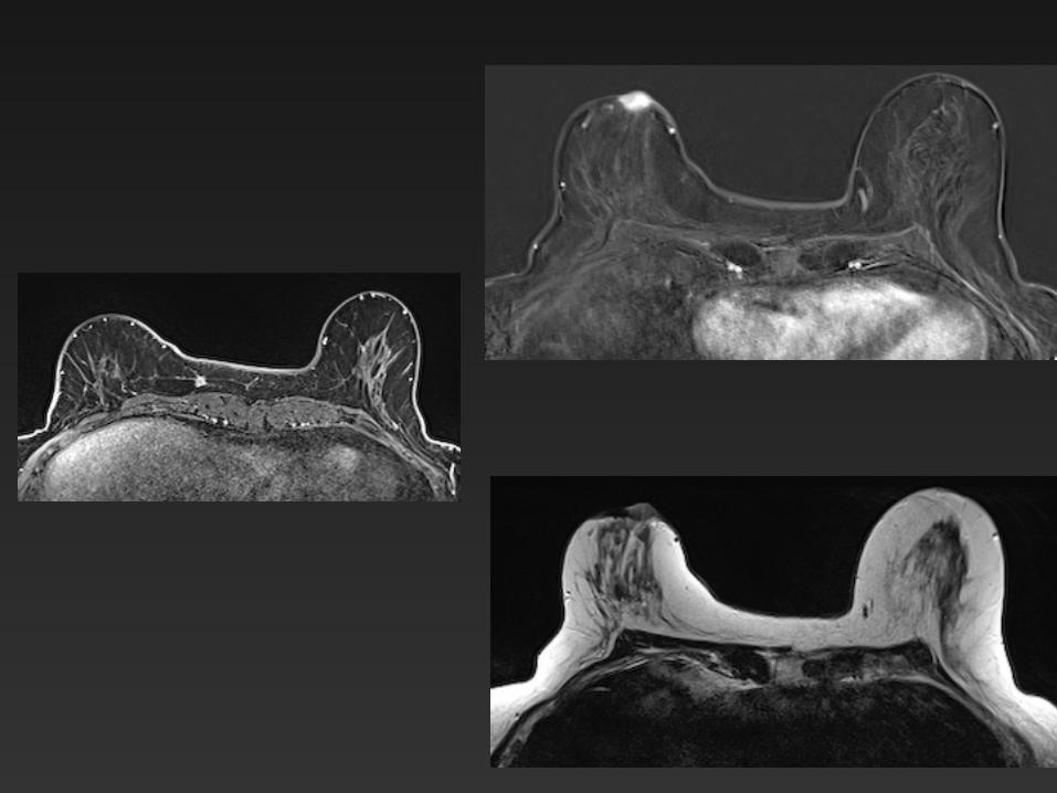
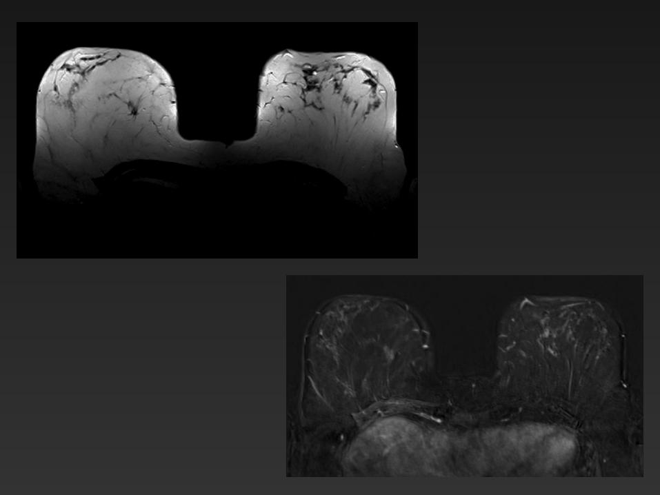
Background Parenchymal Enhancement ( BPE)
• 5 categories - none - minimal - mild - moderate - marked
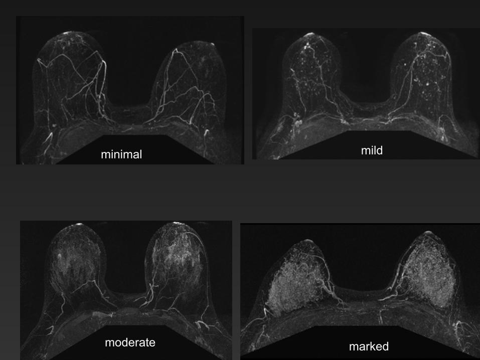
minimal mild
moderate marked
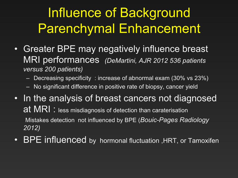
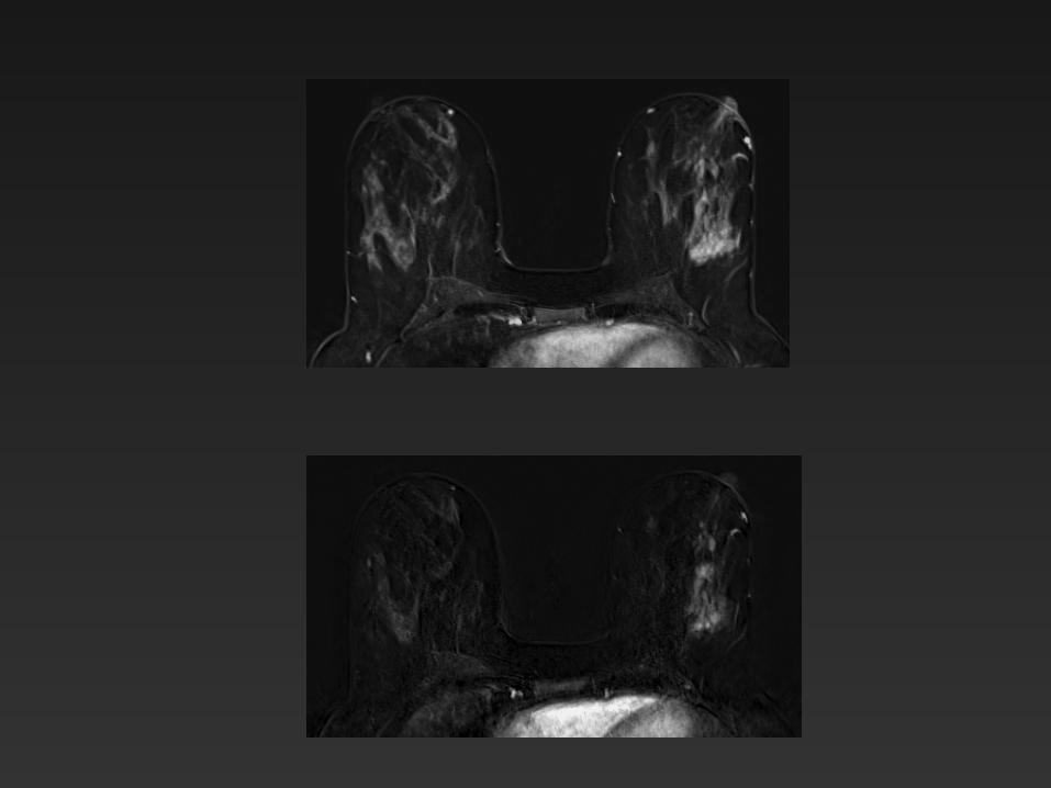
Influence of Background Parenchymal Enhancement
• Greater BPE may negatively influence breast MRI performances (DeMartini, AJR 2012 536 patients versus 200 patients) – Decreasing specificity : increase of abnormal exam (30% vs 23%) – No significant difference in positive rate of biopsy, cancer yield
• In the analysis of breast cancers not diagnosed at MRI : less misdiagnosis of detection than caraterisation
Mistakes detection not influenced by BPE (Bouic-Pages Radiology 2012)
• BPE influenced by hormonal fluctuation ,HRT, or Tamoxifen
Lesions characteristics
• Morphology – Masses – Non-masse enhancement
– Foci (≤ 5mm)
• Enhancements Kinetics
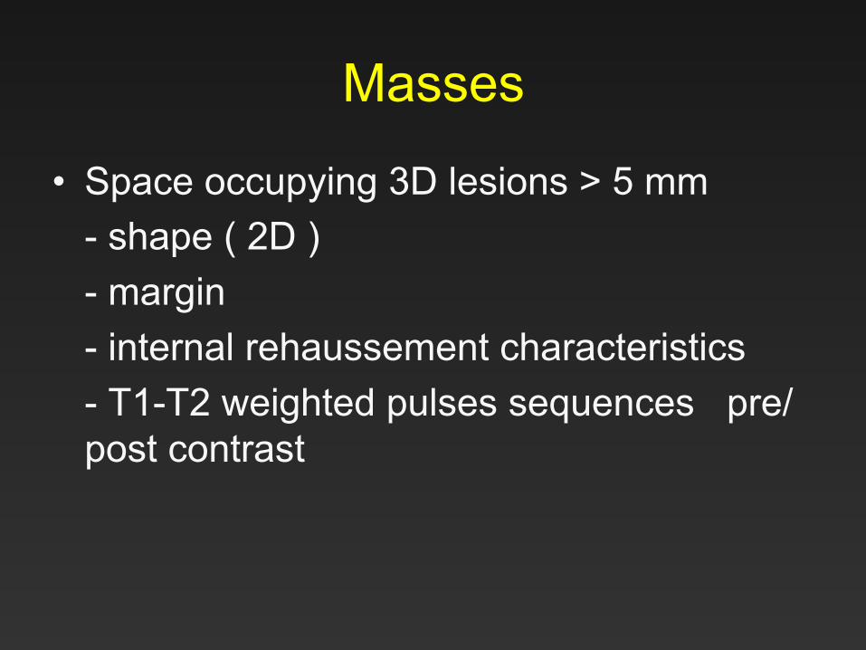
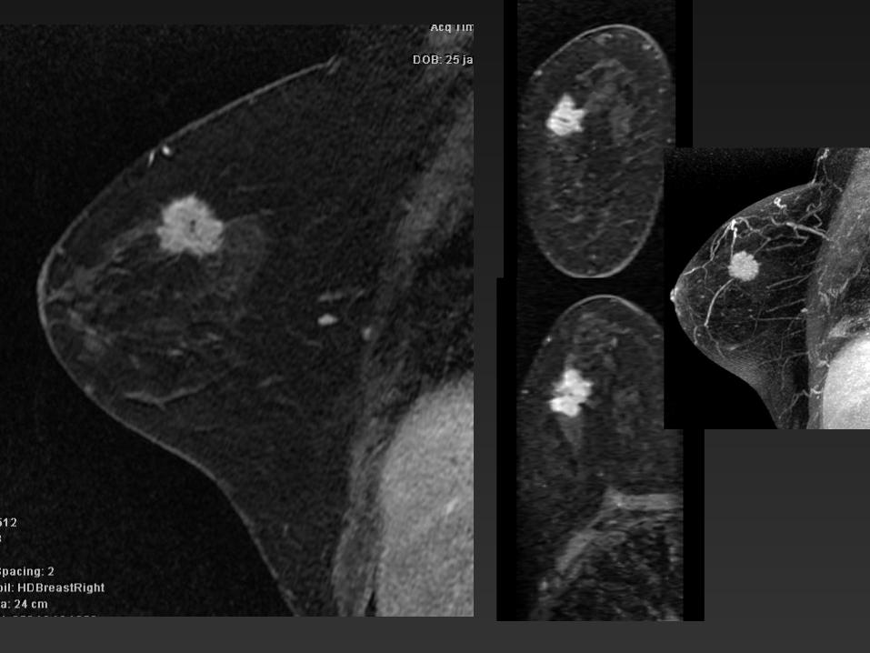
Masses
• Space occupying 3D lesions > 5 mm - shape ( 2D ) - margin - internal rehaussement characteristics - T1-T2 weighted pulses sequences pre/post contrast
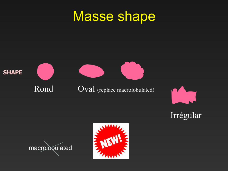
Masse shape
Rond Oval (replace macrolobulated)
Irrégular
SHAPE
macrolobulated
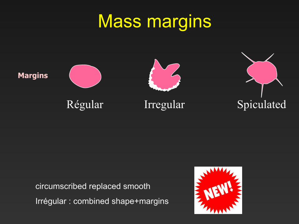
Mass margins
Régular Irregular Spiculated
Margins
circumscribed replaced smooth
Irrégular : combined shape+margins
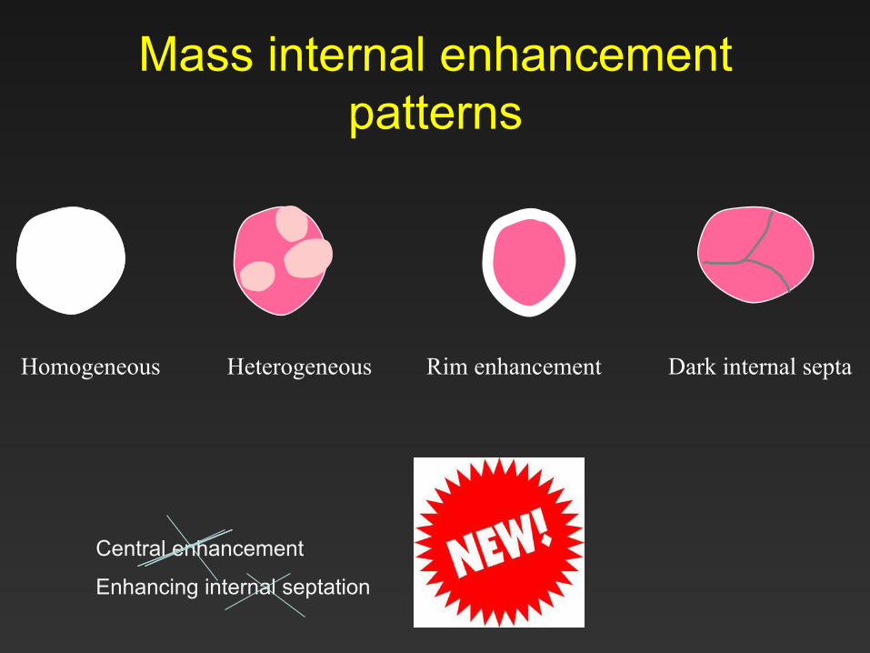
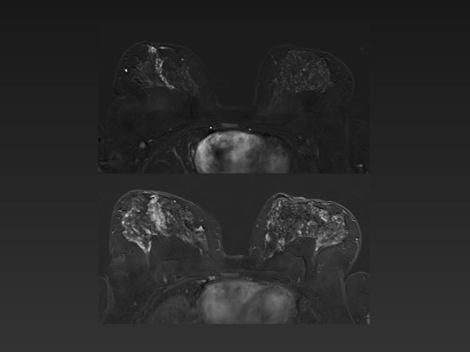
Mass internal enhancement patterns
Homogeneous Heterogeneous Rim enhancement Dark internal septa
Central enhancement
Enhancing internal septation
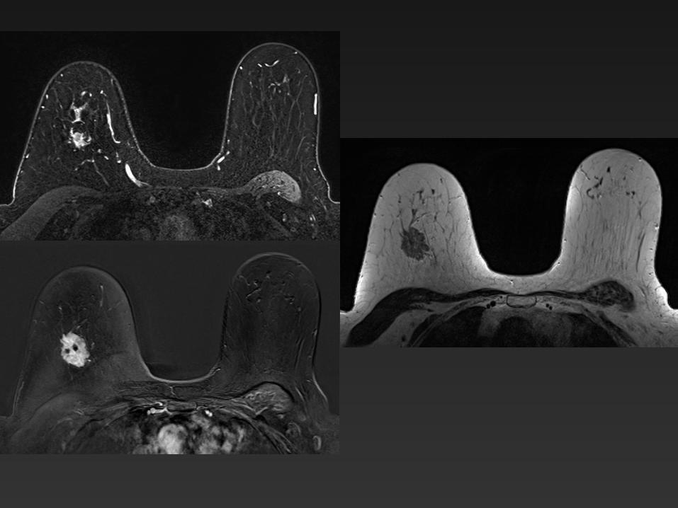
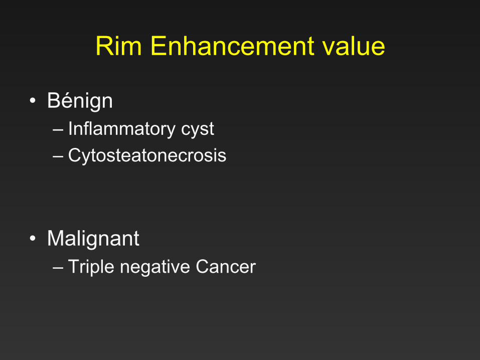
Rim Enhancement value
• Bénign – Inflammatory cyst – Cytosteatonecrosis
• Malignant
– Triple negative Cancer
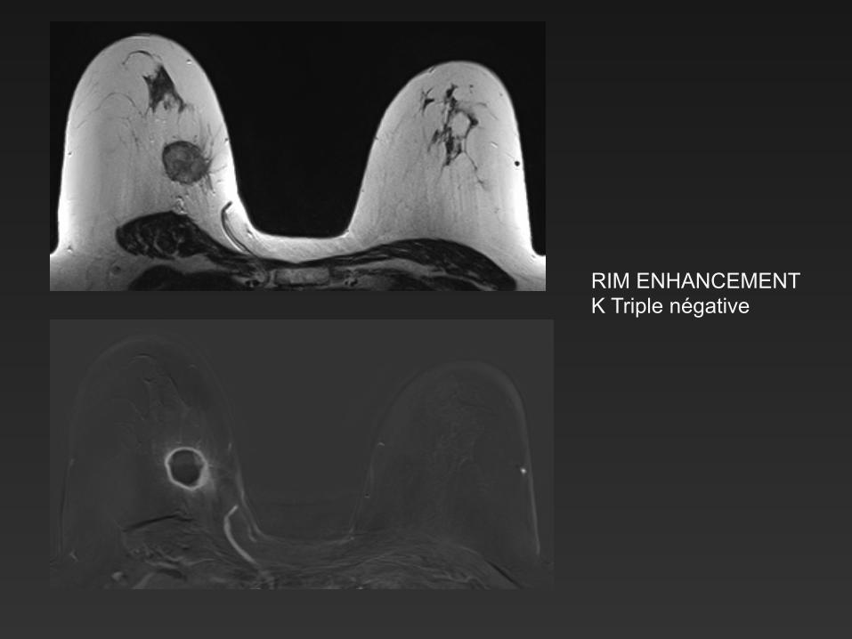
RIM ENHANCEMENT K Triple négative
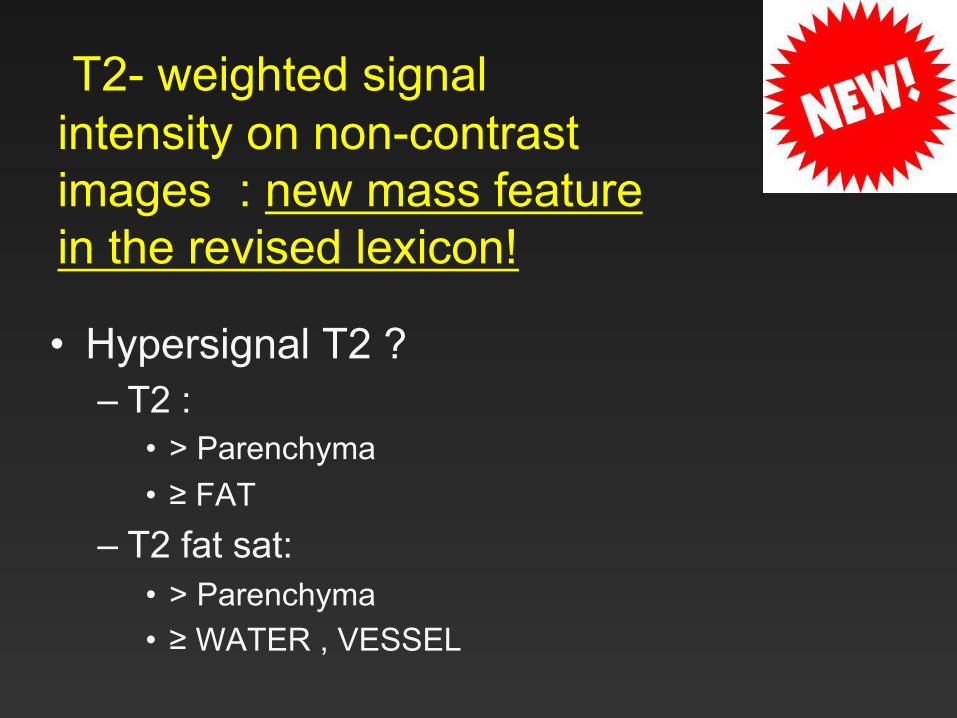
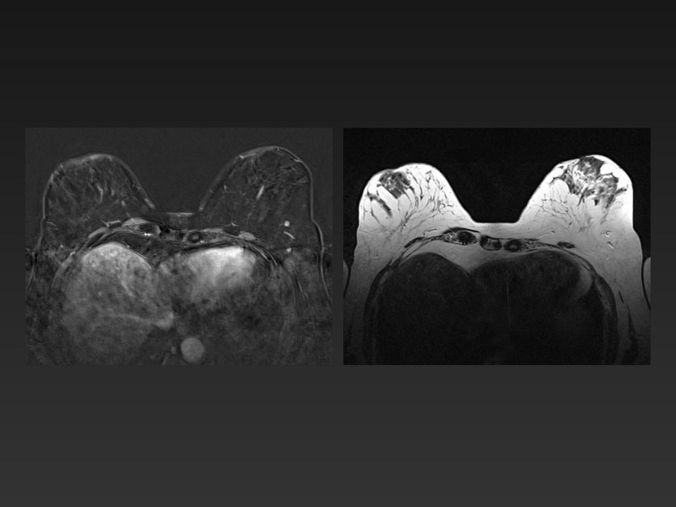
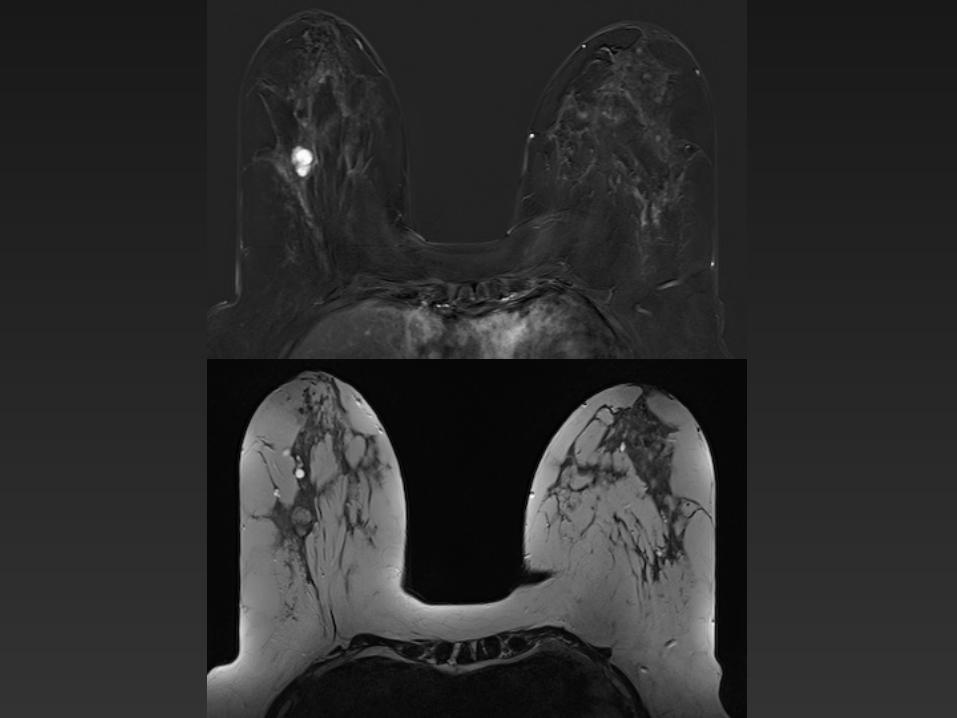
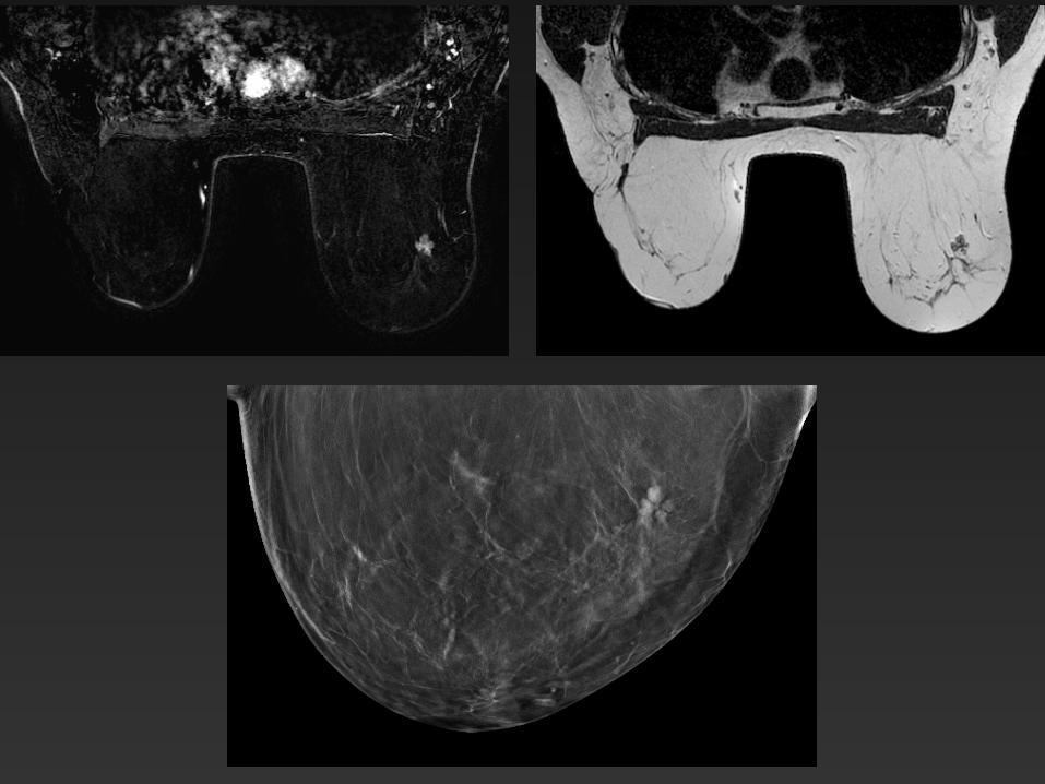
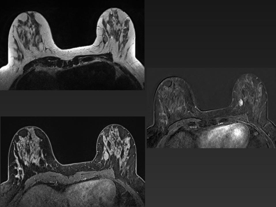
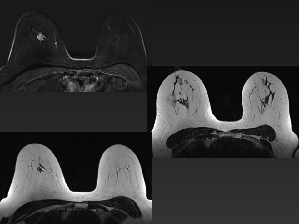
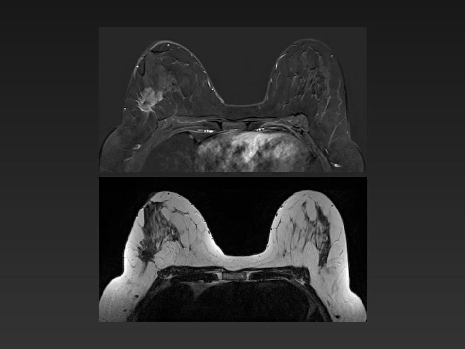
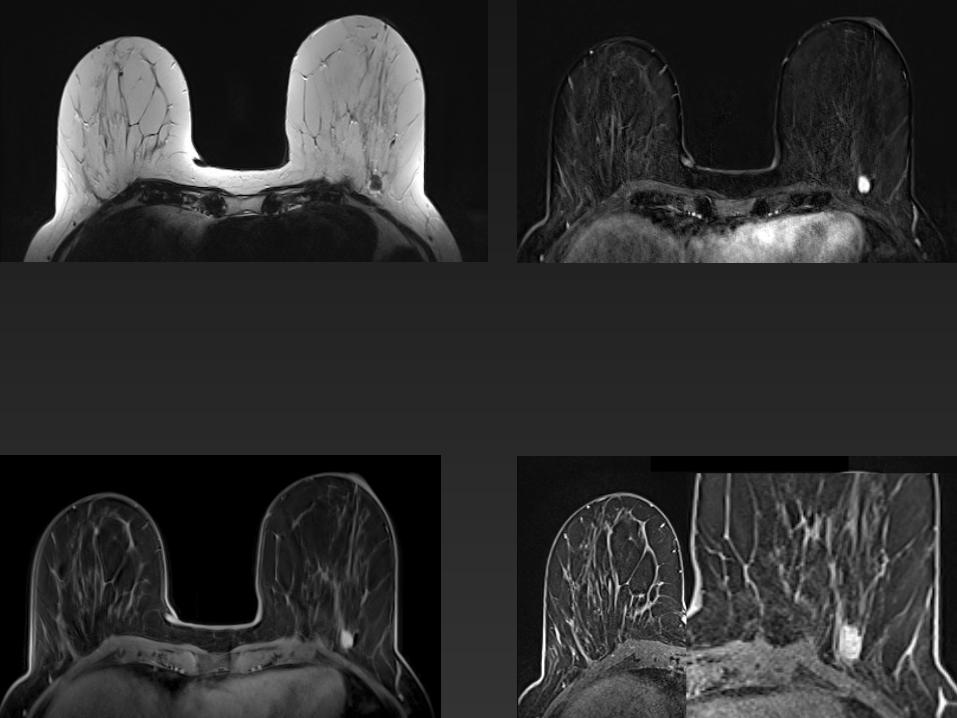
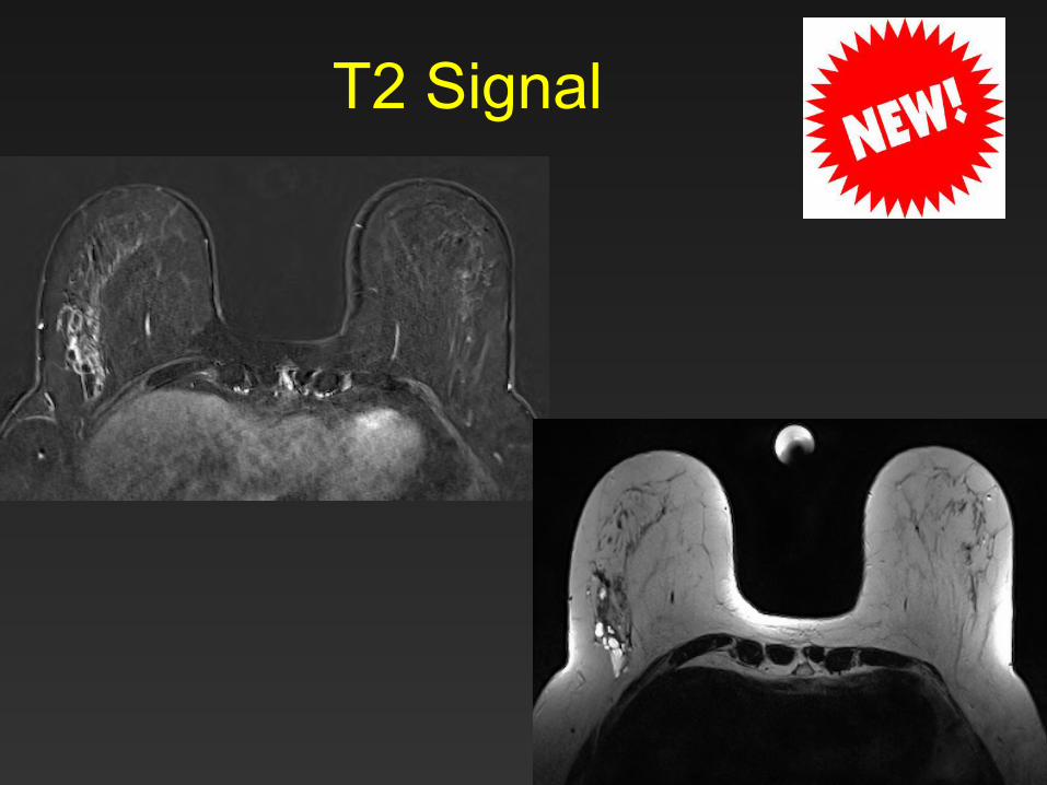
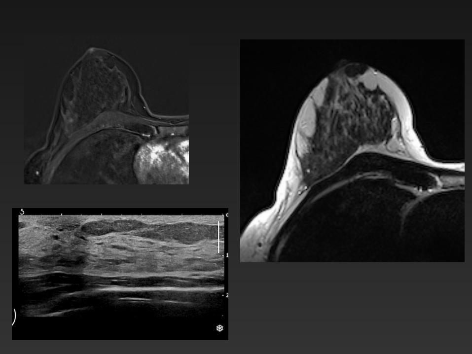
T2- weighted signal intensity on non-contrast images : new mass feature in the revised lexicon!
• Hypersignal T2 ? – T2 :
• > Parenchyma • ≥ FAT
– T2 fat sat: • > Parenchyma • ≥ WATER , VESSEL
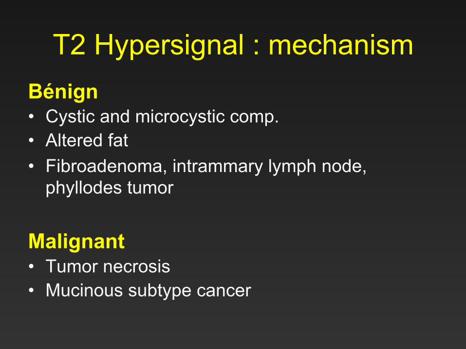
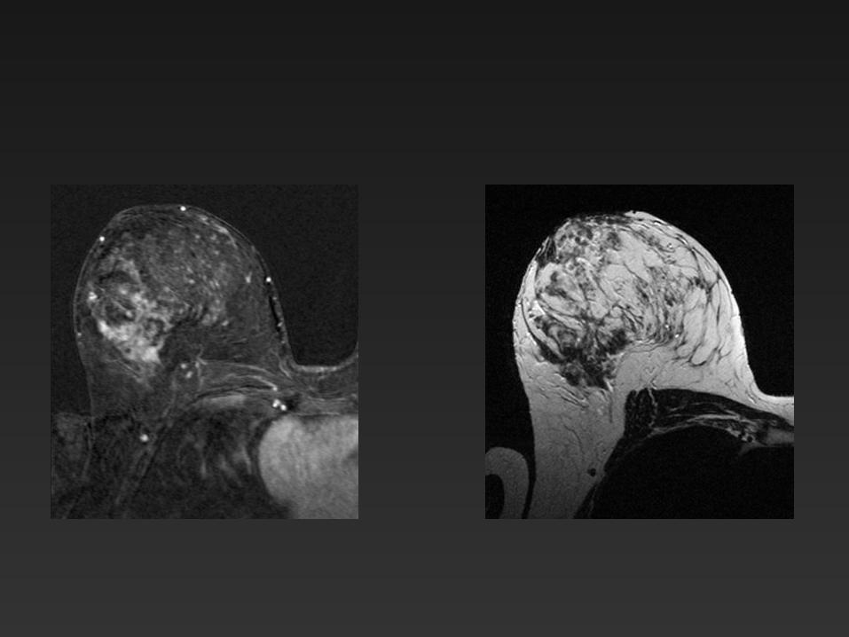
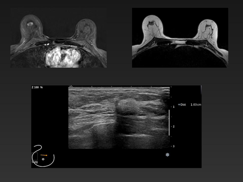
T2 Hypersignal : mechanism Bénign • Cystic and microcystic comp. • Altered fat • Fibroadenoma, intrammary lymph node,
phyllodes tumor Malignant • Tumor necrosis • Mucinous subtype cancer
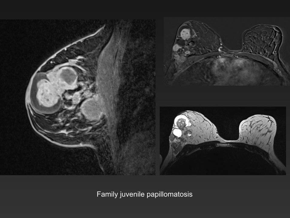
Family juvenile papillomatosis
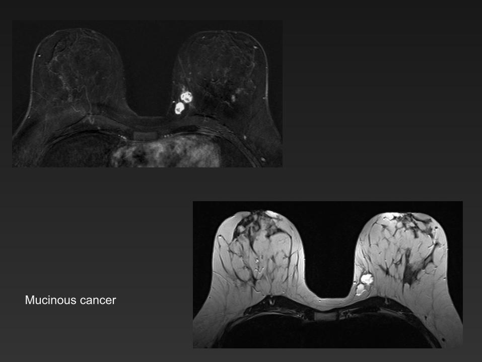
Mucinous cancer
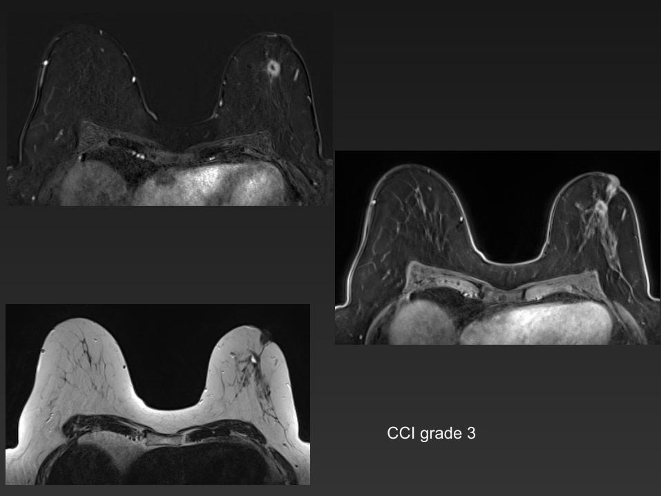
CCI grade 3
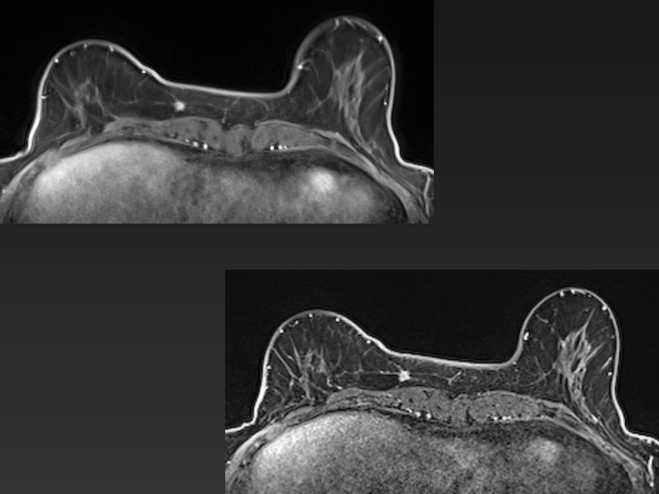
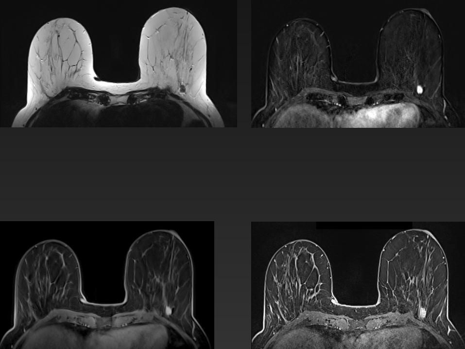
Non-Mass Enhancement
Enhancing area that is not a mass , separate from background parenchymal enhancement may contain interspersed fat
– Distribution (symetric or asymetric) – Internal enhancement characteristics – T2 weighted signal intensity
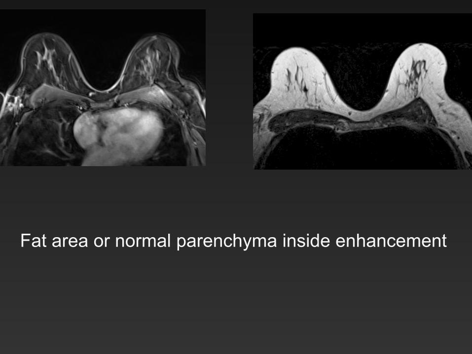
Fat area or normal parenchyma inside enhancement
NME distribution
• Focal area ( <25% Q) • Linear • Branching linear : replace ductal • Segmental • Regional • Multiple regions >2 • Diffuse • Symetric or not
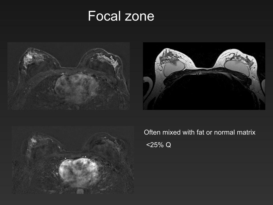
Focal zone
<25% Q
Often mixed with fat or normal matrix
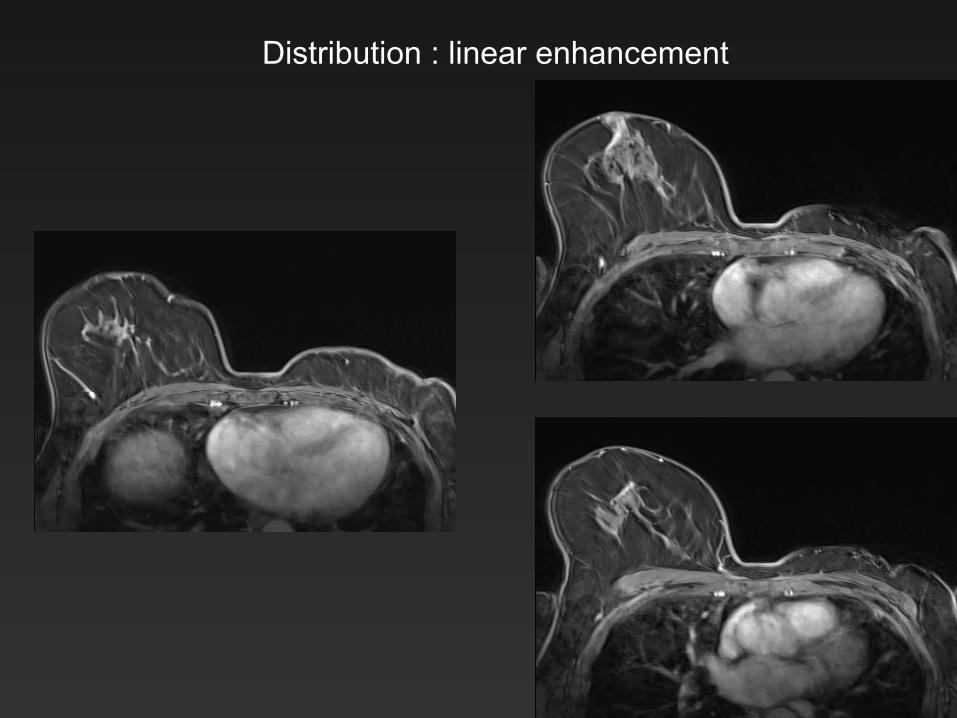
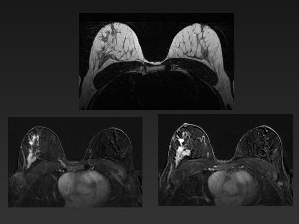
Distribution : linear enhancement
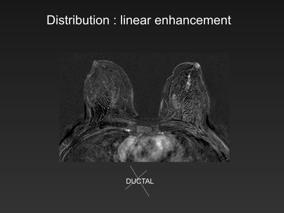
Distribution : linear enhancement
DUCTAL
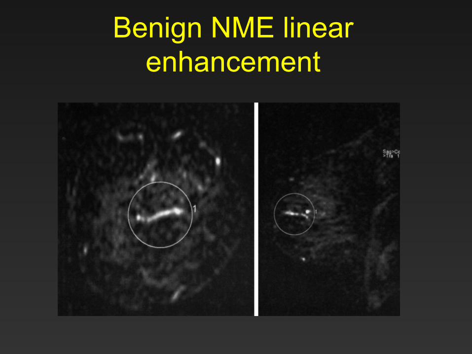
Benign NME linear enhancement
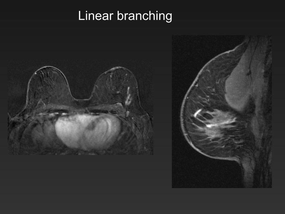
Linear branching
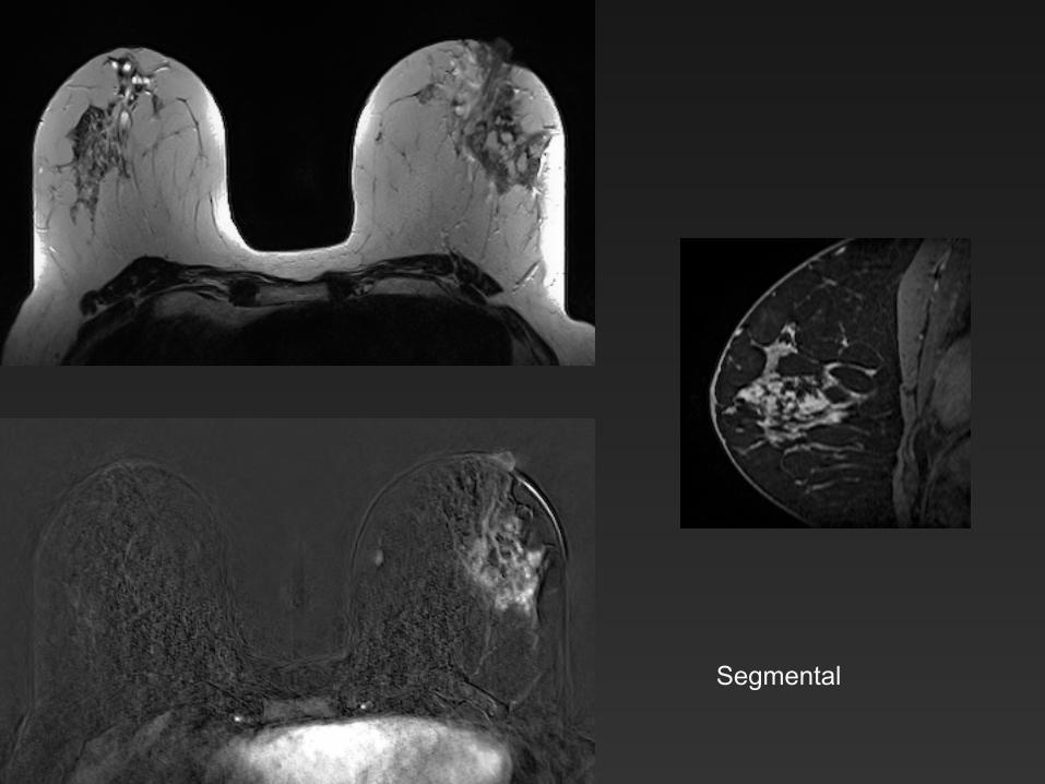
Segmental
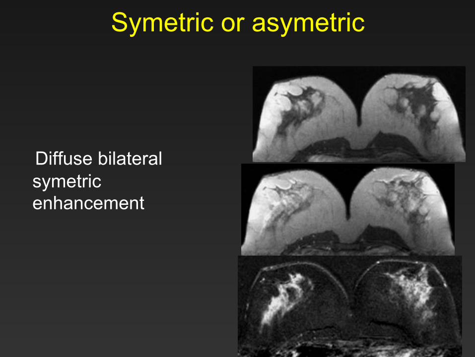
Symetric or asymetric
Diffuse bilateral
symetric enhancement

Symetric or asymetric • Asymetric
Regional, bilateral and asymetric enhancement
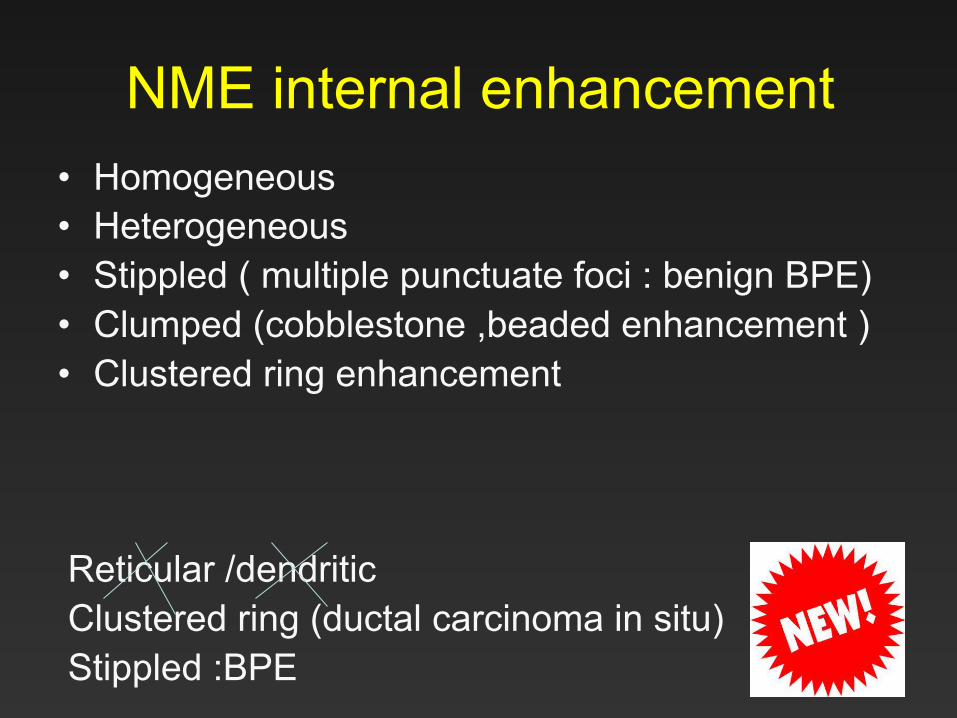
NME internal enhancement • Homogeneous • Heterogeneous • Stippled ( multiple punctuate foci : benign BPE) • Clumped (cobblestone ,beaded enhancement ) • Clustered ring enhancement
Reticular /dendritic Clustered ring (ductal carcinoma in situ) Stippled :BPE
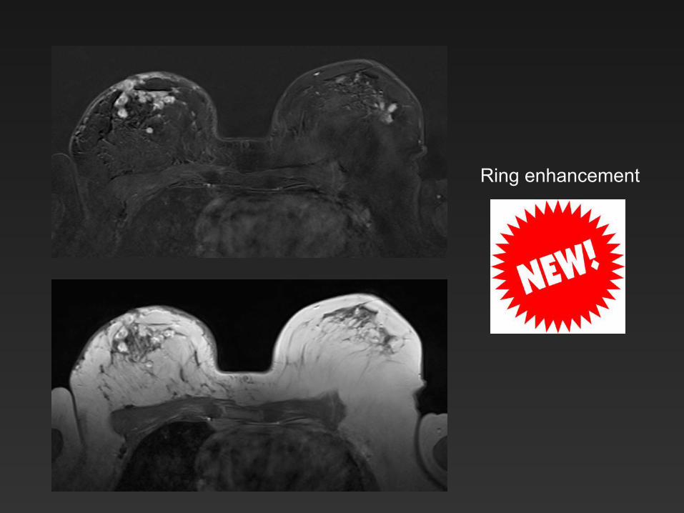
Ring enhancement
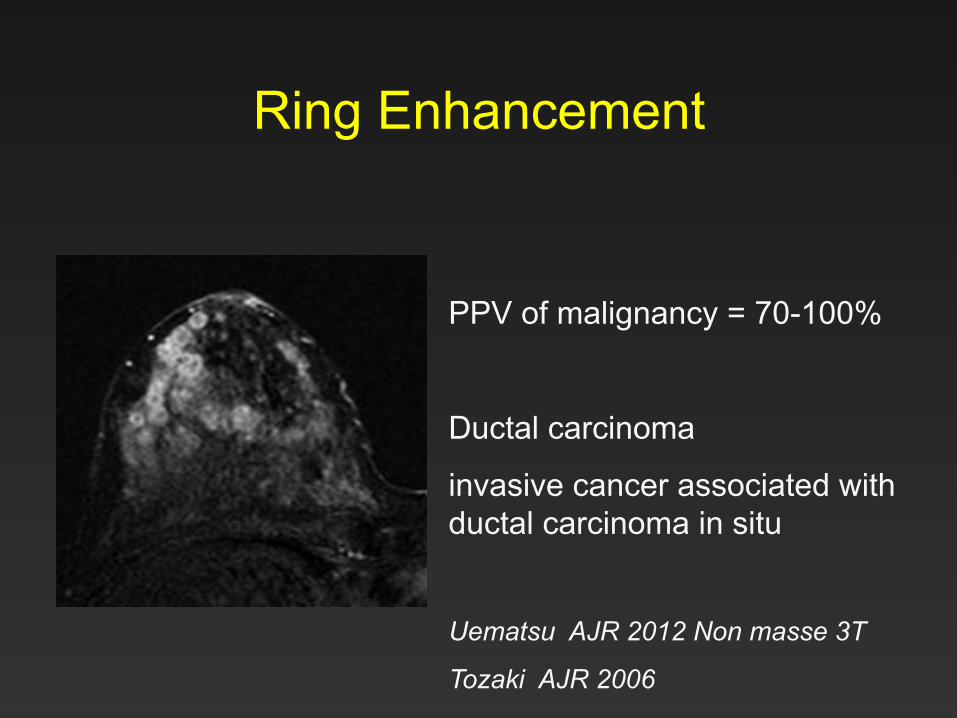
Ring Enhancement
PPV of malignancy = 70-100%
Ductal carcinoma
invasive cancer associated with ductal carcinoma in situ
Uematsu AJR 2012 Non masse 3T
Tozaki AJR 2006
T2 Signal
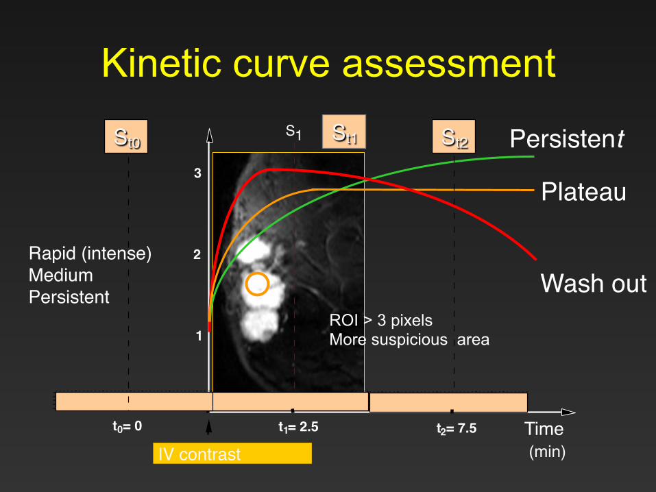
t 0 = 0 t 1 = 2.5 t 2 = 7.5 Time (min)
1
2
3
S 1 S 2
Plateau
Wash out
Persistent S 0
IV contrast"
St2" St1" St0"
Rapid (intense)"Medium"Persistent "
ROI > 3 pixels More suspicious area
Kinetic curve assessment
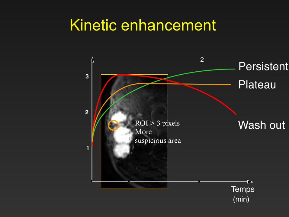
Kinetic enhancement
Temps (min)
1
2
3
2
Plateau
Wash out ROI > 3 pixels More suspicious area
Persistent
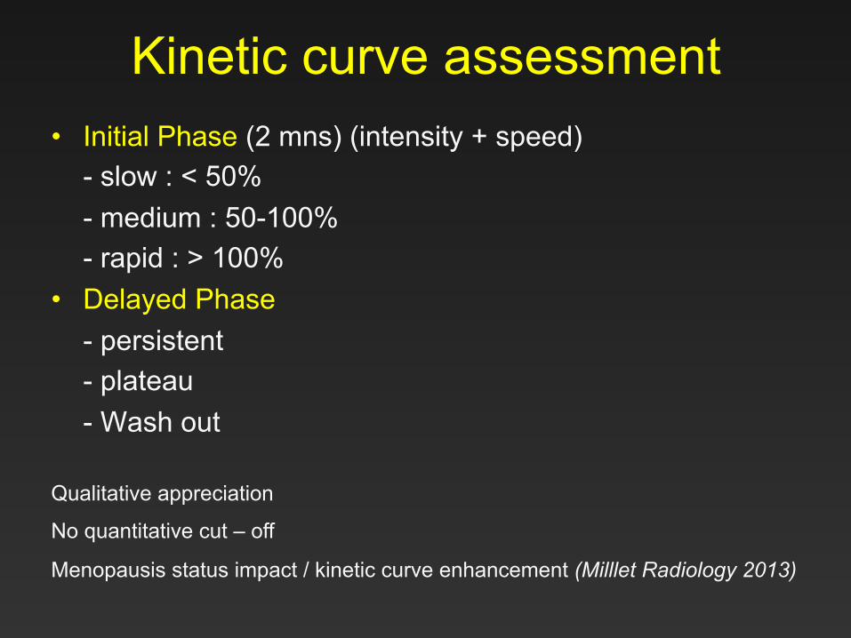
Kinetic curve assessment • Initial Phase (2 mns) (intensity + speed)
- slow : < 50% - medium : 50-100% - rapid : > 100%
• Delayed Phase - persistent - plateau - Wash out
Qualitative appreciation
No quantitative cut – off
Menopausis status impact / kinetic curve enhancement (Milllet Radiology 2013)
Associated findings
• Nipple or skin retraction • Skin thickening ( focal or diffuse) • Edema • Lymphadenopathy • Pectoralis muscle involvement • Precontrast increased ductal signal intensity • Susceptibility artifact related to surgical clips
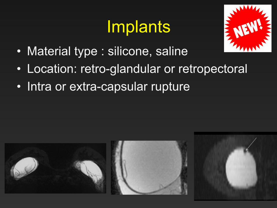
Implants • Material type : silicone, saline • Location: retro-glandular or retropectoral • Intra or extra-capsular rupture
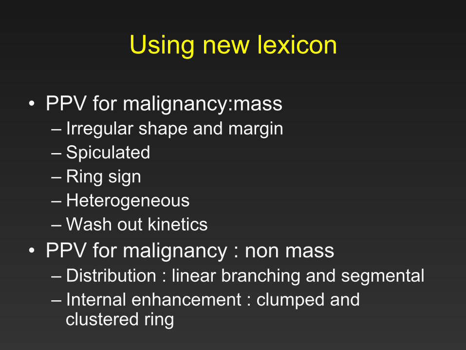
Using new lexicon
• PPV for malignancy:mass – Irregular shape and margin – Spiculated – Ring sign – Heterogeneous – Wash out kinetics
• PPV for malignancy : non mass – Distribution : linear branching and segmental – Internal enhancement : clumped and
clustered ring
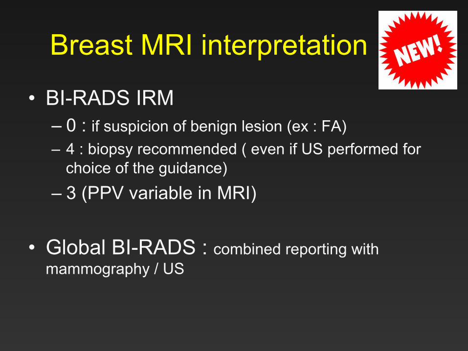
Breast MRI interpretation
• BI-RADS IRM – 0 : if suspicion of benign lesion (ex : FA) – 4 : biopsy recommended ( even if US performed for
choice of the guidance) – 3 (PPV variable in MRI)
• Global BI-RADS : combined reporting with mammography / US
Conclusion
• T2 weighted sequence • Background parenchymal enhancement • Masses / non masses enhancement :
simplification • Breast implant • Management / facilitation patient care : BIRADS
0, 4, 3 • BIRADS global

Thanck you Patrice