Embed Size (px)
Citation preview
CALCIUM & PHOSPHATE CALCIUM & PHOSPHATE METABOLISM METABOLISM
MadhuBilla 1st M.D.S
Contents:Contents:IntroductionDistributionDaily requirementsDietary sourcesFunctionsFactors controlling absorptionHormonal controlOther hormones affecting metabolismClinical importanceConclusion
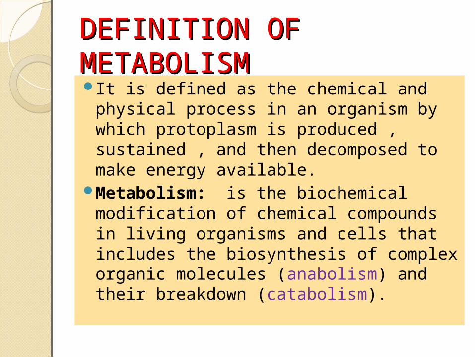
DEFINITION OF DEFINITION OF METABOLISMMETABOLISMIt is defined as the chemical and
physical process in an organism by which protoplasm is produced , sustained , and then decomposed to make energy available.
Metabolism: is the biochemical modification of chemical compounds in living organisms and cells that includes the biosynthesis of complex organic molecules (anabolism) and their breakdown (catabolism).
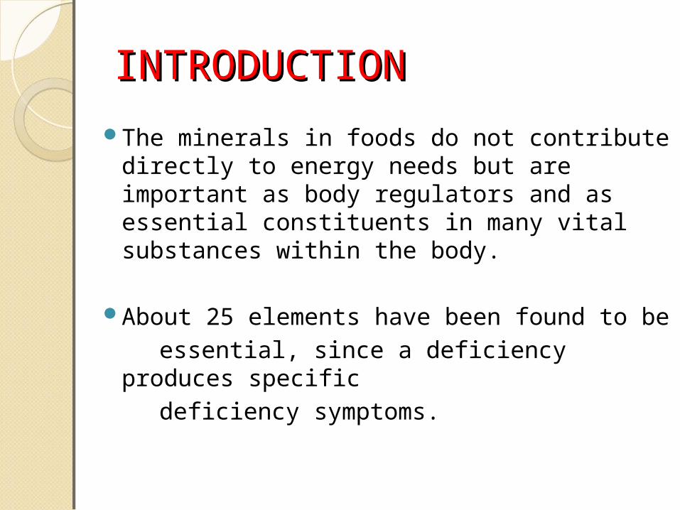
INTRODUCTIONINTRODUCTIONThe minerals in foods do not contribute
directly to energy needs but are important as body regulators and as essential constituents in many vital substances within the body.
About 25 elements have been found to be essential, since a deficiency produces
specific deficiency symptoms.
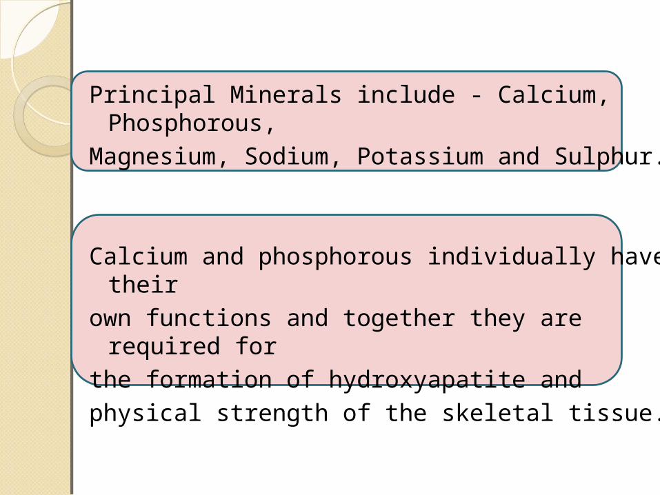
Principal Minerals include - Calcium, Phosphorous,
Magnesium, Sodium, Potassium and Sulphur.
Calcium and phosphorous individually have their
own functions and together they are required for
the formation of hydroxyapatite and physical strength of the skeletal tissue.
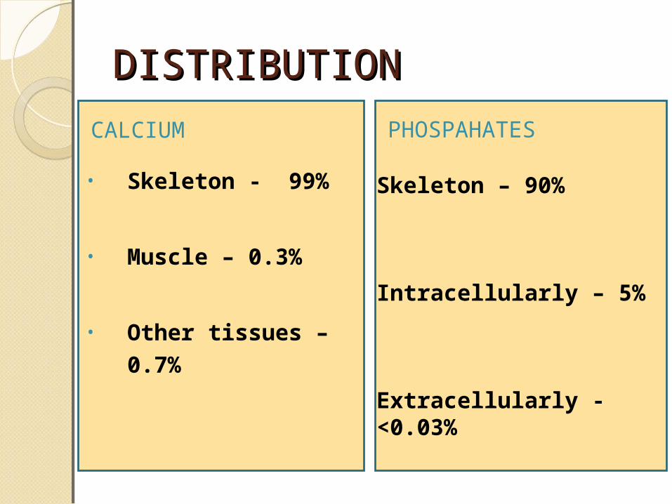
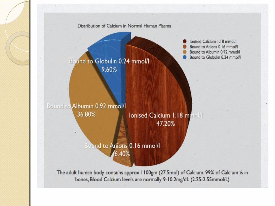
DISTRIBUTIONDISTRIBUTION
• Skeleton - 99%
• Muscle – 0.3%
• Other tissues – 0.7%
CALCIUM PHOSPAHATES
Skeleton – 90%
Intracellularly – 5%
Extracellularly - <0.03%
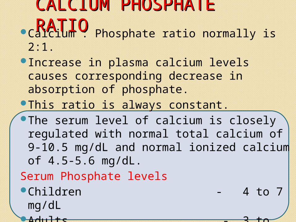
CALCIUM PHOSPHATE CALCIUM PHOSPHATE RATIORATIOCalcium : Phosphate ratio normally is 2:1.
Increase in plasma calcium levels causes corresponding decrease in absorption of phosphate.
This ratio is always constant.The serum level of calcium is closely
regulated with normal total calcium of 9-10.5 mg/dL and normal ionized calcium of 4.5-5.6 mg/dL.
Serum Phosphate levelsChildren - 4 to 7 mg/dLAdults - 3 to 4.5 mg/dL
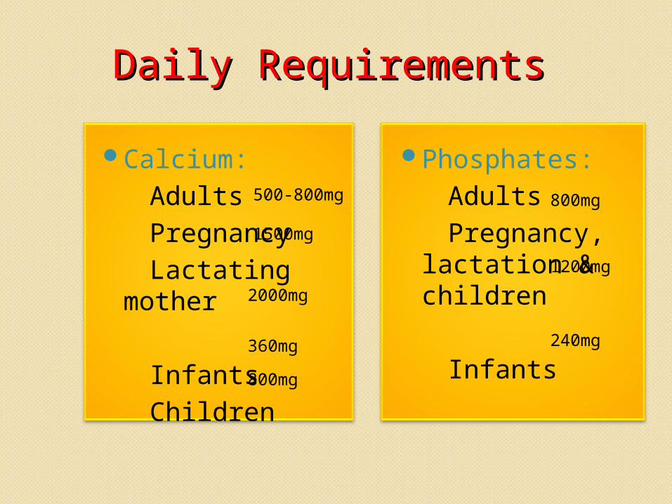
Daily RequirementsDaily RequirementsCalcium: Adults Pregnancy Lactating
mother Infants Children
Phosphates: Adults Pregnancy,
lactation & children
Infants
500-800mg
1500mg
2000mg360mg800mg
1200mg
240mg
800mg
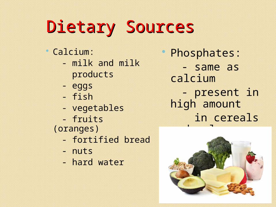
Dietary SourcesDietary Sources Calcium: - milk and milk products - eggs - fish - vegetables - fruits (oranges) - fortified bread - nuts - hard water
Phosphates: - same as calcium - present in high
amount in cereals and pulses - absent in hard water
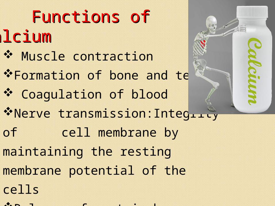
Functions of CalciumFunctions of Calcium Muscle contractionFormation of bone and teeth Coagulation of bloodNerve transmission:Integrity of cell membrane by maintaining the resting membrane potential of the cellsRelease of certain hormones
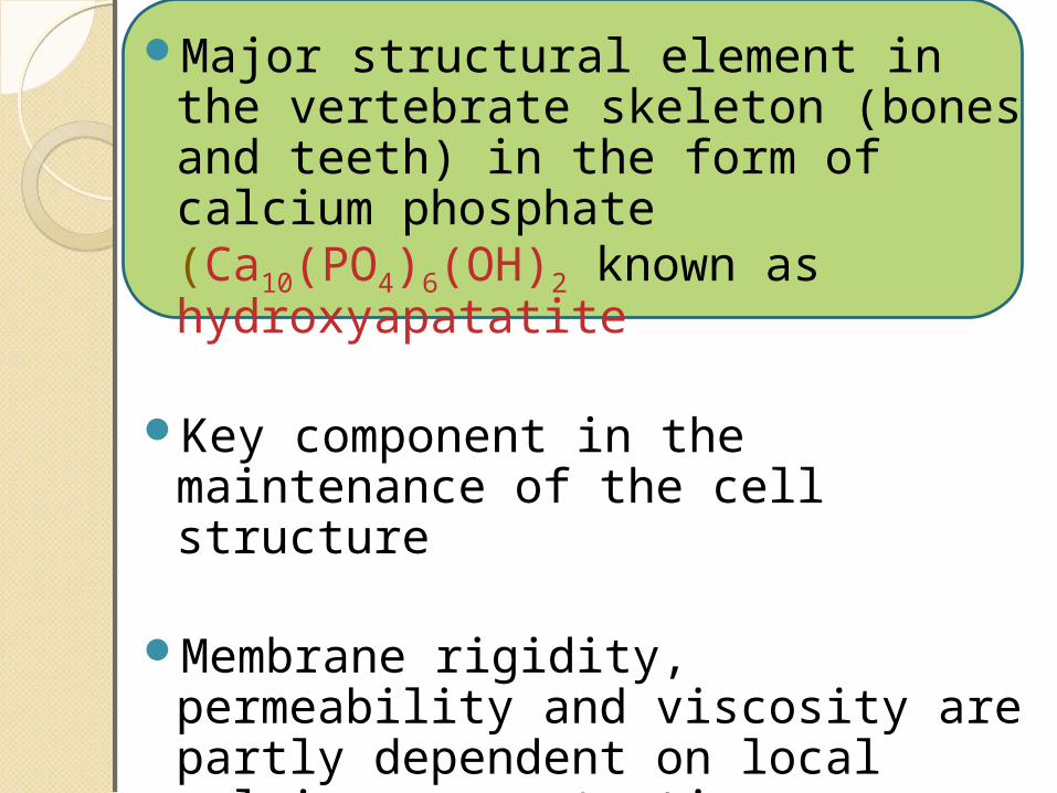
Major structural element in the vertebrate skeleton (bones and teeth) in the form of calcium phosphate (Ca10(PO4)6(OH)2 known as hydroxyapatatite
Key component in the maintenance of the cell structure
Membrane rigidity, permeability and viscosity are partly dependent on local calcium concentrations
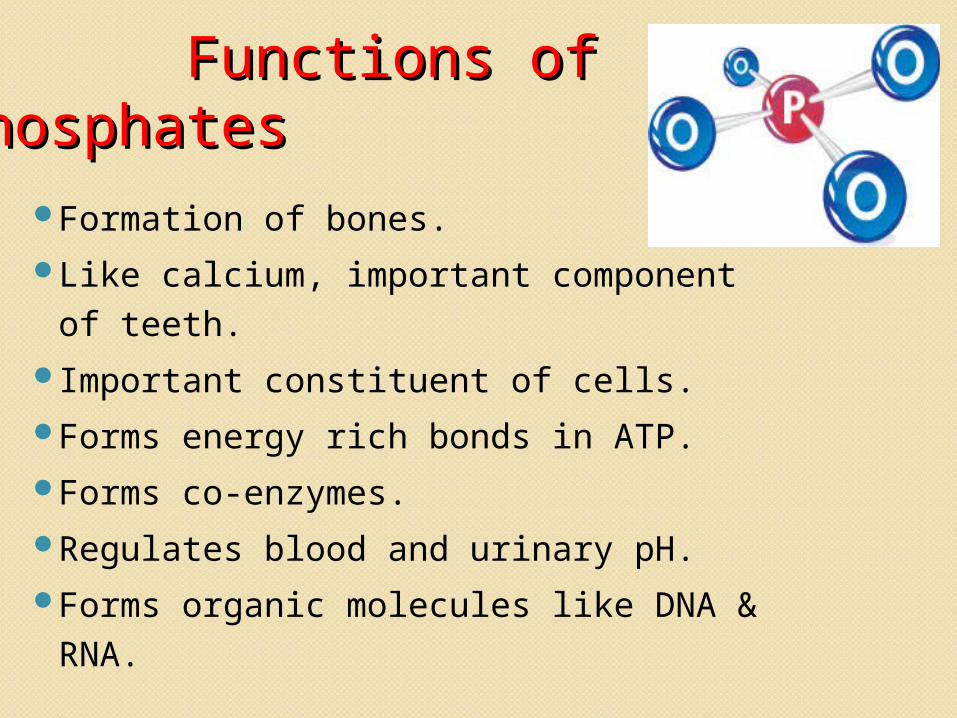
Functions of Functions of PhosphatesPhosphates
Formation of bones.Like calcium, important component of teeth.Important constituent of cells.Forms energy rich bonds in ATP.Forms co-enzymes.Regulates blood and urinary pH.Forms organic molecules like DNA & RNA.
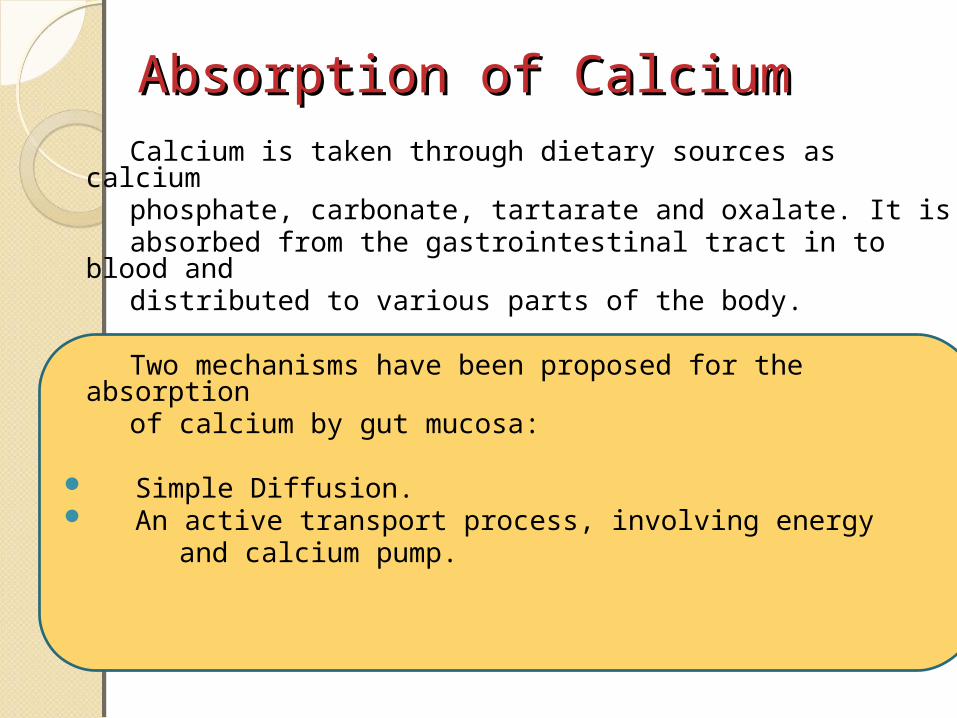
Absorption of CalciumAbsorption of Calcium Calcium is taken through dietary sources as calcium phosphate, carbonate, tartarate and oxalate. It is absorbed from the gastrointestinal tract in to blood and distributed to various parts of the body.
Two mechanisms have been proposed for the absorption
of calcium by gut mucosa:
Simple Diffusion. An active transport process, involving energy and calcium pump.
While passing through the kidney, large quantity of
calcium is filtered in the glomerulus. From the filtrate, 98 to 99% of calcium is reabsorbed in the renal tubules in to blood and only small quantity
is excreted through urine. In the bone, the calcium
may be deposited or resorbed depending upon
the level of calcium in the plasma.
Factors controlling Factors controlling absorption absorption Factors are classified into
1. Those acting on the mucosal cells
2. Those affecting the availability of
calcium and phosphates in the gut.
Factors acting on the Factors acting on the mucosal cellsmucosal cells
Vitamin D
Pregnancy and growth
PTH
VITAMIN-DCalcitriol (1,25-DHCC) is the biologically active form of vit-dIt regulates plasma levels of Ca and P Calcitrial acts at 3 different levels intestine,kidney, bones
Action on Intestines:It increases the intestinal absorption of ca&p iin the intestinal cells calcitriol binds with a cytosolic receptor to form a calcitriol-receptor commplex This complex then approaches the nucleus and interacts with a specific dna leading to synthesis of specific ca binding proteinThis protein increases the ca uptake by intestine
Action on bone:In the osteoblasts of bone calcitriol stimulates ca uptake for deposition as capo4
Action on kidney:It is involved in minimizing the excretion of ca&p through kidney by decreasing their excretion and enhancing reabsorption
• During later stages of pregnancy, greater amount of calcium absorption is seen.
• 50% of this calcium is used for the development of fetal skeleton and the rest is stored in the bones to act as a reserve for lactation.
• This is due to the increased level of placental lactogen and estrogen which stimulates increased hydroxylation of vitamin D.
• In growth there is a increased level of growth hormone. GH acts by increasing calcium absorption. It also increases the renal excretion of calcium and phosphates.
Pregnancy and growth:
• Parathyroid hormone is one of the main hormones controlling Ca+2 absorption.
• It mainly acts by controlling the formation of 1,25 DHCC, which is active form of Vit. D, which is responsible for, increased Ca+2 absorption.
Parathyroid Hormone:
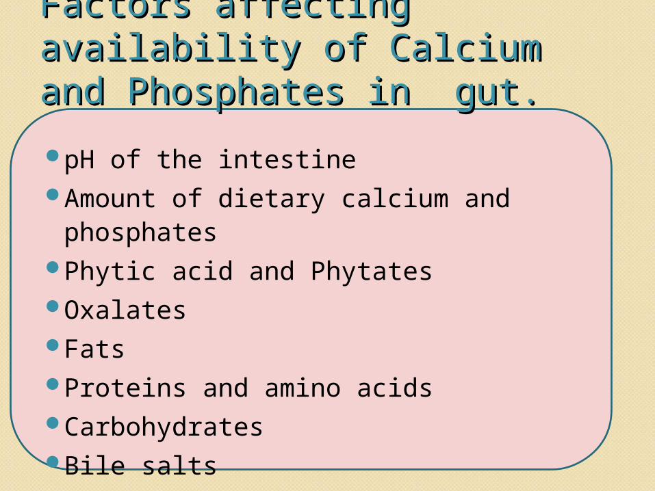
Factors affecting availability Factors affecting availability of Calcium and Phosphates of Calcium and Phosphates in gut.in gut.pH of the intestineAmount of dietary calcium and
phosphatesPhytic acid and PhytatesOxalatesFatsProteins and amino acids CarbohydratesBile salts
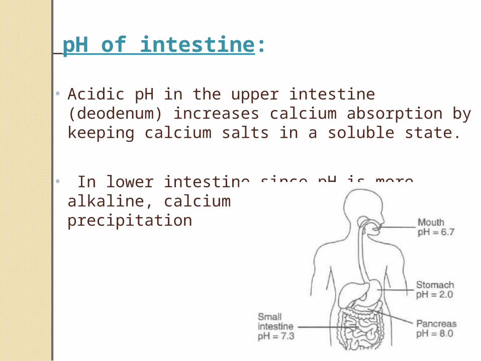
pH of intestine:
• Acidic pH in the upper intestine (deodenum) increases calcium absorption by keeping calcium salts in a soluble state.
• In lower intestine since pH is more alkaline, calcium salts undergoes precipitation
Amount of dietary calcium and phosphates:
• Increased level of calcium and phosphate in diet increases their absorption however up to a certain limit.
• This is because the active process of their absorption can bear with certain amounts of load beyond which the excess would pass out into faeces
Phytic acid and phytates:• They are present in oatmeal, meat and cereals
and are considered anti-calcifying factors as they combine with calcium in the diet thus forming insoluble salts of calcium
Oxalates:• They are present in spinach and rhubarb leaves.
They form oxalate precipitates with calcium present in the diet thus decreasing their availability. Fats:
• They combines with calcium and form insoluble calcium , thus decreasing calcium absorption.
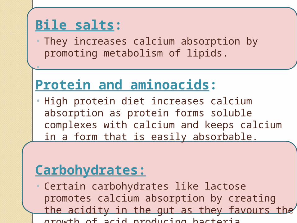
Bile salts:• They increases calcium absorption by promoting
metabolism of lipids.• Protein and aminoacids:• High protein diet increases calcium absorption as
protein forms soluble complexes with calcium and keeps calcium in a form that is easily absorbable.
Carbohydrates:• Certain carbohydrates like lactose promotes
calcium absorption by creating the acidity in the gut as they favours the growth of acid producing bacteria.
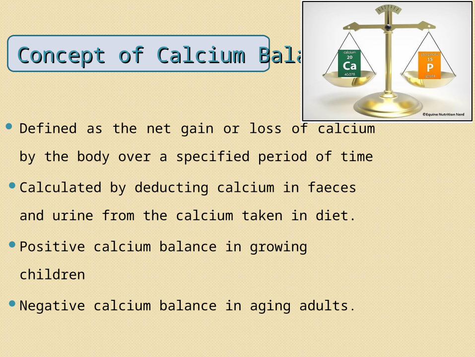
Concept of Calcium BalanceConcept of Calcium Balance
Defined as the net gain or loss of calcium by the body over a specified period of time
Calculated by deducting calcium in faeces and urine from the calcium taken in diet.
Positive calcium balance in growing childrenNegative calcium balance in aging adults.
Hormonal Control of Calcium & Hormonal Control of Calcium & Phosphate metabolismPhosphate metabolism
Three hormones regulate calcium and phosphate metabolism.
Vitamin DPTHCalcitonin
Vitamin DVitamin D Cholecalciferol / D3
Ergocalciferol / D2
Can be called as hormone as it is produced in the skin when exposed to sunlight.
Vitamin D has very little intrinsic biological activity.
Vitamin D itself is not a active substance, instead it must be first converted through a succession of reaction in the liver and the kidneys to the final active product 1, 25 di hydroxycholecalciferol,
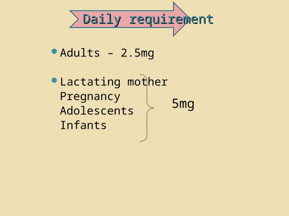
Daily requirementDaily requirement
Adults – 2.5mg
Lactating motherPregnancyAdolescentsInfants
5mg
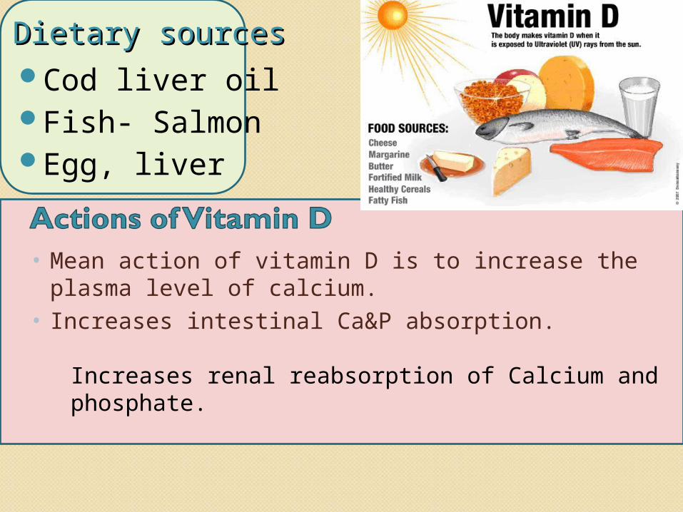
Dietary sourcesDietary sourcesCod liver oilFish- SalmonEgg, liver
• Mean action of vitamin D is to increase the plasma level of calcium.
• Increases intestinal Ca&P absorption. Increases renal reabsorption of Calcium and phosphate.
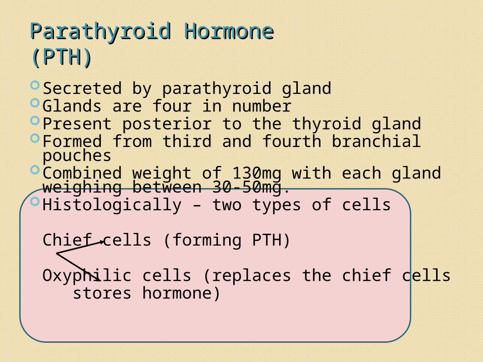
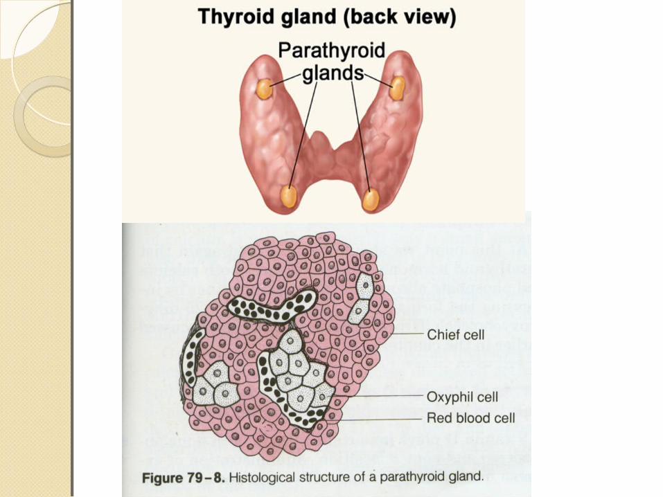
Parathyroid Hormone Parathyroid Hormone (PTH)(PTH)Secreted by parathyroid glandGlands are four in number Present posterior to the thyroid glandFormed from third and fourth branchial pouchesCombined weight of 130mg with each gland weighing between 30-50mg. Histologically – two types of cells
Chief cells (forming PTH)
Oxyphilic cells (replaces the chief cells stores hormone)
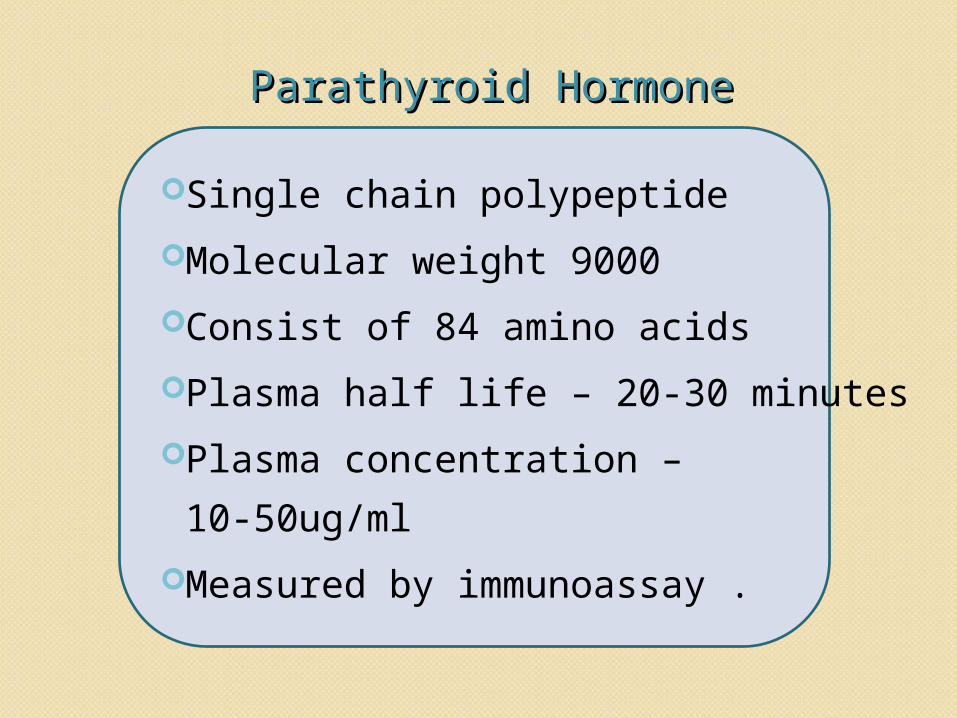
Parathyroid HormoneParathyroid Hormone
Single chain polypeptide Molecular weight 9000Consist of 84 amino acids Plasma half life – 20-30 minutesPlasma concentration – 10-50ug/mlMeasured by immunoassay .
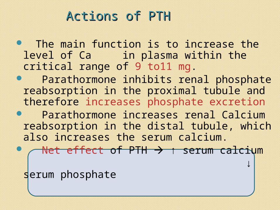
Actions of PTHActions of PTH
The main function is to increase the level of Ca in plasma within the critical range of 9 to11 mg.
Parathormone inhibits renal phosphate reabsorption in the proximal tubule and therefore increases phosphate excretion
Parathormone increases renal Calcium reabsorption in the distal tubule, which also increases the serum calcium.
Net effect of PTH ↑ serum calcium ↓ serum phosphate
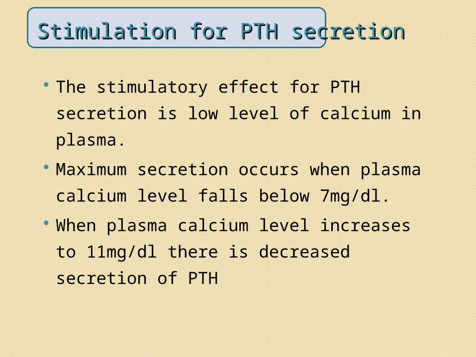
Stimulation for PTH secretionStimulation for PTH secretion
The stimulatory effect for PTH secretion is low level of calcium in plasma.
Maximum secretion occurs when plasma calcium level falls below 7mg/dl.
When plasma calcium level increases to 11mg/dl there is decreased secretion of PTH
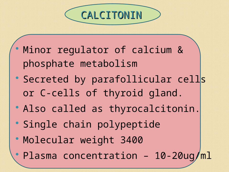
CALCITONINCALCITONIN
Minor regulator of calcium & phosphate metabolism
Secreted by parafollicular cells or C-cells of thyroid gland.
Also called as thyrocalcitonin. Single chain polypeptide Molecular weight 3400 Plasma concentration – 10-20ug/ml
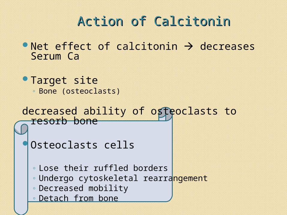
Action of CalcitoninAction of CalcitoninNet effect of calcitonin decreases Serum
Ca
Target site◦ Bone (osteoclasts)
decreased ability of osteoclasts to resorb bone
Osteoclasts cells
◦ Lose their ruffled borders◦ Undergo cytoskeletal rearrangement ◦ Decreased mobility ◦ Detach from bone
Calcitonin is a Physiological Antagonist to
PTH with respect to Calcium.
With respect to Phosphate it has the same
effect as PTH i.e. ↓ Plasma Phosphate level
OTHER HORMONES on CALCIUM OTHER HORMONES on CALCIUM METABOLISMMETABOLISMGROWTH HORMONE INSULIN TESTOSTERONE & OTHER HORMONESLACTOGEN & PROLACTINSTEROIDSTHYROID HORMONES
Increases the intestinal absorption of calcium and
increases its excretion from urineStimulates production of insulin like growth factor in
bone which stimulates protein synthesis in boneStimulates stomatomedian C which acts on cartilage
to increase the length of bones
GROWTH HORMONE
TESTOSTERONE
Testosterone causes differential growth
of cartilage resulting to differential
bone development
Acts on cartilage & increase the bone
growth.
INSULIN• It is an anabolic hormone which favors bone
formation
Thyroid HormoneThyroid HormoneIn infants stimulation of bone
growth
In adultsincreased bone metabolism increased calcium mobilization
GlucocorticoidsGlucocorticoids
Anti vitamin D action, decrease absorption of calcium in intestine
Inhibit protein synthesis and so decrease bone formation
Inhibit new osteoclast formation & decrease the activity of old osteoclasts.
Excretion of Calcium and Excretion of Calcium and PhosphorousPhosphorous Calcium is excreted in the urine, bile, and
digestive secretions. The renal threshold for serum ca is 10
mg/dl. 70-90% of the calcium eliminated from the body is excreted in the feces.The daily loss of calcium in sweat is about
15 mg.
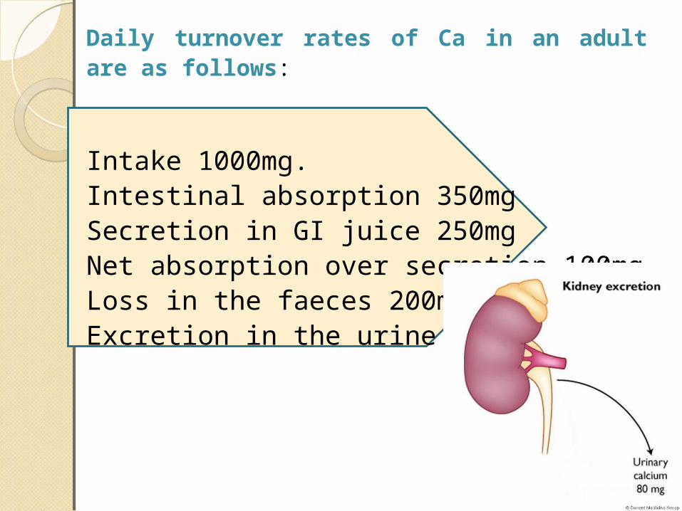
Daily turnover rates of Ca in an adult are as follows:
Intake 1000mg.Intestinal absorption 350mgSecretion in GI juice 250mgNet absorption over secretion 100mgLoss in the faeces 200mgExcretion in the urine 80-100mg
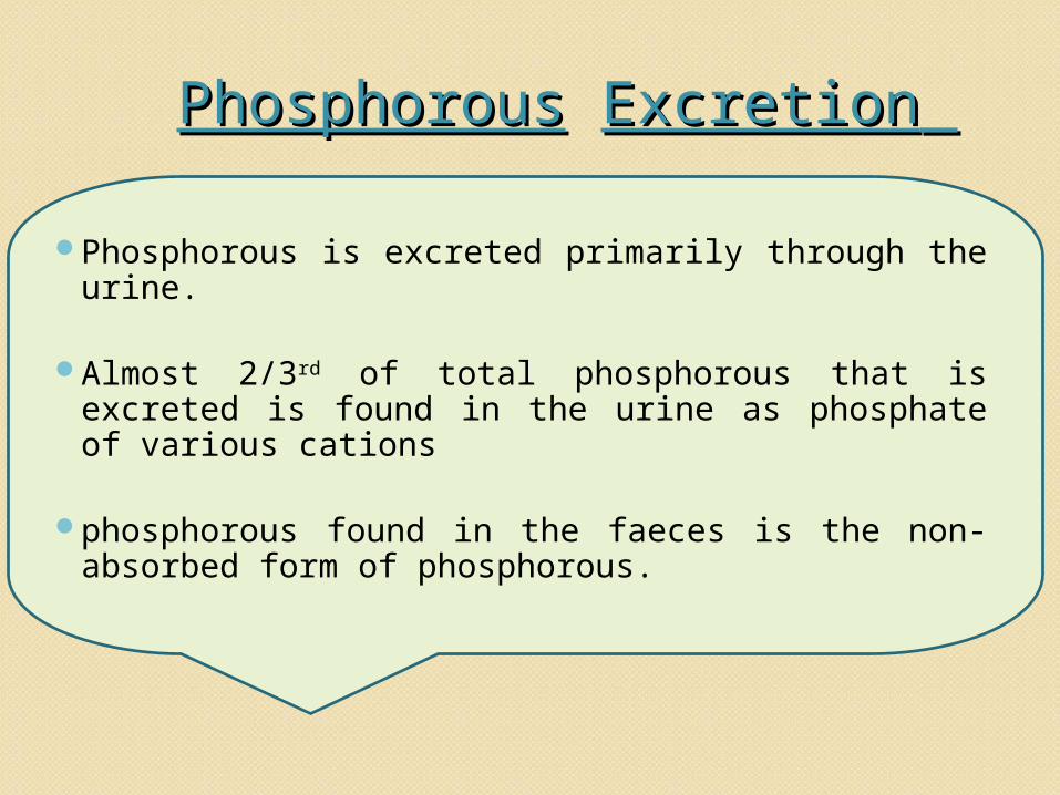
PhosphorousPhosphorous ExcretionExcretion
Phosphorous is excreted primarily through the urine.
Almost 2/3rd of total phosphorous that is excreted is found in the urine as phosphate of various cations
phosphorous found in the faeces is the non-absorbed form of phosphorous.
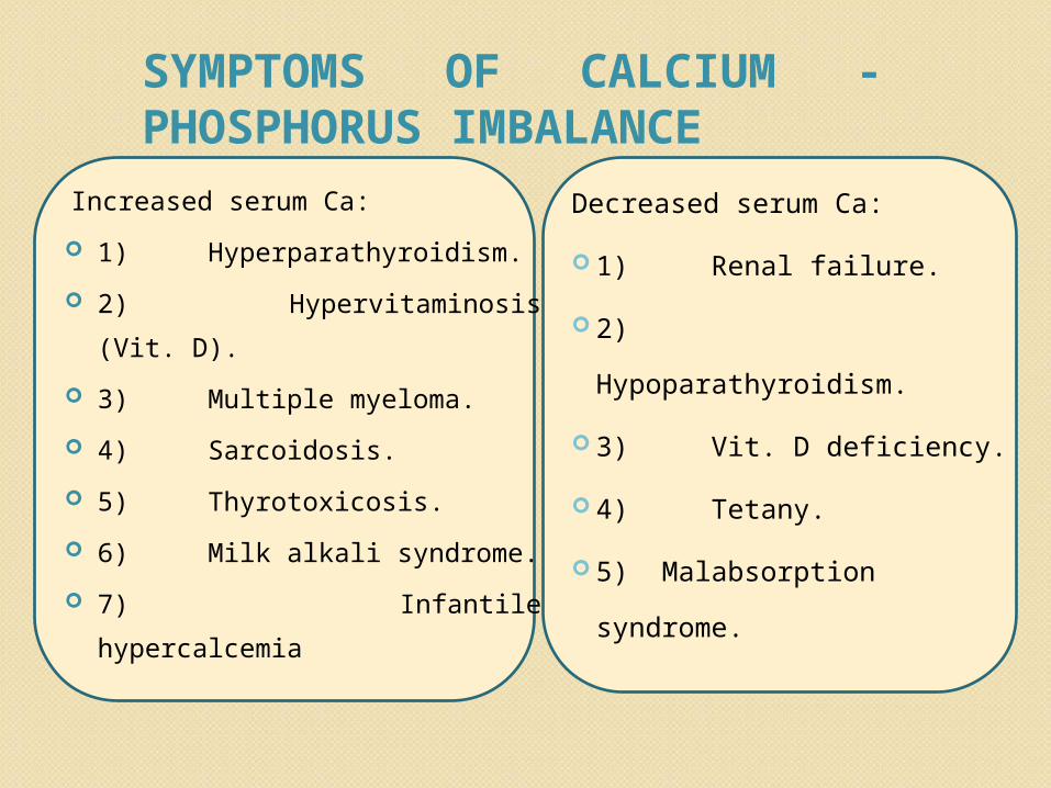
Increased serum Ca: 1) Hyperparathyroidism. 2) Hypervitaminosis (Vit.
D). 3) Multiple myeloma. 4) Sarcoidosis. 5) Thyrotoxicosis. 6) Milk alkali syndrome. 7) Infantile hypercalcemia
Decreased serum Ca: 1) Renal failure. 2) Hypoparathyroidism. 3) Vit. D deficiency. 4) Tetany. 5) Malabsorption
syndrome.
SYMPTOMS OF CALCIUM - PHOSPHORUS IMBALANCE
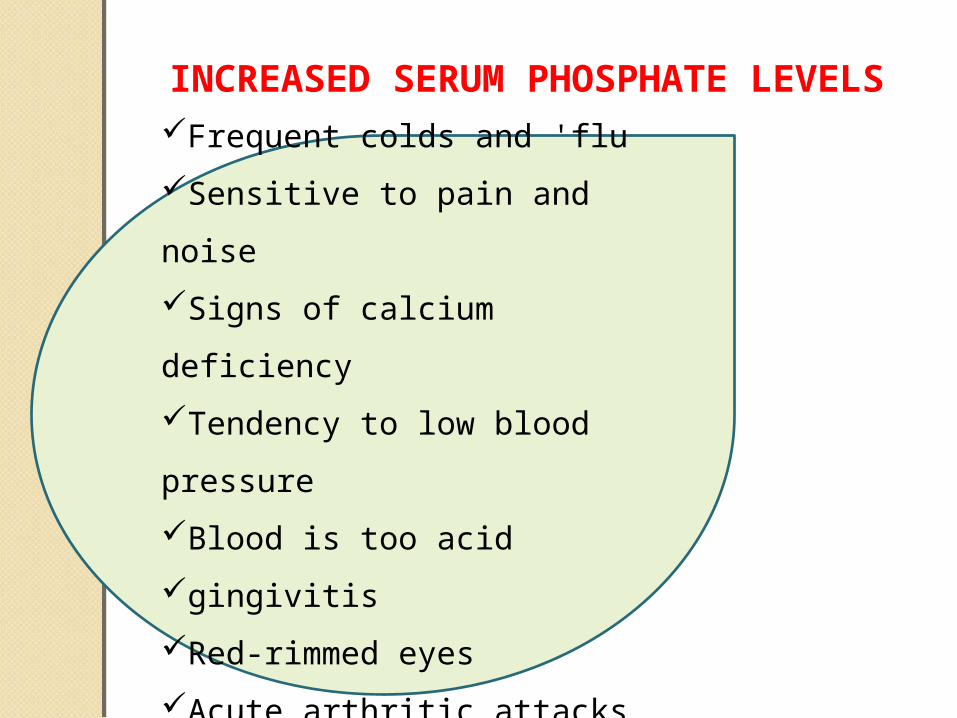
Frequent colds and 'fluSensitive to pain and noiseSigns of calcium deficiencyTendency to low blood pressureBlood is too acidgingivitisRed-rimmed eyesAcute arthritic attacks
INCREASED SERUM PHOSPHATE LEVELS
Clinical ImportanceClinical Importance
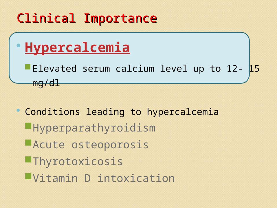
HypercalcemiaElevated serum calcium level up to 12- 15 mg/dl
Conditions leading to hypercalcemiaHyperparathyroidismAcute osteoporosisThyrotoxicosisVitamin D intoxication
Symptoms:
Polyuria, dehydration, confusion, depression, fatigue, nausea/vomiting, anorexia, abdominal pain, and renal calculi
Signs: Diminished reflexes, short QT interval on ECG.
Treatment
ParathyroidectomyHydration.Furosemide, Bisphosphanate, Calcitonin, Mithramycin, Gallium nitrate, Steroids.IV Phosphate.Dialysis.Others- Glucocorticoids, phosphate,
amifostine
HypocalcaemiaDecreased level of calcium in the blood
(<4mg/dl)
Conditions leading to hypocalcaemia
Insufficient dietary calcium
Hypoparathyroidism
Insufficient vitamin D
↑ in calcitonin levels
Symptoms:
Irritability, muscle cramps, depression, bronchospasm, and seizures.
Signs:
Increased reflexes, prolonged QT interval on ECG (the only cause of a prolonged QT with a normal duration of the T wave itself)
Treatment:Asymptomatic
Oral calcium and vitamin D supplementation
Symptomatic
IV calcium gluconate (200mg IV over 10min, then 50-150mg/hr for a total of 15mg/kg)
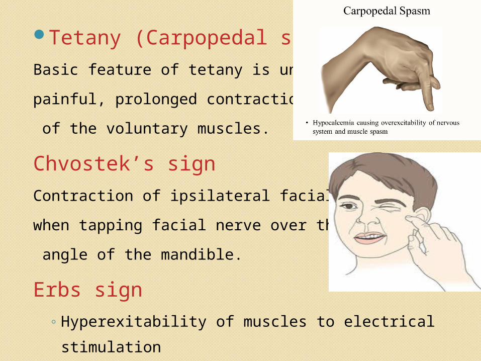
Tetany (Carpopedal spasm)Basic feature of tetany is uncontrolled, painful, prolonged contraction (spasm) of the voluntary muscles.
Chvostek’s signContraction of ipsilateral facial muscles when tapping facial nerve over the angle of the mandible.
Erbs sign◦ Hyperexitability of muscles to electrical stimulation
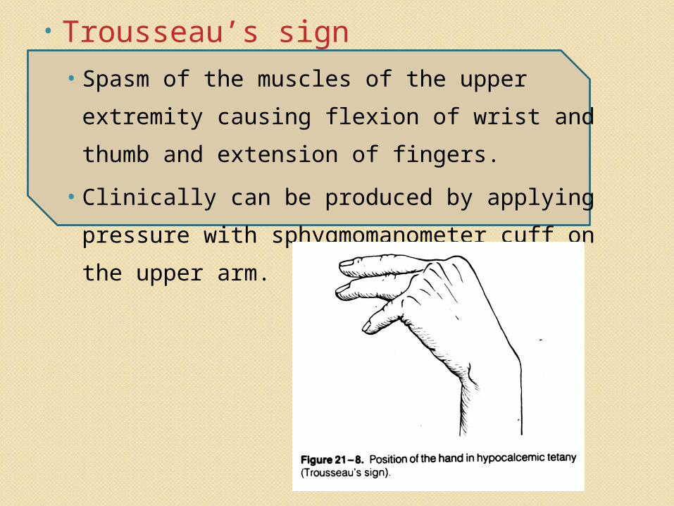
• Trousseau’s sign• Spasm of the muscles of the upper extremity
causing flexion of wrist and thumb and extension of fingers.
• Clinically can be produced by applying pressure with sphygmomanometer cuff on the upper arm.
Vitamin D deficiencyVitamin D deficiency
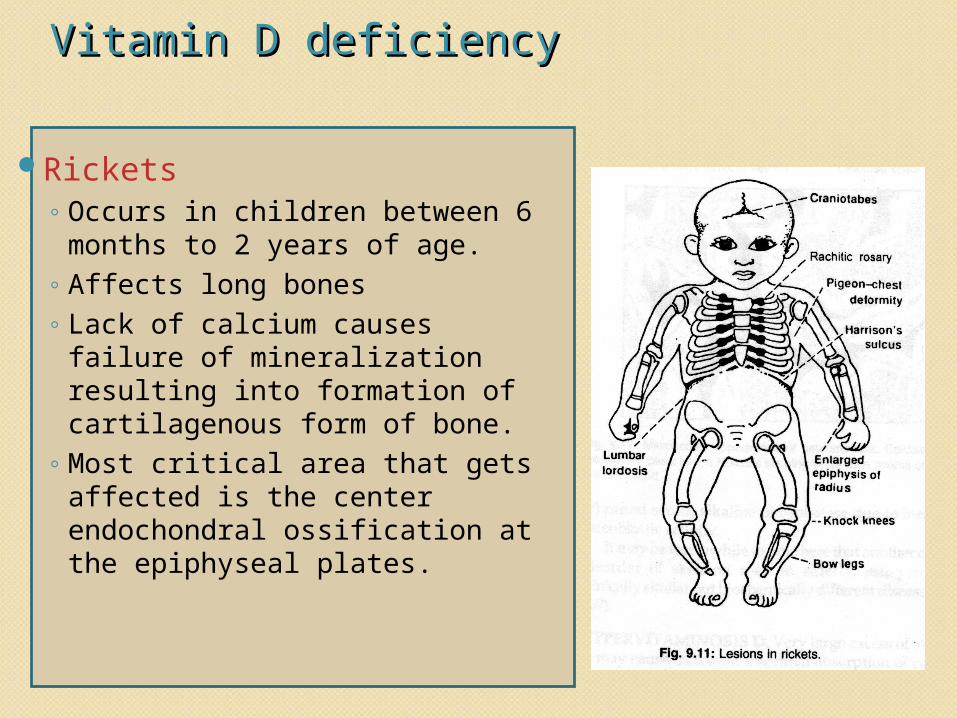
Rickets◦ Occurs in children between 6
months to 2 years of age. ◦ Affects long bones◦ Lack of calcium causes
failure of mineralization resulting into formation of cartilagenous form of bone.
◦ Most critical area that gets affected is the center endochondral ossification at the epiphyseal plates.
Dental findings in RickettsDental findings in RickettsDevelopmental anomalies of enamel
and dentinDelayed eruptionMisalignment of teethIncrease caries indexWide predentin zone and more
interglobular dentinTreatmentDaily administration of 1000 – 4000 units of
vit.D.
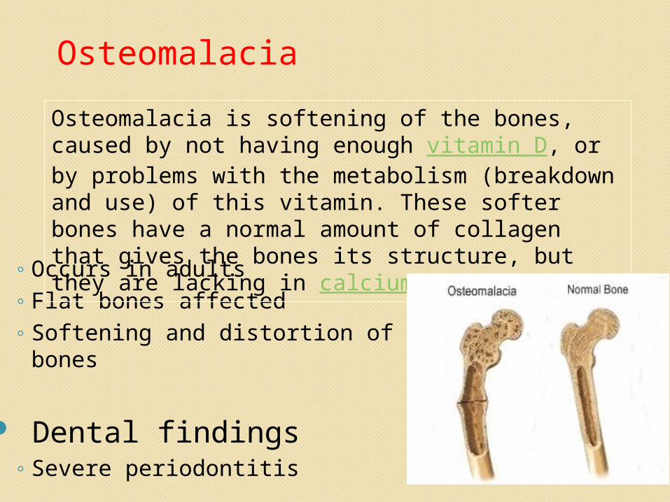
◦ Occurs in adults◦ Flat bones affected◦ Softening and distortion of skeletal bones
Dental findings◦ Severe periodontitis
Osteomalacia is softening of the bones, caused by not having enough vitamin D, or by problems with the metabolism (breakdown and use) of this vitamin. These softer bones have a normal amount of collagen that gives the bones its structure, but they are lacking in calcium
Osteomalacia
OsteoporosisOsteoporosisIt is the most common of all bone diseases in adults , especially in old age.
It is different from osteomalacia and rickets because it results from diminished organic bone matrix
rather than from poor bone calcification.
Characterised by low bone mass, microarchitectural deterioration of bone tissue.
Symptoms:
Fractures of brittle bones occur even after minor accident
Pain due to fractures of vertebrae which may radiate round the trunk , to the buttocks or down the legs
Oral manifestations include alveolar bone loss resulting in ill- fitting dentures and
periodontitis.
Prevention:
Physical activityAndrogen and OestrogenIncreased calcium intake and strontium
and sodium flouride ingestion.
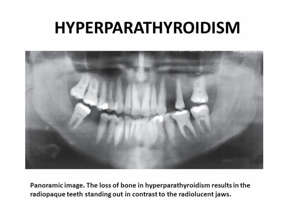
HyperparathyroidismHyperparathyroidism
May be Primary or Secondary. Primary hyperparathyroidism
In Primary form, a primary abnormality of the parathyroid glands causes inappropriate,
excess PTH secretion. Caused mainly by an adenoma of parathyroid.
• Osteitis fibrosa cystica : Bone pain, joint stiffness and pathological fracture are the early symptoms.
• Parathyroid poisoning and Metastatic calcification
• Hypercalcemia.
• Formation of Kidney stones.
Secondary Hyperparathyroidism
In Secondary form, high levels of PTH
occur as a compensation rather than as a primary
abnormality of the parathyroid glands.
It can be caused by Vitamin D deficiency or
chronic renal disease.
Oral Manifestations:
Dehydration Mandibular or maxillary tumors of the
bone, which on biopsy display a brown tumor of von Recklinghausen
Increased incidence of tori; Reduction in indices of cortical bone
leading to osteoporosis (lamina dura and gonial
index);
According to Schour and Massler, malocclusion caused by a sudden drifting with definite spacing of the
teeth may be one of the first signs of the disease.
Radiological features include: Small cystic areas in the calvarium and large/ small sharply defined radioluscencies may be present in the
maxilla/ mandible, typically described as having a ‘ground glass appearance’.
Lamina dura around the teeth may be partially lost Management: involves excision of the parathyroid tumour
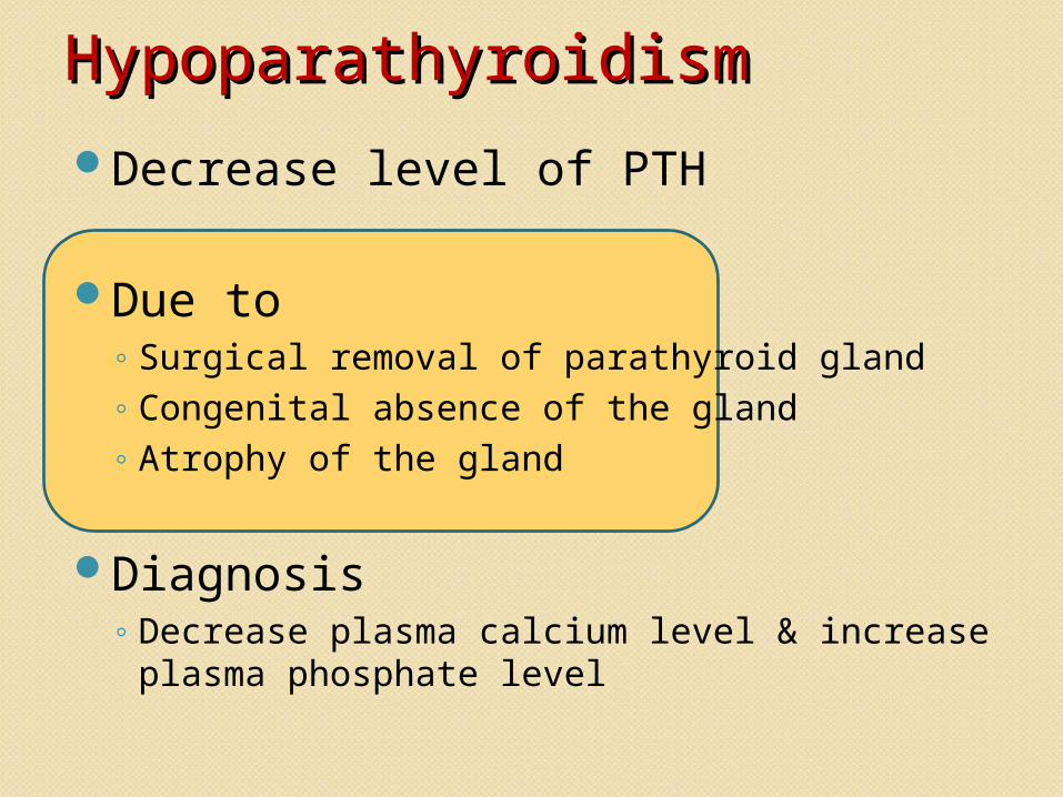
HypoparathyroidismHypoparathyroidismDecrease level of PTH
Due to ◦ Surgical removal of parathyroid gland ◦ Congenital absence of the gland◦ Atrophy of the gland
Diagnosis◦ Decrease plasma calcium level & increase
plasma phosphate level
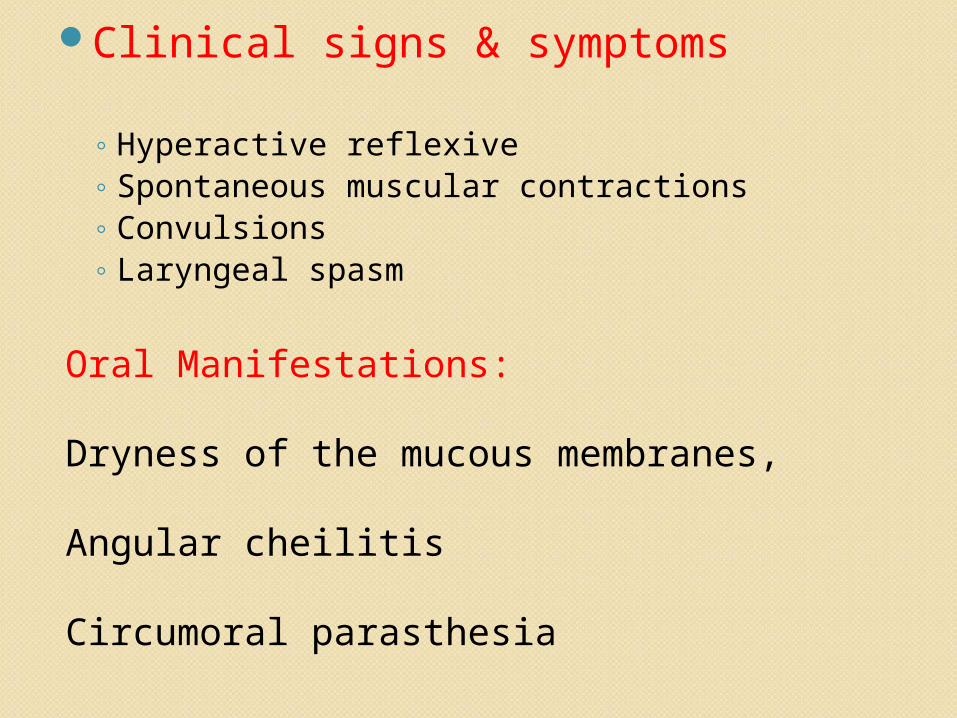
Clinical signs & symptoms
◦ Hyperactive reflexive ◦ Spontaneous muscular contractions◦ Convulsions ◦ Laryngeal spasm
Oral Manifestations:
Dryness of the mucous membranes, Angular cheilitis
Circumoral parasthesia
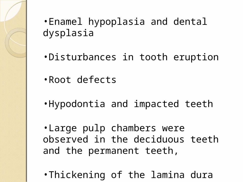
•Enamel hypoplasia and dental dysplasia
•Disturbances in tooth eruption
•Root defects
•Hypodontia and impacted teeth
•Large pulp chambers were observed in the deciduous teeth and the permanent teeth,
•Thickening of the lamina dura was observed in the permanent teeth.
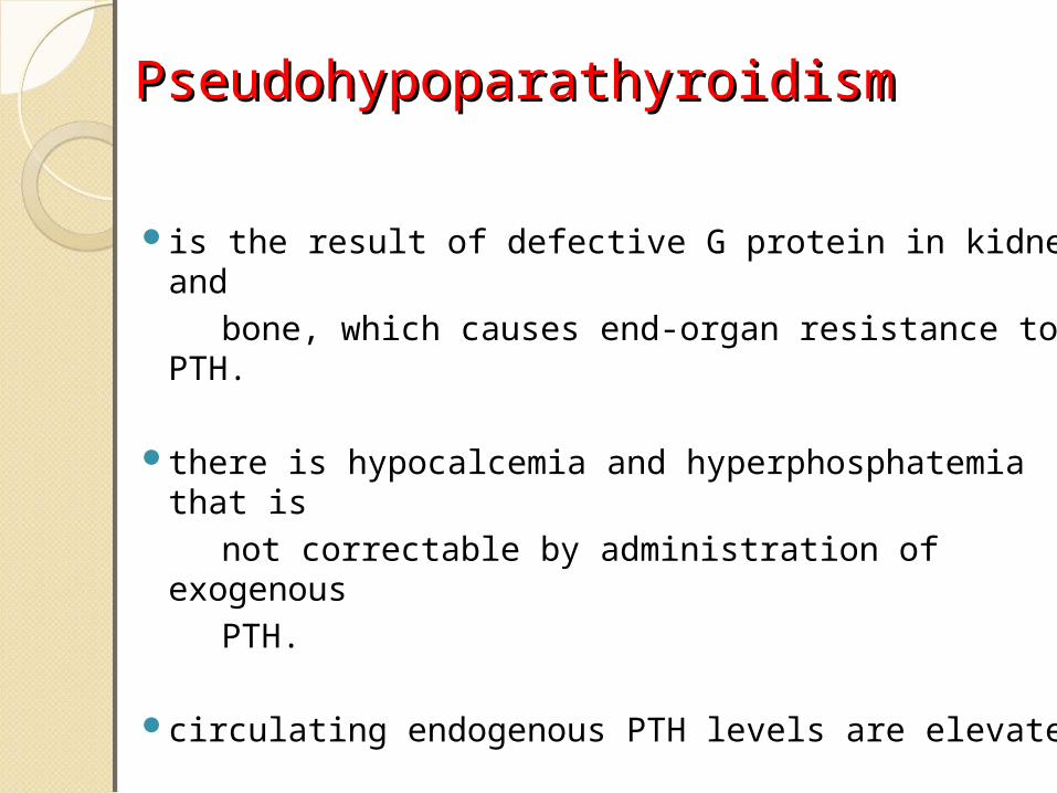
PseudohypoparathyroidismPseudohypoparathyroidism
is the result of defective G protein in kidney and bone, which causes end-organ resistance to PTH.
there is hypocalcemia and hyperphosphatemia that is
not correctable by administration of exogenous PTH.
circulating endogenous PTH levels are elevated.
Management
Administration of extremely large quantities of vitamin D, to as high as 100,000 units per day, along with intake of 1 to 2 grams of calcium, keeps the calcium ion concentration in a normal range.
• Increased intake Diet containing Vit-D• Increased release of P from cells (DM, Acidaemia,
Starvation)• Increased release of P from bone (malignancy,
Renal failure, Increased PTH)• Decreased excretion (Renal failure,
Hyperparathyroidism, Increased growth hormone)
• Phosphate trapping• Respiratory insufficiency• Erythrocyte dysfunction• Nervous Dysfunction• Leukocyte Dysfunction• Metabolic acidosis
Treatment
Treat the underlying causeIn patients with hypoparathyroidism,
calcium and vitamin D supplementation are generally prescribed to correct hypocalcemia.
In hyperphosphatemia due to chemotherapy for
leukemia or lymphoma, vigorous saline diuresis will lead to increased phosphaturia. Alternatively, the administration of acetazolamide, 500 mg every six hours, will enhance renal phosphate excretion through urinary alkalinization and natriuresis.
• Decreased Intake (Starvation, Malabsorption, Vomiting)
• Increased cell uptake (High dietary carbohydrate, Liver disease)
• Increased Excretion (Diuretics, Hypomagnesaemia, Increased PTH)
Oral manifestations:
Histological evidence of widespread formation of globular,hypocalcified dentin, with clefts and
tubular defects occuring in the region of pulphorns.Periapical involvement of grossly normal appearing deciduous and permanent teeth, followed by the development of multiple gingival fistulas.Abnormal cementum and the alveolar bone patternLamina dura is frequently absent or poorly defined.
Radiographic features:
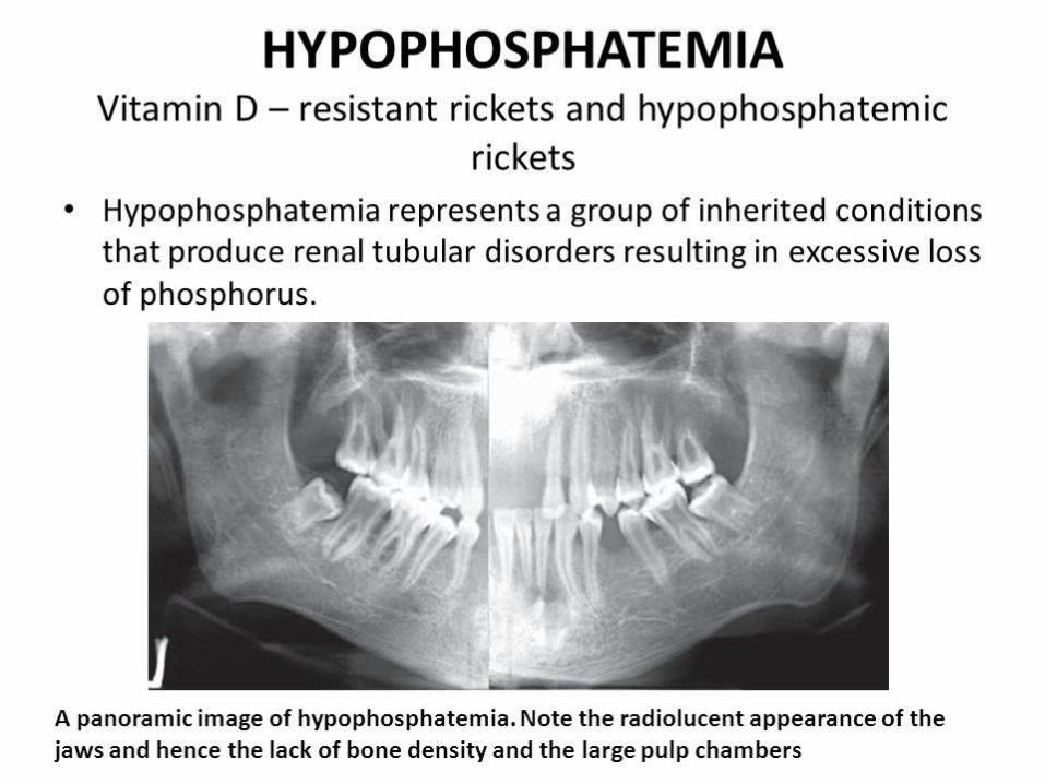
Dental radiographs reveal hypocalcification of teeth and the presence of large pulp chambers and alveolar bone loss.
Histologic features:
Absence of cementum leading to a lack of sound
attachment of the tooth to the bone by periodontal ligament. This lack of
attachment accounts for the early spontaneous exfoliation of the
deciduous teeth.
TreatmentTreat the underlying cause
A total daily amount of 2 to 3 grams of elemental phosphorus may be given in two to four divided
doses, orally.
For patients requiring parenteral administration of
phosphate, an initial phosphate dose of 0.08 mmol per
kg body weight may be given over six hours. The dose may be increased to 0.16 mmol per kg if a patient has serious life-threatening clinical manifestations.
HYPOPHOSPHATASIAHYPOPHOSPHATASIA
The basic disorder is a deficiency of the enzyme alkaline phosphatase in serum or tissues and excretion of phosphoethanolamine in the urine.
Clinical features:
Infantile- severe Rickets, Hypercalciemia, Bone
abnormalities and failure to thrive.
Childhood- Premature exfoliation of deciduous teeth,
increased infection, growth retardation,
rachitic like deformities, pulmonary, GIT and renal abnormalitiesAdult- spontaneous fractures, prior history of rickets and osseous radiolucencies.
Oral manifestations:
Loosening and premature loss of deciduous teeth, chiefly the incisors.
Radiographs show hypocalcification of teeth.
Histogically, the teeth present a unique appearance
characterised by the absence of cementum. Treatment:
Administration of high oral doses of phosphate
results in moderate improvement in bone calcification.
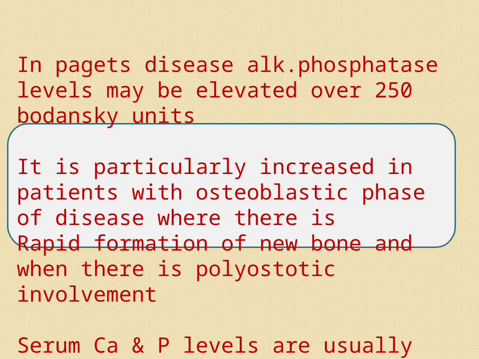
In pagets disease alk.phosphatase levels may be elevated over 250 bodansky units
It is particularly increased in patients with osteoblastic phase of disease where there is Rapid formation of new bone and when there is polyostotic involvement
Serum Ca & P levels are usually within normal limits.
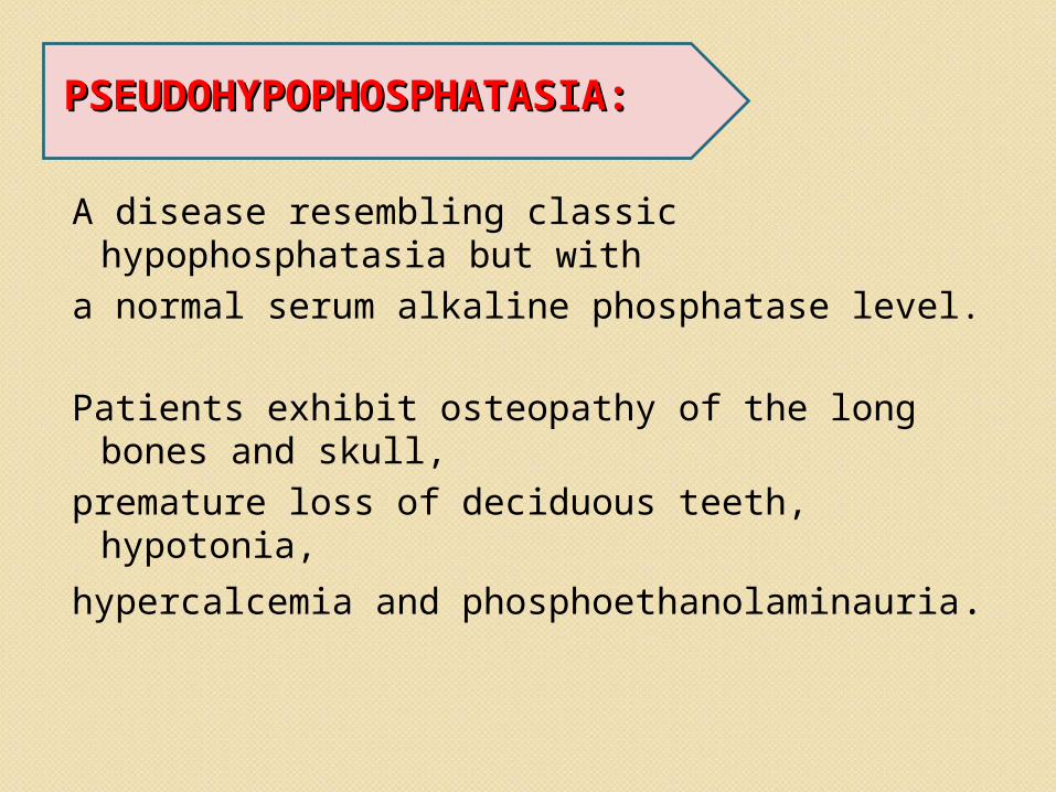
PSEUDOHYPOPHOSPHATASIA:PSEUDOHYPOPHOSPHATASIA:
A disease resembling classic hypophosphatasia but with
a normal serum alkaline phosphatase level.
Patients exhibit osteopathy of the long bones and skull,
premature loss of deciduous teeth, hypotonia, hypercalcemia and phosphoethanolaminauria.
• It has often been supposed that a low intake of calcium, or phosphorous, or both might lead to poor calcification of teeth and possibly, therefore, to an increased risk of dental caries.
• the calcification of the teeth could be affected if calcium was very low in the diet.
• deficiencies in calcium and phosphate
intake do not affect tooth calcification they do reduce that of bone, and they result in mobilization of calcium from already formed bone.
• In pregnancy, when the dietary need of the mother for calcium and phosphate are increased by the demands imposed by the growing fetus, there is mobilization of bone calcium if the dietary supply is insufficient.
ConclusionConclusion
Disturbances in calcium and phosphate intake, excretion and transcellular shift result in deranged metabolism accounting for abnormal serum levels. As a result of the essential role played by these minerals in intra and extracellular metabolism, the clinical manifestations of related disease states are extensive.
Thus, an understanding of the basic mechanism of calcium, phosphate metabolism and pathophysiology of various related disorders is helpful in guiding therapeutic decisions.
ReferencesReferencesTextbook of Biochemistry by U. Satyanarayana,
second edition.Essentials of Medical Physiology by K. Sambulingam, third edition.Textbook of Medical Physiology by Guyton and Hall,
tenth edition.Shafer’s textbook of oral pathology, Fifth edition Burkets oral medicine 11th editionClinical Disorders of Phosphorous Metabolism, West J
Med. 1987 November; 147(5): 569–576 Calcium and Phosphate Metabolism - Annual Review of
Physiology Vol. 36: 361-390 A B Borle


![Free DNA precipitates calcium phosphate apatite crystals ... · phosphorous from intracellular metabolism and pro-calcifying membrane phospholipid-rich micro-particles [8e10]. On](https://img.pdfslide.net/doc/110x75/6031c1e03a8161564e2969ce/free-dna-precipitates-calcium-phosphate-apatite-crystals-phosphorous-from-intracellular.jpg)


![Calcium metabolism [Pharmacology]](https://img.pdfslide.net/doc/110x75/577cc71b1a28aba7119ffe20/calcium-metabolism-pharmacology.jpg)