Embed Size (px)
Citation preview

Color Atlas of Emergency Trauma
This full-color atlas presents nearly 700 images from the largest and busiest trauma centerin North America. The images bring the reader to the bedside of patients with the fullspectrum of common and uncommon traumatic injuries including motor vehicle acci-dents, falls, lacerations, burns, impalements, stabbings, and gunshot wounds. The clinical,operative, and autopsy photographs; x-ray, ultrasound, magnetic resonance imaging, andangiography radiographs; and original illustrations depicting injury patterns will helpguide clinicians in recognizing, prioritizing, and managing trauma patients. Organized bymajor body regions into separate chapters on the head, face, neck, chest, abdomen, mus-culoskeletal system, spine, and soft tissue, this thorough text discusses management guide-lines, emergency workup protocols, and common pitfalls. The Color Atlas of EmergencyTrauma is an essential resource for those involved in trauma care.
Diku P. Mandavia, MD, is Attending Staff Physician in the Department of EmergencyMedicine at Los Angeles County-USC Medical Center and at Cedars-Sinai Medical Cen-ter in Los Angeles. As Clinical Associate Professor of Emergency Medicine at the Uni-versity of Southern California's Keck School of Medicine, Dr. Mandavia specializes inemergency ultrasound and trauma care.
Edward J. Newton, MD, is the former Vice Chairman and current Interim Chairman ofthe Los Angeles County-USC Medical Center's Department of Emergency Medicine. Dr.Newton is also Associate Professor of Clinical Emergency Medicine at the University ofSouthern California's Keck School of Medicine.
Demetrios Demetriades MD, PhD, is Professor of Surgery at the University of SouthernCalifornia's Keck School of Medicine. Dr. Demetriades is also Director of the Trauma Pro-gram and Surgical Intensive Care Unit at the Los Angeles County-USC Medical Center,the largest trauma center in the United States.

ATLAS OF
EMERGENCY TRAUMA
Diku P. Mandavia, MD, FACEP, FRCPCClinical Associate Professor of Emergency MedicineKeck School of MedicineUniversity of Southern California
Attending Staff PhysicianDepartment of Emergency MedicineLos Angeles County-USC Medical CenterDepartment of Emergency MedicineCedars-Sinai Medical CenterLos Angeles, California
Edward J. Newton, MD, FACEP, FRCPCAssociate Professor of Clinical Emergency MedicineKeck School of MedicineUniversity of Southern California
Interim ChairmanDepartment of Emergency MedicineLos Angeles County-USC Medical CenterLos Angeles, California
Demetrios Demetriades, MD, PhD, FACSProfessor of SurgeryKeck School of MedicineUniversity of Southern CaliforniaDirectorDivision of Trauma and Critical CareLos Angeles County-USC Medical CenterLos Angeles, California
CAMBRIDGEUNIVERSITY PRESS

PUBLISHED BY THE PRESS SYNDICATE OF THE UNIVERSITY OF CAMBRIDGEThe Pitt Building, Trumpington Street, Cambridge, United Kingdom
CAMBRIDGE UNIVERSITY PRESSThe Edinburgh Building, Cambridge CB2 2RU, UK40 West 20th Street, New York, NY 10011-4211, USA477 Witliamstown Road, Port Melbourne, VIC 3207, AustraliaRuiz de Alarc6n 13, 28014 Madrid, SpainDock House, The Waterfront, Cape Town 8001, South Africa
http://www.cambridge.org
© Diku P. Mandavia, Edward J. Newton, Demetrios Demetriades
This book is in copyright. Subject to statutory exception
and to the provisions of relevant collective licensing agreements,no reproduction of any part may take place withoutthe written permission of Cambridge University Press.
First published 2003
Printed in Singapore
Typefaces Goudy 11/13 pt. and Stone Sans System QuarkXPress™ [HT]
A catalog record for this book is available from the British Library.
Library of Congress Cataloging in Publication Data
Mandavia, Diku P., 1965-Color atlas of emergency trauma / Diku P. Mandavia, Edward ]. Newton,
Demetrios Demetriades.p. cm.
Includes bibliographical references and index.ISBN 0-521-78148-5 (hardback)
1. Medical emergencies—Atlases. 2. Emergency medical services—Atlases. 3.Wounds and injuries—Atlases. I. Newton, Edward, 1950-11. Demetriades,D e m e t r i o s , 1951—III. Title.
[DNLM: 1. Wounds and Injuries—Atlases. 2. Emergencies—Atlases. WO 517M271c2OO3]
RC86.7 M3478 2003616.02'5'0222—dc21 2002191221
ISBN 0 521 78148 5 hardback
Every effort has been made in preparing this book to provide accurate and up-to-dateinformation that is in accord with accepted standards and practice at the time ofpublication. Nevertheless, the authors, editors, and publisher can make nowarranties that the information contained herein is totally free from error, not leastbecause clinical standards are constantly changing through research and regulation.The authors, editors, and publisher therefore disclaim all liability for direct orconsequential damages resulting from the use of material contained in this book.Readers are strongly advised to pay careful attention to information provided by themanufacturer of any drugs or equipment that they plan to use.

To my parents, my brother Sujal, my precious daughter Neela, and especiallyto my loving wife Katina who has supported me throughout my career.
DM
Dedicated with thanks to my family and colleagues.To my students of trauma and emergency care, may the collected experience
contained in this text assist your pursuit of excellence.
EN
To my parents, my wife Elizabeth, my daughters Alexis and Stefanie,and my son Nicky.
DD

Contents
11.11.21.31.41.51.6
1.7
1.8
1.9
1.101.111.121.13
1.141.15
1.16
22.12.22.3
2.42.52.62.7
Photographic Acknowledgments xi
Forewords xiii
Preface xv
Acknowledgments xvii
HEAD INJURY 1Scalp Injuries 4
Linear Skull Fracture 4Depressed Skull Fracture 6
Open Skull Fracture 8Basilar Skull Fracture 9
Epidural Hematoma 11
Subdural Hematoma 13
Traumatic Subarachnoid Hemorrhage 18
Cerebral Contusion 20
Penetrating Head Injury 22Transtentorial Herniation 24Diffuse Cerebral Edema 26
Pediatric Head Injury 27Axonal Shear Injury 29
Intraventricular Hemorrhage 31
Intraparenchymal Hemorrhage 31
FACIAL INJURY 33Corneal Abrasion 36Ocular Foreign Bodies 36
Hyphema 38
Ruptured Globe 38Retrobulbar Hematoma 40Periorbital Lacerations 41
Orbital Blowout Fracture 41
2.82.92.10
2.11
2.122.13
2.142.15
33.1
3.23.3
3.43.5
3.6
3.73.8
3.93.10
3.113.12
3.13
3.143.15
3.16
Mandible Fracture 44Zygoma Fractures 45
Le Fort Fractures 46
Nasal Injuries 48
Complex Facial Lacerations 49Oral Lacerations 52
Facial Nerve Injury 54Facial Artery Injury 55
NECK INJURY 57
Anatomical Zones of the Neck 59Epidemiology of Penetrating Neck Trauma 59
Physical Examination of PenetratingInjuries of the Neck 60Homer's Syndrome 62
Protocol for Initial Evaluation andManagement of Penetrating Injuriesto the Neck 62
Plain Radiography in Penetrating NeckTrauma 63CT Scan in Penetrating Neck Trauma 65
Evaluation of Vascular Structures inthe Neck 66
Vascular Trauma-Zone I 67Vascular Trauma-Zone III 68
Color Flow Doppler (CFD) 69Angiography for Penetrating Injuriesof the Neck 69
Evaluation of the Aerodigestive Tractin the Neck 74Esophageal Trauma 74Airway Establishment in the Presenceof a Neck Hematoma 76Bleeding Control in the EmergencyDepartment 77
vii
3.17 Operative Approaches 78
3.18 Penetrating Trauma to the Carotid Artery 79
3.19 Penetrating Injuries to the VertebralArtery 80
3.20 Blunt Carotid Injury 80
3.21 Blunt Laryngotracheal Trauma 81
4 THORACIC INJURY 83
4.1 Chest Wall Seatbelt Mark 86
4.2 Fractures of the Upper (First or Second)Ribs 87
4.3 Fractures of Middle (Third to Eighth)Ribs 87
4-4 Fractures of the Lower (Ninth to Twelfth)Ribs 88
4.5 Flail Chest 89
4.6 Pneumo thorax 90
4-7 Tension Pneumothorax 91
4.8 Hemothorax 93
4-9 Thoracostomy Tube Insertion 94
4.10 Autotransfusion of Blood in ChestTrauma 95
4-11 Lung Contusion 964.12 Subcutaneous Emphysema 984-13 Penetrating Cardiac Injury 994-14 Mechanisms of Penetrating Cardiac
Injury 994.15 Survival Factors in Penetrating Cardiac
Injuries 1004.16 Diagnosis of Cardiac Injury 101
4.17 Retained Cardiac Missiles 1034.18 Emergency Department Thoracotomy for
Cardiac Trauma 1044-19 Late Sequelae of Penetrating Cardiac
Injuries 108
4.20 Blunt Cardiac Trauma 109
4.21 Blunt Thoracic Aortic Injury 110
4.22 Penetrating Thoracic Outlet Injuries 1134-23 Transmediastinal Gunshot Wounds 1164.24 Penetrating Diaphragmatic Injuries 1194-25 Blunt Diaphragmatic Injuries 120
4.26 Traumatic Diaphragmatic Hernias 1234-27 Esophageal Injuries 125
4.28 Thoracic Duct Injury 126
4.29 Traumatic Asphyxia 127
5 ABDOMINAL INJURY 129
5.1 Mechanism of Injury in Blunt AbdominalTrauma 132
5.2 Seatbelt Sign 133
5.3 Diagnosis of Hemoperitoneum 133
5.4 Splenic Injuries 137
5.5 Liver Injuries 139
5.6 Pancreatic Injuries 142
5.7 Renal Injuries 144
5.8 Bladder Injuries 146
5.9 Urethral Injuries 148
5.10 Duodenal Injuries 149
5.11 Small-Bowel Injuries 150
5.12 Colorectal Injuries 151
5.13 Mechanism of Injury in PenetratingAbdominal Trauma 151
5.14 Investigations in Penetrating AbdominalInjury 153
5.15 Transpelvic Gunshot Injuries 1555.16 Penetrating Injuries to the Liver 156
5.17 Penetrating Splenic Injuries 158
5.18 Penetrating Pancreatic Injuries 159
5.19 Penetrating Renal Injuries 159
5.20 Penetrating Colorectal Injuries 161
5.21 Abdominal Vascular Injuries 1625.22 Retained Bullets 1645.23 Abdominal Trauma in Pregnancy 164
6 MUSCULOSKELETALINJURY 167
6.1 Classification of Fractures 1696.2 Open Fractures 1706.3 Open Joint Injury 171
6.4 Salter-Harris Classification 172
6.5 Torus and Greenstick Fractures 1746.6 Supracondylar Fracture 175
6.7 Amputations 1766.8 Tendon Injury 1796.9 Peripheral Vascular Injury 1796.10 Peripheral Nerve Injury 182
6.11 Metacarpal Fractures 1846.12 First Metacarpal Fractures (Bennett's and
Rolando's Fractures) 185
Contents
6.13 Scaphoid Fracture 186
6.14 Scapholunate Dislocation 1876.15 Lunate and Perilunate Dislocation 187
6.16 Colles'Fracture 189
6.17 Forearm Fractures 1906.18 Elbow Dislocation 191
6.19 Radial Head Fracture 192
6.20 Humeral Fracture 193
6.21 Shoulder Dislocations 194
6.22 Clavicle Fracture 1966.23 Sternoclavicular Dislocation 198
6.24 Scapula Fracture 199
6.25 Pelvic Fractures 2006.26 Hip Dislocation 203
6.27 Hip Fracture 2066.28 Femoral Shaft Fracture 2086.29 Patellar Fracture 2096.30 Tibial Plateau Fracture 2106.31 Knee Dislocation 211
6.32 Maisonneuve Fracture Complex 2136.33 Ankle Dislocation 213
6.34 Subtalar Dislocation 2146.35 Lisfranc Fracture/Dislocation 215
6.36 Metatarsal Base Fractures 216
6.37 Calcaneal Fracture 217
7 SPINAL INJURY 2197.1 Central Cord Syndrome 2227.2 Brown-Se"quard Syndrome 2237.3 Anterior Cord Syndrome 225
7.4 Complete Spinal Cord Transection 225
7.5 Atlanto-Occipital Dislocation 2287.6 Atlantoaxial Dislocation 2297.7 Rotatory Subluxation of C-l on C-2 230
7.8 C-l Burst Fracture (Jefferson Fracture) 2317.9 Odontoid (Dens) Fractures (C-2) 2337.10 Hangman's Fracture (C-2) 236
7.11 C-3-C-7 Fracture Types 2377.12 Flexion and Extension Teardrop Fracture 238
7.13 Compression or Burst Fracture of the VertebralBody 240
7.14 Clay Shoveler's Fracture 241
7.15 Fractures of the Pedicles, Laminae, and LateralMasses 242
7.16 Facet Dislocation 243
7.17 Cervicothoracic Spinal Injury 246
7.18 Thoracic Spine Injuries 247
7.19 Lumbar Compression Burst Fracture 249
7.20 Chance Fracture 2527.21 Fracture-Dislocation of the Lumbar
Spine 254
7.22 Ligamentous Injuries 2557.23 Pediatric Spinal Injury 256
8 SKIN AND SOFT TISSUEINJURY
8.1 Burn Zones of Injury 261
8.2 First-Degree Burns 2628.3 Second-Degree Burns 263
8.4 Third-Degree Burns 2648.5 Fourth-Degree Burns 2668.6 Extent of Burn Injury 266
8.7 Circumferential Burns 267
8.8 Scald Burns 270
8.9 Chemical Burns 270
8.10 Electrical Injury 272
8.11 Inhalational Injury 2748.12 Dog Bite Injury 2758.13 Cat Bite Injury 2768.14 Human Bite Injury 2768.15 Retained Foreign Body 2788.16 Major Soft Tissue Injury 278
8.17 Compartment Syndrome 279
8.18 High-Pressure Injection Injury 282
Index 285
259
Contents

PhotographicAcknowledgments
Major Contributor
Marie Russell, MDLos Angeles County-USC Medical Center
The authors thank Dr. Marie Russell for her generousphotographic contributions to the text.
Photographic Contributors
Joel Aronowitz, MDCedars-Sinai Medical Center
Paul Carter, MDLos Angeles County-USC Medical Center
Jeff Cohen, MDLos Angeles County-USC Medical Center
John L. Go, MDLos Angeles County-USC Medical Center
Jason Greenspan, MDLos Angeles County-USC Medical Center
Anthony Joseph, MDRoyal Northshore Hospital, Sydney, Australia
Cindy Kallman, MDCedars-Sinai Medical Center
Larry Khoo, MDLos Angeles County-USC Medical Center
William Mallon, MDLos Angeles County-USC Medical Center
Sujal Mandavia, MDCedars-Sinai Medical Center
John Michael, MDBoston, Massachusetts
Peter Mishky, MDNaval Medical Center, San Diego, California
Jack Pulec, MDPulec Ear Clinic, Los Angeles
Paul Prendiville, MDLaguna Beach, California
Bronwyn Pritchard, MDLaguna Beach, California
Jeff Sipsey, MDLos Angeles County-USC Medical Center
Mark Tiara, MDLos Angeles County-USC Medical Center
James Tourge, MDCedars-Sinai Medical Center
xi

Forewords
Though many texts are written in medicine, few have the impact of an atlas that can cap-ture the presence of being at the bedside. The authors share the visual aspects of traumacare and allow the reader to more readily understand textual descriptions. They have pro-duced a comprehensive atlas of trauma that is an excellent reference for physiciansinvolved in trauma care.
The experienced trauma clinicians writing this text present their collective experiencein a visual manner that represents many years of dedicated image collection and collabora-tive efforts from the Department of Emergency Medicine and the Division of Trauma at LosAngeles County-University of Southern California Medical Center. This Center is one ofthe busiest and most active trauma centers in the world.
Gail V. Anderson Sr., MDProfessor and Chairman
Department of Emergency MedicineUniversity of Southern California
There is no end to the writing of books, and their shelf life is limited by the march of under-standing. Surgical atlases are less common and their effect lasts longer. Perhaps this is dueto their dependency on the experience of the authors. Time is required between simply per-forming a procedure and becoming intimately familiar with the procedure. It is the latterthat provides technical insights and make the atlas of value.
Most atlases consist of an artist's conception of the procedures involved. They are a stepremoved from the real world and there are concerns of authenticity. A danger exists thatthe illustrations will encourage action without a thorough understanding of the pathophys-iological principles involved. The latter differentiates knowledge about from knowledge ofthe procedure being described and the problems it is designed to correct. The best atlasescombine a short written description of the principles involved with the actual operativephotographs. They bring the reader a step closer to the actual encounter.
Such is the atlas produced by the Division of Trauma and the Department of EmergencyMedicine at the University of Southern California Keck School of Medicine. The authorshave produced an atlas on trauma consisting of color photographs and accompanied by textthat focuses on the principles involved. Being familiar with the impediments to stoppingduring an operative procedure or a resuscitative effort to record the pathology on cameragives one an appreciation for the efforts employed in accomplishing such a task. Theauthors of this atlas get my highest commendation. I encourage those interested in the sub-ject to enjoy this work while also being educated by it.
Tom R. DeMeester, MDProfessor and Chairman
Department of SurgeryUniversity of Southern California
xiii

Preface
Good trauma care requires a substantial knowledge base and clinical skill. The compre-hension and intuition required to treat trauma injury is gained over many years of clinicalexperience at the bedside of critically injured patients. The aim of this atlas is to share theexperience of the authors from the largest trauma center in the United States and providea solid companion to the many well-written textbooks on trauma management.
This project represents many decades of collective clinical experience. We have assem-bled one of the largest collections of trauma images to help bring the reader "to the bed-side" of the patients. The majority of the photographs originate from the Los AngelesCounty-University of Southern California Trauma Center though some special photo-graphs were donated from outside centers. The acquisition and final assembly of this col-lection of images was a difficult process and they were acquired with the graciouscooperation of our patients. We regularly use these images in our clinical teaching andhope this atlas will supplement other instructional resources in trauma management.
Diku P. Mandavia, MDEdward]. Newton, MD
Demetrios Demetriades, MD, PhD

Acknowledgments
Illustrations and Digital Imaging by:
Robert S. Amaral, MAMedical IllustratorInstructional Imaging CenterKeck School of MedicineUniversity of Southern California
Our appreciation to the following:
Burn Unit, Los Angeles County-USC Medical CenterDepartment of Radiology, Los Angeles County-USC Medical CenterDepartment of Imaging, Cedars-Sinai Medical Center
And to the staff and residents of:
Department of Emergency Medicine & Division of TraumaLos Angeles County-USC Medical CenterDepartment of Emergency Medicine &. Division of TraumaCedars-Sinai Medical Center
xvii


Head Injury
Introduction
Head injury accounts for the majority of deaths andsevere disabilities due to trauma, and complicates themanagement of injury to other systems. Blunt headinjury is most commonly the result of motor vehiclecrashes, auto versus pedestrian collisions, or falls fromsignificant heights. Gunshot wounds cause the vastmajority of penetrating head injuries, although stabwounds and impalement injuries may also be seen.
Clinical Examination
Head injury is classified into mild, moderate, andsevere categories, depending on the patient's Glas-gow Coma Scale (GCS) (see Table 1.1) at the timeof presentation. Mild head injury patients have aGCS of 14-15. Typically these injuries include con-cussion or transient loss of neurologic function, usu-ally a transient loss of consciousness. Concussiondoes not result in any gross pathologic abnormalitiesof the brain, but subtle changes have been describedusing electron microscopy. Although the neurologicexamination is usually normal in terms of quantifi-able deficits, post-concussive neuropsychiatric symp-toms are very common. These include amnesia forthe event, headache, loss of concentration, dizziness,sleep disturbance, and a host of related symptoms.These symptoms resolve within two weeks for thevast majority of patients but may persist for manymonths in a small percentage. "Hard" neurologicfindings such as diplopia, motor weakness, pupillaryabnormalities, and other cranial nerve deficits arenever due to post-concussive syndrome and demandfurther investigation.
Moderate head injury (GCS 8-14) and severe headinjury (GCS <8) comprise a spectrum of injuriesincluding cerebral contusion, diffuse cerebral edema,axonal shear injury, subarachnoid hemorrhage (SAH),
and extra-axial lesions (subdural hematoma andepidural hematoma). The GCS is determined by theseverity of the lesion and its time course.
A rapid but thorough neurologic examination isdone prior to administering paralytic agents. The headis inspected for signs of trauma including lacerations,areas of skull depression, raccoon eyes, Battle's sign,hemotympanum, rhinorrhea, and otorrhea. The pupilsare examined for symmetry and reaction to light; theextraocular movements and other cranial nerve func-tions are assessed; motor and sensory function isassessed for symmetry. The GCS is useful in categorizingpatients as to severity of head injury but is not sufficientto determine the presence or absence of neurologicinjury, as it does not assess pupils, subtle changes inmentation, cranial nerve injury, or skull fractures.
Patients with increased intracranial pressure (ICP)inevitably have some diminution in their typical levelof alertness. As ICP increases further, herniation of theuncus across the tentorium can occur, resulting in com-pression of the ipsilateral cerebral peduncle and ipsilat-eral oculomotor nerve. Consequently, ipsilateral ptosis,restricted extraocular movement, and pupillary dila-tion occur, along with contralateral motor posturing(decorticate, followed by decerebrate, and finally flac-cid paralysis). Once this process begins, there is a verybrief interval for effecting a reversal by lowering ICP.
Skull fractures are common sequelae of both bluntand penetrating head trauma. Although it may be anisolated finding, skull fracture is frequently associatedwith intracranial injury. Closed simple linear frac-tures of the cranial vault are relatively benign inthemselves but signify that substantial force has beenapplied to the cranium, putting its more delicate cere-bral contents at risk. Skull fractures are particularlydangerous in certain anatomic locations, such asacross the middle meningeal arterial groove, acrossdural sinuses, or in the occipital area because theintracranial bleeding associated with these fracturesmay be life threatening.
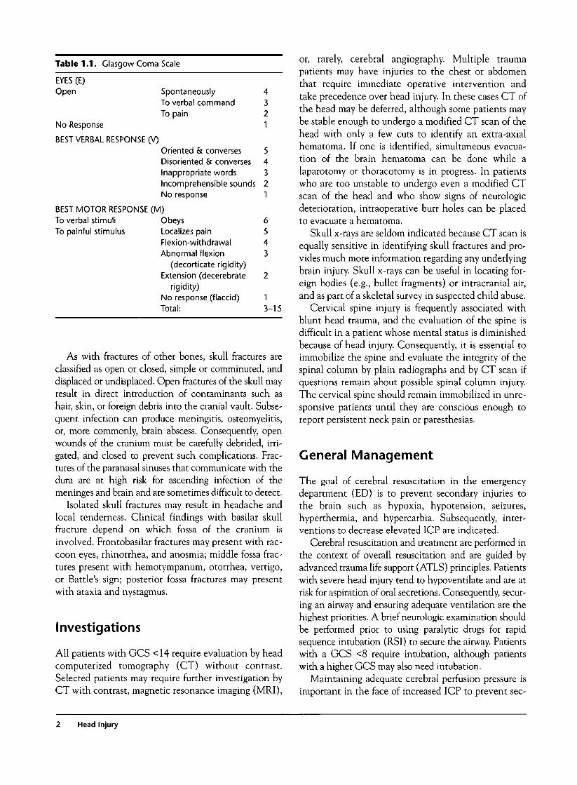
Table 1.1. Glasgow Coma
EYES (E)Open
No Response
BEST VERBAL RESPONSE (V)
Scale
SpontaneouslyTo verbal commandTo pain
Oriented & conversesDisoriented & conversesInappropriate wordsIncomprehensible soundsNo response
BEST MOTOR RESPONSE (M)To verbal stimuliTo painful stimulus
ObeysLocalizes painFlexion-withdrawalAbnormal flexion
(decorticate rigidity)Extension (decerebrate
rigidity)No response (flaccid)Total:
4321
54321
6543
2
13-15
As with fractures of other bones, skull fractures areclassified as open or closed, simple or comminuted, anddisplaced or undisplaced. Open fractures of the skull mayresult in direct introduction of contaminants such ashair, skin, or foreign debris into the cranial vault. Subse-quent infection can produce meningitis, osteomyelitis,or, more commonly, brain abscess. Consequently, openwounds of the cranium must be carefully debrided, irri-gated, and closed to prevent such complications. Frac-tures of the paranasal sinuses that communicate with thedura are at high risk for ascending infection of themeninges and brain and are sometimes difficult to detect.
Isolated skull fractures may result in headache andlocal tenderness. Clinical findings with basilar skullfracture depend on which fossa of the cranium isinvolved. Frontobasilar fractures may present with rac-coon eyes, rhinorrhea, and anosmia; middle fossa frac-tures present with hemotympanum, otorrhea, vertigo,or Battle's sign; posterior fossa fractures may presentwith ataxia and nystagmus.
Investigations
All patients with GCS <14 require evaluation by headcomputerized tomography (CT) without contrast.Selected patients may require further investigation byCT with contrast, magnetic resonance imaging (MRI),
or, rarely, cerebral angiography. Multiple traumapatients may have injuries to the chest or abdomenthat require immediate operative intervention andtake precedence over head injury. In these cases CT ofthe head may be deferred, although some patients maybe stable enough to undergo a modified CT scan of thehead with only a few cuts to identify an extra-axialhematoma. If one is identified, simultaneous evacua-tion of the brain hematoma can be done while alaparotomy or thoracotomy is in progress. In patientswho are too unstable to undergo even a modified CTscan of the head and who show signs of neurologicdeterioration, intraoperative burr holes can be placedto evacuate a hematoma.
Skull x-rays are seldom indicated because CT scan isequally sensitive in identifying skull fractures and pro-vides much more information regarding any underlyingbrain injury. Skull x-rays can be useful in locating for-eign bodies (e.g., bullet fragments) or intracranial air,and as part of a skeletal survey in suspected child abuse.
Cervical spine injury is frequently associated withblunt head trauma, and the evaluation of the spine isdifficult in a patient whose mental status is diminishedbecause of head injury. Consequently, it is essential toimmobilize the spine and evaluate the integrity of thespinal column by plain radiographs and by CT scan ifquestions remain about possible spinal column injury.The cervical spine should remain immobilized in unre-sponsive patients until they are conscious enough toreport persistent neck pain or paresthesias.
General Management
The goal of cerebral resuscitation in the emergencydepartment (ED) is to prevent secondary injuries tothe brain such as hypoxia, hypotension, seizures,hyperthermia, and hypercarbia. Subsequently, inter-ventions to decrease elevated ICP are indicated.
Cerebral resuscitation and treatment are performed inthe context of overall resuscitation and are guided byadvanced trauma life support (ATLS) principles. Patientswith severe head injury tend to hypoventilate and are atrisk for aspiration of oral secretions. Consequently, secur-ing an airway and ensuring adequate ventilation are thehighest priorities. A brief neurologic examination shouldbe performed prior to using paralytic drugs for rapidsequence intubation (RSI) to secure the airway. Patientswith a GCS <8 require intubation, although patientswith a higher GCS may also need intubation.
Maintaining adequate cerebral perfusion pressure isimportant in the face of increased ICP to prevent sec-
Head Injury
ondary ischemic injury. Consequently, measures tomaintain adequate systemic blood pressure are essen-tial and include crystalloid infusion, blood transfusion,thoracotomy, laparotomy, and pressors as indicated.
For patients with evidence of increased ICP or clin-ical signs of actual or impending transtentorial hernia-tion, immediate measures to decrease ICP areindicated. Hyperventilation to reduce pCO2 to a levelof 30-35 mm Hg decreases cerebral blood flow andthus decreases intracranial blood volume, allowing atemporary decrease in ICP. Osmotic diuresis with man-nitol and use of loop diuretics such as furosemide dehy-drate all tissues including brain, again allowing morespace for an expanding hematoma and lowering ICP.These medications must be used with extreme cautionif at all in multiply injured patients, as severe systemichypotension may result. Positioning the head at 30degrees elevation can decrease ICP once the spine hasbeen cleared radiographically. Placement of a ventricu-lostomy to drain cerebrospinal fluid (CSF) has beenshown to be effective not only in treating elevated ICPbut also in following the progress of the patient's condi-tion. Use of hypertonic saline, cerebral or systemicmild hypothermia, and administration of magnesiumsulfate have been shown experimentally to improveoutcome from severe head injury.
Definitive treatment for head injury depends on thenature of the lesion. Closed skull fractures require nospecific treatment, but open fractures should be irri-gated, debrided, and closed. Depressed skull fracturesrequire elevation of the fragment if it is depressedgreater than one bone width, and debridement if thewound is grossly contaminated. Basilar skull fracturesusually heal uneventfully, but patients with rhinorrheaor otorrhea require careful follow-up to ensure that thefistula closes. Most CSF leaks stop within two weeks,but persistent leaks may require a formal dural closure.Most epidural hematomas (EDHs) require surgicalevacuation, although those that are <1 cm can betreated by observation and repeat CT scan if thepatient is asymptomatic. Larger EDHs require cran-iotomy for evacuation. Subdural hematoma (SDH) israrely asymptomatic, and surgical treatment is almostinvariably needed to evacuate the hematoma. Sub-arachnoid hemorrhage is treated with nimodipine todecrease surrounding vasospasm, and measures to
decrease rebleeding are undertaken. Intraventricularhemorrhage may require ventriculostomy to removeblood and CSF, but the prognosis usually remains poor.Patients requiring surgery and those with depressedskull fracture and cerebral contusion should be startedon a course of anticonvulsant medication.
Common Mistakes and Pitfalls
1. Certain patients are at higher risk for intracranialinjury from even relatively minor mechanisms ofinjury. These include the elderly, chronic alco-holics, infants, patients with cerebral atrophy,and those with coagulopathy. A low threshold forobtaining a head CT scan should be maintainedin these patients.
2. Altered mental status, seizures, and focal neuro-logic deficits should not be ascribed to intoxica-tion, dementia, or other chronic conditions ifthere is a history or evidence of head traumapresent.
3. Excessive hyperventilation (to a pCO2 <30 mmHg) should be avoided because it lowers cerebralblood flow to a point that cerebral ischemia canoccur.
4- Delayed presentation of SDH and EDH can occur,often with subtle neurologic signs. Obtaining arepeat CT scan or initial CT scan even weeks afterthe injury is appropriate in selected cases.
5. Subacute SDH may appear isodense with sur-rounding brain five to ten days after the injury.Altering the density values of the CT or use ofcontrast will demonstrate the lesion.
6. Coagulopathy is common with serious headinjury and may result in more severe bleeding,hemorrhage from other noncerebral sites, anddisseminated intravascular coagulopathy (DIC).A baseline coagulation profile should beobtained in patients with serious head injury andrepeated periodically during admission.
7. Child abuse must be suspected in cases of intra-cerebral injury or skull fracture in infants andchildren.
Head Injury
1.1 Scalp Injuries
Commentary
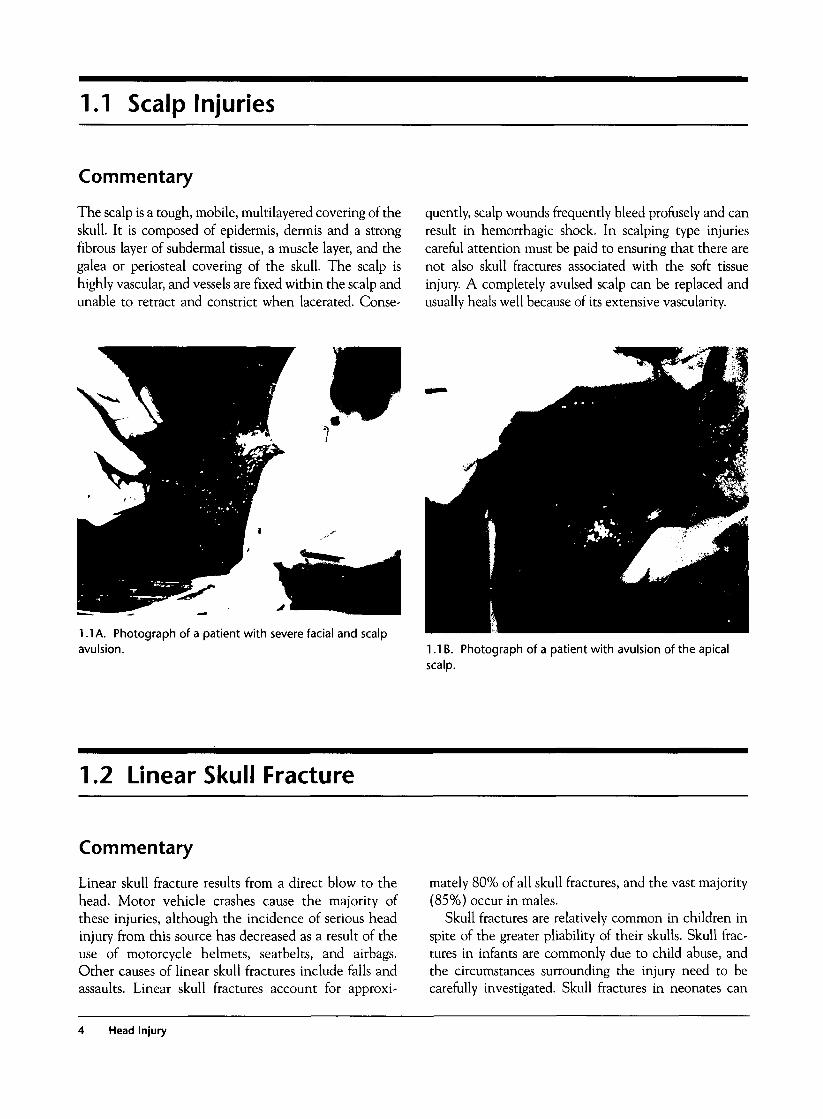
The scalp is a tough, mobile, multilayered covering of theskull. It is composed of epidermis, dermis and a strongfibrous layer of subdermal tissue, a muscle layer, and thegalea or periosteal covering of the skull. The scalp ishighly vascular, and vessels are fixed within the scalp andunable to retract and constrict when lacerated. Conse-
quently, scalp wounds frequently bleed profusely and canresult in hemorrhagic shock. In scalping type injuriescareful attention must be paid to ensuring that there arenot also skull fractures associated with the soft tissueinjury. A completely avulsed scalp can be replaced andusually heals well because of its extensive vascularity.
1.1 A. Photograph of a patient with severe facial and scalpa\/i ilcinnavulsion. L IB . Photograph of a patient with avulsion of the apical
scalp.
1.2 Linear Skull Fracture
Commentary
Linear skull fracture results from a direct blow to thehead. Motor vehicle crashes cause the majority ofthese injuries, although the incidence of serious headinjury from this source has decreased as a result of theuse of motorcycle helmets, seatbelts, and airbags.Other causes of linear skull fractures include falls andassaults. Linear skull fractures account for approxi-
mately 80% of all skull fractures, and the vast majority(85%) occur in males.
Skull fractures are relatively common in children inspite of the greater pliability of their skulls. Skull frac-tures in infants are commonly due to child abuse, andthe circumstances surrounding the injury need to becarefully investigated. Skull fractures in neonates can
Head Injury

also occur in association with a cephalohematoma as aresult of a difficult delivery and use of forceps. Manage-ment of these injuries is generally conservative unlessthe skull fragments are depressed. A "growing" skull frac-ture occasionally occurs in children as a result of theinterposition of a leptomeningeal cyst between the frac-ture edges, and it may require surgery. Healing of a skullfracture takes place over many months, so childrenshould be rechecked approximately six months follow-ing a fracture to ensure that proper healing has occurred.
A skull fracture may occasionally become apparenton physical examination while a scalp laceration isexplored. It is felt as a step-off of the normally smoothskull surface. These fractures should be treated as anyother open fracture with debridement, irrigation, andclosure of each layer of the scalp, including the galea.Antibiotic prophylaxis is indicated, and CT scan shouldbe obtained to delineate the extent of the fracture and todetermine if intracranial injury has occurred.
On plain radiographs a linear skull fracture appearsas a straight or minimally branched lucency if the frag-ments are distracted or as a hyperdensity if the frag-ments overlap. A fracture must be distinguished fromsuture lines and vascular grooves. Suture lines have a
characteristic zigzag appearance and are located in pre-dictable locations. They are of near uniform widththroughout their course. Vascular grooves have sequen-tial bifurcations. Fracture lines can cross either of thesestructures and typically are wider in their center, taper-ing in width toward either end. Plain radiography of theskull is rarely obtained in the current era as CT scan haslargely replaced it. Plain radiography may still be usefulin the assessment of depressed skull fractures or locationof foreign bodies within the cranium, such as bulletfragments or impaled objects. Detection of a fracture onplain films or on physical examination is an indicationfor obtaining a CT scan of the head. The incidence ofbrain injury in the presence of a skull fracture is as highas 12%. Fractures across the middle meningeal vasculargroove are associated with epidural hematoma in asmall percent of cases, but 80% of epidural hematomasare associated with a skull fracture.
CT scan will reveal a linear fracture as a gap in theskull and has the advantage of demonstrating anyunderlying injury to the brain. The sensitivity fordetection of skull fracture with CT scan is very highand comparable to the accuracy of plain radiographs.Patients with linear skull fracture can be discharged ifthey have no other intracerebral injury, a normal neu-rologic examination, and reliable follow-up, althoughchildren are generally admitted for observation.
1.2A. AP skull radiograph showing linear skull fracture(arrow B) caused by a gunshot wound (GSW) to the righttemporal area (arrow A).
1.2B. CT scan with bone windows showing right frontal andleft parietal skull fractures (arrows).
1.2 Linear Skull Fracture

1.2C. Lateral skull radiograph showing a comminutedfracture of the apex of the skull (arrow).
1.2E. Photograph of a semicircular linear skull fracture atcraniotomy (arrows).
1.2D. Linear skull fracture on CT in a patient with a macheteinjury.
1.3 Depressed Skull Fracture
Commentary
The typical depressed skull fracture occurs when alarge amount of force is applied to a small area of theskull (e.g., a blow with a hammer) that results in astellate fracture with a depressed center. Physicalexamination of the skull may reveal a depression inthe skull, but more commonly the fracture is not
palpable because of overlying soft tissue swelling.Unless the view is exactly tangential, plain radi-ographs may not reveal the extent of bone depres-sion. CT scan is highly sensitive and accurate indetecting depressed skull fracture as well as underly-ing brain injury.
Head Injury
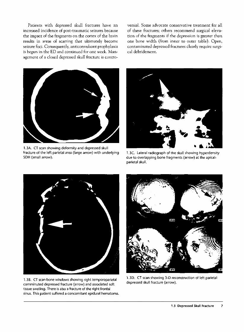
Patients with depressed skull fractures have anincreased incidence of post-traumatic seizures becausethe impact of the fragments on the cortex of the brainresults in areas of scarring that ultimately becomeseizure foci. Consequently, anticonvulsant prophylaxisis begun in the ED and continued for one week. Man-agement of a closed depressed skull fracture is contro-
versial. Some advocate conservative treatment for allof these fractures; others recommend surgical eleva-tion of the fragments if the depression is greater thanone bone width (from inner to outer table). Open,contaminated depressed fractures clearly require surgi-cal debridement.
1.3A. CT scan showing deformity and depressed skullfracture of the left parietal area (large arrow) with underlyingSDH (small arrow).
1.3C. Lateral radiograph of the skull showing hyperdensitydue to overlapping bone fragments (arrow) at the apical-parietal skull.
1.3B. CT scan bone windows showing right temporoparietalcomminuted depressed fracture (arrow) and associated softtissue swelling. There is also a fracture of the right frontalsinus. This patient suffered a concomitant epidural hematoma.
1.3D. CT scan showing 3-D reconstruction of left parietaldepressed skull fracture (arrow).
1.3 Depressed Skull Fracture
1.4 Open Skull Fracture
Commentary
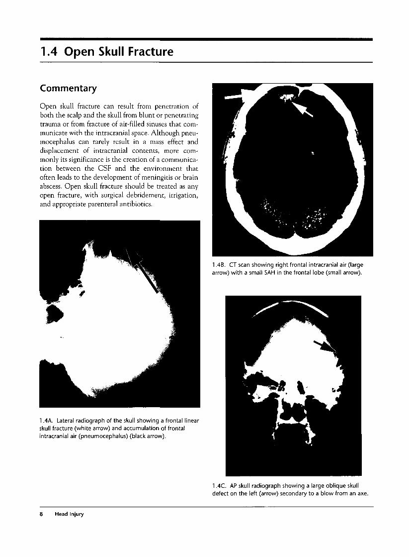
Open skull fracture can result from penetration ofboth the scalp and the skull from blunt or penetratingtrauma or from fracture of air-filled sinuses that com-municate with the intracranial space. Although pneu-mocephalus can rarely result in a mass effect anddisplacement of intracranial contents, more com-monly its significance is the creation of a communica-tion between the CSF and the environment thatoften leads to the development of meningitis or brainabscess. Open skull fracture should be treated as anyopen fracture, with surgical debridement, irrigation,and appropriate parenteral antibiotics.
1.4B. CT scan showing right frontal intracranial air (largearrow) with a small SAH in the frontal lobe (small arrow).
1.4A. Lateral radiograph of the skull showing a frontal linearskull fracture (white arrow) and accumulation of frontalintracranial air (pneumocephalus) (black arrow).
1 AC. AP skull radiograph showing a large oblique skulldefect on the left (arrow) secondary to a blow from an axe.
8 Head Injury

1.4D. Photograph of an open linear skull fracture.
1.5 Basilar Skull Fracture
Commentary
The skull base is divided into three compartments,and the clinical presentation differs depending on thelocation of the fracture. Basilar skull fracture is a clin-ical diagnosis based on physical findings of raccooneyes, Battle's sign, CSF rhinorrhea or otorrhea, orhemotympanum. Injury to cranial nerves that exit thebase of the skull is common, and a careful neurologicexamination is required to seek out these injuries.Occasionally, plain radiographs or CT scan will makethe diagnosis in the absence of these clinical findings,although neither imaging technique is highly sensi-tive. The fracture line may be visualized directly or byindirect evidence such as blood in the sphenoid sinus,the mastoid air cells, or the auditory canal or fractureof the posterior wall of the maxillary sinus. On plain
radiographs, an air-fluid level may be seen in the sphe-noid sinus lying anterior to the pituitary fossa, or thefracture line may be visualized directly.
Because fracture fragments often tear the under-lying dura, leakage of CSF through the nose (rhinor-rhea) or ear canal (otorrhea) is common. If thetympanic membrane remains intact, a hemotympa-num may be seen. Battle's sign occurs because oftracking of blood through the mastoid air cells tothe skin behind the ear, which appears ecchymotic.This sign is often delayed many hours and may notbe apparent on initial examination. Similarly, rac-coon eyes appear sooner but become increasinglyevident with time and may not be apparent on ini-tial examination.
1.5 Basilar Skull Fracture
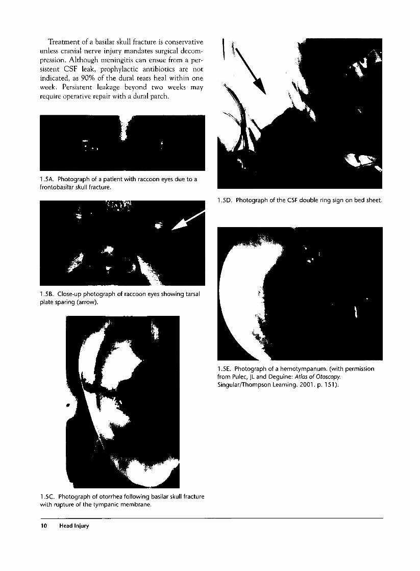
Treatment of a basilar skull fracture is conservativeunless cranial nerve injury mandates surgical decom-pression. Although meningitis can ensue from a per-sistent CSF leak, prophylactic antibiotics are notindicated, as 90% of the dural tears heal within oneweek. Persistent leakage beyond two weeks mayrequire operative repair with a dural patch.
1.5A. Photograph of a patient with raccoon eyes due to afrontobasilar skull fracture.
1.5D. Photograph of the CSF double ring sign on bed sheet.
1.5B. Close-up photograph of raccoon eyes showing tarsalplate sparing (arrow).
1.5E. Photograph of a hemotympanum. (with permissionfrom Pulec, JL and Deguine: Atlas ofOtoscopy.Singular/Thompson Learning. 2001. p. 151).
1.5C. Photograph of otorrhea following basilar skull fracturewith rupture of the tympanic membrane.
10 Head Injury
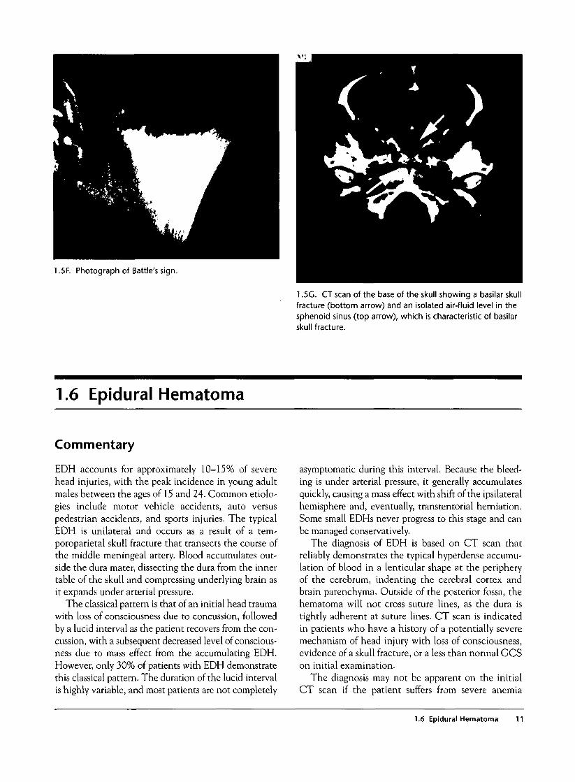
1.5F. Photograph of Battle's sign.
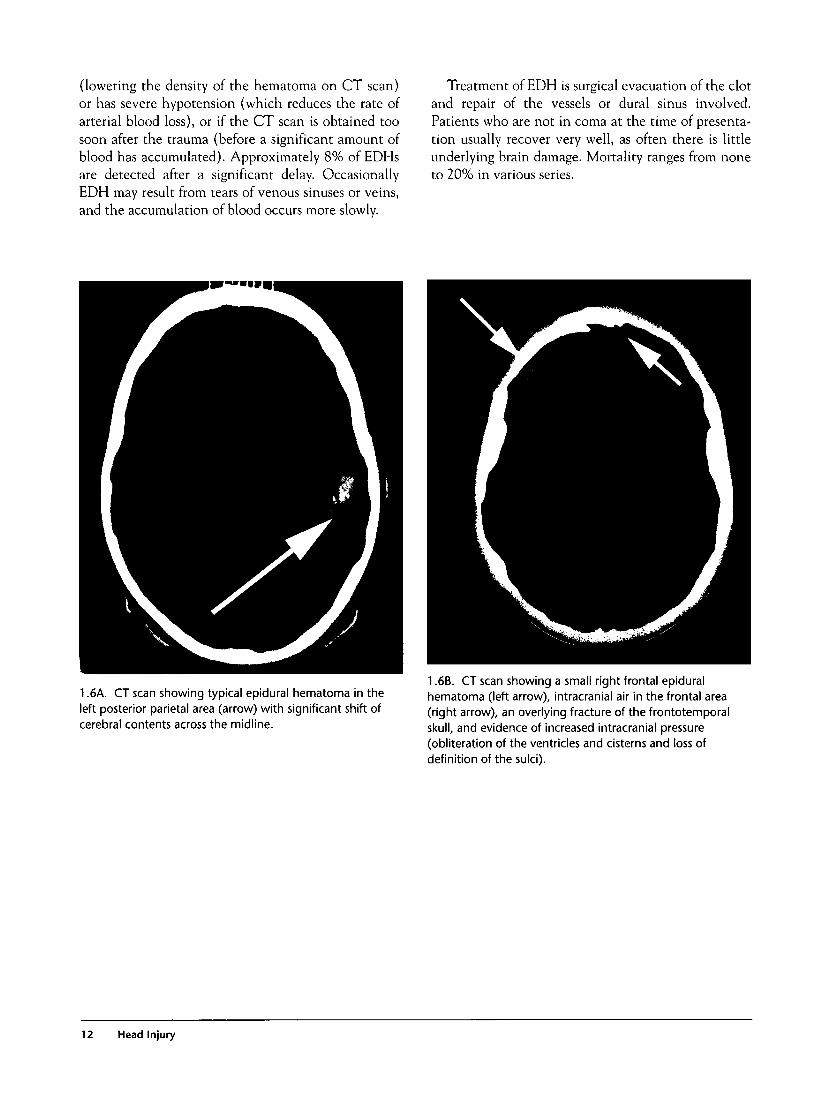
1.5G. CT scan of the base of the skull showing a basilar skullfracture (bottom arrow) and an isolated air-fluid level in thesphenoid sinus (top arrow), which is characteristic of basilarskull fracture.
1.6 Epidural Hematoma
Commentary
EDH accounts for approximately 10-15% of severehead injuries, with the peak incidence in young adultmales between the ages of 15 and 24- Common etiolo-gies include motor vehicle accidents, auto versuspedestrian accidents, and sports injuries. The typicalEDH is unilateral and occurs as a result of a tem-poroparietal skull fracture that transects the course ofthe middle meningeal artery. Blood accumulates out-side the dura mater, dissecting the dura from the innertable of the skull and compressing underlying brain asit expands under arterial pressure.
The classical pattern is that of an initial head traumawith loss of consciousness due to concussion, followedby a lucid interval as the patient recovers from the con-cussion, with a subsequent decreased level of conscious-ness due to mass effect from the accumulating EDH.However, only 30% of patients with EDH demonstratethis classical pattern. The duration of the lucid intervalis highly variable, and most patients are not completely
asymptomatic during this interval. Because the bleed-ing is under arterial pressure, it generally accumulatesquickly, causing a mass effect with shift of the ipsilateralhemisphere and, eventually, transtentorial herniation.Some small EDHs never progress to this stage and canbe managed conservatively.
The diagnosis of EDH is based on CT scan thatreliably demonstrates the typical hyperdense accumu-lation of blood in a lenticular shape at the peripheryof the cerebrum, indenting the cerebral cortex andbrain parenchyma. Outside of the posterior fossa, thehematoma will not cross suture lines, as the dura istightly adherent at suture lines. CT scan is indicatedin patients who have a history of a potentially severemechanism of head injury with loss of consciousness,evidence of a skull fracture, or a less than normal GCSon initial examination.
The diagnosis may not be apparent on the initialCT scan if the patient suffers from severe anemia
1.6 Epidural Hematoma 11
(lowering the density of the hematoma on CT scan)or has severe hypotension (which reduces the rate ofarterial blood loss), or if the CT scan is obtained toosoon after the trauma (before a significant amount ofblood has accumulated). Approximately 8% of EDHsare detected after a significant delay. OccasionallyEDH may result from tears of venous sinuses or veins,and the accumulation of blood occurs more slowly.
Treatment of EDH is surgical evacuation of the clotand repair of the vessels or dural sinus involved.Patients who are not in coma at the time of presenta-tion usually recover very well, as often there is littleunderlying brain damage. Mortality ranges from noneto 20% in various series.
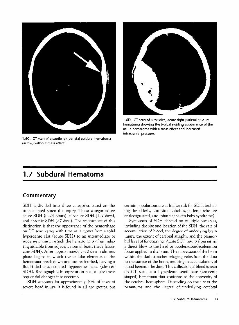
1.6A. CT scan showing typical epidural hematoma in theleft posterior parietal area (arrow) with significant shift ofcerebral contents across the midline.
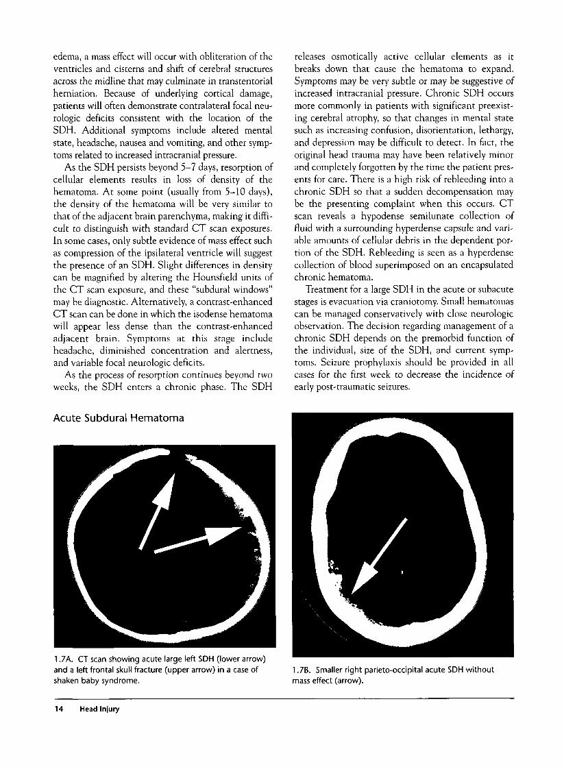
1.6B. CT scan showing a small right frontal epiduralhematoma (left arrow), intracranial air in the frontal area(right arrow), an overlying fracture of the frontotemporalskull, and evidence of increased intracranial pressure(obliteration of the ventricles and cisterns and loss ofdefinition of the sulci).
12 Head Injury
1.6D. CT scan of a massive, acute right parietal epiduralhematoma showing the typical swirling appearance of theacute hematoma with a mass effect and increasedintracranial pressure.
1.6C. CT scan of a subtle left parietal epidural hematoma(arrow) without mass effect.
1.7 Subdural Hematoma
Commentary
SDH is divided into three categories based on thetime elapsed since the injury. These categories areacute SDH (0-24 hours), subacute SDH (1-7 days),and chronic SDH (>7 days). The importance of thisdistinction is that the appearance of the hemorrhageon CT scan varies with time as it moves from a solidhyperdense clot (acute SDH) to an intermediate orisodense phase in which the hematoma is often indis-tinguishable from adjacent normal brain tissue (suba-cute SDH). After approximately 5-10 days a chronicphase begins in which the cellular elements of thehematoma break down and are reabsorbed, leaving afluid-filled encapsulated hypodense mass (chronicSDH). Radiographic interpretation has to take thesesequential changes into account.
SDH accounts for approximately 40% of cases ofsevere head injury. It is found in all age groups, but
certain populations are at higher risk for SDH, includ-ing the elderly, chronic alcoholics, patients who areanticoagulated, and infants (shaken baby syndrome).
Symptoms of SDH depend on multiple variables,including the size and location of the SDH, the rate ofaccumulation of blood, the degree of underlying braininjury, the extent of cerebral atrophy, and the premor-bid level of functioning. Acute SDH results from eithera direct blow to the head or acceleration/decelerationforces applied to the brain. The movement of the brainwithin the skull stretches bridging veins from the durato the surface of the brain, resulting in accumulation ofblood beneath the dura. This collection of blood is seenon CT scan as a hyperdense semilunate (crescent-shaped) hematoma that conforms to the convexity ofthe cerebral hemisphere. Depending on the size of thehematoma and the degree of underlying cerebral
1.7 Subdural Hematoma 13
edema, a mass effect will occur with obliteration of theventricles and cisterns and shift of cerebral structuresacross the midline that may culminate in transtentorialherniation. Because of underlying cortical damage,patients will often demonstrate contralateral focal neu-rologic deficits consistent with the location of theSDH. Additional symptoms include altered mentalstate, headache, nausea and vomiting, and other symp-toms related to increased intracranial pressure.
As the SDH persists beyond 5-7 days, resorption ofcellular elements results in loss of density of thehematoma. At some point (usually from 5-10 days),the density of the hematoma will be very similar tothat of the adjacent brain parenchyma, making it diffi-cult to distinguish with standard CT scan exposures.In some cases, only subtle evidence of mass effect suchas compression of the ipsilateral ventricle will suggestthe presence of an SDH. Slight differences in densitycan be magnified by altering the Hounsfield units ofthe CT scan exposure, and these "subdural windows"may be diagnostic. Alternatively, a contrast-enhancedCT scan can be done in which the isodense hematomawill appear less dense than the contrast-enhancedadjacent brain. Symptoms at this stage includeheadache, diminished concentration and alertness,and variable focal neurologic deficits.
As the process of resorption continues beyond twoweeks, the SDH enters a chronic phase. The SDH
releases osmotically active cellular elements as itbreaks down that cause the hematoma to expand.Symptoms may be very subtle or may be suggestive ofincreased intracranial pressure. Chronic SDH occursmore commonly in patients with significant preexist-ing cerebral atrophy, so that changes in mental statesuch as increasing confusion, disorientation, lethargy,and depression may be difficult to detect. In fact, theoriginal head trauma may have been relatively minorand completely forgotten by the time the patient pres-ents for care. There is a high risk of rebleeding into achronic SDH so that a sudden decompensation maybe the presenting complaint when this occurs. CTscan reveals a hypodense semilunate collection offluid with a surrounding hyperdense capsule and vari-able amounts of cellular debris in the dependent por-tion of the SDH. Rebleeding is seen as a hyperdensecollection of blood superimposed on an encapsulatedchronic hematoma.
Treatment for a large SDH in the acute or subacutestages is evacuation via craniotomy. Small hematomascan be managed conservatively with close neurologicobservation. The decision regarding management of achronic SDH depends on the premorbid function ofthe individual, size of the SDH, and current symp-toms. Seizure prophylaxis should be provided in allcases for the first week to decrease the incidence ofearly post-traumatic seizures.
Acute Subdural Hematoma
1.7A. CT scan showing acute large left SDH (lower arrow)and a left frontal skull fracture (upper arrow) in a case ofshaken baby syndrome.
17B. Smaller right parieto-occipital acute SDH withoutmass effect (arrow).
14 Head Injury
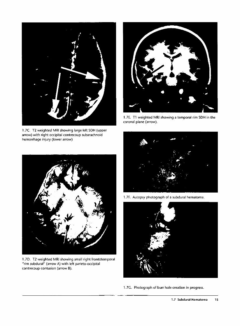
1.7E. T1 weighted MRI showing a temporal rim SDH in thecoronal plane (arrow).
1.7C. T2 weighted MRI showing large left SDH (upperarrow) with right occipital contrecoup subarachnoidhemorrhage injury (lower arrow)
1.7D. T2 weighted MRI showing small right frontotemporal"rim subdural" (arrow A) with left parieto-occipitalcontrecoup contusion (arrow B).
1.7F. Autopsy photograph of a subdural hematoma.
1.7G. Photograph of burr hole creation in progress.
1.7 Subdural Hematoma 15
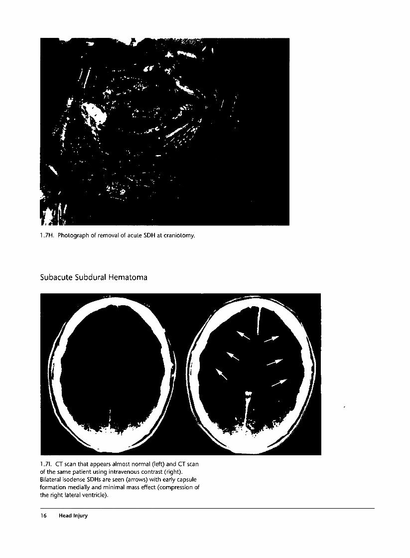
1.7H. Photograph of removal of acute SDH at craniotomy.
Subacute Subdural Hematoma
1.71. CT scan that appears almost normal (left) and CT scanof the same patient using intravenous contrast (right).Bilateral isodense SDHs are seen (arrows) with early capsuleformation medially and minimal mass effect (compression ofthe right lateral ventricle).
16 Head Injury
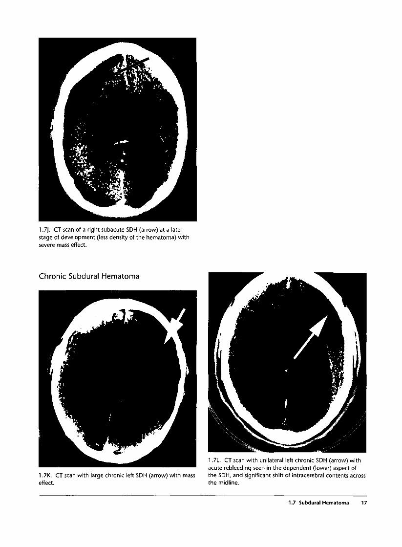
1 J\. CT scan of a right subacute SDH (arrow) at a laterstage of development (less density of the hematoma) withsevere mass effect.
Chronic Subdural Hematoma
1.7K. CT scan with large chronic left SDH (arrow) with masseffect.
1.7L. CT scan with unilateral left chronic SDH (arrow) withacute rebleeding seen in the dependent (lower) aspect ofthe SDH, and significant shift of intracerebral contents acrossthe midline.
1.7 Subdural Hematoma 17
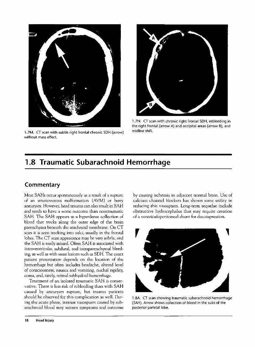
1.7M. CT scan with subtle right frontal chronic SDH (arrow)without mass effect.
1.7N. CT scan with chronic right frontal SDH, rebleeding inthe right frontal (arrow A) and occipital areas (arrow B), andmidline shift.
1.8 Traumatic Subarachnoid Hemorrhage
Commentary
Most SAHs occur spontaneously as a result of a ruptureof an arteriovenous malformation (AVM) or berryaneurysm. However, head trauma can also result in SAHand tends to have a worse outcome than nontraumaticSAH. The SAH appears as a hyperdense collection ofblood that tracks along the outer edge of the brainparenchyma beneath the arachnoid membrane. On CTscan it is seen tracking into sulci, usually in the frontallobes. The CT scan appearance may be very subtle, andthe SAH is easily missed. Often SAH is associated withintraventricular, subdural, and intraparenchymal bleed-ing, as well as with mass lesions such as SDH. The exactpatient presentation depends on the location of thehemorrhage but often includes headache, altered levelof consciousness, nausea and vomiting, nuchal rigidity,coma, and, rarely, retinal subhyaloid hemorrhage.
Treatment of an isolated traumatic SAH is conser-vative. There is less risk of rebleeding than with SAHcaused by aneurysm rupture, but trauma patientsshould be observed for this complication as well. Dur-ing the acute phase, intense vasospasm caused by sub-arachnoid blood may worsen symptoms and outcome
by causing ischemia in adjacent normal brain. Use ofcalcium channel blockers has shown some utility inreducing this vasospasm. Long-term sequelae includeobstructive hydrocephalus that may require creationof a ventriculoperitoneal shunt for decompression.
r
1.8A. CT scan showing traumatic subarachnoid hemorrhage(SAH). Arrow shows collection of blood in the sulci of theposterior parietal lobe.
18 Head Injury
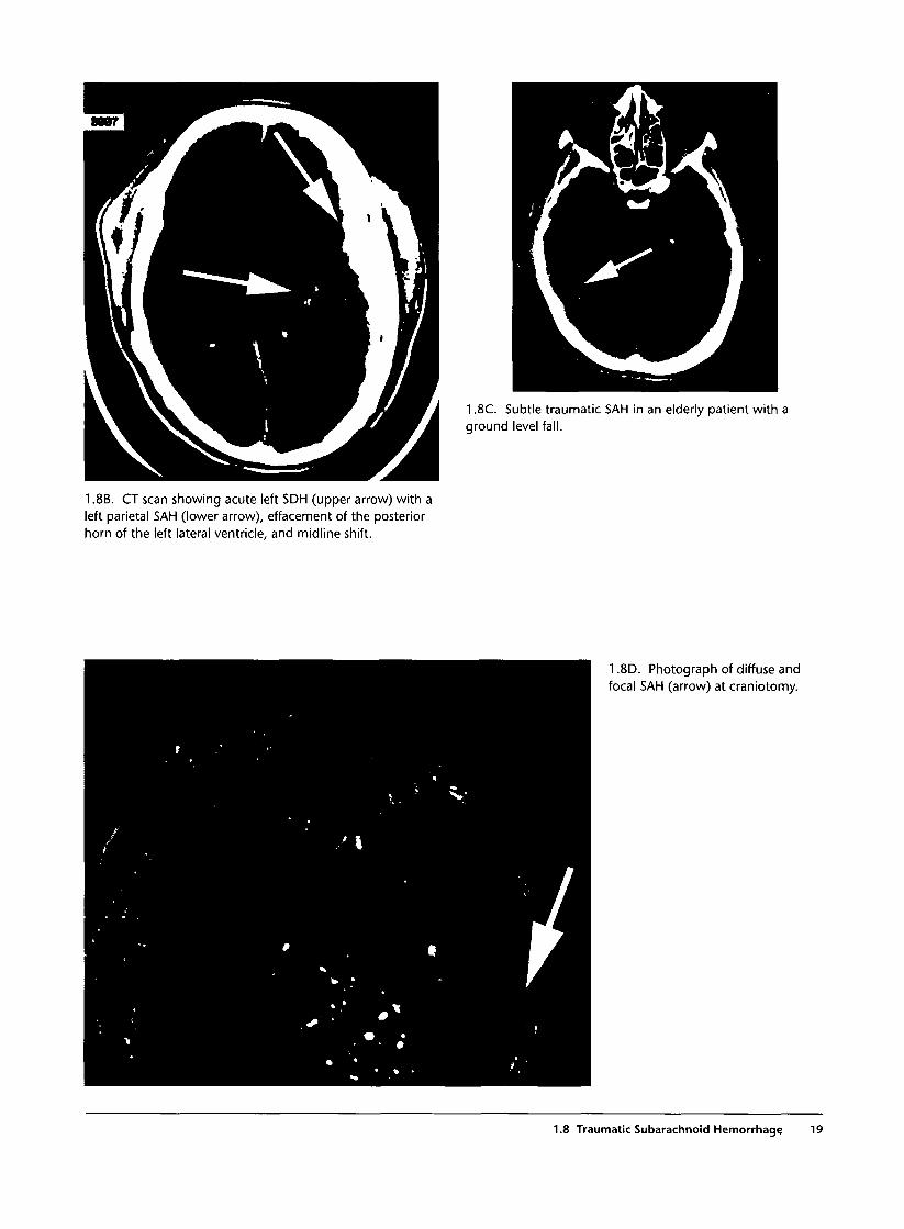
1.8C. Subtle traumatic SAH in an elderly patient with aground level fall.
1.8B. CT scan showing acute left SDH (upper arrow) with aleft parietal SAH (lower arrow), effacement of the posteriorhorn of the left lateral ventricle, and midline shift.
1.8D. Photograph of diffuse andfocal SAH (arrow) at craniotomy.
1.8 Traumatic Subarachnoid Hemorrhage 19
1.9 Cerebral Contusion
Commentary
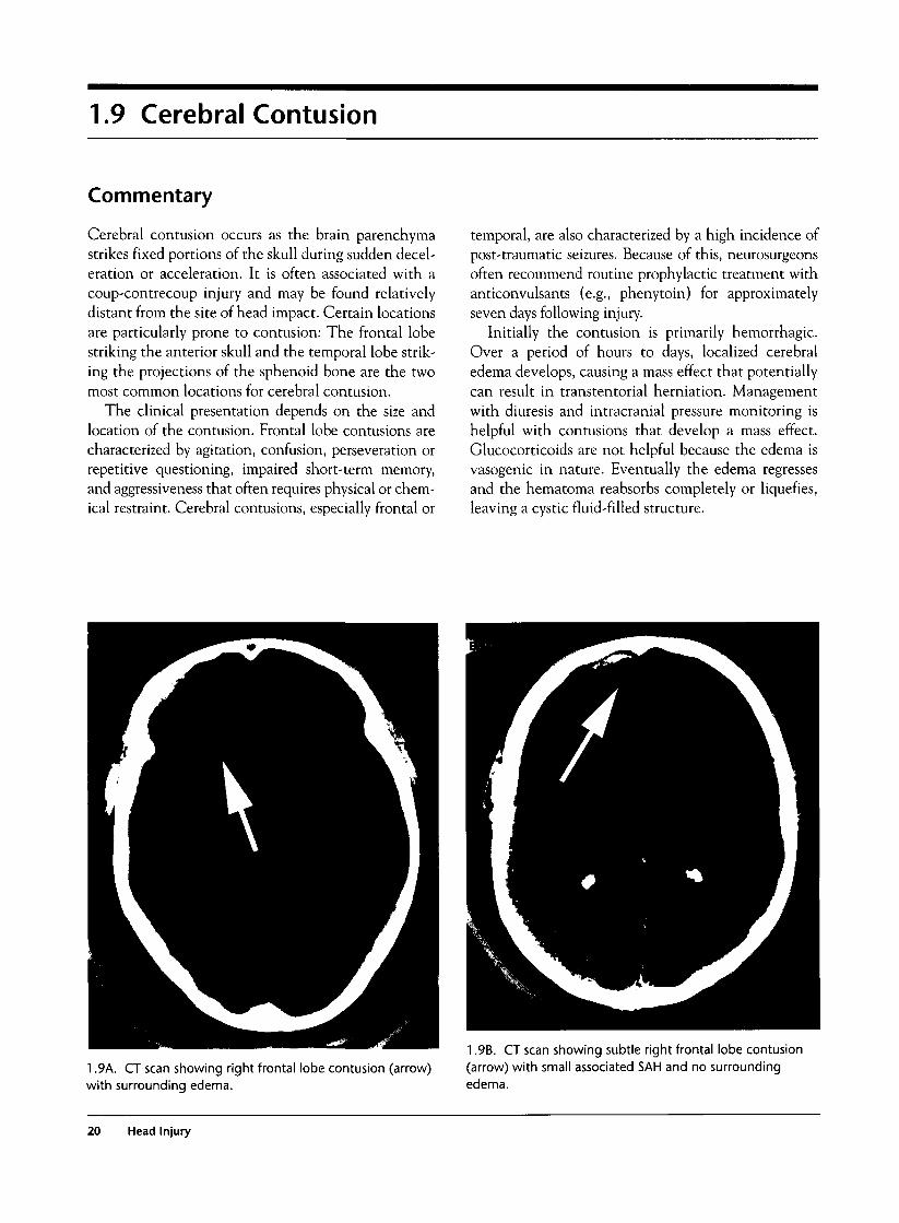
Cerebral contusion occurs as the brain parenchymastrikes fixed portions of the skull during sudden decel-eration or acceleration. It is often associated with acoup-contrecoup injury and may be found relativelydistant from the site of head impact. Certain locationsare particularly prone to contusion: The frontal lobestriking the anterior skull and the temporal lobe strik-ing the projections of the sphenoid bone are the twomost common locations for cerebral contusion.
The clinical presentation depends on the size andlocation of the contusion. Frontal lobe contusions arecharacterized by agitation, confusion, perseveration orrepetitive questioning, impaired short-term memory,and aggressiveness that often requires physical or chem-ical restraint. Cerebral contusions, especially frontal or
temporal, are also characterized by a high incidence ofpost-traumatic seizures. Because of this, neurosurgeonsoften recommend routine prophylactic treatment withanticonvulsants (e.g., phenytoin) for approximatelyseven days following injury.
Initially the contusion is primarily hemorrhagic.Over a period of hours to days, localized cerebraledema develops, causing a mass effect that potentiallycan result in transtentorial herniation. Managementwith diuresis and intracranial pressure monitoring ishelpful with contusions that develop a mass effect.Glucocorticoids are not helpful because the edema isvasogenic in nature. Eventually the edema regressesand the hematoma reabsorbs completely or liquefies,leaving a cystic fluid-filled structure.
1.9A. CT scan showing right frontal lobe contusion (arrow)with surrounding edema.
1.9B. CT scan showing subtle right frontal lobe contusion(arrow) with small associated SAH and no surroundingedema.
20 Head Injury
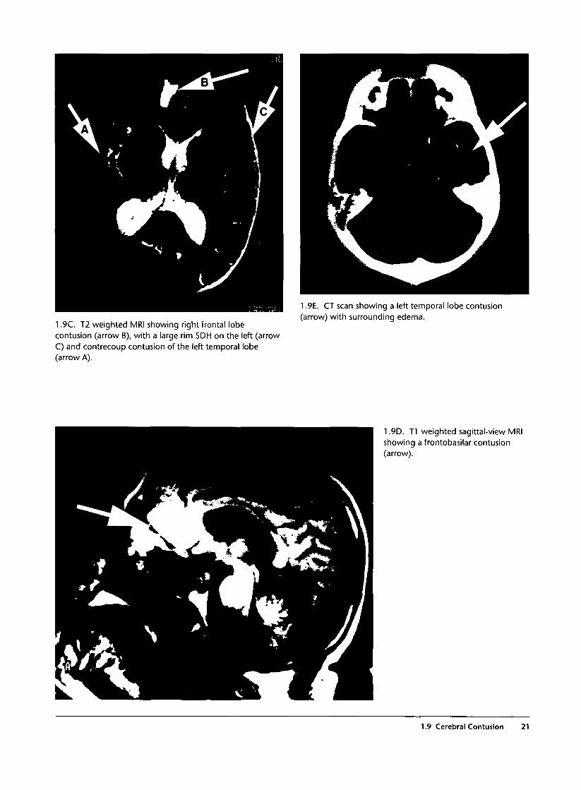
1.9C. T2 weighted MRI showing right frontal lobecontusion (arrow B), with a large rim SDH on the left (arrowC) and contrecoup contusion of the left temporal lobe(arrow A).
1.9E. CT scan showing a left temporal lobe contusion(arrow) with surrounding edema.
1.9D. T1 weighted sagittal-view MRIshowing a frontobasilar contusion(arrow).
1.9 Cerebral Contusion 21

1.10 Penetrating Head Injury
Commentary
The vast majority of penetrating head wounds involveGSWs. These are devastating injuries that frequentlyresult in death or profound disability in survivors. As abullet enters the skull, it produces multiple high-veloc-ity fragments (both from shattered skull bony frag-ments and from bullet fragmentation) that causemultiple injuries. Although the skull absorbs somekinetic energy, the bullet retains sufficient energy tocause a pressure wave once it enters brain parenchyma.This produces a rapidly expanding cavity and subse-quent recoil of brain tissue. The abrupt deformation ofbrain tissue results in laceration and contusion of brainparenchyma, accumulation of blood in the epidural orsubdural spaces, intraparenchymal bleeding, and directlaceration of brain tissue by bullet and bone fragments.Because of the high kinetic energy imparted to thebrain, subsequent cerebral edema is common.
Other types of penetrating injuries involve lesskinetic energy and have a better prognosis. Stabwounds, impalement injuries, and low-velocity shrap-nel wounds can produce all of the same injuries butmost commonly result in open skull fracture and directlaceration of brain parenchyma. Impaled objects resultfrom both accidental trauma and intentional injury.Many different objects may be involved, but knivesand metal rods are the most common. A careful physi-cal examination is indicated with stab wounds of thescalp to ensure that the knife blade has not broken offinside the cranium. Wounds associated with theseinjuries may appear very innocuous and may be missedaltogether as they are often covered with matted hair.Surprisingly, many of these patients are relativelyasymptomatic and often have a normal neurologicexamination in spite of a dramatic presentation. PlainAP and lateral radiographs of the skull will accuratelydelineate the location and depth of penetration ofradiopaque impaled objects. If the patient can fit intothe CT scan gantry without disturbing the impaledobject, the CT scan will demonstrate underlying braininjury, although metal artifact may be problematic.Impaled foreign bodies should be removed only in theoperating room, where prompt vascular control can beobtained once the object is removed.
Management depends on the overall condition of thepatient. Frequently, GSW victims have multiple wounds
of the chest, neck, or abdomen that may take priority interms of restoring hemodynamic stability to the patient.As with open skull fractures, bleeding from a GSW ofthe head may be profuse, but hemorrhagic shock isuncommon with isolated head injuries, and other associ-ated injuries should be sought to explain the shock.Because there is open communication with the environ-ment, an epidural hematoma associated with a penetrat-ing injury may decompress spontaneously. Surgicaldebridement and control of cerebral hemorrhage maybe life saving. The incidence of post-traumaticseizures is more than 50%, and use of anticonvulsantmedication is routine in these cases.
Patients with exposed brain matter, GSW thatcrosses the midline of the brain, severe coagulopathy, orearly transtentorial herniation invariably die of theirwounds and should be considered as potential organdonors.
Gunshot Wound
1.10A. Photograph of a patient with a GSW entry wound onthe right temporal area (lower arrow) with bulgingdeformity of the forehead due to comminuted frontal bonefractures (upper arrow).
22 Head Injury
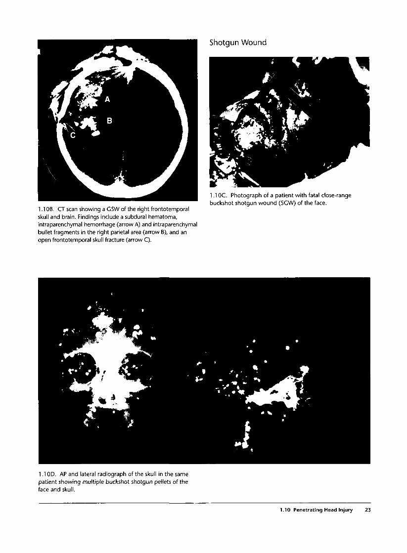
Shotgun Wound
1.1 OB. CT scan showing a GSW of the right frontotemporalskull and brain. Findings include a subdural hematoma,intraparenchymal hemorrhage (arrow A) and intraparenchymalbullet fragments in the right parietal area (arrow B), and anopen frontotemporal skull fracture (arrow C).
1.1OC. Photograph of a patient with fatal close-rangebuckshot shotgun wound (SGW) of the face.
1.1OD. AP and lateral radiograph of the skull in the samepatient showing multiple buckshot shotgun pellets of theface and skull.
1.10 Penetrating Head Injury 23
Stab Wounds and Impalement Injuries
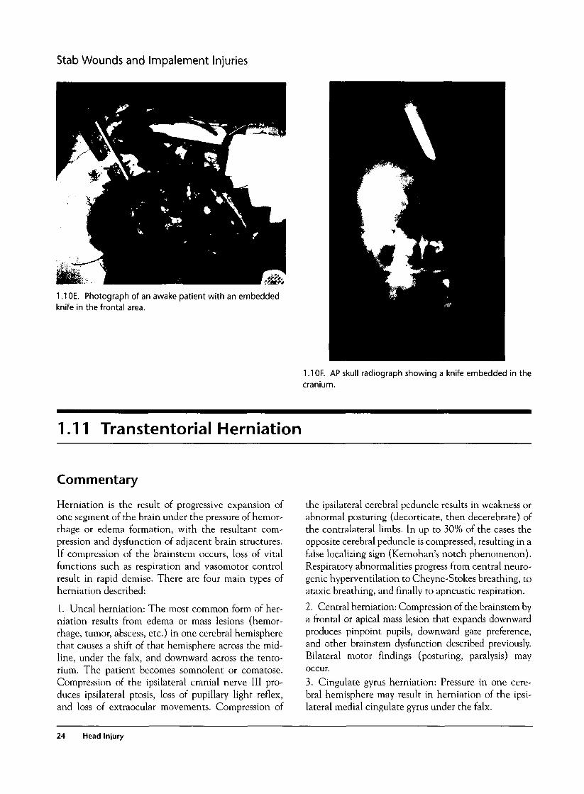
1.1OE. Photograph of an awake patient with an embeddedknife in the frontal area.
1.1 OF. AP skull radiograph showing a knife embedded in thecranium.
1.11 Transtentorial Herniation
Commentary
Herniation is the result of progressive expansion ofone segment of the brain under the pressure of hemor-rhage or edema formation, with the resultant com-pression and dysfunction of adjacent brain structures.If compression of the brainstem occurs, loss of vitalfunctions such as respiration and vasomotor controlresult in rapid demise. There are four main types ofherniation described:
1. Uncal herniation: The most common form of her-niation results from edema or mass lesions (hemor-rhage, tumor, abscess, etc.) in one cerebral hemispherethat causes a shift of that hemisphere across the mid-line, under the falx, and downward across the tento-rium. The patient becomes somnolent or comatose.Compression of the ipsilateral cranial nerve III pro-duces ipsilateral ptosis, loss of pupillary light reflex,and loss of extraocular movements. Compression of
the ipsilateral cerebral peduncle results in weakness orabnormal posturing (decorticate, then decerebrate) ofthe contralateral limbs. In up to 30% of the cases theopposite cerebral peduncle is compressed, resulting in afalse localizing sign (Kemohan's notch phenomenon).Respiratory abnormalities progress from central neuro-genic hyperventilation to Cheyne-Stokes breathing, toataxic breathing, and finally to apneustic respiration.
2. Central herniation: Compression of the brainstem bya frontal or apical mass lesion that expands downwardproduces pinpoint pupils, downward gaze preference,and other brainstem dysfunction described previously.Bilateral motor findings (posturing, paralysis) mayoccur.3. Cingulate gyrus herniation: Pressure in one cere-bral hemisphere may result in herniation of the ipsi-lateral medial cingulate gyrus under the falx.
24 Head Injury
4. Cerebellar tonsillar herniation: Mass lesions oredema of the cerebellum can result in expansion of thecerebellar tonsils into the foramen magnum, com-pressing the posterior brainstem. This presents as sud-den loss of consciousness and loss of brainstemfunction with consequent apnea and hypotension.This condition has extremely high mortality, so cere-bellar lesions must be recognized before the onset ofherniation to salvage the patient.
Patients who achieve hemodynamic stability should betreated with a cerebral resuscitation protocol includingrapid sequence intubation (RSI), moderate hyperventi-lation to a pCC>2 of 35 mm Hg, osmotic and loop diure-sis, sedation with cerebrally protective agents (e.g.,propofol, etomidate, pentobarbital), and mild elevationof the head once the cervical spine is "cleared." Earlyplacement of a ventriculostomy is indicated for moni-toring and control of intracranial pressure by removal ofCSF. Emergency placement of burr holes on the side ofpupillary dysfunction may be successful in draining anEDH. Cerebral perfusion pressure is maintained by infu-sion of fluids and pressors if needed. Complicationsinclude DIC that is often fatal, so a baseline coagulationprofile is obtained in the ED. Once DIC occurs, transfu-
sion of fresh frozen plasma may be helpful, although theprognosis is dismal at this stage.
Cerebral blood flow (CBF) is governed by the rela-tionship of mean arterial pressure (MAP) and intracra-nial pressure (ICP) as follows: CBF=MAP-ICP.Consequently, as ICP increases, every effort must bemade to maintain or elevate MAP. Once ICP exceedsMAP no blood flow to the brain can occur and this isone definition of "brain death".
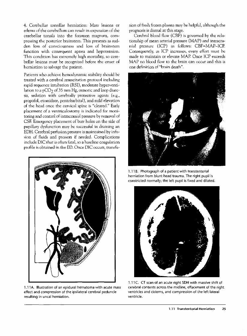
1.11B. Photograph of a patient with transtentorialherniation from blunt head trauma. The right pupil isconstricted normally; the left pupil is fixed and dilated.
1.11 A. Illustration of an epidural hematoma with acute masseffect and compression of the ipsilateral cerebral peduncleresulting in uncal herniation.
1.11C. CT scan of an acute right SDH with massive shift ofcerebral contents across the midline, effacement of the rightventricles and cisterns, and compression of the left lateralventricle.
1.11 Transtentorial Herniation 25
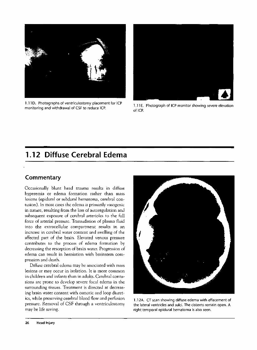
1.11D. Photographs of ventriculostomy placement for ICPmonitoring and withdrawal of CSF to reduce ICP.
1.11E. Photograph of ICP monitor showing severe elevationof ICP.
1.12 Diffuse Cerebral Edema
Commentary
Occasionally blunt head trauma results in diffusehyperemia or edema formation rather than masslesions (epidural or subdural hematoma, cerebral con-tusion). In most cases the edema is primarily vasogenicin nature, resulting from the loss of autoregulation andsubsequent exposure of cerebral arterioles to the fullforce of arterial pressure. Transudation of plasma fluidinto the extracellular compartment results in anincrease in cerebral water content and swelling of theaffected part of the brain. Elevated venous pressurecontributes to the process of edema formation bydecreasing the resorption of brain water. Progression ofedema can result in herniation with brainstem com-pression and death.
Diffuse cerebral edema may be associated with masslesions or may occur in isolation. It is more commonin children and infants than in adults. Cerebral contu-sions are prone to develop severe focal edema in thesurrounding tissues. Treatment is directed at decreas-ing brain water content with osmotic and loop diuret-ics, while preserving cerebral blood flow and perfusionpressure. Removal of CSF through a ventriculostomymay be life saving.
1.12A. CT scan showing diffuse edema with effacement ofthe lateral ventricles and sulci. The cisterns remain open. Aright temporal epidural hematoma is also seen.
26 Head Injury
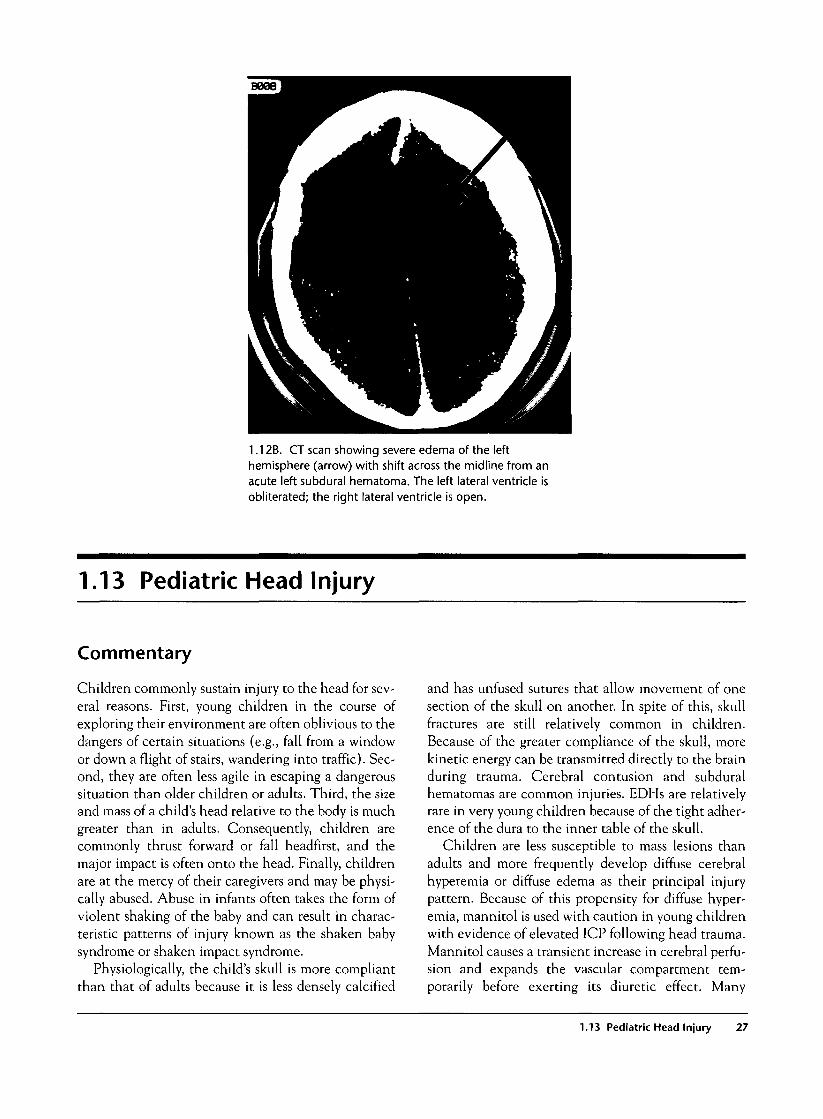
1.12B. CT scan showing severe edema of the lefthemisphere (arrow) with shift across the midline from anacute left subdural hematoma. The left lateral ventricle isobliterated; the right lateral ventricle is open.
1.13 Pediatric Head Injury
Commentary
Children commonly sustain injury to the head for sev-eral reasons. First, young children in the course ofexploring their environment are often oblivious to thedangers of certain situations (e.g., fall from a windowor down a flight of stairs, wandering into traffic). Sec-ond, they are often less agile in escaping a dangeroussituation than older children or adults. Third, the sizeand mass of a child's head relative to the body is muchgreater than in adults. Consequently, children arecommonly thrust forward or fall headfirst, and themajor impact is often onto the head. Finally, childrenare at the mercy of their caregivers and may be physi-cally abused. Abuse in infants often takes the form ofviolent shaking of the baby and can result in charac-teristic patterns of injury known as the shaken babysyndrome or shaken impact syndrome.
Physiologically, the child's skull is more compliantthan that of adults because it is less densely calcified
and has unfused sutures that allow movement of onesection of the skull on another. In spite of this, skullfractures are still relatively common in children.Because of the greater compliance of the skull, morekinetic energy can be transmitted directly to the brainduring trauma. Cerebral contusion and subduralhematomas are common injuries. EDHs are relativelyrare in very young children because of the tight adher-ence of the dura to the inner table of the skull.
Children are less susceptible to mass lesions thanadults and more frequently develop diffuse cerebralhyperemia or diffuse edema as their principal injurypattern. Because of this propensity for diffuse hyper-emia, mannitol is used with caution in young childrenwith evidence of elevated ICP following head trauma.Mannitol causes a transient increase in cerebral perfu-sion and expands the vascular compartment tem-porarily before exerting its diuretic effect. Many
1.13 Pediatric Head Injury 27
authors recommend using loop diuretics and mildhyperventilation rather than mannitol when manag-ing elevated ICP in a young child.
Another injury that occurs almost exclusively inchildren with head trauma is transient cortical blind-ness. The actual incidence of this complication isunknown but is thought to be secondary to vasospasminduced by trauma. Children also manifest cerebralconcussion differently from adults. The "infant con-cussion syndrome" consists of the transient appear-ance of pallor, diaphoresis, vomiting, tachycardia,somnolence, and weakness, often occurring in aninfant after relatively minor head trauma (e.g., fallingoff a changing table). CT scan is invariably normal,and the child often will make an equally dramaticrecovery while still in the emergency department.Small infants may bleed sufficiently into the head todevelop hemorrhagic shock, although this is rare. Sep-aration of cranial sutures, a bulging fontanelle, orincreasing head circumference may rarely be the ini-tial clues to head injury in infants, but the diagnosis ofserious head injury is generally made by CT scan.
The principal concern in evaluating children withhead trauma is to consider the possibility of nonacci-dental trauma. A history incompatible with the severityof injury or that involves activities that the child wouldbe developmentally incapable of performing can be aninitial clue that the injuries may be due to abuse.
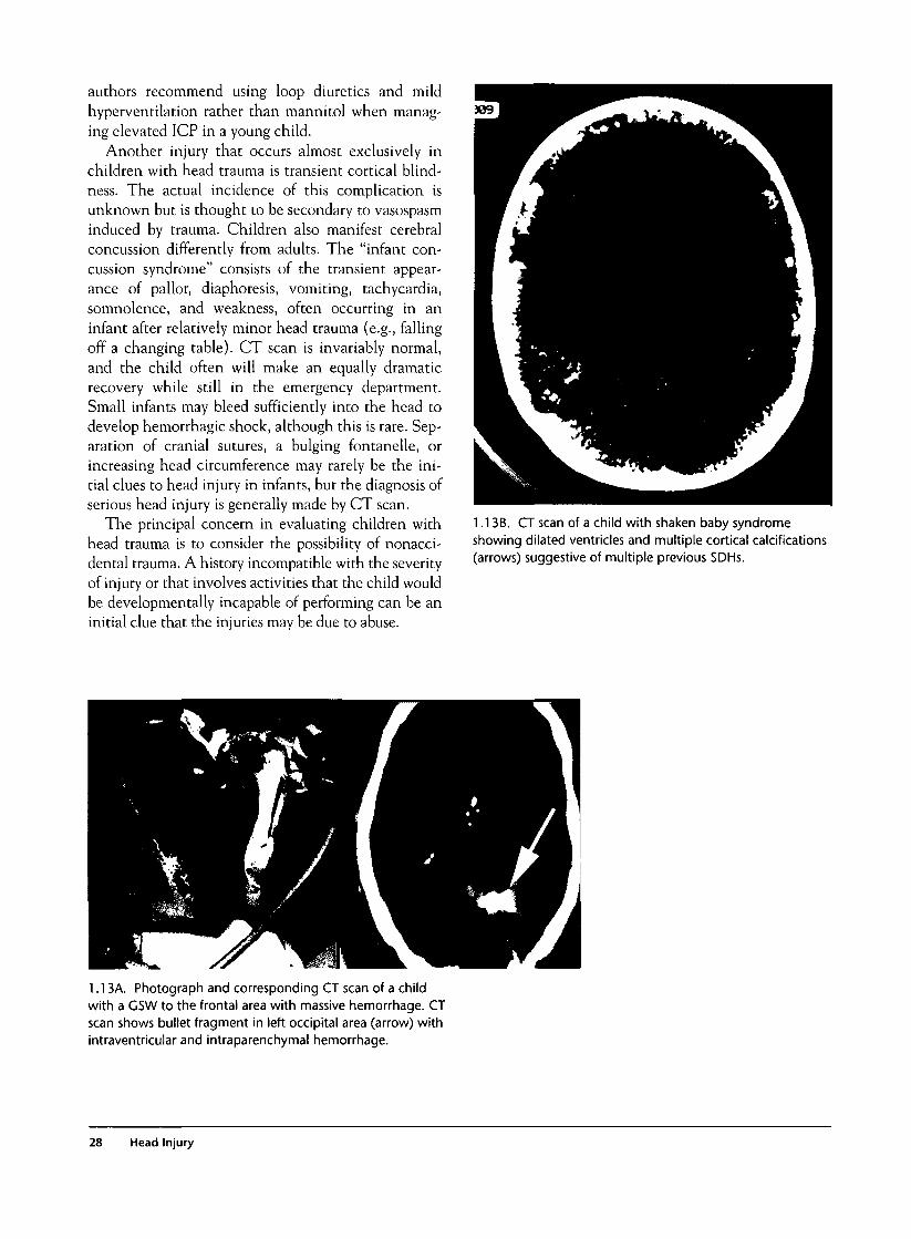
1.13B. CT scan of a child with shaken baby syndromeshowing dilated ventricles and multiple cortical calcifications(arrows) suggestive of multiple previous SDHs.
1.13A. Photograph and corresponding CT scan of a childwith a GSW to the frontal area with massive hemorrhage. CTscan shows bullet fragment in left occipital area (arrow) withintraventricular and intraparenchymal hemorrhage.
28 Head Injury
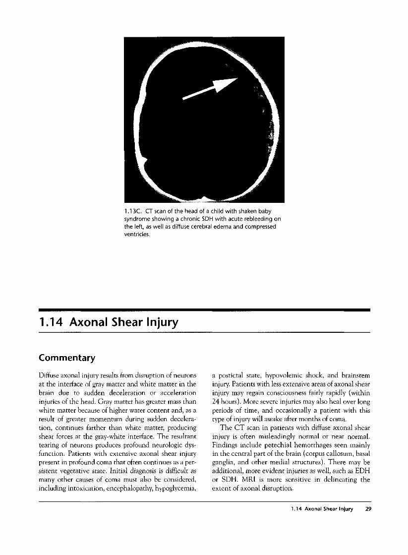
1.13C. CT scan of the head of a child with shaken babysyndrome showing a chronic SDH with acute rebleeding onthe left, as well as diffuse cerebral edema and compressedventricles.
1.14 Axonal Shear Injury
Commentary
Diffuse axonal injury results from disruption of neuronsat the interface of gray matter and white matter in thebrain due to sudden deceleration or accelerationinjuries of the head. Gray matter has greater mass thanwhite matter because of higher water content and, as aresult of greater momentum during sudden decelera-tion, continues farther than white matter, producingshear forces at the gray-white interface. The resultanttearing of neurons produces profound neurologic dys-function. Patients with extensive axonal shear injurypresent in profound coma that often continues as a per-sistent vegetative state. Initial diagnosis is difficult asmany other causes of coma must also be considered,including intoxication, encephalopathy, hypoglycemia,
a postictal state, hypovolemic shock, and brainsteminjury. Patients with less extensive areas of axonal shearinjury may regain consciousness fairly rapidly (within24 hours). More severe injuries may also heal over longperiods of time, and occasionally a patient with thistype of injury will awake after months of coma.
The CT scan in patients with diffuse axonal shearinjury is often misleadingly normal or near normal.Findings include petechial hemorrhages seen mainlyin the central part of the brain (corpus callosum, basalganglia, and other medial structures). There may beadditional, more evident injuries as well, such as EDHor SDH. MRI is more sensitive in delineating theextent of axonal disruption.
1.14 Axonal Shear Injury 29
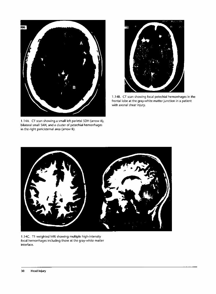
1.14B. CT scan showing focal petechial hemorrhages in thefrontal lobe at the gray-white matter junction in a patientwith axonal shear injury.
1.14A. CT scan showing a small left parietal SDH (arrow A),bilateral small SAH, and a cluster of petechial hemorrhagesin the right pericisternal area (arrow B).
1.14C. T1 weighted MRI showing multiple high-intensityfocal hemorrhages including those at the gray-white matterinterface.
30 Head Injury

1.15 Intraventricular Hemorrhage
Commentary
Hemorrhage into the ventricular system is more com-mon following hypertensive bleeds than after trauma.Occasionally blood from a traumatic lesion (e.g.,SAH) will track into the ventricular system, or lacera-tions of the brain parenchyma may communicatedirectly with the ventricles. The prognosis of intraven-tricular hemorrhage is not as grim as in cases of sponta-neous bleeding, and patients may recover well.Complications include obstructive hydrocephalus thatmay require placement of a ventriculoperitoneal shunt.
1.15A. CT scan showing a small collection of blood in theleft posterior horn of the lateral ventricle (arrow).
1.16 Intraparenchymal Hemorrhage
Commentary
Direct laceration of the brain parenchyma by pene-trating wounds or bone fragments may result in hem-orrhage that is relatively confined to the brainparenchyma. Bleeding in these cases is usually tam-ponaded by surrounding tissue but may become exten-sive enough to produce a midline shift. Exposure ofinjured brain thromboplastin to the circulation resultsin activation of the coagulation cascade and fre-quently produces DIC, which is a poor prognosticsign.
1.16A. CT scan showing accumulation of blood in allventricles as well as intraparenchymal blood on the left(arrow A) and a left EDH (arrow B).
1.16 Intraparenchymal Hemorrhage 31


Facial Injury
Introduction
Soft tissue injuries of the face are common in modernsociety. The majority of serious injuries occur in the con-text of vehicular trauma or assaults. Use of seatbelts andairbags has decreased the frequency but not eliminatedfacial trauma produced by motor vehicle accidents. Inaddition to direct impact of the face against the wind-shield, steering wheel, or dashboard, broken glass frag-ments frequently produce lacerations and eye injuries.
The lower face and neck contain structures thatdefine and maintain the patency of the airway. Conse-quently, facial injuries at times assume the highest pri-ority in trauma management until airway patency andadequate ventilation can be established. Because facialtissues are highly vascularized, massive bleeding intothe oral cavity can occlude the airway, especially whenpatients are obtunded from head injury or intoxication.In the presence of massive bleeding, airway compro-mise may be produced by placing the patient supine forspinal immobilization. Blood, secretions, fragments ofteeth, and foreign bodies must be removed to avoidaspiration and airway occlusion. Although severe facialinjuries are dramatic and often distract the inexperi-enced clinician from more critical tasks, treatment ofmost facial injuries can be safely deferred until life-threatening problems have been addressed.
The face and scalp also contain many structures thatare essential for the function of special senses of sight,smell, taste, and hearing. Human communication isdependent not only on facial structures required forspeech and hearing but also those involved in facialexpression. In addition, many facial landmarks definehuman appearance, and their preservation as intactsymmetrical structures is important cosmetically andpsychologically. Injury to these structures can result indevastating disability that often can be avoided withearly detection and repair.
Special attention is indicated in repairing facialinjuries. Debridement of wound margins should be min-imized, cartilaginous structures should be preserved,
and fine sutures with minimal inflammatory propertiesshould be used in closing the wounds. Complex lacera-tions involving delicate and essential facial structuressuch as the eyelid should be referred to a specialist.
Clinical Examination
1. Examination of the face.After completion of the primary survey, the face is
examined for areas of swelling and tenderness that canindicate underlying fractures. Palpation of the facialbones for crepitus or abnormal motion can locate afracture. Grasping the teeth and pulling forward candemonstrate Le Fort fractures with abnormal motion ofthe alveolar ridge, midface, or whole face. Lacerationsare noted, and massive bleeding is tamponaded bydirect pressure. Blind clamping of bleeding sites is dan-gerous in that it can injure nerves and other structuresthat run in proximity to vessels. Lacerations crossingthe path of the parotid duct mandate examination ofStensen's duct in the mouth (discussed later). Facialasymmetry can be due to direct trauma but also tofacial nerve injury, and an assessment of the muscles offacial expression and facial sensation is made. In coma-tose patients corneal reflexes should be tested to deter-mine these functions.
2. Examination of the eye.Anatomically, the orbit sits relatively protected by
the orbital ridge, malar prominence, and nose. The cil-iary and corneal reflexes rapidly close the eyelid,adding further protection to direct contact with theglobe. Injuries to the eye range from minor (e.g.,corneal abrasion) to critical (e.g., ruptured globe).
Examination of the eye and its adnexa is an impor-tant part of the secondary survey. Victims of motorvehicle crashes often have fragments of glass that canbecome embedded in the eye causing lacerations orcorneal abrasions. Occasionally a patient's refractoryagitation can be cured by treatment of a corneal abra-sion or removal of glass fragments in the eye that were
33
initially unsuspected. Often patients have massive softtissue swelling around the eye that makes examinationdifficult. In these cases devices to hold the eyelids openmust be used and can be improvised by bending paperclips into blunt retractors and gently retracting the lids.Formal measurement of visual acuity may not be possi-ble in the early phases of resuscitation, but an initialestimate of vision can be made by having the patientcount fingers or report light perception. Complete lossof vision in a previously normal eye requires immediateconsultation with an ophthalmologist. The pupils areexamined for symmetry and equality as well as reactionto light. The conjunctivae are assessed for foreign bod-ies and chemosis that can indicate rupture of the globe.A peaked pupil is highly suspicious for rupture of theglobe, and the "peak" often points to the site of rupture.Visible scleral or corneal lacerations may indicate pene-tration of the globe by a foreign object and require radi-ographs or CT of the orbits to detect intraocular foreignbodies. The position of the globe in the orbit is notedfor enophthalmos (blow-out fracture) or exophthalmos(retro-orbital hematoma). Inability to perform allextraocular movements may indicate a brain lesion,peripheral nerve injury, or entrapment of extraocularmuscles. Lacerations involving the lacrimal duct and lidmargins should be noted and referred to an ophthalmol-ogist for repair. A brief fundoscopic examination is per-formed to assess the position of the lens and presence ofblood in the anterior chamber (hyphema) or retina.
3. Examination of the ear.The external ear is inspected for the presence of lac-
erations or hematoma. Cartilaginous lacerations oravulsions require particular attention. The ear canal isexamined with an otoscope for bleeding or CSF otor-rhea indicating a ruptured tympanic membrane andbasilar skull fracture. The tympanic membrane is exam-ined for perforations or accumulation of blood in themiddle ear that is seen as hemotympanum. Although itmay take many hours to appear, inspection of the mas-toid area for Battle's sign is important in detecting basi-lar skull fracture.
4. Examination of the nose.The nose is inspected for lacerations of overlying
skin and of the cartilage. The presence of nasal fractureis often obvious clinically with deformity, crepitus,epistaxis, and tenderness to palpation. The nares areinspected for the presence of epistaxis or hematomaand for CSF drainage.
5. Examination of the mouth.The mouth is inspected for lacerations, avulsion or
fracture of teeth, swelling of the tongue and oral
mucosa, and misalignment of teeth (indicating amandible or maxilla fracture). Blood, loose teeth, andforeign bodies are removed manually or by suction.Simultaneously, an evaluation of the airway is madeexamining for stridor, dysphonia, gagging or drooling,and inability to handle oral secretions. The presence orabsence of a gag reflex in obtunded patients ofteninfluences the decision to intubate the patient to pro-tect against aspiration.
Investigations
After physical examination has indicated areas oflikely injury, specific radiographs or CT scans may beindicated to delineate injuries. Plain radiographs areuseful in detecting most facial fractures and in locatingradiopaque foreign bodies, but CT scan can more accu-rately identify these if the patient is sufficiently stableto undergo this examination. Certain radiographicviews are indicated to clarify specific clinical findingssuch as a submentovertex view to detect zygomaticarch fracture or Panorex views for suspected mandiblefractures. Leakage of CSF from the nose or ear can beassessed by examining the drainage for the presence ofglucose (indicating CSF) or for a double ring sign whenthe drainage is applied to filter paper. Suspicion ofinjury to the lacrimal duct is best confirmed by an oph-thalmologist using fine probes. Instillation of fluores-cein into the conjunctival sac and examination with aUV light source can demonstrate corneal abrasion, andSeidel's test can demonstrate leakage of aqueous humorfrom a ruptured globe. A detailed evaluation of theanterior chamber can be performed on stable patientsusing a slit-lamp examination. Patients with suspectedpost-traumatic glaucoma or retro-orbital hematomashould undergo tonometry to measure intraocular pres-sure, but this test should never be done if there is aposibility of a ruptured globe. Parotid duct lacerationcan be demonstrated by probing the duct or by per-forming a sialogram.
General Management
Airway management is of prime importance whenfacial injuries threaten the ability to ventilate thepatient. Suction of secretions and manual removal offoreign bodies and blood clots may establish airwaypatency, but often endotracheal intubation is indi-cated. Nasotracheal intubation should not beattempted with nasal, basilar skull, or Le Fort fractures
34 Facial Injury
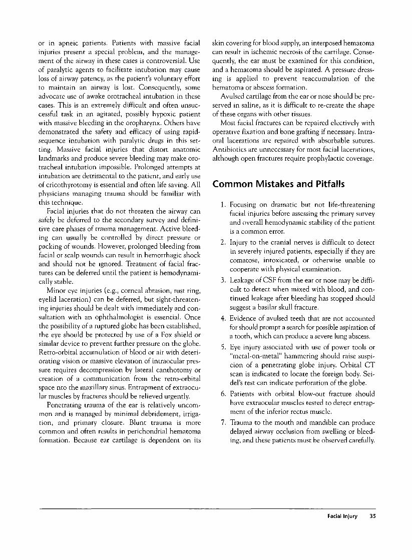
or in apneic patients. Patients with massive facialinjuries present a special problem, and the manage-ment of the airway in these cases is controversial. Useof paralytic agents to facilitate intubation may causeloss of airway patency, as the patient's voluntary effortto maintain an airway is lost. Consequently, someadvocate use of awake orotracheal intubation in thesecases. This is an extremely difficult and often unsuc-cessful task in an agitated, possibly hypoxic patientwith massive bleeding in the oropharynx. Others havedemonstrated the safety and efficacy of using rapid-sequence intubation with paralytic drugs in this set-ting. Massive facial injuries that distort anatomiclandmarks and produce severe bleeding may make oro-tracheal intubation impossible. Prolonged attempts atintubation are detrimental to the patient, and early useof cricothyrotomy is essential and often life saving. Allphysicians managing trauma should be familiar withthis technique.
Facial injuries that do not threaten the airway cansafely be deferred to the secondary survey and defini-tive care phases of trauma management. Active bleed-ing can usually be controlled by direct pressure orpacking of wounds. However, prolonged bleeding fromfacial or scalp wounds can result in hemorrhagic shockand should not be ignored. Treatment of facial frac-tures can be deferred until the patient is hemodynami-cally stable.
Minor eye injuries (e.g., corneal abrasion, rust ring,eyelid laceration) can be deferred, but sight-threaten-ing injuries should be dealt with immediately and con-sultation with an ophthalmologist is essential. Oncethe possibility of a ruptured globe has been established,the eye should be protected by use of a Fox shield orsimilar device to prevent further pressure on the globe.Retro-orbital accumulation of blood or air with deteri-orating vision or massive elevation of intraocular pres-sure requires decompression by lateral canthotomy orcreation of a communication from the retro-orbitalspace nto the maxillary sinus. Entrapment of extraocu-lar muscles by fractures should be relieved urgently.
Penetrating trauma of the ear is relatively uncom-mon and is managed by minimal debridement, irriga-tion, and primary closure. Blunt trauma is morecommon and often results in perichondrial hematomaformation. Because ear cartilage is dependent on its
skin covering for blood supply, an interposed hematomacan result in ischemic necrosis of the cartilage. Conse-quently, the ear must be examined for this condition,and a hematoma should be aspirated. A pressure dress-ing is applied to prevent reaccumulation of thehematoma or abscess formation.
Avulsed cartilage from the ear or nose should be pre-served in saline, as it is difficult to re-create the shapeof these organs with other tissues.
Most facial fractures can be repaired electively withoperative fixation and bone grafting if necessary. Intra-oral lacerations are repaired with absorbable sutures.Antibiotics are unnecessary for most facial lacerations,although open fractures require prophylactic coverage.
Common Mistakes and Pitfalls
1. Focusing on dramatic but not life-threateningfacial injuries before assessing the primary surveyand overall hemodynamic stability of the patientis a common error.
2. Injury to the cranial nerves is difficult to detectin severely injured patients, especially if they arecomatose, intoxicated, or otherwise unable tocooperate with physical examination.
3. Leakage of CSF from the ear or nose may be diffi-cult to detect when mixed with blood, and con-tinued leakage after bleeding has stopped shouldsuggest a basilar skull fracture.
4- Evidence of avulsed teeth that are not accountedfor should prompt a search for possible aspiration ofa tooth, which can produce a severe lung abscess.
5. Eye injury associated with use of power tools or"metal-on-metal" hammering should raise suspi-cion of a penetrating globe injury. Orbital CTscan is indicated to locate the foreign body. Sei-del's test can indicate perforation of the globe.
6. Patients with orbital blow-out fracture shouldhave extraocular muscles tested to detect entrap-ment of the inferior rectus muscle.
7. Trauma to the mouth and mandible can producedelayed airway occlusion from swelling or bleed-ing, and these patients must be observed carefully.
Facial Injury 35
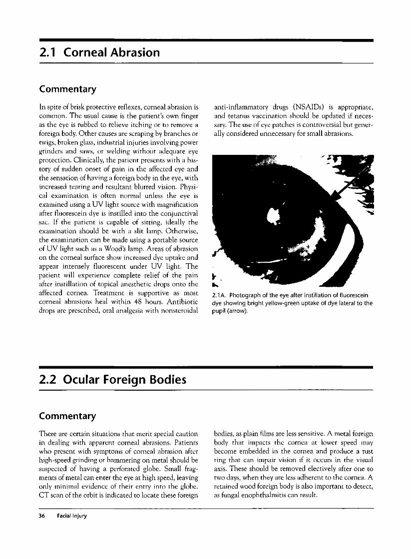
2.1 Corneal Abrasion
Commentary
In spite of brisk protective reflexes, corneal abrasion iscommon. The usual cause is the patient's own fingeras the eye is rubbed to relieve itching or to remove aforeign body. Other causes are scraping by branches ortwigs, broken glass, industrial injuries involving powergrinders and saws, or welding without adequate eyeprotection. Clinically, the patient presents with a his-tory of sudden onset of pain in the affected eye andthe sensation of having a foreign body in the eye, withincreased tearing and resultant blurred vision. Physi-cal examination is often normal unless the eye isexamined using a UV light source with magnificationafter fluorescein dye is instilled into the conjunctivalsac. If the patient is capable of sitting, ideally theexamination should be with a slit lamp. Otherwise,the examination can be made using a portable sourceof UV light such as a Wood's lamp. Areas of abrasionon the corneal surface show increased dye uptake andappear intensely fluorescent under UV light. Thepatient will experience complete relief of the painafter instillation of topical anesthetic drops onto theaffected cornea. Treatment is supportive as mostcorneal abrasions heal within 48 hours. Antibioticdrops are prescribed, oral analgesia with nonsteroidal
anti-inflammatory drugs (NSAIDs) is appropriate,and tetanus vaccination should be updated if neces-sary. The use of eye patches is controversial but gener-ally considered unnecessary for small abrasions.
2.1 A. Photograph of the eye after instillation of fluoresceindye showing bright yellow-green uptake of dye lateral to thepupil (arrow).
2.2 Ocular Foreign Bodies
Commentary
There are certain situations that merit special cautionin dealing with apparent corneal abrasions. Patientswho present with symptoms of corneal abrasion afterhigh-speed grinding or hammering on metal should besuspected of having a perforated globe. Small frag-ments of metal can enter the eye at high speed, leavingonly minimal evidence of their entry into the globe.CT scan of the orbit is indicated to locate these foreign
bodies, as plain films are less sensitive. A metal foreignbody that impacts the cornea at lower speed maybecome embedded in the cornea and produce a rustring that can impair vision if it occurs in the visualaxis. These should be removed electively after one totwo days, when they are less adherent to the cornea. Aretained wood foreign body is also important to detect,as fungal enophthalmitis can result.
36 Facial Injury
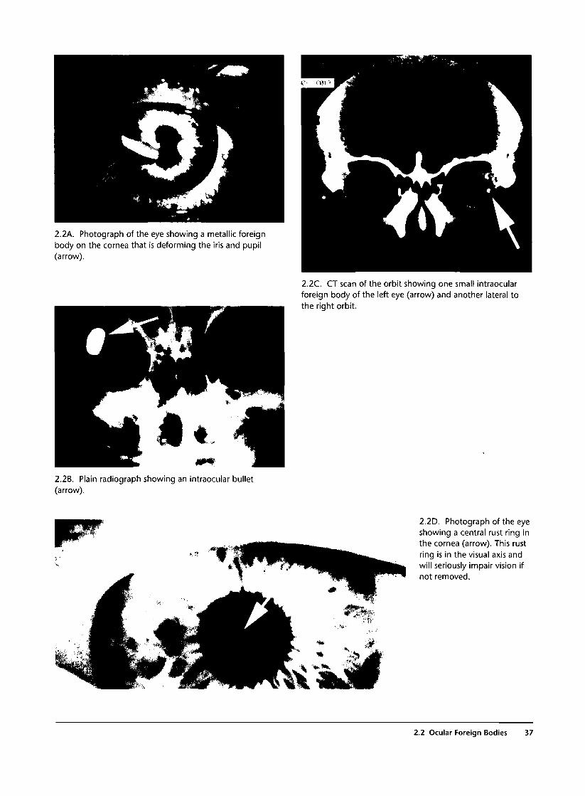
2.2A. Photograph of the eye showing a metallic foreignbody on the cornea that is deforming the iris and pupil(arrow).
2.2C. CT scan of the orbit showing one small intraocularforeign body of the left eye (arrow) and another lateral tothe right orbit.
2.2B. Plain radiograph showing an intraocular bullet(arrow).
2.2D. Photograph of the eyeshowing a central rust ring inthe cornea (arrow). This rustring is in the visual axis andwill seriously impair vision ifnot removed.
2.2 Ocular Foreign Bodies 37
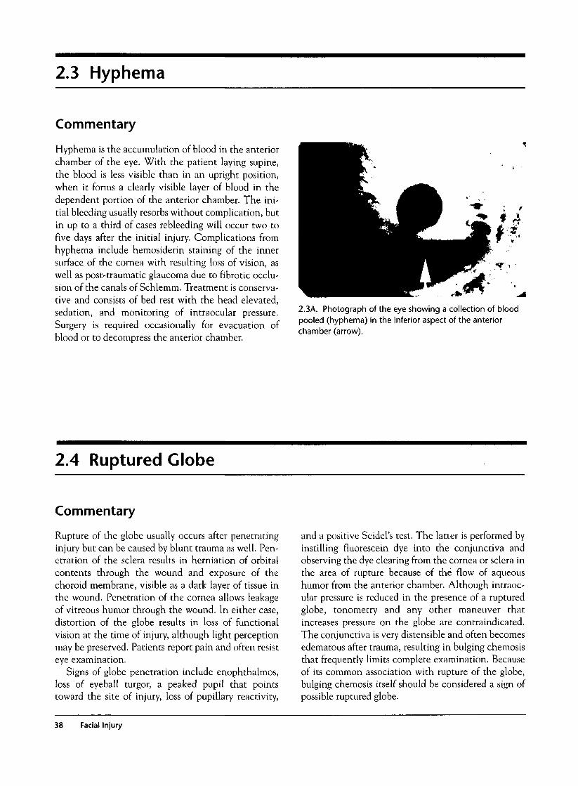
2.3 Hyphema
Commentary
Hyphema is the accumulation of blood in the anteriorchamber of the eye. With the patient laying supine,the blood is less visible than in an upright position,when it forms a clearly visible layer of blood in thedependent portion of the anterior chamber. The ini-tial bleeding usually resorbs without complication, butin up to a third of cases rebleeding will occur two tofive days after the initial injury. Complications fromhyphema include hemosiderin staining of the innersurface of the cornea with resulting loss of vision, aswell as post-traumatic glaucoma due to fibrotic occlu-sion of the canals of Schlemm. Treatment is conserva-tive and consists of bed rest with the head elevated,sedation, and monitoring of intraocular pressure.Surgery is required occasionally for evacuation ofblood or to decompress the anterior chamber.
2.3A. Photograph of the eye showing a collection of bloodpooled (hyphema) in the inferior aspect of the anteriorchamber (arrow).
2.4 Ruptured Globe
Commentary
Rupture of the globe usually occurs after penetratinginjury but can be caused by blunt trauma as well. Pen-etration of the sclera results in herniation of orbitalcontents through the wound and exposure of thechoroid membrane, visible as a dark layer of tissue inthe wound. Penetration of the cornea allows leakageof vitreous humor through the wound. In either case,distortion of the globe results in loss of functionalvision at the time of injury, although light perceptionmay be preserved. Patients report pain and often resisteye examination.
Signs of globe penetration include enophthalmos,loss of eyeball turgor, a peaked pupil that pointstoward the site of injury, loss of pupillary reactivity,
and a positive Seidel's test. The latter is performed byinstilling fluorescein dye into the conjunctiva andobserving the dye clearing from the cornea or sclera inthe area of rupture because of the flow of aqueoushumor from the anterior chamber. Although intraoc-ular pressure is reduced in the presence of a rupturedglobe, tonometry and any other maneuver thatincreases pressure on the globe are contraindicated.The conjunctiva is very distensible and often becomesedematous after trauma, resulting in bulging chemosisthat frequently limits complete examination. Becauseof its common association with rupture of the globe,bulging chemosis itself should be considered a sign ofpossible ruptured globe.
38 Facial Injury
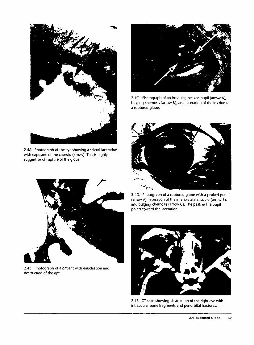
2.4C. Photograph of an irregular, peaked pupil (arrow A),bulging chemosis (arrow B), and laceration of the iris due toa ruptured globe.
2.4A. Photograph of the eye showing a scleral lacerationwith exposure of the choroid (arrow). This is highlysuggestive of rupture of the globe.
2.4D. Photograph of a ruptured globe with a peaked pupil(arrow A), laceration of the inferior/lateral sclera (arrow B),and bulging chemosis (arrow C). The peak in the pupilpoints toward the laceration.
2.4B. Photograph of a patient with enucleation anddestruction of the eye.
2.4E. CT scan showing destruction of the right eye withintraocular bone fragments and periorbital fractures.
2.4 Ruptured Globe 39
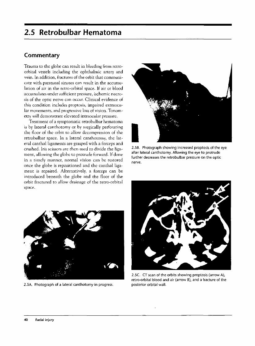
2.5 Retrobulbar Hematoma
Commentary
Trauma to the globe can result in bleeding from retro-orbital vessels including the ophthalmic artery andvein. In addition, fractures of the orbit that communi-cate with paranasal sinuses can result in the accumu-lation of air in the retro-orbital space. If air or bloodaccumulates under sufficient pressure, ischemic necro-sis of the optic nerve can occur. Clinical evidence ofthis condition includes proptosis, impaired extraocu-lar movements, and progressive loss of vision. Tonom-etry will demonstrate elevated intraocular pressure.
Treatment of a symptomatic retrobulbar hematomais by lateral canthotomy or by surgically perforatingthe floor of the orbit to allow decompression of theretrobulbar space. In a lateral canthotomy, the lat-eral canthal ligaments are grasped with a forceps andcrushed. Iris scissors are then used to divide the liga-ment, allowing the globe to protrude forward. If donein a timely manner, normal vision can be restoredonce the globe is repositioned and the canthal liga-ment is repaired. Alternatively, a forceps can beintroduced beneath the globe and the floor of theorbit fractured to allow drainage of the retro-orbitalspace.
2.5B. Photograph showing increased proptosis of the eyeafter lateral canthotomy. Allowing the eye to protrudefurther decreases the retrobulbar pressure on the optic
nerve.
2.5A. Photograph of a lateral canthotomy in progress.
2.5C. CT scan of the orbits showing proptosis (arrow A),retro-orbital blood and air (arrow B), and a fracture of theposterior orbital wall.
40 Facial Injury
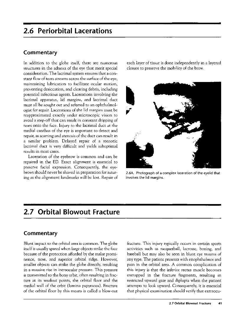
2.6 Periorbital Lacerations
Commentary
In addition to the globe itself, there are numerousstructures in the adnexa of the eye that merit specialconsideration. The lacrimal system ensures that a con-stant flow of tears streams across the surface of the eye,maintaining lubrication to facilitate ocular motion,preventing desiccation, and clearing debris, includingpotential infectious agents. Lacerations involving thelacrimal apparatus, lid margins, and lacrimal ductmust all be sought out and referred to an ophthalmol-ogist for repair. Lacerations of the lid margins must bereapproximated exactly under microscopic vision toavoid a step-off that can result in constant dripping oftears onto the face. Injury to the lacrimal duct at themedial canthus of the eye is important to detect andrepair, as scarring and stenosis of the duct can result ina similar problem. Delayed repair of a stenoticlacrimal duct is very difficult and yields suboptimalresults in most cases.
Laceration of the eyebrow is common and can berepaired in the ED. Exact alignment is essential topreserve facial expression. Consequently, the eye-brows should never be shaved in preparation for sutur-ing as the alignment landmarks will be lost. Repair of
each layer of tissue is done independently in a layeredclosure to preserve the mobility of the brow.
2.6A. Photograph of a complex laceration of the eyelid thatinvolves the lid margins.
2.7 Orbital Blowout Fracture
Commentary
Blunt impact to the orbital area is common. The globeitself is usually spared when large objects strike the facebecause of the protection afforded by the malar promi-nence, nose, and superior orbital ridge. However,smaller objects can strike the globe directly, resultingin a massive rise in intraocular pressure. This pressureis transmitted to the bony orbit, often resulting in frac-ture at its weakest points, the orbital floor and themedial wall of the orbit (lamina papyracea). Fractureof the orbital floor by this means is called a blow-out
fracture. This injury typically occurs in certain sportsactivities such as racquetball, lacrosse, boxing, andbaseball but may also be seen in blunt eye trauma ofany type. The patient presents with enophthalmos andpain in the orbital area. A common complication ofthis injury is that the inferior rectus muscle becomesentrapped in the fracture fragments, resulting inrestricted upward gaze and diplopia when the patientattempts to look upward. Consequently, it is essentialthat physical examination should verify that extraocu-
2.7 Orbital Blowout Fracture 41
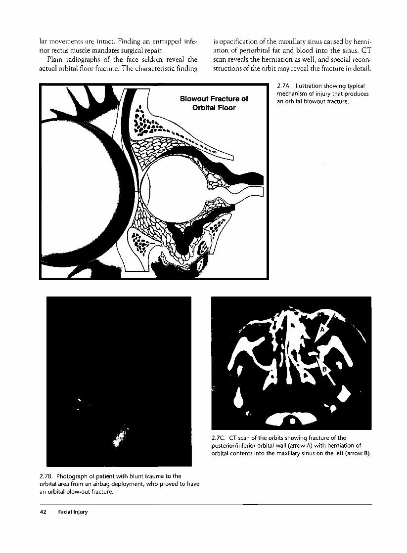
lar movements are intact. Finding an entrapped infe-rior rectus muscle mandates surgical repair.
Plain radiographs of the face seldom reveal theactual orbital floor fracture. The characteristic finding
Blowout Fracture ofOrbital Floor
is opacification of the maxillary sinus caused by herni-ation of periorbital fat and blood into the sinus. CTscan reveals the herniation as well, and special recon-structions of the orbit may reveal the fracture in detail.
2.7k. Illustration showing typicalmechanism of injury that producesan orbital blowout fracture.
2.7C. CT scan of the orbits showing fracture of theposterior/inferior orbital wall (arrow A) with herniation oforbital contents into the maxillary sinus on the left (arrow B).
2.7B. Photograph of patient with blunt trauma to theorbital area from an airbag deployment, who proved to havean orbital blow-out fracture.
42 Facial Injury

2.7D. CT scan showing fracture of the inferiororbital wall with opacification of the rightmaxillary sinus (arrow).
2.7E. Photograph of a patientwith blow-out fracture showingenophthalmos of the left eye.
2.7'F. Photographs of a patient with a blow-out fracture ofthe right eye and subconjunctival hemorrhage. There is noevidence of divergent gaze with the eyes in neutral position.Downward gaze reveals entrapment of the extraocularmuscles of the right eye, resulting in a subtle divergent gaze.
2.7 Orbital Blowout Fracture 43
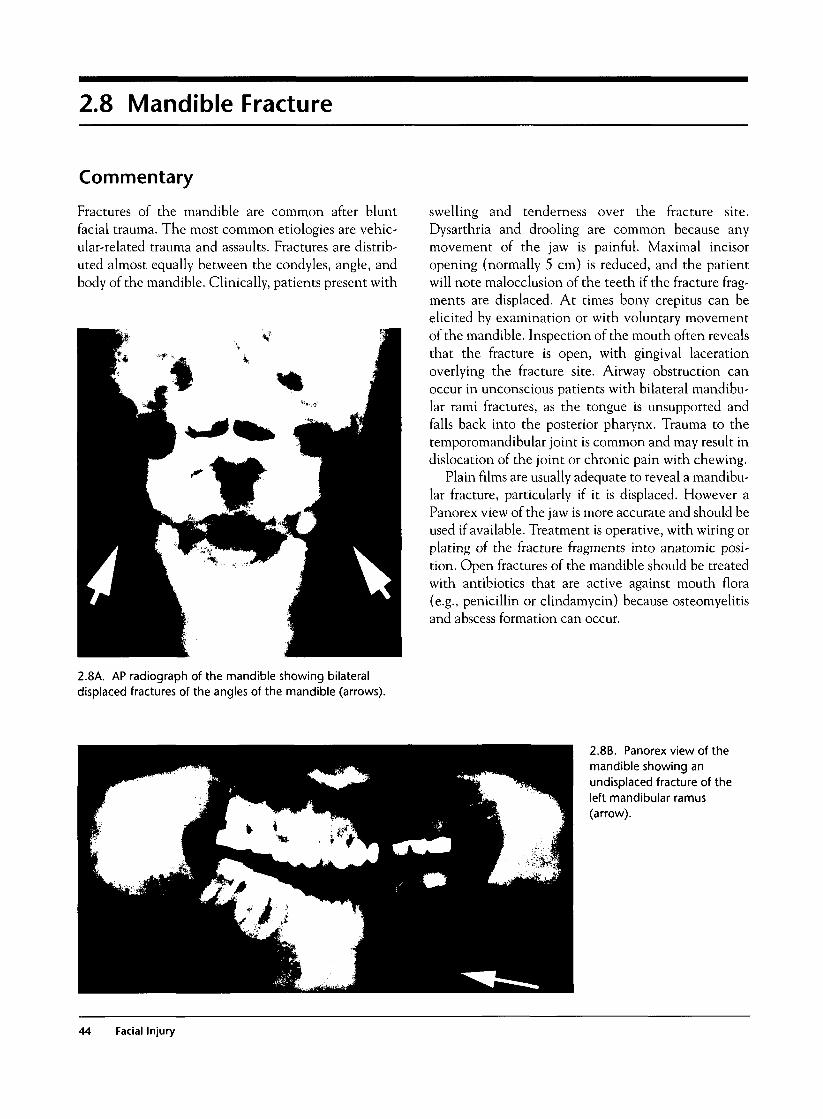
2.8 Mandible Fracture
Commentary
Fractures of the mandible are common after bluntfacial trauma. The most common etiologies are vehic-ular-related trauma and assaults. Fractures are distrib-uted almost equally between the condyles, angle, andbody of the mandible. Clinically, patients present with
swelling and tenderness over the fracture site.Dysarthria and drooling are common because anymovement of the jaw is painful. Maximal incisoropening (normally 5 cm) is reduced, and the patientwill note malocclusion of the teeth if the fracture frag-ments are displaced. At times bony crepitus can beelicited by examination or with voluntary movementof the mandible. Inspection of the mouth often revealsthat the fracture is open, with gingival lacerationoverlying the fracture site. Airway obstruction canoccur in unconscious patients with bilateral mandibu-lar rami fractures, as the tongue is unsupported andfalls back into the posterior pharynx. Trauma to thetemporomandibular joint is common and may result indislocation of the joint or chronic pain with chewing.
Plain films are usually adequate to reveal a mandibu-lar fracture, particularly if it is displaced. However aPanorex view of the jaw is more accurate and should beused if available. Treatment is operative, with wiring orplating of the fracture fragments into anatomic posi-tion. Open fractures of the mandible should be treatedwith antibiotics that are active against mouth flora(e.g., penicillin or clindamycin) because osteomyelitisand abscess formation can occur.
2.8A. AP radiograph of the mandible showing bilateraldisplaced fractures of the angles of the mandible (arrows).
2.8B. Panorex view of themandible showing anundisplaced fracture of theleft mandibular ramus(arrow).
44 Facial Injury
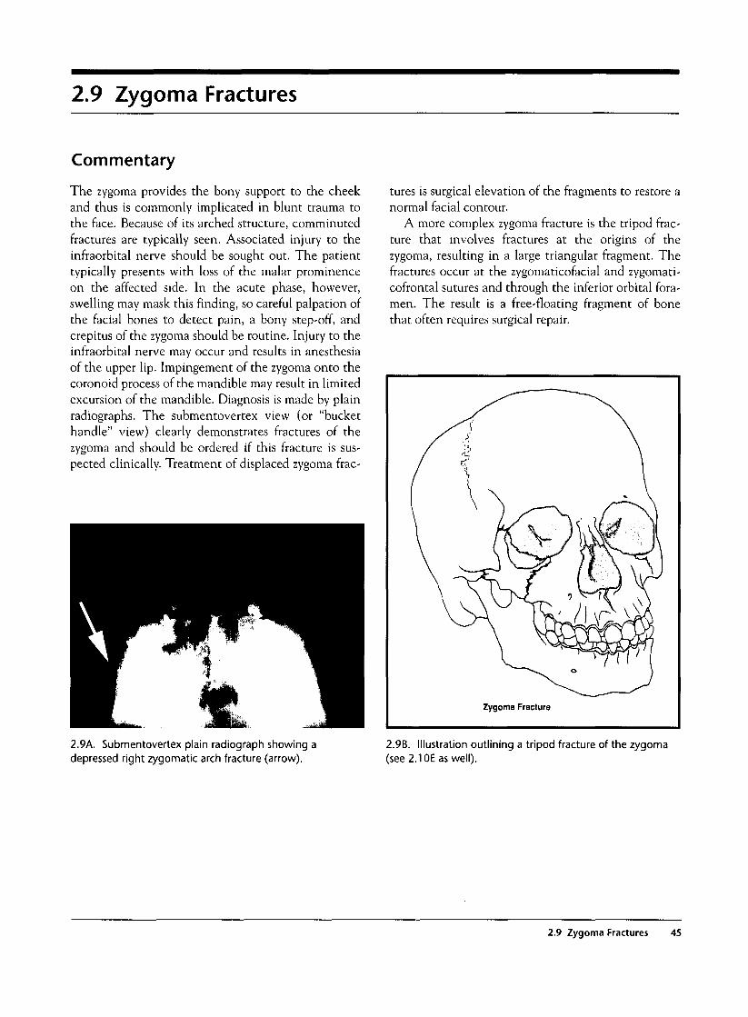
2.9 Zygoma Fractures
Commentary
The zygoma provides the bony support to the cheekand thus is commonly implicated in blunt trauma tothe face. Because of its arched structure, comminutedfractures are typically seen. Associated injury to theinfraorbital nerve should be sought out. The patienttypically presents with loss of the malar prominenceon the affected side. In the acute phase, however,swelling may mask this finding, so careful palpation ofthe facial bones to detect pain, a bony step-off, andcrepitus of the zygoma should be routine. Injury to theinfraorbital nerve may occur and results in anesthesiaof the upper lip. Impingement of the zygoma onto thecoronoid process of the mandible may result in limitedexcursion of the mandible. Diagnosis is made by plainradiographs. The submentovertex view (or "buckethandle" view) clearly demonstrates fractures of thezygoma and should be ordered if this fracture is sus-pected clinically. Treatment of displaced zygoma frac-
tures is surgical elevation of the fragments to restore anormal facial contour.
A more complex zygoma fracture is the tripod frac-ture that involves fractures at the origins of thezygoma, resulting in a large triangular fragment. Thefractures occur at the zygomaticofacial and zygomati-cofrontal sutures and through the inferior orbital fora-men. The result is a free-floating fragment of bonethat often requires surgical repair.
2.9A. Submentovertex plain radiograph showing adepressed right zygomatic arch fracture (arrow).
2.9B. Illustration outlining a tripod fracture of the zygoma(see 2.1 OE as well).
2.9 Zygoma Fractures 45
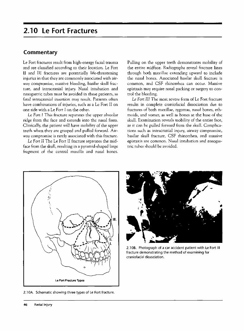
2.10 Le Fort Fractures
Commentary
Le Fort fractures result from high-energy facial traumaand are classified according to their location. Le FortII and III fractures are potentially life-threateninginjuries in that they are commonly associated with air-way compromise, massive bleeding, basilar skull frac-ture, and intracranial injury. Nasal intubation andnasogastric tubes must be avoided in these patients, asfatal intracranial insertion may result. Patients oftenhave combinations of injuries, such as a Le Fort II onone side with a Le Fort I on the other.
Le Fort I This fracture separates the upper alveolarridge from the face and extends into the nasal fossa.Clinically, the patient will have mobility of the upperteeth when they are grasped and pulled forward. Air-way compromise is rarely associated with this fracture.
Le Fort II The Le Fort II fracture separates the mid-face from the skull, resulting in a pyramid-shaped largefragment of the central maxilla and nasal bones.
Pulling on the upper teeth demonstrates mobility ofthe entire midface. Radiographs reveal fracture linesthrough both maxillae extending upward to includethe nasal bones. Associated basilar skull fracture iscommon, and CSF rhinorrhea can occur. Massiveepistaxis may require nasal packing or surgery to con-trol the bleeding.
Le Fort III The most severe form of Le Fort fractureresults in complete craniofacial dissociation due tofractures of both maxillae, zygomas, nasal bones, eth-moids, and vomer, as well as bones at the base of theskull. Examination reveals mobility of the entire face,as it can be pulled forward from the skull. Complica-tions such as intracranial injury, airway compromise,basilar skull fracture, CSF rhinorrhea, and massiveepistaxis are common. Nasal intubation and nasogas-tric tubes should be avoided.
2.1 OB. Photograph of a car accident patient with Le Fortfracture demonstrating the method of examining forcraniofacial dissociation.
2.10A. Schematic showing three types of Le Fort fracture.
46 Facial Injury
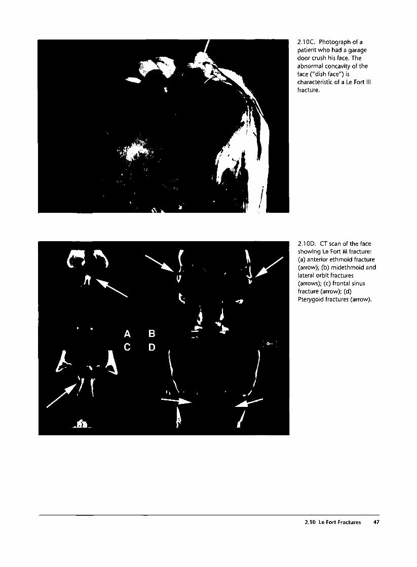
2.1 OC. Photograph of apatient who had a garagedoor crush his face. Theabnormal concavity of theface ("dish face") ischaracteristic of a Le Fort IIIfracture.
2.1 OD. CT scan of the faceshowing Le Fort III fracture:(a) anterior ethmoid fracture(arrow); (b) midethmoid andlateral orbit fractures(arrows); (c) frontal sinusfracture (arrow); (d)Pterygoid fractures (arrow).
2.10 Le Fort Fractures 47
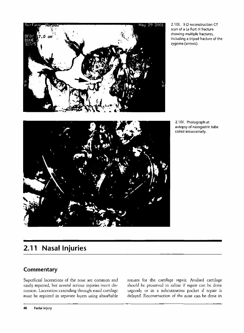
2.1 OE. 3-D reconstruction CTscan of a Le Fort III fractureshowing multiple fractures,including a tripod fracture of thezygoma (arrows).
2.1 OF. Photograph atautopsy of nasogastric tubecoiled intracranially.
2.11 Nasal Injuries
Commentary
Superficial lacerations of the nose are common andeasily repaired, but several serious injuries merit dis-cussion. Laceration extending through nasal cartilagemust be repaired in separate layers using absorbable
sutures for the cartilage repair. Avulsed cartilageshould be preserved in saline if repair can be doneurgently or in a subcutaneous pocket if repair isdelayed. Reconstruction of the nose can be done in
48 Facial Injury
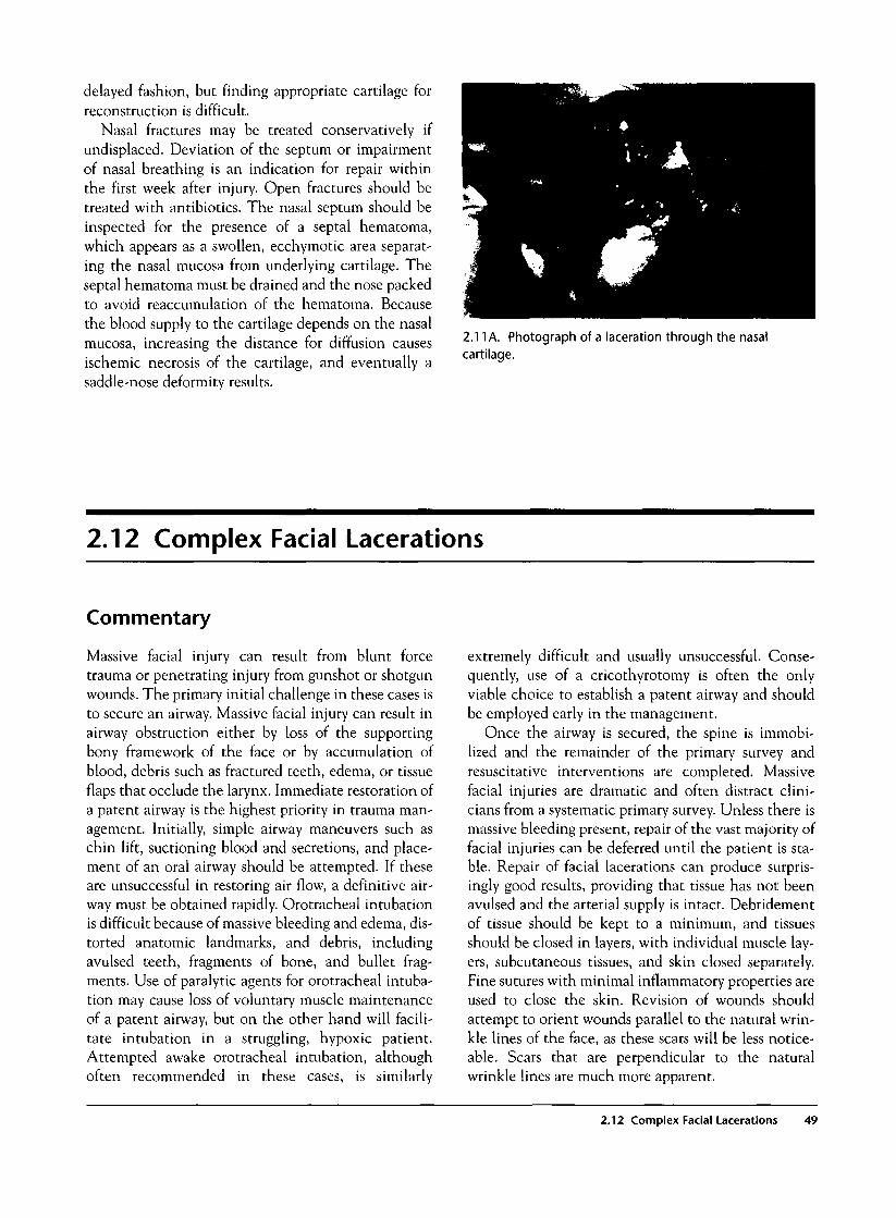
delayed fashion, but finding appropriate cartilage forreconstruction is difficult.
Nasal fractures may be treated conservatively ifundisplaced. Deviation of the septum or impairmentof nasal breathing is an indication for repair withinthe first week after injury. Open fractures should betreated with antibiotics. The nasal septum should beinspected for the presence of a septal hematoma,which appears as a swollen, ecchymotic area separat-ing the nasal mucosa from underlying cartilage. Theseptal hematoma must be drained and the nose packedto avoid reaccumulation of the hematoma. Becausethe blood supply to the cartilage depends on the nasalmucosa, increasing the distance for diffusion causesischemic necrosis of the cartilage, and eventually asaddle-nose deformity results.
2.11 A. Photograph of a laceration through the nasalcartilage.
2.12 Complex Facial Lacerations
Commentary
Massive facial injury can result from blunt forcetrauma or penetrating injury from gunshot or shotgunwounds. The primary initial challenge in these cases isto secure an airway. Massive facial injury can result inairway obstruction either by loss of the supportingbony framework of the face or by accumulation ofblood, debris such as fractured teeth, edema, or tissueflaps that occlude the larynx. Immediate restoration ofa patent airway is the highest priority in trauma man-agement. Initially, simple airway maneuvers such aschin lift, suctioning blood and secretions, and place-ment of an oral airway should be attempted. If theseare unsuccessful in restoring air flow, a definitive air-way must be obtained rapidly. Orotracheal intubationis difficult because of massive bleeding and edema, dis-torted anatomic landmarks, and debris, includingavulsed teeth, fragments of bone, and bullet frag-ments. Use of paralytic agents for orotracheal intuba-tion may cause loss of voluntary muscle maintenanceof a patent airway, but on the other hand will facili-tate intubation in a struggling, hypoxic patient.Attempted awake orotracheal intubation, althoughoften recommended in these cases, is similarly
extremely difficult and usually unsuccessful. Conse-quently, use of a cricothyrotomy is often the onlyviable choice to establish a patent airway and shouldbe employed early in the management.
Once the airway is secured, the spine is immobi-lized and the remainder of the primary survey andresuscitative interventions are completed. Massivefacial injuries are dramatic and often distract clini-cians from a systematic primary survey. Unless there ismassive bleeding present, repair of the vast majority offacial injuries can be deferred until the patient is sta-ble. Repair of facial lacerations can produce surpris-ingly good results, providing that tissue has not beenavulsed and the arterial supply is intact. Debridementof tissue should be kept to a minimum, and tissuesshould be closed in layers, with individual muscle lay-ers, subcutaneous tissues, and skin closed separately.Fine sutures with minimal inflammatory properties areused to close the skin. Revision of wounds shouldattempt to orient wounds parallel to the natural wrin-kle lines of the face, as these scars will be less notice-able. Scars that are perpendicular to the naturalwrinkle lines are much more apparent.
2.12 Complex Facial Lacerations 49
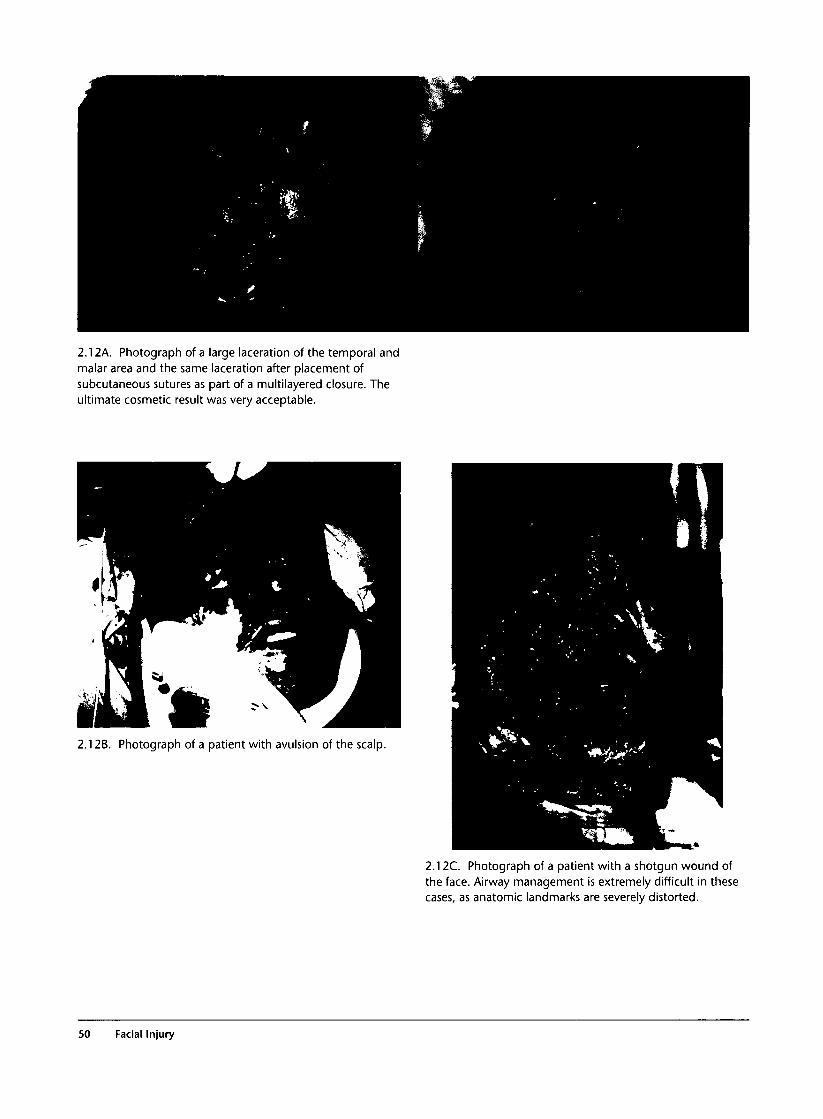
2.12A. Photograph of a large laceration of the temporal andmalar area and the same laceration after placement ofsubcutaneous sutures as part of a multilayered closure. Theultimate cosmetic result was very acceptable.
2.12B. Photograph of a patient with avulsion of the scalp.
2.12C. Photograph of a patient with a shotgun wound ofthe face. Airway management is extremely difficult in thesecases, as anatomic landmarks are severely distorted.
SO Facial Injury
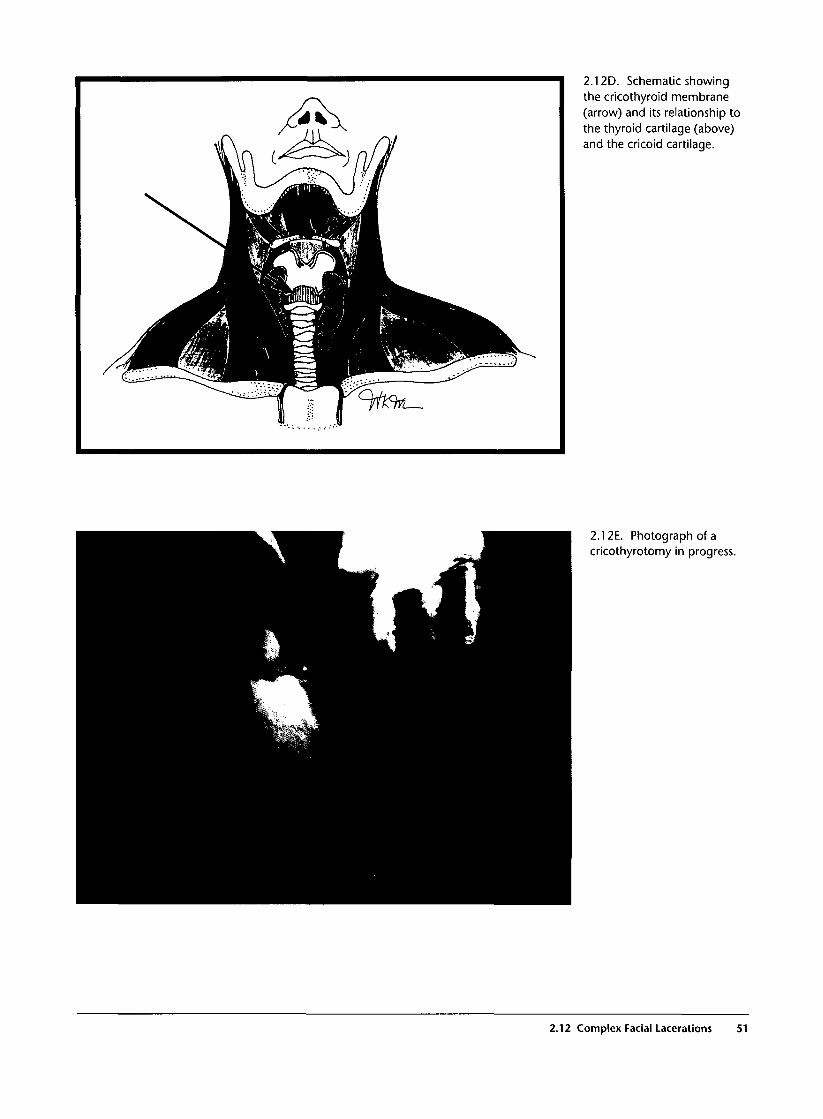
2.12D. Schematic showingthe cricothyroid membrane(arrow) and its relationship tothe thyroid cartilage (above)and the cricoid cartilage.
2.12E. Photograph of acricothyrotomy in progress.
2.12 Complex Facial Lacerations 51
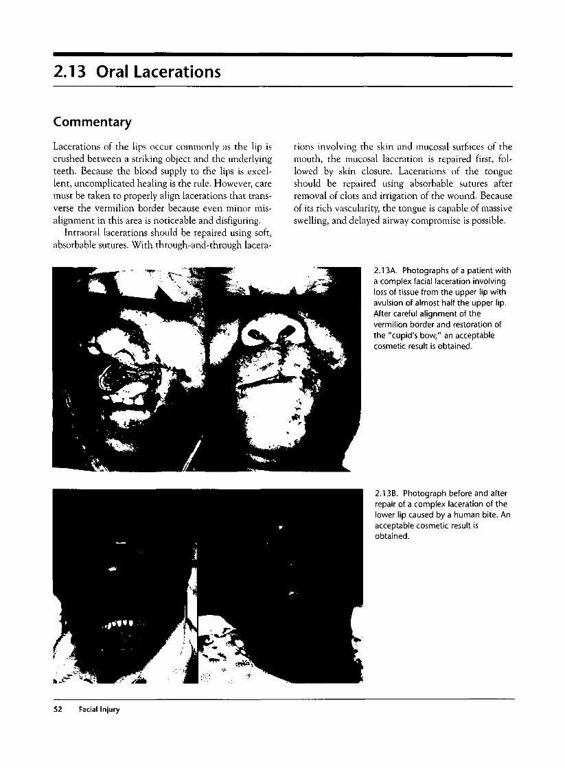
2.13 Oral Lacerations
Commentary
Lacerations of the lips occur commonly as the lip iscrushed between a striking object and the underlyingteeth. Because the blood supply to the lips is excel-lent, uncomplicated healing is the rule. However, caremust be taken to properly align lacerations that trans-verse the vermilion border because even minor mis-alignment in this area is noticeable and disfiguring.
Intraoral lacerations should be repaired using soft,absorbable sutures. With through-and-through lacera-
tions involving the skin and mucosal surfaces of themouth, the mucosal laceration is repaired first, fol-lowed by skin closure. Lacerations of the tongueshould be repaired using absorbable sutures afterremoval of clots and irrigation of the wound. Becauseof its rich vascularity, the tongue is capable of massiveswelling, and delayed airway compromise is possible.
2.13A. Photographs of a patient witha complex facial laceration involvingloss of tissue from the upper lip withavulsion of almost half the upper lip.After careful alignment of thevermilion border and restoration ofthe "cupid's bow," an acceptablecosmetic result is obtained.
2.13B. Photograph before and afterrepair of a complex laceration of thelower lip caused by a human bite. Anacceptable cosmetic result isobtained.
52 Facial Injury
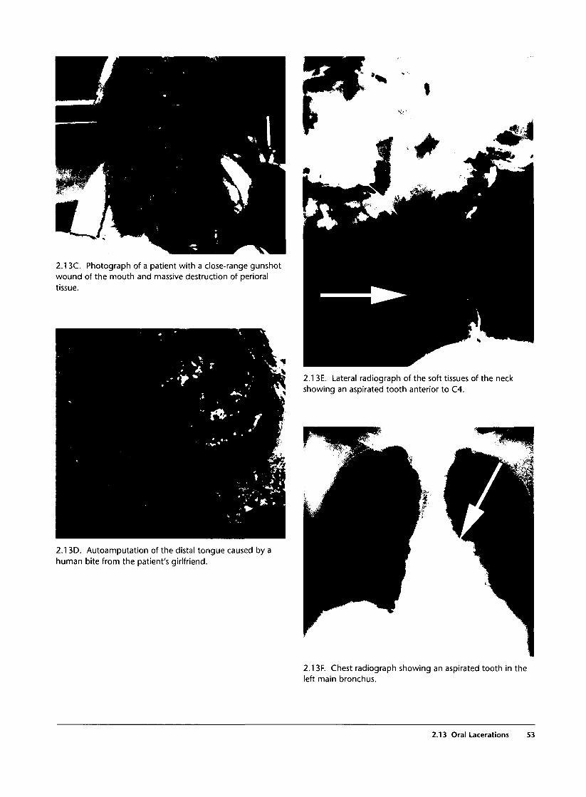
2.13C. Photograph of a patient with a close-range gunshotwound of the mouth and massive destruction of perioraltissue.
2.13E. Lateral radiograph of the soft tissues of the neckshowing an aspirated tooth anterior to C4.
2.13D. Autoamputation of the distal tongue caused by ahuman bite from the patient's girlfriend.
2.13F. Chest radiograph showing an aspirated tooth in theleft main bronchus.
2.13 Oral Lacerations S3

2.14 Facial Nerve Injury
Commentary
Facial nerve injury is most commonly idiopathic inetiology (Bell's palsy). However, penetrating facialtrauma can occasionally result in transection of thefacial nerve as it courses through the face. The facialnerve exits the base of the skull, enters the face justanterior to the tragus of the ear, and continues as alarge trunk for approximately one centimeter before itsubdivides extensively into smaller branches. Lacera-tions in the vicinity of the tragus should prompt care-
ful examination of the muscles of facial expressionbecause facial nerve injury is frequently missed duringthe initial examination of patients with multipletrauma. Injury of the smaller branches of the facialnerve may also merit exploration and surgical repair,depending on the severity of the deficit. Care shouldbe taken to moisten the conjunctiva with artificialtears to avoid desiccation and ulceration of the corneadue to incomplete closure of the eyelid.
2.14A. Photograph of a patient with facial nerve injury onthe left side showing loss of the left nasolabial fold,incomplete eyelid closure, and facial droop.
2.14B. Intraoperative photograph showing a transectedfacial nerve.
54 Facial Injury
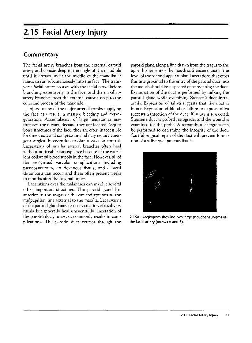
2.15 Facial Artery Injury
Commentary
The facial artery branches from the external carotidartery and courses deep to the angle of the mandibleuntil it crosses under the middle of the mandibularramus to run subcutaneously into the face. The trans-verse facial artery courses with the facial nerve beforebranching extensively in the face, and the maxillaryartery branches from the external carotid deep to thecoronoid process of the mandible.
Injury to any of the major arterial trunks supplyingthe face can result in massive bleeding and exsan-guination. Accumulation of large hematomas maythreaten the airway. Because they are located deep tobony structures of the face, they are often inaccessiblefor direct external compression and may require emer-gent surgical intervention to obtain vascular control.Lacerations of smaller arterial branches often healwithout noticeable consequence because of the excel-lent collateral blood supply in the face. However, all ofthe recognized vascular complications includingpseudoaneurysm, arteriovenous fistula, and delayedthrombosis can occur, and these often present weeksto months after the original injury.
Lacerations over the malar area can involve severalother important structures. The parotid gland liesanterior to the tragus of the ear and extends to themidpupillary line external to the maxilla. Lacerationsof the parotid gland may result in creation of a salivaryfistula but generally heal uneventfully. Laceration ofthe parotid duct, however, commonly results in com-plications. The parotid duct courses through the
parotid gland along a line drawn from the tragus to theupper lip and enters the mouth as Stensen's duct at thelevel of the second upper molar. Lacerations that crossthis line proximal to the entry of the parotid duct intothe mouth should be suspected of transecting the duct.Examination of the duct is performed by milking theparotid gland while examining Stensen's duct intra-orally. Expression of saliva suggests that the duct isintact. Expression of blood or failure to express salivasuggests transection of the duct. If injury is suspected,Stensen's duct is probed retrograde, and the wound isexamined for the probe. Alternately, a sialogram canbe performed to determine the integrity of the duct.Careful surgical repair of the duct will prevent forma-tion of a salivary-cutaneous fistula.
2.15A. Angiogram showing two large pseudoaneurysms ofthe facial artery (arrows A and B).
2.15 Facial Artery Injury 55

Neck Injury
Introduction
Neck injuries, especially penetrating ones, are considereddifficult to evaluate and manage because of the denseconcentration of so many vital structures in a smallanatomical area and the difficult surgical access to manyof these structures. However, very few patients with blunttrauma and only 15-20% of cases with penetratingtrauma require operative treatment. The combination ofa meticulous clinical examination and appropriate inves-tigations can safely identify those patients requiring oper-ative treatment. Advanced trauma life support (ATLS)principles should always be followed.
During the primary survey, the following life-threat-ening conditions in the neck should be identified andtreated:
1. Airway obstruction due to laryngotrachealtrauma or compression by external hematoma
2. Tension pneumothorax
3. Severe active bleeding, externally or in the tho-racic cavity
4. Spinal cord injury or ischemic brain damage dueto carotid artery occlusion
During the secondary survey, the following neckpathologies should be identified and managed:
1. Occult vascular injuries
2. Occult laryngotracheal injuries
3. Occult pharyngoesophageal injuries
4- Cranial or peripheral nerve injuries
5. Small hemopneumothoraces
Clinical Examination
Clinical examination according to a carefully writtenprotocol is the cornerstone of the diagnosis and man-agement. The examination should be systematic andevaluate the vessels, the aerodigestive tract, the spinalcord, the nerves, and the lungs:
1. Vascular structures: "Hard" signs and symptomshighly diagnostic of vascular trauma includeactive bleeding, shock not explained by otherinjuries, expanding or pulsatile hematoma,absent or significantly diminished peripheralpulses, and a bruit. "Soft" signs and symptomssuggestive but not diagnostic of vascular traumainclude mild shock, moderate hematoma, andslow bleeding. This group of patients requires fur-ther investigation.
2. Aerodigestive tract: Hard signs or symptoms highlydiagnostic of significant laryngotracheal traumainclude respiratory distress, air bubbling through aneck wound, and massive hemoptysis. There are nohard signs diagnostic of esophageal trauma. Softsigns and symptoms suspicious of aerodigestivetrauma include subcutaneous emphysema, hoarse-ness, odynophagia, and minor hemoptysis.
3. Nervous system: The examination shouldinclude Glasgow Coma Score, localizing signs,pupils, cranial nerves (VII, IX-XII), spinal cord,brachial plexus (median, ulnar, radial, axillary,musculocutaneous nerves), the phrenic nerve,and the sympathetic chain (Horner's syndrome).
Investigations in Neck Trauma
History and clinical examination will determine theneed and type of investigations in the evaluation ofneck trauma. Patients with hard signs of major vascularor laryngotracheal injuries should be operated on with-out any delay for ancillary investigations. These inves-tigations should be considered only in stable patients.
1. Plain chest and neck films: Look for foreign bodies,fractures, pneumothorax, subcutaneous emphy-sema, and hematoma.
2. Angiography: Angiography should be reservedfor selected cases with soft signs of vasculartrauma when the color flow Doppler study is not
57
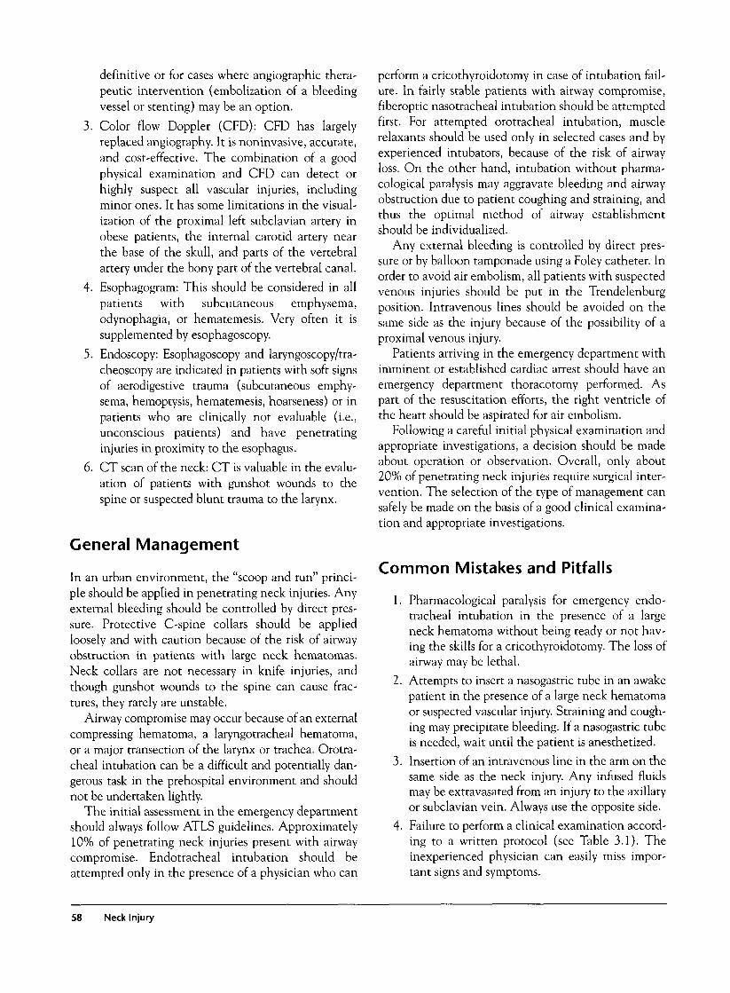
definitive or for cases where angiographic thera-peutic intervention (embolization of a bleedingvessel or stenting) may be an option.
3. Color flow Doppler (CFD): CFD has largelyreplaced angiography. It is noninvasive, accurate,and cost-effective. The combination of a goodphysical examination and CFD can detect orhighly suspect all vascular injuries, includingminor ones. It has some limitations in the visual-ization of the proximal left subclavian artery inobese patients, the internal carotid artery nearthe base of the skull, and parts of the vertebralartery under the bony part of the vertebral canal.
4. Esophagogram: This should be considered in allpatients with subcutaneous emphysema,odynophagia, or hematemesis. Very often it issupplemented by esophagoscopy.
5. Endoscopy: Esophagoscopy and laryngoscopy/tra-cheoscopy are indicated in patients with soft signsof aerodigestive trauma (subcutaneous emphy-sema, hemoptysis, hematemesis, hoarseness) or inpatients who are clinically not evaluable (i.e.,unconscious patients) and have penetratinginjuries in proximity to the esophagus.
6. CT scan of the neck: CT is valuable in the evalu-ation of patients with gunshot wounds to thespine or suspected blunt trauma to the larynx.
General Management
In an urban environment, the "scoop and run" princi-ple should be applied in penetrating neck injuries. Anyexternal bleeding should be controlled by direct pres-sure. Protective C-spine collars should be appliedloosely and with caution because of the risk of airwayobstruction in patients with large neck hematomas.Neck collars are not necessary in knife injuries, andthough gunshot wounds to the spine can cause frac-tures, they rarely are unstable.
Airway compromise may occur because of an externalcompressing hematoma, a laryngotracheal hematoma,or a major transection of the larynx or trachea. Orotra-cheal intubation can be a difficult and potentially dan-gerous task in the prehospital environment and shouldnot be undertaken lightly.
The initial assessment in the emergency departmentshould always follow ATLS guidelines. Approximately10% of penetrating neck injuries present with airwaycompromise. Endotracheal intubation should beattempted only in the presence of a physician who can
perform a cricothyroidotomy in case of intubation fail-ure. In fairly stable patients with airway compromise,fiberoptic nasotracheal intubation should be attemptedfirst. For attempted orotracheal intubation, musclerelaxants should be used only in selected cases and byexperienced intubators, because of the risk of airwayloss. On the other hand, intubation without pharma-cological paralysis may aggravate bleeding and airwayobstruction due to patient coughing and straining, andthus the optimal method of airway establishmentshould be individualized.
Any external bleeding is controlled by direct pres-sure or by balloon tamponade using a Foley catheter. Inorder to avoid air embolism, all patients with suspectedvenous injuries should be put in the Trendelenburgposition. Intravenous lines should be avoided on thesame side as the injury because of the possibility of aproximal venous injury.
Patients arriving in the emergency department withimminent or established cardiac arrest should have anemergency department thoracotomy performed. Aspart of the resuscitation efforts, the right ventricle ofthe heart should be aspirated for air embolism.
Following a careful initial physical examination andappropriate investigations, a decision should be madeabout operation or observation. Overall, only about20% of penetrating neck injuries require surgical inter-vention. The selection of the type of management cansafely be made on the basis of a good clinical examina-tion and appropriate investigations.
Common Mistakes and Pitfalls
1. Pharmacological paralysis for emergency endo-tracheal intubation in the presence of a largeneck hematoma without being ready or not hav-ing the skills for a cricothyroidotomy. The loss ofairway may be lethal.
2. Attempts to insert a nasogastric tube in an awakepatient in the presence of a large neck hematomaor suspected vascular injury. Straining and cough-ing may precipitate bleeding. If a nasogastric tubeis needed, wait until the patient is anesthetized.
3. Insertion of an intravenous line in the arm on thesame side as the neck injury. Any infused fluidsmay be extravasated from an injury to the axillaryor subclavian vein. Always use the opposite side.
4. Failure to perform a clinical examination accord-ing to a written protocol (see Table 3.1). Theinexperienced physician can easily miss impor-tant signs and symptoms.
58 Neck Injury
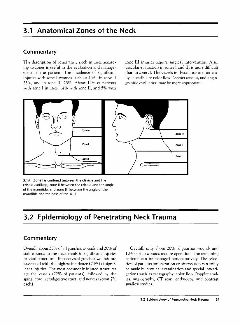
3.1 Anatomical Zones of the Neck
Commentary
The description of penetrating neck injuries accord-ing to zones is useful in the evaluation and manage-ment of the patient. The incidence of significantinjuries with zone I wounds is about 15%, in zone II25%, and in zone III 25%. About 12% of patientswith zone I injuries, 14% with zone II, and 5% with
zone III injuries require surgical intervention. Also,vascular evaluation in zones I and III is more difficultthan in zone II. The vessels in these areas are not eas-ily accessible to color flow Doppler studies, and angio-graphic evaluation may be more appropriate.
3.1 A. Zone I is confined between the clavicle and thecricoid cartilage, zone II between the cricoid and the angleof the mandible, and zone III between the angle of themandible and the base of the skull.
3.2 Epidemiology of Penetrating Neck Trauma
Commentary
Overall, about 35% of all gunshot wounds and 20% ofstab wounds to the neck result in significant injuriesto vital structures. Transcervical gunshot wounds areassociated with the highest incidence (73%) of signif-icant injuries. The most commonly injured structuresare the vessels (22% of patients), followed by thespinal cord, aerodigestive tract, and nerves (about 7%each).
Overall, only about 20% of gunshot wounds and10% of stab wounds require operation. The remainingpatients can be managed nonoperatively. The selec-tion of patients for operation or observation can safelybe made by physical examination and special investi-gations such as radiography, color flow Doppler stud-ies, angiography, CT scan, endoscopy, and contrastswallow studies.
3.2 Epidemiology of Penetrating Neck Trauma 59
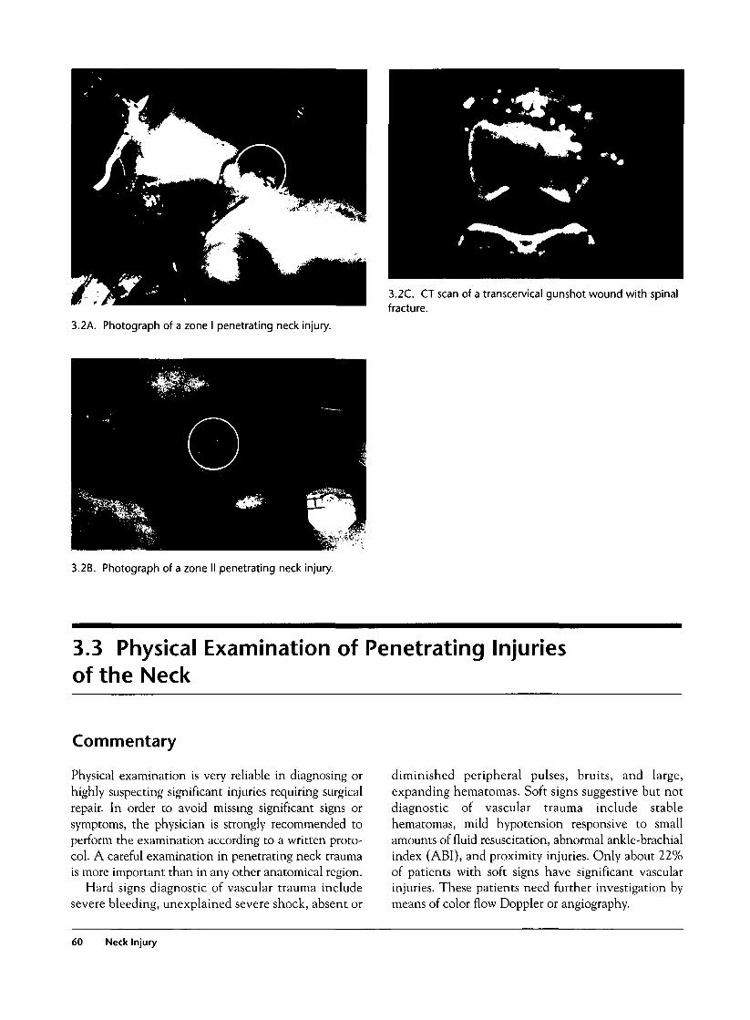
3.2C. CT scan of a transcervical gunshot wound with spinalfracture.
3.2A. Photograph of a zone I penetrating neck injury.
3.2B. Photograph of a zone II penetrating neck injury.
3.3 Physical Examination of Penetrating Injuriesof the Neck
Commentary
Physical examination is very reliable in diagnosing orhighly suspecting significant injuries requiring surgicalrepair. In order to avoid missing significant signs orsymptoms, the physician is strongly recommended toperform the examination according to a written proto-col. A careful examination in penetrating neck traumais more important than in any other anatomical region.
Hard signs diagnostic of vascular trauma includesevere bleeding, unexplained severe shock, absent or
diminished peripheral pulses, bruits, and large,expanding hematomas. Soft signs suggestive but notdiagnostic of vascular trauma include stablehematomas, mild hypotension responsive to smallamounts of fluid resuscitation, abnormal ankle-brachialindex (ABI), and proximity injuries. Only about 22%of patients with soft signs have significant vascularinjuries. These patients need further investigation bymeans of color flow Doppler or angiography.
60 Neck Injury
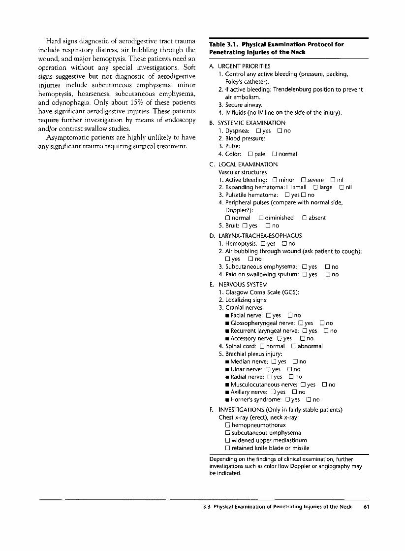
Hard signs diagnostic of aerodigestive tract traumainclude respiratory distress, air bubbling through thewound, and major hemoptysis. These patients need anoperation without any special investigations. Softsigns suggestive but not diagnostic of aerodigestiveinjuries include subcutaneous emphysema, minorhemoptysis, hoarseness, subcutaneous emphysema,and odynophagia. Only about 15% of these patientshave significant aerodigestive injuries. These patientsrequire further investigation by means of endoscopyand/or contrast swallow studies.
Asymptomatic patients are highly unlikely to haveany significant trauma requiring surgical treatment.
Table 3.1. Physical Examination Protocol forPenetrating Injuries of the Neck
A. URGENT PRIORITIES1. Control any active bleeding (pressure, packing,
Foley's catheter).2. If active bleeding: Trendelenburg position to prevent
air embolism.3. Secure airway.4. IV fluids (no IV line on the side of the injury).
B. SYSTEMIC EXAMINATION1. Dyspnea: • yes • no2. Blood pressure:3. Pulse:4. Color: • pale • normal
C. LOCAL EXAMINATIONVascular structures1. Active bleeding: • minor • severe • nil2. Expanding hematoma: • small • large • nil3. Pulsatile hematoma: • yes D no4. Peripheral pulses (compare with normal side,
Doppler?):D normal • diminished D absent
5. Bruit: Dyes • no
D. LARYNX-TRACHEA-ESOPHAGUS1. Hemoptysis: • yes • no2. Air bubbling through wound (ask patient to cough):
• yes • no3. Subcutaneous emphysema: Dyes D no4. Pain on swallowing sputum: • yes • no
E. NERVOUS SYSTEM1. Glasgow Coma Scale (GCS):2. Localizing signs:3. Cranial nerves:
• Facial nerve: • yes • no• Glossopharyngeal nerve: • yes • no• Recurrent laryngeal nerve: • yes • no• Accessory nerve: • yes • no
4. Spinal cord: • normal • abnormal5. Brachial plexus injury:
Median nerve: D yes • noUlnar nerve: Dyes D noRadial nerve: • yes • noMusculocutaneous nerve: • yes • noAxillary nerve: D yes • noHomer's syndrome: • yes • no
F. INVESTIGATIONS (Only in fairly stable patients)Chest x-ray (erect), neck x-ray:
• hemopneumothorax• subcutaneous emphysema• widened upper mediastinum• retained knife blade or missile
Depending on the findings of clinical examination, furtherinvestigations such as color flow Doppler or angiography maybe indicated.
3.3 Physical Examination of Penetrating Injuries of the Neck 61
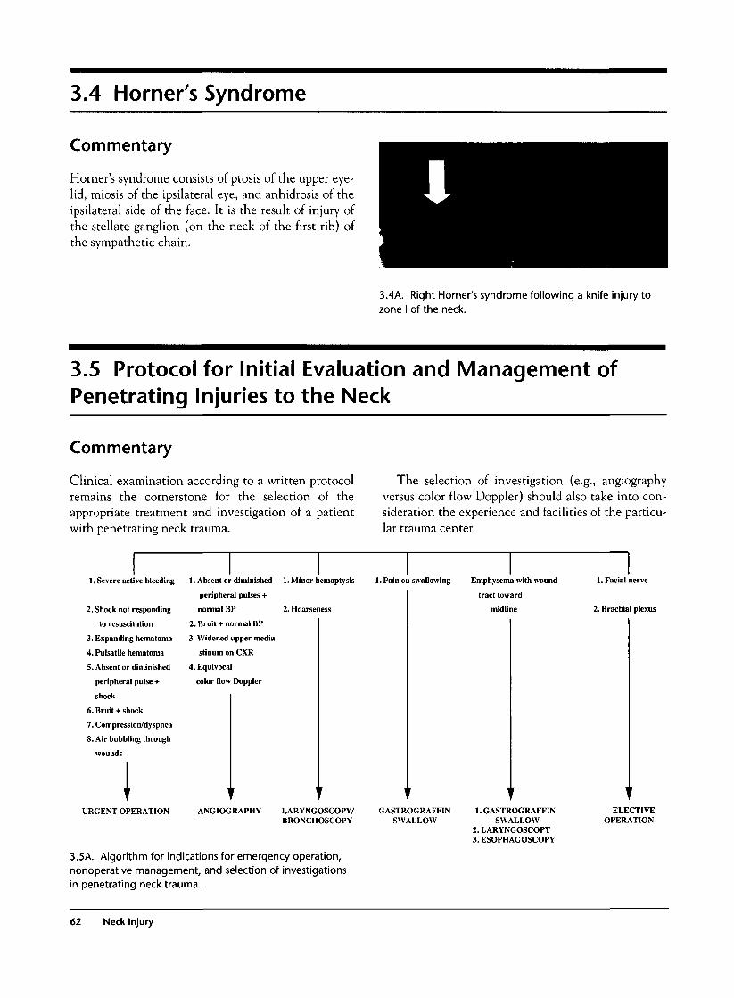
3.4 Homer's Syndrome
Commentary
Homer's syndrome consists of ptosis of the upper eye-lid, miosis of the ipsilateral eye, and anhidrosis of theipsilateral side of the face. It is the result of injury ofthe stellate ganglion (on the neck of the first rib) ofthe sympathetic chain.
3.4A. Right Homer's syndrome following a knife injury tozone I of the neck.
3.5 Protocol for Initial Evaluation and Management ofPenetrating Injuries to the Neck
Commentary
Clinical examination according to a written protocolremains the cornerstone for the selection of theappropriate treatment and investigation of a patientwith penetrating neck trauma.
The selection of investigation (e.g., angiographyversus color flow Doppler) should also take into con-sideration the experience and facilities of the particu-lar trauma center.
1. Severe active bleeding
2. Shock not responding
to resuscitation
3. Expanding hematoma
4. Pulsatile hematoma
5. Absent or diminished
peripheral pulse +
shock
6. Bruit + shock
7. Compression/dyspnea
8. Air bubbling through
wounds
tURGENT OPERATION ANGIOGRAPHY
1. Absent or diminished
peripheral pulses +
normal BP
2. Bruit + normal BP
3. Widened upper media
stinum on CXR
4. Equivocal
color flow Doppler
1. Minor hemoptysis 1. Pain on swallowing
2. Hoarseness
LARYNGOSCOPY/BRONCHOSCOPY
3.5A. Algorithm for indications for emergency operation,nonoperative management, and selection of investigationsin penetrating neck trauma.
Emphysema with wound
tract toward
midline
1. Facial nerve
2. Brachial plexus
\
GASTROGRAFFINSWALLOW
1. GASTROGRAFFINSWALLOW
2. LARYNGOSCOPY3.ESOPHAGOSCOPY
ELECTIVEOPERATION
62 Neck Injury

3.6 Plain Radiography in Penetrating Neck Trauma
Commentary
Anteroposterior and lateral films of the neck shouldbe obtained for all patients with penetrating necktrauma who are hemodynamically stable. Importantradiological findings include hematoma, subcutaneousemphysema, fractures, and foreign bodies. A chest filmis also recommended in all stable patients. In about15% of patients with penetrating neck trauma, there isan associated pneumothorax or hemothorax. A widenedupper mediastinum is suggestive of a hematoma sec-ondary to a vascular injury, and an elevated diaphragmmay be due to a phrenic nerve injury.
3.6B. Plain film of the neck shows a prevertebral hematomaand deviation of the nasogastric tube.
3.6A. Plain film of the neck showing two missiles, one inzone I and another in zone III.
3.6C. AP neck film showing subcutaneous emphysemasecondary to tracheal injury.
3.6 Plain Radiography in Penetrating Neck Trauma 63
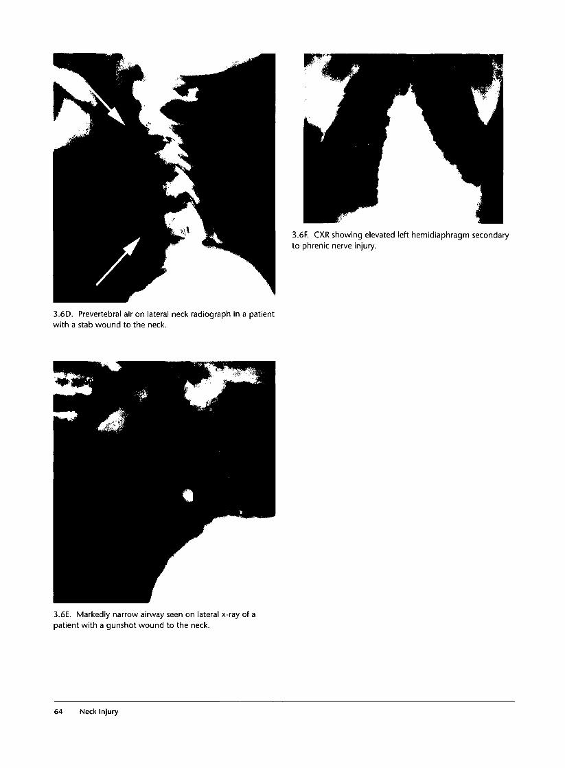
3.6F. CXR showing elevated left hemidiaphragm secondaryto phrenic nerve injury.
3.6D. Prevertebral air on lateral neck radiograph in a patientwith a stab wound to the neck.
3.6E. Markedly narrow airway seen on lateral x-ray of apatient with a gunshot wound to the neck.
64 Neck Injury
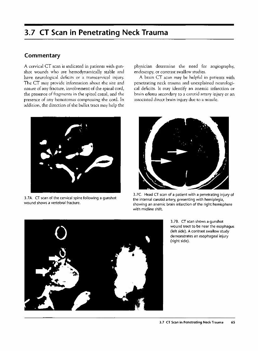
3.7 CT Scan in Penetrating Neck Trauma
Commentary
A cervical CT scan is indicated in patients with gun-shot wounds who are hemodynamically stable andhave neurological deficits or a transcervical injury.The CT may provide information about the site andnature of any fracture, involvement of the spinal cord,the presence of fragments in the spinal canal, and thepresence of any hematomas compressing the cord. Inaddition, the direction of the bullet tract may help the
physician determine the need for angiography,endoscopy, or contrast swallow studies.
A brain CT scan may be helpful in patients withpenetrating neck trauma and unexplained neurologi-cal deficits. It may identify an anemic infarction orbrain edema secondary to a carotid artery injury or anassociated direct brain injury due to a missile.
3.7A. CT scan of the cervical spine following a gunshotwound shows a vertebral fracture.
3.7C. Head CT scan of a patient with a penetrating injury ofthe internal carotid artery, presenting with hemiplegia,showing an anemic brain infarction of the right hemispherewith midline shift.
3.7B. CT scan shows a gunshotwound tract to be near the esophagus(left side). A contrast swallow studydemonstrates an esophageal injury(right side).
3.7 CT Scan in Penetrating Neck Trauma 65

3.7D. Gunshot wound to the neck. CT shows hematoma
with compression and deviation of the nasogastric tube.
3.8 Evaluation of Vascular Structures in the Neck
Commentary
Asymptomatic patients are highly unlikely to havesignificant vascular injuries requiring surgical inter-vention or angiographic embolization. However, inabout 8% of asymptomatic patients investigated byangiography, there is a vascular injury not requiringany type of treatment.
Patients with soft signs of vascular trauma needevaluation by color flow Doppler and, in appropriatecases, angiography. The combination of physicalexamination and color flow Doppler will detectalmost all vascular injuries.
Clinical Examination According to Protocol
Obvious significant vascular injuries?• Severe active bleeding• Shock not responding to fluids• Absent radial pulse
Yes
Operation • Bruit• Widened mediastinum• Shotgun wounds
Yes No
I IAngiogram • Hematoma
• Diminished peripheral pulse• Shock responding to fluids
Yes No
Color flow Doppler(Angiogram if CFD not available)
Observation(CFD optional)
3.8A. Algorithm for evaluation of the vascular structures in
the neck.
66 Neck Injury
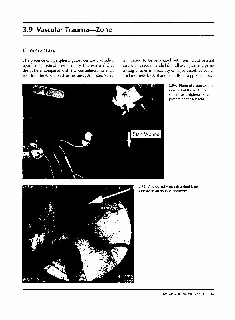
3.9 Vascular Trauma—Zone I
Commentary
The presence of a peripheral pulse does not preclude asignificant proximal arterial injury. It is essential thatthe pulse is compared with the contralateral one. Inaddition, the ABI should be measured. An index >0.90
is unlikely to be associated with significant arterialinjury. It is recommended that all asymptomatic pene-trating injuries in proximity of major vessels be evalu-ated routinely by ABI and color flow Doppler studies.
3.9A. Photo of a stab woundin zone I of the neck. Thevictim has peripheral pulsepresent on the left arm.
3.9B. Angiography reveals a significantsubclavian artery false aneurysm.
3.9 Vascular Trauma—Zone I 67
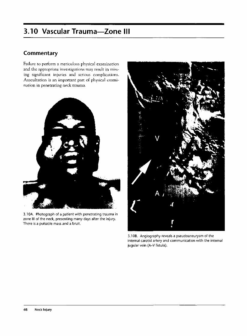
3.10 Vascular Trauma—Zone
Commentary
Failure to perform a meticulous physical examinationand the appropriate investigations may result in miss-ing significant injuries and serious complications.Auscultation is an important part of physical exami-nation in penetrating neck trauma.
3.10A. Photograph of a patient with penetrating trauma inzone III of the neck, presenting many days after the injury.There is a pulsatile mass and a bruit.
3.1 OB. Angiography reveals a pseudoaneurysm of theinternal carotid artery and communication with the internaljugular vein (A-V fistula).
68 Neck Injury
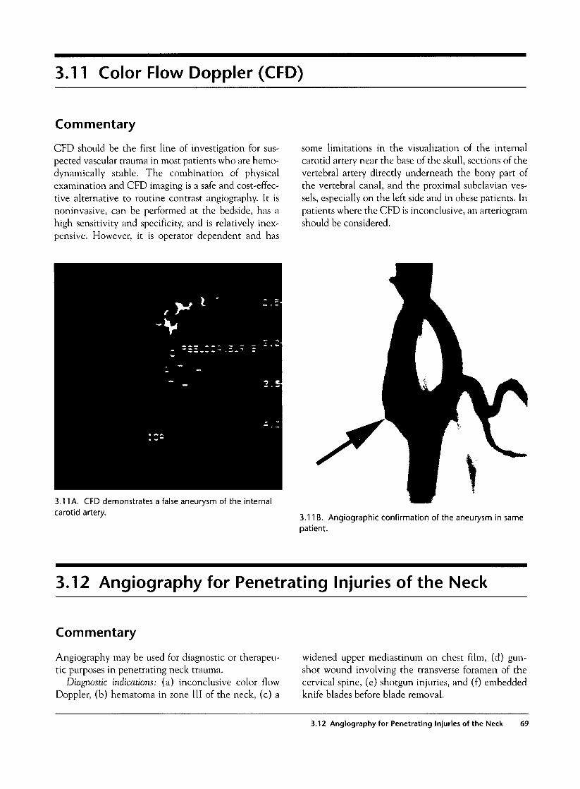
3.11 Color Flow Doppler (CFD)
Commentary
CFD should be the first line of investigation for sus-pected vascular trauma in most patients who are hemo-dynamically stable. The combination of physicalexamination and CFD imaging is a safe and cost-effec-tive alternative to routine contrast angiography. It isnoninvasive, can be performed at the bedside, has ahigh sensitivity and specificity, and is relatively inex-pensive. However, it is operator dependent and has
some limitations in the visualization of the internalcarotid artery near the base of the skull, sections of thevertebral artery directly underneath the bony part ofthe vertebral canal, and the proximal subclavian ves-sels, especially on the left side and in obese patients. Inpatients where the CFD is inconclusive, an arteriogramshould be considered.
3.11 A. CFD demonstrates a false aneurysm of the internalcarotid artery.
3.11B. Angiographic confirmation of the aneurysm in samepatient.
3.12 Angiography for Penetrating Injuries of the Neck
Commentary
Angiography may be used for diagnostic or therapeu-tic purposes in penetrating neck trauma.
Diagnostic indications: (a) inconclusive color flowDoppler, (b) hematoma in zone III of the neck, (c) a
widened upper mediastinum on chest film, (d) gun-shot wound involving the transverse foramen of thecervical spine, (e) shotgun injuries, and (f) embeddedknife blades before blade removal.
3.12 Angiography for Penetrating Injuries of the Neck 69
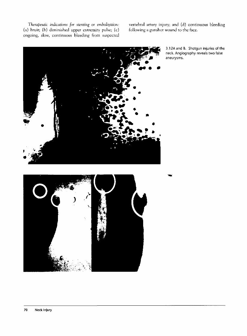
Therapeutic indications for stendng or embolization:(a) bruit; (b) diminished upper extremity pulse; (c)ongoing, slow, continuous bleeding from suspected
vertebral artery injury; and (d) continuous bleedingfollowing a gunshot wound to the face.
3.12A and B. Shotgun injuries of theneck. Angiography reveals two falseaneurysms.
70 Neck Injury
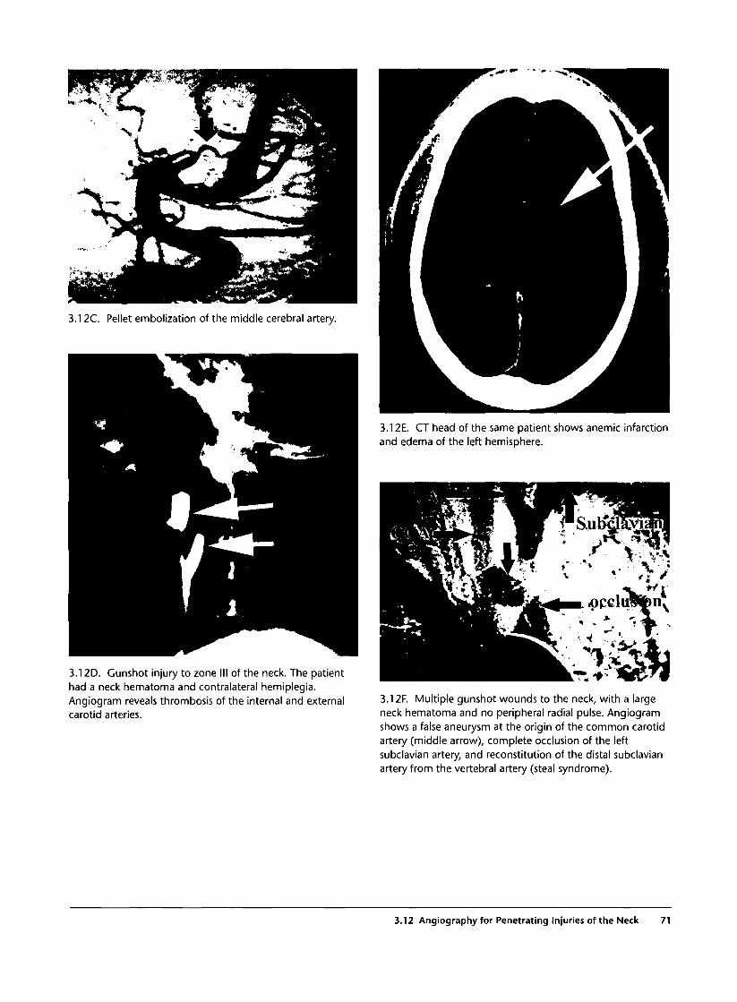
3.12C. Pellet embolization of the middle cerebral artery.
3.12E. CT head of the same patient shows anemic infarctionand edema of the left hemisphere.
3.12D. Gunshot injury to zone III of the neck. The patienthad a neck hematoma and contralateral hemiplegia.Angiogram reveals thrombosis of the internal and externalcarotid arteries.
3.12F. Multiple gunshot wounds to the neck, with a largeneck hematoma and no peripheral radial pulse. Angiogramshows a false aneurysm at the origin of the common carotidartery (middle arrow), complete occlusion of the leftsubclavian artery, and reconstitution of the distal subclavianartery from the vertebral artery (steal syndrome).
3.12 Angiography for Penetrating Injuries of the Neck 71
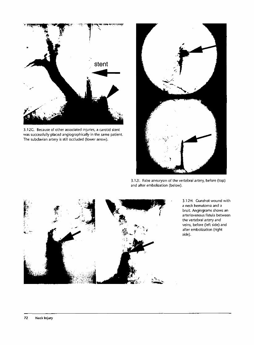
3.12G. Because of other associated injuries, a carotid stentwas successfully placed angiographically in the same patient.The subclavian artery is still occluded (lower arrow).
3.121. False aneurysm of the vertebral artery, before (top)and after embolization (below).
3.12H. Gunshot wound witha neck hematoma and abruit. Angiograms shows anarteriovenous fistula betweenthe vertebral artery andveins, before (left side) andafter embolization (rightside).
72 Neck Injury
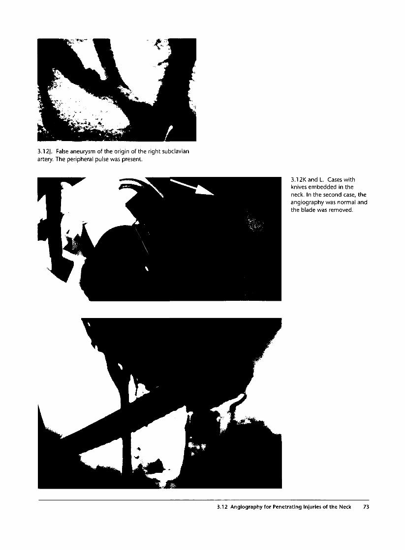
3.12J. False aneurysm of the origin of the right subclavianartery. The peripheral pulse was present.
3.12K and L. Cases withknives embedded in theneck. In the second case, theangiography was normal andthe blade was removed.
3.12 Angiography for Penetrating Injuries of the Neck 73
3.13 Evaluation of the Aerodigestive Tract in the Neck
Commentary
Asymptomatic patients are highly unlikely to have asignificant injury requiring surgical management. Un-evaluable patients or patients with soft symptoms ofaerodigestive injuries need investigation by endoscopyand contrast swallow studies.
Clinical Examination According to Protocol
Obvious significant aerodigestive injuries?• Air bubbling through wound• Major hemoptysis• Respiratory distress
Yes
Operation
No
I• Minor hemoptysis•Hoarseness•Subcutaneous emphysema• Painful swallowing• Proximity in obtunded patient
No Yes
Observation EndoscopyEsophagogram
3.13A. Algorithm for the evaluation of the aerodigestivetract in the neck.
3.14 Esophageal Trauma
Commentary
The esophagus can be evaluated by means of esopha-gography or endoscopy. The combination of the twoinvestigations has a sensitivity of 100%. Rigidesophagoscopy is superior to flexible esophagoscopy in
evaluating the upper esophagus; however, rigidendoscopy can be performed only under general anes-thesia. Thus this investigation is best reserved forpatients undergoing anesthesia for another reason.
74 Neck Injury

3.14A. Subcutaneous emphysema following a gunshotwound to the neck. This finding is an absolute indication forevaluation of the aerodigestive tract.
3.14B. Gastrograffin swallow shows a perforation of thecervical esophagus.
3.14 Esophageal Trauma 75
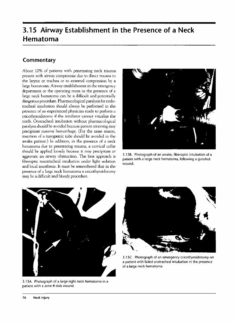
3.15 Airway Establishment in the Presence of a NeckHematoma
Commentary
About 10% of patients with penetrating neck traumapresent with airway compromise due to direct trauma tothe larynx or trachea or to external compression by alarge hematoma. Airway establishment in the emergencydepartment or the operating room in the presence of alarge neck hematoma can be a difficult and potentiallydangerous procedure. Pharmacological paralysis for endo-tracheal intubation should always be performed in thepresence of an experienced physician ready to perform acricothyroidotomy if the intubator cannot visualize thecords. Orotracheal intubation without pharmacologicalparalysis should be avoided because patient straining mayprecipitate massive hemorrhage. (For the same reason,insertion of a nasogastric tube should be avoided in theawake patient.) In addition, in the presence of a neckhematoma due to penetrating trauma, a cervical collarshould be applied loosely because it may precipitate oraggravate an airway obstruction. The best approach isfiberoptic nasotracheal intubation under light sedationand local anesthesia. It must be remembered that in thepresence of a large neck hematoma a cricothyroidotomymay be a difficult and bloody procedure.
3.15B. Photograph of an awake, fiberoptic intubation of apatient with a large neck hematoma, following a gunshotwound.
3.15C. Photograph of an emergency cricothyroidotomy ona patient with failed orotracheal intubation in the presenceof a large neck hematoma.
3.15A. Photograph of a large right neck hematoma in apatient with a zone II stab wound.
76 Neck Injury
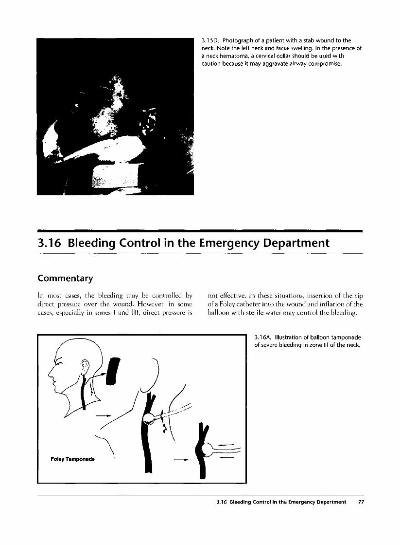
3.15D. Photograph of a patient with a stab wound to theneck. Note the left neck and facial swelling. In the presence ofa neck hematoma, a cervical collar should be used withcaution because it may aggravate airway compromise.
3.16 Bleeding Control in the Emergency Department
Commentary
In most cases, the bleeding may be controlled bydirect pressure over the wound. However, in somecases, especially in zones I and III, direct pressure is
not effective. In these situations, insertion of the tipof a Foley catheter into the wound and inflation of theballoon with sterile water may control the bleeding.
3.16A. Illustration of balloon tamponadeof severe bleeding in zone III of the neck.
3.16 Bleeding Control in the Emergency Department 77
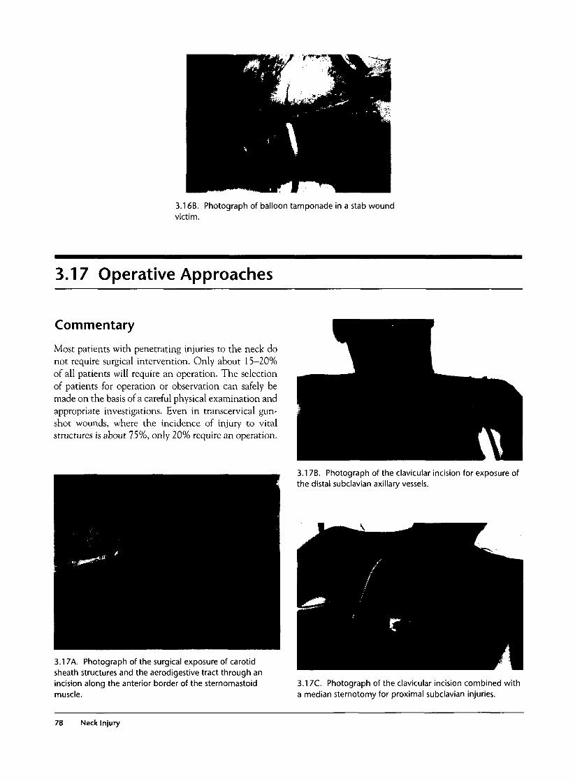
3.16B. Photograph of balloon tamponade in a stab woundvictim.
3.17 Operative Approaches
Commentary
Most patients with penetrating injuries to the neck donot require surgical intervention. Only about 15-20%of all patients will require an operation. The selectionof patients for operation or observation can safely bemade on the basis of a careful physical examination andappropriate investigations. Even in transcervical gun-shot wounds, where the incidence of injury to vitalstructures is about 75%, only 20% require an operation.
3.17B. Photograph of the clavicular incision for exposure ofthe distal subclavian axillary vessels.
3.17A. Photograph of the surgical exposure of carotidsheath structures and the aerodigestive tract through anincision along the anterior border of the sternomastoidmuscle.
3.17C. Photograph of the clavicular incision combined witha median sternotomy for proximal subclavian injuries.
78 Neck Injury

3.18 Penetrating Trauma to the Carotid Artery
Commentary
Traumatic thrombosis of the common or internal carotidartery may result in ipsilateral anemic infarction andbrain edema, although in some cases this may be welltolerated. Early revascularization, ideally within the first2—4 hours, is associated with good results. Delayed revas-cularization after the establishment of an anemicinfarction may worsen the brain edema or lead to a hem-orrhagic infarction. Early surgical reconstructionremains the mainstay of management of penetratingcarotid injuries. In selected cases such as in high internalcarotid injuries, where surgical exposure is difficult,angiographic stenting may be a good alternative.
3.18B. CT of the brain with severe anemic infarction andbrain edema following thrombosis of the internal carotidartery due to a gunshot wound.
! • '
3.18A. Angiogram showing gunshot wound-relatedocclusion of the internal carotid artery.
3.18C. CT of the brain showing hemorrhagic infarctionfollowing delayed revascularization of a thrombosed carotidartery.
3.18 Penetrating Trauma to the Carotid Artery 79
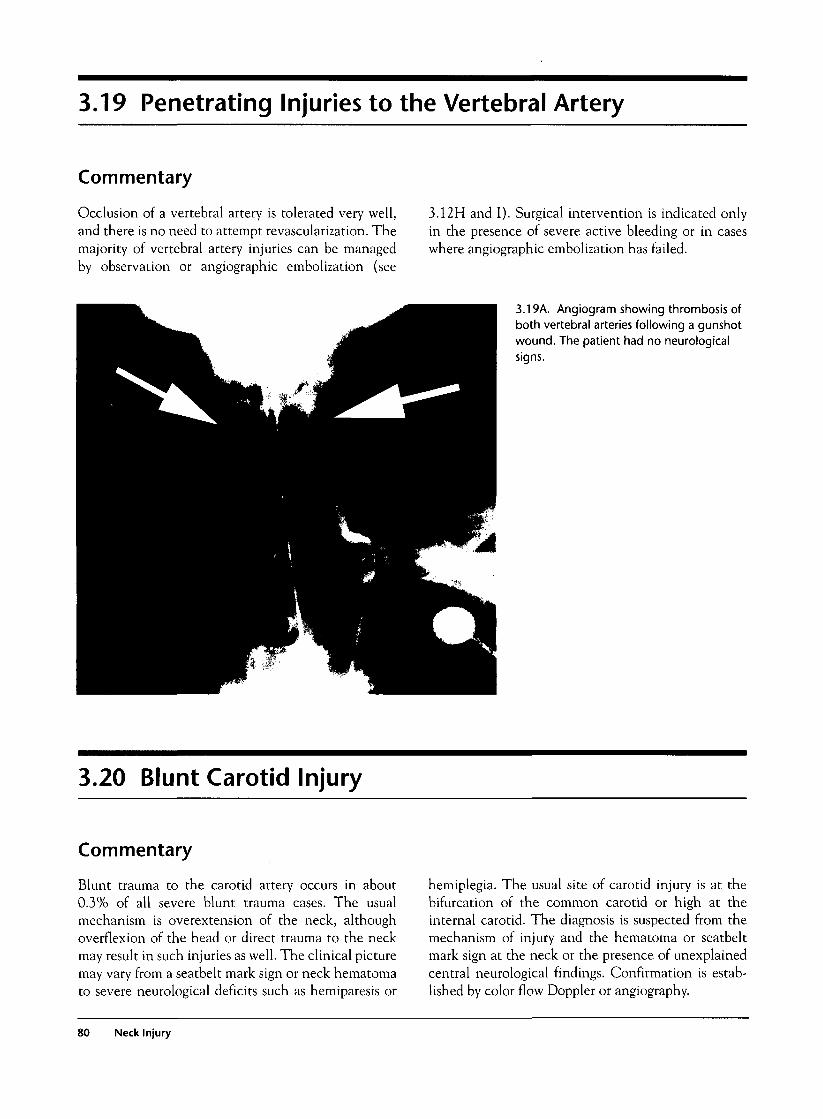
3.19 Penetrating Injuries to the Vertebral Artery
Commentary
Occlusion of a vertebral artery is tolerated very well,and there is no need to attempt revascularization. Themajority of vertebral artery injuries can be managedby observation or angiographic embolization (see
3.12H and I). Surgical intervention is indicated onlyin the presence of severe active bleeding or in caseswhere angiographic embolization has failed.
3.19A. Angiogram showing thrombosis ofboth vertebral arteries following a gunshotwound. The patient had no neurologicalsigns.
3.20 Blunt Carotid Injury
Commentary
Blunt trauma to the carotid artery occurs in about0.3% of all severe blunt trauma cases. The usualmechanism is overextension of the neck, althoughoverflexion of the head or direct trauma to the neckmay result in such injuries as well. The clinical picturemay vary from a seatbelt mark sign or neck hematomato severe neurological deficits such as hemiparesis or
hemiplegia. The usual site of carotid injury is at thebifurcation of the common carotid or high at theinternal carotid. The diagnosis is suspected from themechanism of injury and the hematoma or seatbeltmark sign at the neck or the presence of unexplainedcentral neurological findings. Confirmation is estab-lished by color flow Doppler or angiography.
80 Neck Injury
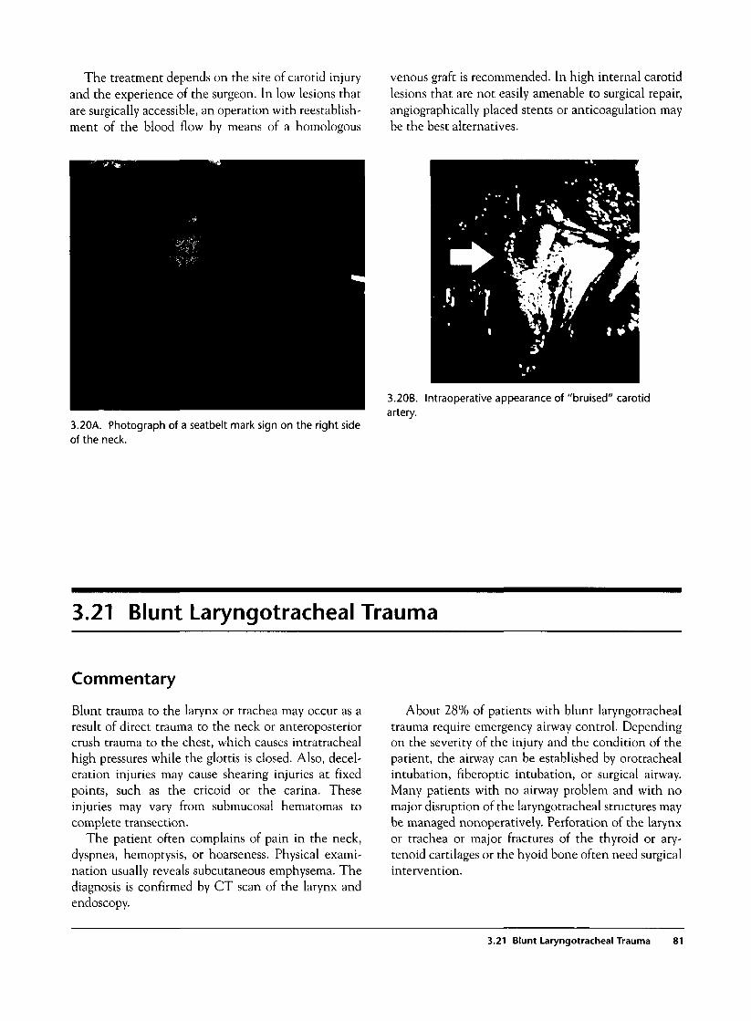
The treatment depends on the site of carotid injuryand the experience of the surgeon. In low lesions thatare surgically accessible, an operation with reestablish-ment of the blood flow by means of a homologous
venous graft is recommended. In high internal carotidlesions that are not easily amenable to surgical repair,angiographically placed stents or anticoagulation maybe the best alternatives.
3.20A. Photograph of a seatbelt mark sign on the right sideof the neck.
3.20B. Intraoperative appearance of "bruised" carotidartery.
3.21 Blunt Laryngotracheal Trauma
Commentary
Blunt trauma to the larynx or trachea may occur as aresult of direct trauma to the neck or anteroposteriorcrush trauma to the chest, which causes intratrachealhigh pressures while the glottis is closed. Also, decel-eration injuries may cause shearing injuries at fixedpoints, such as the cricoid or the carina. Theseinjuries may vary from submucosal hematomas tocomplete transection.
The patient often complains of pain in the neck,dyspnea, hemoptysis, or hoarseness. Physical exami-nation usually reveals subcutaneous emphysema. Thediagnosis is confirmed by CT scan of the larynx andendoscopy.
About 28% of patients with blunt laryngotrachealtrauma require emergency airway control. Dependingon the severity of the injury and the condition of thepatient, the airway can be established by orotrachealintubation, fiberoptic intubation, or surgical airway.Many patients with no airway problem and with nomajor disruption of the laryngotracheal structures maybe managed nonoperatively. Perforation of the larynxor trachea or major fractures of the thyroid or ary-tenoid cartilages or the hyoid bone often need surgicalintervention.
3.21 Blunt Laryngotracheal Trauma 81

3.21 A. Photograph showing subcutaneous emphysemafollowing blunt trauma to the neck and laryngeal fracture.
3.21 B. CT scan of the neck confirms fracture of the thyroidcartilage.
82 Neck Injury
Thoracic Injury
Introduction
Chest trauma is estimated to be the primary cause ofdeath in 25% of traumatic mortalities and a con-tributing factor in another 25% of deaths. Goodunderstanding of the pathophysiology of chest traumaand timely selection of the appropriate investigationsand treatment are all critical components for optimaloutcome.
Clinical Examination
Advanced trauma life support (ATLS) principles areparticularly important in the initial evaluation andmanagement of the chest trauma patient.
During the primary survey, there are six life-threatening conditions that need to be identified andtreated:
1. Airway obstruction
2. Tension pneumothorax
3. Open pneumothorax with a "sucking wound"
4. Flail chest
5. Massive hemothorax
6. Cardiac tamponade
During the secondary survey, there are another sixpotentially lethal chest injuries that should be identi-fied and treated. The diagnosis of these conditionsmay need more complex and time-consuming investi-gations.
1. Lung contusion
2. Myocardial contusion
3. Aortic rupture
4. Diaphragmatic rupture
5. Tracheobronchial rupture
6. Esophageal injury
Investigations
History and clinical examination will determine thetype and timing of investigations necessary for the safeand efficient evaluation of the chest trauma patient.Very often, in unstable patients, therapeutic interven-tions such as thoracostomy tube insertion or thoraco-tomy may be initiated without any investigations.
Investigations are necessary in fairly stable patientswhen the diagnosis is uncertain. The following investi-gations may be useful in chest trauma:
1. Chest x-ray: Ideally it should be taken during deepexpiration and erect position for more accuratedetection of small pneumothoraces, small hemoth-oraces, and mediastinal abnormalities. In manycases this is not possible because of associatedhemodynamic instability, depressed level of con-sciousness, or concern about spinal injury. The fol-lowing radiological findings should be sought:pneumothorax, hemothorax, lung contusion, sub-cutaneous or mediastinal emphysema, fractures,mediastinal widening, enlarged cardiac shadow,pneumopericardium, elevated diaphragm or suspi-cious shadows suggestive of diaphragmatic hernia,free air under the diaphragm, and foreign bodies.
2. Electrocardiogram (ECG): ECG should be per-formed in most blunt chest trauma patients andselected penetrating injuries. It is essential in thediagnosis of blunt cardiac trauma or myocardialinfarction.
3. Trauma ultrasound: This is a very important inves-tigation and should be available in the emergencydepartment. Emergency physicians and traumasurgeons should be trained in its use, as it allowsearly diagnosis of cardiac tamponade or hemoperi-toneum at the bedside.
4. Pericardiocentesis: This diagnostic procedure haslittle or no value in the evaluation of suspectedcardiac trauma in a modern trauma center as it is
83
associated with an unacceptably high incidence offalse-negative results because of clot formation inthe pericardium. In addition, it is a potentiallydangerous procedure because of the risk of myocar-dial or coronary vessel perforation, especially ifperformed in the absence of hemopericardium.
5. Subxiphoid pericardial window: This major inva-sive procedure has been used by many surgeons todiagnose cardiac tamponade. With bedside echocar-diography to evaluate the pericardium, it has verylittle or no place in a modern trauma center.
6. Chest computed tomography (CT): CT is veryvaluable in blunt trauma in the evaluation of themediastinum, the thoracic spine, and suspectedlung contusions. All stable blunt trauma patientswith a suspicious mechanism of injury (high-speedaccidents, falls from height) or those with anabnormal mediastinum on chest films should beevaluated by spiral CT scan for aortic rupture. Spi-ral CT scan is highly sensitive and specific indetecting aortic rupture and has replaced aortogra-phy to a great extent. CT scan is also useful in theevaluation of suspected lung contusions or persist-ent opacifications on chest x-ray following inser-tion of a thoracostomy tube. In these cases, thedifferential diagnosis between atelectasis, contu-sion, and residual hemothorax may be difficult orimpossible on the basis of clinical examinationand chest x-ray.
CT scan also has a definitive role in the evalua-tion of selected patients with penetrating chesttrauma. In patients with transmediastinal gunshotwounds who are hemodynamically stable, a spiralCT scan with thin cuts may be useful in identifyingthe bullet track and in determining the need forfurther investigations such as aortography orendoscopy. If the direction of the bullet track isaway from the aorta, esophagus, and other impor-tant mediastinal structures, no further investiga-tions are necessary.
7. Angiography: Aortography is still used by manycenters as the standard investigation for suspectedaortic injuries. In some trauma centers, it haslargely been replaced by spiral CT scan, and aor-tography is reserved for cases with indeterminateCT scan findings.
Angiography also has a role for selected pene-trating injuries of the thoracic inlet with suspectedinnominate or subclavian vascular injuries. In someof these cases with subclavian false aneurysms orarteriovenous fistulae, angiographically placed
stents may provide the definitive management ofthe injury.
8. Color flow Doppler: Color flow Doppler ultrasoundis a very good noninvasive vascular study that canreliably evaluate the subclavian and neck vesselsand is recommended for hemodynamically stablepatients with thoracic inlet penetrating injuries. Itsmajor weaknesses are its operator-dependent accu-racy and inability to visualize the left proximal sub-clavian vessels, especially in obese patients.
9. Endoscopy: Esophagoscopy and bronchoscopy maybe necessary for suspected aerodigestive tractinjuries, usually as a result of mediastinal penetrat-ing trauma.
10. Laparoscopy: Laparoscopy has a definitive role inthe evaluation of the diaphragm in asymptomaticpatients with left thoracoabdominal or right ante-rior thoracoabdominal penetrating injuries. Failureto recognize and repair a diaphragmatic injury mayresult in a diaphragmatic hernia at a later stage.Right posterior thoracoabdominal injuries do notneed evaluation and repair of any diaphragmaticlaceration because the presence of the liver pro-tects against herniation of intra-abdominal organs.The procedure may be performed under generalanesthesia or under sedation and local anesthesia.
11. Thoracoscopy: Thoracoscopy is used in the evalu-ation of diaphragmatic injuries, especially in theposterior region. It is particularly useful in caseswith residual hemothorax that can be evacuatedthrough the port. It has the disadvantage of need-ing double lumen intubation and lung collapse,which may be technically demanding and some-times not well tolerated by the patient.
12. Troponin levels: Serial troponin measurements havereplaced cardiac enzymes (e.g., CPK-MB) for moni-toring in suspected myocardial contusion. Thoughroutinely used, troponin levels do not correlate withthe severity of the myocardial trauma.
13. Arterial blood gas: Arterial blood gases are indi-cated in all severe chest trauma cases in order toassess severity, plan treatment, and monitorprogress of treatment.
General Management
In an urban environment, there is no place for prehos-pital attempts to stabilize patients with severe chesttrauma, and the principle of "scoop and run" should be
84 Thoracic Injury
applied. The patient should be placed on a spinalboard, receive oxygen by mask, and be transferredwithout any delay to the nearest trauma hospital.Intravenous access should be attempted in the ambu-lance en route. In patients with severe respiratory dis-tress and clinical suspicion of tension pneumothorax, aneedle thoracostomy may be attempted. Open suckingwounds should be covered with a clean square gauzetaped only on three sides to avoid a tension pneumoth-orax. Patients with respiratory failure or a depressedlevel of consciousness will need prehospital intubationor bag valve mask ventilation en route. In the emer-gency department, the primary survey will determinethe need and type of immediate treatment. Patientswith imminent or established cardiac arrest should bemanaged with an emergency department resuscitativethoracotomy. Any bleeding is controlled by sutures orclamping. Aortic cross-clamping is performed, and car-diac massage is initiated. Transfusions with O negativeblood, cardiac drugs, and defibrillation are adminis-tered as necessary. If cardiac activity returns, the opera-tion is completed in the operating room.
Patients with severe hypotension and suspicion ofcardiovascular trauma should be taken directly to theoperating room with minimal investigations. A traumaultrasound is very valuable in determining the source ofhypotension in patients with multiple injuries as it canevaluate the pericardial, pleural, and peritoneal spaces.
Hemodynamically stable patients are examinedcarefully, and further investigations are performed asindicated. Thoracostomy tubes, analgesia, intubation,and mechanical ventilation may be necessary.
Elderly patients with multiple rib fractures despite anormal respiratory status on initial examination shouldbe admitted to the intensive care unit for close moni-toring, as these patients are prone to deteriorate a fewhours after admission. Adequate analgesia by means ofepidural or patient-controlled analgesia are criticalcomponents of the management of these cases.
Indications for Thoracotomy
Fewer than 10% of blunt chest traumas, about 15% ofstab wounds, and about 20% of gunshot wounds
require thoracotomy. The majority of patients withchest trauma can safely be managed with a thoracos-tomy tube and other supportive treatment. The indica-tions for emergency thoracotomy are generally thefollowing:
• Cardiac arrest or imminent cardiac arrest
• Evidence of cardiovascular injury, such as severehypotension or severe active external or internalbleeding
• Immediate blood loss in the thoracostomy tubemore than 1,000-1,200 ml
• Diagnosis of aortic rupture or esophageal or tracheo-bronchial injury
Semielective thoracotomies are indicated for largeresidual hemothoraces or persistent major air leaks.
Common Mistakes and Pitfalls
1. Elderly patients with multiple rib fractures mayseem stable on admission, but rapid respiratory dete-rioration may occur a few hours later. Close moni-toring in an ICU environment is critical.
2. Patients with flail chest may seem stable on admis-sion. Decompensation with severe respiratory fail-ure may occur during prolonged radiologicalinvestigation with potentially catastrophic conse-quences. Liberal early intubation is recommendedbefore these patients are transferred to the radiologysuite.
3. A widened mediastinum following a traffic accidentor a fall from height may be due to rupture of thethoracic aorta or to fractures of the thoracic spine.
4- Many diaphragmatic injuries may be asymptomatic,and the chest films may be normal or nondiagnostic.For left thoracoabdominal or anterior right thora-coabdominal penetrating injuries, routine laparoscopyshould be perfomied on all asymptomatic patients.
Thoracic Injury 85
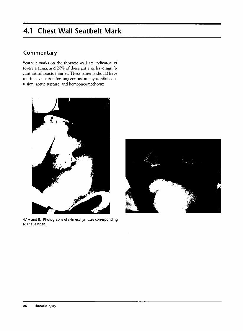
4.1 Chest Wall Seatbelt Mark
Commentary
Seatbelt marks on the thoracic wall are indicators ofsevere trauma, and 20% of these patients have signifi-cant intra thoracic injuries. These patients should haveroutine evaluation for lung contusion, myocardial con-tusion, aortic rupture, and hemopneumothorax.
4.1 A and B. Photographs of skin ecchymoses correspondingto the seatbelt.
86 Thoracic Injury

4.2 Fractures of the Upper (First or Second) Ribs
Commentary
Fractures of the upper ribs, especially the first rib, areassociated with a high incidence of subclavian vascu-lar injuries. A color flow Doppler evaluation of thevessels will help exclude this injury. The forcerequired to fracture an upper rib is severe, and thus
mediastinal structures, including the aorta, are also athigh risk with this mechanism. Children have a muchmore compliant chest wall, and thus any rib fracturesare significant.
4.2A. CXR with fractures of the upper ribs. 4.2B. CXR showing upper rib fractures in a small child.
4.3 Fractures of Middle (Third to Eighth) Ribs
Commentary
Rib fractures are diagnosed clinically (pain aggravatedby breathing or coughing, pain on anteroposterior chestcompression) or radiologically. Fractures at the costo-chondral junction may not show on x-rays. Associatedinjuries include hemopneumothorax, lung contusion,cardiac trauma, and diaphragmatic tear. Pain relief isextremely important, especially in elderly patients withmultiple rib fractures, in order to prevent atelectasis andpneumonia. Patient-controlled analgesia (PCA) orepidural anesthesia should be used in multiple fractures.
Rib fractures in children are uncommon and signifysevere impact to the chest wall, and there is a highincidence of underlying lung contusion.
4.3A. Plain radiograph of multiple rib fractures withunderlying lung contusion.
4.3 Fractures of Middle (Third to Eighth) Ribs 87

4.4 Fractures of the Lower (Ninth to Twelfth) Ribs
Commentary
Fractures of the lower ribs are often associated withinjuries to the kidneys, liver, or spleen. Liberal use ofabdominal CT scan is highly recommended.
4.4A. Radiograph showingfractures of the right lowerribs.
4.4B. Intraoperative photograph of the associated liverinjury in the same patient.
88 Thoracic Injury
4.5 Flail Chest
Commentary
Flail chest is the result of anterior or lateral doublefractures of at least three adjacent ribs. In most casesthere is an underlying lung contusion. Clinically theflail segment moves paradoxically to normal chestwall motion, and the patient may be in respiratory dis-tress. All patients with flail chest should have a thora-costomy tube inserted and be monitored in an ICUsetting with serial blood gases and continuous pulse
oximetry. Analgesia by means of patient-controlled orepidural anesthesia should always be used. Mechani-cal ventilation is necessary for respiratory failure orimminent failure. Internal, operative fracture fixationhas been used in selected cases without underlyinglung contusion and in patients having difficulty wean-ing off mechanical ventilation.
4.5A. Illustration of flail chest. Double fractures of at leastthree adjacent ribs are required in order to produce a flailchest.
4.5C. Photograph of a flail sternal segment. Note themidsternal depression.
4.5B. CXR showing a flail chest with underlying lungcontusion.
4.5 Flail Chest 89
4.6 Pneumothorax
Commentary
A pneumothorax may be due to blunt or penetratingtrauma. Small pneumothoraces are usually asympto-matic and can safely be managed nonoperatively, pro-vided the patient does not need mechanical ventilationor air transportation. In these cases, any size pneumoth-orax should be treated with a thoracostomy tube toavoid the creation of tension pneumothorax. Large
pneumothoraces may cause respiratory distress, and ten-sion pneumothoraces can cause cardiorespiratory fail-ure. The diagnosis of simple pneumothorax is usuallymade by plain chest film. An erect chest x-ray in deepexpiration is the most suitable film to identify smallpneumothoraces.
r
4.6A. Large left pneumothorax on chest x-ray. Arrow pointsto collapsed lung.
4.6B. Bilateral pneumothoraces onCT scan. Arrows point topneumothorax.
90 Thoracic Injury
4.7 Tension Pneumothorax
Commentary
In tension pneumothorax, air leaks into the pleuralcavity with no escape avenue on account of a one-wayvalve effect. It is a life-threatening condition becauseof the severe cardiorespiratory failure that ensues. Thepatient is panicky and has dyspnea, cyanosis, shock,and distended neck veins. The trachea is shifted tothe opposite side, there are no breath sounds in theaffected hemithorax, and there is hyperresonance onpercussion. Immediate lifesaving therapy is needle
decompression of the chest, followed by formal thora-costomy tube insertion. The mere suspicion of a ten-sion pneumothorax is an absolute indication for aneedle decompression whenever a thoracostomy tubecannot be inserted immediately (prehospital stage,areas outside the emergency department or operativeroom). A thoracostomy tube should be subsequentlyinserted as soon as possible.
Tension Pneumothorax
4.7A. Illustration showing mechanism of tensionpneumothorax: Air under tension collapses the lung,expands the hemithorax, depresses the diaphragm, andpushes the heart toward the opposite side. These changescause acute cardiorespiratory failure.
4.7B. CXR showing a large tension pneumothorax on theleft side, mediastinal shift to the opposite side, anddownward displacement of the left hemidiaphragm.
4.7 Tension Pneumothorax 91

4.7C. Tension pneumothoraxon CT scan.
4.7D. Photograph showing athoracostomy needle inplace, below the middle ofthe clavicle.
92 Thoracic Injury
4.8 Hemothorax
Commentary
A hemothorax can be due to blunt or penetrating trauma.Large hemothoraces may present with hypovolemia ordyspnea, but small hemothoraces can be asymptomatic.On physical examination, the breath sounds are dimin-ished, there is dullness on percussion, and the affectedhemithorax moves poorly. A chest x-ray, preferably in theerect position, confirms the diagnosis, although manytimes it cannot distinguish between a hemothorax andintrapulmonary hematoma, contusion, or atelectasis.
The treatment of significant hemothoraces is thora-costomy tube insertion through the fourth or fifthintercostal space, in the midaxillary line. A thoraco-tomy should be considered if the initial thoracostomytube output exceeds 1,000-1,500 ml of blood, or if thepatient is hemodynamically unstable.
Significant residual hemothorax following thoracos-tomy tube insertion should be evaluated by means of CTscan and evacuated within 3-5 days. Early evacuation iseasy and can be performed with thoracoscopy or a smallanterolateral thoracotomy. Thrombolytic agents, such asstreptokinase or urokinase may be effective in somecases and should be the first line of treatment before anoperative approach is attempted. Delayed evacuation isdifficult because of clot organization and inflammation,and it requires a thoracotomy with decortication.
An undrained significant hemothorax is associatedwith increased risk of empyema and may cause respira-tory compromise due to fibrosis.
4.8B. Gunshot wound to the right chest with a suspected"residual hemothorax" on CXR two days after injury.
4.8C. Chest CT scan of the same patient showsintrapulmonary hematomas and parenchymal lacerations.
4.8A. Chest radiograph with extensive opacification of lefthemithorax due to massive hemothorax with mediastinalshift to the opposite site.
4.8D. Residual hemothorax suspected on chest radiographyand confirmed by CT scan of the chest.
4.8 Hemothorax 93
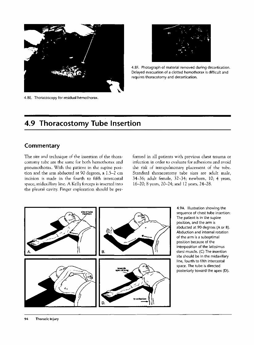
4.8F. Photograph of material removed during decortication.Delayed evacuation of a clotted hemothorax is difficult andrequires thoracotomy and decortication.
4.8E. Thoracoscopy for residual hemothorax.
4.9 Thoracostomy Tube Insertion
Commentary
The site and technique of the insertion of the thora-costomy tube are the same for both hemothorax andpneumothorax. With the patient in the supine posi-tion and the arm abducted at 90 degrees, a 1.5-2 cmincision is made in the fourth to fifth intercostalspace, midaxillary line. A Kelly forceps is inserted intothe pleural cavity. Finger exploration should be per-
formed in all patients with previous chest trauma orinfection in order to evaluate for adhesions and avoidthe risk of intrapulmonary placement of the tube.Standard thoracostomy tube sizes are adult male,34-36; adult female, 32-34; newborn, 10; 4 years,16-20; 8 years, 20-24; and 12 years, 24-28.
towardsap*x of lui
D.
4.9A. Illustration showing thesequence of chest tube insertion:The patient is in the supineposition, and the arm isabducted at 90 degrees (A or B).Abduction and internal rotationof the arm is a suboptimalposition because of theinterposition of the latissimusdorsi muscle. (C) The insertionsite should be in the midaxillaryline, fourth to fifth intercostalspace. The tube is directedposteriorly toward the apex (D).
94 Thoracic Injury
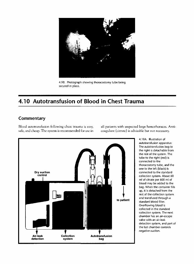
4.9B. Photograph showing thoracostomy tube beingsecured in place.
4.10 Autotransfusion of Blood in Chest Trauma
Commentary
Blood autotransfusion following chest trauma is easy,safe, and cheap. The system is recommended for use in
all patients with suspected large hemothoraces. Anti-coagulant (citrate) is advisable but not necessary.
to patient
Air-leakdetection
Collectionsystem
Autotransfusionbag
4.10A. Illustration ofautotransfusion apparatus:The autotransfusion bag tothe right is detachable fromthe rest of the system. Thetube to the right (red) isconnected to thethoracostomy tube, and theone to the left (black) isconnected to the standardcollection system. About 60ml of citrate per 600 ml ofblood may be added to thebag. When the container fillsup, it is detached from therest of the collection systemand transfused through astandard blood filter.Overflowing blood iscollected in the standardcollection system. The nextchamber has an air-escapevalve with an air-leakdetection system, and part ofthe last chamber controlsnegative suction.

4.1 OB. Photograph of blood collected with theautotransfusion technique ready for transfusion.
4.11 Lung Contusion
Commentary
Lung contusion may occur after blunt trauma to thechest, rapid deceleration injuries, or gunshot woundsto the lung. Associated rib fractures are a commonfinding in adults but unusual in children. The symp-toms vary from minor chest discomfort or hemoptysisto severe dyspnea, massive hemoptysis, and respiratoryfailure. The treatment is symptomatic, and mechani-cal ventilation may be necessary. Independent lungventilation may be necessary in patients who do notrespond to conventional ventilation.
The radiological diagnosis of a lung contusion isnot always easy with plain chest radiography becauseit has a similar appearance to hemothorax or pneumo-nia. CT scan delineates lung parenchyma well andprovides an accurate diagnosis.
4.11 A. Chest x-ray with large opacification in the right lung,highly suspicious of lung contusion.
96 Thoracic Injury

4.11B. Chest CT scan in above caseconfirms the presence of lungcontusion.
4.11C. Gunshot wound tothe right chest. The chestfilm cannot distinguishbetween a hemothorax anda lung contusion. The CTscan clearly demonstrates alarge contusion without anysignificant hemothorax.
4.11D. Photograph showing bronchial bleeding inrespiratory tubing in severe lung contusion.
4.11 Lung Contrusion 97

4.11E. Intraoperative photograph of a lung contusion.
4.12 Subcutaneous Emphysema
Commentary
Subcutaneous emphysema may be secondary to a pneu-mothorax or aerodigestive tract perforation. Inappro-priately placed thoracostomy tubes with a fenestrationoutside the pleural cavity may also result in extensiveemphysema. The first step in evaluating the cause ofthe emphysema is to check the position of any thora-
costomy tube. For massive, unexplained emphysema,endoscopy and esophagography should be performed toevaluate the aerodigestive tract. Subcutaneous emphy-sema rarely causes symptoms, and the treatment isdirected toward the underlying cause.
4.12A. Photograph showing extensive subcutaneousemphysema involving the chest, neck, and face.
4.12B. Chest x-ray showing extensive subcutaneousemphysema.
98 Thoracic Injury
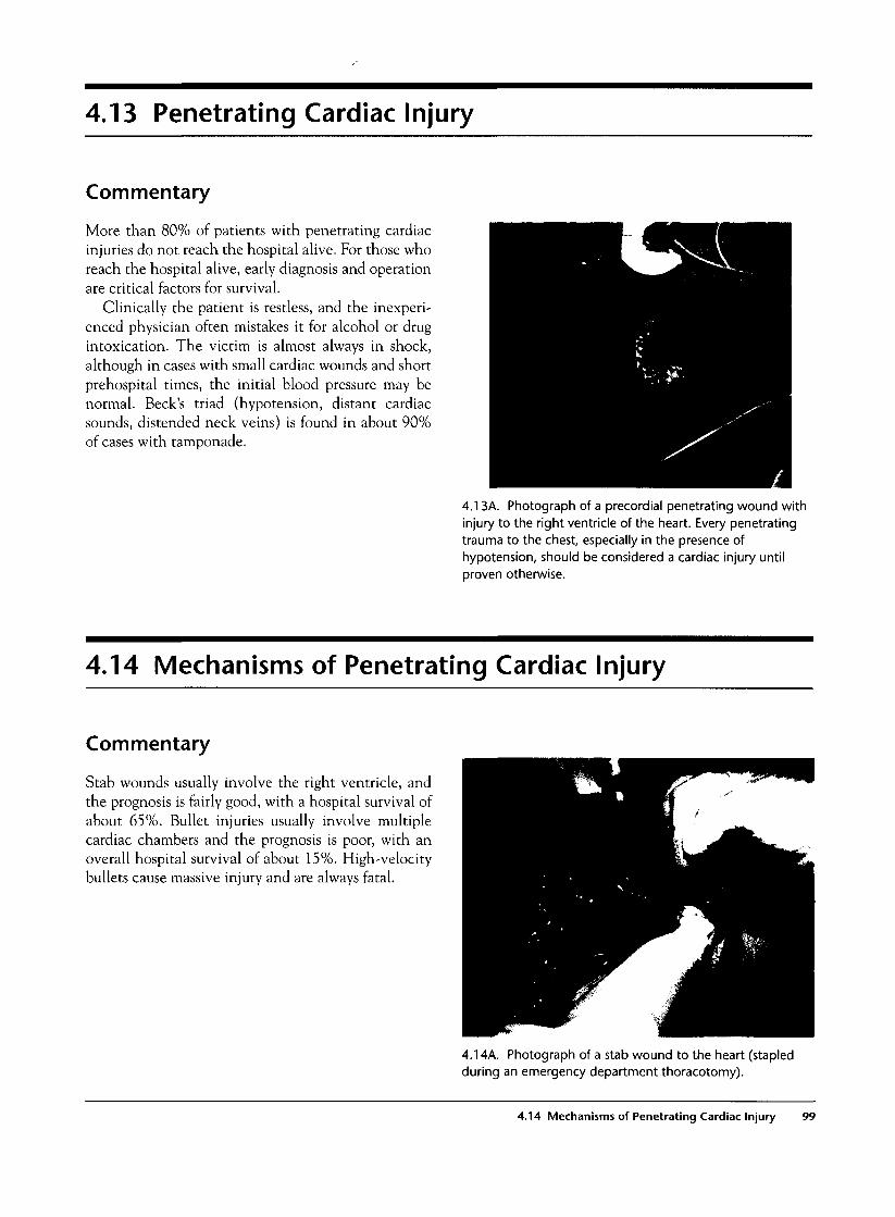
4.13 Penetrating Cardiac Injury
Commentary
More than 80% of patients with penetrating cardiacinjuries do not reach the hospital alive. For those whoreach the hospital alive, early diagnosis and operationare critical factors for survival.
Clinically the patient is restless, and the inexperi-enced physician often mistakes it for alcohol or drugintoxication. The victim is almost always in shock,although in cases with small cardiac wounds and shortprehospital times, the initial blood pressure may benormal. Beck's triad (hypotension, distant cardiacsounds, distended neck veins) is found in about 90%of cases with tamponade.
4.13A. Photograph of a precordial penetrating wound withinjury to the right ventricle of the heart. Every penetratingtrauma to the chest, especially in the presence ofhypotension, should be considered a cardiac injury untilproven otherwise.
4.14 Mechanisms of Penetrating Cardiac Injury
Commentary
Stab wounds usually involve the right ventricle, andthe prognosis is fairly good, with a hospital survival ofabout 65%. Bullet injuries usually involve multiplecardiac chambers and the prognosis is poor, with anoverall hospital survival of about 15%. High-velocitybullets cause massive injury and are always fatal.
4.14A. Photograph of a stab wound to the heart (stapledduring an emergency department thoracotomy).
4.14 Mechanisms of Penetrating Cardiac Injury 99
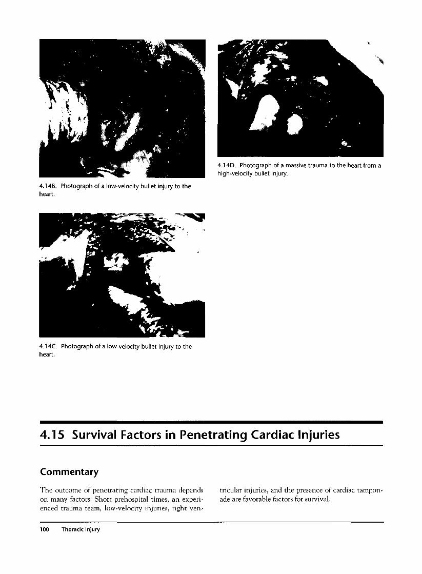
\
4.14D. Photograph of a massive trauma to the heart from ahigh-velocity bullet injury.
4.14B. Photograph of a low-velocity bullet injury to theheart.
4.14C. Photograph of a low-velocity bullet injury to theheart.
4.15 Survival Factors in Penetrating Cardiac Injuries
Commentary
The outcome of penetrating cardiac trauma dependson many factors: Short prehospital times, an experi-enced trauma team, low-velocity injuries, right ven-
tricular injuries, and the presence of cardiac tampon-ade are favorable factors for survival.
100 Thoracic Injury

4.15B. Photograph of the heart during thoracotomy withrepair of a 6-cm-long laceration following a gunshot wound.Such injuries are usually fatal.
4.15A. Autopsy photograph of the heart. The right ventriclehas a relatively thick wall (4 mm) with relatively lowpressures. This is an optimal combination for survival. Theleft ventricle has a thick wall (12 mm) but high pressures,which result in a tense tamponade or rapid exsanguination,and thus the prognosis is poor.
4.16 Diagnosis of Cardiac Injury
Commentary
The diagnosis of penetrating cardiac injury is usuallyclinical and can be confirmed by trauma ultrasoundperformed in the emergency department by emer-gency physicians or surgeons, and thus it should bepart of the standard primary survey. A chest film maybe helpful in about half the cases with cardiac trauma.Radiological signs suggestive of cardiac traumainclude an enlarged cardiac shadow, a widened uppermediastinum (due to a dilated superior vena cava),and pneumopericardium.
An ECG may be diagnostic in about 30% of cases.It may show low-voltage QRS complexes, elevated STsegments, inverted T waves, electrical alternans, andother nonspecific findings. Subxiphoid windows aremajor invasive procedures and have a diminished role,and pericardiocentesis has almost no role in a moderntrauma center. The blood in the pericardial sac usuallyclots, and this results in high incidence of false-nega-tive pericardiocentesis.
4.16A. Trauma ultrasound showing a circumferentialpericardial effusion.
4.16 Diagnosis of Cardiac Injury 101
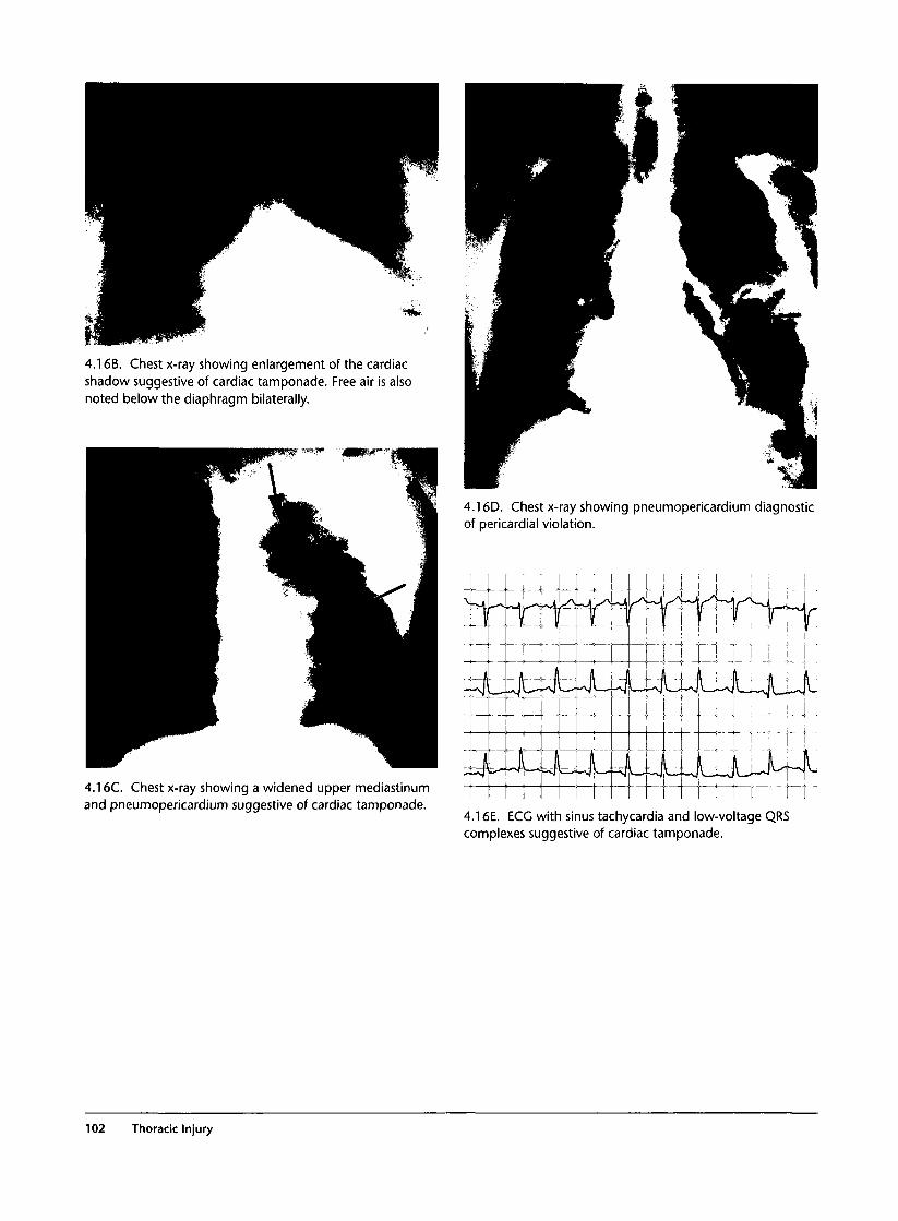
4.16B. Chest x-ray showing enlargement of the cardiacshadow suggestive of cardiac tamponade. Free air is alsonoted below the diaphragm bilaterally.
4.16C. Chest x-ray showing a widened upper mediastinumand pneumopericardium suggestive of cardiac tamponade.
4.16D. Chest x-ray showing pneumopericardium diagnosticof pericardial violation.
r\ i4.16E. ECG with sinus tachycardia and low-voltage QRScomplexes suggestive of cardiac tamponade.
102 Thoracic Injury
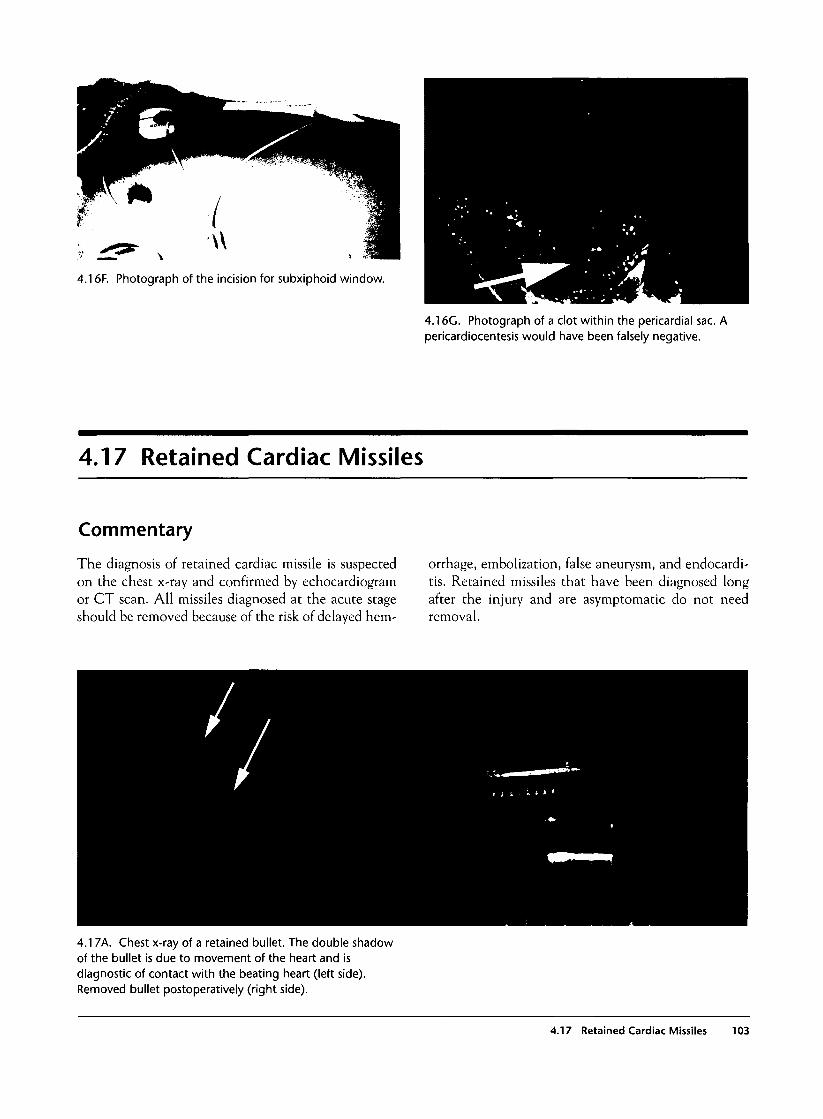
4.16F. Photograph of the incision for subxiphoid window.
4.16C. Photograph of a clot within the pericardial sac. Apericardiocentesis would have been falsely negative.
4.17 Retained Cardiac Missiles
Commentary
The diagnosis of retained cardiac missile is suspectedon the chest x-ray and confirmed by echocardiogramor CT scan. All missiles diagnosed at the acute stageshould be removed because of the risk of delayed hem-
orrhage, embolization, false aneurysm, and endocardi-tis. Retained missiles that have been diagnosed longafter the injury and are asymptomatic do not needremoval.
4.17k. Chest x-ray of a retained bullet. The double shadowof the bullet is due to movement of the heart and isdiagnostic of contact with the beating heart (left side).Removed bullet postoperatively (right side).
4.17 Retained Cardiac Missiles 103
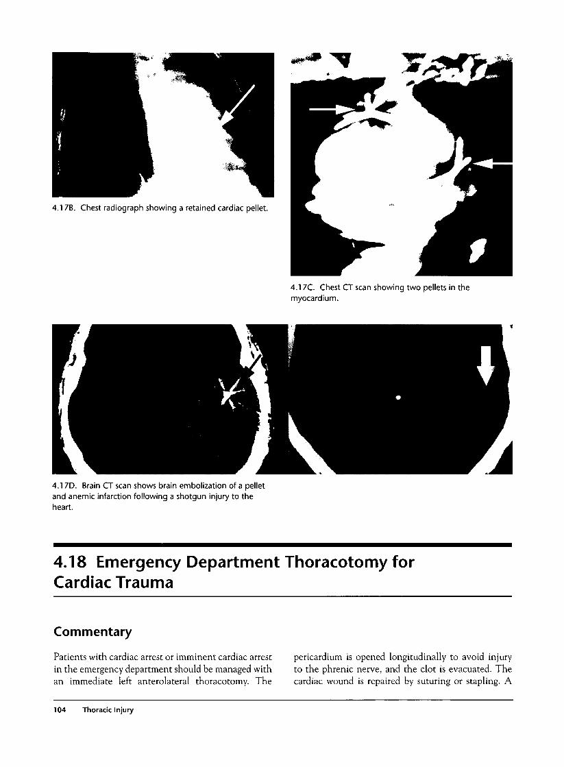
4.17B. Chest radiograph showing a retained cardiac pellet.
4.17C. Chest CT scan showing two pellets in themyocardium.
4.17D. Brain CT scan shows brain embolization of a pelletand anemic infarction following a shotgun injury to theheart.
4.18 Emergency Department Thoracotomy forCardiac Trauma
Commentary
Patients with cardiac arrest or imminent cardiac arrestin the emergency department should be managed withan immediate left anterolateral thoracotomy. The
pericardium is opened longitudinally to avoid injuryto the phrenic nerve, and the clot is evacuated. Thecardiac wound is repaired by suturing or stapling. A
104 Thoracic Injury

Foley catheter balloon may be useful in achievingtemporary control of bleeding. In the presence of car-diac arrest, the aorta is cross-clamped just above thediaphragm, and direct cardiac massage is performed.Adrenaline or defibrillation may be administeredwhen the heart is full, and their use in an empty heartreduces the chances of successful resuscitation. Inter-nal cardiac pacing may be useful in cases that do notrespond to conventional treatment. If cardiac activity
returns, the operation is completed in the operatingroom. The survival rate for emergency departmentthoracotomy is about 10%.
Indications of emergency department thoracotomyinclude cardiac arrest or imminent cardiac arrest dueto both penetrating and blunt trauma, excluding fatalhead injuries. Some of the survivors suffer brain death,but they may become useful organ donors.
Thoracotomy Incision
4.18A. Illustration of the incisionfor an emergency departmentleft anterolateral thoracotomy.
4.18B. Photograph of a healingemergency department thoracotomyincision just below the nipple.
4.18 Emergency Department Thoracotomy for Cardiac Trauma 105
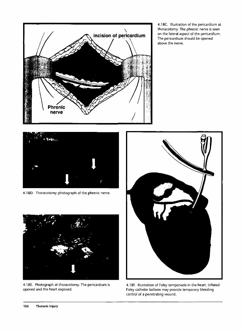
If A&
1 \ \^|'3 \ Pnrenic
^ nerve
\>T\ incision of pericardium
• U P\ \ r j
" 1
4.18C. Illustration of the pericardium atthoracotomy. The phrenic nerve is seenon the lateral aspect of the pericardium.The pericardium should be openedabove the nerve.
4.18D. Thoracotomy photograph of the phrenic nerve.
4.18E. Photograph at thoracotomy. The pericardium isopened and the heart exposed.
4.18F. Illustration of Foley tamponade in the heart. InflatedFoley catheter balloon may provide temporary bleedingcontrol of a penetrating wound.
106 Thoracic Injury
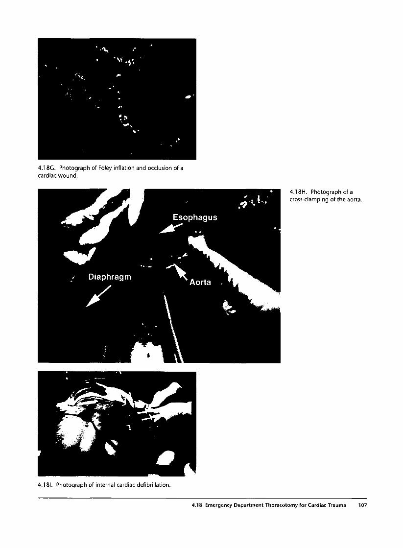
4.18C. Photograph of Foley inflation and occlusion of acardiac wound.
4.18H. Photograph of across-clamping of the aorta.
4.181. Photograph of internal cardiac defibrillation.
4.18 Emergency Department Thoracotomy for Cardiac Trauma 107
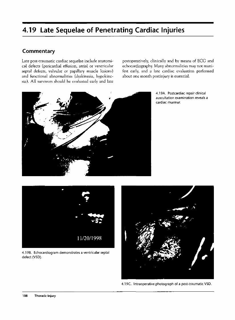
4.19 Late Sequelae of Penetrating Cardiac Injuries
Commentary
Late post-traumatic cardiac sequelae include anatomi-cal defects (pericardial effusion, atrial or ventricularseptal defects, valvular or papillary muscle lesions)and functional abnormalities (dyskinesia, hypokine-sia). All survivors should be evaluated early and late
postoperatively, clinically and by means of ECG andechocardiography. Many abnormalities may not mani-fest early, and a late cardiac evaluation performedabout one month postinjury is essential.
4.19A. Postcardiac repair clinicalauscultation examination reveals acardiac murmur.
4.19B. Echocardiogram demonstrates a ventricular septaldefect (VSD).
4.19C. Intraoperative photograph of a post-traumatic VSD.
108 Thoracic Injury
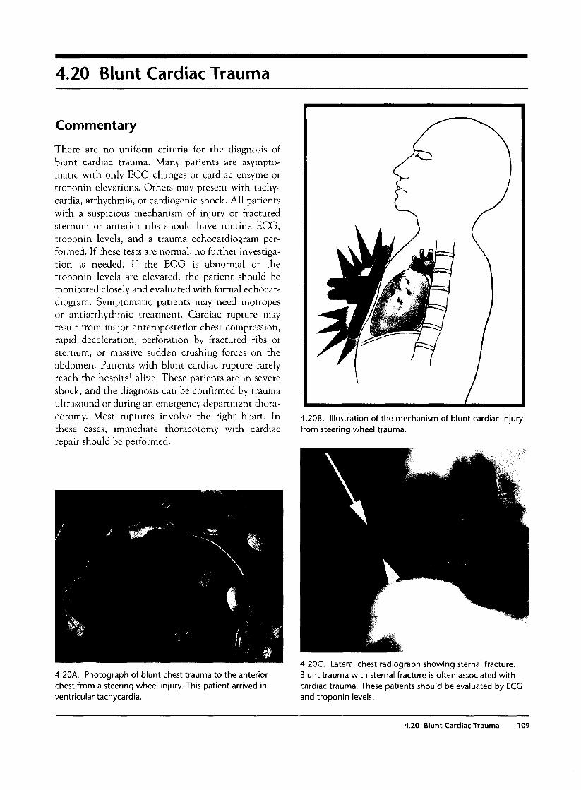
4.20 Blunt Cardiac Trauma
Commentary
There are no uniform criteria for the diagnosis ofblunt cardiac trauma. Many patients are asympto-matic with only ECG changes or cardiac enzyme ortroponin elevations. Others may present with tachy-cardia, arrhythmia, or cardiogenic shock. All patientswith a suspicious mechanism of injury or fracturedsternum or anterior ribs should have routine ECG,troponin levels, and a trauma echocardiogram per-formed. If these tests are normal, no further investiga-tion is needed. If the ECG is abnormal or thetroponin levels are elevated, the patient should bemonitored closely and evaluated with formal echocar-diogram. Symptomatic patients may need inotropesor antiarrhythmic treatment. Cardiac rupture mayresult from major anteroposterior chest compression,rapid deceleration, perforation by fractured ribs orsternum, or massive sudden crushing forces on theabdomen. Patients with blunt cardiac rupture rarelyreach the hospital alive. These patients are in severeshock, and the diagnosis can be confirmed by traumaultrasound or during an emergency department thora-cotomy. Most ruptures involve the right heart. Inthese cases, immediate thoracotomy with cardiacrepair should be performed.
4.20B. Illustration of the mechanism of blunt cardiac injuryfrom steering wheel trauma.
4.20A. Photograph of blunt chest trauma to the anteriorchest from a steering wheel injury. This patient arrived inventricular tachycardia.
4.20C. Lateral chest radiograph showing sternal fracture.Blunt trauma with sternal fracture is often associated withcardiac trauma. These patients should be evaluated by ECGand troponin levels.
4.20 Blunt Cardiac Trauma 109
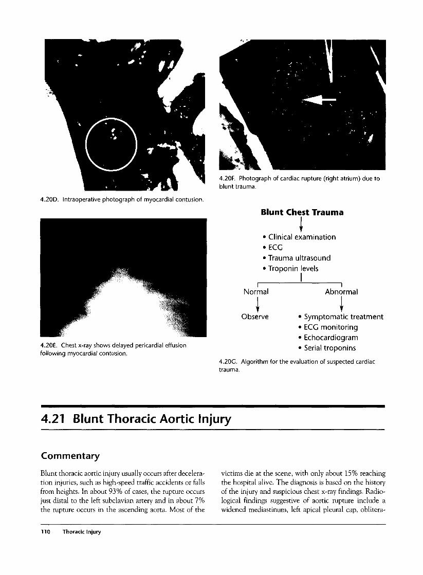
4.20D. Intraoperative photograph of myocardial contusion.
4.20F. Photograph of cardiac rupture (right atrium) due toblunt trauma.
Blunt Chest Trauma
• Clinical examination• ECG• Trauma ultrasound• Troponin levels
4.20E. Chest x-ray shows delayed pericardial effusionfollowing myocardial contusion.
Normal Abnormal
I \Observe • Symptomatic treatment
• ECC monitoring• Echocardiogram• Serial troponins
4.20G. Algorithm for the evaluation of suspected cardiactrauma.
4.21 Blunt Thoracic Aortic Injury
Commentary
Blunt thoracic aortic injury usually occurs after decelera-tion injuries, such as high-speed traffic accidents or fallsfrom heights. In about 93% of cases, the rupture occursjust distal to the left subclavian artery and in about 7%the rupture occurs in the ascending aorta. Most of the
victims die at the scene, with only about 15% reachingthe hospital alive. The diagnosis is based on the historyof the injury and suspicious chest x-ray findings. Radio-logical findings suggestive of aortic rupture include awidened mediastinum, left apical pleural cap, oblitera-
110 Thoracic Injury
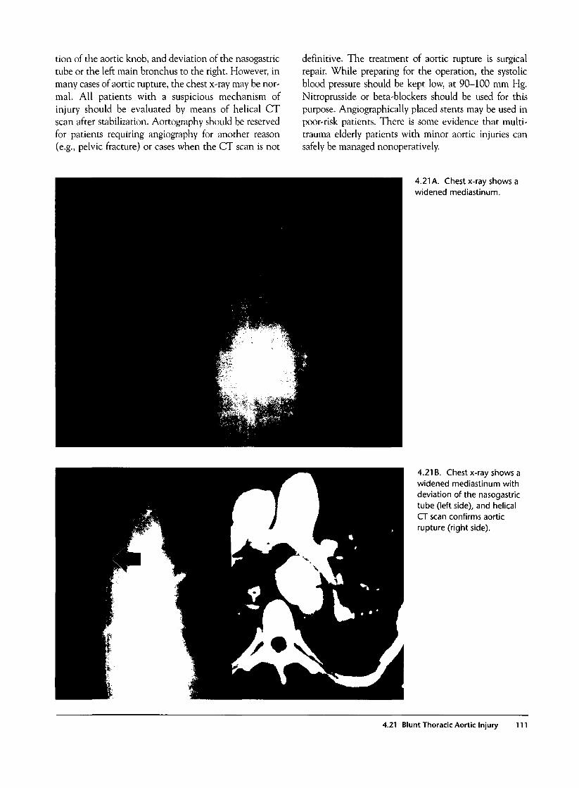
tion of the aortic knob, and deviation of the nasogastrictube or the left main bronchus to the right. However, inmany cases of aortic rupture, the chest x-ray may be nor-mal. All patients with a suspicious mechanism ofinjury should be evaluated by means of helical CTscan after stabilization. Aortography should be reservedfor patients requiring angiography for another reason(e.g., pelvic fracture) or cases when the CT scan is not
definitive. The treatment of aortic rupture is surgicalrepair. While preparing for the operation, the systolicblood pressure should be kept low, at 90-100 mm Hg.Nitroprusside or beta-blockers should be used for thispurpose. Angiographically placed stents may be used inpoor-risk patients. There is some evidence that multi-trauma elderly patients with minor aortic injuries cansafely be managed nonoperatively.
4.21 A. Chest x-ray shows awidened mediastinum.
4.21 B. Chest x-ray shows awidened mediastinum withdeviation of the nasogastrictube (left side), and helicalCT scan confirms aorticrupture (right side).
4.21 Blunt Thoracic Aortic Injury 111
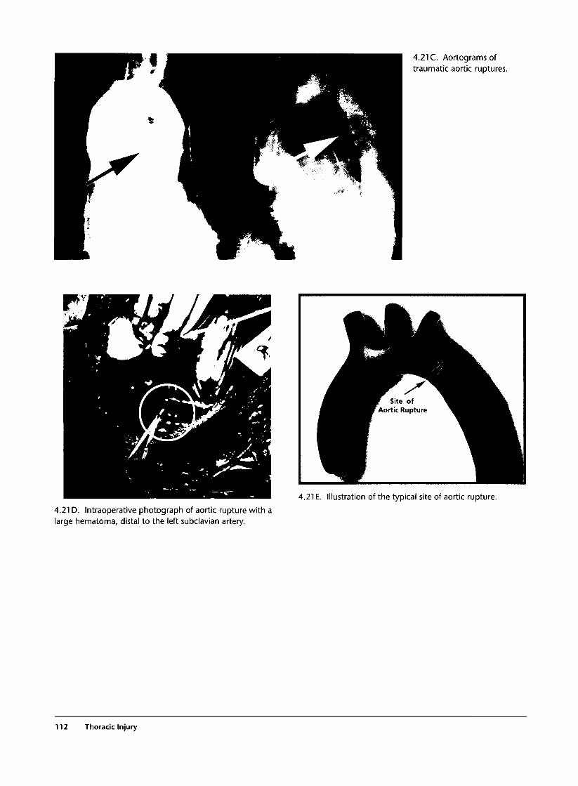
4.21 C. Aortograms oftraumatic aortic ruptures.
4.21 E. Illustration of the typical site of aortic rupture.
4.21 D. Intraoperative photograph of aortic rupture with alarge hematoma, distal to the left subclavian artery.
112 Thoracic Injury
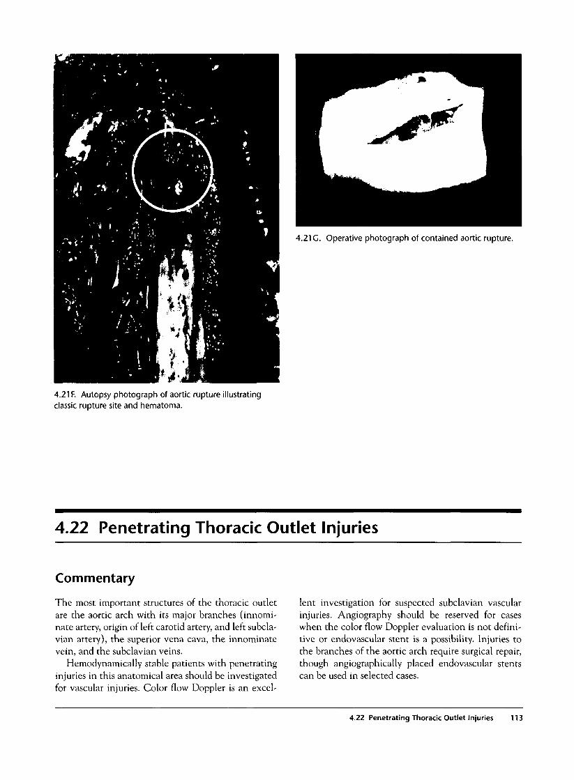
4.21 G. Operative photograph of contained aortic rupture.
4.21 F. Autopsy photograph of aortic rupture illustratingclassic rupture site and hematoma.
4.22 Penetrating Thoracic Outlet Injuries
Commentary
The most important structures of the thoracic outletare the aortic arch with its major branches (innomi-nate artery, origin of left carotid artery, and left subcla-vian artery), the superior vena cava, the innominatevein, and the subclavian veins.
Hemodynamically stable patients with penetratinginjuries in this anatomical area should be investigatedfor vascular injuries. Color flow Doppler is an excel-
lent investigation for suspected subclavian vascularinjuries. Angiography should be reserved for caseswhen the color flow Doppler evaluation is not defini-tive or endovascular stent is a possibility. Injuries tothe branches of the aortic arch require surgical repair,though angiographically placed endovascular stentscan be used in selected cases.
4.22 Penetrating Thoracic Outlet Injuries 113
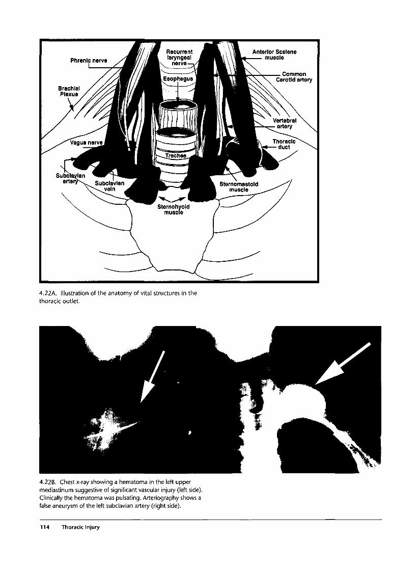
MPhrenic nerve /^Mll
^W9kSubcTaylan ^ ^ r T ~ ^ M B" ^ " ^ ^ S u b c l a v l a n ^k
\ \ v e l n ^M
/ U Recurrent ^^H
^^•Esophagusj^HT
HISternohyold /
muscle /
i \ \ M Anterior Scalene
!\\!f^—muscie
^ Common^ k \ ^ K \ \ \ Carotid artery
Sternomastold ^^ymuscle ^ — /
^ ^ /
A.22k. Illustration of the anatomy of vital structures in thethoracic outlet.
4.22B. Chest x-ray showing a hematoma in the left uppermediastinum suggestive of significant vascular injury (left side).Clinically the hematoma was pulsating. Arteriography shows afalse aneurysm of the left subclavian artery (right side).
114 Thoracic Injury
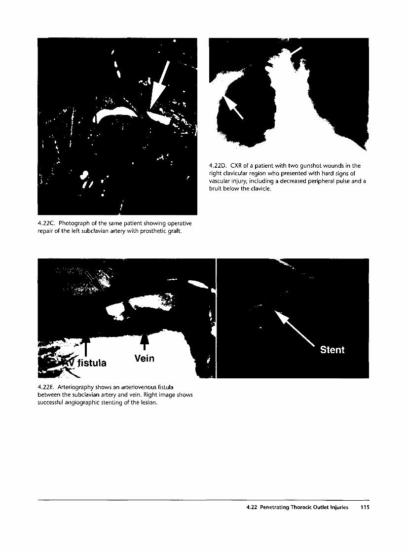
4.22D. CXR of a patient with two gunshot wounds in theright clavicular region who presented with hard signs ofvascular injury, including a decreased peripheral pulse and abruit below the clavicle.
4.22C. Photograph of the same patient showing operativerepair of the left subclavian artery with prosthetic graft.
4.22E. Arteriography shows an arteriovenous fistulabetween the subclavian artery and vein. Right image showssuccessful angiographic stenting of the lesion.
4.22 Penetrating Thoracic Outlet Injuries 115
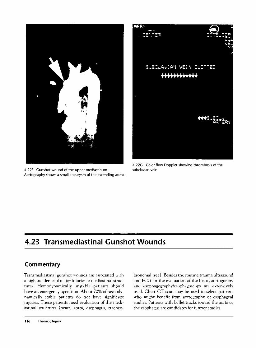
4.22F. Gunshot wound of the upper mediastinum.Aortography shows a small aneurysm of the ascending aorta.
4.22G. Color flow Doppler showing thrombosis of thesubclavian vein.
4.23 Transmediastinal Gunshot Wounds
Commentary
Transmediastinal gunshot wounds are associated witha high incidence of major injuries to mediastinal struc-tures. Hemodynamically unstable patients shouldhave an emergency operation. About 70% of hemody-namically stable patients do not have significantinjuries. These patients need evaluation of the medi-astinal structures (heart, aorta, esophagus, tracheo-
bronchial tree). Besides the routine trauma ultrasoundand ECG for the evaluation of the heart, aortographyand esophagography/esophagoscopy are extensivelyused. Chest CT scan may be used to select patientswho might benefit from aortography or esophagealstudies. Patients with bullet tracks toward the aorta orthe esophagus are candidates for further studies.
116 Thoracic Injury
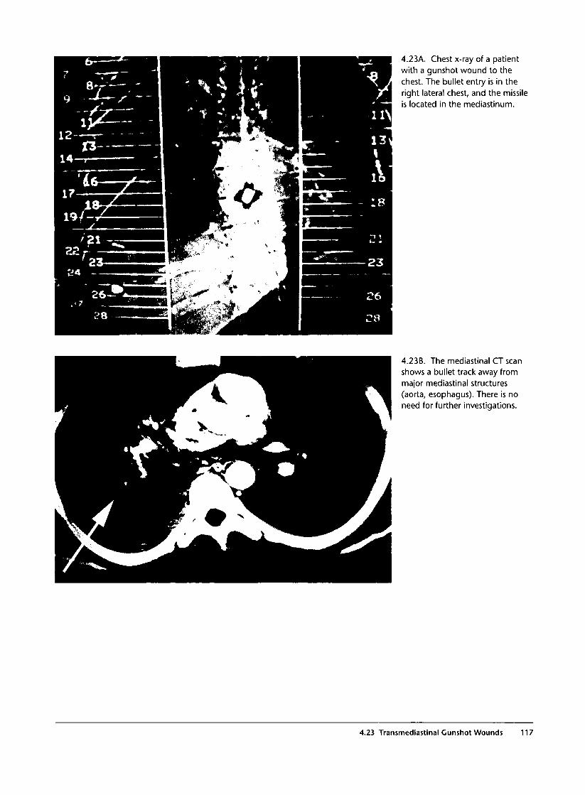
4.23A. Chest x-ray of a patientwith a gunshot wound to thechest. The bullet entry is in theright lateral chest, and the missileis located in the mediastinum.
4.23B. The mediastinal CT scanshows a bullet track away frommajor mediastinal structures(aorta, esophagus). There is noneed for further investigations.
4.23 Transmediastinal Gunshot Wounds 117
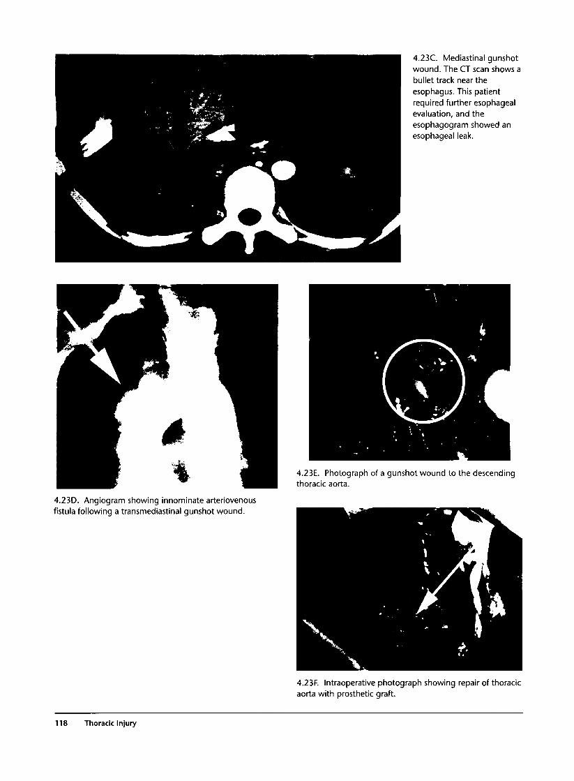
4.23C. Mediastinal gunshotwound. The CT scan shows abullet track near theesophagus. This patientrequired further esophagealevaluation, and theesophagogram showed anesophageal leak.
4.23E. Photograph of a gunshot wound to the descendingthoracic aorta.
4.23D. Angiogram showing innominate arteriovenousfistula following a transmediastinal gunshot wound.
4.23F. Intraoperative photograph showing repair of thoracicaorta with prosthetic graft.
118 Thoracic Injury
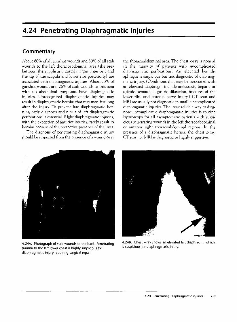
4.24 Penetrating Diaphragmatic Injuries
Commentary
About 60% of all gunshot wounds and 30% of all stabwounds to the left thoracoabdominal area (the areabetween the nipple and costal margin anteriorly andthe tip of the scapula and lower ribs posteriorly) areassociated with diaphragmatic injuries. About 13% ofgunshot wounds and 26% of stab wounds to this areawith no abdominal symptoms have diaphragmaticinjuries. Unrecognized diaphragmatic injuries mayresult in diaphragmatic hernias that may manifest longafter the injury. To prevent late diaphragmatic her-nias, early diagnosis and repair of left diaphragmaticperforations is essential. Right diaphragmatic injuries,with the exception of anterior injuries, rarely result inhernias because of the protective presence of the liver.
The diagnosis of penetrating diaphragmatic injuryshould be suspected from the presence of a wound over
the thoracoabdominal area. The chest x-ray is normalin the majority of patients with uncomplicateddiaphragmatic perforations. An elevated hemidi-aphragm is suspicious but not diagnostic of diaphrag-matic injury. (Conditions that may be associated withan elevated diaphragm include atelectasis, hepatic orsplenic hematoma, gastric dilatation, fractures of thelower ribs, and phrenic nerve injury.) CT scan andMRI are usually not diagnostic in small, uncomplicateddiaphragmatic injuries. The most reliable way to diag-nose uncomplicated diaphragmatic injuries is routinelaparoscopy for all asymptomatic patients with suspi-cious penetrating wounds in the left thoracoabdominalor anterior right thoracoabdominal regions. In thepresence of a diaphragmatic hernia, the chest x-ray,CT scan, or MRI is diagnostic or highly suggestive.
4.24A. Photograph of stab wounds to the back. Penetratingtrauma to the left lower chest is highly suspicious fordiaphragmatic injury requiring surgical repair.
4.24B. Chest x-ray shows an elevated left diaphragm, whichis suspicious for diaphragmatic injury.
4.24 Penetrating Diaphragmatic Injuries 119
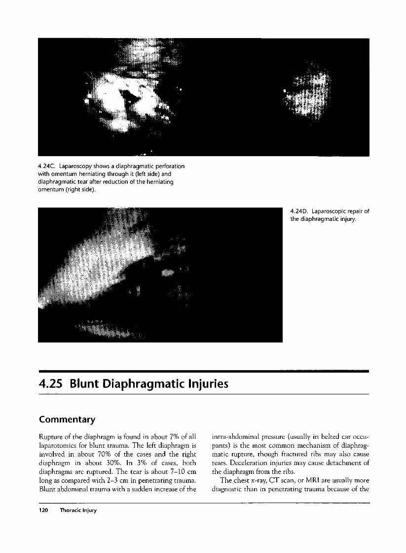
4.24C. Laparoscopy shows a diaphragmatic perforationwith omentum herniating through it (left side) anddiaphragmatic tear after reduction of the herniatingomentum (right side).
4.24D. Laparoscopic repair ofthe diaphragmatic injury.
4.25 Blunt Diaphragmatic Injuries
Commentary
Rupture of the diaphragm is found in about 7% of alllaparotomies for blunt trauma. The left diaphragm isinvolved in about 70% of the cases and the rightdiaphragm in about 30%. In 3% of cases, bothdiaphragms are ruptured. The tear is about 7-10 cmlong as compared with 2-3 cm in penetrating trauma.Blunt abdominal trauma with a sudden increase of the
intra-abdominal pressure (usually in belted car occu-pants) is the most common mechanism of diaphrag-matic rupture, though fractured ribs may also causetears. Deceleration injuries may cause detachment ofthe diaphragm from the ribs.
The chest x-ray, CT scan, or MRI are usually morediagnostic than in penetrating trauma because of the
120 Thoracic Injury
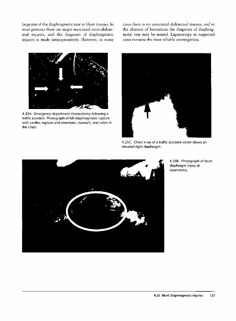
large size of the diaphragmatic tear in blunt trauma. Inmost patients there are major associated intra-abdom-inal injuries, and the diagnosis of diaphragmaticinjuries is made intraoperatively. However, in many
cases there is no associated abdominal trauma, and inthe absence of herniation the diagnosis of diaphrag-matic tear may be missed. Laparoscopy in suspectedcases remains the most reliable investigation.
4.25A. Emergency department thoracotomy following atraffic accident. Photograph of left diaphragmatic rupturewith cardiac rupture and omentum, stomach, and colon inthe chest.
4.25C. Chest x-ray of a traffic accident victim shows anelevated right diaphragm.
4.25B. Photograph of bluntdiaphragm injury atlaparotomy.
4.25 Blunt Diaphragmatic Injuries 121
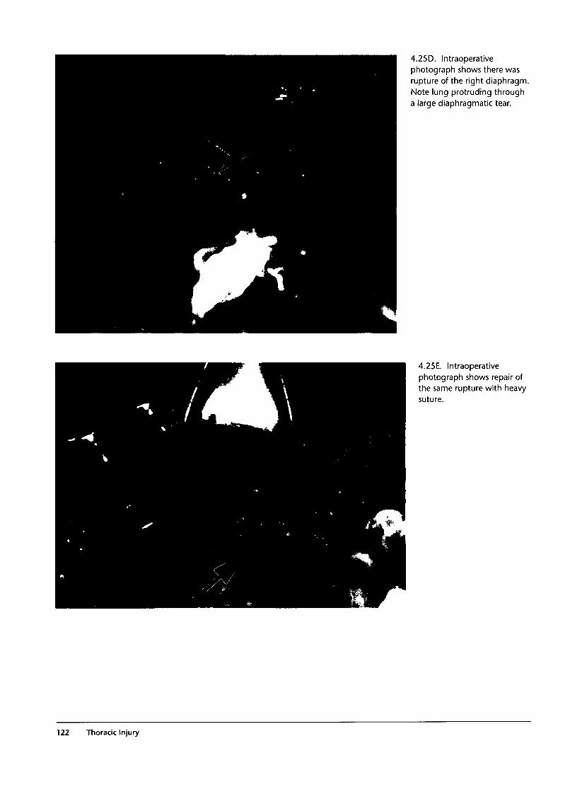
4.25D. Intraoperativephotograph shows there wasrupture of the right diaphragm.Note lung protruding througha large diaphragmatic tear.
4.25E. Intraoperativephotograph shows repair ofthe same rupture with heavysuture.
122 Thoracic Injury
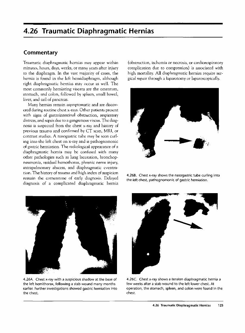
4.26 Traumatic Diaphragmatic Hernias
Commentary
Traumatic diaphragmatic hernias may appear withinminutes, hours, days, weeks, or many years after injuryto the diaphragm. In the vast majority of cases, thehernia is found in the left hemidiaphragm, althoughright diaphragmatic hernias may occur as well. Themost commonly herniating viscera are the omentum,stomach, and colon, followed by spleen, small bowel,liver, and tail of pancreas.
Many hernias remain asymptomatic and are discov-ered during routine chest x-rays. Other patients presentwith signs of gastrointestinal obstruction, respiratorydistress, and sepsis due to a gangrenous viscus. The diag-nosis is suspected from the chest x-ray and history ofprevious trauma and confirmed by CT scan, MRI, orcontrast studies. A nasogastric tube may be seen curl-ing into the left chest on x-ray and is pathognomonicof gastric herniation. The radiological appearance of adiaphragmatic hernia may be confused with manyother pathologies such as lung laceration, bronchop-neumonia, residual hemothorax, phrenic nerve injury,intrapulmonary abscess, and diaphragmatic eventra-tion. The history of trauma and high index of suspicionremain the cornerstone of early diagnosis. Delayeddiagnosis of a complicated diaphragmatic hernia
(obstruction, ischemia or necrosis, or cardiorespiratorycomplication due to compression) is associated withhigh mortality. All diaphragmatic hernias require sur-gical repair through a laparotomy or laparoscopically.
4.26B. Chest x-ray shows the nasogastric tube curling intothe left chest, pathognomonic of gastric herniation.
4.26A. Chest x-ray with a suspicious shadow at the base ofthe left hemithorax, following a stab wound many monthsearlier. Further investigations showed gastric herniation intothe chest.
4.26C. Chest x-ray shows a tension diaphragmatic hernia afew weeks after a stab wound to the left lower chest. Atoperation, the stomach, spleen, and colon were found in thechest.
4.26 Traumatic Diaphragmatic Hernias 123
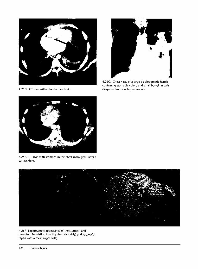
4.26D. CT scan with colon in the chest.
4.26G. Chest x-ray of a large diaphragmatic herniacontaining stomach, colon, and small bowel, initiallydiagnosed as bronchopneumonia.
4.26E. CT scan with stomach in the chest many years after acar accident.
4.26F. Laparoscopic appearance of the stomach andomentum herniating into the chest (left side) and successfulrepair with a mesh (right side).
124 Thoracic Injury

4.26H. Chest x-ray of a patient with diaphragmaticeventration. The differential diagnosis from a diaphragmatichernia is difficult and can be done only with laparoscopy.
4.261. CT scan of a post-traumatic infected pulmonaryhematoma, which may be confused with a diaphragmatichernia.
4.27 Esophageal Injuries
Commentary
Esophageal injuries should be suspected in all penetrat-ing proximity injuries, especially if the x-ray showsmediastinal or subcutaneous emphysema in the neck. Insuspected cases, esophagography and/or esophagoscopyshould be performed. The combination of both investi-gations is highly sensitive in the identification of
esophageal injuries. Early diagnosis and surgical repairare critical for good outcome. Delayed diagnosis is asso-ciated with a high incidence of sepsis and mortality.
4.27A. Penetrating upper mediastinal injury with extensiveneck and mediastinal emphysema. Gastrograffin swallowshows an esophageal perforation.
4.27B. Photograph of rigid esophagoscopy for suspectedesophageal injury.
4.27 Esophageal Injuries 125
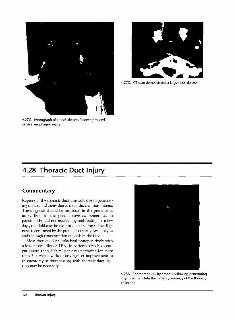
4.27D. CT scan demonstrates a large neck abscess.
4.27C. Photograph of a neck abscess following missedcervical esophageal injury.
4.28 Thoracic Duct Injury
Commentary
Rupture of the thoracic duct is usually due to penetrat-ing trauma and rarely due to blunt deceleration trauma.The diagnosis should be suspected in the presence ofmilky fluid in the pleural cavities. Sometimes inpatients who did not receive any oral feeding for a fewdays, the fluid may be clear or blood-stained. The diag-nosis is confirmed by the presence of many lymphocytesand the high concentration of lipids in the fluid.
Most thoracic duct leaks heal nonoperatively witha low-fat oral diet or TPN. In patients with high out-put (more than 500 ml per day) persisting for morethan 2-3 weeks without any sign of improvement, athoracotomy or thoracoscopy with thoracic duct liga-tion may be necessary.
4.28A. Photograph of chylothorax following penetratingchest trauma. Note the milky appearance of the thoraciccollection.
126 Thoracic Injury
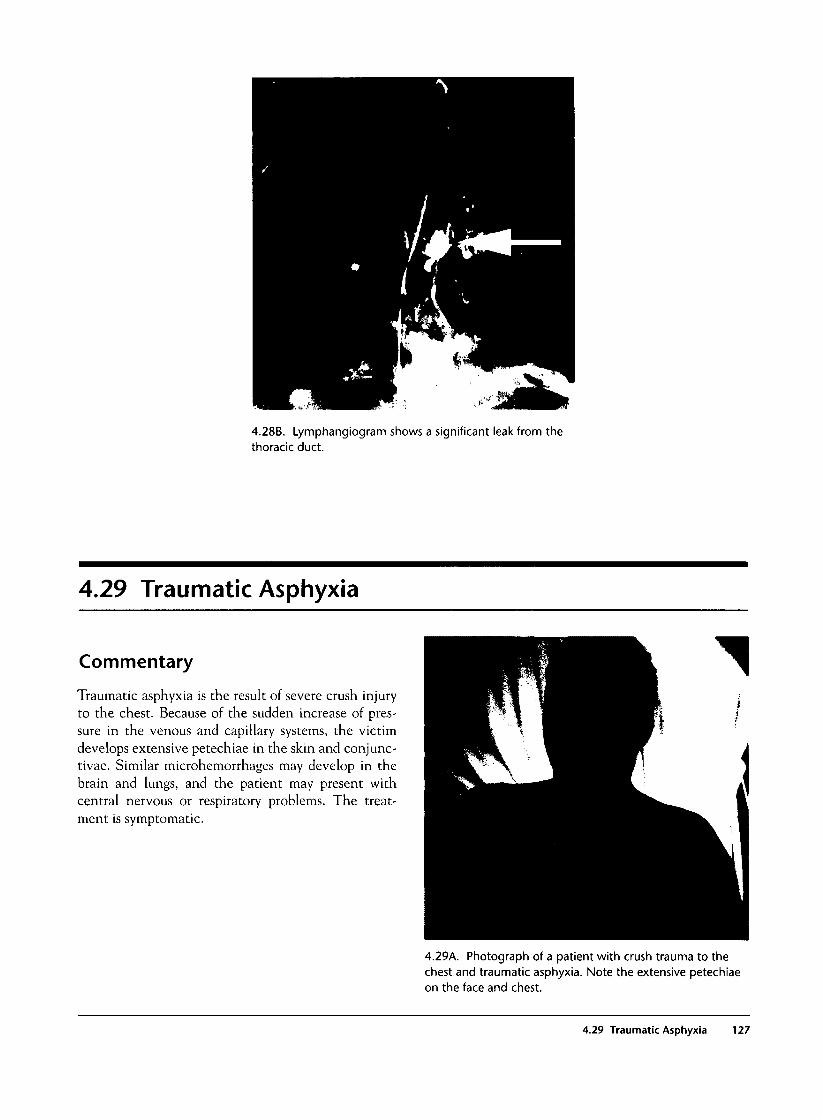
4.28B. Lymphangiogram shows a significant leak from thethoracic duct.
4.29 Traumatic Asphyxia
Commentary
Traumatic asphyxia is the result of severe crush injuryto the chest. Because of the sudden increase of pres-sure in the venous and capillary systems, the victimdevelops extensive petechiae in the skin and conjunc-tivae. Similar microhemorrhages may develop in thebrain and lungs, and the patient may present withcentral nervous or respiratory problems. The treat-ment is symptomatic.
4.29A. Photograph of a patient with crush trauma to thechest and traumatic asphyxia. Note the extensive petechiaeon the face and chest.
4.29 Traumatic Asphyxia 127

4.29B and C. Photographs of extensive petechiae on theface and conjunctival hemorrhages in an adult and a childwith traumatic asphyxia.
128 Thoracic Injury
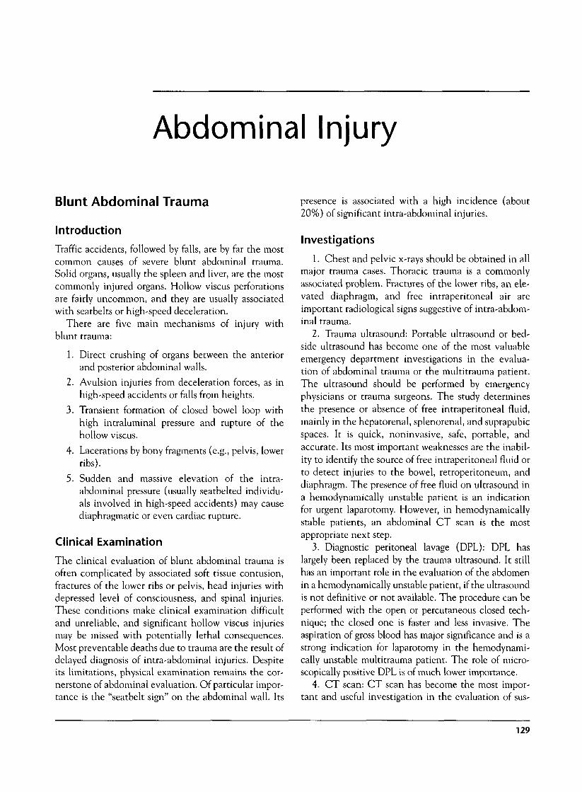
Abdominal Injury
Blunt Abdominal Trauma
Introduction
Traffic accidents, followed by falls, are by far the mostcommon causes of severe blunt abdominal trauma.Solid organs, usually the spleen and liver, are the mostcommonly injured organs. Hollow viscus perforationsare fairly uncommon, and they are usually associatedwith seatbelts or high-speed deceleration.
There are five main mechanisms of injury withblunt trauma:
1. Direct crushing of organs between the anteriorand posterior abdominal walls.
2. Avulsion injuries from deceleration forces, as inhigh-speed accidents or falls from heights.
3. Transient formation of closed bowel loop withhigh intraluminal pressure and rupture of thehollow viscus.
4- Lacerations by bony fragments (e.g., pelvis, lowerribs).
5. Sudden and massive elevation of the intra-abdominal pressure (usually seatbelted individu-als involved in high-speed accidents) may causediaphragmatic or even cardiac rupture.
Clinical Examination
The clinical evaluation of blunt abdominal trauma isoften complicated by associated soft tissue contusion,fractures of the lower ribs or pelvis, head injuries withdepressed level of consciousness, and spinal injuries.These conditions make clinical examination difficultand unreliable, and significant hollow viscus injuriesmay be missed with potentially lethal consequences.Most preventable deaths due to trauma are the result ofdelayed diagnosis of intra-abdominal injuries. Despiteits limitations, physical examination remains the cor-nerstone of abdominal evaluation. Of particular impor-tance is the "seatbelt sign" on the abdominal wall. Its
presence is associated with a high incidence (about20%) of significant intra-abdominal injuries.
Investigations
1. Chest and pelvic x-rays should be obtained in allmajor trauma cases. Thoracic trauma is a commonlyassociated problem. Fractures of the lower ribs, an ele-vated diaphragm, and free intraperitoneal air areimportant radiological signs suggestive of intra-abdom-inal trauma.
2. Trauma ultrasound: Portable ultrasound or bed-side ultrasound has become one of the most valuableemergency department investigations in the evalua-tion of abdominal trauma or the multitrauma patient.The ultrasound should be performed by emergencyphysicians or trauma surgeons. The study determinesthe presence or absence of free intraperitoneal fluid,mainly in the hepatorenal, splenorenal, and suprapubicspaces. It is quick, noninvasive, safe, portable, andaccurate. Its most important weaknesses are the inabil-ity to identify the source of free intraperitoneal fluid orto detect injuries to the bowel, retroperitoneum, anddiaphragm. The presence of free fluid on ultrasound ina hemodynamically unstable patient is an indicationfor urgent laparotomy. However, in hemodynamicallystable patients, an abdominal CT scan is the mostappropriate next step.
3. Diagnostic peritoneal lavage (DPL): DPL haslargely been replaced by the trauma ultrasound. It stillhas an important role in the evaluation of the abdomenin a hemodynamically unstable patient, if the ultrasoundis not definitive or not available. The procedure can beperformed with the open or percutaneous closed tech-nique; the closed one is faster and less invasive. Theaspiration of gross blood has major significance and is astrong indication for laparotomy in the hemodynami-cally unstable multitrauma patient. The role of micro-scopically positive DPL is of much lower importance.
4. CT scan: CT scan has become the most impor-tant and useful investigation in the evaluation of sus-
129

pected blunt abdominal trauma. It is very sensitive indiagnosing solid organ injuries. In addition to the siteand size of solid organ injury, it may give valuableinformation about the presence of active bleeding orfalse aneurysms, provided intravenous contrast hasbeen administered. The role of CT scan in the diagno-sis of hollow viscus perforation is rather limited, and upto 30-40% of injuries may be missed. CT scan findingssuggesting bowel injury include pneumoperitoneum,thickened bowel wall, extravasation of oral contrast,and free intraperitoneal fluid in the absence of solidorgan injury.
5. Intravenous pyelogram (IVP): One-shot IVP inthe emergency department has little or no role in theevaluation of abdominal trauma. Formal IVP haslargely been replaced by contrast CT scan, though itmay still have a role in suspected ureteric injuries.
6. Angiography: This is an important diagnosticand therapeutic tool in selected cases of abdominaltrauma, such as in suspected bleeding from pelvic frac-tures or complex liver injuries.
7. Magnetic resonance cholangiopancreatography(MRCP): This is a good test for evaluation of theintegrity of the pancreatic duct in suspected pancreatictrauma.
8. Urinalysis: All fairly stable patients should be eval-uated for hematuria. Gross or symptomatic microscopichematuria should be evaluated by means of contrast CTscan and cystogram. Asymptomatic microscopic hema-turia does not need further evaluation.
9. Serum amylase: This may be a useful laboratorytest for suspected pancreaticoduodenal trauma,although its sensitivity and specificity are fairly low.Normal levels do not reliably exclude pancreatictrauma, and it is elevated in only about 70% of caseswith proven pancreatic trauma. Serial measurementsare more useful than the initial values.
General Management
The initial evaluation and management should followthe standard ATLS guidelines. Trauma ultrasoundstudies should be part of the standard evaluation of allsuspected abdominal blunt trauma. Hemodynamicallyunstable patients or those with signs of peritonitisshould be taken to the operating room with no delay.Time-consuming investigations should be reservedonly for the patient who is hemodynamically stableand whose diagnosis is uncertain.
In the multitrauma patient, there is often combinedabdominal and head trauma. The timing of evaluationand management of the injuries of these anatomical
areas is important and can have a major effect on out-come. In the presence of hemodynamic instability, theabdomen should be evaluated by trauma ultrasound orDPL. If there is evidence of free intraperitoneal fluid, thepatient should have a laparotomy first and a head CTscan postoperatively. If there are localizing neurologicalsigns, burr holes should be considered during laparotomybefore CT evaluation. In the hemodynamically stablepatient with peritonitis and GCS <12 or localizing signs,a CT scan of the head should precede laparotomy, underclose monitoring. It is rare that both a laparotomy and acraniotomy are required simultaneously.
Many carefully selected patients with solid organinjury (liver, spleen, kidney) can safely be managednonoperatively, provided that they are hemodynami-cally stable and have no signs of peritonitis. Overall,about 70% of blunt liver injuries, about 80% of splenicinjuries, and about 90% of renal injuries can be man-aged nonoperatively.
Common Mistakes and Pitfalls
1. Delayed diagnosis of hollow viscus perforation ina clinically unevaluable patient (e.g., associated headinjury, spinal cord injury, or intoxication) is a commonproblem. The presence of a seatbelt sign should raisethe index of suspicion. Look for subtle CT scan find-ings, such as bowel edema, intraperitoneal gas, and freeintraperitoneal fluid in the absence of a solid organinjury. They are highly suspicious findings, andexploratory laparotomy should be considered. Repeatabdominal CT scan or DPL should be considered insuspicious cases.
2. Missed pancreaticoduodenal injuries are notori-ous for their fairly silent clinical presentation. The ini-tial CT scan may miss the injury, and the serumamylase may be normal. In suspicious cases, repeat theCT scan 6-10 hours later and perform serial amylasedeterminations.
Penetrating Abdominal Trauma
Introduction
The initial evaluation of penetrating abdominaltrauma is usually very different from evaluation ofblunt trauma. Penetrating trauma, especially gunshotwounds, is much more likely to be associated with life-threatening major vascular injuries than blunt trauma.
Knife injuries to the anterior abdomen are associ-ated with significant intra-abdominal injuries in about
130 Abdominal Injury

50% of patients. In gunshot wounds, about 80% ofcases have major injuries requiring surgical repair.
Clinical Examination
Physical examination is usually reliable in identifyingpatients with significant intra-abdominal injuries. Thephysician must remember that patients with a shortprehospital time may seem hemodynamically stabledespite severe active intra-abdominal bleeding, andthus serial examinations are critical in the initiallyasymptomatic patient. Tachycardia and elevated dias-tole pressure are suspicious markers of bleeding, andthese patients should be reevaluated every few minutesduring admission. Of note, the presence of hemoperi-toneum in the absence of hollow viscus perforationvery often does not produce peritoneal signs.
Investigations
Very few investigations are needed in the evaluation ofpenetrating abdominal trauma. Radiological studiesshould be reserved for fairly stable patients.
1. Chest x-ray should be obtained in selectedpatients with stab wounds or gunshot wounds with sus-pected thoracic involvement. The x-ray may showhemopneumothoraces, an elevated diaphragm, free airunder the diaphragm, and foreign bodies.
2. Abdominal x-rays have no role in stab wounds tothe abdomen, and they should not be obtained. How-ever, they may be useful in gunshot wounds with noexit or suspected spinal or pelvic fractures.
3. CT scan studies may be useful in evaluatingpatients with gunshot wounds who are hemodynami-cally stable and have a soft abdomen. The CT scanmay show a bullet track away from any major structuresand avoid an unnecessary operation. Some trauma cen-ters practice nonoperative management of gunshotwounds to the liver or kidney in carefully selectedpatients who are hemodynamically stable and have asoft abdomen. CT scanning of these patients candemonstrate the extent of the solid organ injury andmay identify false aneurysms or active bleeding.
4- Bedside ultrasound can identify patients withsurgical amounts of hemoperitoneum. This can be use-ful in deciding which patients are stable candidates forfurther evaluation by CT. In addition, the pericardiumcan be examined in patients with proximity wounds.
5. Angiography may play an important diagnosticand therapeutic role in patients with liver or kidneyinjuries when the CT scan is suspicious for false
aneurysms, arteriovenous fistula, or active bleeding.Similarly, in patients who have undergone damagecontrol operations for severe liver injuries, angio-graphic embolization of any remaining bleeding vesselsmay be life saving.
6. Diagnostic laparoscopy has a definitive role inthe evaluation of suspected diaphragmatic injuries (seeChapter 4).
General Management
One of the most important goals in managing penetrat-ing abdominal injuries is to avoid nontherapeuticlaparotomies and yet not miss any significant injuries.Patients presenting with hemodynamic instability orsigns of peritonitis require an emergency operationwithout any delay. Patients with minimal or equivocalabdominal signs should be monitored with serial clini-cal examinations and continuous hemodynamicrecordings. Further investigations, such as CT scan-ning or diagnostic laparoscopy, should be performed inappropriate cases. If the patient develops hemody-namic instability or signs of peritonitis, an exploratorylaparotomy should be performed. Patients who remainasymptomatic are discharged after 48-72 hours ofobservation. The selective nonoperative managementpolicy can be applied in both stab wounds and gunshotwounds, provided the patient is clinically evaluable.
Common Mistakes and Pitfalls
1. Patients with short prehospital times may seemhemodynamically stable on admission, despite signifi-cant active intra-abdominal bleeding. Once a decisionfor laparotomy has been made, no time should bewasted for further investigations.
2. Penetrating injuries to the diaphragm are usuallyclinically and radiologically silent. All patients withleft or anterior right thoracoabdominal injuries whoare asymptomatic should be evaluated by diagnosticlaparoscopy.
3. Patients with associated severe head or spinalcord injuries and those undergoing general anesthesiafor an extra-abdominal operation cannot be evaluatedclinically. In these groups of patients, the presence of adeep penetrating abdominal wound should be an indi-cation for laparotomy, irrespective of signs or symptoms.
4. Patients selected for observation and nonopera-tive management should not receive analgesics or pro-phylactic antibiotics because of the risk of maskingimportant signs and symptoms.
Abdominal Injury 131
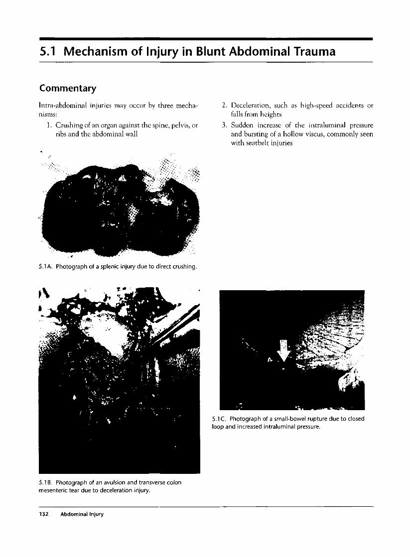
5.1 Mechanism of Injury in Blunt Abdominal Trauma
Commentary
Intra-abdominal injuries may occur by three mecha-nisms:
1. Crushing of an organ against the spine, pelvis, orribs and the abdominal wall
2. Deceleration, such as high-speed accidents orfalls from heights
3. Sudden increase of the intraluminal pressureand bursting of a hollow viscus, commonly seenwith seatbelt injuries
5.1 A. Photograph of a splenic injury due to direct crushing.
t\
5.1 C. Photograph of a small-bowel rupture due to closedloop and increased intraluminal pressure.
5.1 B. Photograph of an avulsion and transverse colonmesenteric tear due to deceleration injury.
132 Abdominal Injury
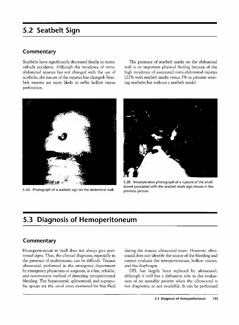
5.2 Seatbelt Sign
Commentary
Seatbelts have significantly decreased deaths in motorvehicle accidents. Although the incidence of intra-abdominal injuries has not changed with the use ofseatbelts, the nature of the injuries has changed: Seat-belt wearers are more likely to suffer hollow viscusperforation.
The presence of seatbelt marks on the abdominalwall is an important physical finding because of thehigh incidence of associated intra-abdominal injuries(22% with seatbelt marks versus 3% in patients wear-ing seatbelts but without a seatbelt mark).
5.2A. Photograph of a seatbelt sign on the abdominal wall.
5.2B. Intraoperative photograph of a rupture of the smallbowel associated with the seatbelt mark sign shown in theprevious picture.
5.3 Diagnosis of Hemoperitoneum
Commentary
Hemoperitoneum in itself does not always give peri-toneal signs. Thus, the clinical diagnosis, especially inthe presence of multitrauma, can be difficult. Traumaultrasound, performed in the emergency departmentby emergency physicians or surgeons, is a fast, reliable,and noninvasive method of detecting intraperitonealbleeding. The hepatorenal, splenorenal, and suprapu-bic spaces are the usual areas examined for free fluid
during the trauma ultrasound exam. However, ultra-sound does not identify the source of the bleeding andcannot evaluate the retroperitoneum, hollow viscera,and the diaphragm.
DPL has largely been replaced by ultrasound,although it still has a definitive role in the evalua-tion of an unstable patient when the ultrasound isnot diagnostic or not available. It can be performed
5.3 Diagnosis of Hemoperitoneum 133

with the open technique or the closed guide-wiretechnique.
CT scan should be considered in hemodynamicallystable patients. Under these circumstances, it is the mostvaluable investigation. It can also identify the source ofthe bleeding and visualize the retroperitoneum.
5.3A. Normal trauma ultrasound depicts the liver and theright kidney with no free fluid between the two organs (K =kidney, L = liver).
5.3B. Trauma ultrasound shows fluid (blood) between theliver and the kidney.
134 Abdominal Injury

IK'ii•a
5.3C. Illustration of opendiagnostic peritoneal lavage(DPL) technique.
5.3E. Photograph of gross blood aspirated from DPLcatheter.
5.3D. Photograph of the introduction of the DPL catheterunder direct vision, using the open technique.
5.3 Diagnosis of Hemoperitoneum 135
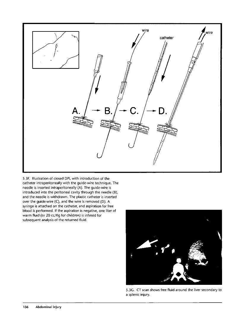
5.3F. Illustration of closed DPL with introduction of thecatheter intraperitoneally with the guide-wire technique. Theneedle is inserted intraperitoneally (A). The guide-wire isintroduced into the peritoneal cavity through the needle (B),and the needle is withdrawn. The plastic catheter is insertedover the guide-wire (C), and the wire is removed (D). Asyringe is attached on the catheter, and aspiration for freeblood is performed. If the aspiration is negative, one liter ofwarm fluid (or 20 cc/Kg for children) is infused forsubsequent analysis of the returned fluid.
5.3G. CT scan shows free fluid around the liver secondary toa splenic injury.
136 Abdominal Injury
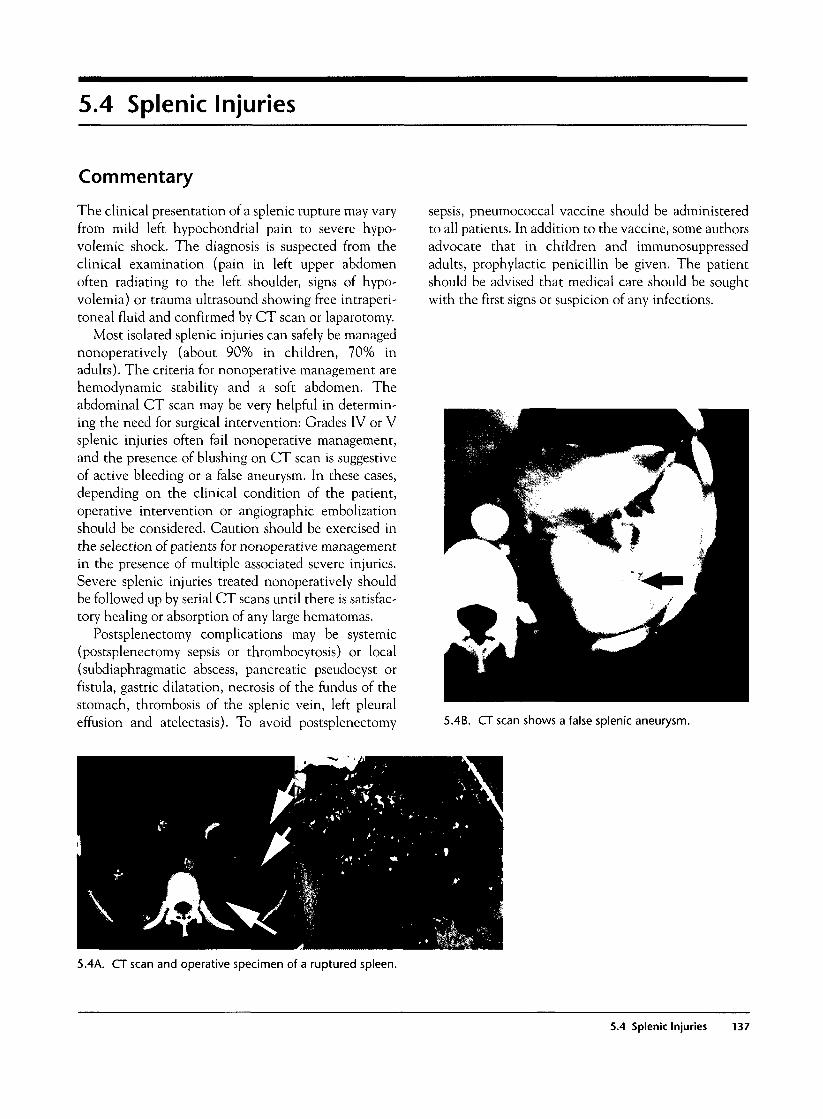
5.4 Splenic Injuries
Commentary
The clinical presentation of a splenic rupture may varyfrom mild left hypochondrial pain to severe hypo-volemic shock. The diagnosis is suspected from theclinical examination (pain in left upper abdomenoften radiating to the left shoulder, signs of hypo-volemia) or trauma ultrasound showing free intraperi-toneal fluid and confirmed by CT scan or laparotomy.
Most isolated splenic injuries can safely be managednonoperatively (about 90% in children, 70% inadults). The criteria for nonoperative management arehemodynamic stability and a soft abdomen. Theabdominal CT scan may be very helpful in determin-ing the need for surgical intervention: Grades IV or Vsplenic injuries often fail nonoperative management,and the presence of blushing on CT scan is suggestiveof active bleeding or a false aneurysm. In these cases,depending on the clinical condition of the patient,operative intervention or angiographic embolizationshould be considered. Caution should be exercised inthe selection of patients for nonoperative managementin the presence of multiple associated severe injuries.Severe splenic injuries treated nonoperatively shouldbe followed up by serial CT scans until there is satisfac-tory healing or absorption of any large hematomas.
Postsplenectomy complications may be systemic(postsplenectomy sepsis or thrombocytosis) or local(subdiaphragmatic abscess, pancreatic pseudocyst orfistula, gastric dilatation, necrosis of the fundus of thestomach, thrombosis of the splenic vein, left pleuraleffusion and atelectasis). To avoid postsplenectomy
sepsis, pneumococcal vaccine should be administeredto all patients. In addition to the vaccine, some authorsadvocate that in children and immunosuppressedadults, prophylactic penicillin be given. The patientshould be advised that medical care should be soughtwith the first signs or suspicion of any infections.
5.4B. CT scan shows a false splenic aneurysm.
5.4A. CT scan and operative specimen of a ruptured spleen.
5.4 Splenic Injuries 137
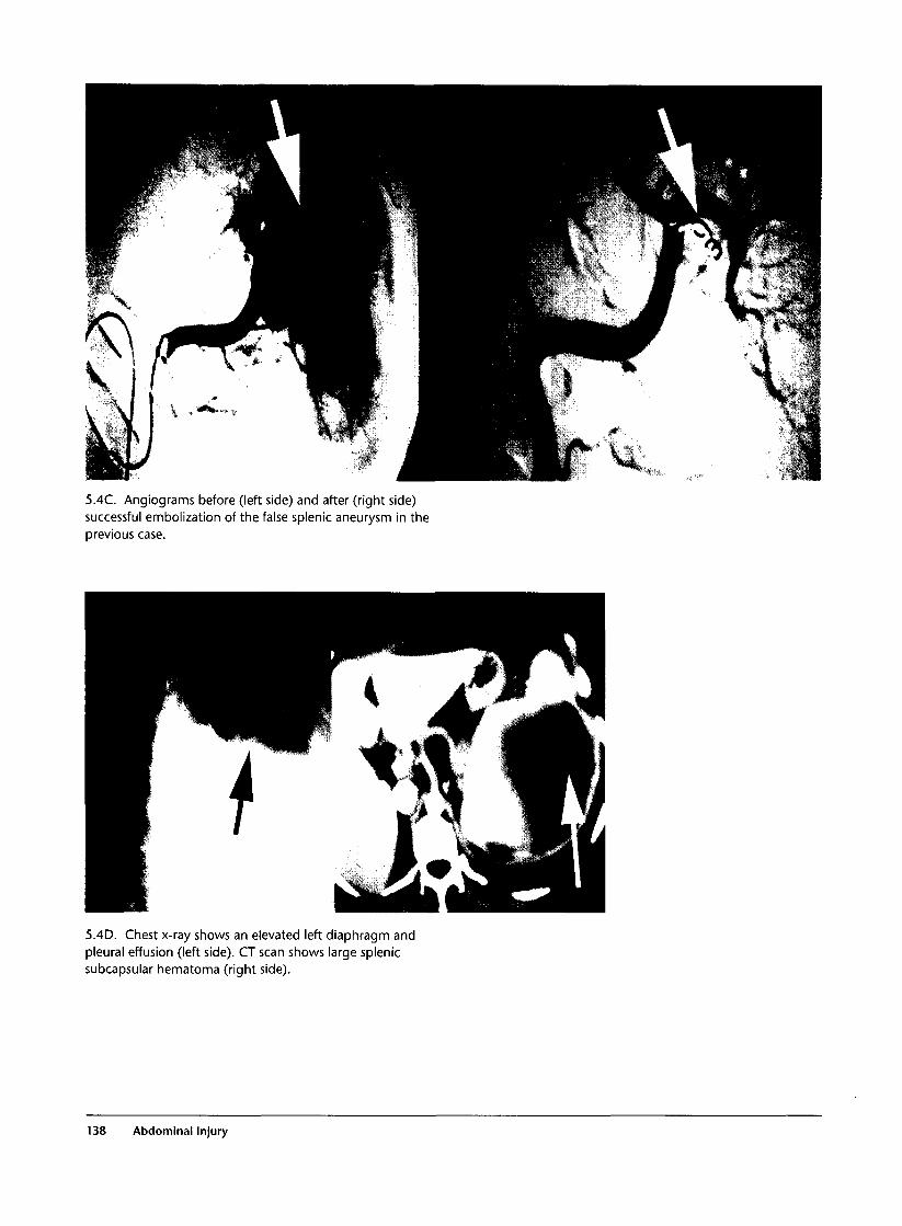
5.4C. Angiograms before (left side) and after (right side)successful embolization of the false splenic aneurysm in theprevious case.
5.4D. Chest x-ray shows an elevated left diaphragm andpleural effusion (left side). CT scan shows large splenicsubcapsular hematoma (right side).
138 Abdominal Injury
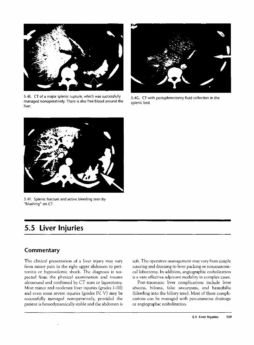
5.4E. CT of a major splenic rupture, which was successfullymanaged nonoperatively. There is also free blood around theliver.
5.4G. CTwith postsplenectomy fluid collection in thesplenic bed.
5.4F. Splenic fracture and active bleeding seen by"blushing" on CT.
5.5 Liver Injuries
Commentary
The clinical presentation of a liver injury may varyfrom minor pain in the right upper abdomen to peri-tonitis or hypovolemic shock. The diagnosis is sus-pected from the physical examination and traumaultrasound and confirmed by CT scan or laparotomy.Most minor and moderate liver injuries (grades I-III)and even some severe injuries (grades IV, V) may besuccessfully managed nonoperatively, provided thepatient is hemodynamically stable and the abdomen is
soft. The operative management may vary from simplesuturing and draining to liver packing or nonanatomi-cal lobectomy. In addition, angiographic embolizationis a very effective adjuvant modality in complex cases.
Post-traumatic liver complications include liverabscess, biloma, false aneurysms, and hemobilia(bleeding into the biliary tree). Most of these compli-cations can be managed with percutaneous drainageor angiographic embolization.
5.5 Liver Injuries 139

5.5A. Abdominal CT scan shows a false aneurysm in theliver.
5.5B. Angiography before(left side) and after (rightside) embolization of theprevious aneurysm.
5.5D. CT scan of large subcapsular liver hematoma.
5.5C. CT scan of a subcapsular liver hematoma.
140 Abdominal Injury
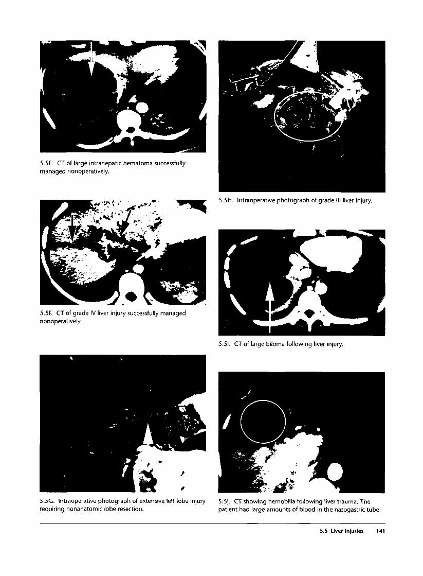
5.5E. CT of large intrahepatic hematoma successfullymanaged nonoperatively.
5.5H. Intraoperative photograph of grade III liver injury.
5.5F. CT of grade IV liver injury successfully managednonoperatively.
5.51. CT of large biloma following liver injury.
5.5G. Intraoperative photograph of extensive left lobe injrequiring nonanatomic lobe resection.
injury 5.5J. CT showing hemobilia following liver trauma. Thepatient had large amounts of blood in the nasogastric tube.
5.5 Liver Injuries 141
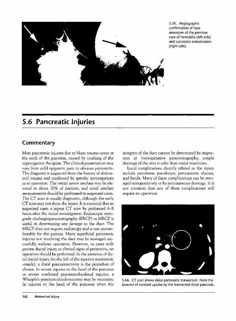
5.5K. Angiographicconfirmation of falseaneurysm of the previouscase of hemobilia (left side)and successful embolization(right side).
5.6 Pancreatic Injuries
Commentary
Most pancreatic injuries due to blunt trauma occur atthe neck of the pancreas, caused by crushing of theorgan against the spine. The clinical presentation mayvary from mild epigastric pain to obvious peritonitis.The diagnosis is suspected from the history of abdom-inal trauma and confirmed by specific investigationsor at operation. The initial serum amylase may be ele-vated in about 70% of patients, and serial amylasemeasurements should be performed in suspected cases.The CT scan is usually diagnostic, although the earlyCT scan may not show the injury. It is essential that insuspected cases a repeat CT scan be performed 6-8hours after the initial investigation. Endoscopic retro-grade cholangiopancreatography (ERCP) or MRCP isuseful in determining any damage to the duct. TheMRCP does not require endoscopy and is not uncom-fortable for the patient. Many superficial pancreaticinjuries not involving the duct may be managed suc-cessfully without operation. However, in cases withproven ductal injury or clinical signs of peritonitis, anoperation should be performed. In the presence of dis-tal ductal injury (to the left of the superior mesentericvessels), a distal pancreatectomy is the procedure ofchoice. In severe injuries to the head of the pancreasor severe combined pancreatoduodenal injuries, aWhipple's pancreatoduodenectomy may be necessary.In injuries to the head of the pancreas when the
integrity of the duct cannot be determined by inspec-tion or intraoperative pancreatography, simpledrainage of the area is safer than major resections.
Local complications directly related to the injuryinclude pancreatic pseudocyst, pancreatitis, abscess,and fistula. Many of these complications can be man-aged nonoperatively or by percutaneous drainage. It isnot common that any of these complications willrequire an operation.
5.6A. CT scan shows distal pancreatic transection. Note theabsence of contrast uptake by the transected distal pancreas.
142 Abdominal Injury
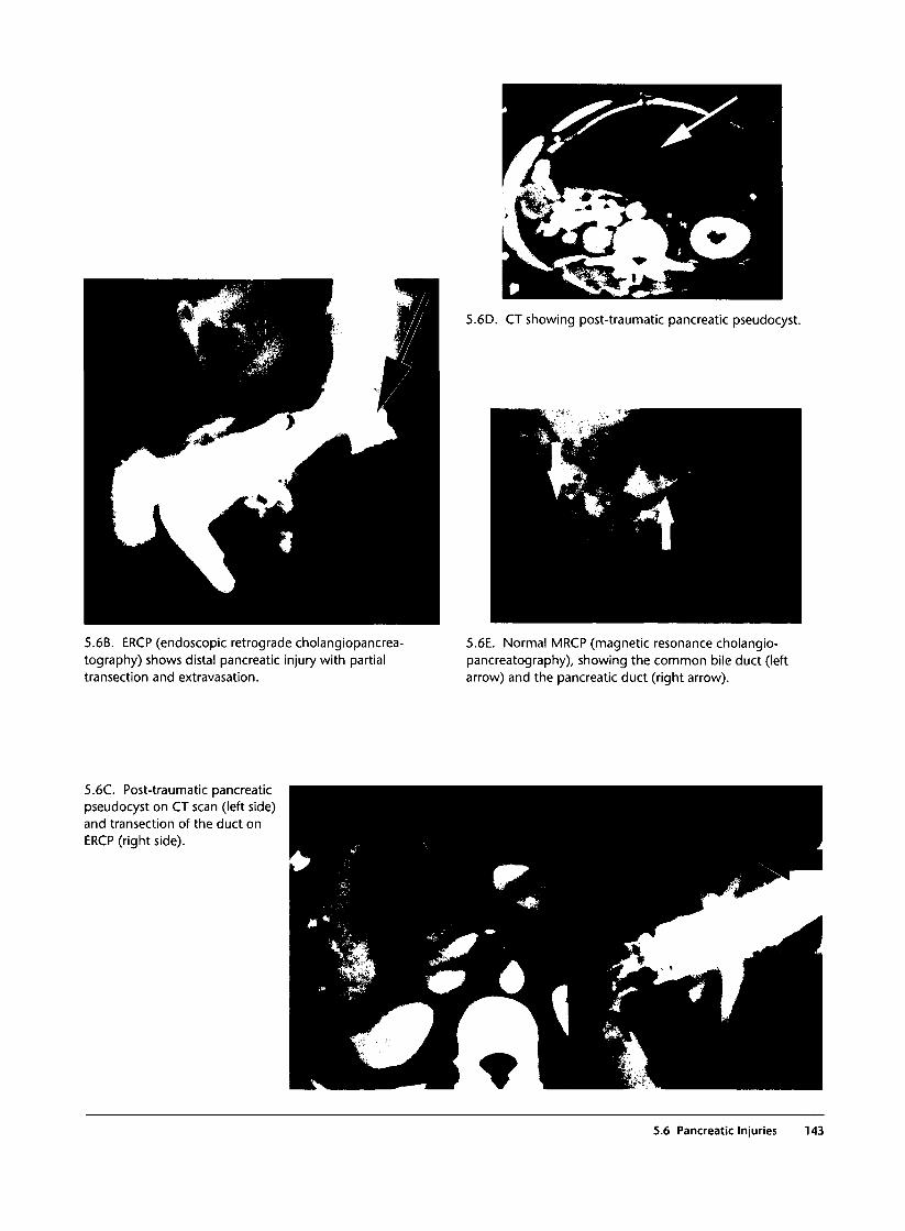
5.6D. CT showing post-traumatic pancreatic pseudocyst.
5.6B. ERCP (endoscopic retrograde cholangiopancrea-tography) shows distal pancreatic injury with partialtransection and extravasation.
5.6E. Normal MRCP (magnetic resonance cholangio-pancreatography), showing the common bile duct (leftarrow) and the pancreatic duct (right arrow).
5.6C. Post-traumatic pancreaticpseudocyst on CT scan (left side)and transection of the duct onERCP (right side).
5.6 Pancreatic Injuries 143
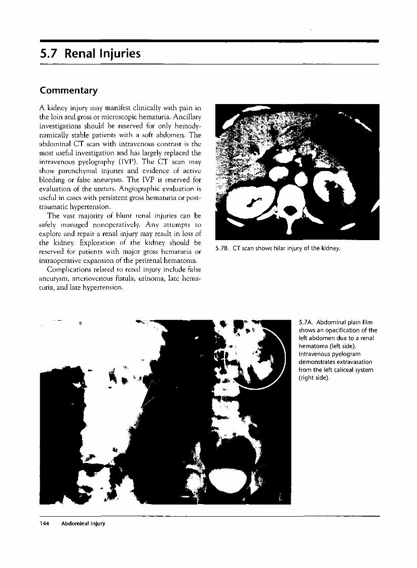
5.7 Renal Injuries
Commentary
A kidney injury may manifest clinically with pain inthe loin and gross or microscopic hematuria. Ancillaryinvestigations should be reserved for only hemody-namically stable patients with a soft abdomen. Theabdominal CT scan with intravenous contrast is themost useful investigation and has largely replaced theintravenous pyelography (IVP). The CT scan mayshow parenchymal injuries and evidence of activebleeding or false aneurysm. The IVP is reserved forevaluation of the ureters. Angiographic evaluation isuseful in cases with persistent gross hematuria or post-traumatic hypertension.
The vast majority of blunt renal injuries can besafely managed nonoperatively. Any attempts toexplore and repair a renal injury may result in loss ofthe kidney. Exploration of the kidney should bereserved for patients with major gross hematuria orintraoperative expansion of the perirenal hematoma.
Complications related to renal injury include falseaneurysm, arteriovenous fistula, urinoma, late hema-turia, and late hypertension.
5.7B. CT scan shows hilar injury of the kidney.
5.7A. Abdominal plain filmshows an opacification of theleft abdomen due to a renalhematoma (left side).Intravenous pyelogramdemonstrates extravasationfrom the left caliceal system(right side).
144 Abdominal Injury
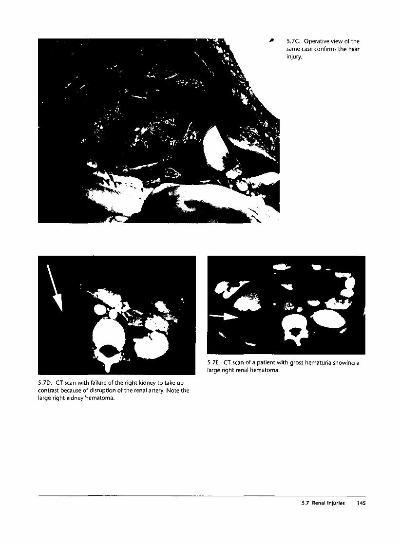
5.7C. Operative view of thesame case confirms the hilarinjury.
5.7E. CT scan of a patient with gross hematuria showing alarge right renal hematoma.
5.7D. CT scan with failure of the right kidney to take upcontrast because of disruption of the renal artery. Note thelarge right kidney hematoma.
5.7 Renal Injuries 145
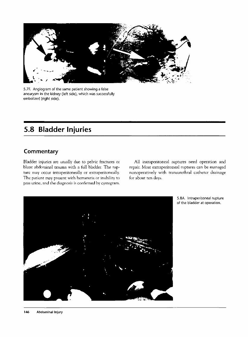
5.7F. Angiogram of the same patient showing a falseaneurysm in the kidney (left side), which was successfullyembolizer! (right side).
5.8 Bladder Injuries
Commentary
Bladder injuries are usually due to pelvic fractures orblunt abdominal trauma with a full bladder. The rup-ture may occur intraperitoneally or extraperitoneally.The patient may present with hematuria or inability topass urine, and the diagnosis is confirmed by cystogram.
All intraperitoneal ruptures need operation andrepair. Most extraperitoneal ruptures can be managednonoperatively with transurethral catheter drainagefor about ten days.
5.8A. Intraperitoneal ruptureof the bladder at operation.
146 Abdominal Injury
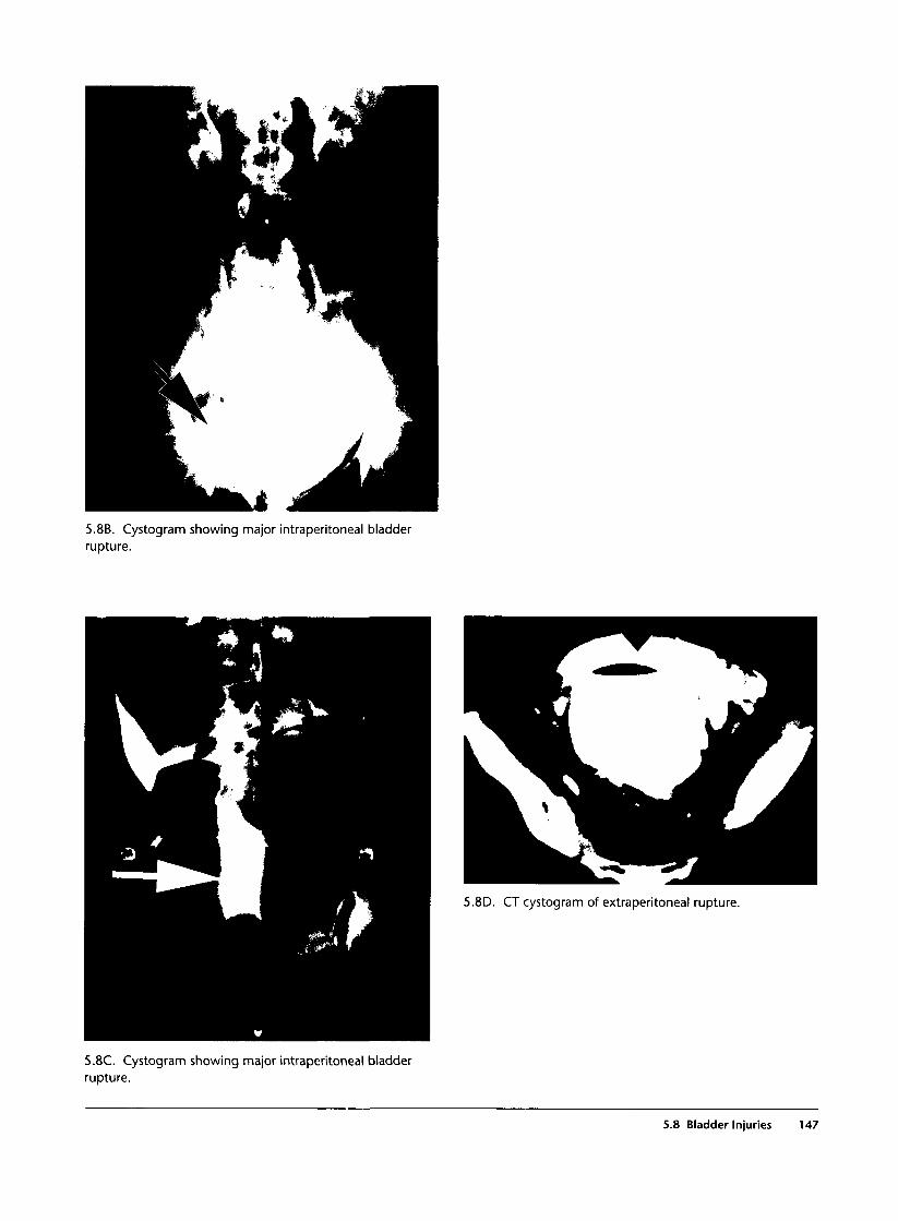
5.8B. Cystogram showing major intraperitoneal bladderrupture.
5.8D. CT cystogram of extraperitoneal rupture.
5.8C. Cystogram showing major intraperitoneal bladderrupture.
5.8 Bladder Injuries 147
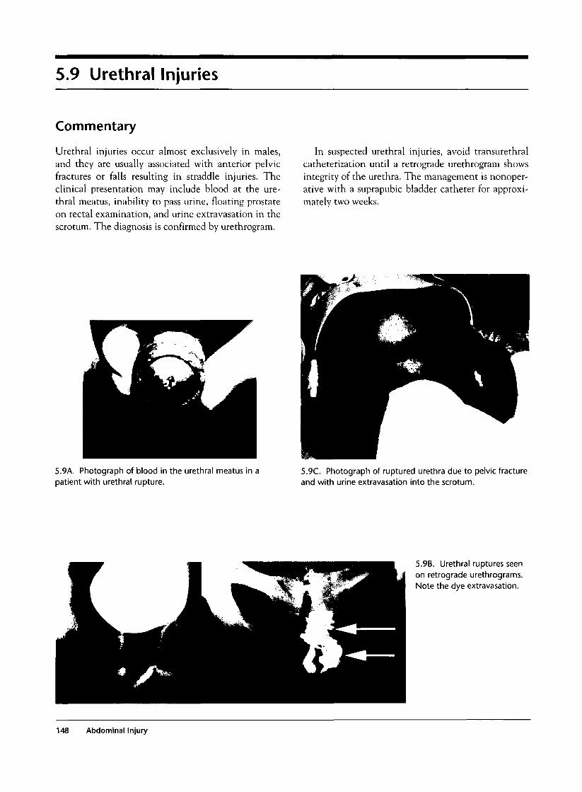
5.9 Urethral Injuries
Commentary
Urethral injuries occur almost exclusively in males,and they are usually associated with anterior pelvicfractures or falls resulting in straddle injuries. Theclinical presentation may include blood at the ure-thral meatus, inability to pass urine, floating prostateon rectal examination, and urine extravasation in thescrotum. The diagnosis is confirmed by urethrogram.
In suspected urethral injuries, avoid transurethralcatheterization until a retrograde urethrogram showsintegrity of the urethra. The management is nonoper-ative with a suprapubic bladder catheter for approxi-mately two weeks.
5.9A. Photograph of blood in the urethral meatus in apatient with urethral rupture.
5.9C. Photograph of ruptured urethra due to pelvic fractureand with urine extravasation into the scrotum.
5.9B. Urethral ruptures seenon retrograde urethrograms.Note the dye extravasation.
148 Abdominal Injury

5.9D. CT cystogram with massive extravasation ofcontrast in the scrotum and extraperitoneal tissues,secondary to posterior urethral rupture.
5.10 Duodenal Injuries
Commentary
Blunt duodenal trauma may occur as a result of directtrauma to the abdomen that compresses the duode-num against the vertebral column or following decel-eration injuries. The injury usually involves theretroperitoneal part of the duodenum. The clinicalexamination may be difficult due to subtle signs andsymptoms because of the retroperitoneal location ofthe rupture. The initial presentation may includeminor epigastric tenderness and tachycardia, and signsof peritonitis may appear a few hours later.
Laboratory tests are of little help in the early diagnosisof duodenal rupture. The serum amylase may be elevated
and should be monitored at 4- to 6-hour intervals. Plainfilms may show retroperitoneal air, especially around theupper pole of the right kidney. CT scan with oral con-trast or Gastrograffin meal are the definitive studies.
Early diagnosis and surgical repair remain the corner-stones for a good outcome. Common postoperative com-plications include local infection and fistula formation.
5.10A. CT scan of retroperitoneal duodenal rupture withfree air around the kidney.
5.1 OB. CT scan with oral contrast shows contrastextravasation in the mid-abdomen and between kidney andliver (lower arrow). A small amount of free intraperitoneal airis seen in the anterior abdomen (upper arrow).
5.10 Duodenal Injuries 149
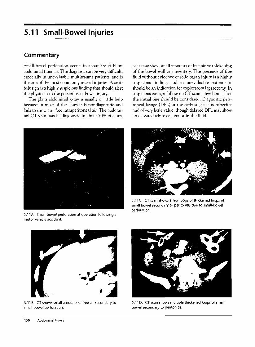
5.11 Small-Bowel Injuries
Commentary
Small-bowel perforation occurs in about 3% of bluntabdominal traumas. The diagnosis can be very difficult,especially in unevaluable multitrauma patients, and isthe one of the most commonly missed injuries. A seat-belt sign is a highly suspicious finding that should alertthe physician to the possibility of bowel injury.
The plain abdominal x-ray is usually of little helpbecause in most of the cases it is nondiagnostic andfails to show any free intraperitoneal air. The abdomi-nal CT scan may be diagnostic in about 70% of cases,
as it may show small amounts of free air or thickeningof the bowel wall or mesentery. The presence of freefluid without evidence of solid organ injury is a highlysuspicious finding, and in unevaluable patients itshould be an indication for exploratory laparotomy. Insuspicious cases, a follow-up CT scan a few hours afterthe initial one should be considered. Diagnostic peri-toneal lavage (DPL) at the early stages is nonspecificand of very little value, though delayed DPL may showan elevated white cell count in the fluid.
5.11 A. Small-bowel perforation at operation following amotor vehicle accident.
5.11C. CT scan shows a few loops of thickened loops ofsmall bowel secondary to peritonitis due to small-bowelperforation.
5.11B. CT shows small amounts of free air secondary tosmall-bowel perforation.
5.11D. CT scan shows multiple thickened loops of smallbowel secondary to peritonitis.
150 Abdominal Injury

5.12 Colorectal Injuries
Commentary
Blunt colorectal trauma may occur in association withpelvic fractures or high-speed deceleration accidents.The diagnosis is usually easy in intraperitonealinjuries because of the presence of peritonitis. How-ever, in extraperitoneal injuries, the physical findingsmay be unremarkable, and the diagnosis may be
delayed. The diagnosis should be suspected in severepelvic fractures, especially in the presence of blood onrectal examination. A Gastrograffin enema and acareful sigmoidoscopy without excessive insufflationcan confirm the injury. Early operation is essential toreduce the possibility of sepsis.
5.12A and B. Photographs of extensive sigmoid coloninjuries, including devascularization, secondary todeceleration injuries after high-speed car accidents.
5.13 Mechanism of Injury in PenetratingAbdominal Trauma
Commentary
A distinction should be made between low-velocityand high-velocity penetrating abdominal injuriesbecause the severity of the injury, the treatment, andthe prognosis are different. High-velocity injuriescause extensive damage by direct laceration, produc-tion of a shock wave, and transient cavitation. Almostall cases require a laparotomy. Low-velocity injuries
(knives, most civilian handguns) cause damage onlyby direct laceration, and often there is no significantintra-abdominal injury requiring surgical repair.Although many centers recommend routine laparo-tomy, many others practice selective nonoperativemanagement for all low-velocity injuries. Criteria fornonoperative management are hemodynamic stability
5.13 Mechanism of Injury in Penetrating Abdominal Trauma 151
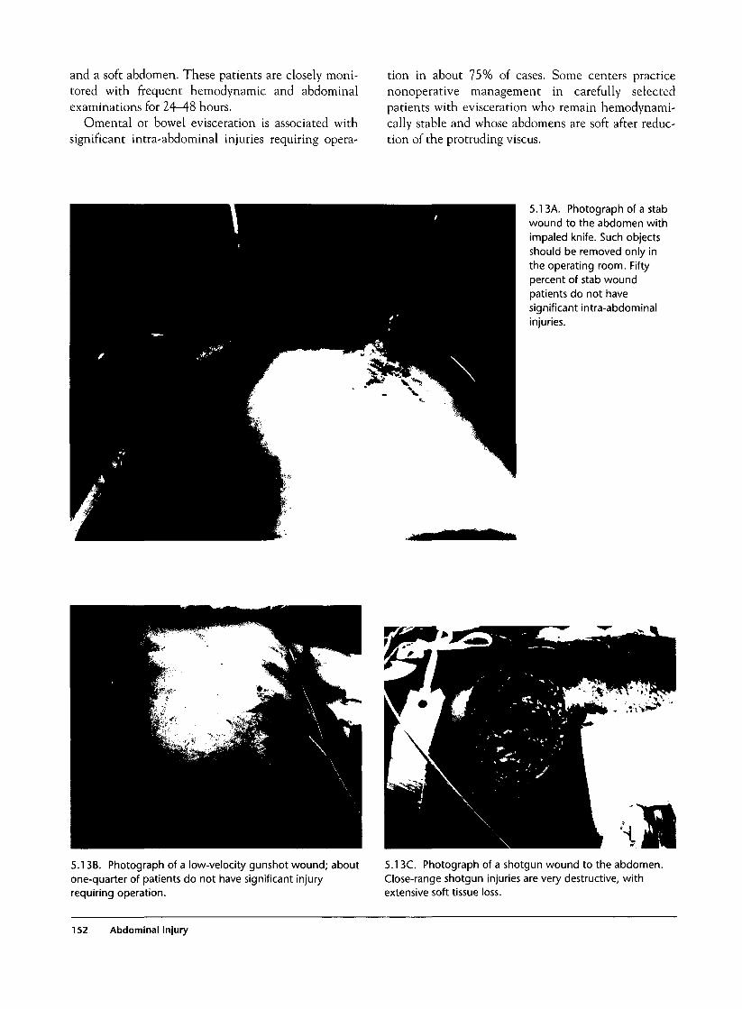
and a soft abdomen. These patients are closely moni-tored with frequent hemodynamic and abdominalexaminations for 24-48 hours.
Omental or bowel evisceration is associated withsignificant intra-abdominal injuries requiring opera-
tion in about 75% of cases. Some centers practicenonoperative management in carefully selectedpatients with evisceration who remain hemodynami-cally stable and whose abdomens are soft after reduc-tion of the protruding viscus.
5.13A. Photograph of a stabwound to the abdomen withimpaled knife. Such objectsshould be removed only inthe operating room. Fiftypercent of stab woundpatients do not havesignificant intra-abdominalinjuries.
5.13B. Photograph of a low-velocity gunshot wound; aboutone-quarter of patients do not have significant injuryrequiring operation.
5.13C. Photograph of a shotgun wound to the abdomen.Close-range shotgun injuries are very destructive, withextensive soft tissue loss.
152 Abdominal Injury
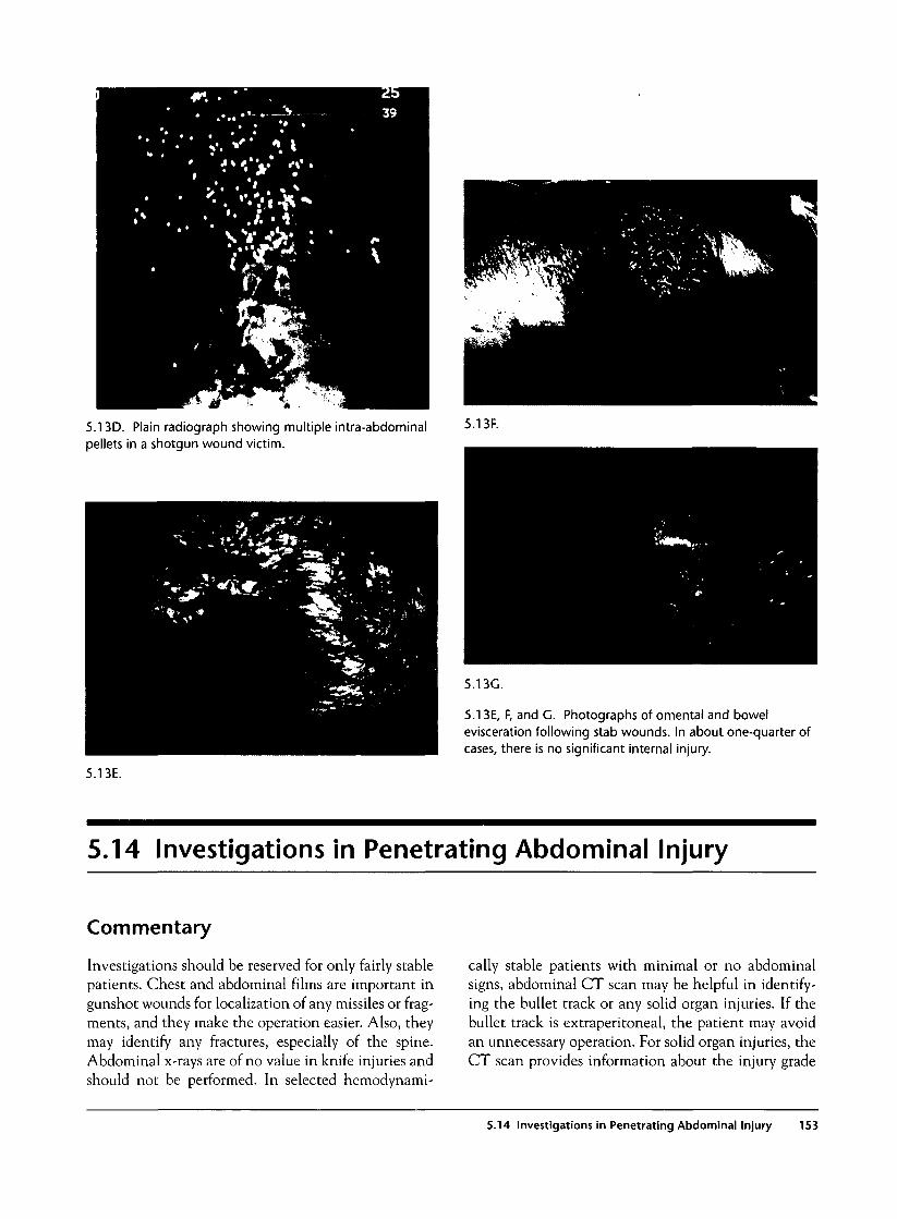
5.13D. Plain radiograph showing multiple intra-abdominalpellets in a shotgun wound victim.
5.13F.
5.13E, F, and G. Photographs of omental and bowelevisceration following stab wounds. In about one-quarter ofcases, there is no significant internal injury.
5.13E.
5.14 Investigations in Penetrating Abdominal Injury
Commentary
Investigations should be reserved for only fairly stablepatients. Chest and abdominal films are important ingunshot wounds for localization of any missiles or frag-ments, and they make the operation easier. Also, theymay identify any fractures, especially of the spine.Abdominal x-rays are of no value in knife injuries andshould not be performed. In selected hemodynami-
cally stable patients with minimal or no abdominalsigns, abdominal CT scan may be helpful in identify-ing the bullet track or any solid organ injuries. If thebullet track is extraperitoneal, the patient may avoidan unnecessary operation. For solid organ injuries, theCT scan provides information about the injury grade
5.14 Investigations in Penetrating Abdominal Injury 153
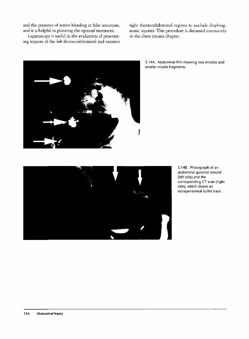
and the presence of active bleeding or false aneurysm,and it is helpful in planning the optimal treatment.
Laparoscopy is useful in the evaluation of penetrat-ing injuries of the left thoracoabdominal and anterior
right thoracoabdominal regions to exclude diaphrag-matic injuries. This procedure is discussed extensivelyin the chest trauma chapter.
5.14A. Abdominal film showing two missiles andsmaller missile fragments.
5.14B. Photograph of anabdominal gunshot wound(left side) and thecorresponding CT scan (rightside), which shows anextraperitoneal bullet track.
154 Abdominal Injury
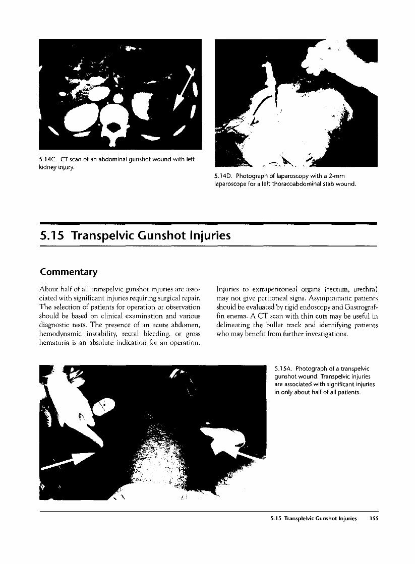
5.14C. CT scan of an abdominal gunshot wound with leftkidney injury.
5.14D. Photograph of laparoscopy with a 2-mmlaparoscope for a left thoracoabdominal stab wound.
5.15 Transpelvic Gunshot Injuries
Commentary
About half of all transpelvic gunshot injuries are asso-ciated with significant injuries requiring surgical repair.The selection of patients for operation or observationshould be based on clinical examination and variousdiagnostic tests. The presence of an acute abdomen,hemodynamic instability, rectal bleeding, or grosshematuria is an absolute indication for an operation.
Injuries to extraperitoneal organs (rectum, urethra)may not give peritoneal signs. Asymptomatic patientsshould be evaluated by rigid endoscopy and Gastrograf-fin enema. A CT scan with thin cuts may be useful indelineating the bullet track and identifying patientswho may benefit from further investigations.
5.15A. Photograph of a transpelvicgunshot wound. Transpelvic injuriesare associated with significant injuriesin only about half of all patients.
5.15 Transplelvic Gunshot Injuries 155
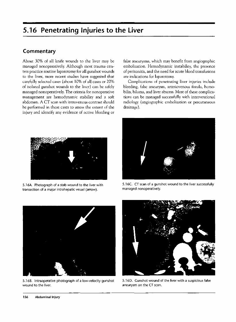
5.16 Penetrating Injuries to the Liver
Commentary
About 30% of all knife wounds to the liver may bemanaged nonoperatively. Although most trauma cen-ters practice routine laparotomy for all gunshot woundsto the liver, more recent studies have suggested thatcarefully selected cases (about 10% of all cases or 20%of isolated gunshot wounds to the liver) can be safelymanaged nonoperatively. The criteria for nonoperativemanagement are hemodynamic stability and a softabdomen. A CT scan with intravenous contrast shouldbe performed in these cases to assess the extent of theinjury and identify any evidence of active bleeding or
false aneurysms, which may benefit from angiographicembolization. Hemodynamic instability, the presenceof peritonitis, and the need for acute blood transfusionsare indications for laparotomy.
Complications of penetrating liver injuries includebleeding, false aneurysm, arteriovenous fistula, hemo-bilia, biloma, and liver abscess. Most of these complica-tions can be managed successfully with interventionalradiology (angiographic embolization or percutaneousdrainage).
5.16A. Photograph of a stab wound to the liver withtransection of a major intrahepatic vessel (arrow).
5.16C. CT scan of a gunshot wound to the liver successfullymanaged nonoperatively.
5.16B. Intraoperative photograph of a low-velocity gunshotwound to the liver.
5.16D. Gunshot wound of the liver with a suspicious falseaneurysm on the CT scan.
156 Abdominal Injury
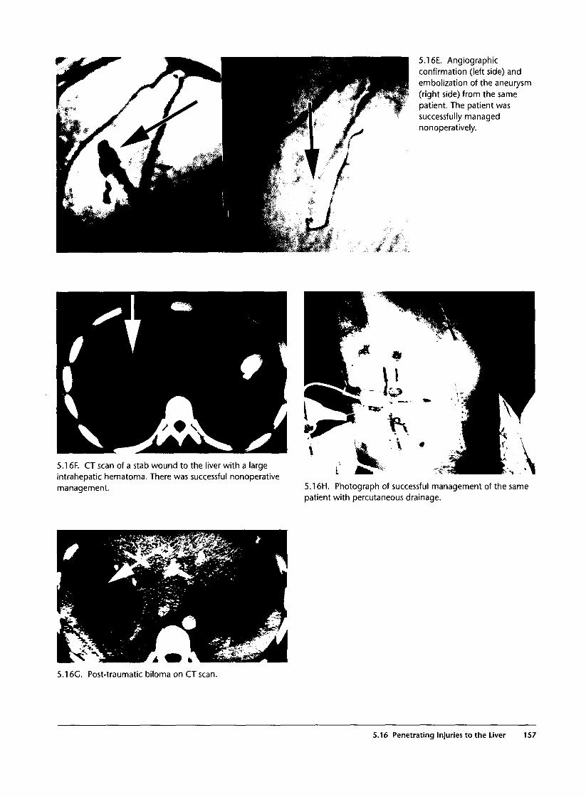
5.16E. Angiographicconfirmation (left side) andembolization of the aneurysm(right side) from the samepatient. The patient wassuccessfully managednonoperatively.
5.16F. CT scan of a stab wound to the liver with a largeintrahepatic hematoma. There was successful nonoperativemanagement. 5.16H. Photograph of successful management of the same
patient with percutaneous drainage.
5.16G. Post-traumatic biloma on CT scan.
5.16 Penetrating Injuries to the Liver 157
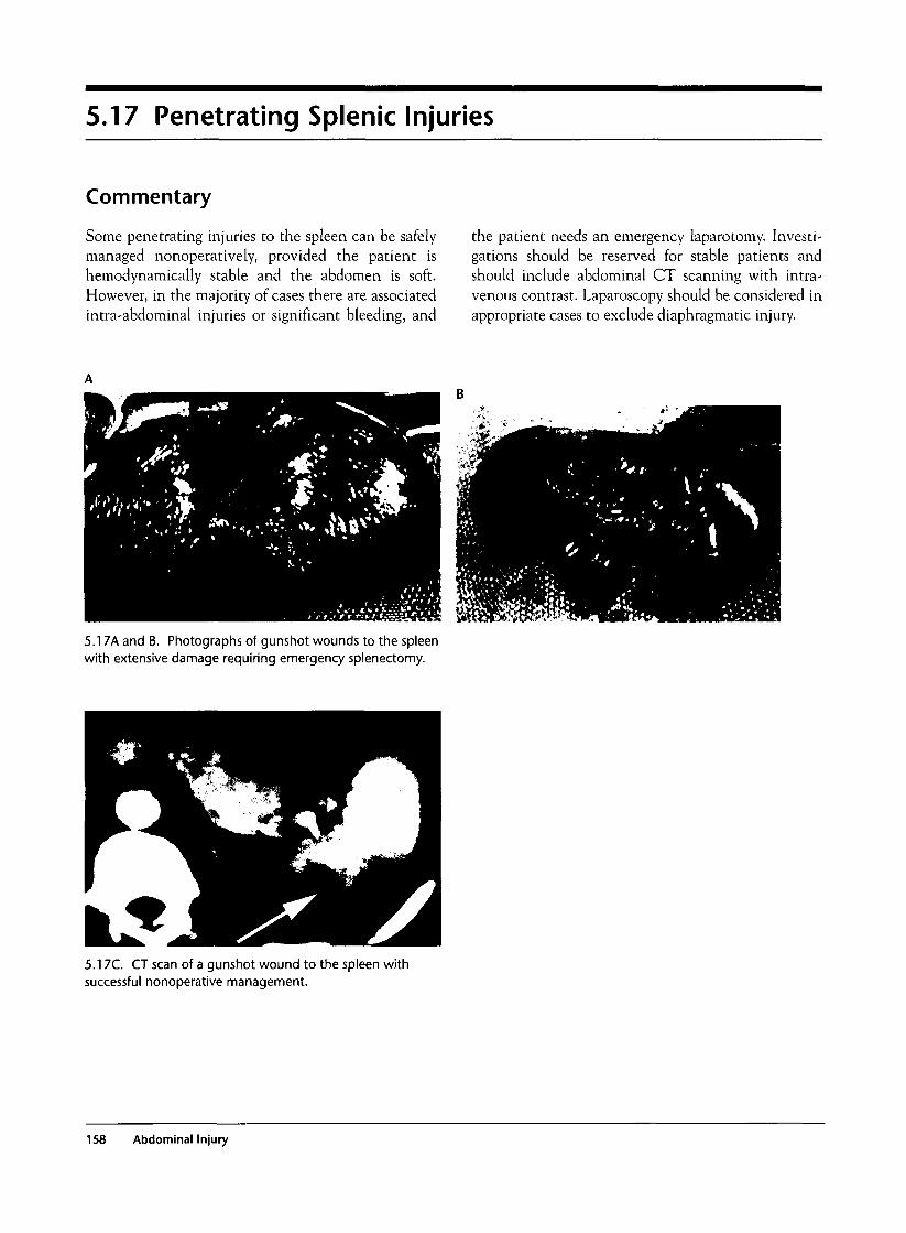
5.17 Penetrating Splenic Injuries
Commentary
Some penetrating injuries to the spleen can be safelymanaged nonoperatively, provided the patient ishemodynamically stable and the abdomen is soft.However, in the majority of cases there are associatedintra-abdominal injuries or significant bleeding, and
the patient needs an emergency laparotomy. Investi-gations should be reserved for stable patients andshould include abdominal CT scanning with intra-venous contrast. Laparoscopy should be considered inappropriate cases to exclude diaphragmatic injury.
5.17A and B. Photographs of gunshot wounds to the spleenwith extensive damage requiring emergency splenectomy.
5.17C. CT scan of a gunshot wound to the spleen withsuccessful nonoperative management.
158 Abdominal Injury
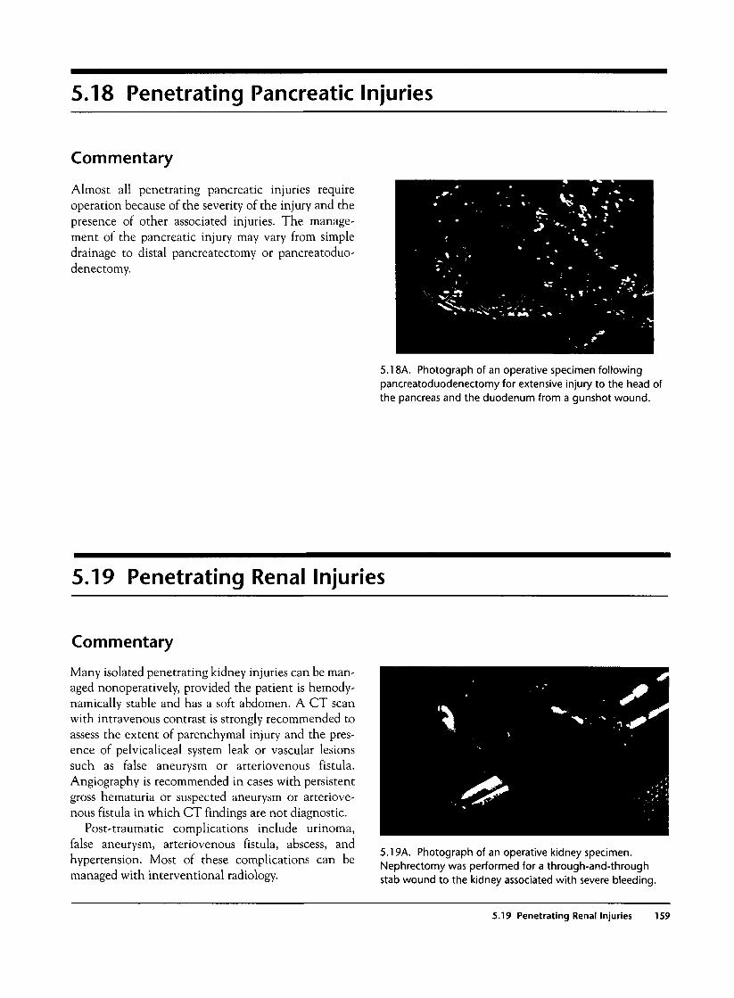
5.18 Penetrating Pancreatic Injuries
Commentary
Almost all penetrating pancreatic injuries requireoperation because of the severity of the injury and thepresence of other associated injuries. The manage-ment of the pancreatic injury may vary from simpledrainage to distal pancreatectomy or pancreatoduo-denectomy.
5.18A. Photograph of an operative specimen followingpancreatoduodenectomy for extensive injury to the head ofthe pancreas and the duodenum from a gunshot wound.
5.19 Penetrating Renal Injuries
Commentary
Many isolated penetrating kidney injuries can be man-aged nonoperatively, provided the patient is hemody-namically stable and has a soft abdomen. A CT scanwith intravenous contrast is strongly recommended toassess the extent of parenchymal injury and the pres-ence of pelvicaliceal system leak or vascular lesionssuch as false aneurysm or arteriovenous fistula.Angiography is recommended in cases with persistentgross hematuria or suspected aneurysm or arteriove-nous fistula in which CT findings are not diagnostic.
Post-traumatic complications include urinoma,false aneurysm, arteriovenous fistula, abscess, andhypertension. Most of these complications can bemanaged with interventional radiology.
5.19A. Photograph of an operative kidney specimen.Nephrectomy was performed for a through-and-throughstab wound to the kidney associated with severe bleeding.
5.19 Penetrating Renal Injuries 159
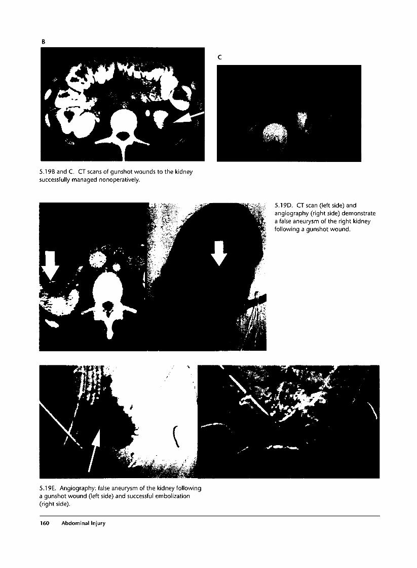
5.19B and C. CT scans of gunshot wounds to the kidneysuccessfully managed nonoperatively.
5.19D. CT scan (left side) andangiography (right side) demonstratea false aneurysm of the right kidneyfollowing a gunshot wound.
5.19E. Angiography: false aneurysm of the kidney followinga gunshot wound (left side) and successful embolization(right side).
160 Abdominal Injury
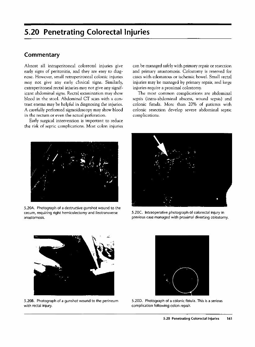
5.20 Penetrating Colorectal Injuries
Commentary
Almost all intraperitoneal colorectal injuries giveearly signs of peritonitis, and they are easy to diag-nose. However, small retroperitoneal colonic injuriesmay not give any early clinical signs. Similarly,extraperitoneal rectal injuries may not give any signif-icant abdominal signs. Rectal examination may showblood in the stool. Abdominal CT scan with a con-trast enema may be helpful in diagnosing the injuries.A carefully performed sigmoidoscopy may show bloodin the rectum or even the actual perforation.
Early surgical intervention is important to reducethe risk of septic complications. Most colon injuries
can be managed safely with primary repair or resectionand primary anastomosis. Colostomy is reserved forcases with edematous or ischemic bowel. Small rectalinjuries may be managed by primary repair, and largeinjuries require a proximal colostomy.
The most common complications are abdominalsepsis (intra-abdominal abscess, wound sepsis) andcolonic fistula. More than 20% of patients withcolonic resection develop severe abdominal septiccomplications.
5.20A. Photograph of a destructive gunshot wound to thececum, requiring right hemicolectomy and ileotransverseanastomosis.
5.20C. Intraoperative photograph of colorectal injury inprevious case managed with proximal diverting colostomy.
5.20B. Photograph of a gunshot wound to the perineumwith rectal injury.
5.20D. Photograph of a colonic fistula. This is a seriouscomplication following colon repair.
5.20 Penetrating Colorectal Injuries 161
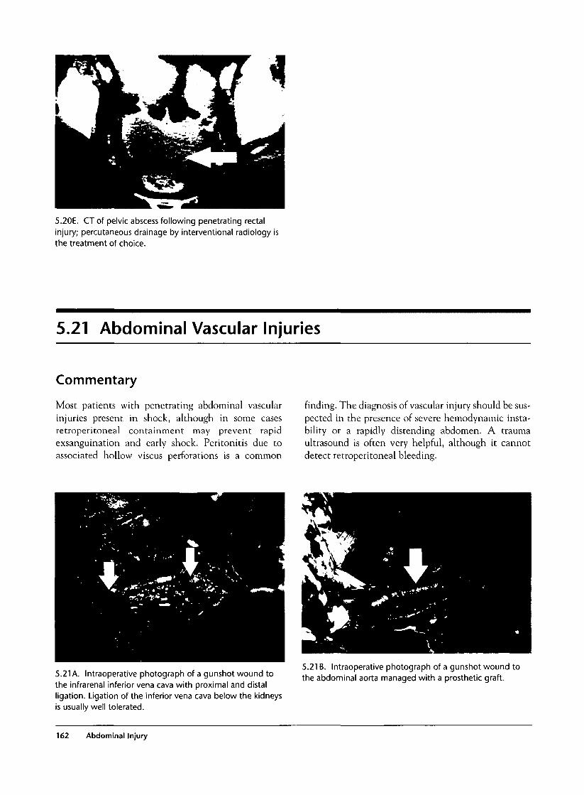
5.20E. CT of pelvic abscess following penetrating rectalinjury; percutaneous drainage by interventional radiology isthe treatment of choice.
5.21 Abdominal Vascular Injuries
Commentary
Most patients with penetrating abdominal vascularinjuries present in shock, although in some casesretroperitoneal containment may prevent rapidexsanguination and early shock. Peritonitis due toassociated hollow viscus perforations is a common
finding. The diagnosis of vascular injury should be sus-pected in the presence of severe hemodynamic insta-bility or a rapidly distending abdomen. A traumaultrasound is often very helpful, although it cannotdetect retroperitoneal bleeding.
5.21 A. Intraoperative photograph of a gunshot wound tothe infrarenal inferior vena cava with proximal and distalligation. Ligation of the inferior vena cava below the kidneysis usually well tolerated.
5.21 B. Intraoperative photograph of a gunshot wound tothe abdominal aorta managed with a prosthetic graft.
162 Abdominal Injury
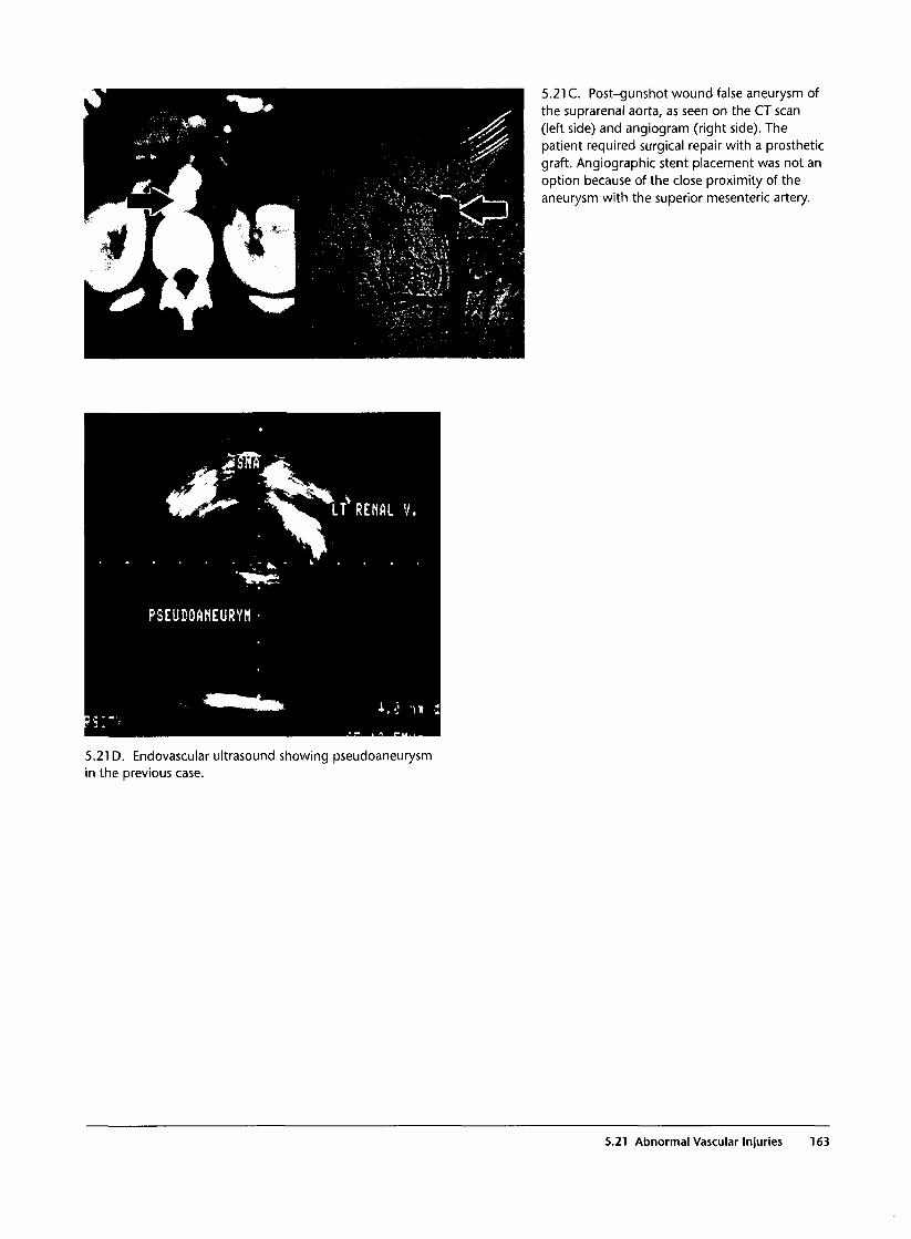
5.21 C. Post-gunshot wound false aneurysm ofthe suprarenal aorta, as seen on the CT scan(left side) and angiogram (right side). Thepatient required surgical repair with a prostheticgraft. Angiographic stent placement was not anoption because of the close proximity of theaneurysm with the superior mesenteric artery.
5.21 D. Endovascular ultrasound showing pseudoaneurysmin the previous case.
5.21 Abnormal Vascular Injuries 163
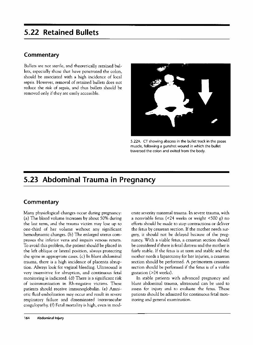
5.22 Retained Bullets
Commentary
Bullets are not sterile, and theoretically retained bul-lets, especially those that have penetrated the colon,should be associated with a high incidence of localsepsis. However, removal of retained bullets does notreduce the risk of sepsis, and thus bullets should beremoved only if they are easily accessible.
5.22A. CT showing abscess in the bullet track in the psoasmuscle, following a gunshot wound in which the bullettraversed the colon and exited from the body.
5.23 Abdominal Trauma in Pregnancy
Commentary
Many physiological changes occur during pregnancy:(a) The blood volume increases by about 50% duringthe last term, and the trauma victim may lose up toone-third of her volume without any significanthemodynamic changes, (b) The enlarged uterus com-presses the inferior vena and impairs venous return.To avoid this problem, the patient should be placed inthe left oblique or lateral position, always protectingthe spine in appropriate cases, (c) In blunt abdominaltrauma, there is a high incidence of placenta abrup-tion. Always look for vaginal bleeding. Ultrasound isvery insensitive for abruption, and continuous fetalmonitoring is indicated, (d) There is a significant riskof isoimmunization in Rh-negative victims. Thesepatients should receive immunoglobulin. (e) Amni-otic fluid embolization may occur and result in severerespiratory failure and disseminated intravascularcoagulopathy. (f) Fetal mortality is high, even in mod-
erate severity maternal trauma. In severe trauma, witha nonviable fetus (<24 weeks or weight <500 g) noefforts should be made to stop contractions or deliverthe fetus by cesarean section. If the mother needs sur-gery, it should not be delayed because of the preg-nancy. With a viable fetus, a cesarean section shouldbe considered if there is fetal distress and the mother isfairly stable. If the fetus is at term and stable and themother needs a laparotomy for her injuries, a cesareansection should be performed. A perimortem cesareansection should be performed if the fetus is of a viablegestation (>24 weeks).
In stable patients with advanced pregnancy andblunt abdominal trauma, ultrasound can be used toassess for injury and to evaluate the fetus. Thesepatients should be admitted for continuous fetal mon-itoring and general examination.
164 Abdominal Injury
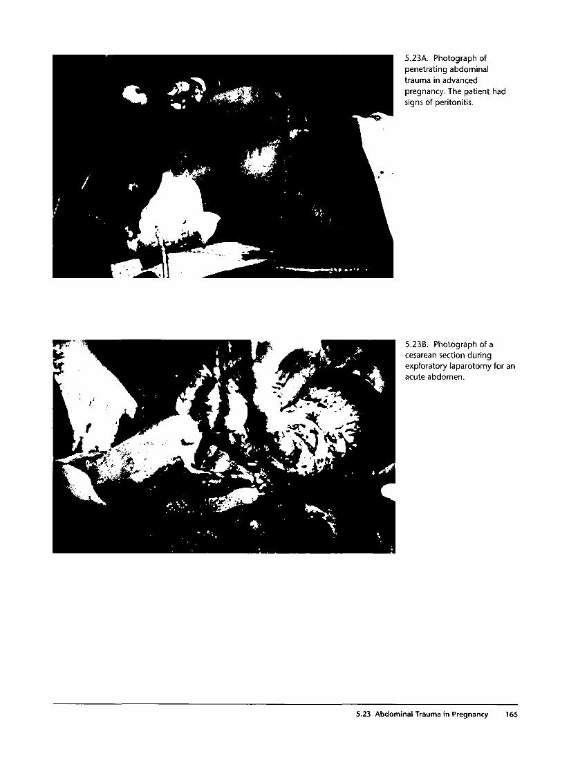
5.23A. Photograph ofpenetrating abdominaltrauma in advancedpregnancy. The patient hadsigns of peritonitis.
Sir 5.23B. Photograph of acesarean section duringexploratory laparotomy for anacute abdomen.
5.23 Abdominal Trauma in Pregnancy 165
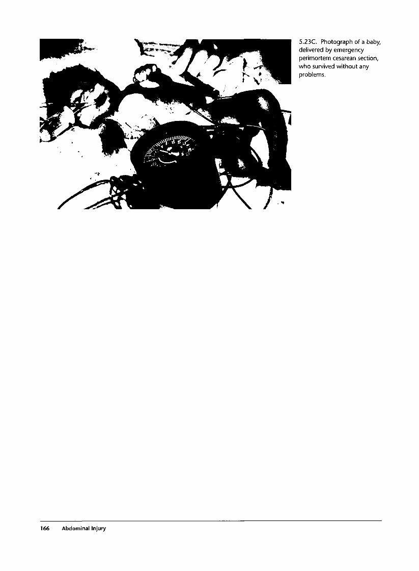
5.23C. Photograph of a baby,delivered by emergencyperimortem cesarean section,who survived without anyproblems.
166 Abdominal Injury
Musculoskeletal Injury
Introduction
Orthopedic injuries are found in approximately 85% ofblunt trauma victims, and thus expert knowledge intheir emergency care is necessary. Many injuries arealso acutely life-threatening or limb-threatening andthus need to be evaluated and treated in an expeditedfashion. Despite the importance of early treatment, thestandard primary survey evaluation with the ABCDEapproach is necessary to detect other injuries with agreater priority. During the primary survey, the onlyattention to musculoskeletal injury is acute hemor-rhage control with direct pressure.
Clinical Examination
The physical exam is an integral component of detect-ing acute orthopedic injuries. The overlying skinshould be examined for contusions and lacerations.Lacerations need a more detailed evaluation for neu-rovascular injury, tendon injury, foreign bodies, andproximity to fracture sites. Cool, pale skin may indicateacute vascular insufficiency. Capillary refill and periph-eral pulses should be checked and compared with theunaffected limb. In some cases Doppler ultrasound maybe required to detect poor pulses. The compartmentsshould be palpated for firmness that may indicate anacute compartment syndrome. General range ofmotion and areas of tenderness help guide necessaryradiographs. Careful peripheral nerve exam is alsoimportant, as nerve injury may be part of the injurycomplex. Some injuries may not present with any frac-ture, but ligamentous injury is important to detect.
Investigations
1. Plain radiographs are necessary for most orthope-dic injuries. Fractures are usually evident on plain radi-
ographs, but occasionally the fracture is occult, requir-ing further imaging. An AP pelvis film is standard inmultisystem blunt trauma, and other radiographsshould be guided by the clinical exam. X-rays shouldview the affected bone in at least two planes and pro-vide a full view of the involved bone, including thejoints above and below. Depending on the spectrum ofinjuries, full radiographic imaging may need to bedelayed to stabilize the patient from life-threateninginjuries.
2. Computed tomography (CT) scanning is useful inevaluating patients with continued pain and with nor-mal plain radiographs, such as in acute hip injuries. CTscan provides much more detail of an identified injuryand allows 3D reformatting.
3. Magnetic resonance imaging (MRI) is rarelyneeded to acutely evaluate musculoskeletal injury. It isuseful in detecting occult fractures, as in hip injuries,and is also useful in delineating ligamentous and carti-laginous injury, such as may be seen with acute kneeinjury. It has a prominent role in spinal injuries.
4. Bone scanning at 72 hours is occasionally used tofollow up patients who may have occult fractures of thescaphoid.
5. Doppler ultrasound and arteriography, in con-junction with the ankle-brachial index, are used toevaluate patients with possible vascular injury.
6. Intracompartment pressures can easily be meas-ured with portable equipment and can help guide themanagement of patients with possible compartmentsyndrome.
General Management
Life-threatening injuries receive priority, and thusmany orthopedic emergencies may receive more exten-sive evaluation and treatment in the postoperativephase. Some orthopedic injuries require special men-tion as they are life-threatening and need similar prior-
167
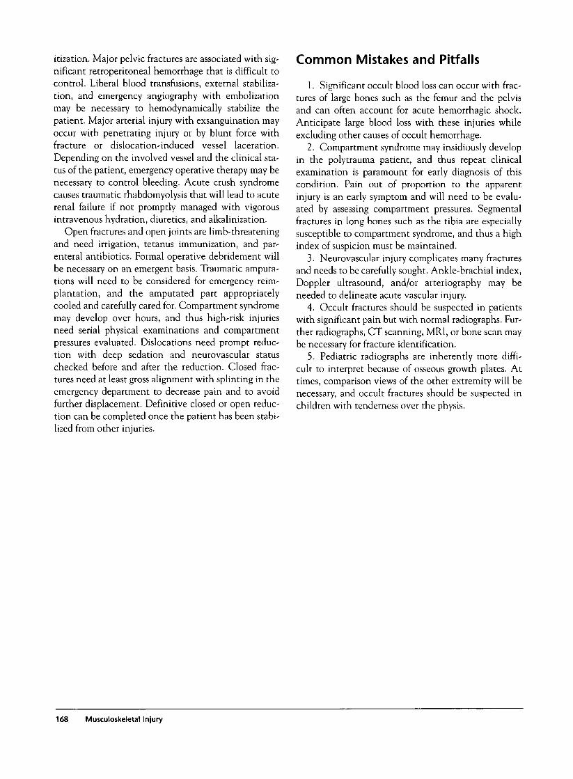
itization. Major pelvic fractures are associated with sig-nificant retroperitoneal hemorrhage that is difficult tocontrol. Liberal blood transfusions, external stabiliza-tion, and emergency angiography with embolizationmay be necessary to hemodynamicaHy stabilize thepatient. Major arterial injury with exsanguination mayoccur with penetrating injury or by blunt force withfracture or dislocation-induced vessel laceration.Depending on the involved vessel and the clinical sta-tus of the patient, emergency operative therapy may benecessary to control bleeding. Acute crush syndromecauses traumatic rhabdomyolysis that will lead to acuterenal failure if not promptly managed with vigorousintravenous hydration, diuretics, and alkalinization.
Open fractures and open joints are limb-threateningand need irrigation, tetanus immunization, and par-enteral antibiotics. Formal operative debridement willbe necessary on an emergent basis. Traumatic amputa-tions will need to be considered for emergency reim-plantation, and the amputated part appropriatelycooled and carefully cared for. Compartment syndromemay develop over hours, and thus high-risk injuriesneed serial physical examinations and compartmentpressures evaluated. Dislocations need prompt reduc-tion with deep sedation and neurovascular statuschecked before and after the reduction. Closed frac-tures need at least gross alignment with splinting in theemergency department to decrease pain and to avoidfurther displacement. Definitive closed or open reduc-tion can be completed once the patient has been stabi-lized from other injuries.
Common Mistakes and Pitfalls
1. Significant occult blood loss can occur with frac-tures of large bones such as the femur and the pelvisand can often account for acute hemorrhagic shock.Anticipate large blood loss with these injuries whileexcluding other causes of occult hemorrhage.
2. Compartment syndrome may insidiously developin the polytrauma patient, and thus repeat clinicalexamination is paramount for early diagnosis of thiscondition. Pain out of proportion to the apparentinjury is an early symptom and will need to be evalu-ated by assessing compartment pressures. Segmentalfractures in long bones such as the tibia are especiallysusceptible to compartment syndrome, and thus a highindex of suspicion must be maintained.
3. Neurovascular injury complicates many fracturesand needs to be carefully sought. Ankle-brachial index,Doppler ultrasound, and/or arteriography may beneeded to delineate acute vascular injury.
4. Occult fractures should be suspected in patientswith significant pain but with normal radiographs. Fur-ther radiographs, CT scanning, MRI, or bone scan maybe necessary for fracture identification.
5. Pediatric radiographs are inherently more diffi-cult to interpret because of osseous growth plates. Attimes, comparison views of the other extremity will benecessary, and occult fractures should be suspected inchildren with tenderness over the physis.
168 Musculoskeletal Injury
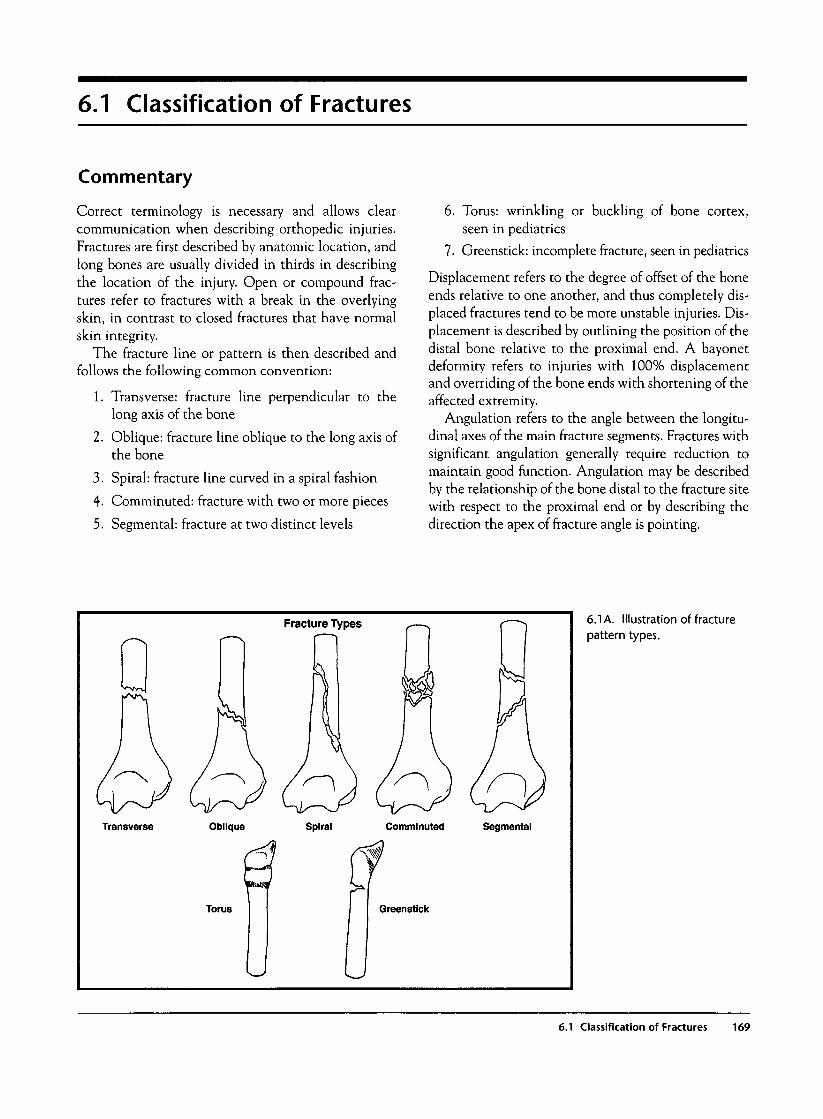
6.1 Classification of Fractures
Commentary
Correct terminology is necessary and allows clearcommunication when describing orthopedic injuries.Fractures are first described by anatomic location, andlong bones are usually divided in thirds in describingthe location of the injury. Open or compound frac-tures refer to fractures with a break in the overlyingskin, in contrast to closed fractures that have normalskin integrity.
The fracture line or pattern is then described andfollows the following common convention:
1. Transverse: fracture line perpendicular to thelong axis of the bone
2. Oblique: fracture line oblique to the long axis ofthe bone
3. Spiral: fracture line curved in a spiral fashion
4- Comminuted: fracture with two or more pieces
5. Segmental: fracture at two distinct levels
6. Torus: wrinkling or buckling of bone cortex,seen in pediatrics
7. Greenstick: incomplete fracture, seen in pediatrics
Displacement refers to the degree of offset of the boneends relative to one another, and thus completely dis-placed fractures tend to be more unstable injuries. Dis-placement is described by outlining the position of thedistal bone relative to the proximal end. A bayonetdeformity refers to injuries with 100% displacementand overriding of the bone ends with shortening of theaffected extremity.
Angulation refers to the angle between the longitu-dinal axes of the main fracture segments. Fractures withsignificant angulation generally require reduction tomaintain good function. Angulation may be describedby the relationship of the bone distal to the fracture sitewith respect to the proximal end or by describing thedirection the apex of fracture angle is pointing.
Fracture Types
Transverse Oblique Spiral Comminuted Segmental
Torus Greenstick
6.1 A. Illustration of fracture
pattern types.
6.1 Classification of Fractures 169
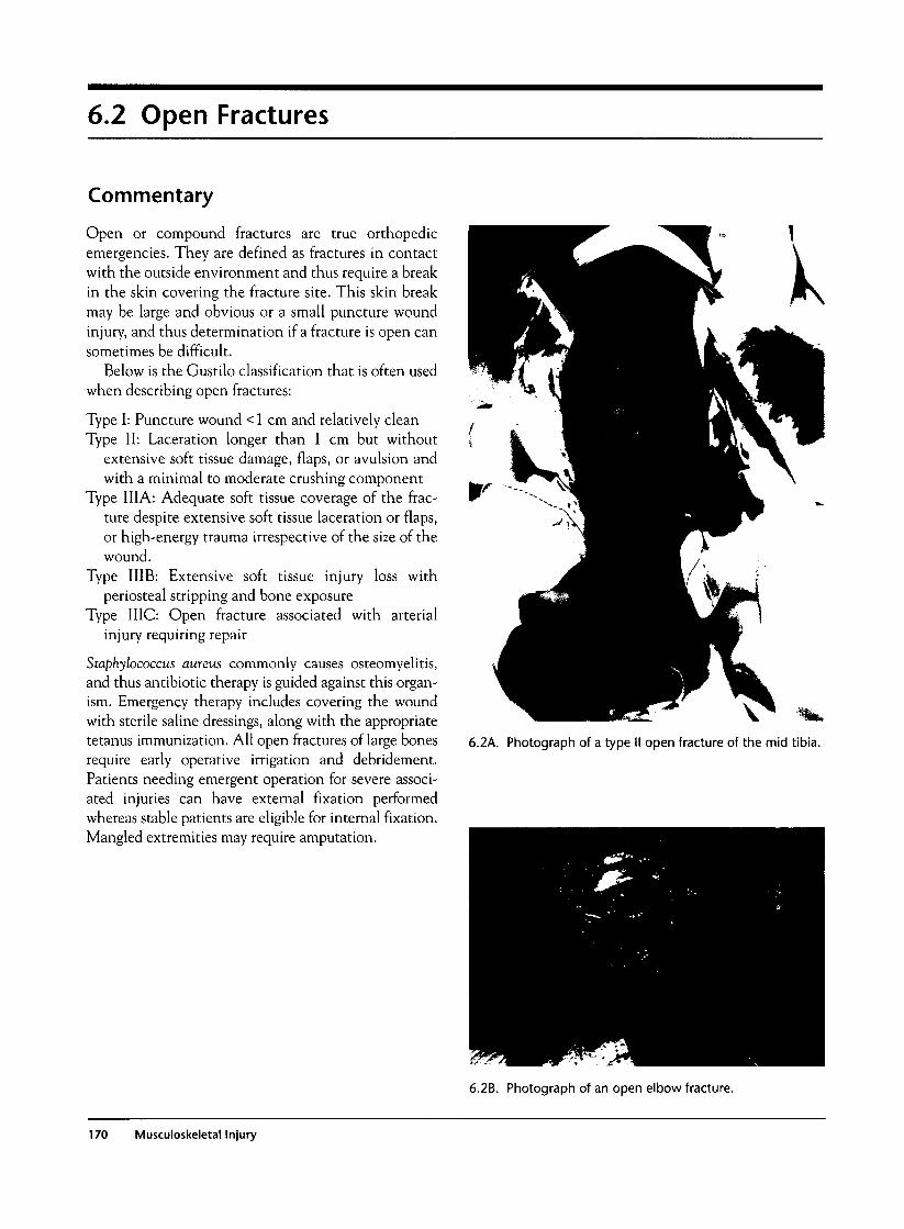
6.2 Open Fractures
Commentary
Open or compound fractures are true orthopedicemergencies. They are defined as fractures in contactwith the outside environment and thus require a breakin the skin covering the fracture site. This skin breakmay be large and obvious or a small puncture woundinjury, and thus determination if a fracture is open cansometimes be difficult.
Below is the Gustilo classification that is often usedwhen describing open fractures:
Type I: Puncture wound <1 cm and relatively cleanType II: Laceration longer than 1 cm but without
extensive soft tissue damage, flaps, or avulsion andwith a minimal to moderate crushing component
Type IIIA: Adequate soft tissue coverage of the frac-ture despite extensive soft tissue laceration or flaps,or high-energy trauma irrespective of the size of thewound.
Type IIIB: Extensive soft tissue injury loss withperiosteal stripping and bone exposure
Type IIIC: Open fracture associated with arterialinjury requiring repair
Staphylococcus aureus commonly causes osteomyelitis,and thus antibiotic therapy is guided against this organ-ism. Emergency therapy includes covering the woundwith sterile saline dressings, along with the appropriatetetanus immunization. All open fractures of large bonesrequire early operative irrigation and debridement.Patients needing emergent operation for severe associ-ated injuries can have external fixation performedwhereas stable patients are eligible for internal fixation.Mangled extremities may require amputation.
6.2A. Photograph of a type II open fracture of the mid tibia.
6.2B. Photograph of an open elbow fracture.
170 Musculoskeletal Injury
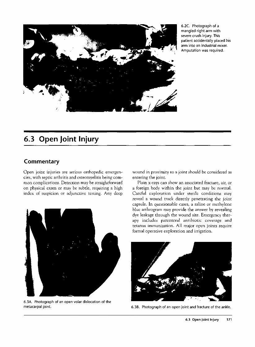
6.2C. Photograph of amangled right arm withsevere crush injury. Thispatient accidentally placed hisarm into an industrial mixer.Amputation was required.
6.3 Open Joint Injury
Commentary
Open joint injuries are serious orthopedic emergen-cies, with septic arthritis and osteomyelitis being com-mon complications. Detection may be straightforwardon physical exam or may be subtle, requiring a highindex of suspicion or adjunctive testing. Any deep
wound in proximity to a joint should be considered asentering the joint.
Plain x-rays can show an associated fracture, air, ora foreign body within the joint but may be normal.Careful exploration under sterile conditions mayreveal a wound track directly penetrating the jointcapsule. In questionable cases, a saline or methyleneblue arthrogram may provide the answer by revealingdye leakage through the wound site. Emergency ther-apy includes parenteral antibiotic coverage andtetanus immunization. All major open joints requireformal operative exploration and irrigation.
6.3A. Photograph of an open volar dislocation of themetacarpal joint. 6.3B. Photograph of an open joint and fracture of the ankle.
6.3 Open Joint Injury 171
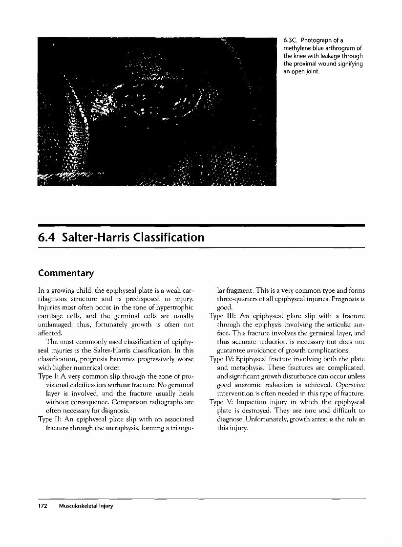
6.3C. Photograph of amethylene blue arthrogram ofthe knee with leakage throughthe proximal wound signifyingan open joint.
6.4 Salter-Harris Classification
Commentary
In a growing child, the epiphyseal plate is a weak car-tilaginous structure and is predisposed to injury.Injuries most often occur in the zone of hypertrophiccartilage cells, and the germinal cells are usuallyundamaged; thus, fortunately growth is often notaffected.
The most commonly used classification of epiphy-seal injuries is the Salter-Harris classification. In thisclassification, prognosis becomes progressively worsewith higher numerical order.Type I: A very common slip through the zone of pro-
visional calcification without fracture. No germinallayer is involved, and the fracture usually healswithout consequence. Comparison radiographs areoften necessary for diagnosis.
Type II: An epiphyseal plate slip with an associatedfracture through the metaphysis, forming a triangu-
lar fragment. This is a very common type and formsthree-quarters of all epiphyseal injuries. Prognosis isgood.
Type III: An epiphyseal plate slip with a fracturethrough the epiphysis involving the articular sur-face. This fracture involves the germinal layer, andthus accurate reduction is necessary but does notguarantee avoidance of growth complications.
Type IV: Epiphyseal fracture involving both the plateand metaphysis. These fractures are complicated,and significant growth disturbance can occur unlessgood anatomic reduction is achieved. Operativeintervention is often needed in this type of fracture.
Type V: Impaction injury in which the epiphysealplate is destroyed. They are rare and difficult todiagnose. Unfortunately, growth arrest is the rule inthis injury.
172 Musculoskeletal Injury

6.4A. Illustration of the Salter-Harris pediatric fractureclassification.
6.4B. Radiograph of Salter-Harris II fracture involving theproximal phalanx of the thumb.
6.4C. Radiograph of Salter-Harris III fracture of the ankle.
6.4 Salter-Harris Classification 173

6.4D. Radiograph of Salter-Harris IV fracture of the distalradius.
6.5 Torus and Greenstick Fractures
Commentary
Pediatric bones are much less brittle than adult bonesand thus are less likely to have complete fracturesthrough the bone. A torus fracture is an incompletefracture with a small fold in the cortex. It is often seen
at the end of long bones. A greenstick fracture is anincomplete angulated fracture of a long bone recog-nized by a bowing appearance. These pediatric vari-ants are common and can easily be missed.
6.5B. Radiograph of a greenstick fracture of the forearm.
6.5A. Radiograph of a distal radius torus fracture.
174 Musculoskeletal Injury

6.6 Supracondylar Fracture
Commentary
Distal humeral fractures that are proximal to the epi-condyles are termed supracondylar fractures, and mostof these fractures occur in children age 5-10 years. Inchildren, the ligaments and joint capsule are strongerthan the bone and thus hyperextension injury oftencauses bone fracture, while adults often suffer a poste-rior dislocation of the elbow with a similar mechanism.
Supracondylar fractures are of two common types:flexion and extension, with extension fractures theoverwhelming majority. These extension injuries areoften the result of a fall on an arm with the elbow fullyextended. On exam, the elbow will be swollen, oftenwith a joint effusion and with significant pain and ten-derness. In addition, the olecranon will be moreprominent as it is attached to the posteriorly displaced
distal fragment. Careful neurovascular examination ofthe arm is necessary as many of these fractures arecomplicated with brachial artery and median, radial,or ulnar nerve injury. In addition, compartment syn-drome can be seen with displaced fractures and needsto be considered.
Radiographically, these fractures are often detectedon the lateral view of the elbow. Because many ofthese fractures are transverse they may not be readilyvisible on the AP view. In addition, up to 25% of thesefractures are of the greenstick variety with the poste-rior cortex remaining intact. The only abnormalityseen may be a posterior fat pad sign or an abnormalanterior humeral line. A high degree of suspicion forsupracondylar fracture must be maintained for anychild with acute elbow trauma, pain, and normal radi-ography. Most undisplaced fractures are treated non-operatively with casting, and most displaced fractureswill undergo percutaneous pinning.
6.6k. Photograph of a swollen elbow of a child suffering asupracondylar fracture.
6.6B. Radiograph of a displaced extension supracondylarfracture.
6.6 Supracondylar Fracture 175

6.6C. Radiograph of an undisplaced transversesupracondylar fracture.
6.6D. Illustration of the anterior humeral line. Normally thisline will intersect the middle of the capitellum. With anextension fracture, this line will intersect the anterior one-third of the capitellum or pass entirely anteriorly.
6.7 Amputations
Commentary
Amputations are devastating injuries and requireexpert knowledge in the care of the patient and theamputated part. The area affected is important in thedecision to consider reimplantation. Lower extremityreimplantation is rarely indicated, given the frequencyof associated crush injury and the efficacy of currentprostheses. In contrast, upper extremity amputations,especially involving the thumb, are often reimplanted,given the severe disability that occurs with the loss ofthat single digit. Time elapsed since the injury is alsoan important consideration. Reimplantation is lesslikely to be successful if the warm ischemia (room tem-perature) time has been >6-8 hours. If the part hasbeen properly cooled and cared for, then this windowof time may be successfully extended to 12-24 hours.In addition, clean, sharp amputations are more likelyto be successful than crush injuries. In general, all
amputated parts should be considered as candidates forreimplantation, and even severely crushed parts can beused for skin coverage.
The amputated part must be cared for properly tomaximize the chance of successful reimplantation.The part should be handled minimally with no anti-septics. In addition, no debridement should occur.The part should be irrigated with normal saline andthen sterile saline-soaked gauze used to cover the part.It should then be placed in a watertight plastic bagand the bag placed in an ice water mixture.
The patient's stump should also be irrigated withnormal saline and direct pressure used to control anybleeding. No antiseptics should be used, but prophy-lactic antibiotics and tetanus immunization should beadministered.
176 Musculoskeletal Injury
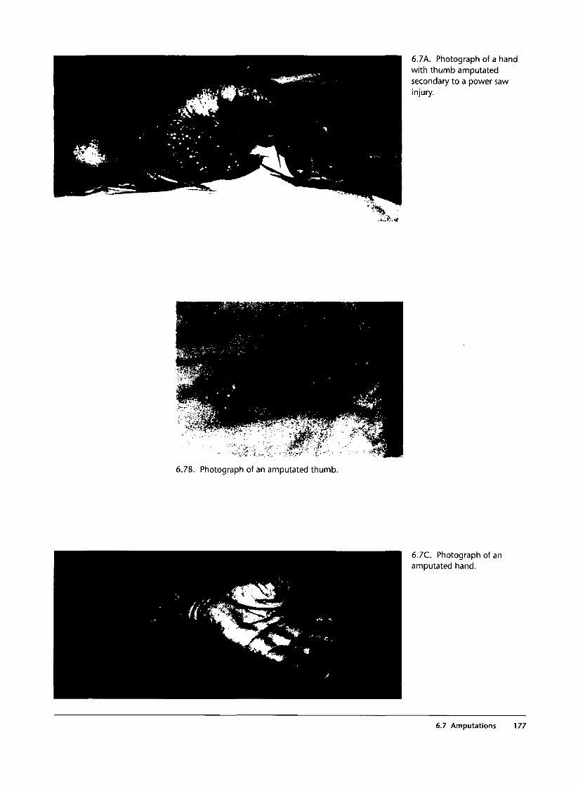
6.7A. Photograph of a handwith thumb amputatedsecondary to a power sawinjury.
6.7%. Photograph of an amputated thumb.
6.7C. Photograph of anamputated hand.
6.7 Amputations 177
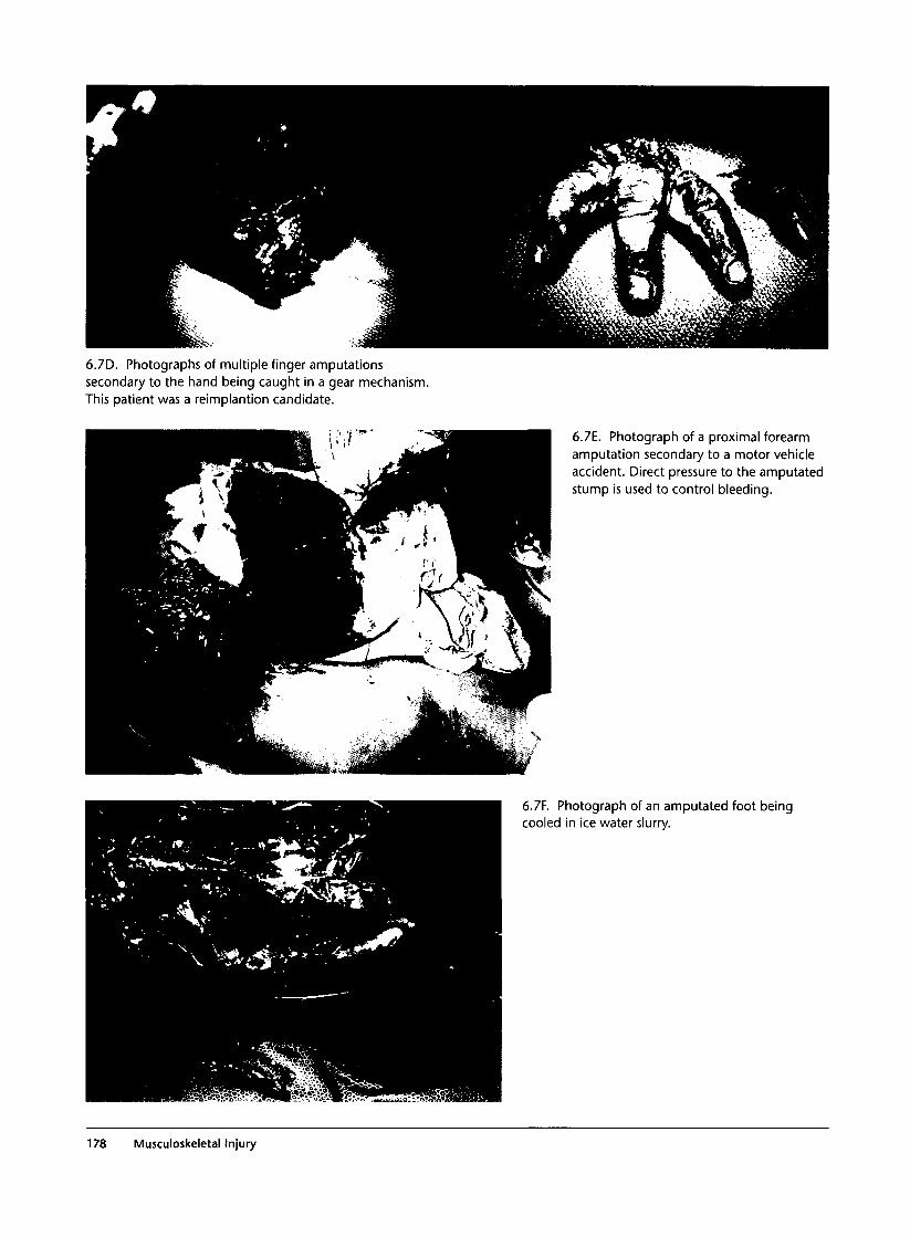
6.7D. Photographs of multiple finger amputationssecondary to the hand being caught in a gear mechanism.This patient was a reimplantion candidate.
6.7E. Photograph of a proximal forearmamputation secondary to a motor vehicleaccident. Direct pressure to the amputatedstump is used to control bleeding.
6.7F. Photograph of an amputated foot beingcooled in ice water slurry.
178 Musculoskeletal Injury
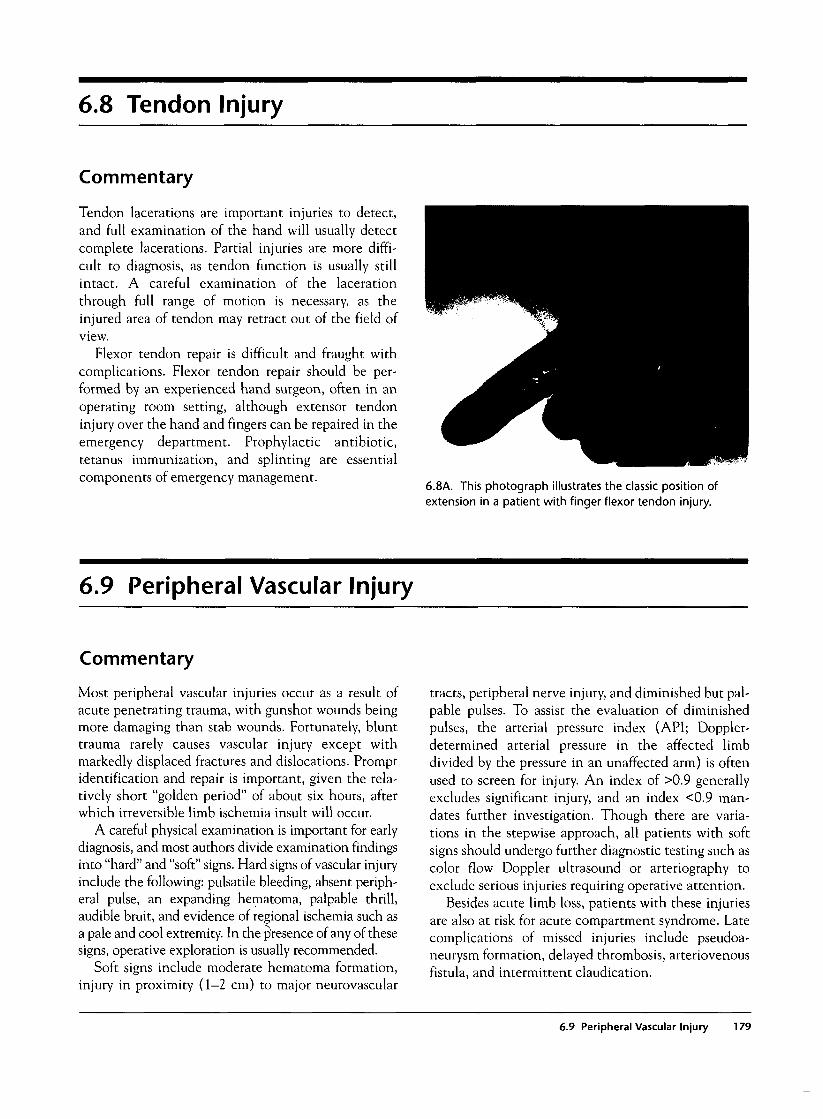
6.8 Tendon Injury
Commentary
Tendon lacerations are important injuries to detect,and full examination of the hand will usually detectcomplete lacerations. Partial injuries are more diffi-cult to diagnosis, as tendon function is usually stillintact. A careful examination of the lacerationthrough full range of motion is necessary, as theinjured area of tendon may retract out of the field ofview.
Flexor tendon repair is difficult and fraught withcomplications. Flexor tendon repair should be per-formed by an experienced hand surgeon, often in anoperating room setting, although extensor tendoninjury over the hand and fingers can be repaired in theemergency department. Prophylactic antibiotic,tetanus immunization, and splinting are essentialcomponents of emergency management.
6.8A. This photograph illustrates the classic position ofextension in a patient with finger flexor tendon injury.
6.9 Peripheral Vascular Injury
Commentary
Most peripheral vascular injuries occur as a result ofacute penetrating trauma, with gunshot wounds beingmore damaging than stab wounds. Fortunately, blunttrauma rarely causes vascular injury except withmarkedly displaced fractures and dislocations. Promptidentification and repair is important, given the rela-tively short "golden period" of about six hours, afterwhich irreversible limb ischemia insult will occur.
A careful physical examination is important for earlydiagnosis, and most authors divide examination findingsinto "hard" and "soft" signs. Hard signs of vascular injuryinclude the following: pulsatile bleeding, absent periplveral pulse, an expanding hematoma, palpable thrill,audible bruit, and evidence of regional ischemia such asa pale and cool extremity. In the presence of any of thesesigns, operative exploration is usually recommended.
Soft signs include moderate hematoma formation,injury in proximity (1-2 cm) to major neurovascular
tracts, peripheral nerve injury, and diminished but pal-pable pulses. To assist the evaluation of diminishedpulses, the arterial pressure index (API; Doppler-determined arterial pressure in the affected limbdivided by the pressure in an unaffected arm) is oftenused to screen for injury. An index of >0.9 generallyexcludes significant injury, and an index <0.9 man-dates further investigation. Though there are varia-tions in the stepwise approach, all patients with softsigns should undergo further diagnostic testing such ascolor flow Doppler ultrasound or arteriography toexclude serious injuries requiring operative attention.
Besides acute limb loss, patients with these injuriesare also at risk for acute compartment syndrome. Latecomplications of missed injuries include pseudoa-neurysm formation, delayed thrombosis, arteriovenousfistula, and intermittent claudication.
6.9 Peripheral Vascular Injury 179
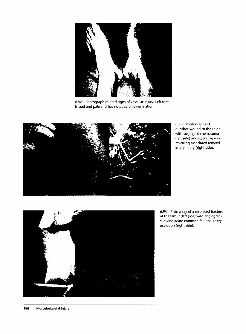
6.9A. Photograph of hard signs of vascular injury: Left footis cool and pale and has no pulse on examination.
6.9B. Photographs ofgunshot wound to the thighwith large groin hematoma(left side) and operative viewrevealing associated femoralartery injury (right side).
6.9C. Plain x-ray of a displaced fractureof the femur (left side) with angiogramshowing acute common femoral arteryocclusion (right side).
180 Musculoskeletal Injury

6.9F. Photograph of operative repair of the femoral arteryinjury in the previous patient.
6.9D. Angiogram of shotgun pellet injury to the knee withpopliteal artery injury.
6.9G. Complicating false aneurysm seen on angiogram.
6.9E. Photograph of a gunshot wound to the knee.
6.9 Peripheral Vascular Injury 181
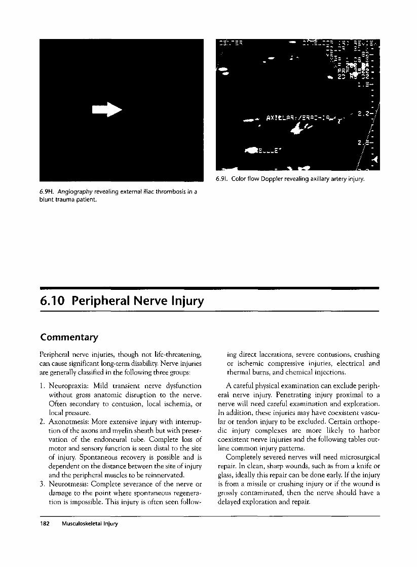
6.91. Color flow Doppler revealing axillary artery injury.
6.9H. Angiography revealing external iliac thrombosis in ablunt trauma patient.
6.10 Peripheral Nerve Injury
Commentary
Peripheral nerve injuries, though not life-threatening,can cause significant long-term disability. Nerve injuriesare generally classified in the following three groups:
1. Neuropraxia: Mild transient nerve dysfunctionwithout gross anatomic disruption to the nerve.Often secondary to contusion, local ischemia, orlocal pressure.
2. Axonotmesis: More extensive injury with interrup-tion of the axons and myelin sheath but with preser-vation of the endoneural tube. Complete loss ofmotor and sensory function is seen distal to the siteof injury. Spontaneous recovery is possible and isdependent on the distance between the site of injuryand the peripheral muscles to be reinnervated.
3. Neurotmesis: Complete severance of the nerve ordamage to the point where spontaneous regenera-tion is impossible. This injury is often seen follow-
ing direct lacerations, severe contusions, crushingor ischemic compressive injuries, electrical andthermal burns, and chemical injections.
A careful physical examination can exclude periph-eral nerve injury. Penetrating injury proximal to anerve will need careful examination and exploration.In addition, these injuries may have coexistent vascu-lar or tendon injury to be excluded. Certain orthope-dic injury complexes are more likely to harborcoexistent nerve injuries and the following tables out-line common injury patterns.
Completely severed nerves will need microsurgicalrepair. In clean, sharp wounds, such as from a knife orglass, ideally this repair can be done early. If the injuryis from a missile or crushing injury or if the wound isgrossly contaminated, then the nerve should have adelayed exploration and repair.
182 Musculoskeletal Injury
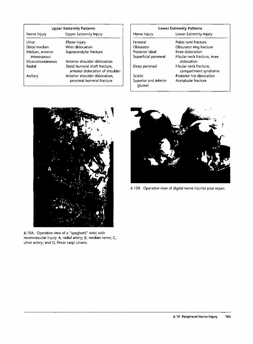
Upper
Nerve Injury
UlnarDistal medianMedian, anterior
interosseousMusculocutaneousRadial
Axillary
Extremity Patterns
Upper Extremity Injury
Elbow injuryWrist dislocationSupracondylar fracture
Anterior shoulder dislocationDistal humeral shaft fracture,
anterior dislocation of shoulderAnterior shoulder dislocation,
proximal humeral fracture
Lower
Nerve Injury
FemoralObturatorPosterior tibialSuperficial peroneal
Deep peroneal
SciaticSuperior and inferior
gluteal
Extremity Patterns
Lower Extremity Injury
Pubic rami fractureObturator ring fractureKnee dislocationFibular neck fracture, knee
dislocationFibular neck fracture,
compartment syndromePosterior hip dislocationAcetabular fracture
6.1 OB. Operative view of digital nerve injuries post repair.
6.1 OA. Operative view of a "spaghetti" wrist withneurovascular injury. A, radial artery; B, median nerve; C,ulnar artery; and D, flexor carpi ulnaris.
6.10 Peripheral Nerve Injury 183
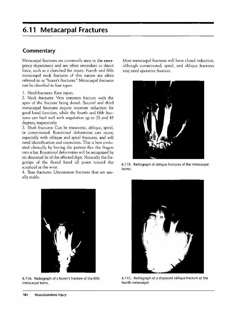
6.11 MetacarpaI Fractures
Commentary
Metacarpal fractures are commonly seen in the emer-gency department and are often secondary to directforce, such as a clenched fist injury. Fourth and fifthmetacarpal neck fractures of this nature are oftenreferred to as "boxer's fractures." Metacarpal fracturescan be classified in four types:
1. Head fractures: Rare injury.2. Neck fractures: Very common fracture with theapex of the fracture being dorsal. Second and thirdmetacarpal fractures require accurate reduction forgood hand function, while the fourth and fifth frac-tures can heal well with angulation up to 35 and 45degrees, respectively.3. Shaft fractures: Can be transverse, oblique, spiral,or comminuted. Rotational deformities can occur,especially with oblique and spiral fractures, and willneed identification and correction. This is best evalu-ated clinically by having the patient flex the fingersinto a fist. Rotational deformities will be recognized byan abnormal lie of the affected digit. Normally the fin-gertips of the flexed hand all point toward thescaphoid at the wrist.4- Base fractures: Uncommon fractures that are usu-ally stable.
Most metacarpal fractures will have closed reduction,although comminuted, spiral, and oblique fracturesmay need operative fixation.
6.11B. Radiograph of oblique fractures of the metacarpalbones.
6.11 A. Radiograph of a boxer's fracture of the fifthmetacarpal bone.
6.11 C. Radiograph of a displaced oblique fracture of thefourth metacarpal.
184 Musculoskeletal Injury

6.11D. Photograph of arotational injury evident onphysical exam. All fingertipsshould normally point towardthe scaphoid. The patient'sright fifth digit has arotational abnormality.
6.12 First Metacarpal Fractures(Bennett's and Rolando's Fractures)
Commentary
First metacarpal fractures are classified as articular orless commonly nonarticular, with the latter beingeither transverse or oblique. Articular fractures arefurther described in two common patterns.
Bennett's fracture is an oblique fracture thatinvolves the carpometacarpal joint. It is usually due todirect axial force on a partially flexed thumb, such asin a fistfight. The characteristic finding is a small tri-angular proximal fragment that remains anatomicallycorrect while the shaft is displaced in a dorsal-radialfashion by the pull of the abductor pollicis longus andthe abductor pollicis. Anatomic reduction is impor-tant, and these fractures are usually treated with per-cutaneous pinning or open reduction.
Rolando's fracture is a comminuted fracture at thebase of the first metacarpal. It is a relatively rare frac-ture often caused by a similar mechanism to a Ben-nett's fracture. The treatment is controversial and mayvary from closed to open reduction, depending on thedegree of comminution and displacement.
6.12A. Radiograph of a Bennett's fracture.
6.12 First Metacarpal Fractures 185
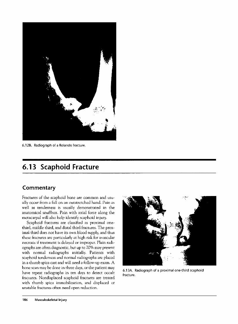
6.12B. Radiograph of a Rolando fracture.
6.13 Scaphoid Fracture
Commentary
Fractures of the scaphoid bone are common and usu-ally occur from a fall on an outstretched hand. Pain aswell as tenderness is usually demonstrated in theanatomical snuffbox. Pain with axial force along themetacarpal will also help identify scaphoid injury.
Scaphoid fractures are classified as proximal one-third, middle third, and distal third fractures. The prox-imal third does not have its own blood supply, and thusthese fractures are particularly at high risk for avascularnecrosis if treatment is delayed or improper. Plain radi-ographs are often diagnostic, but up to 20% may presentwith normal radiographs initially. Patients withscaphoid tenderness and normal radiographs are placedin a thumb spica cast and will need a follow-up exam. Abone scan may be done in three days, or the patient mayhave repeat radiographs in ten days to detect occultfractures. Nondisplaced scaphoid fractures are treatedwith thumb spica immobilization, and displaced orunstable fractures often need open reduction.
6.13A. Radiograph of a proximal one-third scaphoidfracture.
186 Musculoskeletal Injury
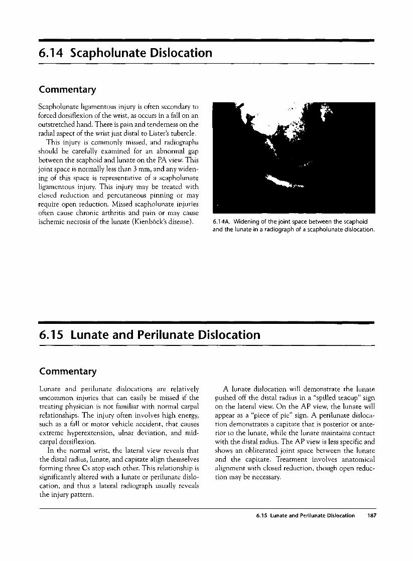
6.14 Scapholunate Dislocation
Commentary
Scapholunate ligamentous injury is often secondary toforced dorsiflexion of the wrist, as occurs in a fall on anoutstretched hand. There is pain and tenderness on theradial aspect of the wrist just distal to Lister's tubercle.
This injury is commonly missed, and radiographsshould be carefully examined for an abnormal gapbetween the scaphoid and lunate on the PA view. Thisjoint space is normally less than 3 mm, and any widen-ing of this space is representative of a scapholunateligamentous injury. This injury may be treated withclosed reduction and percutaneous pinning or mayrequire open reduction. Missed scapholunate injuriesoften cause chronic arthritis and pain or may causeischemic necrosis of the lunate (Kienbock's disease). 6.14A. Widening of the joint space between the scaphoid
and the lunate in a radiograph of a scapholunate dislocation.
6.15 Lunate and Perilunate Dislocation
Commentary
Lunate and perilunate dislocations are relativelyuncommon injuries that can easily be missed if thetreating physician is not familiar with normal carpalrelationships. The injury often involves high energy,such as a fall or motor vehicle accident, that causesextreme hyperextension, ulnar deviation, and mid-carpal dorsiflexion.
In the normal wrist, the lateral view reveals thatthe distal radius, lunate, and capitate align themselvesforming three Cs atop each other. This relationship issignificantly altered with a lunate or perilunate dislo-cation, and thus a lateral radiograph usually revealsthe injury pattern.
A lunate dislocation will demonstrate the lunatepushed off the distal radius in a "spilled teacup" signon the lateral view. On the AP view, the lunate willappear as a "piece of pie" sign. A perilunate disloca-tion demonstrates a capitate that is posterior or ante-rior to the lunate, while the lunate maintains contactwith the distal radius. The AP view is less specific andshows an obliterated joint space between the lunateand the capitate. Treatment involves anatomicalalignment with closed reduction, though open reduc-tion may be necessary.
6.15 Lunate and Perilunate Dislocation 187
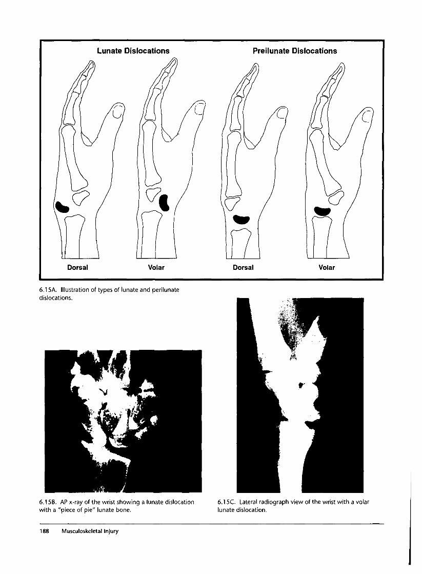
Lunate Dislocations Preilunate Dislocations
Dorsal Volar Dorsal Volar
6.15A. Illustration of types of lunate and perilunatedislocations.
6.15B. AP x-ray of the wrist showing a lunate dislocationwith a "piece of pie" lunate bone.
6.15C. Lateral radiograph view of the wrist with a volarlunate dislocation.
188 Musculoskeletal Injury
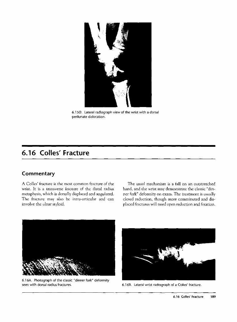
6.15D. Lateral radiograph view of the wrist with a dorsalperilunate dislocation.
6.16 Colles' Fracture
Commentary
A Colles' fracture is the most common fracture of thewrist. It is a transverse fracture of the distal radiusmetaphysis, which is dorsally displaced and angulated.The fracture may also be intra-articular and caninvolve the ulnar styloid.
The usual mechanism is a fall on an outstretchedhand, and the wrist may demonstrate the classic "din-ner fork" deformity on exam. The treatment is usuallyclosed reduction, though more comminuted and dis-placed fractures will need open reduction and fixation.
6.16A. Photograph of the classic "dinner fork" deformityseen with dorsal radius fractures. 6.16B. Lateral wrist radiograph of a Colles' fracture.
6.16 Colles' Fracture 189
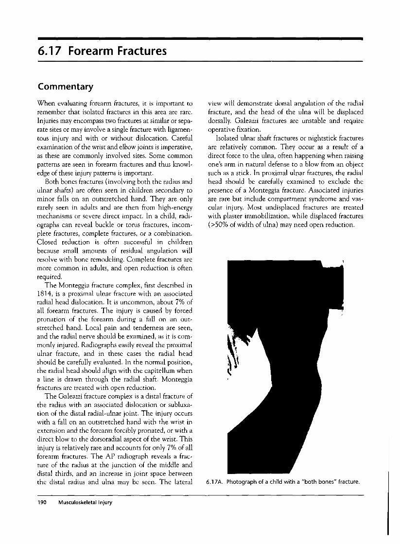
6.17 Forearm Fractures
Commentary
When evaluating forearm fractures, it is important toremember that isolated fractures in this area are rare.Injuries may encompass two fractures at similar or sepa-rate sites or may involve a single fracture with ligamen-tous injury and with or without dislocation. Carefulexamination of the wrist and elbow joints is imperative,as these are commonly involved sites. Some commonpatterns are seen in forearm fractures and thus knowl-edge of these injury patterns is important.
Both bones fractures (involving both the radius andulnar shafts) are often seen in children secondary tominor falls on an outstretched hand. They are onlyrarely seen in adults and are then from high-energymechanisms or severe direct impact. In a child, radi-ographs can reveal buckle or torus fractures, incom-plete fractures, complete fractures, or a combination.Closed reduction is often successful in childrenbecause small amounts of residual angulation willresolve with bone remodeling. Complete fractures aremore common in adults, and open reduction is oftenrequired.
The Monteggia fracture complex, first described in1814, is a proximal ulnar fracture with an associatedradial head dislocation. It is uncommon, about 7% ofall forearm fractures. The injury is caused by forcedpronation of the forearm during a fall on an out-stretched hand. Local pain and tenderness are seen,and the radial nerve should be examined, as it is com-monly injured. Radiographs easily reveal the proximalulnar fracture, and in these cases the radial headshould be carefully evaluated. In the normal position,the radial head should align with the capitellum whena line is drawn through the radial shaft. Monteggiafractures are treated with open reduction.
The Galeazzi fracture complex is a distal fracture ofthe radius with an associated dislocation or subluxa-tion of the distal radial-ulnar joint. The injury occurswith a fall on an outstretched hand with the wrist inextension and the forearm forcibly pronated, or with adirect blow to the dorsoradial aspect of the wrist. Thisinjury is relatively rare and accounts for only 7% of allforearm fractures. The AP radiograph reveals a frac-ture of the radius at the junction of the middle anddistal thirds, and an increase in joint space betweenthe distal radius and ulna may be seen. The lateral
view will demonstrate dorsal angulation of the radialfracture, and the head of the ulna will be displaceddorsally. Galeazzi fractures are unstable and requireoperative fixation.
Isolated ulnar shaft fractures or nightstick fracturesare relatively common. They occur as a result of adirect force to the ulna, often happening when raisingone's arm in natural defense to a blow from an objectsuch as a stick. In proximal ulnar fractures, the radialhead should be carefully examined to exclude thepresence of a Monteggia fracture. Associated injuriesare rare but include compartment syndrome and vas-cular injury. Most undisplaced fractures are treatedwith plaster immobilization, while displaced fractures(>50% of width of ulna) may need open reduction.
6.17A. Photograph of a child with a "both bones" fracture.
190 Musculoskeletal Injury
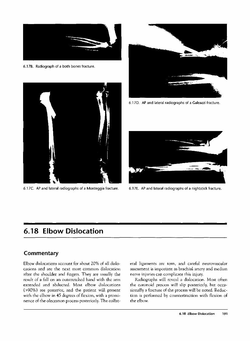
6.17B. Radiograph of a both bones fracture.
6.17D. AP and lateral radiographs of a Galeazzi fracture.
6.17C. AP and lateral radiographs of a Monteggia fracture. 6.17E. AP and lateral radiographs of a nightstick fracture.
6.18 Elbow Dislocation
Commentary
Elbow dislocations account for about 20% of all dislo-cations and are the next most common dislocationafter the shoulder and fingers. They are usually theresult of a fall on an outstretched hand with the armextended and abducted. Most elbow dislocations(>90%) are posterior, and the patient will presentwith the elbow in 45 degrees of flexion, with a promi-nence of the olecranon process posteriorly. The collat-
eral ligaments are torn, and careful neurovascularassessment is important as brachial artery and mediannerve injuries can complicate this injury.
Radiographs will reveal a dislocation. Most oftenthe coronoid process will slip posteriorly, but occa-sionally a fracture of the process will be noted. Reduc-tion is performed by countertraction with flexion ofthe elbow.
6.18 Elbow Dislocation 191
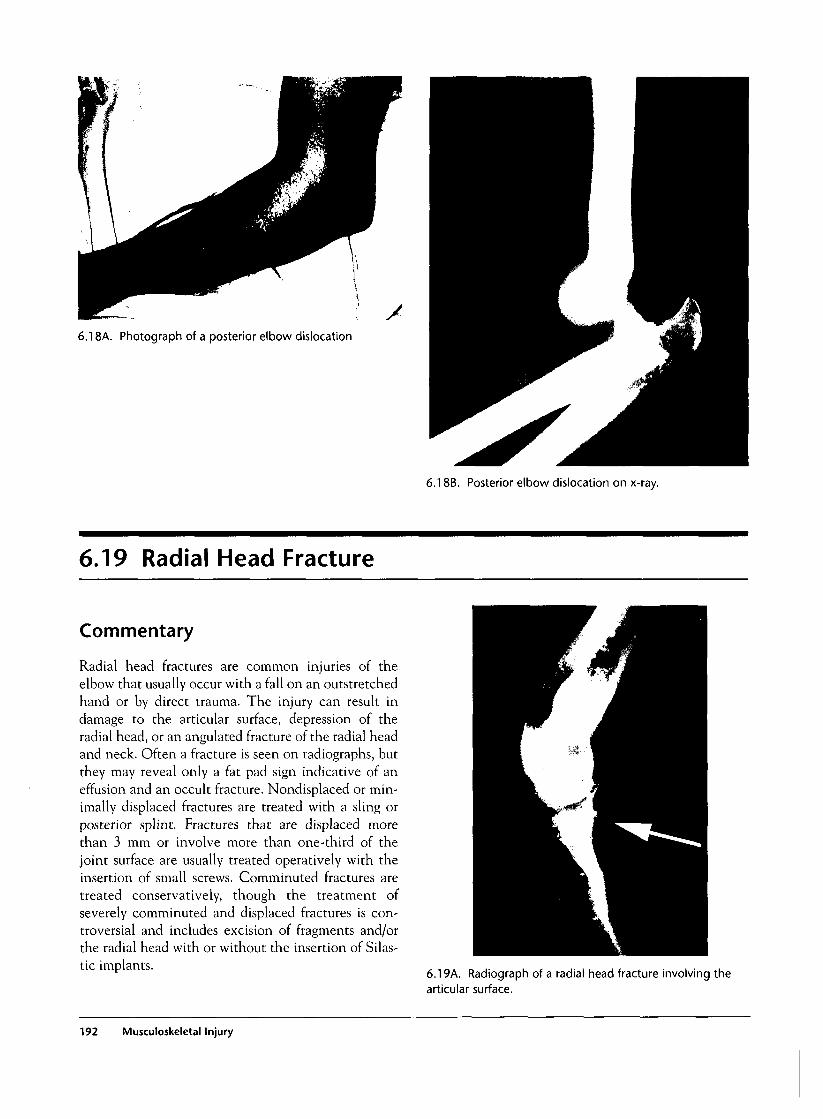
6.18A. Photograph of a posterior elbow dislocation
6.18B. Posterior elbow dislocation on x-ray.
6.19 Radial Head Fracture
Commentary
Radial head fractures are common injuries of theelbow that usually occur with a fall on an outstretchedhand or by direct trauma. The injury can result indamage to the articular surface, depression of theradial head, or an angulated fracture of the radial headand neck. Often a fracture is seen on radiographs, butthey may reveal only a fat pad sign indicative of aneffusion and an occult fracture. Nondisplaced or min-imally displaced fractures are treated with a sling orposterior splint. Fractures that are displaced morethan 3 mm or involve more than one-third of thejoint surface are usually treated operatively with theinsertion of small screws. Comminuted fractures aretreated conservatively, though the treatment ofseverely comminuted and displaced fractures is con-troversial and includes excision of fragments and/orthe radial head with or without the insertion of Silas-tic implants.
6.19A. Radiograph of a radial head fracture involving thearticular surface.
192 Musculoskeletal Injury
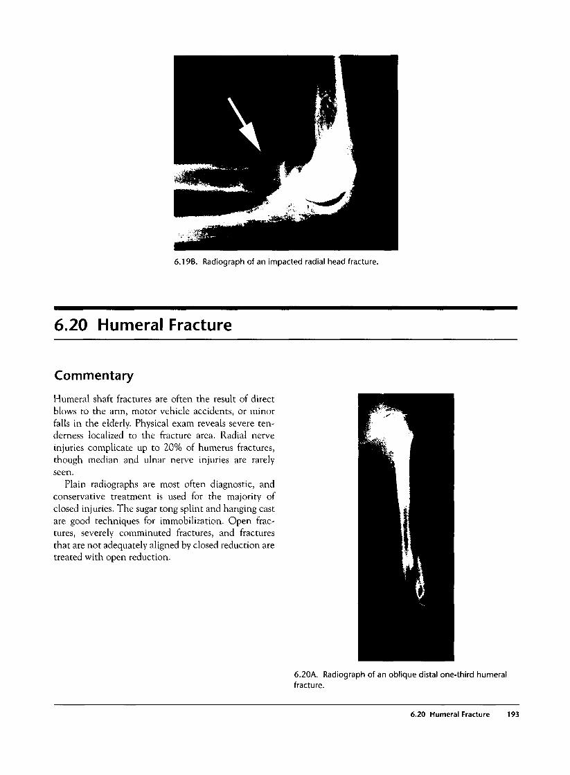
6.19B. Radiograph of an impacted radial head fracture.
6.20 Humeral Fracture
Commentary
Humeral shaft fractures are often the result of directblows to the arm, motor vehicle accidents, or minorfalls in the elderly. Physical exam reveals severe ten-derness localized to the fracture area. Radial nerveinjuries complicate up to 20% of humerus fractures,though median and ulnar nerve injuries are rarelyseen.
Plain radiographs are most often diagnostic, andconservative treatment is used for the majority ofclosed injuries. The sugar tong splint and hanging castare good techniques for immobilization. Open frac-tures, severely comminuted fractures, and fracturesthat are not adequately aligned by closed reduction aretreated with open reduction.
6.20A. Radiograph of an oblique distal one-third humeralfracture.
6.20 Humeral Fracture 193
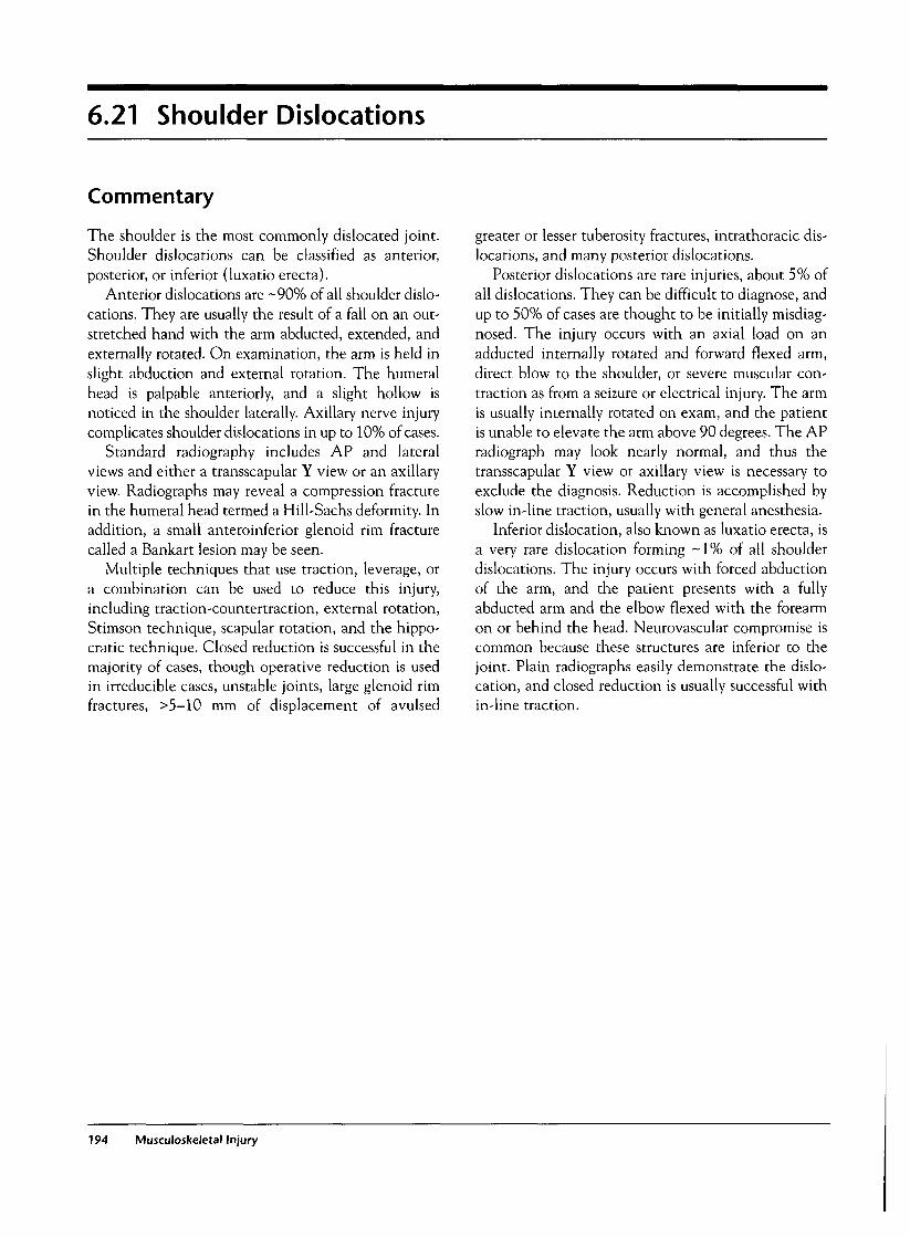
6.21 Shoulder Dislocations
Commentary
The shoulder is the most commonly dislocated joint.Shoulder dislocations can be classified as anterior,posterior, or inferior (luxatio erecta).
Anterior dislocations are -90% of all shoulder dislo-cations. They are usually the result of a fall on an out-stretched hand with the arm abducted, extended, andexternally rotated. On examination, the arm is held inslight abduction and external rotation. The humeralhead is palpable anteriorly, and a slight hollow isnoticed in the shoulder laterally. Axillary nerve injurycomplicates shoulder dislocations in up to 10% of cases.
Standard radiography includes AP and lateralviews and either a transscapular Y view or an axillaryview. Radiographs may reveal a compression fracturein the humeral head termed a Hill-Sachs deformity. Inaddition, a small anteroinferior glenoid rim fracturecalled a Bankart lesion may be seen.
Multiple techniques that use traction, leverage, ora combination can be used to reduce this injury,including traction-countertraction, external rotation,Stimson technique, scapular rotation, and the hippo-cratic technique. Closed reduction is successful in themajority of cases, though operative reduction is usedin irreducible cases, unstable joints, large glenoid rimfractures, >5—10 mm of displacement of avulsed
greater or lesser tuberosity fractures, intrathoracic dis-locations, and many posterior dislocations.
Posterior dislocations are rare injuries, about 5% ofall dislocations. They can be difficult to diagnose, andup to 50% of cases are thought to be initially misdiag-nosed. The injury occurs with an axial load on anadducted internally rotated and forward flexed arm,direct blow to the shoulder, or severe muscular con-traction as from a seizure or electrical injury. The armis usually internally rotated on exam, and the patientis unable to elevate the arm above 90 degrees. The APradiograph may look nearly normal, and thus thetransscapular Y view or axillary view is necessary toexclude the diagnosis. Reduction is accomplished byslow in-line traction, usually with general anesthesia.
Inferior dislocation, also known as luxatio erecta, isa very rare dislocation forming - 1 % of all shoulderdislocations. The injury occurs with forced abductionof the arm, and the patient presents with a fullyabducted arm and the elbow flexed with the forearmon or behind the head. Neurovascular compromise iscommon because these structures are inferior to thejoint. Plain radiographs easily demonstrate the dislo-cation, and closed reduction is usually successful within-line traction.
194 Musculoskeletal Injury
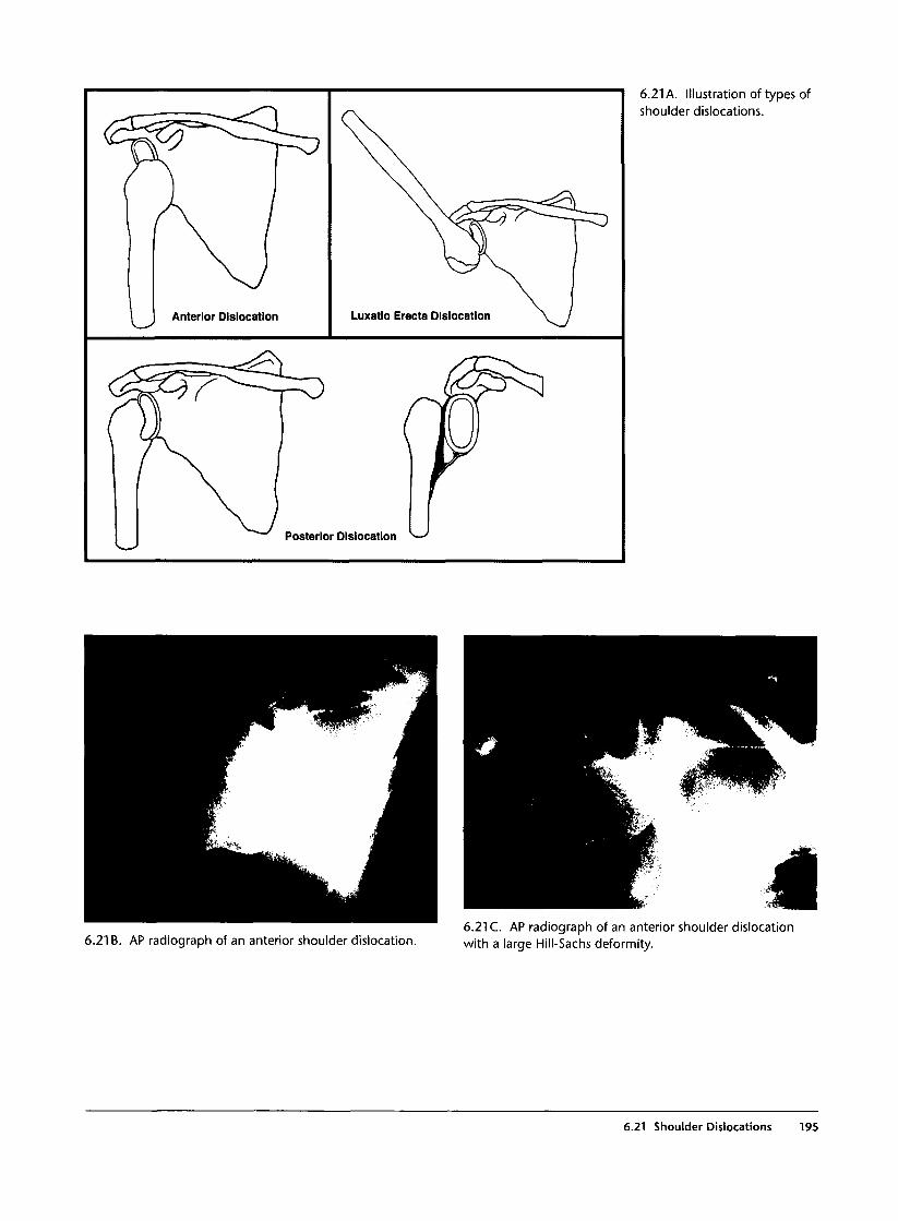
Posterior Dislocation
6.21 A. Illustration of types ofshoulder dislocations.
6.21 B. AP radiograph of an anterior shoulder dislocation.6.21 C. AP radiograph of an anterior shoulder dislocationwith a large Hill-Sachs deformity.
6.21 Shoulder Dislocations 195
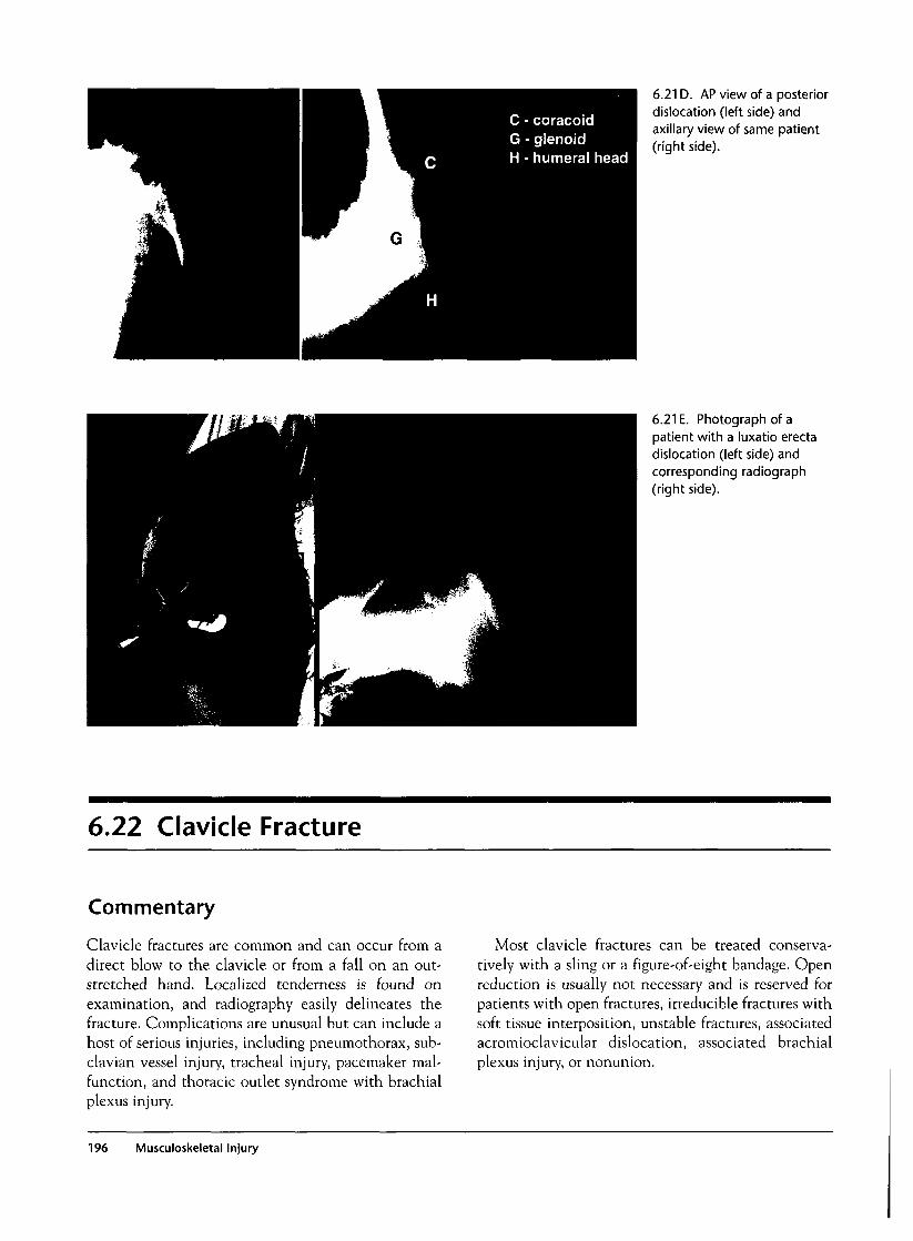
6.21 D. AP view of a posteriordislocation (left side) andaxillary view of same patient(right side).
6.21 E. Photograph of apatient with a luxatio erectadislocation (left side) andcorresponding radiograph(right side).
6.22 Clavicle Fracture
Commentary
Clavicle fractures are common and can occur from adirect blow to the clavicle or from a fall on an out-stretched hand. Localized tenderness is found onexamination, and radiography easily delineates thefracture. Complications are unusual but can include ahost of serious injuries, including pneumothorax, sub-clavian vessel injury, tracheal injury, pacemaker mal-function, and thoracic outlet syndrome with brachialplexus injury.
Most clavicle fractures can be treated conserva-tively with a sling or a figure-of-eight bandage. Openreduction is usually not necessary and is reserved forpatients with open fractures, irreducible fractures withsoft tissue interposition, unstable fractures, associatedacromioclavicular dislocation, associated brachialplexus injury, or nonunion.
196 Musculoskeletal Injury
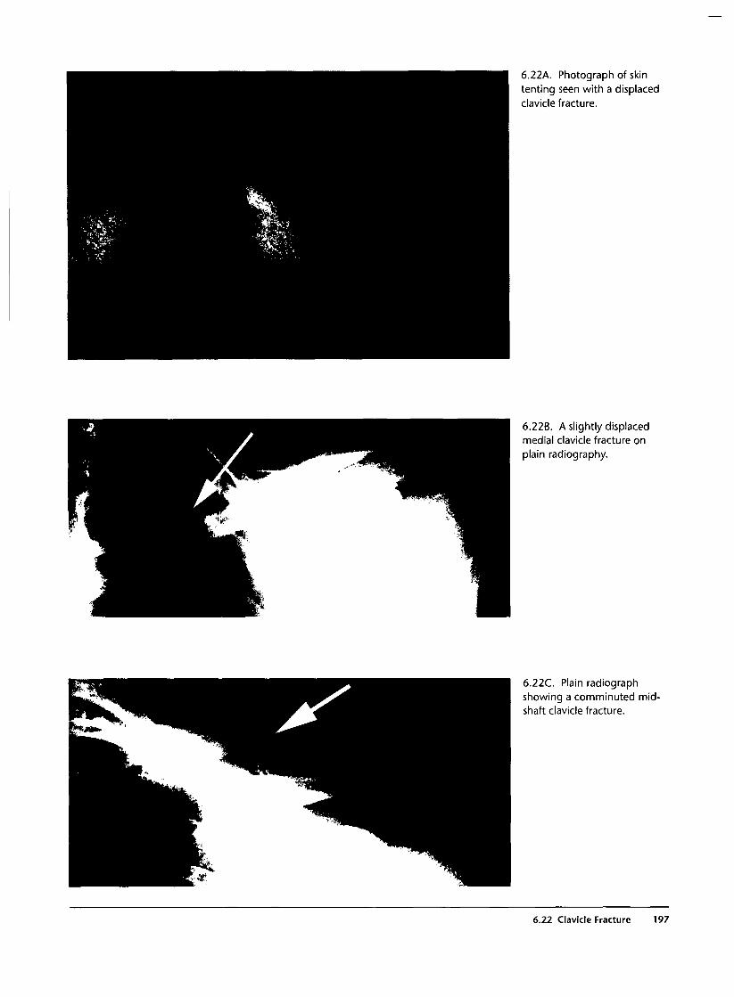
6.22A. Photograph of skintenting seen with a displacedclavicle fracture.
6.22B. A slightly displacedmedial clavicle fracture onplain radiography.
6.22C. Plain radiographshowing a comminuted mid-shaft clavicle fracture.
6.22 Clavicle Fracture 197
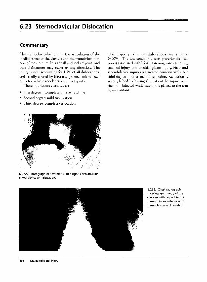
6.23 Sternoclavicular Dislocation
Commentary
The sternoclavicular joint is the articulation of themedial aspect of the clavicle and the manubrium por-tion of the sternum. It is a "ball and socket" joint, andthus dislocations may occur in any direction. Theinjury is rare, accounting for 1.5% of all dislocations,and usually caused by high-energy mechanisms suchas motor vehicle accidents or contact sports.
These injuries are classified as:
• First degree: incomplete injury/stretching
• Second degree: mild subluxation
• Third degree: complete dislocation
The majority of these dislocations are anterior(-90%). The less commonly seen posterior disloca-tion is associated with life-threatening vascular injury,tracheal injury, and brachial plexus injury. First- andsecond-degree injuries are treated conservatively, butthird-degree injuries require reduction. Reduction isaccomplished by having the patient lie supine withthe arm abducted while traction is placed to the armby an assistant.
6.23A. Photograph of a woman with a right-sided anteriorsternoclavicular dislocation.
6.23B. Chest radiographshowing asymmetry of theclavicles with respect to thesternum in an anterior rightsternoclavicular dislocation.
198 Musculoskeletal Injury
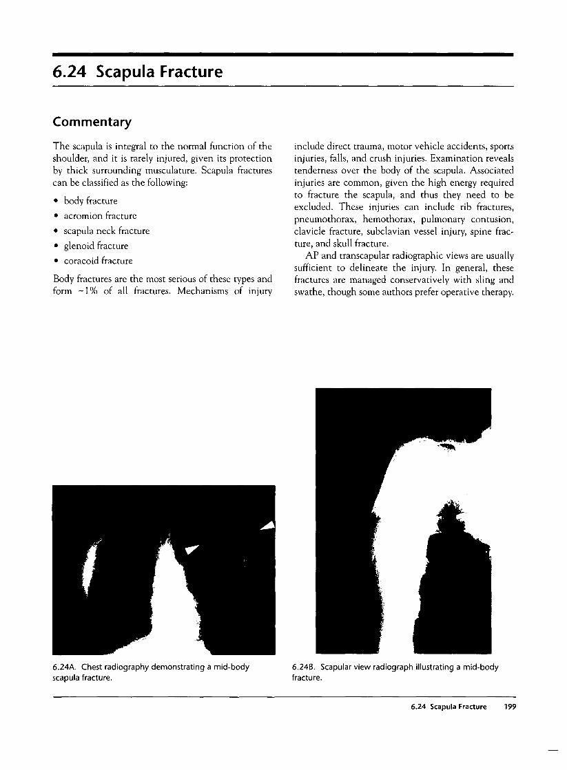
6.24 Scapula Fracture
Commentary
The scapula is integral to the normal function of theshoulder, and it is rarely injured, given its protectionby thick surrounding musculature. Scapula fracturescan be classified as the following:
• body fracture
• acromion fracture
• scapula neck fracture
• glenoid fracture
• coracoid fracture
Body fractures are the most serious of these types andform - 1 % of all fractures. Mechanisms of injury
include direct trauma, motor vehicle accidents, sportsinjuries, falls, and crush injuries. Examination revealstenderness over the body of the scapula. Associatedinjuries are common, given the high energy requiredto fracture the scapula, and thus they need to beexcluded. These injuries can include rib fractures,pneumothorax, hemothorax, pulmonary contusion,clavicle fracture, subclavian vessel injury, spine frac-ture, and skull fracture.
AP and transcapular radiographic views are usuallysufficient to delineate the injury. In general, thesefractures are managed conservatively with sling andswathe, though some authors prefer operative therapy.
6.24A. Chest radiography demonstrating a mid-bodyscapula fracture.
6.24B. Scapular view radiograph illustrating a mid-bodyfracture.
6.24 Scapula Fracture 199

6.25 Pelvic Fractures
Commentary
Pelvic fractures account for significant morbidity andmortality, and their emergency management is challeng-ing. Though minor pubic rami fractures are often seen inthe elderly with ground-level falls, major pelvic fracturesusually require significant force, and thus motor vehicleaccidents, falls, and auto-versus-pedestrian accidents arethe leading causes. Overall mortality is 5-15%, andother extrapelvic injuries are commonly seen.
The pelvic bones form a sturdy ring, but when abreak in this ring occurs, especially if at two places,complications can be significant, with the most seri-ous being acute hemorrhage within the retroperi-toneal space. In the male, a urethral transection isidentified clinically by the presence of blood at theurethral meatus, a scrotal hematoma, or a high-ridingprostate. The presence of any of these signs mandatesa retrograde urethrogram to exclude urethral injurybefore placement of a Foley catheter. In addition, thebladder, rectum, vagina, uterus, and sacral nerve rootsare all at high risk of injury from pelvic bone fractures.
Persistent retroperitoneal bleeding can be extremelydifficult to manage, accounting for 65% of deaths from
pelvic fractures, and therefore prompt identification ofthe bleeding source in the pelvic fracture victim inshock is critical. These seriously injured patients arealso at high risk for intraperitoneal, intrathoracic, andlong bone fractures that could account for extensiveblood loss. Bedside ultrasound, computed tomography,and diagnostic peritoneal lavage are all useful adjunctsin the proper identification of the bleeding source.Proper disposition is imperative, as laparotomy has alimited role in hemorrhage control in the modernmanagement of pelvic fractures and associatedretroperitoneal bleeding and may worsen mortality. Inaddition to aggressive blood resuscitation, tightlywrapped sheets, special pelvic binders, or a MAST suitcan be helpful in stabilizing the pelvis. Emergencyexternal fixation is most helpful in stabilizing anteriorarch fractures but less helpful for bleeding associatedwith posterior pelvic injuries. Serious retroperitonealbleeding is best treated by angiography; selectiveembolization of small pelvic arteries may be lifesavingin the critical patient.
ilium fracture with extensionto the sacroiliac joint
sacroillac jointfracture-dislocation
iliac wing fracture
pubic symphysisseparation
ischial fractures
Letournel Classification of Pelvic Fractures
6.25A. Illustration of Letournel'sanatomic classification of pelvicfractures.
200 Musculoskeletal Injury

6.25B. Pelvic x-ray showing bilateral pubic rami fractures.6.25D. Pelvic x-ray showing unstable Malgaigne fracturewith bilateral pubic rami fractures and left sacroiliac jointdisruption.
6.25C. Pelvic x-ray showing unstable Malgaigne fracturewith widening of symphysis pubis, right acetabular fracture,left rami fracture, and left sacroiliac joint disruption.
6.25E. Photograph of blood at urethral meatus withsuprapubic cystostomy.
6.25F. Retrograde urethrogram with gross dye extravasationindicating urethral injury.
6.25 Pelvic Fractures 201
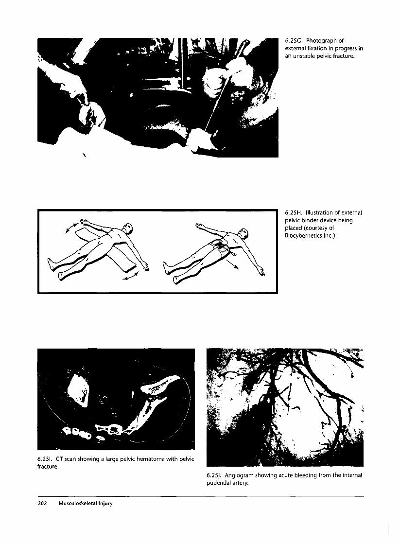
6.25C. Photograph ofexternal fixation in progress inan unstable pelvic fracture.
6.25H. Illustration of externalpelvic binder device beingplaced (courtesy ofBiocybernetics Inc.).
6.251. CT scan showing a large pelvic hematoma with pelvicfracture.
6.25). Angiogram showing acute bleeding from the internalpudendal artery.
202 Musculoskeletal Injury

6.25K. Photograph of an open pelvic fracturefrom a motor vehicle accident.
6.26 Hip Dislocation
Commentary
Hip dislocations occur from high-energy trauma suchas motor vehicle accidents and are true orthopedicemergencies because of the risk of ischemic necrosis tothe femoral head. Because many patients present withsevere associated trauma, other injuries may take pri'ority, but the hip dislocation should be addressedbefore other orthopedic concerns. Ninety percent ofall hip dislocations are posterior, with only about 10%being anterior dislocations. Rarely seen are centraldislocations in which there is a concomitant fracturein the floor of the acetabulum. Given the energyinvolved, a femoral head fracture may also be present,and radiographs should provide good views of the
femoral head and neck. In addition, the sciatic nerveis at risk with posterior dislocations, whereas thefemoral nerve and vessels may be damaged with ante-rior dislocations, and thus the affected lower extrem-ity requires a careful neurovascular examination.
Early closed reduction is paramount, as the femoralhead has a limited blood supply. The average rate ofavascular necrosis is 20%, and the risk rises to 50% inreductions performed twelve hours or more after theinjury. Reduction can usually be accomplished usingthe Allis or Stimson techniques (see 6.26D and E)with appropriate deep sedation, though some patientswill require operative reduction.
6.26 Hip Dislocation 203
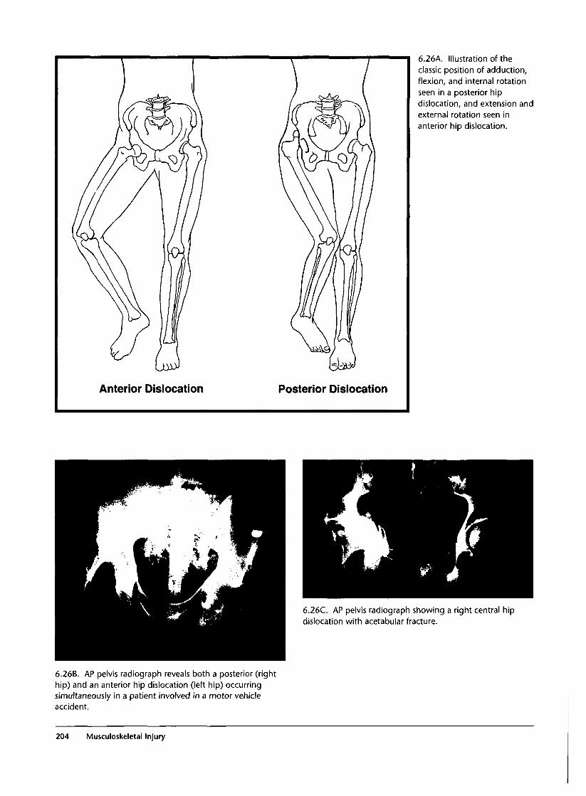
Anterior Dislocation Posterior Dislocation
6.26A. Illustration of theclassic position of adduction,flexion, and internal rotationseen in a posterior hipdislocation, and extension andexternal rotation seen inanterior hip dislocation.
6.26C. AP pelvis radiograph showing a right central hipdislocation with acetabular fracture.
6.26B. AP pelvis radiograph reveals both a posterior (righthip) and an anterior hip dislocation (left hip) occurringsimultaneously in a patient involved in a motor vehicleaccident.
204 Musculoskeletal Injury

6.26D. Illustration of Allisclosed reduction techniqueused for posterior dislocations.
Stimson Technique
6.26E. Illustration of Stimsonclosed reduction techniqueused for posterior dislocations.
6.26 Hip Dislocation 205
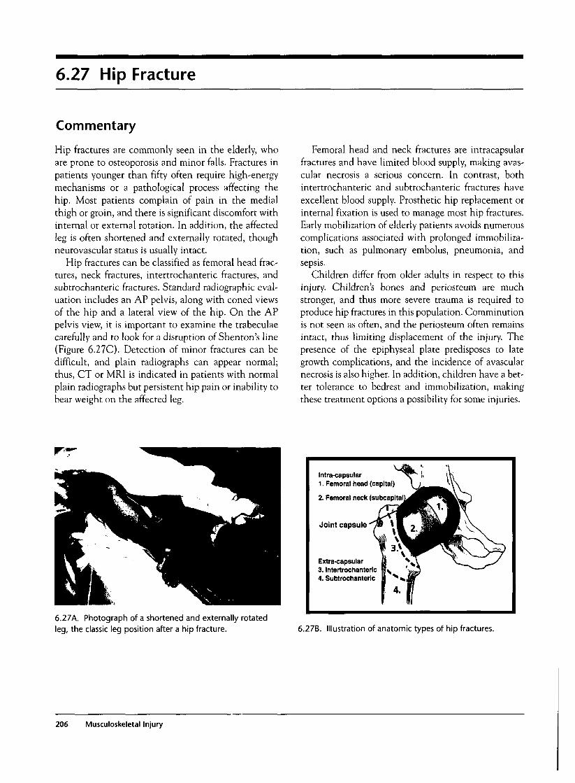
6.27 Hip Fracture
Commentary
Hip fractures are commonly seen in the elderly, whoare prone to osteoporosis and minor falls. Fractures inpatients younger than fifty often require high-energymechanisms or a pathological process affecting thehip. Most patients complain of pain in the medialthigh or groin, and there is significant discomfort withinternal or external rotation. In addition, the affectedleg is often shortened and externally rotated, thoughneurovascular status is usually intact.
Hip fractures can be classified as femoral head frac-tures, neck fractures, intertrochanteric fractures, andsubtrochanteric fractures. Standard radiographic eval-uation includes an AP pelvis, along with coned viewsof the hip and a lateral view of the hip. On the APpelvis view, it is important to examine the trabeculaecarefully and to look for a disruption of Shenton's line(Figure 6.27C). Detection of minor fractures can bedifficult, and plain radiographs can appear normal;thus, CT or MRI is indicated in patients with normalplain radiographs but persistent hip pain or inability tobear weight on the affected leg.
Femoral head and neck fractures are intracapsularfractures and have limited blood supply, making avas-cular necrosis a serious concern. In contrast, bothintertrochanteric and subtrochanteric fractures haveexcellent blood supply. Prosthetic hip replacement orinternal fixation is used to manage most hip fractures.Early mobilization of elderly patients avoids numerouscomplications associated with prolonged immobiliza-tion, such as pulmonary embolus, pneumonia, andsepsis.
Children differ from older adults in respect to thisinjury. Children's bones and periosteum are muchstronger, and thus more severe trauma is required toproduce hip fractures in this population. Comminutionis not seen as often, and the periosteum often remainsintact, thus limiting displacement of the injury. Thepresence of the epiphyseal plate predisposes to lategrowth complications, and the incidence of avascularnecrosis is also higher. In addition, children have a bet-ter tolerance to bedrest and immobilization, makingthese treatment options a possibility for some injuries.
6.27A. Photograph of a shortened and externally rotatedleg, the classic leg position after a hip fracture.
intra-capsular ^ r ^ \>1. Femoral head (capital) \ i
2. Femoral neck (subcapitaN^fl
Jkrw\Joint capsule ̂ M * K 7 I
Extra-capsular (1 ^ s \3. Intertrochanteric iff^ ' v ^4. Subtrochanteric K N ^ J j f
1 4. 1/
6.27B. Illustration of anatomic types of hip fractures.
206 Musculoskeletal Injury

6.27C. Illustration of Shenton's line.
6.27E. Hip radiograph of a intertrochanteric hip fracture.
6.27F. Hip radiograph of a subtrochanteric hip fracture.
6.27D. Hip radiograph of a femoral neck fracture.
6.27 Hip Fracture 207
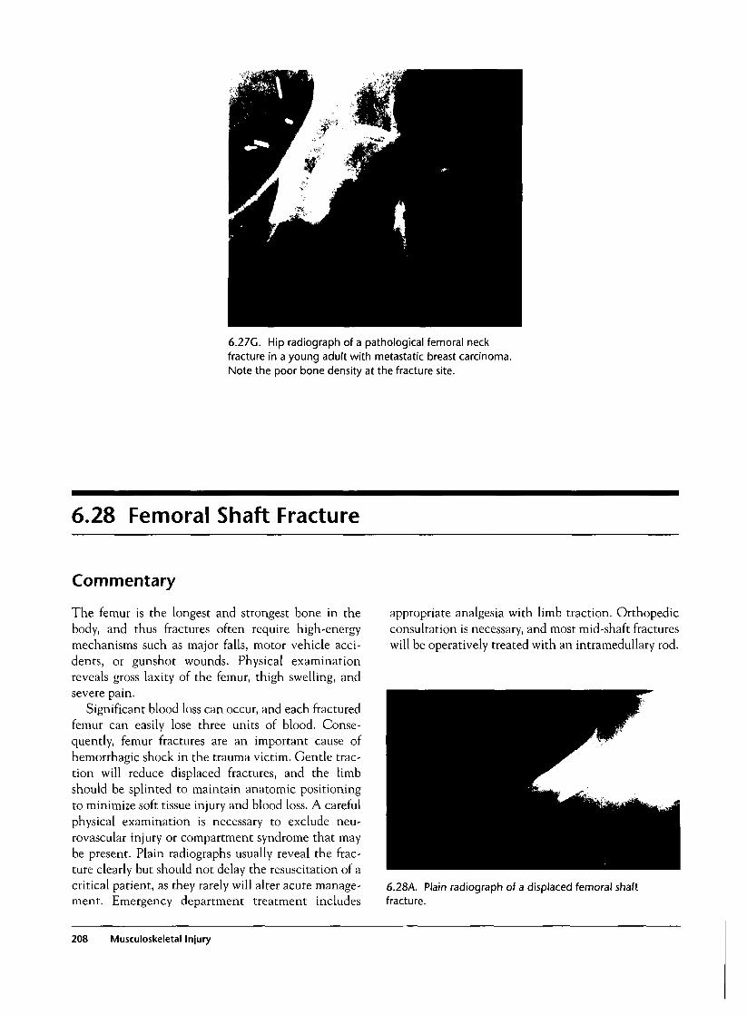
6.27G. Hip radiograph of a pathological femoral neckfracture in a young adult with metastatic breast carcinoma.Note the poor bone density at the fracture site.
6.28 Femoral Shaft Fracture
Commentary
The femur is the longest and strongest bone in thebody, and thus fractures often require high-energymechanisms such as major falls, motor vehicle acci-dents, or gunshot wounds. Physical examinationreveals gross laxity of the femur, thigh swelling, andsevere pain.
Significant blood loss can occur, and each fracturedfemur can easily lose three units of blood. Conse-quently, femur fractures are an important cause ofhemorrhagic shock in the trauma victim. Gentle trac-tion will reduce displaced fractures, and the limbshould be splinted to maintain anatomic positioningto minimize soft tissue injury and blood loss. A carefulphysical examination is necessary to exclude neu-rovascular injury or compartment syndrome that maybe present. Plain radiographs usually reveal the frac-ture clearly but should not delay the resuscitation of acritical patient, as they rarely will alter acute manage-ment. Emergency department treatment includes
appropriate analgesia with limb traction. Orthopedicconsultation is necessary, and most mid-shaft fractureswill be operatively treated with an intramedullary rod.
6.28A. Plain radiograph of a displaced femoral shaftfracture.
208 Musculoskeletal Injury
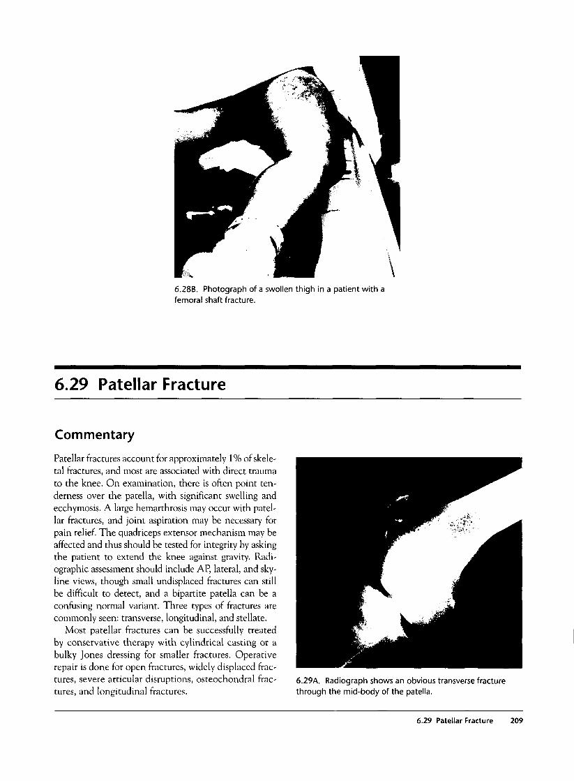
\
6.28B. Photograph of a swollen thigh in a patient with afemoral shaft fracture.
6.29 Patellar Fracture
Commentary
Patellar fractures account for approximately 1% of skele-tal fractures, and most are associated with direct traumato the knee. On examination, there is often point ten-derness over the patella, with significant swelling andecchymosis. A large hemarthrosis may occur with patel-lar fractures, and joint aspiration may be necessary forpain relief. The quadriceps extensor mechanism may beaffected and thus should be tested for integrity by askingthe patient to extend the knee against gravity. Radi-ographic assessment should include AP, lateral, and sky-line views, though small undisplaced fractures can stillbe difficult to detect, and a bipartite patella can be aconfusing normal variant. Three types of fractures arecommonly seen: transverse, longitudinal, and stellate.
Most patellar fractures can be successfully treatedby conservative therapy with cylindrical casting or abulky Jones dressing for smaller fractures. Operativerepair is done for open fractures, widely displaced frac-tures, severe articular disruptions, osteochondral frac-tures, and longitudinal fractures.
6.29A. Radiograph shows an obvious transverse fracturethrough the mid-body of the patella.
6.29 Patellar Fracture 209

6.30 Tibial Plateau Fracture
Commentary
Tibial plateau fractures are often the result of directshear forces. With most car bumpers being twentyinches high, the tibial plateau is at high risk in auto-versus-pedestrian accidents. Swelling, hemarthrosis,point tenderness, and pain with any motion of theknee make weight bearing impossible. Ligamentousinjury may coexist and thus should be sought out onthe clinical examination. Standard AP and lateralradiographs with oblique views will make the diagno-sis in most cases, but occasionally CT scanning may beneeded to further define the injury. The medialplateau is stronger, and thus fractures usually involvethe lateral plateau. Generally the fractures are of twovarieties: split and/or depressed. Split fractures tend tooccur in younger patients with stronger bone, anddepressed fractures more often occur in the elderlypatient with osteoporosis.
As tibial plateau fractures involve the articular sur-face of a major joint, they are at high risk for peristentpain and arthritis and thus should be managed by anorthopedic surgeon. In the past, these fractures havegenerally been casted, but because of the residual stiff-ness often associated with casting, surgical interven-tion and early mobilization are increasingly being used.
6.30B. This AP view of the knee reveals a lateral split plateaufracture.
Split Depressed Split & Depressed
Tibial Plateau Fractures
6.30A. Illustration of types ofplateau fractures.
210 Musculoskeletal Injury
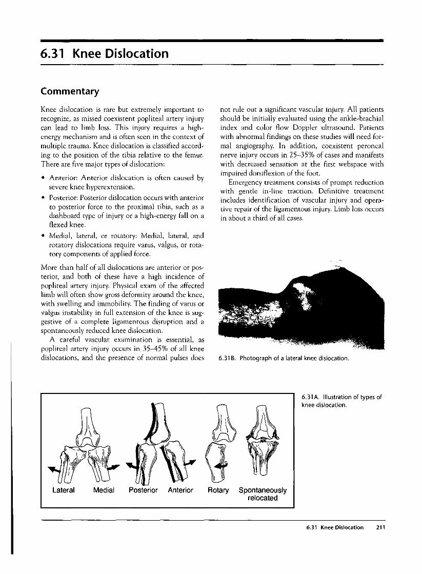
6.31 Knee Dislocation
Commentary
Knee dislocation is rare but extremely important torecognize, as missed coexistent popliteal artery injurycan lead to limb loss. This injury requires a high-energy mechanism and is often seen in the context ofmultiple trauma. Knee dislocation is classified accord-ing to the position of the tibia relative to the femur.There are five major types of dislocation:
• Anterior: Anterior dislocation is often caused bysevere knee hyperextension.
• Posterior: Posterior dislocation occurs with anteriorto posterior force to the proximal tibia, such as adashboard type of injury or a high-energy fall on aflexed knee.
• Medial, lateral, or rotatory: Medial, lateral, androtatory dislocations require varus, valgus, or rota-tory components of applied force.
More than half of all dislocations are anterior or pos-terior, and both of these have a high incidence ofpopliteal artery injury. Physical exam of the affectedlimb will often show gross deformity around the knee,with swelling and immobility. The finding of varus orvalgus instability in full extension of the knee is sug-gestive of a complete ligamentous disruption and aspontaneously reduced knee dislocation.
A careful vascular examination is essential, aspopliteal artery injury occurs in 35-45% of all kneedislocations, and the presence of normal pulses does
not rule out a significant vascular injury. All patientsshould be initially evaluated using the ankle-brachialindex and color flow Doppler ultrasound. Patientswith abnormal findings on these studies will need for-mal angiography. In addition, coexistent peronealnerve injury occurs in 25-35% of cases and manifestswith decreased sensation at the first webspace withimpaired dorsiflexion of the foot.
Emergency treatment consists of prompt reductionwith gentle in-line traction. Definitive treatmentincludes identification of vascular injury and opera-tive repair of the ligamentous injury. Limb loss occursin about a third of all cases.
6.31 B. Photograph of a lateral knee dislocation.
Lateral Medial Posterior Anterior Rotary Spontaneouslyrelocated
6.31 A. Illustration of types ofknee dislocation.
6.31 Knee Dislocation 211
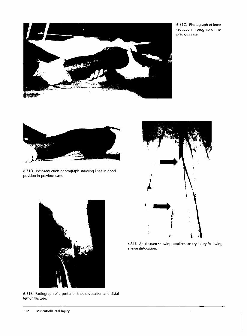
6.31 C. Photograph of kneereduction in progress of theprevious case.
6.31 D. Post-reduction photograph showing knee in goodposition in previous case.
6.31 F. Angiogram showing popliteal artery injury followinga knee dislocation.
6.31 E. Radiograph of a posterior knee dislocation and distalfemur fracture.
212 Musculoskeletal Injury
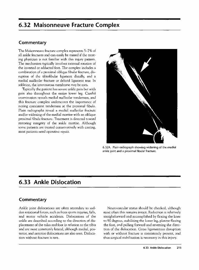
6.32 Maisonneuve Fracture Complex
Commentary
The Maisonneuve fracture complex represents 5-7% ofall ankle fractures and can easily be missed if the treat-ing physician is not familiar with this injury pattern.The mechanism typically involves external rotation ofthe inverted or adducted foot. The complex includes acombination of a proximal oblique fibular fracture, dis-ruption of the tibiofibular ligament distally, and amedial malleolar fracture or deltoid ligament tear. Inaddition, the interosseous membrane may be torn.
Typically the patient has severe ankle pain but withpain also throughout the entire lower leg. Carefulexamination reveals medial malleolar tenderness, andthis fracture complex underscores the importance ofnoting coexistent tenderness at the proximal fibula.Plain radiographs reveal a medial malleolar fractureand/or widening of the medial mortise with an obliqueproximal fibula fracture. Treatment is directed towardrestoring integrity of the ankle mortise. Althoughsome patients are treated conservatively with casting,most patients need operative repair.
6.32A. Plain radiograph showing widening of the medialankle joint and a proximal fibular fracture.
6.33 Ankle Dislocation
Commentary
Ankle joint dislocations are often secondary to sud-den rotational forces, such as from sports injuries, falls,and motor vehicle accidents. Dislocations of theankle are described according to the direction of dis-placement of the talus and foot in relation to the tibiaand are most commonly lateral, although medial, pos-terior, and anterior dislocations are also seen. Disloca-tion without fracture is rare.
Neurovascular status should be checked, althoughmost often this remains intact. Reduction is relativelystraightforward and accomplished by flexing the kneeto 90 degrees, stabilizing the lower leg, plantar flexingthe foot, and pulling forward and reversing the direc-tion of the dislocation. Gross ligamentous disruptionwith or without fracture is consistently present, andthus surgical stabilization is necessary in this injury.
6.33 Ankle Dislocation 213
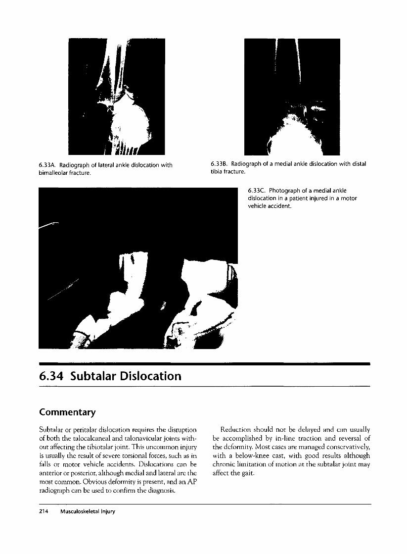
6.33A. Radiograph of lateral ankle dislocation withbimalleolar fracture.
6.33B. Radiograph of a medial ankle dislocation with distaltibia fracture.
6.33C. Photograph of a medial ankledislocation in a patient injured in a motorvehicle accident.
6.34 Subtalar Dislocation
Commentary
Subtalar or peritalar dislocation requires the disruptionof both the talocalcaneal and talonavicular joints with-out affecting the tibiotalar joint. This uncommon injuryis usually the result of severe torsional forces, such as infalls or motor vehicle accidents. Dislocations can beanterior or posterior, although medial and lateral are themost common. Obvious deformity is present, and an APradiograph can be used to confirm the diagnosis.
Reduction should not be delayed and can usuallybe accomplished by in-line traction and reversal ofthe deformity. Most cases are managed conservatively,with a below-knee cast, with good results althoughchronic limitation of motion at the subtalar joint mayaffect the gait.
214 Musculoskeletal Injury
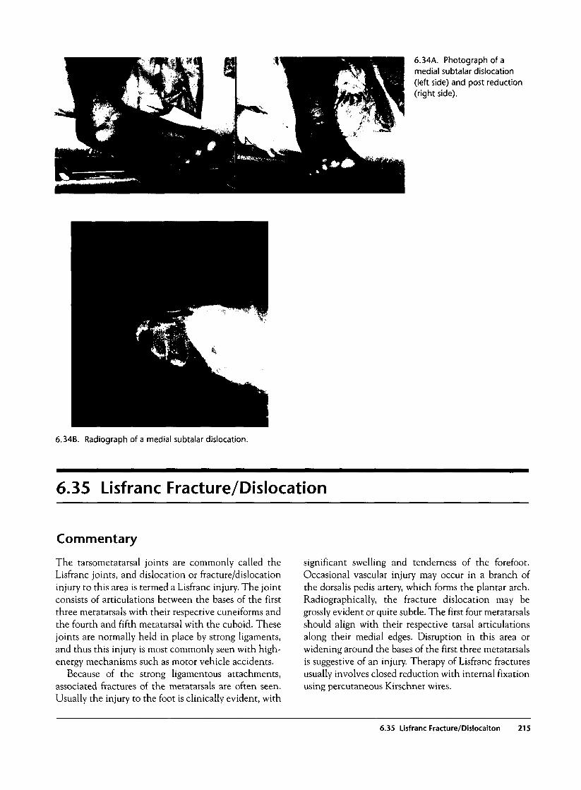
6.34A. Photograph of amedial subtalar dislocation(left side) and post reduction(right side).
6.34B. Radiograph of a medial subtalar dislocation.
6.35 Lisfranc Fracture/Dislocation
Commentary
The tarsometatarsal joints are commonly called theLisfranc joints, and dislocation or fracture/dislocationinjury to this area is termed a Lisfranc injury. The jointconsists of articulations between the bases of the firstthree metatarsals with their respective cuneiforms andthe fourth and fifth metatarsal with the cuboid. Thesejoints are normally held in place by strong ligaments,and thus this injury is most commonly seen with high-energy mechanisms such as motor vehicle accidents.
Because of the strong ligamentous attachments,associated fractures of the metatarsals are often seen.Usually the injury to the foot is clinically evident, with
significant swelling and tenderness of the forefoot.Occasional vascular injury may occur in a branch ofthe dorsalis pedis artery, which forms the plantar arch.Radiographically, the fracture dislocation may begrossly evident or quite subtle. The first four metatarsalsshould align with their respective tarsal articulationsalong their medial edges. Disruption in this area orwidening around the bases of the first three metatarsalsis suggestive of an injury. Therapy of Lisfranc fracturesusually involves closed reduction with internal fixationusing percutaneous Kirschner wires.
6.35 Lisfranc Fracture/Dislocaiton 215
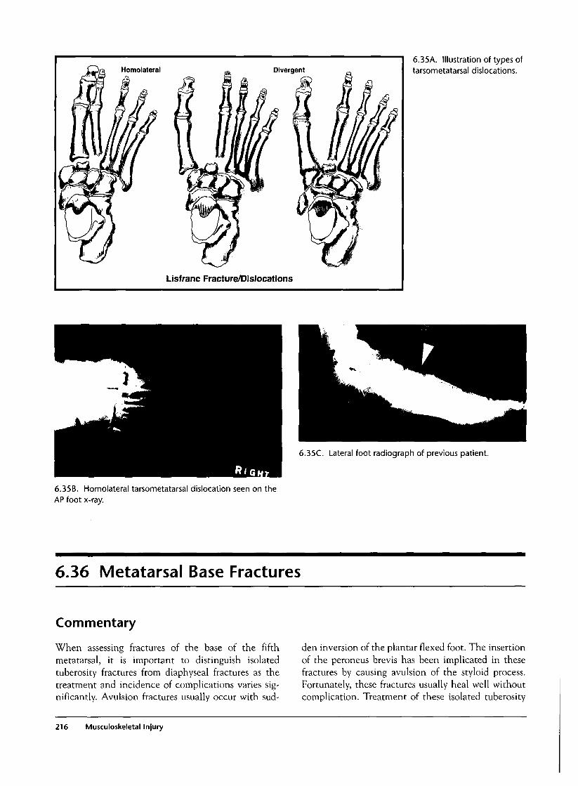
Divergent
Lisfranc Fracture/Dislocations
6.35A. Illustration of types oftarsometatarsal dislocations.
6.35C. Lateral foot radiograph of previous patient.
6.35B. Homolateral tarsometatarsal dislocation seen on theAP foot x-ray.
6.36 Metatarsal Base Fractures
Commentary
When assessing fractures of the base of the fifthmetatarsal, it is important to distinguish isolatedtuberosity fractures from diaphyseal fractures as thetreatment and incidence of complications varies sig-nificantly. Avulsion fractures usually occur with sud-
den inversion of the plantar flexed foot. The insertionof the peroneus brevis has been implicated in thesefractures by causing avulsion of the styloid process.Fortunately, these fractures usually heal well withoutcomplication. Treatment of these isolated tuberosity
216 Musculoskeletal Injury
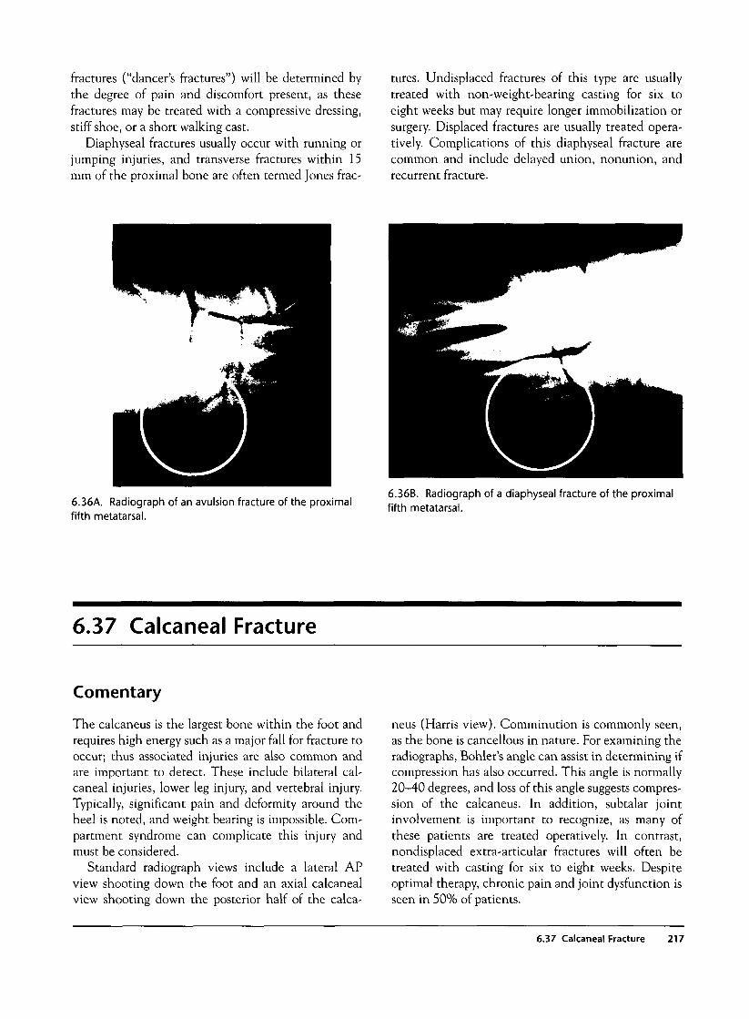
fractures ("dancer's fractures") will be determined bythe degree of pain and discomfort present, as thesefractures may be treated with a compressive dressing,stiff shoe, or a short walking cast.
Diaphyseal fractures usually occur with running orjumping injuries, and transverse fractures within 15mm of the proximal bone are often termed Jones frac-
tures. Undisplaced fractures of this type are usuallytreated with non-weight-bearing casting for six toeight weeks but may require longer immobilization orsurgery. Displaced fractures are usually treated opera-tively. Complications of this diaphyseal fracture arecommon and include delayed union, nonunion, andrecurrent fracture.
6.36A. Radiograph of an avulsion fracture of the proximalfifth metatarsal.
6.36B. Radiograph of a diaphyseal fracture of the proximalfifth metatarsal.
6.37 Calcaneal Fracture
Comentary
The calcaneus is the largest bone within the foot andrequires high energy such as a major fall for fracture tooccur; thus associated injuries are also common andare important to detect. These include bilateral cal-caneal injuries, lower leg injury, and vertebral injury.Typically, significant pain and deformity around theheel is noted, and weight bearing is impossible. Com-partment syndrome can complicate this injury andmust be considered.
Standard radiograph views include a lateral APview shooting down the foot and an axial calcanealview shooting down the posterior half of the calca-
neus (Harris view). Comminution is commonly seen,as the bone is cancellous in nature. For examining theradiographs, Bohler's angle can assist in determining ifcompression has also occurred. This angle is normally20-40 degrees, and loss of this angle suggests compres-sion of the calcaneus. In addition, subtalar jointinvolvement is important to recognize, as many ofthese patients are treated operatively. In contrast,nondisplaced extra-articular fractures will often betreated with casting for six to eight weeks. Despiteoptimal therapy, chronic pain and joint dysfunction isseen in 50% of patients.
6.37 Calcaneal Fracture 217
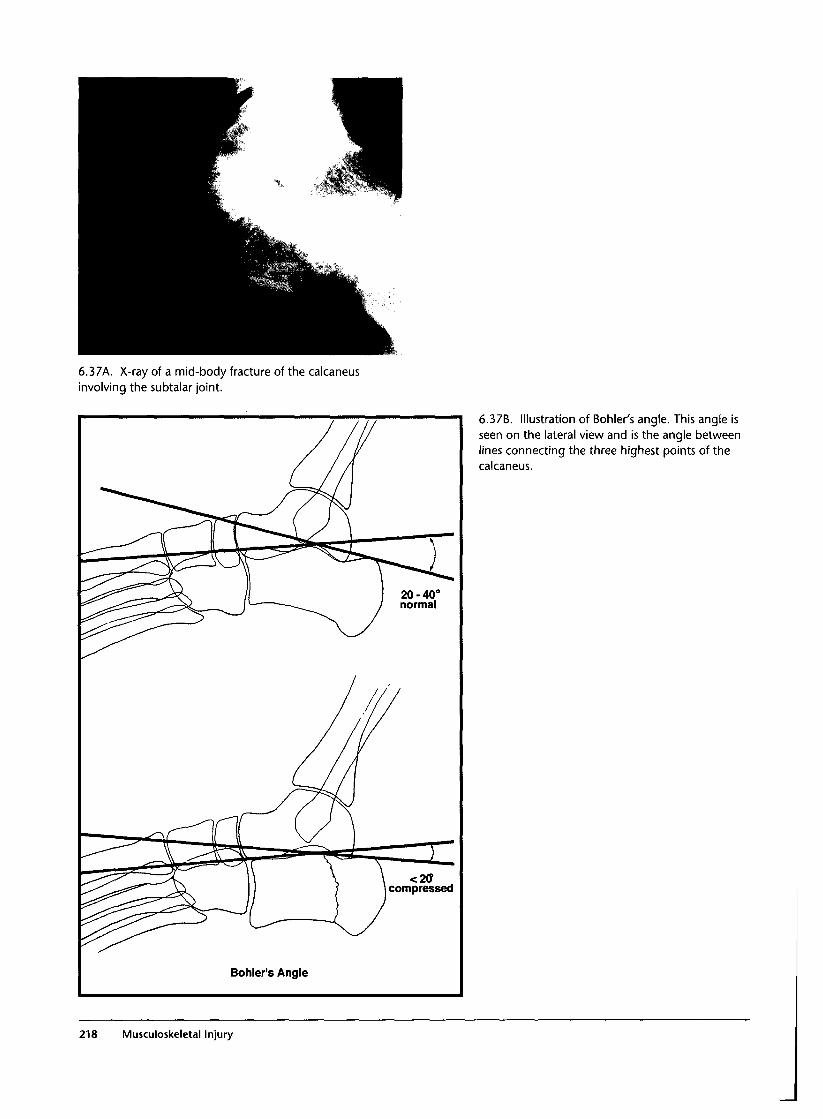
6.37A. X-ray of a mid-body fracture of the calcaneusinvolving the subtalar joint.
Bohler's Angle
6.37B. Illustration of Bohler's angle. This angle isseen on the lateral view and is the angle betweenlines connecting the three highest points of thecalcaneus.
218 Musculoskeletal Injury
Spinal Injury
Introduction
One of the most devastating consequences of trauma isspinal cord injury. In the United States, approximately10,000 spinal cord injuries yearly result in permanentdisability. Although spinal fractures can occur in anyage group, the peak incidence is in males from ages 18to 25. Certain conditions predispose to spinal fractureor dislocation: old age, rheumatoid arthritis, osteoporo-sis, Down's syndrome, and spinal stenosis. Even rela-tively minor mechanisms can result in spinal fracturein these groups. Forces that injure the spinal columninclude flexion, extension, axial loading, shear force,and rotational acceleration.
Clinical Examination
All multiple trauma victims must undergo evaluationfor possible spinal column fracture, dislocation, orspinal cord injury. Blunt trauma patients should havethe spine immobilized at first medical contact andremain in spinal immobilization until the integrity ofthe cord and spinal column can be verified. In patientswith multiple severe injuries, this verification can bedeferred until more critical injuries have beenaddressed, provided that immobilization of the spine ismaintained.
Patients with spinal fractures experience pain, andexamination will reveal tenderness. However, patientswho are unable to report pain because of concomitanthead injury or intoxication may harbor occult spinalinjuries and should remain immobilized until they canbe accurately evaluated. Patients with spinal cord injurymanifest symptoms according to the spinal cord levelaffected. With complete cord transection, all motor andsensory function below the level of the lesion is lost.The highest intact sensory level should be marked onthe patient to determine whether the cord lesion is pro-gressing proximally on subsequent examinations. Acareful motor examination should corroborate the level
of the cord lesion. Assessment of rectal tone and peri-anal sensation is important in detecting any sparing oflower cord segments that significantly improves theprognosis. Spinal shock is common in the immediateperiod after injury and consists of loss of all spinalreflexes and flaccid paralysis below the level of thelesion. During this phase the bulbocavernosus reflex(anal sphincter contraction with stimulation of theglans or urethra) is absent. No prognostication regard-ing the presence or level of the spinal cord lesion can bemade while spinal shock is present. Once the bulbocav-ernosus reflex returns, spinal shock is resolved, reflexesbecome spastic, and the lesion is complete. Priapism iscommon in males after complete cord transection butresolves quickly in most cases.
Neurogenic shock is the hemodynamic effect ofsympathetic denervation that results in vasodilationand hypotension. Because the sympathetic innervationto the heart is similarly disrupted by high cord injuries,reflex tachycardia does not occur in response tohypotension, and the patient is usually hypotensiveand bradycardic. Regardless of the presence of neuro-genic shock, hypotension must be assumed to be due tohemorrhage, and a diligent search for sources of bloodloss must be made.
Partial cord syndromes may present with a confusingpattern of neurologic deficits. In central cord syn-drome, the hands and arms sustain a much denserbilateral paralysis than the lower extremities. InBrown-Sequard syndrome, pain and temperature sen-sory loss occurs contralaterally, whereas motor loss andall other sensory loss is ipsilateral. With anterior spinalcord syndrome, there is preservation of posterior col-umn function (position and vibration sense) bilater-ally, but all other functions are lost. The syndrome ofspinal cord injury without radiographic abnormality(SCIWORA) can occur in any age group but is partic-ularly common in children. Neurologic deficits may bedelayed many hours, and by definition, radiographs arenormal. Consequently, it is difficult to make this diag-nosis on initial evaluation. Patients who report persist-
219
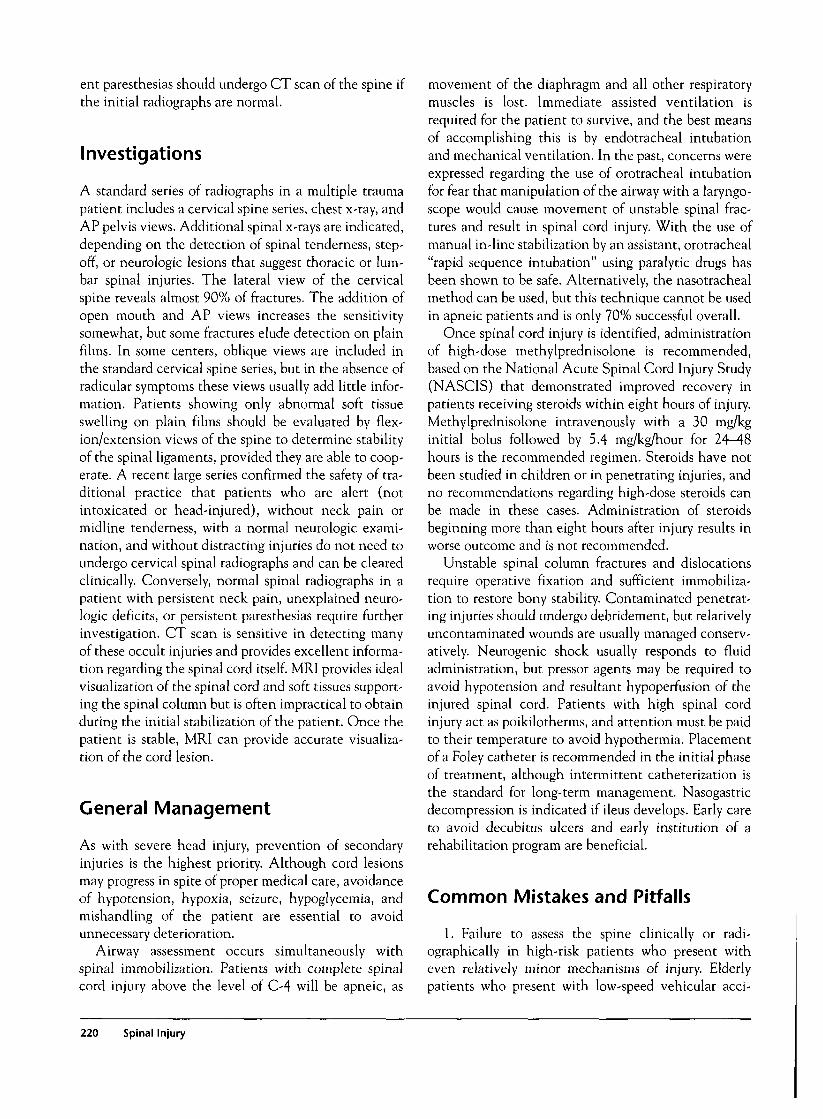
ent paresthesias should undergo CT scan of the spine ifthe initial radiographs are normal.
Investigations
A standard series of radiographs in a multiple traumapatient includes a cervical spine series, chest x-ray, andAP pelvis views. Additional spinal x-rays are indicated,depending on the detection of spinal tenderness, step-off, or neurologic lesions that suggest thoracic or lum-bar spinal injuries. The lateral view of the cervicalspine reveals almost 90% of fractures. The addition ofopen mouth and AP views increases the sensitivitysomewhat, but some fractures elude detection on plainfilms. In some centers, oblique views are included inthe standard cervical spine series, but in the absence ofradicular symptoms these views usually add little infor-mation. Patients showing only abnormal soft tissueswelling on plain films should be evaluated by flex-ion/extension views of the spine to determine stabilityof the spinal ligaments, provided they are able to coop-erate. A recent large series confirmed the safety of tra-ditional practice that patients who are alert (notintoxicated or head-injured), without neck pain ormidline tenderness, with a normal neurologic exami-nation, and without distracting injuries do not need toundergo cervical spinal radiographs and can be clearedclinically. Conversely, normal spinal radiographs in apatient with persistent neck pain, unexplained neuro-logic deficits, or persistent paresthesias require furtherinvestigation. CT scan is sensitive in detecting manyof these occult injuries and provides excellent informa-tion regarding the spinal cord itself. MRI provides idealvisualization of the spinal cord and soft tissues support-ing the spinal column but is often impractical to obtainduring the initial stabilization of the patient. Once thepatient is stable, MRI can provide accurate visualiza-tion of the cord lesion.
General Management
As with severe head injury, prevention of secondaryinjuries is the highest priority. Although cord lesionsmay progress in spite of proper medical care, avoidanceof hypotension, hypoxia, seizure, hypoglycemia, andmishandling of the patient are essential to avoidunnecessary deterioration.
Airway assessment occurs simultaneously withspinal immobilization. Patients with complete spinalcord injury above the level of C-4 will be apneic, as
movement of the diaphragm and all other respiratorymuscles is lost. Immediate assisted ventilation isrequired for the patient to survive, and the best meansof accomplishing this is by endotracheal intubationand mechanical ventilation. In the past, concerns wereexpressed regarding the use of orotracheal intubationfor fear that manipulation of the airway with a laryngo-scope would cause movement of unstable spinal frac-tures and result in spinal cord injury. With the use ofmanual in-line stabilization by an assistant, orotracheal"rapid sequence intubation" using paralytic drugs hasbeen shown to be safe. Alternatively, the nasotrachealmethod can be used, but this technique cannot be usedin apneic patients and is only 70% successful overall.
Once spinal cord injury is identified, administrationof high-dose methylprednisolone is recommended,based on the National Acute Spinal Cord Injury Study(NASCIS) that demonstrated improved recovery inpatients receiving steroids within eight hours of injury.Methylprednisolone intravenously with a 30 mg/kginitial bolus followed by 5.4 mg/kg/hour for 24-48hours is the recommended regimen. Steroids have notbeen studied in children or in penetrating injuries, andno recommendations regarding high-dose steroids canbe made in these cases. Administration of steroidsbeginning more than eight hours after injury results inworse outcome and is not recommended.
Unstable spinal column fractures and dislocationsrequire operative fixation and sufficient immobiliza-tion to restore bony stability. Contaminated penetrat-ing injuries should undergo debridement, but relativelyuncontaminated wounds are usually managed conserv-atively. Neurogenic shock usually responds to fluidadministration, but pressor agents may be required toavoid hypotension and resultant hypoperfusion of theinjured spinal cord. Patients with high spinal cordinjury act as poikilotherms, and attention must be paidto their temperature to avoid hypothermia. Placementof a Foley catheter is recommended in the initial phaseof treatment, although intermittent catheterization isthe standard for long-term management. Nasogastricdecompression is indicated if ileus develops. Early careto avoid decubitus ulcers and early institution of arehabilitation program are beneficial.
Common Mistakes and Pitfalls
1. Failure to assess the spine clinically or radi-ographically in high-risk patients who present witheven relatively minor mechanisms of injury. Elderlypatients who present with low-speed vehicular acci-
220 Spinal Injury
dents or ground-level falls can have fractured theirbrittle or osteoporotic spines. A low threshold forobtaining radiographs should be maintained in traumainvolving high-risk patients.
2. Failure to recognize partial cord syndromes. Insome series, the vast majority of patients with centralcord syndrome were initially misdiagnosed because theywere intoxicated or because they were thought to bemalingering or have conversion disorder. Paralysis ofboth upper extremities without simultaneous paralysis ofthe lower extremities was thought by these clinicians tobe anatomically impossible because they were unawareof the presentation of the central cord syndrome.
3. Mishandling of the patient with spinal fracture.One older study found that almost half of patients whodeveloped neurologic deficits did so after contact withmedical personnel. Although some of these deficitswould occur due to progressive edema or ischemia, oth-ers were certainly caused by inadequate care in main-taining spinal immobilization during transfer of thepatient from stretcher to x-ray table or CT scanner.
4. Intoxicated or head-injured patients should havespinal precautions maintained until they can reliably
report pain or neurologic deficits. Patients who reportsuch symptoms require further investigation even ifplain radiographs are normal.
5. There is often confusion between spinal shock(loss of reflexes and motor/sensory function for up to24 hours after injury) and neurogenic shock (hemody-namic effects of sympathectomy due to high spinalcord lesions manifesting as bradycardia and hypoten-sion). These terms are not interchangeable and shouldbe used correctly to avoid miscommunication.
6. Chance fracture of the lumbar spine is frequentlyassociated with a small-intestinal injury that may beinitially occult.
7. Patients who present with calcaneal fracture(s)after jumping from a height should be screened forcoexistent lumbar compression fractures because thesefractures are commonly associated.
8. Approximately 10% of spinal fractures are associ-ated with another non-contiguous spine fracture, thusthe finding of any spinal fracture should initiate asearch for other fractures elsewhere in the spine.
Spinal Injury 221
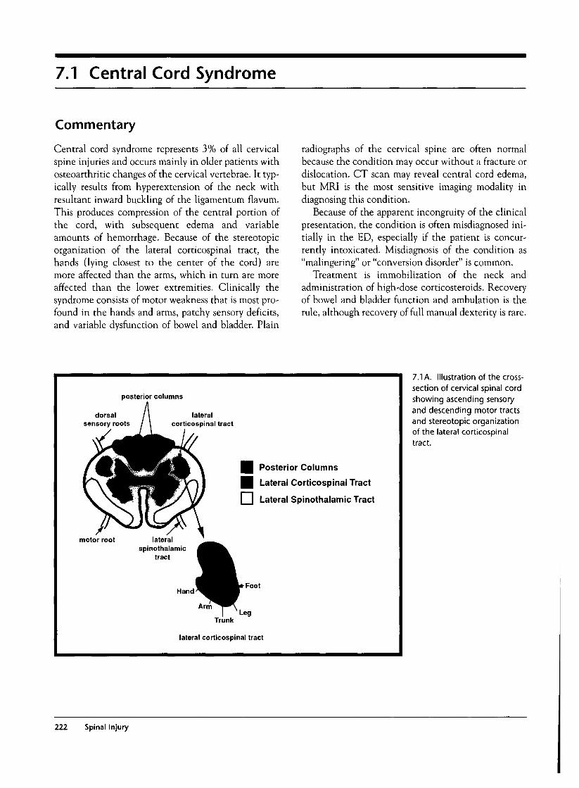
7.1 Central Cord Syndrome
Commentary
Central cord syndrome represents 3% of all cervicalspine injuries and occurs mainly in older patients withosteoarthritic changes of the cervical vertebrae. It typ-ically results from hyperextension of the neck withresultant inward buckling of the ligamentum flavum.This produces compression of the central portion ofthe cord, with subsequent edema and variableamounts of hemorrhage. Because of the stereotopicorganization of the lateral corticospinal tract, thehands (lying closest to the center of the cord) aremore affected than the arms, which in turn are moreaffected than the lower extremities. Clinically thesyndrome consists of motor weakness that is most pro-found in the hands and arms, patchy sensory deficits,and variable dysfunction of bowel and bladder. Plain
radiographs of the cervical spine are often normalbecause the condition may occur without a fracture ordislocation. CT scan may reveal central cord edema,but MRI is the most sensitive imaging modality indiagnosing this condition.
Because of the apparent incongruity of the clinicalpresentation, the condition is often misdiagnosed ini-tially in the ED, especially if the patient is concur-rently intoxicated. Misdiagnosis of the condition as"malingering" or "conversion disorder" is common.
Treatment is immobilization of the neck andadministration of high-dose corticosteroids. Recoveryof bowel and bladder function and ambulation is therule, although recovery of full manual dexterity is rare.
posterior columns
dorsalsensory roots
lateralcorticospinal tract
Posterior Columns
Lateral Corticospinal Tract
I | Lateral Spinothalamic Tract
motor root lateralspinothalamic
tract
HandFoot
Trunk
lateral corticospinal tract
7.1 A. Illustration of the cross-section of cervical spinal cordshowing ascending sensoryand descending motor tractsand stereotopic organizationof the lateral corticospinaltract.
222 Spinal Injury

Central Cord Syndrome
7.1 B. Illustration of cross-section of cervical spinal cord. Thered area shows section of the spinal cord affected in centralcord syndrome.
7.1 C. T2 weighted cervicothoracic MRI showinghemorrhagic contusion of the central cervical cord (blackarrow) and edema of the adjacent spinal cord (white arrow).
7.2 Brown-Sequard Syndrome
Commentary
Brown-Se"quard syndrome is due to an incompletetransection or hemisection of the spinal cord, usuallycaused by penetrating trauma. It consists of a charac-teristic constellation of findings with loss of pain andtemperature contralateral to the side of the injury andloss of motor function (corticospinal tract), lighttouch (spinothalamic tract), position, and vibrationsensation (posterior columns) ipsilateral to the injury.Although true Brown-Sequard injury is due to anexact hemisection of the spinal cord, the clinical find-ings may vary if more or less of the cord is transected.
As with other penetrating injuries, the use of highdoses of corticosteroids has not been found to be ben-eficial in prospective series, although it is still some-times used in individual cases. Broad-spectrum
antibiotic coverage is indicated, and surgical debride-ment may be required. Impaled objects should alwaysbe removed in the operating room. Functional recov-ery is often surprisingly good in spite of the permanentcord lesions.
Neurologic Deficits Distal to Site of Hemisectionof Spinal cord in Brown-Sequard Syndrome
Light touchPosition/vibrationMotor functionPain sensationTemperature sensation
IpsilateralIpsilateralIpsilateralContralateralContralateral
7.2 Brown Sequard Syndrome 223
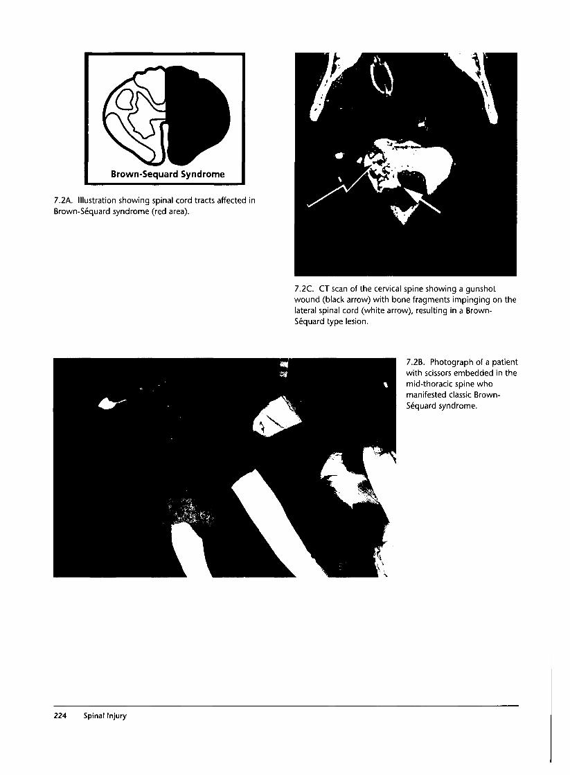
Brown-Sequard Syndrome
7.2k. Illustration showing spinal cord tracts affected inBrown-Sequard syndrome (red area).
7.2C. CT scan of the cervical spine showing a gunshotwound (black arrow) with bone fragments impinging on thelateral spinal cord (white arrow), resulting in a Brown-Sequard type lesion.
7.2%. Photograph of a patientwith scissors embedded in themid-thoracic spine whomanifested classic Brown-Sequard syndrome.
224 Spinal Injury

7.3 Anterior Cord Syndrome
Commentary
Anterior cord syndrome results from damage to theanterior spinal artery, producing infarction of the ante-rior spinal tracts, or by direct laceration of the anteriorpart of the cord by retropulsed bony fragments. Poste-rior column function (position and vibration sense) ispreserved. Clinically, there is bilateral loss of motorfunction, light touch, pain, and temperature sensationbelow the level of the injury.
Anterior cord syndrome is usually seen in elderlypatients in conjunction with medical conditions, par-ticularly arterial embolization from the heart. How-ever, it may also result from prolonged cross clampingof the aorta, prolonged hypotension, or direct injuryfrom bone fragments or foreign bodies.
Anterior Cord Syndrome
7.3A. Schematic drawing of spinal cord tracts affected inanterior cord syndrome.
7.4 Complete Spinal Cord Transection
Commentary
Complete transection of the spinal cord is a devastat-ing injury. Transection above the level of C-4 resultsin respiratory failure as the nerve supply to both thediaphragm (C-3-5) and intercostal muscles (Tl-12)is lost. Such patients frequently expire in the fieldunless immediate ventilation is provided.
At more distal levels, acute spinal cord transectionresults in complete flaccid motor paralysis and anes-thesia below the level of the injury. Deep tendonreflexes are absent distal to the lesion. Examinationwill reveal urinary retention and diminished or absentrectal sphincter tone. In males, transient priapism isvery common and indicates complete cord transec-tion, although it often resolves by the time the patientarrives in the ED.
Loss of sympathetic innervation results in loss ofvasomotor tone and hypotension. In cervical cordinjuries, loss of sympathetic innervation to the heartprevents the normal response of reflex tachycardia.
Consequently, the hemodynamic picture of hypoten-sion, inappropriate bradycardia, and warm, flushedskin constitutes the classic syndrome of neurogenicshock. Inability to vasoconstrict also prevents the nor-mal response to cold stress, and these patients are atrisk for hypothermia. Treatment is initially directed atrestoring intravascular volume and ensuring thathypotension is not due to occult blood loss. If volumeinfusion does not restore adequate blood pressure,pressor agents such as dopamine are indicated toimprove perfusion and consequently the survival ofthe spinal cord proximal to the transection.
During the acute phase of spinal cord injury, all dis-tal reflexes are absent, and the patient is said to be in"spinal shock" (not to be confused with neurogenicshock with its hemodynamic manifestations). To con-firm that all reflexes are absent, the most distal reflexarc or bulbocavernosus reflex is examined. Stimula-tion of the glans or clitoris or tugging on a Foley
7.4 Complete Spinal Cord Transection 225
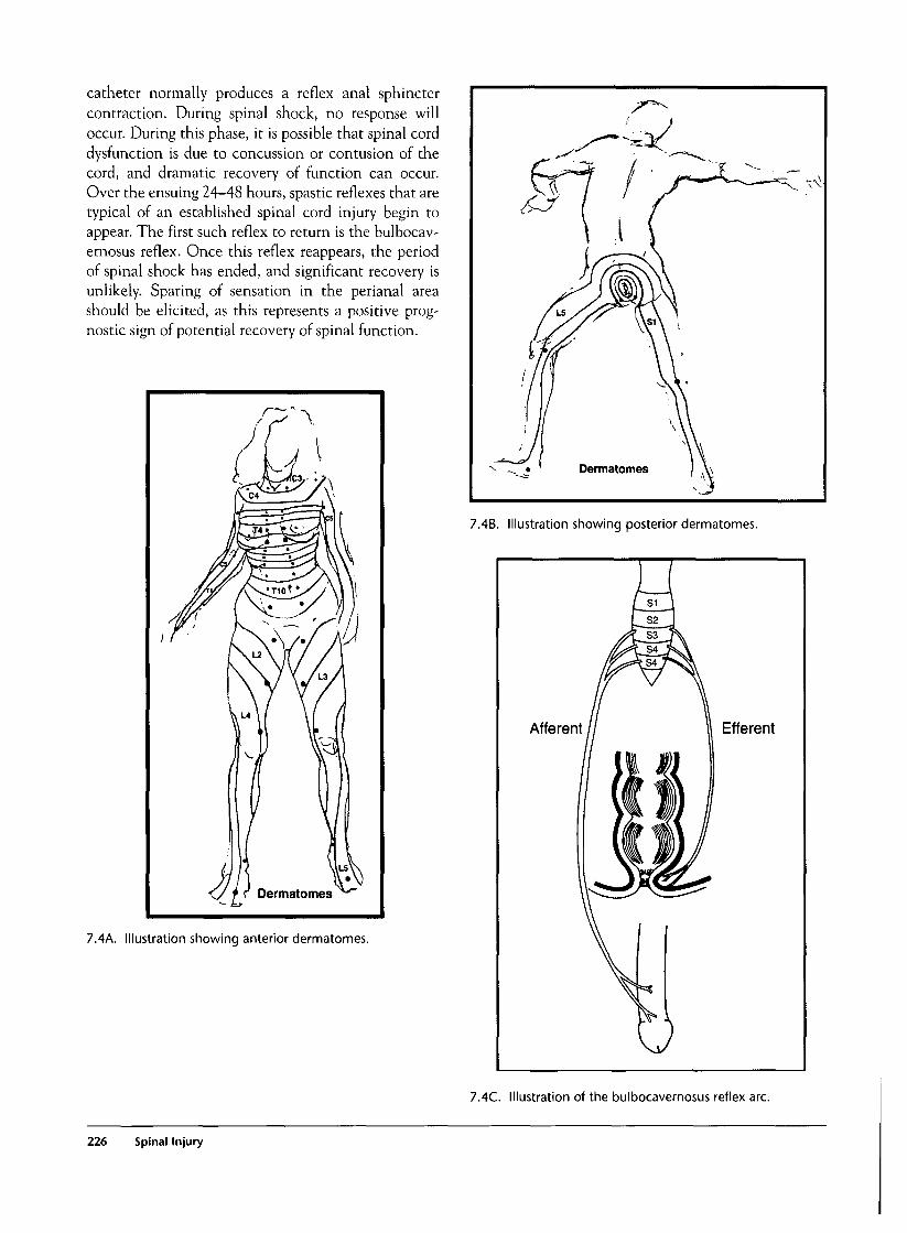
catheter normally produces a reflex anal sphinctercontraction. During spinal shock, no response willoccur. During this phase, it is possible that spinal corddysfunction is due to concussion or contusion of thecord, and dramatic recovery of function can occur.Over the ensuing 24-48 hours, spastic reflexes that aretypical of an established spinal cord injury begin toappear. The first such reflex to return is the bulbocav-ernosus reflex. Once this reflex reappears, the periodof spinal shock has ended, and significant recovery isunlikely. Sparing of sensation in the perianal areashould be elicited, as this represents a positive prog-nostic sign of potential recovery of spinal function.
7.4B. Illustration showing posterior dermatomes.
7.4A. Illustration showing anterior dermatomes.
S1
Afferent Efferent
7.4C. Illustration of the bulbocavemosus reflex arc.
226 Spinal Injury

7.4D. Photograph showing priapism in a child with complete transection of the spinal cord.
7.4E. Autopsy photograph of severe distraction of the spinal column and extrusion of the spinal cord.
7.4 Complete Spinal Cord Transection 227
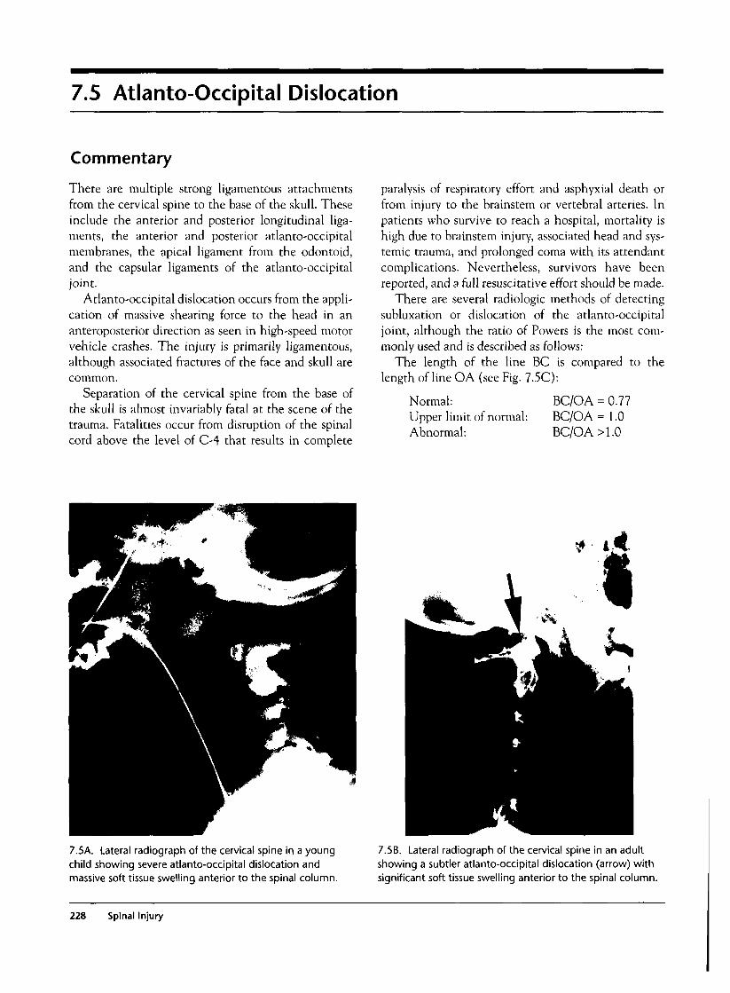
7.5 Atlanto-Occipital Dislocation
Commentary
There are multiple strong ligamentous attachmentsfrom the cervical spine to the base of the skull. Theseinclude the anterior and posterior longitudinal liga-ments, the anterior and posterior atlanto-occipitalmembranes, the apical ligament from the odontoid,and the capsular ligaments of the atlanto-occipitaljoint.
Atlanto-occipital dislocation occurs from the appli-cation of massive shearing force to the head in ananteroposterior direction as seen in high-speed motorvehicle crashes. The injury is primarily ligamentous,although associated fractures of the face and skull arecommon.
Separation of the cervical spine from the base ofthe skull is almost invariably fatal at the scene of thetrauma. Fatalities occur from disruption of the spinalcord above the level of C-4 that results in complete
paralysis of respiratory effort and asphyxial death orfrom injury to the brainstem or vertebral arteries. Inpatients who survive to reach a hospital, mortality ishigh due to brainstem injury, associated head and sys-temic trauma, and prolonged coma with its attendantcomplications. Nevertheless, survivors have beenreported, and a full resuscitative effort should be made.
There are several radiologic methods of detectingsubluxation or dislocation of the atlanto-occipitaljoint, although the ratio of Powers is the most com-monly used and is described as follows:
The length of the line BC is compared to thelength of line OA (see Fig. 7.5C):
Normal:Upper limit of normal:Abnormal:
BC/OA = 0.77BC/OA = 1.0BC/OA > 1.0
7.5A. Lateral radiograph of the cervical spine in a youngchild showing severe atlanto-occipital dislocation andmassive soft tissue swelling anterior to the spinal column.
f • 4,
7.5B. Lateral radiograph of the cervical spine in an adultshowing a subtler atlanto-occipital dislocation (arrow) withsignificant soft tissue swelling anterior to the spinal column.
228 Spinal Injury

Cranlocervical Relationships
7.5C. Schematic drawing illustrating the measurementsused in calculating the "ratio of Powers" to detect atlanto-occipital dislocation.
7.6 Atlantoaxial Dislocation
Commentary
Atlantoaxial dislocation is a rare injury of unclearmechanism. There is disruption of the transverse andalar ligaments that connect the odontoid process tothe ring of C-l, which displaces superiorly (see Fig.7.9A). Neurologic deficits are uncommon with supe-rior dislocation (as shown) or when associated withfracture of the odontoid, although the condition isunstable due to disruption of the ligaments. Anteriordislocation is usually fatal as the spinal cord is com-pressed against the intact odontoid.
7.6A. Lateral radiograph of the cervical spine in a childshowing atlantoaxial dislocation. The ring of C-1 (A) is seenlying completely above the tip of the odontoid process (B).
7.6 Atlantoaxial Dislocation 229
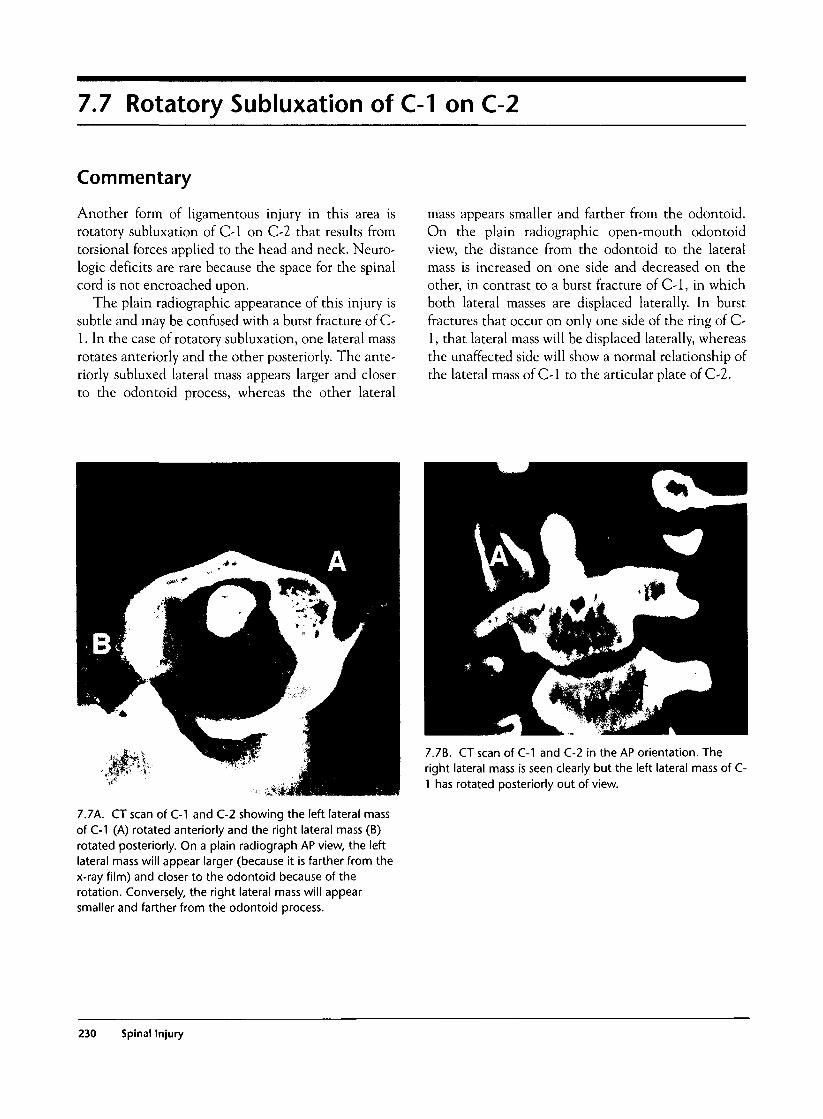
7.7 Rotatory Subluxation of C-1 on C-2
Commentary
Another form of ligamentous injury in this area isrotatory subluxation of C-1 on C-2 that results fromtorsional forces applied to the head and neck. Neuro-logic deficits are rare because the space for the spinalcord is not encroached upon.
The plain radiographic appearance of this injury issubtle and may be confused with a burst fracture of C-1. In the case of rotatory subluxation, one lateral massrotates anteriorly and the other posteriorly. The ante-riorly subluxed lateral mass appears larger and closerto the odontoid process, whereas the other lateral
mass appears smaller and farther from the odontoid.On the plain radiographic open-mouth odontoidview, the distance from the odontoid to the lateralmass is increased on one side and decreased on theother, in contrast to a burst fracture of C-1, in whichboth lateral masses are displaced laterally. In burstfractures that occur on only one side of the ring of C-1, that lateral mass will be displaced laterally, whereasthe unaffected side will show a normal relationship ofthe lateral mass of C-1 to the articular plate of C-2.
7.7A. CT scan of C-1 and C-2 showing the left lateral massof C-1 (A) rotated anteriorly and the right lateral mass (B)rotated posteriorly. On a plain radiograph AP view, the leftlateral mass will appear larger (because it is farther from thex-ray film) and closer to the odontoid because of therotation. Conversely, the right lateral mass will appearsmaller and farther from the odontoid process.
7.7%. CT scan of C-1 and C-2 in the AP orientation. Theright lateral mass is seen clearly but the left lateral mass of C-1 has rotated posteriorly out of view.
230 Spinal Injury
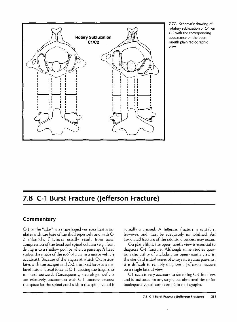
7.7C. Schematic drawing ofrotatory subluxation of C-1 onC-2 with the correspondingappearance on the open-mouth plain radiographicview.
7.8 C-1 Burst Fracture (Jefferson Fracture)
Commentary
C-1 or the "atlas" is a ring-shaped vertebra that artic-ulates with the base of the skull superiorly and with C-2 inferiorly. Fractures usually result from axialcompression of the head and spinal column (e.g., fromdiving into a shallow pool or when a passenger's headstrikes the inside of the roof of a car in a motor vehicleaccident). Because of the angles at which C-1 articu-lates with the occiput and C-2, the axial force is trans-lated into a lateral force at C-1, causing the fragmentsto burst outward. Consequently, neurologic deficitsare relatively uncommon with C-1 fracture becausethe space for the spinal cord within the spinal canal is
actually increased. A Jefferson fracture is unstable,however, and must be adequately immobilized. Anassociated fracture of the odontoid process may occur.
On plain films, the open-mouth view is essential todiagnose C-1 fracture. Although some studies ques-tion the utility of including an open-mouth view inthe standard initial series of x-rays in trauma patients,it is difficult to reliably diagnose a Jefferson fractureon a single lateral view.
CT scan is very accurate in detecting C-1 fracturesand is indicated for any suspicious abnormalities or forinadequate visualization on plain radiographs.
7.8 C-1 Burst Fracture (Jefferson Fracture) 231
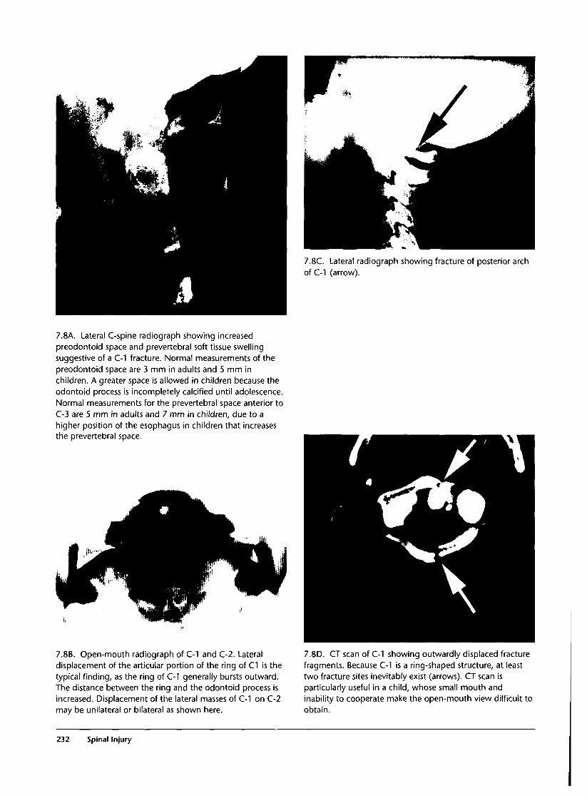
7.8C. Lateral radiograph showing fracture of posterior archof C-1 (arrow).
7.8A. Lateral C-spine radiograph showing increasedpreodontoid space and prevertebral soft tissue swellingsuggestive of a C-1 fracture. Normal measurements of thepreodontoid space are 3 mm in adults and 5 mm inchildren. A greater space is allowed in children because theodontoid process is incompletely calcified until adolescence.Normal measurements for the prevertebral space anterior toC-3 are 5 mm in adults and 7 mm in children, due to ahigher position of the esophagus in children that increasesthe prevertebral space.
7.8B. Open-mouth radiograph of C-1 and C-2. Lateraldisplacement of the articular portion of the ring of C1 is thetypical finding, as the ring of C-1 generally bursts outward.The distance between the ring and the odontoid process isincreased. Displacement of the lateral masses of C-1 on C-2may be unilateral or bilateral as shown here.
7.8D. CT scan of C-1 showing outwardly displaced fracturefragments. Because C-1 is a ring-shaped structure, at leasttwo fracture sites inevitably exist (arrows). CT scan isparticularly useful in a child, whose small mouth andinability to cooperate make the open-mouth view difficult toobtain.
232 Spinal Injury
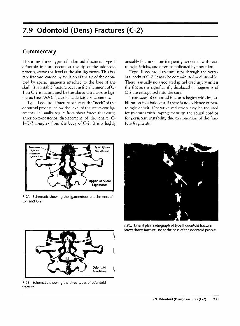
7.9 Odontoid (Dens) Fractures (C-2)
Commentary
There are three types of odontoid fracture. Type Iodontoid fracture occurs at the tip of the odontoidprocess, above the level of the alar ligaments. This is arare fracture, caused by avulsion of the tip of the odon-toid by apical ligaments attached to the base of theskull. It is a stable fracture because the alignment of C-1 on C-2 is maintained by the alar and transverse liga-ments (see 7.9A). Neurologic deficit is uncommon.
Type II odontoid fracture occurs at the "neck" of theodontoid process, below the level of the transverse lig-aments. It usually results from shear forces that causeanterior-to-posterior displacement of the entire C-l-C-2 complex from the body of C-2. It is a highly
unstable fracture, more frequently associated with neu-rologic deficits, and often complicated by nonunion.
Type III odontoid fracture runs through the verte-bral body of C-2. It may be comminuted and unstable.There is usually no associated spinal cord injury unlessthe fracture is significantly displaced or fragments ofC-2 are retropulsed into the canal.
Treatment of odontoid fractures begins with immo-bilization in a halo vest if there is no evidence of neu-rologic deficit. Operative reduction may be requiredfor fractures with impingement on the spinal cord orfor persistent instability due to nonunion of the frac-ture fragments.
Transverseligament
Accessoryligament
7.9k. Schematic showing the ligamentous attachments ofC-1 and C-2.
Odontoidfractures
7.9C. Lateral plain radiograph of type II odontoid fracture.Arrow shows fracture line at the base of the odontoid process.
7.9B. Schematic showing the three types of odontoidfracture.
7.9 Odontoid (Dens) Fractures (C-2) 233
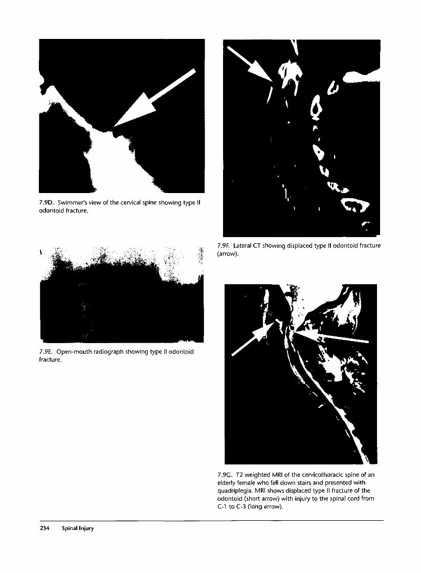
7.9D. Swimmer's view of the cervical spine showing type Iodontoid fracture.
7.9F. Lateral CT showing displaced type II odontoid fracture(arrow).
7.9E. Open-mouth radiograph showing type II odontoidfracture.
7.9G. T2 weighted MRI of the cervicothoracic spine of anelderly female who fell down stairs and presented withquadriplegia. MRI shows displaced type II fracture of theodontoid (short arrow) with injury to the spinal cord fromC-1 to C-3 (long arrow).
234 Spinal Injury
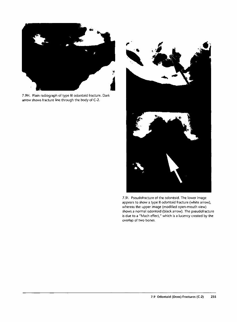
7.9H. Plain radiograph of type III odontoid fracture. Darkarrow shows fracture line through the body of C-2.
7.91. Pseudofracture of the odontoid. The lower imageappears to show a type II odontoid fracture (white arrow),whereas the upper image (modified open-mouth view)shows a normal odontoid (black arrow). The pseudofractureis due to a "Mach effect," which is a lucency created by theoverlap of two bones.
7.9 Odontoid (Dens) Fractures (C-2) 235

7.10 Hangman's Fracture (C-2)
Commentary
Hangman's fracture is traumatic spondylolisthesis ofthe pars interarticularis of C-2. In common usage,however, the term is applied to bilateral pedicle frac-tures of C-2 as well. The fracture results from hyperex-tension of the neck with compression of the posteriorelements of C-2 by the hyperextending occiput. Thisis the type of injury produced by a judicial hanging,hence the name. Displaced hangman's fractures are
commonly associated with severe neurologic deficits.Cord lesions at this level result in paralysis of respira-tory muscles and therefore apnea, as well as completeparalysis and sensory loss distal to the lesion.
Nevertheless, patients may sustain this injury with-out any injury to the spinal cord. These patientsshould be immobilized in a halo vest and often requireoperative fixation.
7.1 OA. Lateral C-spine radiograph showing bilateral pediclefracture (hangman's fracture) of C-2. Undisplacedhangman's fracture.
7.1 OB. Moderately displaced hangman's fracture.
236 Spinal Injury
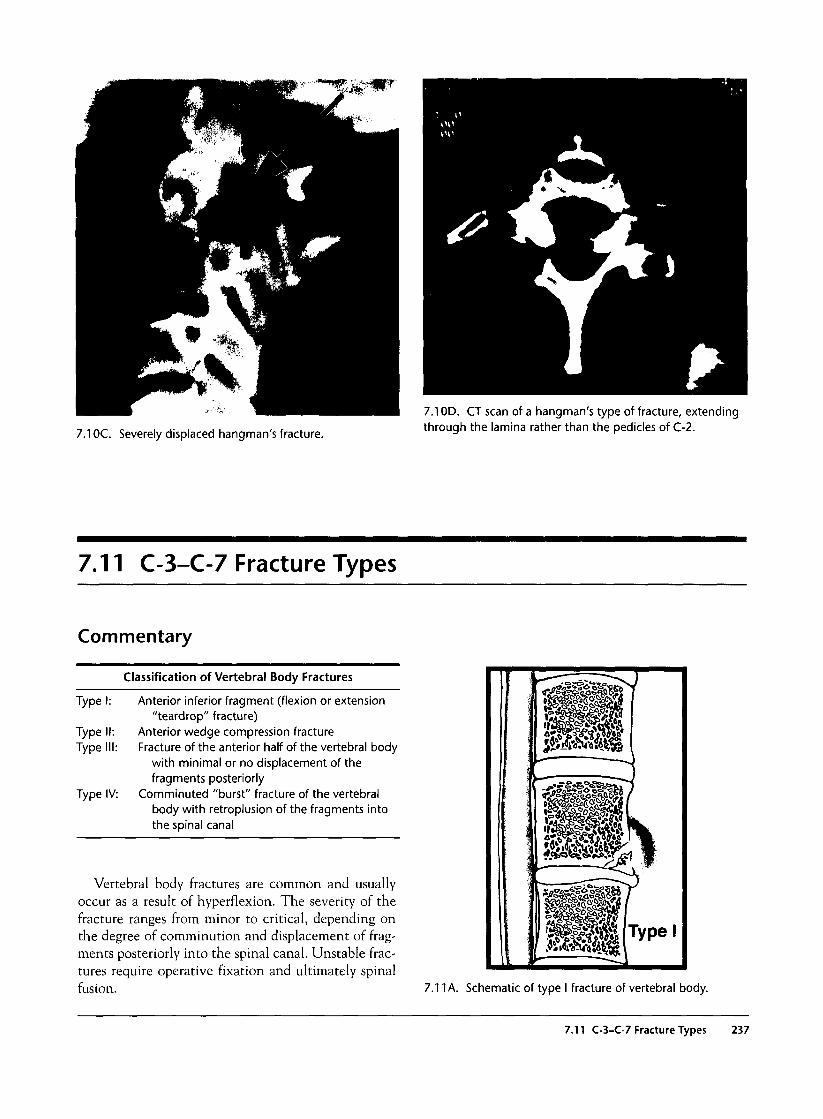
7.1 OC. Severely displaced hangman's fracture.
7.1 OD. CT scan of a hangman's type of fracture, extendingthrough the lamina rather than the pedicles of C-2.
7.11 C-3-C-7 Fracture Types
Commentary
Classification of Vertebral Body Fractures
Type I: Anterior inferior fragment (flexion or extension"teardrop" fracture)
Type II: Anterior wedge compression fractureType III: Fracture of the anterior half of the vertebral body
with minimal or no displacement of thefragments posteriorly
Type IV: Comminuted "burst" fracture of the vertebralbody with retroplusion of the fragments intothe spinal canal
Vertebral body fractures are common and usuallyoccur as a result of hyperflexion. The severity of thefracture ranges from minor to critical, depending onthe degree of comminution and displacement of frag-ments posteriorly into the spinal canal. Unstable frac-tures require operative fixation and ultimately spinalfusion. 7.11 A. Schematic of type I fracture of vertebral body.
7.11 C-3-C-7 Fracture Types 237
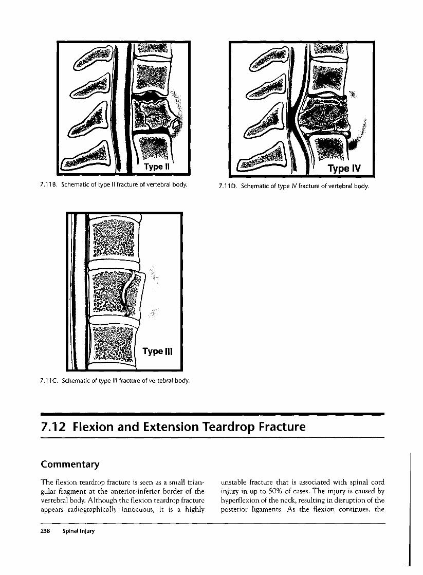
7.11B. Schematic of type II fracture of vertebral body. 7.11D. Schematic of type IV fracture of vertebral body.
7.11C. Schematic of type III fracture of vertebral body.
7.12 Flexion and Extension Teardrop Fracture
Commentary
The flexion teardrop fracture is seen as a small trian-gular fragment at the anterior-inferior border of thevertebral body. Although the flexion teardrop fractureappears radiographically innocuous, it is a highly
unstable fracture that is associated with spinal cordinjury in up to 50% of cases. The injury is caused byhyperflexion of the neck, resulting in disruption of theposterior ligaments. As the flexion continues, the
238 Spinal Injury
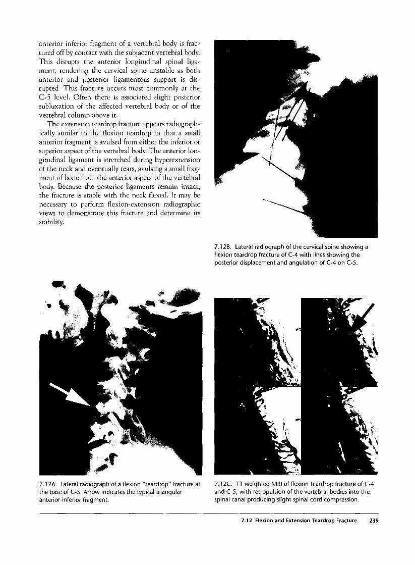
anterior inferior fragment of a vertebral body is frac-tured off by contact with the subjacent vertebral body.This disrupts the anterior longitudinal spinal liga-ment, rendering the cervical spine unstable as bothanterior and posterior ligamentous support is dis-rupted. This fracture occurs most commonly at theC-5 level. Often there is associated slight posteriorsubluxation of the affected vertebral body or of thevertebral column above it.
The extension teardrop fracture appears radiograph-ically similar to the flexion teardrop in that a smallanterior fragment is avulsed from either the inferior orsuperior aspect of the vertebral body. The anterior lon-gitudinal ligament is stretched during hyperextensionof the neck and eventually tears, avulsing a small frag-ment of bone from the anterior aspect of the vertebralbody. Because the posterior ligaments remain intact,the fracture is stable with the neck flexed. It may benecessary to perform flexion-extension radiographicviews to demonstrate this fracture and determine itsstability.
7.12B. Lateral radiograph of the cervical spine showing aflexion teardrop fracture of C-4 with lines showing theposterior displacement and angulation of C-4 on C-5.
7.12A. Lateral radiograph of a flexion "teardrop" fracture atthe base of C-5. Arrow indicates the typical triangularanterior-inferior fragment.
7.12C. T1 weighted MRI of flexion teardrop fracture of C-4and C-5, with retropulsion of the vertebral bodies into thespinal canal producing slight spinal cord compression.
7.12 Flexion and Extension Teardrop Fracture 239
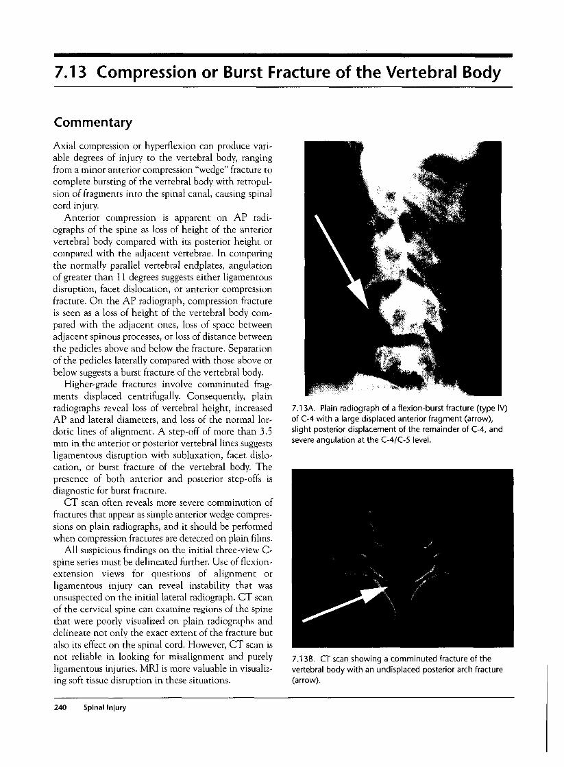
7.13 Compression or Burst Fracture of the Vertebral Body
Commentary
Axial compression or hyperflexion can produce vari-able degrees of injury to the vertebral body, rangingfrom a minor anterior compression "wedge" fracture tocomplete bursting of the vertebral body with retropul-sion of fragments into the spinal canal, causing spinalcord injury.
Anterior compression is apparent on AP radi-ographs of the spine as loss of height of the anteriorvertebral body compared with its posterior height orcompared with the adjacent vertebrae. In comparingthe normally parallel vertebral endplates, angulationof greater than 11 degrees suggests either ligamentousdisruption, facet dislocation, or anterior compressionfracture. On the AP radiograph, compression fractureis seen as a loss of height of the vertebral body com-pared with the adjacent ones, loss of space betweenadjacent spinous processes, or loss of distance betweenthe pedicles above and below the fracture. Separationof the pedicles laterally compared with those above orbelow suggests a burst fracture of the vertebral body.
Higher-grade fractures involve comminuted frag-ments displaced centrifugally. Consequently, plainradiographs reveal loss of vertebral height, increasedAP and lateral diameters, and loss of the normal lor-dotic lines of alignment. A step-off of more than 3.5mm in the anterior or posterior vertebral lines suggestsligamentous disruption with subluxation, facet dislo-cation, or burst fracture of the vertebral body. Thepresence of both anterior and posterior step-offs isdiagnostic for burst fracture.
CT scan often reveals more severe comminution offractures that appear as simple anterior wedge compres-sions on plain radiographs, and it should be performedwhen compression fractures are detected on plain films.
All suspicious findings on the initial three-view C-spine series must be delineated further. Use of flexion-extension views for questions of alignment orligamentous injury can reveal instability that wasunsuspected on the initial lateral radiograph. CT scanof the cervical spine can examine regions of the spinethat were poorly visualized on plain radiographs anddelineate not only the exact extent of the fracture butalso its effect on the spinal cord. However, CT scan isnot reliable in looking for misalignment and purelyligamentous injuries. MRI is more valuable in visualiz-ing soft tissue disruption in these situations.
7.13A. Plain radiograph of a flexion-burst fracture (type IV)of C-4 with a large displaced anterior fragment (arrow),slight posterior displacement of the remainder of C-4, andsevere angulation at the C-4/C-5 level.
7.13B. CT scan showing a comminuted fracture of thevertebral body with an undisplaced posterior arch fracture(arrow).
240 Spinal Injury
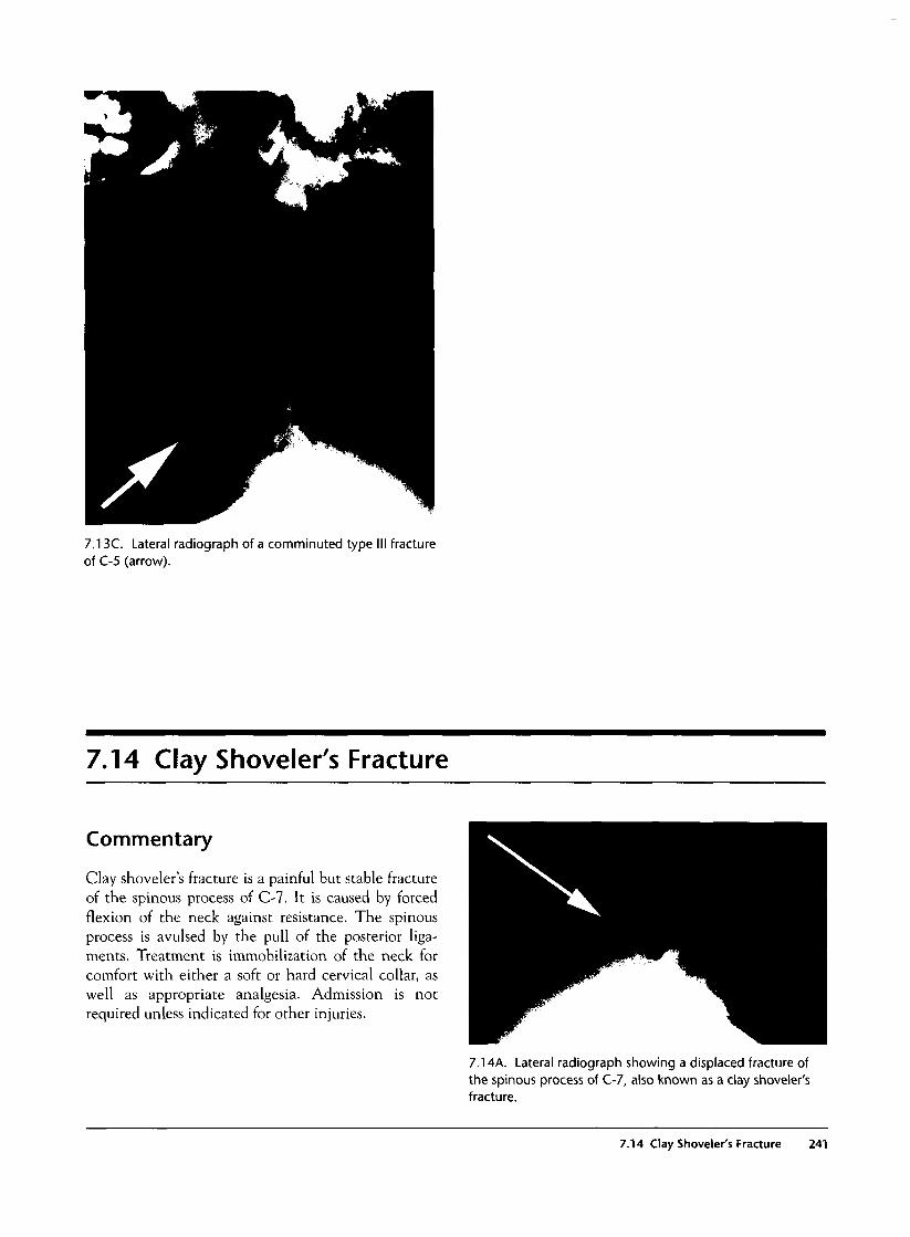
7.13C. Lateral radiograph of a comminuted type Iof C-5 (arrow).
I fracture
7.14 Clay Shoveler's Fracture
Commentary
Clay shoveler's fracture is a painful but stable fractureof the spinous process of C-7. It is caused by forcedflexion of the neck against resistance. The spinousprocess is avulsed by the pull of the posterior liga-ments. Treatment is immobilization of the neck forcomfort with either a soft or hard cervical collar, aswell as appropriate analgesia. Admission is notrequired unless indicated for other injuries.
7.14A. Lateral radiograph showing a displaced fracture ofthe spinous process of C-7, also known as a clay shoveler'sfracture.
7.14 Clay Shoveler's Fracture 241
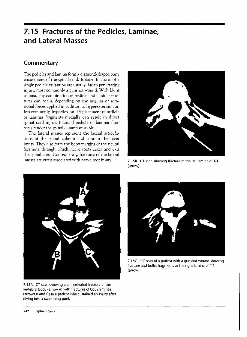
7.15 Fractures of the Pedicles, Laminae,and Lateral Masses
Commentary
The pedicles and lamina form a diamond-shaped bonyencasement of the spinal cord. Isolated fractures of asingle pedicle or lamina are usually due to penetratinginjury, most commonly a gunshot wound. With blunttrauma, any combination of pedicle and laminar frac-tures can occur, depending on the angular or rota-tional forces applied in addition to hyperextension or,less commonly, hyperflexion. Displacement of pedicleor laminar fragments medially can result in directspinal cord injury. Bilateral pedicle or laminar frac-tures render the spinal column unstable.
The lateral masses represent the lateral articula-tions of the spinal column and contain the facetjoints. They also form the bony margins of the neuralforamina through which nerve roots enter and exitthe spinal cord. Consequently, fractures of the lateralmasses are often associated with nerve root injury. 7.15B. CT scan showing fracture of the left lamina of T-1
(arrow).
7.15C. CT scan of a patient with a gunshot wound showingfracture and bullet fragments at the right lamina of T-1(arrow).
7.15A. CT scan showing a comminuted fracture of thevertebral body (arrow A) with fractures of both laminae(arrows B and C) in a patient who sustained an injury afterdiving into a swimming pool.
242 Spinal Injury

7.16 Facet Dislocation
Commentary
Unilateral facet dislocation results from forced flexionof the neck with a rotational component to one sideor the other. If sufficient flexion or rotation occurs,one superior facet will become locked anterior to theinferior facet on that side. The opposite side will shownormal alignment of the facets. Oblique radiographsof the spine will often more clearly demonstrate thedislocation. Subluxation of the vertebral bodies is lessthan 50%, and approximately 25% will have associ-ated spinal cord injury. Reduction is accomplished bysedation and simple traction in most cases.
Bilateral facet dislocation results from forced flex-ion of the neck, causing the superior facets to glide
anteriorly on their inferior counterparts. Once thefacet joints reach the peak of the inferior facet, theyare said to be "perched" atop the inferior facet. If thesuperior facet continues anteriorly, it becomes lockedin the dislocated position. Bilateral facet dislocationresults in greater than 50% subluxation of the superiorvertebral body on the inferior one and almostinevitably results in complete spinal cord injury.
Plain radiographs are usually sufficient to demon-strate facet dislocation, but CT scan will also demon-strate this injury and may reveal unsuspected fracturesof the facets, lateral masses, pedicles, or laminae.
7.16A. Lateral radiograph of the C-spine showingsubluxation of C-5 on C-6 due to unilateral facet dislocationof C-5.
7.16B. Oblique view of unilateral facet dislocation.
7.16 Facet Dislocation 243
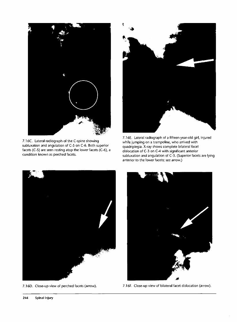
7.16C. Lateral radiograph of the C-spine showingsubluxation and angulation of C-5 on C-6. Both superiorfacets (C-5) are seen resting atop the lower facets (C-6), acondition known as perched facets.
7.16E. Lateral radiograph of a fifteen-year-old girl, injuredwhile jumping on a trampoline, who arrived withquadriplegia. X-ray shows complete bilateral facetdislocation of C-3 on C-4 with significant anteriorsubluxation and angulation of C-3. (Superior facets are lyinganterior to the lower facets; see arrow.)
7.16D. Close-up view of perched facets (arrow). 7.16F. Close-up view of bilateral facet dislocation (arrow).
244 Spinal Injury
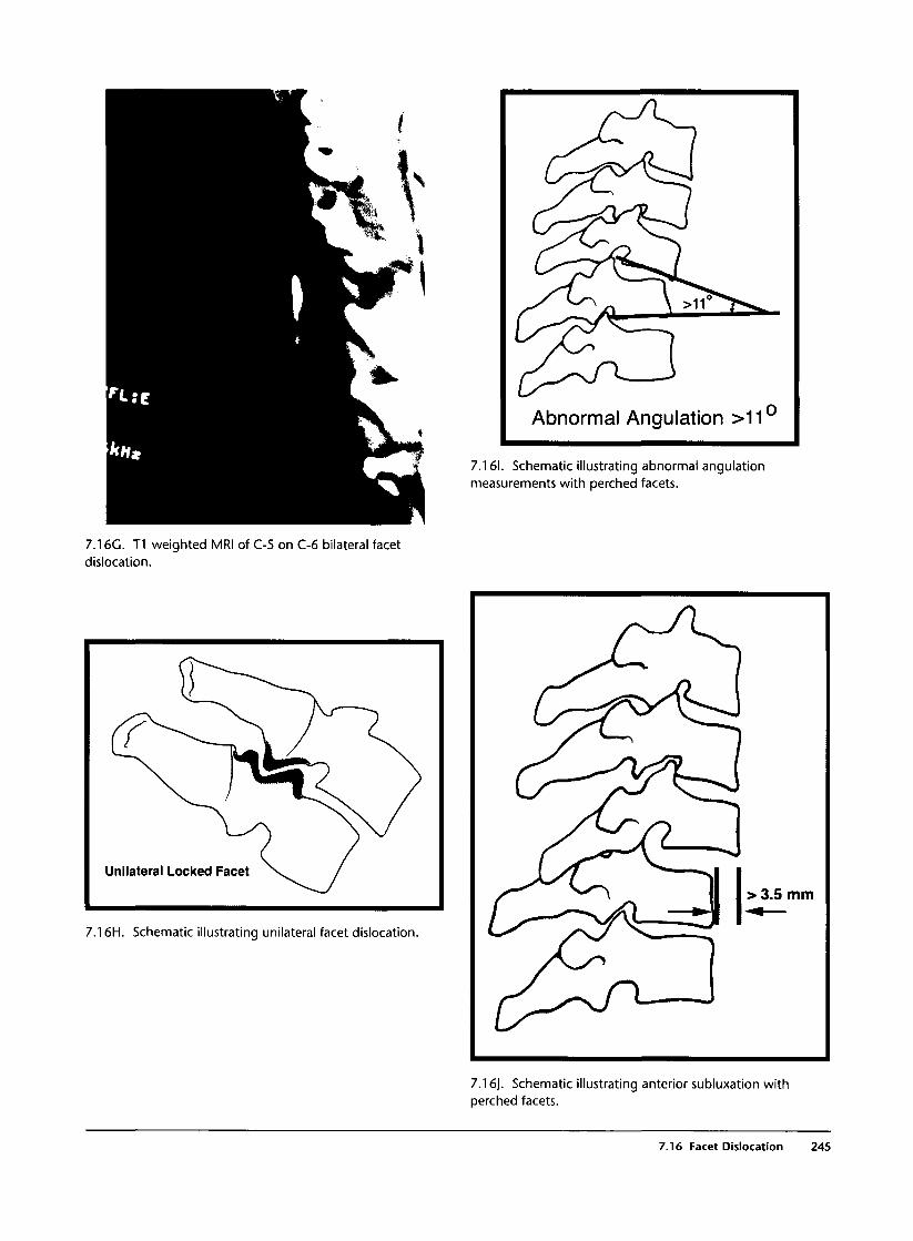
Abnormal Angulation >11
7.161. Schematic illustrating abnormal angulationmeasurements with perched facets.
7.16G. T1 weighted MRI of C-5 on C-6 bilateral facetdislocation.
Unilateral Locked Facet
7.16H. Schematic illustrating unilateral facet dislocation
7.16J. Schematic illustrating anterior subluxation withperched facets.
7.16 Facet Dislocation 245

7.17 Cervicothoracic Spinal Injury
Commentary
The junction of C-7 and T-l is a relatively commonlocation for spinal injury, up to 20% of spinal fracturesin some series. Consequently, it is essential to visualizethis area with plain radiographs on the initial exami-nation prior to clearing the cervical spine. Visualiza-tion of this area is difficult to accomplish in children,patients with large, muscular shoulders, and thosewith upper extremity injuries that make it difficult topull on the arms for a lateral x-ray. Unless the lateralx-ray extends to the top of T-l, a swimmer's view isoften required to verify alignment in this area. Detec-tion of fractures with the swimmer's view is difficultbecause of superimposed bones and soft tissues, andCT scan should be used when visualization of the cer-vicothoracic junction is not possible with conven-tional radiography or when suspicious areas areidentified on the swimmer's view.
7.17B. Repeat lateral view of the same patient with a clearlyapparent bilateral facet dislocation of C-6 on C-7 (arrow)and significant subluxation of C-6 on C-7.
7.1 7A. Lateral cervical spine radiograph appearing normalto the top of C-6 level.
7.17C. Swimmer's view showing bilateral facet dislocationat the cervicothoracic junction.
246 Spinal Injury
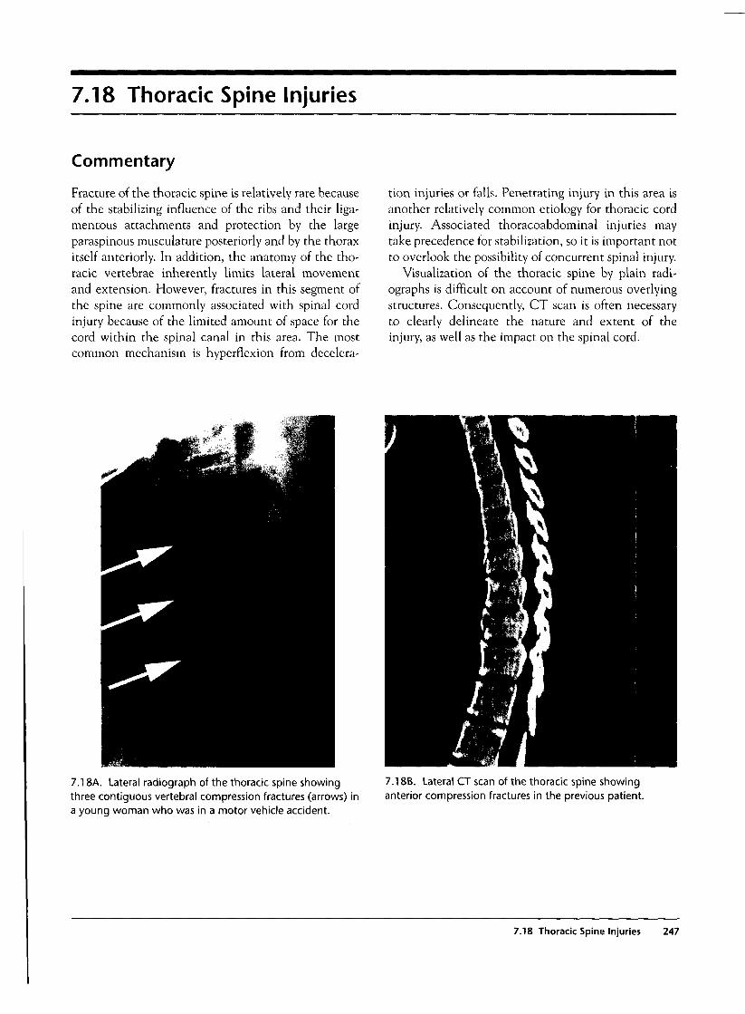
7.18 Thoracic Spine Injuries
Commentary
Fracture of the thoracic spine is relatively rare becauseof the stabilizing influence of the ribs and their liga-mentous attachments and protection by the largeparaspinous musculature posteriorly and by the thoraxitself anteriorly. In addition, the anatomy of the tho-racic vertebrae inherently limits lateral movementand extension. However, fractures in this segment ofthe spine are commonly associated with spinal cordinjury because of the limited amount of space for thecord within the spinal canal in this area. The mostcommon mechanism is hyperflexion from decelera-
tion injuries or falls. Penetrating injury in this area isanother relatively common etiology for thoracic cordinjury. Associated thoracoabdominal injuries maytake precedence for stabilization, so it is important notto overlook the possibility of concurrent spinal injury.
Visualization of the thoracic spine by plain radi-ographs is difficult on account of numerous overlyingstructures. Consequently, CT scan is often necessaryto clearly delineate the nature and extent of theinjury, as well as the impact on the spinal cord.
7.18A. Lateral radiograph of the thoracic spine showingthree contiguous vertebral compression fractures (arrows) ina young woman who was in a motor vehicle accident.
7.18B. Lateral CT scan of the thoracic spine showinganterior compression fractures in the previous patient.
7.18 Thoracic Spine Injuries 247
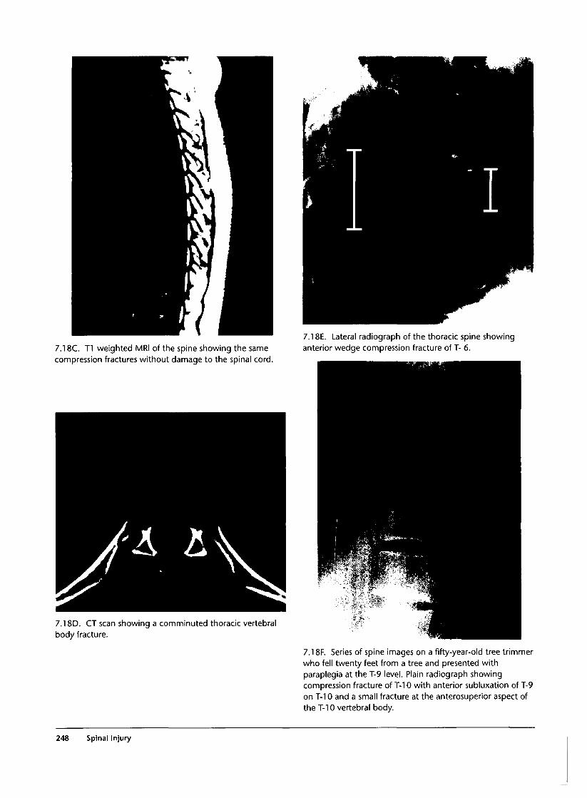
7.18C. T1 weighted MRI of the spine showing the samecompression fractures without damage to the spinal cord.
7.18E. Lateral radiograph of the thoracic spine showinganterior wedge compression fracture of T- 6.
7.18D. CT scan showing a comminuted thoracic vertebralbody fracture.
7.18F. Series of spine images on a fifty-year-old tree trimmerwho fell twenty feet from a tree and presented withparaplegia at the T-9 level. Plain radiograph showingcompression fracture of T-10 with anterior subluxation of T-9on T-10 and a small fracture at the anterosuperior aspect ofthe T-10 vertebral body.
248 Spinal Injury
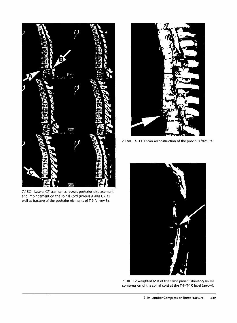
7.18G. Lateral CT scan series reveals posterior displacementand impingement on the spinal cord (arrows A and C), aswell as fracture of the posterior elements of T-9 (arrow B).
7.18H. 3-D CT scan reconstruction of the previous fracture.
7.181. T2 weighted MRI of the same patient showing severecompression of the spinal cord at the T-9-T-10 level (arrow).
7.19 Lumbar Compression Burst Fracture 249
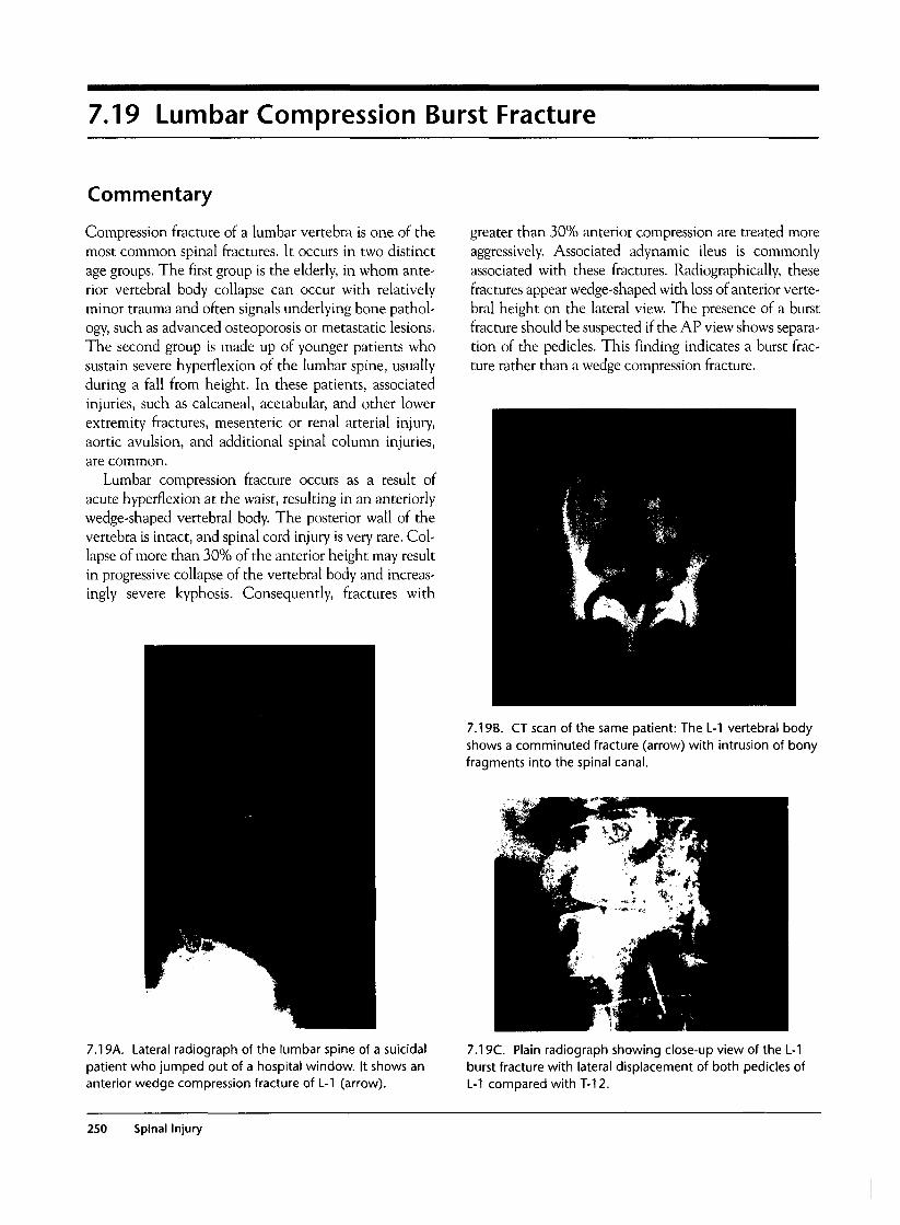
7.19 Lumbar Compression Burst Fracture
Commentary
Compression fracture of a lumbar vertebra is one of themost common spinal fractures. It occurs in two distinctage groups. The first group is the elderly, in whom ante-rior vertebral body collapse can occur with relativelyminor trauma and often signals underlying bone pathol-ogy, such as advanced osteoporosis or metastatic lesions.The second group is made up of younger patients whosustain severe hyperflexion of the lumbar spine, usuallyduring a fall from height. In these patients, associatedinjuries, such as calcaneal, acetabular, and other lowerextremity fractures, mesenteric or renal arterial injury,aortic avulsion, and additional spinal column injuries,are common.
Lumbar compression fracture occurs as a result ofacute hyperflexion at the waist, resulting in an anteriorlywedge-shaped vertebral body. The posterior wall of thevertebra is intact, and spinal cord injury is very rare. Col-lapse of more than 30% of the anterior height may resultin progressive collapse of the vertebral body and increas-ingly severe kyphosis. Consequently, fractures with
greater than 30% anterior compression are treated moreaggressively. Associated adynamic ileus is commonlyassociated with these fractures. Radiographically, thesefractures appear wedge-shaped with loss of anterior verte-bral height on the lateral view. The presence of a burstfracture should be suspected if the AP view shows separa-tion of the pedicles. This finding indicates a burst frac-ture rather than a wedge compression fracture.
7.19B. CT scan of the same patient: The L-l vertebral bodyshows a comminuted fracture (arrow) with intrusion of bonyfragments into the spinal canal.
7.19A. Lateral radiograph of the lumbar spine of a suicidalpatient who jumped out of a hospital window. It shows ananterior wedge compression fracture of L-1 (arrow).
7.19C. Plain radiograph showing close-up view of the L-1burst fracture with lateral displacement of both pedicles ofL-1 compared with T-12.
250 Spinal Injury
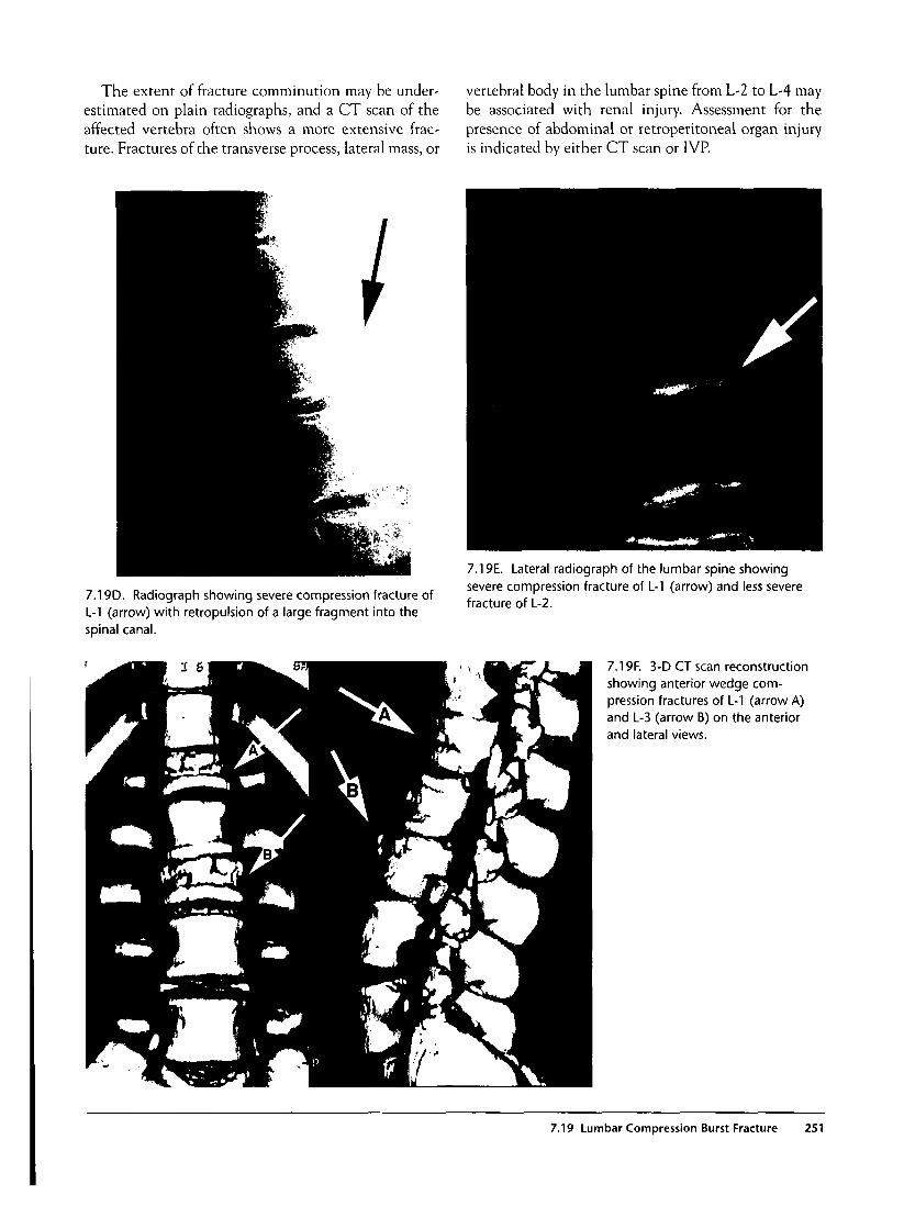
The extent of fracture comminution may be under-estimated on plain radiographs, and a CT scan of theaffected vertebra often shows a more extensive frac-ture. Fractures of the transverse process, lateral mass, or
vertebral body in the lumbar spine from L-2 to L-4 maybe associated with renal injury. Assessment for thepresence of abdominal or retroperitoneal organ injuryis indicated by either CT scan or 1VP.
7.19D. Radiograph showing severe compression fracture ofL-l (arrow) with retropulsion of a large fragment into thespinal canal.
7.19E. Lateral radiograph of the lumbar spine showingsevere compression fracture of L-1 (arrow) and less severefracture of L-2.
7.19F. 3-D CT scan reconstructionshowing anterior wedge com-pression fractures of L-1 (arrow A)and L-3 (arrow B) on the anteriorand lateral views.
7.19 Lumbar Compression Burst Fracture 251
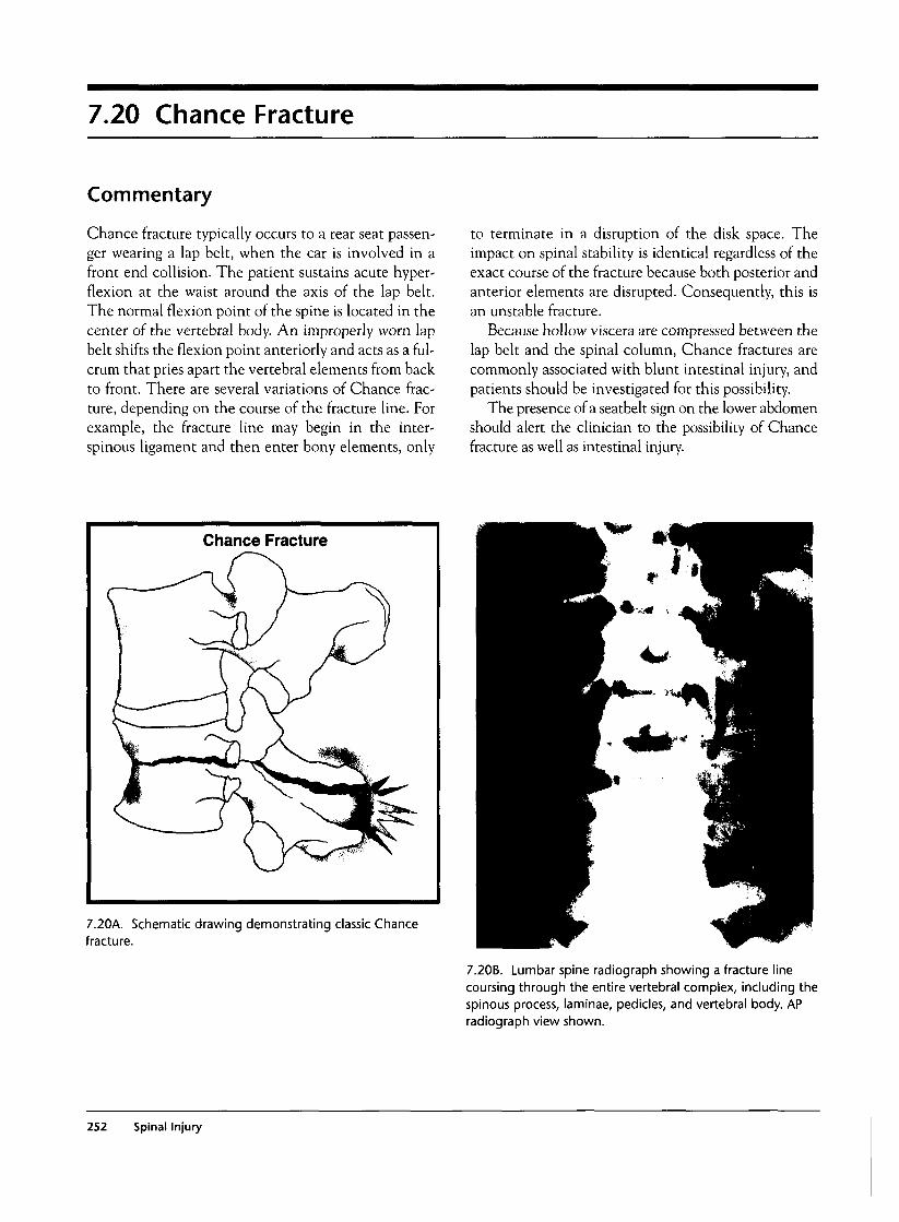
7.20 Chance Fracture
Commentary
Chance fracture typically occurs to a rear seat passen-ger wearing a lap belt, when the car is involved in afront end collision. The patient sustains acute hyper-flexion at the waist around the axis of the lap belt.The normal flexion point of the spine is located in thecenter of the vertebral body. An improperly worn lapbelt shifts the flexion point anteriorly and acts as a ful-crum that pries apart the vertebral elements from backto front. There are several variations of Chance frac-ture, depending on the course of the fracture line. Forexample, the fracture line may begin in the inter-spinous ligament and then enter bony elements, only
to terminate in a disruption of the disk space. Theimpact on spinal stability is identical regardless of theexact course of the fracture because both posterior andanterior elements are disrupted. Consequently, this isan unstable fracture.
Because hollow viscera are compressed between thelap belt and the spinal column, Chance fractures arecommonly associated with blunt intestinal injury, andpatients should be investigated for this possibility.
The presence of a seatbelt sign on the lower abdomenshould alert the clinician to the possibility of Chancefracture as well as intestinal injury.
7.20k. Schematic drawing demonstrating classic Chancefracture.
7.20B. Lumbar spine radiograph showing a fracture linecoursing through the entire vertebral complex, including thespinous process, laminae, pedicles, and vertebral body. APradiograph view shown.
252 Spinal Injury
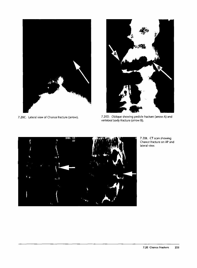
7.20C. Lateral view of Chance fracture (arrow). 7.20D. Oblique showing pedicle fracture (arrow A) andvertebral body fracture (arrow B).
7.20E. CT scan showingChance fracture on AP andlateral view.
7.20 Chance Fracture 253
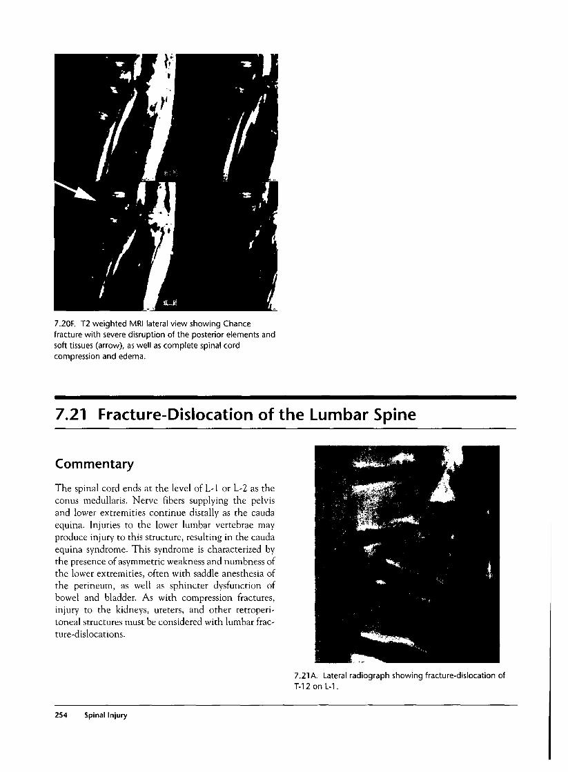
7.20F. T2 weighted MRI lateral view showing Chancefracture with severe disruption of the posterior elements andsoft tissues (arrow), as well as complete spinal cordcompression and edema.
7.21 Fracture-Dislocation of the Lumbar Spine
Commentary
The spinal cord ends at the level of L-l or L-2 as theconus medullaris. Nerve fibers supplying the pelvisand lower extremities continue distally as the caudaequina. Injuries to the lower lumbar vertebrae mayproduce injury to this structure, resulting in the caudaequina syndrome. This syndrome is characterized bythe presence of asymmetric weakness and numbness ofthe lower extremities, often with saddle anesthesia ofthe perineum, as well as sphincter dysfunction ofbowel and bladder. As with compression fractures,injury to the kidneys, ureters, and other retroperi-toneal structures must be considered with lumbar frac-ture-dislocations.
7.21 A. Lateral radiograph showing fracture-dislocation ofT-12 on L-1.
254 Spinal Injury
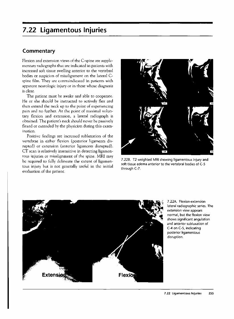
7.22 Ligamentous Injuries
Commentary
Flexion and extension views of the C-spine are supple-mentary radiographs that are indicated in patients withincreased soft tissue swelling anterior to the vertebralbodies or suspicion of misalignment on the lateral C-spine film. They are contraindicated in patients withapparent neurologic injury or in those whose diagnosisis clear.
The patient must be awake and able to cooperate.He or she should be instructed to actively flex andthen extend the neck up to the point of experiencingpain and no further. At the point of maximal volun-tary flexion and extension, a lateral radiograph isobtained. The patient's neck should never be passivelyflexed or extended by the physician during this exam-ination.
Positive findings are increased subluxation of thevertebrae in either flexion (posterior ligaments dis-rupted) or extension (anterior ligaments disrupted).CT scan is relatively insensitive in detecting ligamen-tous injuries or misalignment of the spine. MRI maybe required to fully delineate the extent of ligamen-tous injury but is not generally useful in the initialevaluation of the patient.
7.22B. T2 weighted MRI showing ligamentous injury andsoft tissue edema anterior to the vertebral bodies of C-5through C-7.
7.22A. Flexion-extensionlateral radiographic series. Theextension view appearsnormal, but the flexion viewshows significant angulationand anterior subluxation ofC-4 on C-5, indicatingposterior ligamentousdisruption.
7.22 Ligamentous Injuries 255
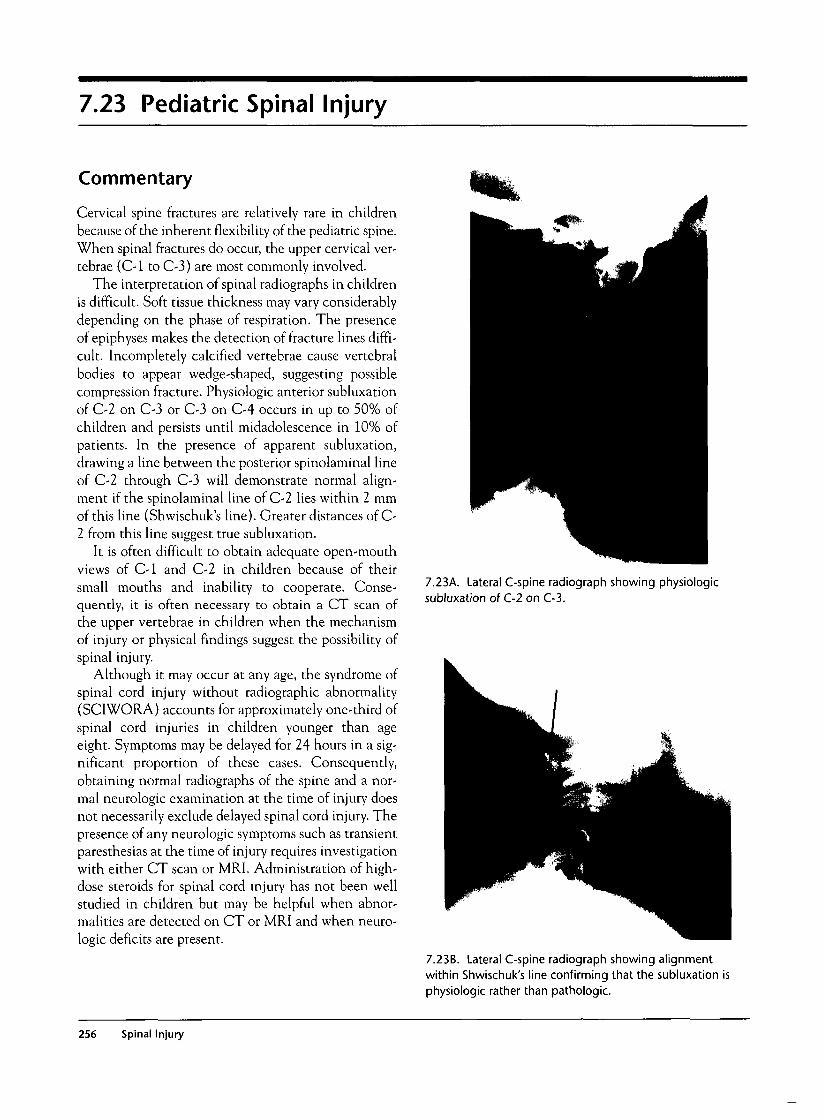
7.23 Pediatric Spinal Injury
Commentary
Cervical spine fractures are relatively rare in childrenbecause of the inherent flexibility of the pediatric spine.When spinal fractures do occur, the upper cervical ver-tebrae (C-l to C-3) are most commonly involved.
The interpretation of spinal radiographs in childrenis difficult. Soft tissue thickness may vary considerablydepending on the phase of respiration. The presenceof epiphyses makes the detection of fracture lines diffi-cult. Incompletely calcified vertebrae cause vertebralbodies to appear wedge-shaped, suggesting possiblecompression fracture. Physiologic anterior subluxationof C-2 on C-3 or C-3 on C-4 occurs in up to 50% ofchildren and persists until midadolescence in 10% ofpatients. In the presence of apparent subluxation,drawing a line between the posterior spinolaminal lineof C-2 through C-3 will demonstrate normal align-ment if the spinolaminal line of C-2 lies within 2 mmof this line (Shwischuk's line). Greater distances of C-2 from this line suggest true subluxation.
It is often difficult to obtain adequate open-mouthviews of C-l and C-2 in children because of theirsmall mouths and inability to cooperate. Conse-quently, it is often necessary to obtain a CT scan ofthe upper vertebrae in children when the mechanismof injury or physical findings suggest the possibility ofspinal injury.
Although it may occur at any age, the syndrome ofspinal cord injury without radiographic abnormality(SCIWORA) accounts for approximately one-third ofspinal cord injuries in children younger than ageeight. Symptoms may be delayed for 24 hours in a sig-nificant proportion of these cases. Consequently,obtaining normal radiographs of the spine and a nor-mal neurologic examination at the time of injury doesnot necessarily exclude delayed spinal cord injury. Thepresence of any neurologic symptoms such as transientparesthesias at the time of injury requires investigationwith either CT scan or MRI. Administration of high-dose steroids for spinal cord injury has not been wellstudied in children but may be helpful when abnor-malities are detected on CT or MRI and when neuro-logic deficits are present.
7.23A. Lateral C-spine radiograph showing physiologicsubluxation of C-2 on C-3.
7.23B. Lateral C-spine radiograph showing alignmentwithin Shwischuk's line confirming that the subluxation isphysiologic rather than pathologic.
256 Spinal Injury
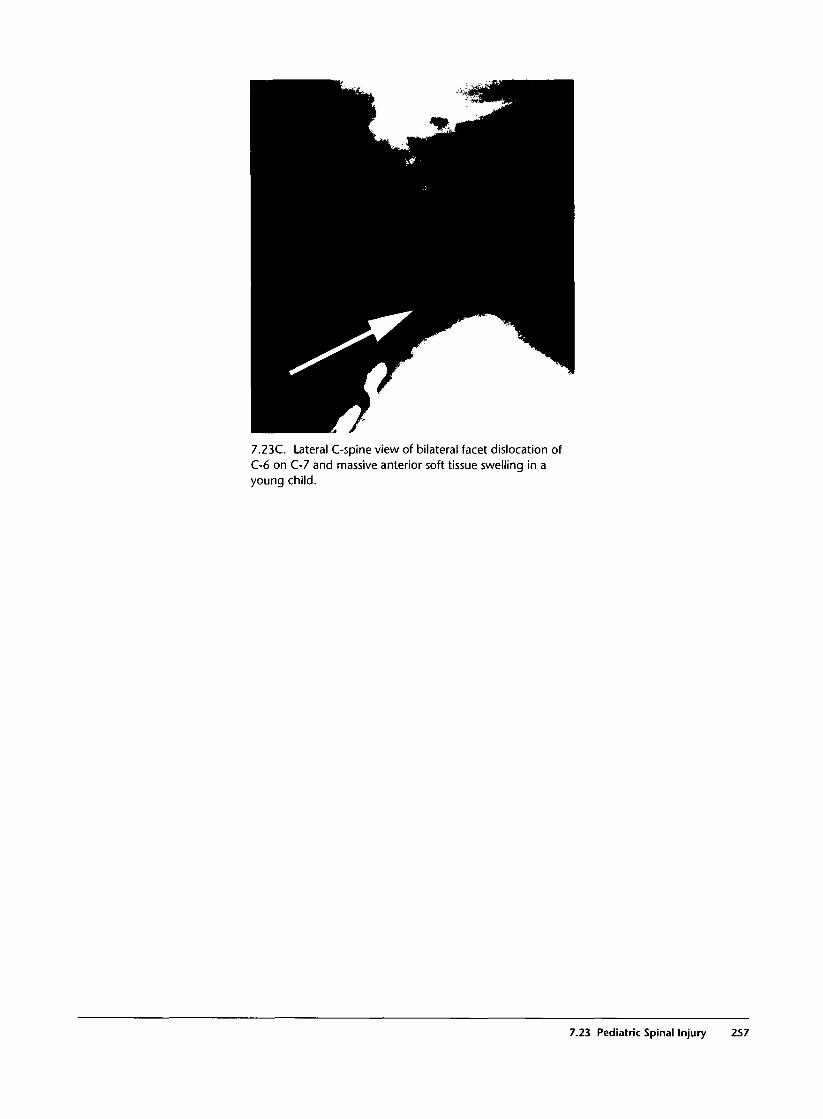
7.23C. Lateral C-spine view of bilateral facet dislocation ofC-6 on C-7 and massive anterior soft tissue swelling in ayoung child.
7.23 Pediatric Spinal Injury 257

Skin and Soft Tissue Injury
Introduction
The skin, being the largest organ and exposed, is verysusceptible to injury, ranging from contusions to deepextensive burns. Furthermore, deeper soft tissue injurycan lead to extensive blood loss, tendon injury, neu-rovascular trauma, and acute compartment syndrome.More than 1.25 million burns are evaluated in emer-gency departments across the United States annually,and these burns account for about 12,000 deaths eachyear. Burn injuries to the skin can be cosmetically andfunctionally devastating, and they account for manyyears of productive lives lost.
Acute burn evaluation requires special expertise andknowledge, given the unique complications theseinjuries can have. Careful history is important. Patientsinvolved in closed-space fires with poor ventilation areat especially high risk for inhalational injury and carbonmonoxide poisoning. The clinician needs to considerassociated blunt trauma injury that may be related to afall or an attempt to escape the flames. Suicide attemptvictims may also have ingested poisons, and thus acoexistent toxicological emergency must be considered.
Clinical Examination
The standard primary survey with ABCDE evaluationis paramount. During this survey, careful attention tofacial burns and possible inhalational injury is neces-sary, as these patients may require immediate airwaycontrol. Impaired lung ventilation may be detected bythe presence of chest burn eschar or bronchospasm.Signs of shock secondary to third-space fluid loss and/orblood loss from another associated injury may be noted.A brief neurological examination is important to deter-mine the possibility of head or spinal injury, and duringthe exposure evaluation all clothing should be removedto evaluate the extent of skin and soft tissue injury.
To evaluate burn injury, both the extent and thedepth are important to determine. Percentage of bodysurface area burned can be calculated by using the rule
of nines or the Lund-Browder chart. In this calcula-tion, only second- and third-degree burns are included.The presence of skin blisters signifies a second-degreeburn, and insensate waxy or charred skin signifies afull-thickness or third-degree burn. Circumferentialburn injury can cause inadequate ventilation if itinvolves the chest or can cause acute compartmentsyndrome of an extremity. Early detection of this acutelimb threat involves careful palpation of the compart-ments and serial evaluation of neurovascular status.
Deep soft tissue lacerations will need exploration todetermine the extent of injury and any possible com-plications. Foreign bodies need to be excluded andbony injury detected by radiographs. Meticulous ten-don and nerve examinations are important to detectsubtle injury.
Investigations
History and examination will determine the need fordiagnostic tests but common investigations needed inmany of these patients are as follows:
1. Arterial blood gas and pulse oximetry. Earlydetection of hypoxia and/or hypercapnia in patientswith inhalational injury is facilitated with these tests.
2. Carbon monoxide (CO) level. Patients in closed-space fires are at high risk for CO poisoning, and thusthis level should be included in blood gas analysis.
3. Chest radiography. Though the chest radiographmay be normal in early inhalational injury, it maydemonstrate pulmonary abnormalities consistent withpulmonary edema.
4. Doppler stethoscope. In patients with extensiveskin burns and extremity edema, peripheral pulses maybe difficult to palpate, and a Doppler stethoscope mayhelp detect weak pulses.
5. Compartment pressure measurement. In equivo-cal cases of suspected compartment syndrome, a bed-side compartment pressure can be measured to helpclinical management.
259
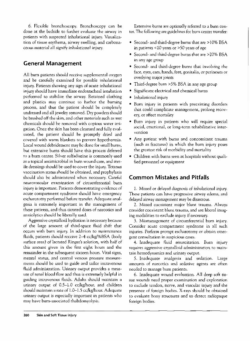
6. Flexible bronchoscopy. Bronchoscopy can bedone at the bedside to further evaluate the airway inpatients with suspected inhalational injury. Visualiza-tion of tissue erythema, airway swelling, and carbona-ceous material all signify inhalational injury.
General Management
All burn patients should receive supplemental oxygenand be carefully examined for possible inhalationalinjury. Patients showing any sign of acute inhalationalinjury should have immediate endotracheal intubationperformed to stabilize the airway. Retained clothingand plastics may continue to harbor the burningprocess, and thus the patient should be completelyundressed and all jewelry removed. Dry powders shouldbe brushed off the skin, and other materials such as wetchemicals should be removed with copious water irri-gation. Once the skin has been cleansed and fully eval-uated, the patient should be promptly dried andcovered with warm blankets to prevent hypothermia.Local wound debridement may be done for small burns,but extensive burns should have this process deferredto a burn center. Silver sulfadiazine is commonly usedas a topical antimicrobial in burn wound care, and ster-ile dressings should be used to cover the injury. Tetanusvaccination status should be obtained, and prophylaxisshould also be administered when necessary. Carefulneurovascular examination of circumferential burninjury is important. Patients demonstrating evidence ofacute compartment syndrome should have emergencyescharotomy performed before transfer. Adequate anal-gesia is extremely important in the management ofthese patients, and thus titrated doses of narcotics andanxiolytics should be liberally used.
Aggressive crystalloid hydration is necessary becauseof the large amount of third-space fluid shift thatoccurs with burn injury. In addition to maintenancefluids, patients should receive 2-4 cc/kg/%BSA (bodysurface area) of lactated Ringer's solution, with half ofthis amount given in the first eight hours and theremainder in the subsequent sixteen hours. Vital signs,mental status, and central venous pressure measure-ments should be used to guide and tailor intravenousfluid administration. Urinary output provides a meas-ure of renal blood flow and thus is extremely helpful inguiding intravenous fluids. Adults should maintain aurinary output of 0.5-1.0 cc/kg/hour, and childrenshould maintain a rate of 1.0-1.5 cc/kg/hour. Adequateurinary output is especially important in patients whomay have burn-associated rhabdomyolysis.
Extensive burns are optimally referred to a burn cen-ter. The following are guidelines for burn center transfer:
• Second- and third-degree burns that are >10% BSAin patients <10 years or >50 years of age
• Second- and third-degree burns that are >20% BSAin any age group
• Second- and third-degree burns that involving theface, eyes, ears, hands, feet, genitalia, or perineum oroverlying major joints
• Third-degree burn >5% BSA in any age group
• Significant electrical and chemical burns
• Inhalational injury
• Burn injury in patients with preexisting disordersthat could complicate management, prolong recov-ery, or affect mortality
• Burn injury in patients who will require specialsocial, emotional, or long-term rehabilitative inter-vention
• Any patient with burns and concomitant trauma(such as fractures) in which the burn injury posesthe greatest risk of morbidity and mortality
• Children with burns seen at hospitals without quali-fied personnel or equipment
Common Mistakes and Pitfalls
1. Missed or delayed diagnosis of inhalational injury.These patients can have progressive airway edema, anddelayed airway management may be disastrous.
2. Missed coexistent major blunt trauma. Alwaysconsider coexistent blunt trauma, and use liberal imag-ing modalities to exclude injury if necessary.
3. Mismanagement of circumferential burn injury.Consider acute compartment syndrome in all suchinjuries. Perform prompt escharotomy or obtain emer-gent consultation in suspicious cases.
4. Inadequate fluid resuscitation. Burn injuryrequires aggressive crystalloid administration to main-tain hemodynamics and urinary output.
5. Inadequate analgesia and sedation. Largeamounts of narcotics and sedative agents are oftenneeded to manage burn patients.
6. Inadequate wound evaluation. All deep soft tis-sue wounds need proper examination and explorationto exclude tendon, nerve, and vascular injury and thepresence of foreign bodies. X-rays should be obtainedto evaluate bony structures and to detect radiopaqueforeign bodies.
260 Skin and Soft Tissue Injury

8.1 Burn Zones of Injury
Commentary
Tissue destruction after acute thermal injury is a con-sequence of heat intensity, duration of exposure, andtissue conductance. This heat injury classically causesthree concentric zones of injury in the pathologicmodel. The central area is where the most intenseburn occurs and cells have been coagulated. Thus, thisarea is termed the zone of coagulation, an area of burnthat has definite tissue destruction. The outermostzone is described as the zone of hyperemia and oftenrepresents tissue that will survive the injury. This
hyperemia will resolve in 7-10 days, but to the inex-perienced clinician this may be confused with celluli-tis of a burn wound. The intermediate area is the zoneof stasis and represents tissue that may or may not sur-vive the burn injury. Progressive tissue loss secondaryto dermal ischemia over the following 24-72 hourswill determine the extent of injury in this zone. Bloodflow may return to this area, but more often progres-sive thrombosis results in deepening and widening ofthe necrotic tissue.
Xhair follicle
i1
\ IJJJJ
i
epidermis
dermis
adipose
8.1 A. Illustration of normalskin layers.
8.1 Burn Zones of Injury 261
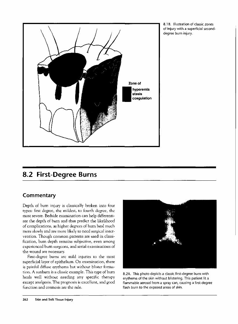
Zone of
hyperemiastasiscoagulation
8.1 B. Illustration of classic zonesof injury with a superficial second-degree burn injury.
8.2 First-Degree Burns
Commentary
Depth of burn injury is classically broken into fourtypes: first degree, the mildest, to fourth degree, themost severe. Bedside examination can help differenti-ate the depth of burn and thus predict the likelihoodof complications, as higher degrees of burn heal muchmore slowly and are more likely to need surgical inter-vention. Though common patterns are used in classi-fication, burn depth remains subjective, even amongexperienced burn surgeons, and serial examinations ofthe wound are necessary.
First-degree burns are mild injuries to the mostsuperficial layer of epithelium. On examination, thereis painful diffuse erythema but without blister forma-tion. A sunburn is a classic example. This type of burnheals well without needing any specific therapyexcept analgesia. The prognosis is excellent, and goodfunction and cosmesis are the rule.
8.2A. This photo depicts a classic first-degree burn witherythema of the skin without blistering. This patient lit aflammable aerosol from a spray can, causing a first-degreeflash burn to the exposed areas of skin.
262 Skin and Soft Tissue Injury

8.3 Second-Degree Burns
Commentary
Second-degree burns involve injury to the dermislayer. Typically, blisters are present, and the wound isextremely painful. These second-degree burns can beclassified further into superficial (top one-third der-mis) and deep (bottom two-thirds dermis) burns.Superficial burns have pink tissue under the blisters,and deep burns have pink and white mottling of thesame areas. The distinction of superficial and deepsecond-degree burns is important, as deeper sectionsof the dermis house skin appendages, hair shafts, andsweat and sebaceous glands that initiate reepithelial-ization of the burned area. Thus, burns affecting theseimportant structures heal more slowly and with poorresults. Typically, superficial second-degree burns willheal in less than three weeks, while deep burns heal inthree to nine weeks with significant scar formation.
The first step of medical therapy is cleansing theburned tissue of loose debris, blisters, and foreign bod-
ies with saline and sterile gauzes after administrationof adequate analgesia. Superficial second-degree burnsthat are expected to heal within two to three weeksare often treated with topical antibiotic in the form of1% silver sulfadiazine cream. The wound is then cov-ered with a loose, bulky dressing, with frequentrecleansing, reapplication of topical antibiotic, anddressing changes.
Because of slow and poor healing, deep second-degree wounds are considered candidates for early sur-gical therapy. Tangential excison is performed toremove the necrotic eschar so that the wound isexcised down to viable tissue. Ideally, at this pointimmediate allografting is performed. If a donor site isnot available, then fresh or cryopreserved human allo-graft can provide physiological wound closure untilrejection occurs two to four weeks later. Subsequently,the wound can be covered with reharvested allograft.
Zone of
hyperemiastasiscoagulation
8.3A. Illustration of cross sectionof a deep second-degree burn ofthe skin.
8.3 Second-Degree Burns 263
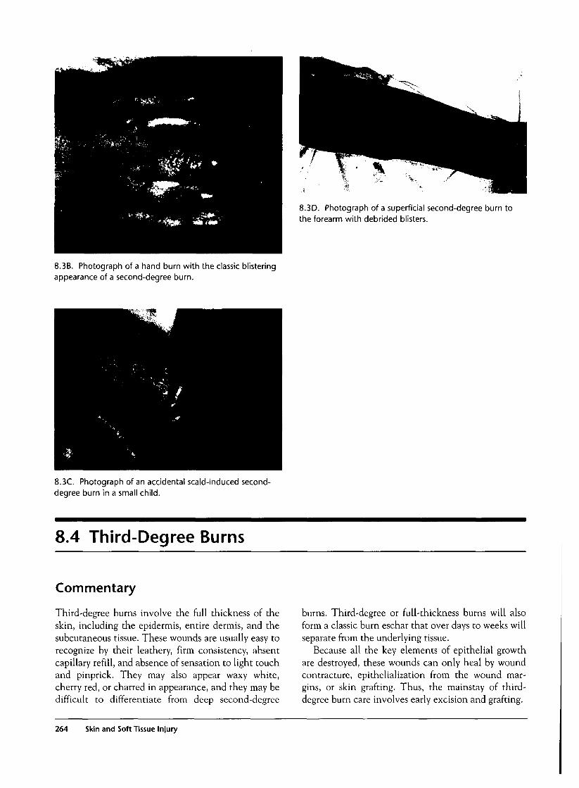
8.3D. Photograph of a superficial second-degree burn tothe forearm with debrided blisters.
8.3B. Photograph of a hand burn with the classic blisteringappearance of a second-degree burn.
8.3C. Photograph of an accidental scald-induced second-degree burn in a small child.
8.4 Third-Degree Burns
Commentary
Third-degree burns involve the full thickness of theskin, including the epidermis, entire dermis, and thesubcutaneous tissue. These wounds are usually easy torecognize by their leathery, firm consistency, absentcapillary refill, and absence of sensation to light touchand pinprick. They may also appear waxy white,cherry red, or charred in appearance, and they may bedifficult to differentiate from deep second-degree
burns. Third-degree or full-thickness burns will alsoform a classic burn eschar that over days to weeks willseparate from the underlying tissue.
Because all the key elements of epithelial growthare destroyed, these wounds can only heal by woundcontracture, epithelialization from the wound mar-gins, or skin grafting. Thus, the mainstay of third-degree burn care involves early excision and grafting.
264 Skin and Soft Tissue Injury
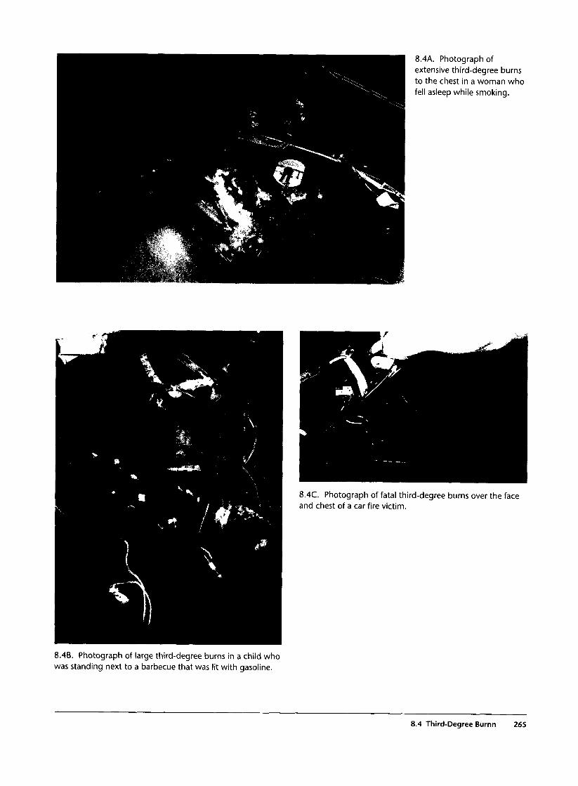
8.4A. Photograph ofextensive third-degree burnsto the chest in a woman whofell asleep while smoking.
8.4C. Photograph of fatal third-degree burns over the faceand chest of a car fire victim.
8.4B. Photograph of large third-degree burns in a child whowas standing next to a barbecue that was lit with gasoline.
8.4 Third-Degree Burnn 265
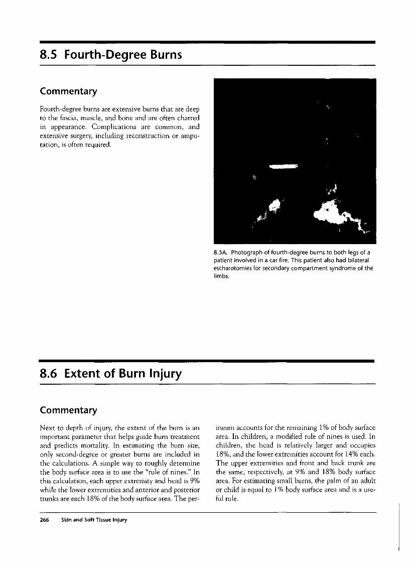
8.5 Fourth-Degree Burns
Commentary
Fourth-degree burns are extensive burns that are deepto the fascia, muscle, and bone and are often charredin appearance. Complications are common, andextensive surgery, including reconstruction or ampu-tation, is often required.
8.5A. Photograph of fourth-degree burns to both legs of apatient involved in a car fire. This patient also had bilateralescharotomies for secondary compartment syndrome of thelimbs.
8.6 Extent of Burn Injury
Commentary
Next to depth of injury, the extent of the burn is animportant parameter that helps guide burn treatmentand predicts mortality. In estimating the burn size,only second-degree or greater burns are included inthe calculations. A simple way to roughly determinethe body surface area is to use the "rule of nines." Inthis calculation, each upper extremity and head is 9%while the lower extremities and anterior and posteriortrunks are each 18% of the body surface area. The per-
ineum accounts for the remaining 1 % of body surfacearea. In children, a modified rule of nines is used. Inchildren, the head is relatively larger and occupies18%, and the lower extremities account for 14% each.The upper extremities and front and back trunk arethe same, respectively, at 9% and 18% body surfacearea. For estimating small burns, the palm of an adultor child is equal to 1% body surface area and is a use-ful rule.
266 Skin and Soft Tissue Injury
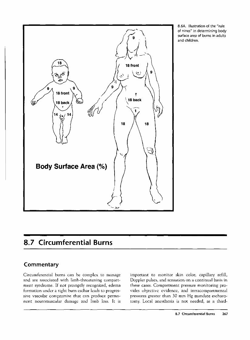
Body Surface Area (%
8.6A. Illustration of the "ruleof nines" in determining bodysurface area of burns in adultsand children.
8.7 Circumferential Burns
Commentary
Circumferential burns can be complex to manageand are associated with limb-threatening compart-ment syndrome. If not promptly recognized, edemaformation under a tight burn eschar leads to progres-sive vascular compromise that can produce perma-nent neuromuscular damage and limb loss. It is
important to monitor skin color, capillary refill,Doppler pulses, and sensation on a continual basis inthese cases. Compartment pressure monitoring pro-vides objective evidence, and intracompartmentalpressures greater than 30 mm Hg mandate escharo-tomy. Local anesthesia is not needed, as a third-
8.7 Circumferential Burns 267
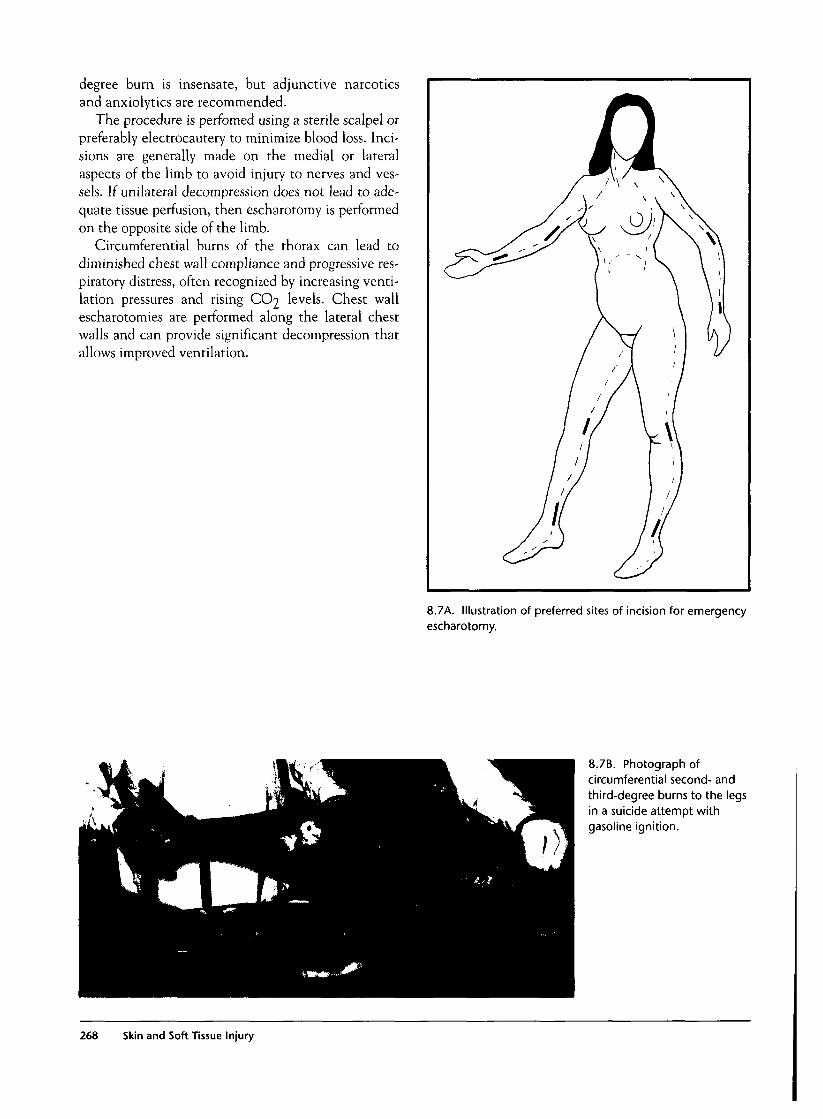
degree burn is insensate, but adjunctive narcoticsand anxiolytics are recommended.
The procedure is perfomed using a sterile scalpel orpreferably electrocautery to minimize blood loss. Inci-sions are generally made on the medial or lateralaspects of the limb to avoid injury to nerves and ves-sels. If unilateral decompression does not lead to ade-quate tissue perfusion, then escharotomy is performedon the opposite side of the limb.
Circumferential burns of the thorax can lead todiminished chest wall compliance and progressive res-piratory distress, often recognized by increasing venti-lation pressures and rising CO2 levels. Chest wallescharotomies are performed along the lateral chestwalls and can provide significant decompression thatallows improved ventilation.
8.7A. Illustration of preferred sites of incision for emergencyescharotomy.
8.7B. Photograph ofcircumferential second- andthird-degree burns to the legsin a suicide attempt withgasoline ignition.
268 Skin and Soft Tissue Injury
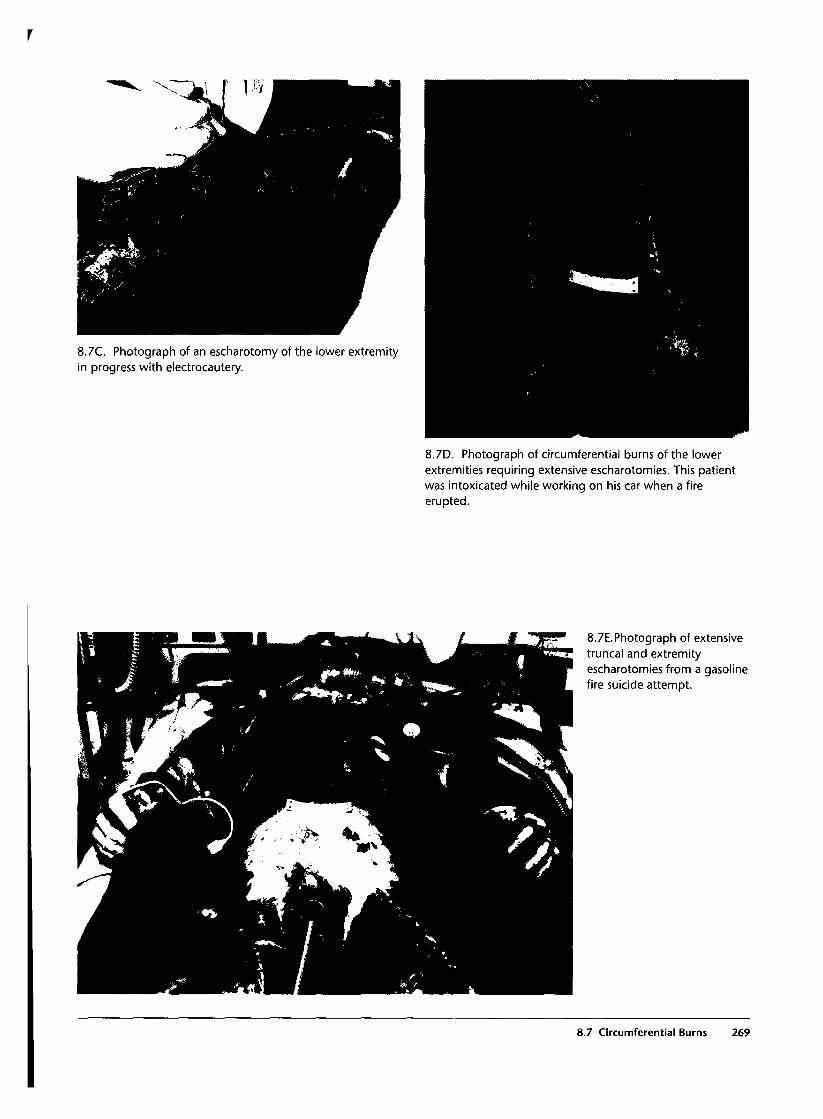
r
8.7C. Photograph of an escharotomy of the lower extremityin progress with electrocautery.
8.7D. Photograph of circumferential burns of the lowerextremities requiring extensive escharotomies. This patientwas intoxicated while working on his car when a fireerupted.
8.7E.Photograph of extensivetruncal and extremityescharotomies from a gasolinefire suicide attempt.
8.7 Circumferential Burns 269
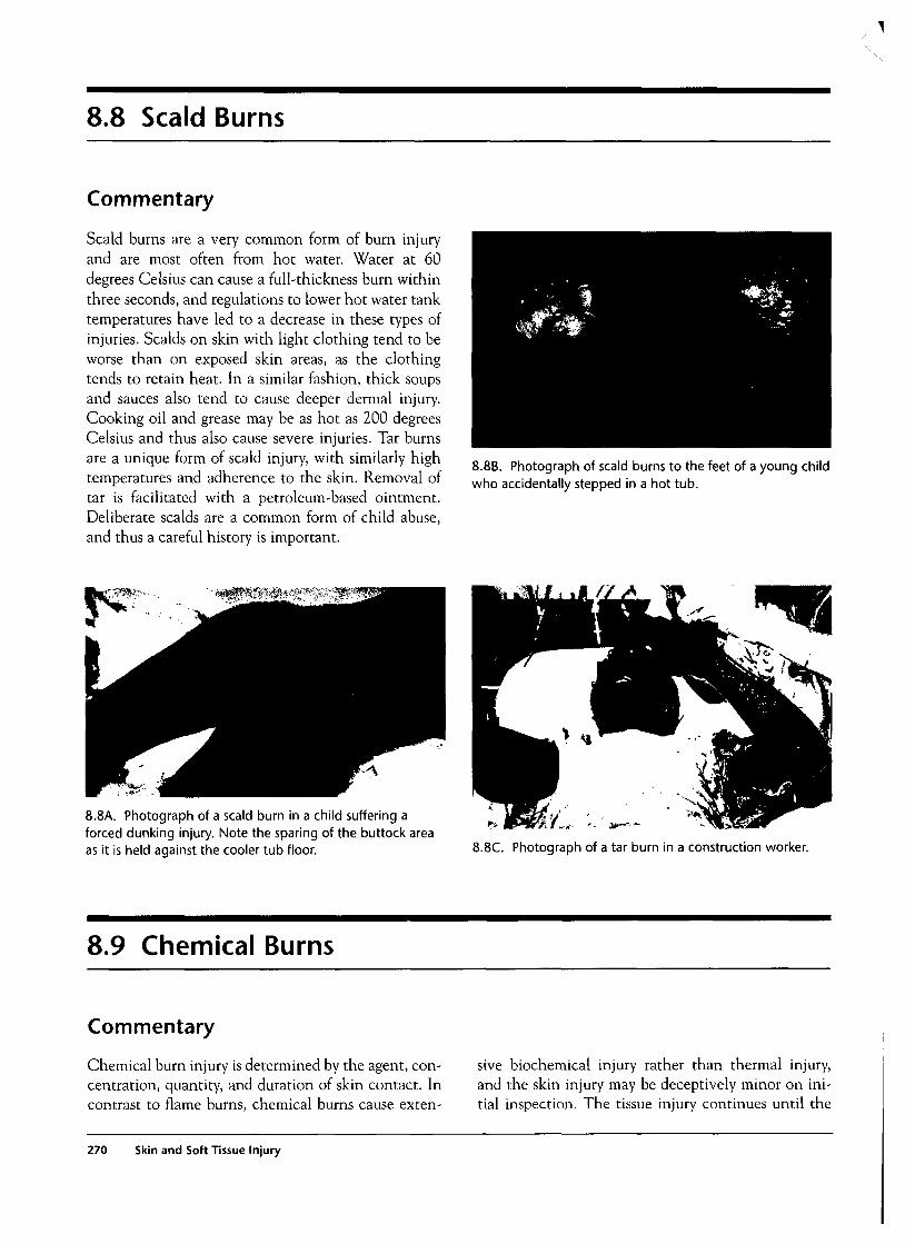
8.8 Scald Burns
Commentary
Scald burns are a very common form of burn injuryand are most often from hot water. Water at 60degrees Celsius can cause a full-thickness burn withinthree seconds, and regulations to lower hot water tanktemperatures have led to a decrease in these types ofinjuries. Scalds on skin with light clothing tend to beworse than on exposed skin areas, as the clothingtends to retain heat. In a similar fashion, thick soupsand sauces also tend to cause deeper dermal injury.Cooking oil and grease may be as hot as 200 degreesCelsius and thus also cause severe injuries. Tar burnsare a unique form of scald injury, with similarly hightemperatures and adherence to the skin. Removal oftar is facilitated with a petroleum-based ointment.Deliberate scalds are a common form of child abuse,and thus a careful history is important.
8.8B. Photograph of scald burns to the feet of a young childwho accidentally stepped in a hot tub.
8.8A. Photograph of a scald burn in a child suffering aforced dunking injury. Note the sparing of the buttock areaas it is held against the cooler tub floor. 8.8C. Photograph of a tar burn in a construction worker.
8.9 Chemical Burns
Commentary
Chemical burn injury is determined by the agent, con-centration, quantity, and duration of skin contact. Incontrast to flame burns, chemical burns cause exten-
sive biochemical injury rather than thermal injury,and the skin injury may be deceptively minor on ini-tial inspection. The tissue injury continues until the
270 Skin and Soft Tissue Injury
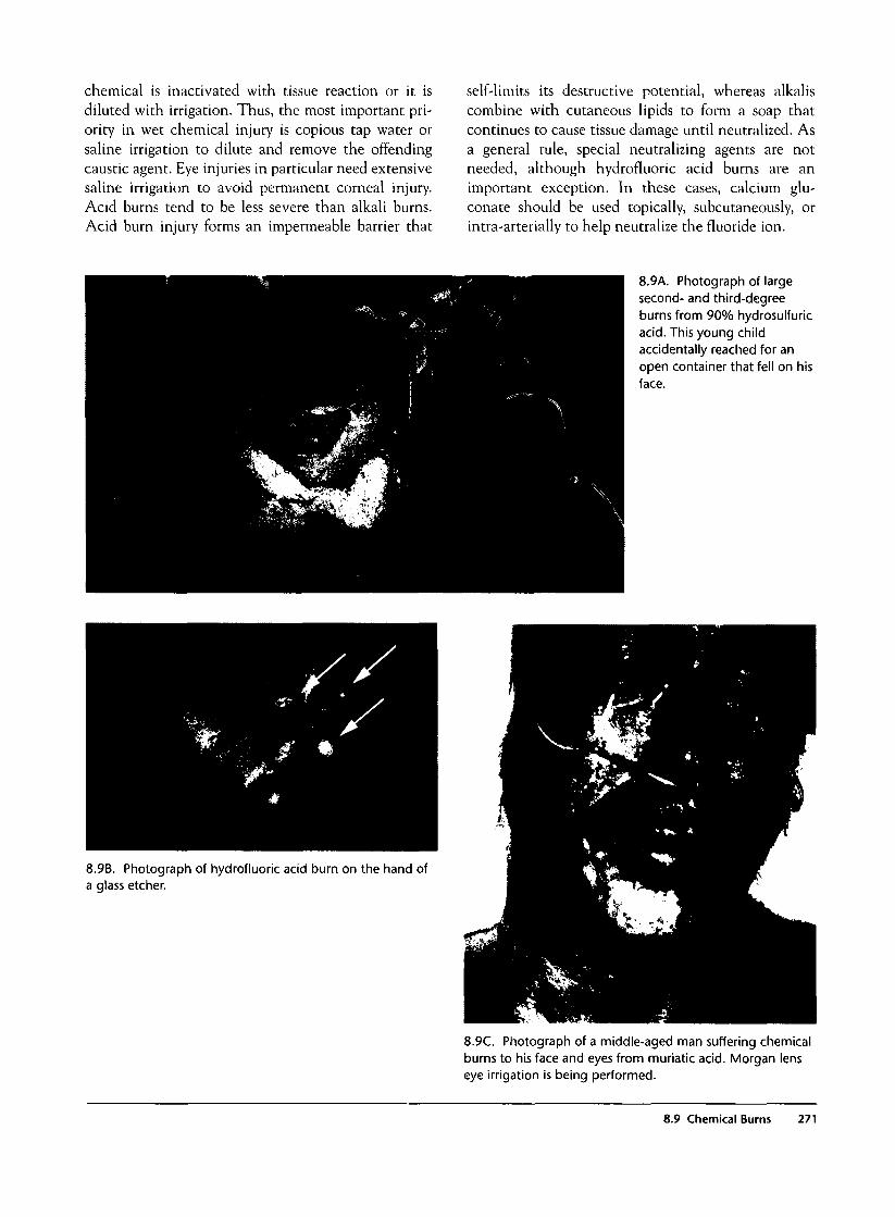
chemical is inactivated with tissue reaction or it isdiluted with irrigation. Thus, the most important pri-ority in wet chemical injury is copious tap water orsaline irrigation to dilute and remove the offendingcaustic agent. Eye injuries in particular need extensivesaline irrigation to avoid permanent corneal injury.Acid burns tend to be less severe than alkali burns.Acid burn injury forms an impermeable barrier that
self-limits its destructive potential, whereas alkaliscombine with cutaneous lipids to form a soap thatcontinues to cause tissue damage until neutralized. Asa general rule, special neutralizing agents are notneeded, although hydrofluoric acid burns are animportant exception. In these cases, calcium glu-conate should be used topically, subcutaneously, orintra-arterially to help neutralize the fluoride ion.
8.9A. Photograph of largesecond- and third-degreeburns from 90% hydrosulfuricacid. This young childaccidentally reached for anopen container that fell on hisface.
8.9B. Photograph of hydrofluoric acid burn on the hand ofa glass etcher.
8.9C. Photograph of a middle-aged man suffering chemicalburns to his face and eyes from muriatic acid. Morgan lenseye irrigation is being performed.
8.9 Chemical Burns 271
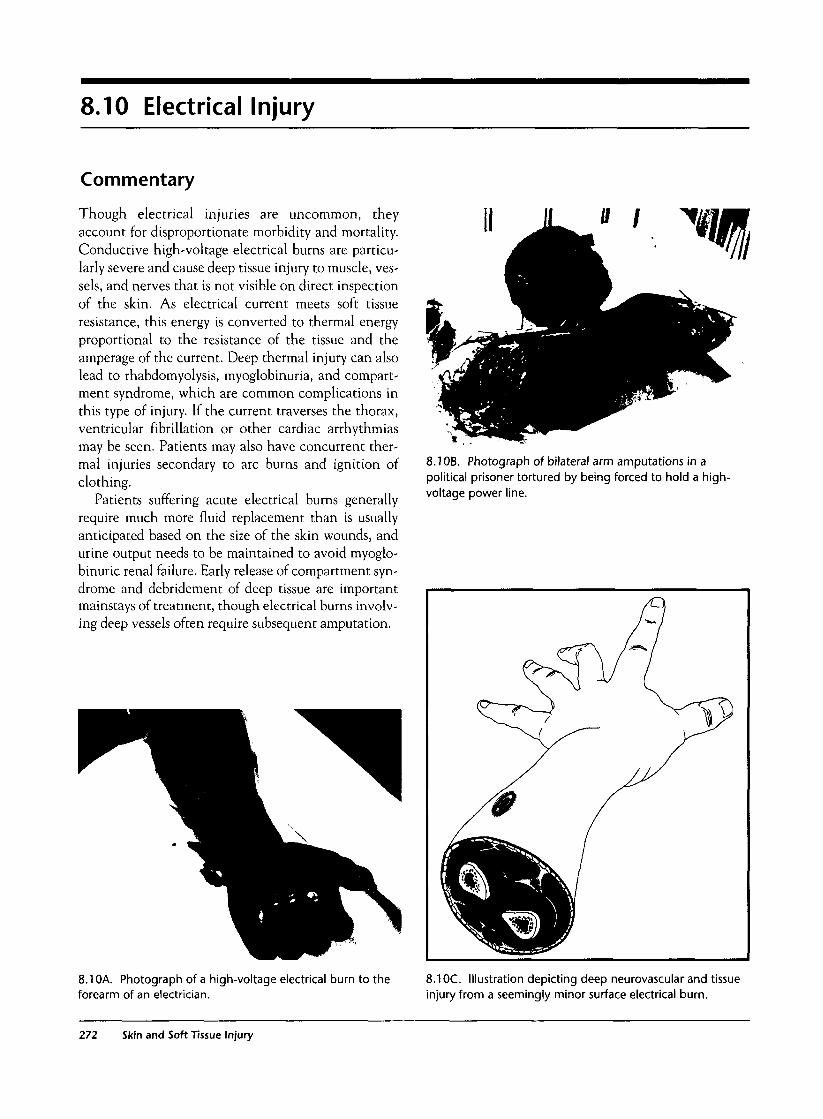
8.10 Electrical Injury
Commentary
Though electrical injuries are uncommon, theyaccount for disproportionate morbidity and mortality.Conductive high-voltage electrical burns are particu-larly severe and cause deep tissue injury to muscle, ves-sels, and nerves that is not visible on direct inspectionof the skin. As electrical current meets soft tissueresistance, this energy is converted to thermal energyproportional to the resistance of the tissue and theamperage of the current. Deep thermal injury can alsolead to rhabdomyolysis, myoglobinuria, and compart-ment syndrome, which are common complications inthis type of injury. If the current traverses the thorax,ventricular fibrillation or other cardiac arrhythmiasmay be seen. Patients may also have concurrent ther-mal injuries secondary to arc burns and ignition ofclothing.
Patients suffering acute electrical burns generallyrequire much more fluid replacement than is usuallyanticipated based on the size of the skin wounds, andurine output needs to be maintained to avoid myoglo-binuric renal failure. Early release of compartment syn-drome and debridement of deep tissue are importantmainstays of treatment, though electrical burns involv-ing deep vessels often require subsequent amputation.
8.1 OB. Photograph of bilateral arm amputations in apolitical prisoner tortured by being forced to hold a high-voltage power line.
8.10A. Photograph of a high-voltage electrical burn to theforearm of an electrician.
8.1 OC. Illustration depicting deep neurovascular and tissueinjury from a seemingly minor surface electrical burn.
272 Skin and Soft Tissue Injury
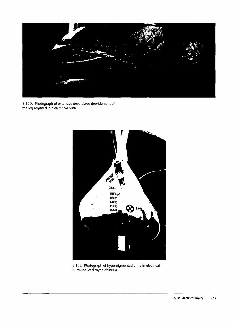
8.1 OD. Photograph of extensive deep tissue debridement ofthe leg required in a electrical burn.
8.1 OE. Photograph of hyperpigmented urine in electricalburn-induced myoglobinuria.
8.10 Electrical Injury 273

8.11 Inhalational Injury
Commentary
Smoke inhalational injury should be suspected in allpatients in fires that occur within a closed space. Hun-dreds of toxic substances are present in smoke particles,and inhalation causes direct epithelial damage leading toedema, submucosal hemorrhage, and necrosis, mostly inthe lower airways. Microatelectasis and interstitial andalveolar edema subsequently occur and can lead to respi-ratory failure. One of the most significant early compli-cations is upper airway edema, which, if not treated earlywith intubation, can lead to airway obstruction.
Patients may present with a combination of cough,shortness of breath, hoarseness, and wheezing. Facialburns and singed facial hairs may be seen, and, if present,carbonaceous sputum is a very specific sign. In question-able cases, fiberoptic bronchoscopy provides direct visu-alization of the affected airways for definitive diagnosis.Early chest radiographs are often normal, while delayedradiographs show pulmonary edema in advanced cases.Supportive care with humidified oxygen and beta-ago-nists for wheezing suffice for most minor cases, but intu-bation and ventilatory support are necessary for seriousdisease. Of particular note, significant carbon monoxideand cyanide poisoning can lead to lethal inhalations.
8.11B. Chest radiograph with noncardiogenic pulmonaryedema in a patient with severe inhalational injury.
8.11 A. Photograph of a facial bum with burned nasal hairsin a young adult with severe inhalational injury.
8.11C. Photograph showing the laryngotracheal view ofinhalational injury at autopsy. Note the extensive erythemaand carbonaceous material throughout the airway.
274 Skin and Soft Tissue Injury
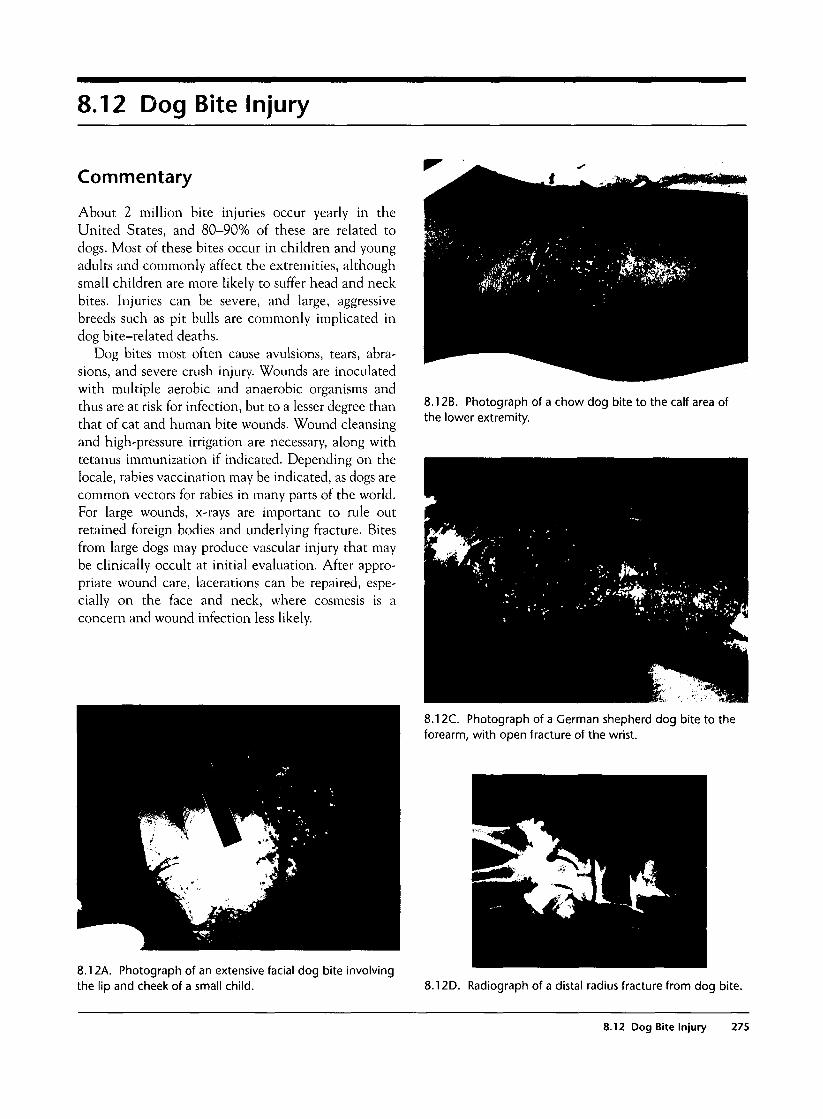
8.12 Dog Bite Injury
Commentary
About 2 million bite injuries occur yearly in theUnited States, and 80-90% of these are related todogs. Most of these bites occur in children and youngadults and commonly affect the extremities, althoughsmall children are more likely to suffer head and neckbites. Injuries can be severe, and large, aggressivebreeds such as pit bulls are commonly implicated indog bite-related deaths.
Dog bites most often cause avulsions, tears, abra-sions, and severe crush injury. Wounds are inoculatedwith multiple aerobic and anaerobic organisms andthus are at risk for infection, but to a lesser degree thanthat of cat and human bite wounds. Wound cleansingand high-pressure irrigation are necessary, along withtetanus immunization if indicated. Depending on thelocale, rabies vaccination may be indicated, as dogs arecommon vectors for rabies in many parts of the world.For large wounds, x-rays are important to rule outretained foreign bodies and underlying fracture. Bitesfrom large dogs may produce vascular injury that maybe clinically occult at initial evaluation. After appro-priate wound care, lacerations can be repaired, espe-cially on the face and neck, where cosmesis is aconcern and wound infection less likely.
8.12B. Photograph of a chow dog bite to the calf area ofthe lower extremity.
8.12C. Photograph of a German shepherd dog bite to theforearm, with open fracture of the wrist.
8.12A. Photograph of an extensive facial dog bite involvingthe lip and cheek of a small child. 8.12D. Radiograph of a distal radius fracture from dog bite.
8.12 Dog Bite Injury 275
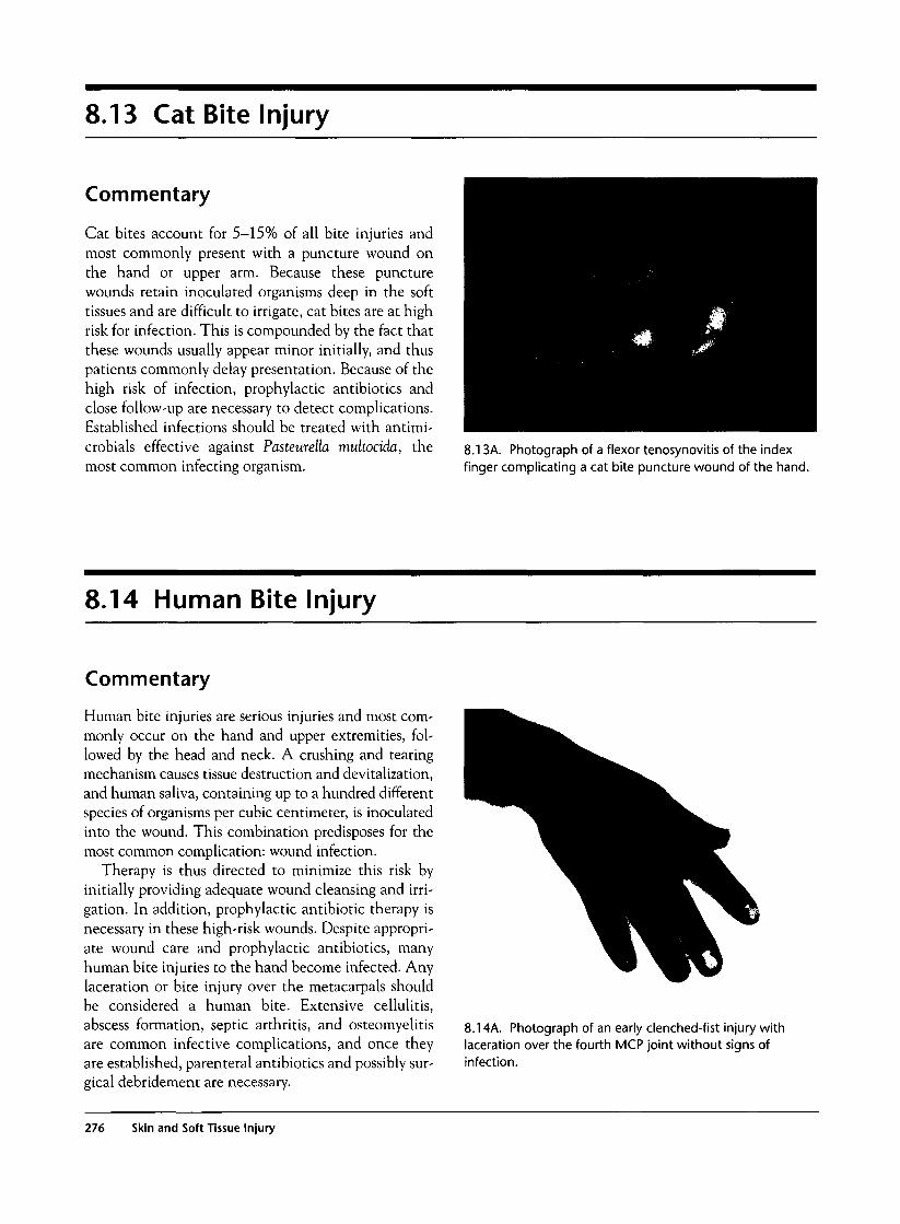
8.13 Cat Bite Injury
Commentary
Cat bites account for 5-15% of all bite injuries andmost commonly present with a puncture wound onthe hand or upper arm. Because these puncturewounds retain inoculated organisms deep in the softtissues and are difficult to irrigate, cat bites are at highrisk for infection. This is compounded by the fact thatthese wounds usually appear minor initially, and thuspatients commonly delay presentation. Because of thehigh risk of infection, prophylactic antibiotics andclose follow-up are necessary to detect complications.Established infections should be treated with antimi-crobials effective against Pasteurella multocida, themost common infecting organism.
8.13A. Photograph of a flexor tenosynovitis of the indexfinger complicating a cat bite puncture wound of the hand.
8.14 Human Bite Injury
Commentary
Human bite injuries are serious injuries and most com-monly occur on the hand and upper extremities, fol-lowed by the head and neck. A crushing and tearingmechanism causes tissue destruction and devitalization,and human saliva, containing up to a hundred differentspecies of organisms per cubic centimeter, is inoculatedinto the wound. This combination predisposes for themost common complication: wound infection.
Therapy is thus directed to minimize this risk byinitially providing adequate wound cleansing and irri-gation. In addition, prophylactic antibiotic therapy isnecessary in these high-risk wounds. Despite appropri-ate wound care and prophylactic antibiotics, manyhuman bite injuries to the hand become infected. Anylaceration or bite injury over the metacarpals shouldbe considered a human bite. Extensive cellulitis,abscess formation, septic arthritis, and osteomyelitisare common infective complications, and once theyare established, parenteral antibiotics and possibly sur-gical debridement are necessary.
8.14A. Photograph of an early clenched-fist injury withlaceration over the fourth MCP joint without signs ofinfection.
276 Skin and Soft Tissue Injury
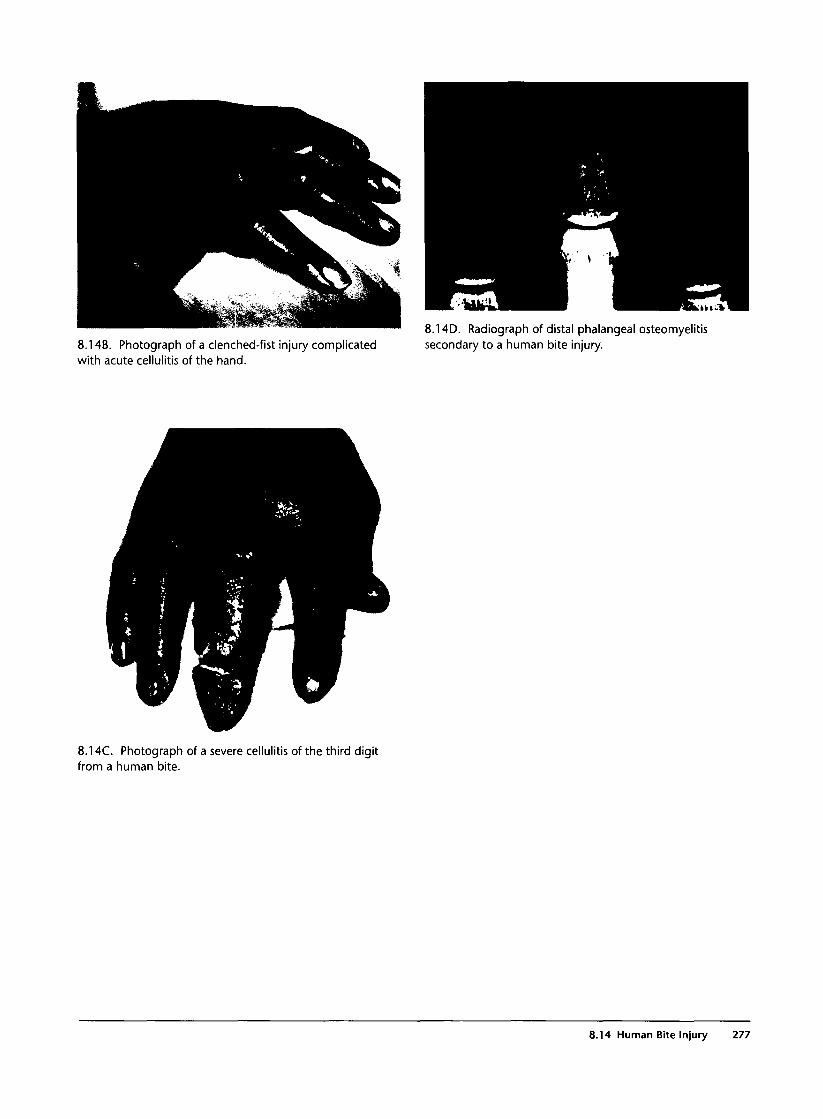
8.14B. Photograph of a clenched-fist injury complicatedwith acute cellulitis of the hand.
8.14D. Radiograph of distal phalangeal osteomyelitissecondary to a human bite injury.
8.14C. Photograph of a severe cellulitis of the third digitfrom a human bite.
8.14 Human Bite Injury 277
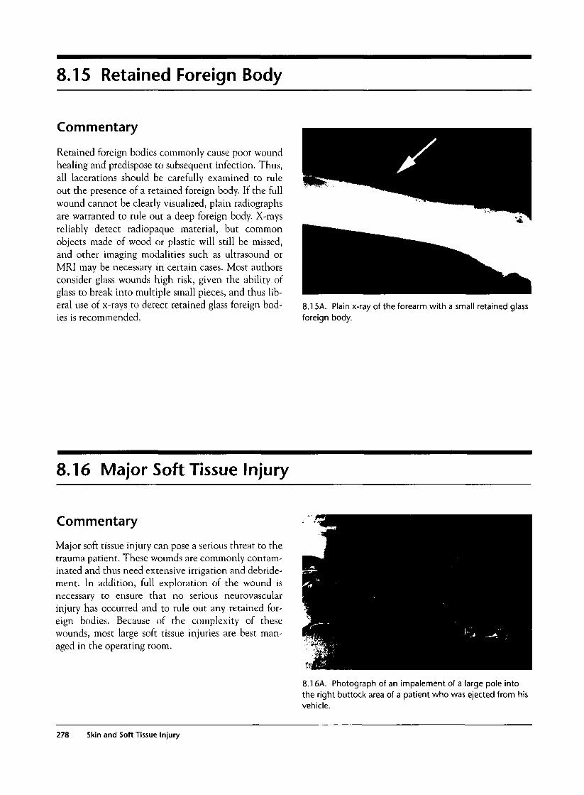
8.15 Retained Foreign Body
Commentary
Retained foreign bodies commonly cause poor woundhealing and predispose to subsequent infection. Thus,all lacerations should be carefully examined to ruleout the presence of a retained foreign body. If the fullwound cannot be clearly visualized, plain radiographsare warranted to rule out a deep foreign body. X-raysreliably detect radiopaque material, but commonobjects made of wood or plastic will still be missed,and other imaging modalities such as ultrasound orMRI may be necessary in certain cases. Most authorsconsider glass wounds high risk, given the ability ofglass to break into multiple small pieces, and thus lib-eral use of x-rays to detect retained glass foreign bod-ies is recommended.
8.15A. Plain x-ray of the forearm with a small retained glassforeign body.
8.16 Major Soft Tissue Injury
Commentary
Major soft tissue injury can pose a serious threat to thetrauma patient. These wounds are commonly contam-inated and thus need extensive irrigation and debride-ment. In addition, full exploration of the wound isnecessary to ensure that no serious neurovascularinjury has occurred and to rule out any retained for-eign bodies. Because of the complexity of thesewounds, most large soft tissue injuries are best man-aged in the operating room.
8.16A. Photograph of an impalement of a large pole intothe right buttock area of a patient who was ejected from hisvehicle.
278 Skin and Soft Tissue Injury
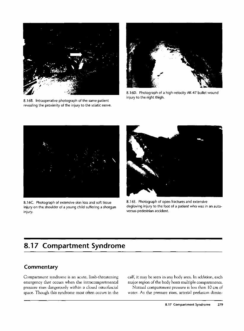
8.16B. Intraoperative photograph of the same patientrevealing the proximity of the injury to the sciatic nerve.
8.16D. Photograph of a high-velocity AK-47 bullet woundinjury to the right thigh.
8.16C. Photograph of extensive skin loss and soft tissueinjury on the shoulder of a young child suffering a shotguninjury.
8.16E. Photograph of open fractures and extensivedegloving injury to the foot of a patient who was in an auto-versus-pedestrian accident.
8.17 Compartment Syndrome
Commentary
Compartment syndrome is an acute, limb-threateningemergency that occurs when the intracompartmentalpressure rises dangerously within a closed osteofascialspace. Though this syndrome most often occurs in the
calf, it may be seen in any body area. In addition, eachmajor region of the body hosts multiple compartments.
Normal compartment pressure is less than 10 cm ofwater. As the pressure rises, arterial perfusion dimin-
8.17 Compartment Syndrome 279

ishes and at a critical point stops completely. The mus-cle and nerve are at severe risk for permanent injury.Fractures of the mid-shaft tibia are especially at veryhigh risk, though many other etiologies of compart-ment syndrome exist, such as vascular injuries, burns,excessive exercise, infiltrated infusions, hematomas,tight dressings or casts, and prolonged immobilizationor pressure on a limb. In the majority of cases, the risein compartment pressure is related to tissue edema orintracompartmental bleeding, and the compliance ofthe fascia and compartment steadily decrease as theintracompartmental pressure rises. At an intracom-partmental pressure of 20-30 mm Hg, capillaries areoccluded and tissue ischemia results.
Classic teaching includes evaluating for the six Psassociated with compartment syndrome: pain, pallor,paresthesias, poikilothermia, paralysis, and pulseless-ness. Unfortunately, many of these signs are late, andpermanent damage may already have occurred by thetime these signs appear. Deep burning pain that is outof proportion to the apparent injury is an importantearly clue to the condition, along with an increase ofthe pain on passive stretching of the involved musclegroup. Prompt orthopedic consultation is necessary in
suspected cases. Compartment pressure measurementis a helpful adjunct in borderline cases and can easilybe done by placing a needle pressure transducer withinthe compartment in question. When compartmentsyndrome is considered, the pressures of all the com-partments in that extremity should be measured.Though compartment pressures help diagnose the syn-drome, the patient's clinical symptoms and signsshould also be taken into account when making thediagnosis.
Pressures less than 20 mm Hg generally do not causeacute compartment syndrome but may warrant admis-sion and serial examination. Pressures greater than 30mm Hg are an indication for definitive therapy in theform of fasciotomy. Pressures between 20 and 30 mmHg are considered to be in a "gray zone" area.
Complications of compartment syndrome includemuscle necrosis and infection. Adequate hydration isimportant, as muscle breakdown can lead to rhab-domyolysis and myoglobinuric renal failure. In thelong term, necrotic muscle is slowly converted to anelastic fibrous tissue, which leads to the developmentof Volkmann's contractures.
8.17B. Photograph of delayed capillary refill in the foot. Alate finding in an acute compartment syndrome.
8.17A. Radiograph of a comminuted multisegment tibialshaft fracture at high risk for compartment syndrome.
280 Skin and Soft Tissue Injury
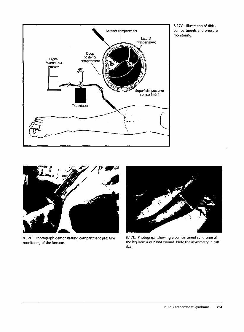
Anterior compartment
Lateralcompartment
DigitalManometer
Deepposterior
compartment
Superficial posteriorcompartment
8.17C. Illustration of tibialcompartments and pressuremonitoring.
8.17D. Photograph demonstrating compartment pressuremonitoring of the forearm.
8.17E. Photograph showing a compartment syndrome ofthe leg from a gunshot wound. Note the asymmetry in calfsize.
8.17 Compartment Syndrome 281
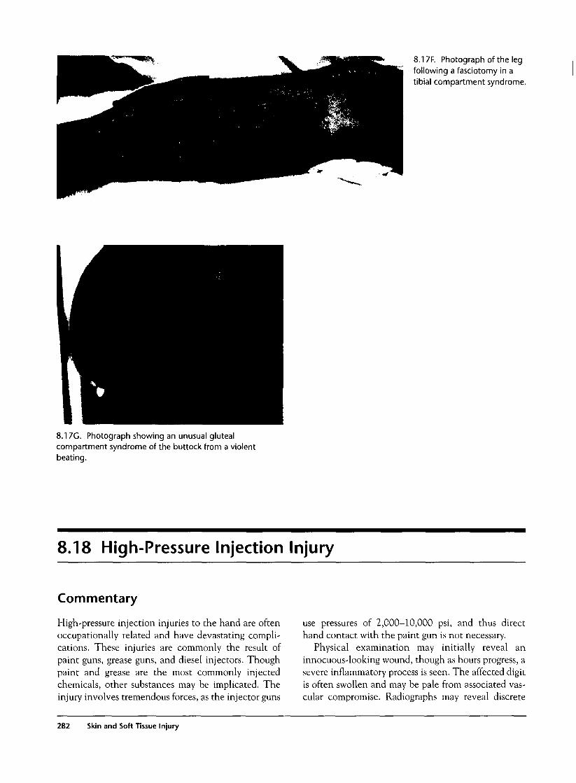
8.17F. Photograph of the legfollowing a fasciotomy in atibial compartment syndrome.
8.17C. Photograph showing an unusual glutealcompartment syndrome of the buttock from a violentbeating.
8.18 High-Pressure Injection Injury
Commentary
High-pressure injection injuries to the hand are oftenoccupationally related and have devastating compli-cations. These injuries are commonly the result ofpaint guns, grease guns, and diesel injectors. Thoughpaint and grease are the most commonly injectedchemicals, other substances may be implicated. Theinjury involves tremendous forces, as the injector guns
use pressures of 2,000-10,000 psi, and thus directhand contact with the paint gun is not necessary.
Physical examination may initially reveal aninnocuous-looking wound, though as hours progress, asevere inflammatory process is seen. The affected digitis often swollen and may be pale from associated vas-cular compromise. Radiographs may reveal discrete
282 Skin and Soft Tissue Injury
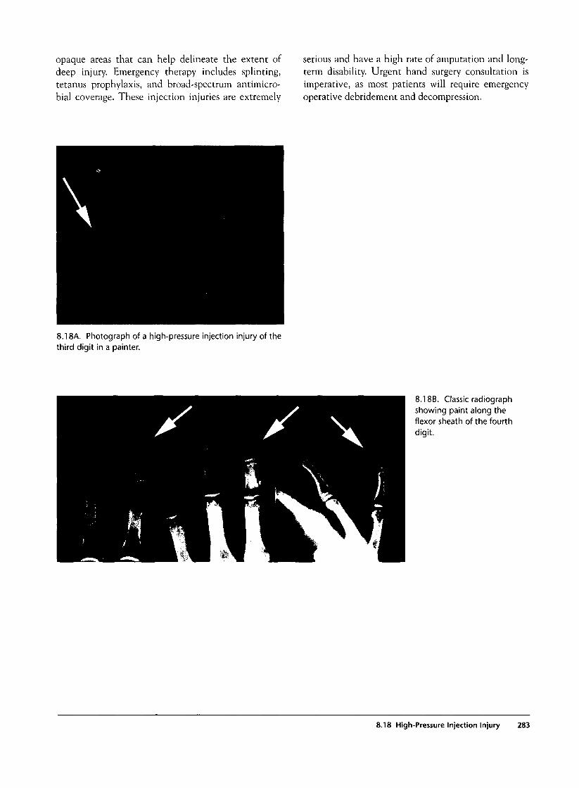
opaque areas that can help delineate the extent ofdeep injury. Emergency therapy includes splinting,tetanus prophylaxis, and broad-spectrum antimicro-bial coverage. These injection injuries are extremely
serious and have a high rate of amputation and long-term disability. Urgent hand surgery consultation isimperative, as most patients will require emergencyoperative debridement and decompression.
8.18A. Photograph of a high-pressure injection injury of thethird digit in a painter.
8.18B. Classic radiographshowing paint along theflexor sheath of the fourthdigit.
8.18 High-Pressure Injection Injury 283


ndex
Abdominal trauma, 129-166bladder injuries in, 146, 146f-147f, 200blunt, 129-130, 132-151
associated injuries in, 129clinical examination in, 129common mistakes and pitfalls in, 130general management of, 130investigations in, 129-130mechanism of injury in, 129, 132, 132f
colorectal injuries in. See Colorectaltrauma
diaphragmatic injuries in. See Diaphrag-matic trauma
duodenal injuries in, 149, 149f, 159fwith head trauma, 130, 131hemoperitoneum in, 131, 133-134,
134f-136fkidney injuries in. See Kidney injuriesliver injuries in. See Liver injuriespancreatic injuries in, 130, 142,
142f-143f, 159,159fpenetrating, 130-131, 151-164
clinical examination in, 131common mistakes and pitfalls in, 131general management of, 131in gunshot wounds. See Gunshot
wounds, of abdomeninvestigations in, 131, 153-154,
154f-155fmechanism of injury in, 151-152,
152f-153fvascular, 162, 162f-163f
in pregnancy, 164, 165f-166fseatbelt marks in, 129, 130, 133, 133f
and Chance fracture of spine, 252and small bowel injuries, 133f, 150
small bowel injuries in, 132f, 133f,149f-150f, 149-150
splenic injuries in. See Splenic traumaurethral injuries in, 148, 148f-149f, 200,
201fAbrasion of cornea, 36, 36f
examination in, 33, 34, 36management of, 35, 36
Acetabular fractures, 200fwith hip dislocation, 203, 204fnerve injuries in, 183t
Acid chemical injuries, 271, 271f
Aerodigestive injuriesin neck trauma, 57, 58, 74-76
algorithms on evaluation and manage-ment in, 62f, 74, 74f
physical examination protocol in, 61,61t
in thoracic trauma, 84subcutaneous emphysema in, 98f
Airway managementin burns and inhalational injuries, 259,
260, 274in facial trauma, 33, 34-35
in complex lacerations, 49, 50f, 5Ifin mandible fractures, 44
in neck trauma, 57, 58blunt laryngotracheal, 81in hematoma, 58, 76, 76f-77f
in spinal trauma, 220in thoracic trauma, 83, 85
Alkali chemical injuries, 271Allis reduction technique in hip disloca-
tion, 203, 2O5fAmputations, traumatic, 176, 177f-178f
general management in, 168Amylase serum levels
in duodenal injuries, 130, 149in pancreatic injuries, 130, 142
Angiographyin abdominal trauma, 130, 131
in aortic injury, 163fin liver injury, 139, 140f, 142f, 156, 157fin renal injury, 144, 146f, 159, 160fin splenic injury, 138f
in facial artery injury, 55fin knee dislocation, 211, 212fin musculoskeletal trauma, 167, 168
in peripheral vascular injuries, 179,180f-182f
in neck trauma, 57-58, 69-70in carotid artery injury, 68f, 69f,
71f-72f, 79fcompared to color flow Doppler imag-
ing, 69, 69fembolization in, 70, 72f, 80indications for, 62f, 66, 66f, 69-70penetrating, 69-70, 7Of-73fstent placement in, 70, 72fin vertebral artery injury, 80, 80f
in zone I, 67fin zone III, 68f
in pelvic fractures, 200, 2O2fin thoracic trauma, 84
in aortic injury, 111, 112fin thoracic outlet injury, 113, 114f,
115f, 116fin transmediastinal gunshot wounds,
116, 118fAnkle
dislocation of, 213, 214ffractures of, 213, 214f
Maisonneuve complex in, 213, 213fSalter-Harris 111, 173f
open injury of, 17IfAnterior cord syndrome, 219, 225, 225fAortic injuries
in abdominal trauma, 162f-163fin thoracic trauma, 84
blunt, 110-111, 11 lf-113findications for thoracotomy in, 85in penetrating thoracic outlet injuries,
113, 116fsites of rupture in, 110, 112f, 113fin transmediastinal gunshot wounds,
116, 118fwidened mediastinum in, 85, 110,
11 IfAortography, 84
in blunt thoracic trauma, 111, 112fArterial blood gas analysis
in inhalational injury, 259in thoracic trauma, 84
Asphyxia in thoracic trauma, 127,127f-128f
Atlantoaxial dislocation, 229, 229fAtlantoaxial rotatory subluxation, 230,
230f-231fAtlanto-occipital dislocation, 228,
228f-229fAtlas, cervical
in atlantoaxial dislocation, 229, 229fin atlanto-occipital dislocation, 228,
228f-229fJefferson fracture of, 231, 232frotatory subluxation of, 230, 230f-231f
Autotransfusion of blood in thoracictrauma, 95, 95f-96f
285

Axillary artery injury, 182fAxillary nerve injuries in shoulder disloca-
tion, 183t, 194Axis, cervical
in atlantoaxial dislocation, 229, 229fhangman's fracture of, 236, 236f-237fodontoid process of. See Odontoid processrotatory subluxation of atlas on, 230,
230f-231fAxon injury, 182
in head trauma, 1, 29, 30fAxonotmesis, 182
Balloon tamponade for bleeding controlin neck trauma, 77, 77f-78fin thoracic trauma, 105, 106f-107f
Battles' sign in head trauma, 1, 2with basilar skull fracture, 9, llf, 34
Beck's triad in cardiac injuries, 99Bell's palsy, 54Bennett's fracture, 185, 185fBiloma in liver injury
blunt, 139, 141fpenetrating, 156, 157f
Bite wounds, 275f-277f, 275-276cat, 276, 276fdog, 275, 275fhuman, 276, 276f-277f
of hand, 276, 276f-277foral lacerations in, 52f, 53f
Bladder traumablunt, 146, 146f-147fin pelvic fractures, 146, 200
Blow-out fractures of orbit, 41-42, 42f-43fexamination in, 34, 35, 41-42mechanism of injury in, 41, 42f
Blunt traumaof abdomen, 129-130, 132-151. See also
Abdominal trauma, bluntof head, 1of neck, 57, 80-82
carotid artery injury in, 80-81, 81flaryngotracheal injury in, 81, 82f
of thoraxalgorithm on evaluation of, 1 lOfaortic injury in, 110-111, 11 If—113fcardiac injury in, 109, 109f-110f
Body surface area in burns, 259, 260, 266,267f
Bohler's angle in calcaneal fractures, 217,218f
Boxer's fracture, 184, 184fBronchoscopy in inhalational injury, 260,
274Brown-Se'quard syndrome, 219, 223, 223t,
224fBulbocavernous reflex in complete spinal
cord transection, 225-226, 227fBurns, 259-274
body surface area involved in, 259, 260,266, 267f
in chemical injuries, 260, 270-271, 27Ifin children. See Children, burns incircumferential, 267-268, 268f-269f
clinical examination in, 259escharotomy in, 260, 267-268, 268f,
269fgeneral management in, 260
clinical examination in, 259common mistakes and pitfalls in, 260in electrical injuries, 260, 272, 272f-273fextent of injury in, 266, 267ffirst-degree, 262, 262ffourth-degree, 266, 266fgeneral management of, 260with inhalational injuries. See Inhala-
tional injuriesinvestigations in, 259-260rule of nines in, 259, 266, 267fin scald injuries, 270, 27Of
in children, 264f, 270, 27Ofsecond-degree, 259, 260, 263,
263f-264fin chemical injuries, 27Ifcircumferential, 268fdeep, 263, 263fsuperficial, 263zones of injury in, 262f, 263f
third-degree, 259, 260, 264, 265fin chemical injuries, 27Ifcircumferential, 268f
zones of injury in, 261, 261f-262fBurst fractures, vertebral
cervical, 237, 240, 240f-241fof atlas, 231, 232f
lumbar, 249-250, 250f-251fButtock area
compartment syndrome of, 282fmajor soft tissue injury of, 278fscald burn of, 27Of
Calcaneal fractures, 217, 218fwith spinal injury, 221
Canthotomy, lateral, in retrobulbarhematoma, 40, 40f
Carbon monoxide levels in burns andinhalational injury, 259
Cardiac injuries. See Heart injuriesCarotid artery injuries
angiography in, 68f, 69f, 71f-72f, 79fin blunt trauma, 80-81, 81fcolor flow Doppler imaging in, 69, 69fcomputed tomography in, 65f, 79fpenetrating, 79, 79fpseudoaneurysm in, 68f, 69fthrombosis in, 79, 79f
Cat bite injuries, 276, 276fCauda equina syndrome, 254Cecum, gunshot wound of, 161fCentral cord syndrome, 222, 222f-223f
clinical examination in, 219, 221Central transtentorial herniation, 24Cerebellar tonsillar herniation, 25
Cerebrospinal fluiddouble ring sign
in basilar skull fracture, lOfin facial trauma, 34
otorrhea and rhinorrheain facial trauma, 34, 35in head trauma, 1, 2, 9, lOf, 34
Cervical collar in penetrating neck injurieswith hematoma, 76, 77f
Cervical spinal trauma, 228-241atlantoaxial dislocation in, 229, 229fatlantoaxial rotatory subluxation in, 230,
230f-231fatlanto-occipital dislocation in, 228,
228f-229fratio of powers in, 228, 229f
burst fractures in, 237, 240, 24Of-241fof atlas, 231, 232f
central cord syndrome in, 222, 222f-223fin cervicothoracic injuries, 246, 246fin children, 256, 256f-257f
atlantoaxial dislocation in, 229fatlanto-occipital dislocation in, 228fJefferson fracture of atlas in, 232f
clay shoveler's fracture in, 241, 24Ifcomplete spinal cord transection in, 225facet dislocation in, 243f-245fgeneral management in, 220hangman's fracture in, 236, 236f-237fwith head injuries, 2investigations in, 220ligament injuries in, 255, 255f. See also
Ligament injuries in cervical spinaltrauma
odontoid fractures in, 233, 233f-235fin penetrating neck injuries, 65fteardrop fractures in, 237, 238-239, 239fvertebral body fractures in, 237f-241f,
237-240types of, 237, 237f-238f
Cervicothoracic spinal trauma, 246, 246fChance fracture of spine, 221, 252,
252f-254fChemical burns, 260, 270-271, 271fChest trauma, 83-128. See also Thoracic
traumaChild abuse
head trauma in, 27, 28, 28f-29fcommon mistakes and pitfalls in, 3investigations in, 2in shaken baby syndrome, 13, 14f, 27,
28f, 29fskull fractures in, 4subdural hematoma in, 13, 14f, 28f,
29fscald burns in, 270, 27Of
Childrenburns in, 260
body surface area involved in, 266,267f
in chemical injuries, 27If
286 Index

in scald injuries, 264f, 270, 27Ofsecond-degree, 264f, 27Ifthird-degree, 265f
dog bite wounds in, 275, 275ffractures in, 168, 169
common mistakes and pitfalls in, 168in forearm, 190, 190fgreenstick and torus, 169, 174, 174fin hip, 206Salter-Harris classification of, 172,
173f-174fsupracondylar, 175, 175f
head trauma in, 27-28, 28f-29fin abused child. See Child abuse, head
trauma indiffuse cerebral edema in, 26, 27, 29fskull fractures in, 4-5, 27subdural hematoma in, 13, 14f, 27,
28f, 29fmajor soft tissue injury in, 279fspinal trauma in, 219, 256, 256f-257f
atlantoaxial dislocation in, 229fatlanto-occipital dislocation in, 228fcervicothoracic, 246complete spinal cord transection in,
227ffacet dislocation in, 244fgeneral management of, 220Jefferson fracture of cervical atlas in,
232fwithout radiographic abnormality, 256
splenic injury in, blunt, 137thoracic trauma in
asphyxia in, 128frib fractures in, 87, 87f
Cholangiopancreatographyendoscopic retrograde, in blunt pancre-
atic injuries, 142, 143fmagnetic resonance
normal appearance in, 143fin pancreatic injuries, blunt, 130, 142
Chylothorax in thoracic duct injury, 126fCingulate gyms hemiation, 25Circumferential burns, 267-268, 268f-269f
clinical examination in, 259escharotomy in, 260, 267-268, 268f, 269fgeneral management in, 260
Clavicledislocation from sternum, 198, 198ffractures of, 196, 197f
Clay shoveler's fracture, 241, 241fClenched-fist injury, 276f-277fCoagulation disorders in head trauma, 3,
25,31Colles' fracture, 189, 189fColor flow Doppler imaging. See Doppler
imaging, color flowColorectal trauma
blunt, 151,151fmechanism of injury in, 132f
penetrating, 161, 161f-162f
in gunshot wounds, 161f, 164, 164fCompartment syndrome, 279-280,
280f-282fin burns, 259, 260, 280
circumferential, 267in electrical injuries, 272fourth-degree, 266f
in calcaneal fractures, 217clinical examination in, 167common mistakes and pitfalls in, 168, 260gluteal, 282fpressure measurements in, 167, 259, 267,
279-280, 28Ifsigns of, 168, 280tibial, 281f, 282f
Compression fractures, vertebralcervical, 240, 240f-241flumbar, 249-250, 250f-251fthoracic, 247f-248f
Computed tomographyin abdominal trauma, 129-130, 131,
153-154,154f-155fin bladder injuries, 147fin colorectal injuries, 161, 162fin duodenal injuries, 149, 149fin hemoperitoneum, 134, 136fin kidney injuries, 144, 144f-145f,
159, 160fin liver injuries, 139, 140f-141f, 156,
156f, 157fin pancreatic injuries, 142, 142f-143fin small bowel injuries, 150, 150fin splenic injuries, 137, 137f-139f,
158, 158fin transpelvic gunshot injuries, 155in urethral injuries, 149fin vascular injuries, 163f
in facial trauma, 34in globe rupture, 39fin Le Fort fracture, 47f, 48fin ocular foreign bodies, 34, 35, 36, 37fin orbital blow-out fractures, 42, 42f, 43fin retrobulbar hematoma, 40f
in head trauma, 2, 3in axonal sheer injury, 29, 30fin basilar skull fracture, 9, 1 Ifin cerebral contusion, 20f, 2Ifin children, 28, 28f-29fin depressed skull fractures, 6, 7fin diffuse cerebral edema, 26f-27fin epidural hematoma, 11-12, 12f-13fin intraparenchymal hemorrhage, 3 Ifin intraventricular hemorrhage, 3Ifin linear skull fractures, 5, 5f, 6fin open skull fracture, 8fin penetrating injuries, 22, 23fin subarachnoid hemorrhage, 18,
18f-19fin subdural hematoma, 13-14, 14f,
16f-18fin transtentorial hemiation, 25f
in musculoskeletal trauma, 167, 168in neck trauma, 58
blunt laryngotracheal, 82fin carotid artery injuries, 65f, 79fin penetrating injuries, 60f, 65,
65f-66fin pelvic fractures, 200, 2O2fin spinal trauma, 220
in atlantoaxial rotatory subluxation,230f
in Brown-Se'quard syndrome, 224fin burst fractures, cervical, 240,
240fin Chance fractures, 253fin hangman's fractures, 237fin Jefferson fractures, 231, 23 2fin lamina fractures, thoracic, 242fin lumbar compression fractures, 250,
250f, 25Ifin odontoid process fractures, 234fthoracic, 247, 247f-249f
in thoracic trauma, 84in aortic injuries, blunt, 111, 11 Ifin diaphragmatic injuries, 119, 120,
123, 124f, 125fin esophageal injuries, 126fin hemothorax, 93, 93fin lung contusion, 84, 96, 97fin pneumothorax, 90f, 92fin retained cardiac missiles, 103,
104fspinal, 247, 247f-249fin transmediastinal gunshot wounds,
116, 117f, 118fConcussion in head trauma, 1
in children, 28Contusion
cerebral, 1,20, 2Of-2Ifin children, 27diffuse cerebral edema in, 26management of, 3, 20
of lung, 83,96, 96f-98fcomputed tomography in, 84, 96, 97fin rib fractures, 87, 87f, 89, 89f, 96
myocardial, 83, 84, HOfCorneal abrasion, 36, 36f
examination in, 33, 34, 36management of, 35, 36
Corticosteroid therapy in spinal trauma,220
Cricothyrotomyin facial lacerations, 49, 5 Ifin neck injuries, 58, 76, 76f
Crush injuries, 168, 17Ifof thorax, asphyxia in, 127, 127f
Cystographyin bladder injuries, 146, 147fin urethral injuries, 149f
Dancer's fracture, 217Dermatomes, 226f
Index 287
Diaphragmatic trauma, 120-121, 131blunt, 120-121, 121f-122f
rupture of diaphragm in, 120clinical examination in, 83
Diaphragmatic trauma (continued)hernia in, 84. 123, 123f-125f
laparoscopy in, 120f, 123, 124f, 125fin penetrating injuries, 119, 120f,
123finvestigations in, 84, 85penetrating, 119, 119f—120f
hernia in, 119, 120f, I23fDislocations
of ankle, 213, 214fof elbow, 191, 192fgeneral management in, 168of hip, 183c, 203, 2O4f-2O5fof knee, 183t, 211, 211f-212flunate and perilunate, 187, 188f-189fof metacarpal joint, open, 17Ifnerve injuries in, 183tperipheral vascular injuries in, 179scapholunate, 187, 187fof shoulder, 183t, 194, 195f-196fspinal
atlantoaxial, 229, 229fatlanto-occipital, 228, 228f-229fclinical examination in, 219facet, 243, 243f-245f, 246f, 257fgeneral management in, 220
sternoclavicular, 198, 198fsubtalar, 214, 215f
Dog bite injuries, 275, 275fDoppler imaging, color flow
in axillary artery injury, 182fin neck trauma, 58, 69, 69f
in carotid artery injury, 69, 69findications for, 66, 66f, 69in zone 1, 67
in thoracic trauma, 84in penetrating thoracic outlet injury,
113, U6fin rib fractures, 87
Duodenal traumablunt, 149, 149fpenetrating, in gunshot wound, 159f
Ear injuriesexamination in, 34management of, 35otorrhea in, 1, 2, 34, 35
in basilar skull fracture, 9, lOf, 34Echocardiography in heart trauma
blunt, 109penetrating, 108, 108f
Edema, cerebral, 1, 26, 26f-27fin children, 26, 27, 29fin penetrating head injury, 22transtentorial herniation in, 24
Effusions, pericardial, in heart trauma, lOlf,108, HOf
Elbowdislocation of, 191,192fnerve injuries at, 183t, 191open fracture of, 170fradial head fractures at, 192, 192f-193f
Electrical injuries, 260, 272, 272f-273fElectrocardiography in thoracic trauma, 83
in blunt cardiac injury, 109, HOfin penetrating cardiac injury, 101, 102f,
108Embolization in vascular injuries
of kidney, 160fof liver, 139, HOf, 142f
penetrating, 156, 157fof neck, 70, 72f, 80
Emphysemamediastinal, in esophageal injury, 125,
125fsubcutaneous
in aerodigestive tract trauma, 74f,75f
in blunt laryngotracheal trauma, 82fclinical examination in, 57in esophageal injury, 125investigations in, 58in penetrating neck trauma, 61, 75fradiography in, 63, 63f, 98fin thoracic trauma, 98, 98f, 125
Endoscopy
in neck injuries, 58indications for, 74, 74f
retrograde cholangiopancreatography in,in pancreatic injuries, 142, 143f
in thoracic trauma, 84Epidural hematoma. See Hematoma,
epiduralEpiphyseal injuries, Salter-Harris classifica-
tion of, 172, 173f-174fEscharotomy in circumferential burns, 260,
267-268, 268f, 269fEsophageal injury
in neck trauma, 57, 74, 75fabscess of neck in, 126falgorithm on initial evaluation and
management in, 62fcomputed tomography in, 65finvestigations in, 58physical examination protocol in, 61t
in thoracic trauma, 83, 84, 125,125f-126f
in transmediastinal gunshot wounds,116, 118f
Esophagography, 58, 74, 74f, 125indications for, 74fin transmediastinal gunshot wounds, 116,
118fEsophagoscopy
in neck trauma, 58indications for, 62f, 74
in thoracic trauma, 84, 125, 125fEthmoid fractures, 46, 47f
Eye injuriesin chemical burns, 271, 27Ifcorneal abrasion in, 36, 36f
examination of, 33, 34, 36management of, 35, 36
examination in, 33-34, 35foreign bodies in, 33-34, 35, 36, 37fglobe rupture in, 33, 34, 35, 38, 39fhyphema in, 34, 38, 38fmanagement of, 35periorbital lacerations in, 41, 4If
Eyebrow lacerations, 41Eyelid lacerations, 41, 41f
Facet dislocation in spinal trauma, 243,243f-245f
cervicothoracic, 246fin children, 25 7f
Facial artery injury, 55, 55fFacial nerve injury, 54, 54f
examination in, 33, 54Facial trauma, 33-55
avulsion of scalp and face in, 4f, 50fin burns, 259, 260
in chemical injuries, 27 Ifwith inhalational injury, 274, 274fthird-degree, 265f
clinical examination in, 33-34common mistakes and pitfalls in, 35complex lacerations in, 49, 5Of-51fin dog bites, 275, 275feye injuries in. See Eye injuriesfacial artery injury in, 55, 55ffacial nerve injury in, 33, 54, 54fgeneral management of, 33, 34-35in gunshot wounds, 23f, 49, 50f
oral injuries in, 53fhyphema in, 34, 38, 38finvestigations in, 34Le Fort fractures in, 46, 46f-48fmandible fractures in, 34, 44, 44fnasal injuries in, 34, 35, 48-49, 49foral lacerations in, 52, 52f-53forbital fractures in. See Orbital fracturesperiorbital lacerations in, 41, 4Ifretrobulbar hematoma in, 40, 40fzygoma fractures in, 45, 45f, 48f
Femoral artery injury, 180f, 18IfFemoral nerve injuries, 183t, 203Femur
avascular necrosis of head, 203fractures of, 203, 206, 206f-208f
with knee dislocation, 212fin shaft, 208, 208f-209f
Fibula fracturesMaisonneuve complex in, 213, 213fnerve injuries in, 183t
Finger injuries. See HandFlail chest, 83, 85, 89, 89fFluid management in burns, 260
in electrical injuries, 272
288 Index

Foot injuriesamputation in, traumatic, 176, 178fcalcaneal fractures in, 217, 218f, 221compartment syndrome in, 280fLisfranc fracture/dislocation in, 215, 216fmetatarsal fractures in, 216-217, 217fin scald burns, 27Ofof soft tissues, major trauma in, 279f
Forearm
amputation of, traumatic, 178fburns of, 264f
in electrical injuries, 272fcompartment pressure in, 281fdog bite of, 275ffractures of, 190, 190f-191f
greenstick, 174fretained glass foreign body in, 278f
Foreign bodiesin eye, 33-34, 35, 36, 37f
examination in, 33-34, 35retained, 278, 278f
glass particles in, 278, 278fFractures, 169-176
acetabular, 200fwith hip dislocation, 203, 204fnerve injuries in, 183c
of ankle, 213, 214fMaisonneuve complex in, 213, 213fSalter-Harris III, 173f
Bennett's, 185, 185fboxer's, 184, 184fcalcaneal, 217, 218f
with spinal injury, 221in children. See Children, fractures inclassification of, 169, 169f, 170
Gustilo, 170Salter-Harris, 172, 173f-174f
of clavicle, 196, 197fColles', 189, 189fcommon mistakes and pitfalls in, 168dancer's, 217of femur, 203,206, 2O6f-2O8f
with knee dislocation, 212fin shaft, 208, 208f-209f
of fibulaMaisonneuve complex in, 213, 213fnerve injuries in, 183t
of forearm, 174f, 190, 190f-191fGaleazzi, 190, 191fgeneral management in, 168greenstick, 169, 169f, 174, 174fof hip, 183t, 206, 206f-208fof humerus. See Humerus fracturesinvestigations in, 167Le Fort, 33, 46, 46f-48fLisfranc, 215, 216fMalgaigne, 201fof mandible, 34,44, 44fof maxilla, 34, 46metacarpal, 184, 184f-185f
of thumb, 185, 185f-186f
metatarsal, 216-217, 217fMonteggia, 190, 19 Ifnerve injuries in, 183cof nose, 46, 49open or compound, 169, 170, 17Of-171forbital. See Orbital fracturesof patella, 209, 209fpelvic. See Pelvic fracturesperipheral vascular injuries in, 179, 180fof pubic rami, 183t, 200, 200f, 201fof radius. See Radius fracturesof ribs. See Rib fracturesRolando's, 185, 186fscaphoid, 186, 186fof scapula, 199, 199fof skull. See Skull fracturesspinal, 219
burst fractures in. See Burst fractures,vertebral
cervicothoracic, 246Chance fracture in, 221, 252,
252f-254fin children, 256clay shoveler's fracture in, 241, 24Ifclinical examination in, 219common mistakes and pitfalls in, 221general management of, 220in gunshot wounds, 65f, 242, 242fhangman's fracture in, 236, 236f-237finvestigations in, 220Jefferson's fracture in, 231, 232flumbar fracture-dislocation in, 254,
254fof odontoid process, 233, 233f-235fteardrop fractures in, cervical, 237,
238-239, 239fthoracic, 247, 247f-249fin vertebral body, 237f-241f, 237-240,
247f_249fof sternum, blunt cardiac injury in, 109,
109fof thyroid cartilage, 81, 82fof tibia. See Tibia fracturestorus, 169, 169f, 174, 174fof ulna, 190, 190f-191fof zygoma, 45, 45f, 48f
Galeazzi fractures, 190, 191fGases, arterial blood
in inhalational injury, 259in thoracic trauma, 84
Glasgow Coma Scale in head trauma, 1, 2, 2tGlass foreign bodies, retained, 278, 278fGlobe rupture in eye injuries, 33, 34, 38,
39fexamination of, 33, 34, 38management of, 35
Gluteal compartment syndrome, 282fGreenstick fractures, 169, 169f, 174, 174fGunshot wounds
of abdomen, 130, 131
colorectal injury in, 161f, 164, 164finvestigations in, 153, 154f-155fliver injury in, 156, 156f-157fmechanism of injury in, 151, 152f-153fpancreatic and duodenal injury in,
159frenal injury in, 155f, 160fretained bullets in, 164, 164fsplenic injury in, 158ftranspelvic, 155, 155fvascular injuries in, 162f-163f
compartment syndrome in, 28Ifof face, 23f, 49, 50f
oral injuries in, 53fof head, 1,22, 22f-23f
in children, 28fskull fracture in, 5f
ofknee, 181fmajor soft tissue injury in, 279fof neck, 59, 60f
angiography in, 69, 70, 7Of-72f, 79fcarotid artery injury in, 79fcomputed tomography in, 65, 65f-66f,
79femphysema in, subcutaneous, 75fhematoma in, 76finvestigations in, 58operative approaches in, 78radiography in, 64f
peripheral vascular injuries in, 179, 180f,181f
spinal trauma in, 65f, 242, 242fof thorax
cardiac injury in, 99, lOOf, 103,103f-104f
diaphragmatic injury in, 119hemothorax in, 93flung contusion in, 97fretained missiles in, 103, 103f-104fthoracic outlet injury in, 115f, 116ftransmediastinal, 84, 116, 117f-l 18f
Gustilo classification of open fractures, 170
Handamputation of, traumatic, 176, 177f-178fbite wounds of
from cat, 276, 276ffrom human, 276, 276f-277f
burns of, 264fin chemical injuries, 27If
clenched-fist injury of, 276f-277fhigh-pressure injection injury of,
282-283,283fcompartment syndrome in, 28Of
metacarpal dislocation in, open, 17Ifmetacarpal fractures of, 184, I84f-185f
in thumb, 185, 185f-186fnerve injuries in, 183tphalangeal fractures of thumb in, 173fscaphoid fractures of, 186, 186ftendon injuries of, 179, 179f
Index 289
Hangman's fracture of cervical axis, 236,236f-237f
Head trauma, 1-31with abdominal trauma, 130, 131axonal shear injury in, 1, 29, 30fcerebral contusion in. See Contusion,
cerebralcerebral edema in. See Edema, cerebralin children. See Children, head trauma inclinical examination in, 1-2common mistakes and pitfalls in, 3epidural hematoma in. See Hematoma,
epiduralgeneral management of, 2-3Glasgow Coma Scale in, 1, 2,2chemorrhage in. See Hemorrhage, in head
traumaintracranial pressure increase in. See
Intracranial pressure increase inhead trauma
investigations in, 2penetrating, 1, 22, 22f-24f
in children, 28fskull fracture in, 5f, 22
scalp injury in, 4, 4fskull fractures in. See Skull fractureswith spinal injuries, 2, 221subdural hematoma in. See Hematoma,
subduraltranstentorial herniation in, 3, 24-25,
25f-26fHeart injuries
blunt, 109, 109f-110falgorithm on evaluation of, 1 lOfmechanism of injury in, 109, 109f
clinical examination in, 83emergency department thoracotomy in,
104-105, 105f-107fin gunshot wounds, 99, lOOf
retained missiles in, 103, 103f-104finvestigations in, 83-84myocardial contusion in, 83, 84, 1 lOfpenetrating, 99-108
diagnosis of, 101, 101f-103flate sequelae in, 108, 108fmechanisms of, 99, 99f-100fretained missiles in, 103, 103f-104fsurvival factors in, 100, lOlfsymptoms in, 99
Hematoma
epidural, 1,3, 11-12, 12f-13flucid interval in, 11management of, 3, 12in penetrating head trauma, 22in skull fracture, 5, 7f, 11,12funcal herniation in, 25f
in liver injury, 140f-141f, 157fin neck injuries
airway management in, 58, 76, 76f-77fangiography in, 69, 7If, 72fcervical collar use in, 76, 77f
computed tomography in, 66fradiography in, 63, 63f
in renal injuries, 144, 144f, 145fretrobulbar, 40, 40f
lateral canthotomy in, 40, 40fretro-orbital, 34, 35in splenic injury, 137, 138fsubdural, 1,3, 13-14, 14f-18f
acute, 13, 14f-16fin axonal sheer injury, 29, 30fin cerebral contusion, 2Ifin children, 13, 14f, 27, 28f, 29fchronic, 13, 14, 17f-18fdiffuse cerebral edema in, 26, 27fmanagement of, 3, 14, 15f-16fin penetrating head trauma, 23frebleeding in, 14, 17f, 18fin shaken baby syndrome, 13, 14f, 28f,
29fin skull fracture, 7f, 14fsubacute, 13, 16f-17fin subarachnoid hemorrhage, 18, 19ftranstentorial herniation in, 25f
Hematuria in blunt abdominal trauma, 130of bladder, 146of kidneys, 144, 145f
Hemobilia in liver injury, 139, 141f-142fHemoperitoneum in abdominal trauma,
133-134, 134f-136fpenetrating, 131
Hemoptysis in neck injuries, 57, 58, 61, 74fHemorrhage
in facial trauma, 33, 35hyphema in, 38, 38fretrobulbar hematoma in, 40, 40f
in femoral shaft fractures, 208in head trauma
in axonal sheer injury, 29, 30fin children, 28, 28fin coagulopathy, 3epidural hematoma in, 11-12, 12f-13fintraparenchymal, 31, 3 Ifintraventricular, 3, 31, 3Ifin penetrating injuries, 22, 23f, 31in scalp injuries, 4subarachnoid. See Subarachnoid hem-
orrhagesubdural hematoma in, 13-14, 14f-18ftranstentorial herniation in, 24
in neck trauma, control in emergencydepartment, 58, 77, 77f-78f
in pelvic fractures, 168, 200, 202fin thoracic trauma
autotransfusion of blood in, 95,95f-96f
balloon tamponade in, 105, 106f-107findications for thoracotomy in, 85
Hemothorax in thoracic trauma, 83, 93,93f-94f
autotransfusion of blood in, 95thoracostomy tube insertion in, 93, 94
thoracotomy and decortication in, 93,94f
Hemotympanum in head trauma, 1, 2in basilar skull fracture, 9, lOf
Herniationdiaphragmatic, 84, 123, 123f-125f
laparoscopy in, 120f, 123, 124f, 125fin penetrating injuries, 119, 120f, 123f
transtentorial, in head trauma, 3, 24-25,25f-26f
management of, 25, 26ftypes of, 24-25
High-pressure injection injury, 282-283,283f
compartment syndrome in, 280fHill-Sachs deformity, 194, 195fHip
dislocation of, 203, 204f-205fanterior, 203, 204fnerve injuries in, 183t, 203posterior, 203, 204f, 2O5freduction techniques in, 203, 2O5f
fractures of, 206, 206f-208fin children, 206classification of, 206, 206fnerve injuries in, 183tShenton's line in, 206, 2O7f
Hoarseness in neck injuries, 57, 58, 61, 74fHomer's syndrome in neck injuries, 57, 62,
62fHuman bite injuries, 276, 276f-277f
of hand, 276, 276f-277foral, 52f, 53f
Humerus fractures, 193, 193fHill-Sachs deformity in, 194, 195fnerve injuries in, 183t, 193supracondylar, 175, 175f-176f
anterior humeral line in, 175, 176fnerve injuries in, 183t
Hydrofluoric acid burns, 271, 27IfHyphema, 34, 38, 38fHypotension in thoracic trauma, 85, 99
Ilium fractures, 200fInhalational injuries, 259, 260, 274, 274f
clinical examination in, 259common mistakes and pitfalls in, 260general management of, 260investigations in, 259, 260
Innominate arteriovenous fistula in trans-mediastinal gunshot wounds, 118f
Intracranial pressure increase in headtrauma, 1
in children, 27-28in epidural hematoma, 12f, 13fmanagement of, 2-3transtentorial herniation in, 24-25,
25f-26fIntraparenchymal hemorrhage, 31, 3IfIntraventricular hemorrhage, 3, 31, 3IfIschium fractures, 200f
290 Index

Jefferson fracture of cervical atlas, 231, 232fJoint injuries
dislocations in. See Dislocationsopen, 171, 171f-172f
Jones fracture, 217
Kidney injuriesblunt, 144,144f-146f
general management of, 130pseudoaneurysm in, 144, 146f
penetrating, 159, 159f-160fin gunshot wounds, 155f, 160finvestigations in, 131pseudoaneurysm in, 159, 160f
Kneedislocation of, 211, 21 lf-212f
nerve injuries in, 183treduction in, 211, 212ftypes of, 211, 21 Ifvascular injuries in, 211, 212f
gunshot wounds of, 18Ifopen injury of, 172fpatellar fractures of, 209, 209ftibial plateau fractures of, 210, 210f
Lacerationsfacial, 34
complex, 49, 50f-5Iffacial artery injury in, 55facial nerve injury in, 54of nose, 48-49, 49foral, 52, 52f-53fperiorbital, 41, 41f
in open fractures, 170tendon, 179
Lacrimal duct lacerations, 34, 41Lamina fractures, vertebral, 242, 242fLaparoscopy
in abdominal injuries, penetrating, 131,154, I55f
in diaphragmatic injuries, 84blunt, 121and hernia, 120f, 123, 124f, 125fpenetrating, 119, 120f
Laparotomyin abdominal trauma, 130
in pregnancy, 164, 165fin diaphragmatic injury, blunt, 120, 12If
Laryngoscopy in neck injuries, 58indications for, 62f
Laryngotracheal injuriesairway management in, 76in blunt trauma, 81, 82fclinical examination in, 57general management of, 58investigations in, 58physical examination protocol in, 611
Lavage, peritoneal, in abdominal trauma,129, 130
in hemoperitoneum, 133-134, 135f-136fin small bowel injury, 150
Le Fort fractures, 46, 46f-48fexamination in, 33types of, 46,46f-48f
Letournel classification of pelvic fractures,200f
Ligament injuries in cervical spinal trauma,255,255f
atlantoaxial dislocation in, 229, 229fatlantoaxial rotatory subluxation in, 230,
230f-231fatlanto-occipital dislocation in, 228,
228f-229fLip lacerations, 52, 52f-53fLisfranc fracture/dislocation, 215, 216fLiver injuries
blunt, 129, 139, 140f-142fgeneral management of, 130investigations in, 130pseudoaneurysm in, 139, 140f, 142f
penetrating, 156, 156f-157finvestigations in, 131pseudoaneurysm in, 156, 156f
in rib fractures, 88, 88fLower extremity
amputation in, traumatic, 176, 178fankle injuries in. See Anklefoot injuries in. See Foot injuriesknee injuries in. See Kneenerve injuries in, 183tvascular injuries in, 180f— 181 f
Lumbar spinal trauma
Chance fracture in, 221, 252, 252f-254fcompression burst fractures in, 249-250,
25Of-251ffracture-dislocation in, 254, 254finvestigations in, 220
Lunate dislocations, 187, 188fLung contusion, 83, 96, 96f-98f
computed tomography in, 84, 96, 97fin rib fractures, 87, 87f, 89, 89f, 96
Luxatio erecta shoulder dislocation, 194,195f, 196f
Lymphangiography in thoracic duct injury,127f
Mach effect, 235fMagnetic resonance imaging
cholangiopancreatography innormal appearance in, 143fin pancreatic injuries, blunt, 130, 142
in head trauma, 2in axonal sheer injury, 29, 30fin cerebral contusion, 2Ifin subdural hematoma, 215f
in musculoskeletal trauma, 167, 168in spinal trauma, 220
in central cord syndrome, 222, 223fin cervical teardrop fractures, 239fin Chance fracture, 254fin facet dislocation, 245fin ligament injuries, 255, 255f
in odontoid process fractures, 234fthoracic, 248f, 249f
in thoracic traumain diaphragmatic injuries, 119, 120,
123spinal, 248f, 249f
Maisonneuve fracture complex, 213, 213fMalgaigne fractures, 20IfMandible fractures, 34, 44, 44fMaxilla fractures, 34, 46Median nerve injuries, 183c, 193Mediastinum
gunshot wounds of, 84, 116, 117f-l 18fstructures in, 116widened, in aortic injuries, 85, 110, 11 If
Metacarpal dislocation, open, 171fMetacarpal fractures, 184, 184f-185f
of thumb, 185, 185f-186fMetatarsal bones
fractures of base, 216-217, 217fin tarsometatarsal dislocations, 215, 216f
Methylprednisolone in spinal trauma, 220Monteggia fractures, 190, 19IfMotor vehicle accidents
cardiac injury in, blunt, 109, 109ffacial injuries in, 33head injuries in, 1
skull fractures in, 4major soft tissue injury in, 278f-279fseatbelt marks in. See Seatbelt marks
Mouth injuries. See Oral injuriesMusculoskeletal trauma, 167-218
amputations in, 168, 176, 177f-178fclinical examination in, 167common mistakes and pitfalls in, 168dislocations in. See Dislocationsfractures in. See Fracturesgeneral management of, 167-168investigations in, 167Lisfranc injury in, 215, 216fnerve injury in, peripheral, 182, 183f,
183ttendon injury in, 179, 179fvascular injury in, peripheral, 179,
180f-182fMyocardial contusion, 83, 84, 1 lOfMyoglobinuria in electrical injuries, 272,
273f
Nasal injuries. See Nose injuriesNeck trauma, 57-82
aerodigestive injuries in, 57, 58, 74-76algorithms on evaluation and manage-
ment in, 62f, 74, 74fphysical examination protocol in, 61,
61cblunt, 57, 80-82
of carotid artery, 80-81, 8 Iflaryngotracheal, 81, 82f
of cervical spine. See Cervical spinaltrauma
Index 291
Neck trauma (continued)clinical examination in, 57common mistakes and pitfalls in, 58esophageal injury in. See Esophageal
injury, in neck traumageneral management of, 58hematoma in. See Hematoma, in neck
injuriesHomer's syndrome in, 57, 62, 62finvestigations in, 57-58laryngotracheal injuries in, 57, 58, 61t,
76blunt, 81, 82f
penetrating, 57, 59-80airway management in, 76, 76f-77fanatomical zones in, 59, 59f, 60fangiography in, 62f, 69-70, 7Of-73f,
79fof carotid artery, 79, 79fcervical collar use in, 76, 77fcomputed tomography in, 60f, 65,
65f-66f, 79fepidemiology of, 59, 60fgeneral management of, 58in gunshot wounds. See Gunshot
wounds, of neckoperative approaches to, 58, 62f, 78,
78fphysical examination in, 60-61, 611protocol for initial evaluation and
management of, 62, 62fradiography in, 63, 63f-64fof vertebral artery, 80, 80f
vascular injuries in, 66-73algorithms on evaluation and manage-
ment in, 62f, 66, 66fangiography in, 57-58, 69-70,
7Of-73f, 79fballoon tamponade in, 77, 77f-78fof carotid artery, 79, 79f, 80-81, 81fclinical examination in, 57, 66fcolor flow Doppler in, 58, 69, 69fcomputed tomography in, 65f, 79fcontrol of bleeding in emergency
department, 58, 77, 77f-78fembolization in, 70, 72f, 80general management of, 58physical examination protocol in, 60,
61tstent placement in, 70, 72f, 79of vertebral artery, 72f, 80, 80fin zone I, 67, 67fin zone II, 68, 68f
Nerve injuriesof axillary nerve, in shoulder dislocation,
183t, 194axonal sheer, 1,29, 30fin electrical burns, 272, 272fof facial nerve, 33, 54, 54fof femoral nerve, 183t, 203in lower extremity, 183c
of median nerve, 183t, 193in musculoskeletal trauma, 182, 183f,
183tclinical examination in, 167common mistakes and pitfalls in, 168
in neck traumaclinical examination in, 57Homer's syndrome in, 62, 62fphysical examination protocol in, 611protocol for initial evaluation and
management of, 62fradiography in, 63, 64f
of phrenic nerve, 63, 64fof radial nerve, in humerus fractures,
183t, 193of sciatic nerve, in hip dislocation, 183c,
203in spinal trauma, 219
anterior cord syndrome in, 225, 225fBrown-Sequard syndrome in, 219, 223,
223c, 224fcentral cord syndrome in, 222,
222f-223fcommon mistakes and pitfalls in, 221in complete spinal cord transection,
225-226, 226f-227fgeneral management in, 220
of ulnar nerve, 183c, 193in upper extremity, 183c
Neuropraxia, 182Neurotmesis, 182Nightstick fracture of forearm, 190, 191fNose injuries, 48^9 , 49f
examination in, 34fractures in, 46, 49lacerations in, 48-49, 49fmanagement of, 35, 48-49
Odontoid processin atlantoaxial dislocation, 229, 229ffractures of, 233, 233f-235f
types of, 233, 233fpseudofracture of, 235f
Odynophagia in neck injuries, 57, 58, 61,74f
Oral injuriesexaminarion in, 34, 35lacerations in, 52, 52f-53fmandible fractures in, 34, 44, 44fmaxilla fractures in, 34, 46tooth aspiration in, 53f
Orbital fracturesblow-out type, 41-42, 42f—43f
examination in, 34, 35, 41-42mechanism of injury in, 41, 42f
Le Fort III, 47fretrobulbar hematoma in, 40, 40f
Otorrhea, cerebrospinal fluidin facial trauma, 34, 35in head trauma, 1, 2
in basilar skull fracture, 9, lOf, 34
Pancreatic traumablunt, 142, 142f-143f
investigations in, 130penetrating, 159, 159f
Parotid duct lacerations, 33, 34, 55Patellar fractures, 209, 209fPedicle fractures, vertebral, 242Pelvic fractures, 129, 130, 200, 200f-203f
bladder injuries in, 146, 200hemorrhage in, 168, 200, 2O2fLetournel classification of, 200fmanagement of, 168, 200, 2O2fnerve injuries in, 183topen, 203f
urethral injuries in, 148, 148f, 200, 201fPenetrating trauma
of abdomen, 130-131, 151-164. See alsoAbdominal trauma, penetrating
in gunshot wounds. See Gunshot woundsof head, 1,22, 22f-24f
in children, 28fskull fracture in, 5f, 22
of neck, 57, 59-80. See also Neck trauma,penetrating
spinal, Brown-Sequard syndrome in, 223,224f
in stab wounds. See Stab woundsof thorax. See Thoracic trauma, penetrat-
ingPericardial effusions in heart trauma, lOlf,
108, HOfPericardial window, subxiphoid, 84, 101,
103fPericardiocentesis in thoracic trauma,
83-84, 101, 103fPerilunate dislocations, 187, 188f-189fPerineum, gunshot wound of, 161fPeriorbital lacerations, 41, 41fPeripheral nerve injuries, 182, 183f, 183tPeripheral vascular injuries, 179, 180f-182fPeritoneal lavage in abdominal trauma,
129, 130in hemoperitoneum, 133-134, 135f-136fin small bowel injury, 150
Peritonitis in abdominal trauma, 131colorectal, 151, 161of small bowel, 149, 150fvascular, 162
Phalangeal fracture of thumb, Salter-Harristype II, 173f
Phrenic nerveinjury of, 63, 64fin thoracotomy, 106f
Pneumocephalus in open skull fracture, 8,8f
Pneumopericardium in thoracic trauma,102f
Pneumothorax, 83, 90, 90fgeneral management of, 85subcutaneous emphysema in, 98tension, 91,91 f-92f
292 Index

thoracostomy tube insertion in, 90, 91,92f, 94
Popliteal artery injury, 181fin knee dislocation, 211, 212f
Pregnancy, abdominal trauma in, 164,165f-166f
Priapism in spinal trauma, 225, 227fPseudoaneurysm
of carotid artery, 68f, 69fof facial artery, 55fin liver injuries
blunt, 139, HOf, 142fpenetrating, 156, 156f
in renal injuriesblunt, 144, 146fpenetrating, 159, 160f
in splenic injuries, blunt, 137, 137f, 138fof subclavian artery, 67f, 73f, 114f-115fof vertebral artery, 72f
Pseudocyst of pancreas, post-traumatic,
142, 143fPseudofracture of odontoid, 235fPubic rami fractures, 200, 200f, 201f
nerve injuries in, 183tPudendal artery injury, 2O2fPyelography, intravenous, in blunt abdomi-
nal trauma, 130of kidney, 144, 144f
Raccoon eyes in head trauma, 1, 2in basilar skull fracture, 9, lOf
Radial nerve injuries in humerus fractures,183t, 193
Radiographyin abdominal trauma
blunt, 129penetrating, 131, 153, 154f
in ankle fracture, 173fin aortic injury, 110-111, 11 Ifin atlantoaxial dislocation, 229fin atlantoaxial rotatory subluxation, 230,
230fin atlanto-occipital dislocation, 228, 228fin burst fracture, cervical, 240, 240f-241fin calcaneal fracture, 217, 218fin cardiac trauma
blunt, 109f, 11 Ofpenetrating, 101, 102f, 103,
103f-104fin retained missiles, 103, 103f-104f
in Chance fracture, 252f-253fin clavicle fracture, 196, 197fin clay shoveler's fracture, 241fin Colles'fracture, 189fin diaphragmatic injury
blunt, 120, 121fwith hernia, 123, 123f, 124f, 125fpenetrating, 119, 119f
in elbow dislocation, 191, 192fin emphysema, subcutaneous, 98fin facial trauma, 34
in femoral fracture, 206, 2O7f-2O8fwith knee dislocation, 212fin shaft, 208, 208f
in flail chest, 89fin forearm fracture, 174f, 190, 19 Ifin glass foreign body, retained, 278, 278fin hangman's fracture, 236f—237fin head trauma, 2
penetrating, 22, 23f, 24fin hemothorax, 93, 93fin high-pressure injection injury of hand,
282-283, 283fin hip dislocation, 2O4fin hip fracture, 206, 2O7f-2O8fin humerus fracture, 193, 193f
supracondylar, 175, 175f-176fin inhalational injury, 259, 274, 274fin Jefferson fracture, 231, 232fin knee dislocation, 212fin Lisfranc fracture/dislocation, 215,
216fin lumbar compression fracture, 250,
25Of-251fin lumbar fracture-dislocation, 254fin lunate and perilunate dislocation, 187,
188f-189fin lung contusion, 96, 96fin Maisonneuve fracture complex, 213,
213fin mandible fracture, 44, 44fin metacarpal fracture, 184f
of thumb, 185f-186fin metatarsal fracture, 217fin musculoskeletal trauma, 167in neck trauma, 57
penetrating, 63, 63f-64fin odontoid process fracture, 233f-235fin patellar fracture, 209, 209fin pelvic fracture, 201fin phalangeal fracture of thumb, 173fin pneumothorax, 90, 90f, 9Ifin radius fracture
of head, 192, 192f-193fSalter-Harris IV, 174fin shaft, 190, 191ftorus, 174f
in rib fracture, 87, 87f, 88f, 89fin scaphoid fracture, 186, 186fin scapholunate dislocation, 187, 187fin scapula fracture, 199, 199fin shoulder dislocation, 194, 195f-196fin skull fracture
basilar, 9depressed, 7flinear, 5, 5f, 6fopen,8f
in spinal trauma, 220cervicothoracic, 246, 246fin children, 256, 256f-257fin facet dislocation, 243, 243f-244fin ligament injuries, 255, 255f
and spinal cord injury without radi-ographic abnormality, 219, 256
thoracic, 247, 247f-248fin sternoclavicular dislocation, 198fin teardrop fracture, cervical, 238, 239,
239fin thoracic outlet injuries, penetrating,
115fin thoracic trauma, 83
spinal, 247, 247f-248fin tibia fracture
with ankle dislocation, 214fin plateau, 210, 21 Of
in tooth aspiration, 53fin transmediastinal gunshot wounds,
117fin zygoma fracture, 45, 45f
Radionuclide scans in musculoskeletaltrauma, 167
Radius fracturesdistal
Colles', 189, 189fin dog bite, 275fSalter-Harris IV, 174torus, 174f
in head portion, 192, 192f-193fin shaft, 190, 190f-191f
Rectal trauma. See Colorectal traumaReflex testing in complete spinal cord tran-
section, 225-226, 227fRenal injuries. See Kidney injuriesRetrobulbar hematoma, 40, 40fRhinorrhea, cerebrospinal fluid
in facial trauma, 35in head trauma, 1, 2
in basilar skull fracture, 9Rib fractures, 85, 87f-88f, 87-88
and abdominal trauma, 129flail chest in, 89, 89fof lower ribs, 88, 88flung contusion in, 87, 87f, 89, 89f, 96of middle ribs, 87, 87fof upper ribs, 87, 87f
Rolando's fracture, 185, 186fRule of nines in burns, 259, 266, 267fRust ring in eye, 35, 37f
Sacroiliac fracture-dislocation, 200f, 20IfSalter-Harris classification of fractures, 172,
173f-174fScald burns, 270, 27Of
in children, 264f, 270, 27OfScalp injuries, 4, 4f
avulsion of face and scalp in, 4f, 50fScaphoid bone
dislocation from lunate, 187, 187ffractures of, 186, 186f
Scapholunate dislocation, 187, 187fScapula fractures, 199, 199fSciatic nerve injuries in hip dislocation,
183t, 203
Index 293

Seatbelt markson abdomen, 129, 130, 133, 133f
Chance fracture of spine in, 252small bowel injury in, 133f, 150
on chest wall, 86, 86fon neck, carotid injury in, 80, 81f
Seidel's test in eye injuries, 34, 35, 38Shaken baby syndrome, 27, 28f, 29f
subdural hematoma in, 13, 14f, 28f, 29fShenton's line in hip fractures, 206, 2O7fShock
in burns, 259neurogenic, 219, 220, 221
in complete spinal cord transection,225
spinal, 219, 221in complete spinal cord transection,
225-226Shotgun wounds
of abdomen, 152f-153fofface and skull, 23f, 49, 50fof knee, 181fmajor soft tissue injury in, 279fof neck, 69, 70fof thorax, retained cardiac missiles in,
104fShoulder dislocation, 194, 195f-196f
Hill-Sachs deformity in, 194, 195fnerve injuries in, 183t, 194types of, 194, 195f
Shwischuk's line, 256, 256fSkin and soft tissue injuries, 259-283
in bites, 275f-277f, 275-276in burns, 259-274. See also Burnsclinical examination in, 259common mistakes and pitfalls in, 260compartment syndrome in, 279-280,
280f-282fofface, 33general management of, 260in high-pressure injection, 282-283,
283finvestigations in, 259-260major soft tissue trauma in, 278,
278f-279fretained foreign bodies in, 278, 278f
Skull fractures, 1-2, 4-10basilar, 2,9-10, 10f-l If
Battles' sign in, 9, 1 If, 34management of, 3, 10otorrhea in, 9, lOf, 34
in children, 4-5, 27clinical examination in, 1-2depressed, 3, 6-7, 7fepidural hematoma in, 5, 7f, 11, 12fin Le Fort fractures, 46linear, 1,4-5, 5f-6fopen, 2, 8, 8f-9f
management of, 3, 8in penetrating head injury, 22, 22f, 23fsubdural hematoma in, 7f, 14f
Small bowel traumablunt, 149f-150f, 149-150
mechanism of injury in, 132fseatbelt sign in, 133f, 150
in gunshot wound, 159fSmoke inhalation, 274, 274fSoft tissue injuries, 259-283. See also Skin
and soft tissue injuriesSpinal trauma, 219-257
anterior cord syndrome in, 219, 225, 225fBrown-Sequard syndrome in, 219, 223,
223t, 224fcauda equina syndrome in, 254central cord syndrome in, 219, 221, 222,
222f-223fcervical, 228-241. See also Cervical
spinal traumacervicothoracic, 246, 246fChance fracture in, 221, 252, 252f-254fin children. See Children, spinal trauma
inclinical examination in, 219—220common mistakes and pitfalls in,
220-221complete spinal cord transection in, 219,
225-226, 226f-227ffacet dislocation in, 243, 243f-245f
cervicothoracic, 246fin children, 257f
general management of, 220in gunshot wounds, 65f, 242, 242fwith head injuries, 2, 221investigations in, 220lateral mass fractures in, 242ligament injuries in, 255, 255f
atlantoaxial dislocation in, 229, 229fatlantoaxial rotatory subluxation in,
230, 230f-231fatlanto-occipital dislocation in, 228,
228f-229flumbar. See Lumbar spinal traumapedicle or lamina fractures in, 242, 242fthoracic, 247, 247f-249f
investigations in, 220lamina fractures in, 242f
without radiographic abnormalities, 219,256
Splenic traumablunt, 129, 137, 137f-139f
general management in, 130mechanism of injury in, 132fpseudoaneurysm in, 137, 137f, 138frupture in, 137, 137f, 139f
hemoperitoneum in, 136fpenetrating, 158, 158f
Stab woundsof abdomen, 130-131
investigations in, 153, 155fliver injury in, 156, 156f, 157fmechanism of injury in, 151, 152f,
153f
of head, 22, 24fof neck, 59
angiography in, 69, 73fcontrol of bleeding in, 78fhematoma and airway management in,
76f, 77fHomer's syndrome in, 62fradiography in, 64fin zone I, 67f
spinal, Brown-SSquard syndrome in, 224fof thorax
cardiac injury in, 99, 99fdiaphragmatic injury in, 119, 119f, 123f
Steal syndrome in vascular injuries of neck,71f
Stent placement in vascular injuriesin neck, 70, 72f, 79in thoracic outlet, 115f
Stemoclavicular dislocation, 198, 198fSternum
dislocation from clavicle, 198, 198ffractures of, cardiac injury in, 109, 109f
Stimson reduction technique in hip dislo-cation, 203, 2O5f
Subarachnoid hemorrhage, 1, 18, 18f-19fin cerebral contusion, 20fmanagement of, 3, 18in skull fracture, 8f
Subclavian arteryfistula with subclavian vein, 115focclusion of, 7If—72fin penetrating thoracic outlet injuries,
113, 114f-115fpseudoaneurysm of, 67f, 73f, 114f-115f
Subclavian veinfistula with subclavian artery, 115fthrombosis of, 116f
Subdural hematoma. See Hematoma,subdural
Subluxation, rotatory atlantoaxial, 230,230f-231f
Subtalar jointin calcaneal fractures, 217, 218fdislocation of, 214, 215f
Subxiphoid pericardial window, 84, 101,103f
Sunburn, 262Supracondylar fractures of humerus, 175,
175f-176fanterior humeral line in, 175, 176fnerve injuries in, 183t
Tamponade, cardiac, 83, 84electrocardiography in, 102fin penetrating cardiac injuries, 99, 100,
lOlfradiography in, 102f
Tarsometatarsal joints, Lisfranc fracture/dis-location of, 215, 216f
Teardrop fractures, cervical, 237, 238-239,239f
294 Index

Tendon injuries, 179, 179fThoracic duct injury, 126, 126f-127fThoracic outlet
penetrating injuries of, 113, 114f—116fstructures in, 113, 114f
Thoracic spinal trauma, 247, 247f-249fin cervicothoracic injuries, 246, 246finvestigations in, 220lamina fractures in, 242f
Thoracic trauma, 83-128with abdominal trauma, 129asphyxia in, 127, 127f-128fautotransfusion of blood in, 95, 95f-96fblunt
algorithm on evaluation of, 1 lOfa o r t i c injury i n , 1 1 0 - 1 1 1 , 1 1 l f - 1 1 3 fcardiac injury in, 109, 109f-110f
in burnscircumferential, 268third-degree, 265f
clinical examination in, 83common mistakes and pitfalls in, 85diaphragmatic injuries in. See Diaphrag-
matic traumaemphysema in, subcutaneous, 98, 98f, 125esophageal injury in, 83, 84, 125,
125f-126fin gunshot wounds, 116, 118f
flail chest in, 83, 89, 89fgeneral management of, 84-85heart injuries in. See Heart injurieshemothorax in, 83, 93, 93f-94f, 95investigations in, 83-84lung contusion in, 83, 84, 96, 96f-98f
in rib fractures, 87, 87f, 89, 89f, 96penetrating
cardiac injury in, 99-108diaphragmatic injury in, 119,
119f-120fin gunshot wounds. See Gunshot
wounds, of thoraxthoracic outlet injury in, 113, 114f— 116f
pneumothorax in, 83, 85, 90, 90ftension, 91,91 f-92f
rib fractures in. See Rib fracturesseatbelt mark in, 86, 86fspinal. See Thoracic spinal traumathoracic duct injury in, 126, 126f-127fthoracostomy tube insertion in, 94,
94f-95fthoracotomy in. See Thoracotomy in
thoracic traumavascular injuries in
angiography in. See Angiography, inthoracic trauma
aortic. See Aortic injuries, in thoracictrauma
autotransfusion of blood in, 95, 95f-96fDoppler imaging in, 84, 87, 113, 116fgeneral management of, 85in penetrating thoracic outlet trauma,
113, 114f-116f
in rib fractures, 87Thoracoscopy in thoracic trauma, 84
in hemothorax, 94fThoracostomy tube insertion, 94, 94f-95f
in flail chest, 89in hemothorax, 93, 94inappropriate, subcutaneous emphysema
in, 98in pneumothorax, 90, 91, 92f, 94sequence in, 94f
Thoracotomy in thoracic traumain cardiac injury, 104-105, 105f-107fin diaphragmatic injury, 12Ifin hemothorax, 83, 94findications for, 85, 104, 105operative technique in emergency
department, 104-105, 105f-107fThrombosis
in carotid artery injury, 79, 79fof subclavian vein, 116fin vertebral artery injury, 80f
Thumbamputation of, traumatic, 177fmetacarpal fracture of, 185, 185f-186fphalangeal fracture of, Salter-Harris II,
173fThyroid cartilage fracture, 81, 82fTibia fractures
distal, with ankle dislocation, 214fopen, 170fin plateau, 210, 210f
Tongue lacerations, 52, 53fTooth injuries, 34, 35
aspiration of tooth in, 53fTorus fractures, 169, 169f, 174, 174fTracheal injuries, 81. See also Laryngotra-
cheal injuriesTracheoscopy, 58Transfusions, and autotransfusion in tho-
racic trauma, 95, 95f-96fTransmediastinal gunshot wounds, 84, 116,
117f-118fTranstentorial herniation in head trauma,
3, 24-25Troponin levels in cardiac trauma, 109, HOf
in myocardial contusion, 84
Ulna fractures in shaft, 190, 190f-191fUlnar nerve injuries, 183t, 193Ultrasonography
in abdominal traumablunt, 129, 130in hemoperitoneum, 133, 134fpenetrating, 131
in musculoskeletal trauma, 167, 168in thoracic trauma, 83, 85
in blunt cardiac injury, HOfin penetrating cardiac injury, 101, lOlf
Uncal herniation, 24Upper extremity
amputation in, traumatic, 176, 177f-178f
forearm injuries in. See Forearmhand injuries in. See Handnerve injuries in, 183ttendon injuries in, 179, 179fvascular injuries in, 182f
Ureteral trauma, 130Urethral trauma
blunt, 148,148f-149fin pelvic fractures, 148, 148f, 200, 20If
Urinalysis in blunt abdominal trauma, 130
Vascular traumain abdominal injuries
angiography in, 130, 131, 163fblunt, 130penetrating, 131, 162, 162f-163f
of aorta. See Aortic injuriesof axillary artery, 182fof carotid artery. See Carotid artery
injuriesin electrical burns, 272, 272fof facial artery, 55, 55fof femoral artery, 180f, 181fin femoral fractures, 208in knee dislocation, 211, 212fin musculoskeletal injuries, 179,
180f-182fclinical examination in, 167common mistakes and pitfalls in, 168general management of, 168
in neck injuries, 66-73. See also Necktrauma, vascular injuries in
in pelvic fractures, 168, 200, 2O2fof popliteal artery, 181f
in knee dislocation, 211, 212fof pudendal artery, 202fin thoracic injuries. See Thoracic trauma,
vascular injuries inVena cava, inferior, gunshot wound of, 162fVentricular septal defects, post-traumatic,
108, 108fVentriculostomy in transtentorial hernia-
tion, 25, 26fVertebral artery trauma, 80, 80f
fistula with vertebral vein in, 72fpseudoaneurysm in, 72f
Vertebral body fracturescervical, 237f-241f, 237-240thoracic, 247f-249f
Vertebral vein fistula with vertebral artery,72f
Wrist injuriesColles' fracture in, 189, 189flunate and perilunate dislocation in, 187,
188f-189fneurovascular, 183f, 183tscapholunate dislocation in, 187, 187f
Zygoma fractures, 45, 45fLe Fort III, 46, 48ftripod type, 45, 45f, 48f
Index 295