Embed Size (px)
Citation preview
Congenital Heart disease and vascular abnormality
Jeetendra
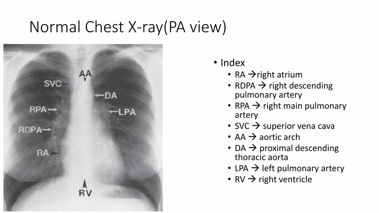
Normal Chest X-ray(PA view)
• Index• RA right atrium• RDPA right descending
pulmonary artery• RPA right main pulmonary
artery• SVC superior vena cava• AA aortic arch • DA proximal descending
thoracic aorta• LPA left pulmonary artery• RV right ventricle
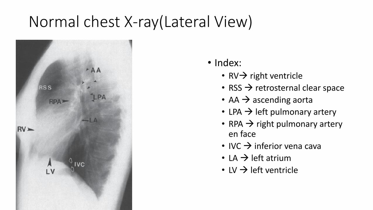
Normal chest X-ray(Lateral View)
• Index:• RV right ventricle
• RSS retrosternal clear space
• AA ascending aorta
• LPA left pulmonary artery
• RPA right pulmonary artery en face
• IVC inferior vena cava
• LA left atrium
• LV left ventricle
Congenital Heart Disease
• Transposition of the Great Vessels• Most common cyanotic congenital heart lesion
• 5%–7% of congenital cardiac malformations
• isolated in 90%
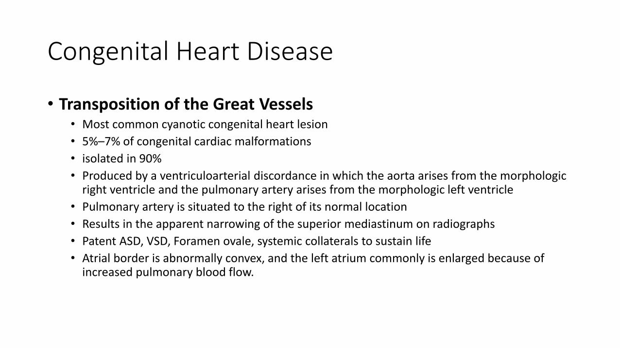
• Produced by a ventriculoarterial discordance in which the aorta arises from the morphologic right ventricle and the pulmonary artery arises from the morphologic left ventricle
• Pulmonary artery is situated to the right of its normal location
• Results in the apparent narrowing of the superior mediastinum on radiographs
• Patent ASD, VSD, Foramen ovale, systemic collaterals to sustain life
• Atrial border is abnormally convex, and the left atrium commonly is enlarged because of increased pulmonary blood flow.
Transposition of great vessels EGG ON STRING SIGN
Total Anomalous Pulmonary Venous Return
• Occurs when the pulmonary veins fail to drain into the left atrium and instead form an aberrant connection with some other cardiovascular structure
• 2% of cardiac malformations
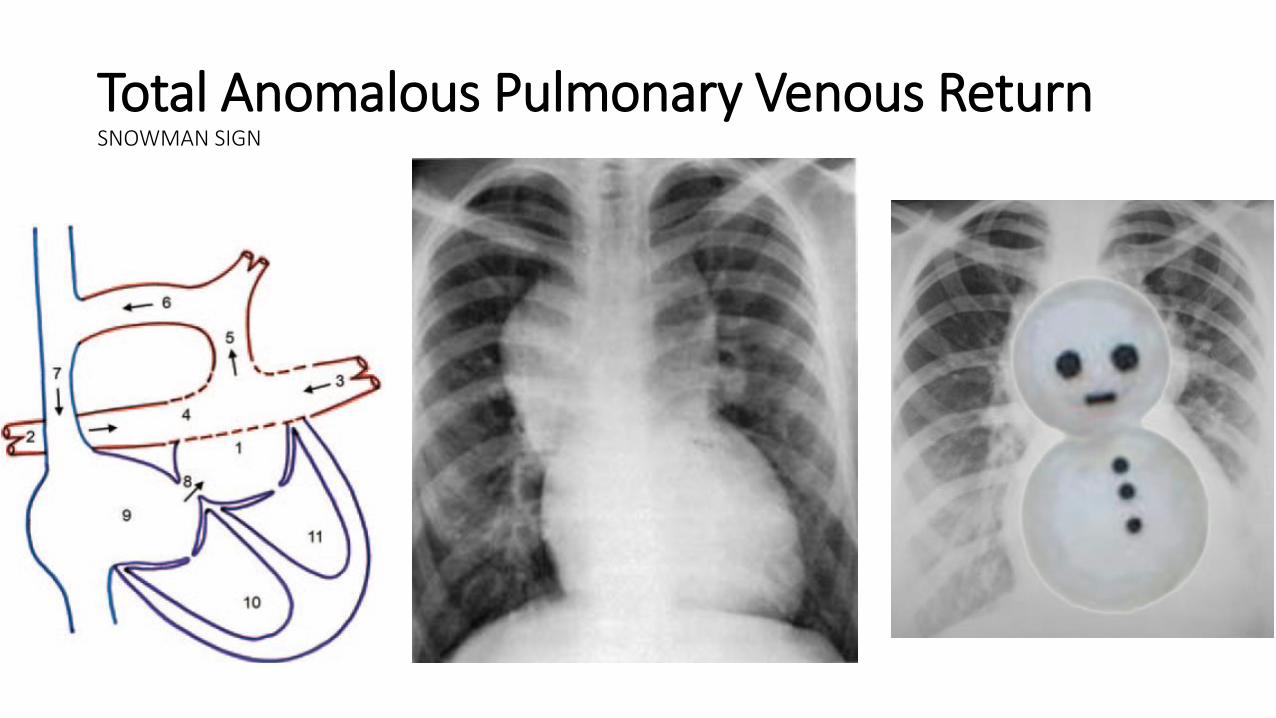
Total Anomalous Pulmonary Venous ReturnSNOWMAN SIGN
Partial Anomalous Pulmonary Venous Return
• Anomalous pulmonary vein drains any or all of the lobes of the right lung
• Vein curves outward along the right cardiac border, usually from the middle of the lung to the cardiophrenic angle, and usually empties into the inferior vena cava but also may drain into the portal vein, hepatic vein, or right atrium
• Size of the vein generally increases as it descends.
• Characteristic appearance of the vein has led to its comparison to a scimitar
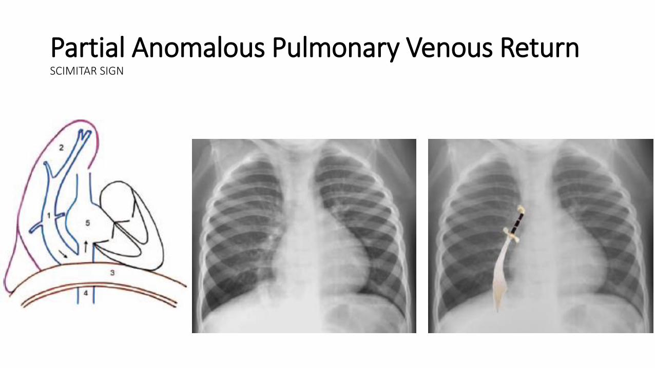
Partial Anomalous Pulmonary Venous ReturnSCIMITAR SIGN
Endocardial Cushion Defects
• Interruption of the normal development of the endocardial tissues during gestation
• Endocardial cushion forms the lower portion of the atrial septum, the upper portion of the interventricular septum, and the septal leaflets of the mitral valve and the tricuspid valve
• 4% of all cases of congenital heart disease
• Gooseneck-shaped deformity• Caused by a deficiency of both the conus and sinus portions of the interventricular
septum, with narrowing of the left ventricular outflow tract. • Characteristic shape by concavity of the interventricular septum below the mitral
valve, along with the elongation and narrowing of the left ventricular outflow tract
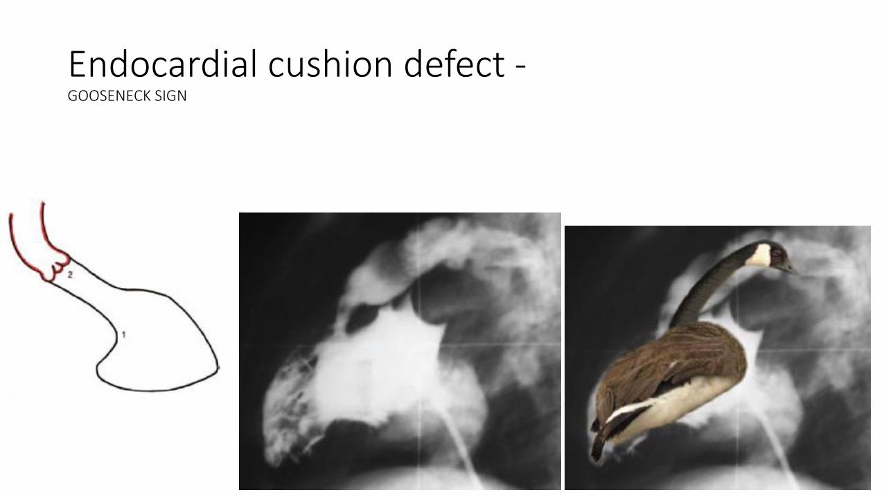
Endocardial cushion defect -GOOSENECK SIGN
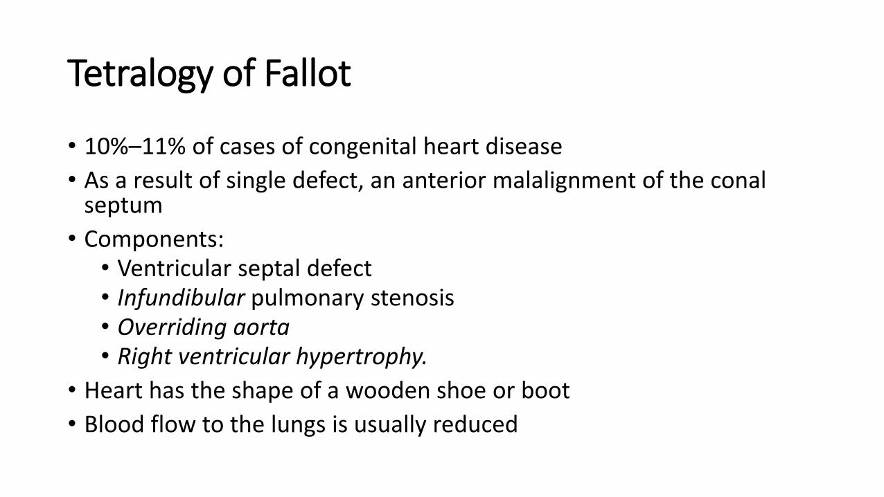
Tetralogy of Fallot
• 10%–11% of cases of congenital heart disease
• As a result of single defect, an anterior malalignment of the conalseptum
• Components:• Ventricular septal defect• Infundibular pulmonary stenosis• Overriding aorta• Right ventricular hypertrophy.
• Heart has the shape of a wooden shoe or boot
• Blood flow to the lungs is usually reduced
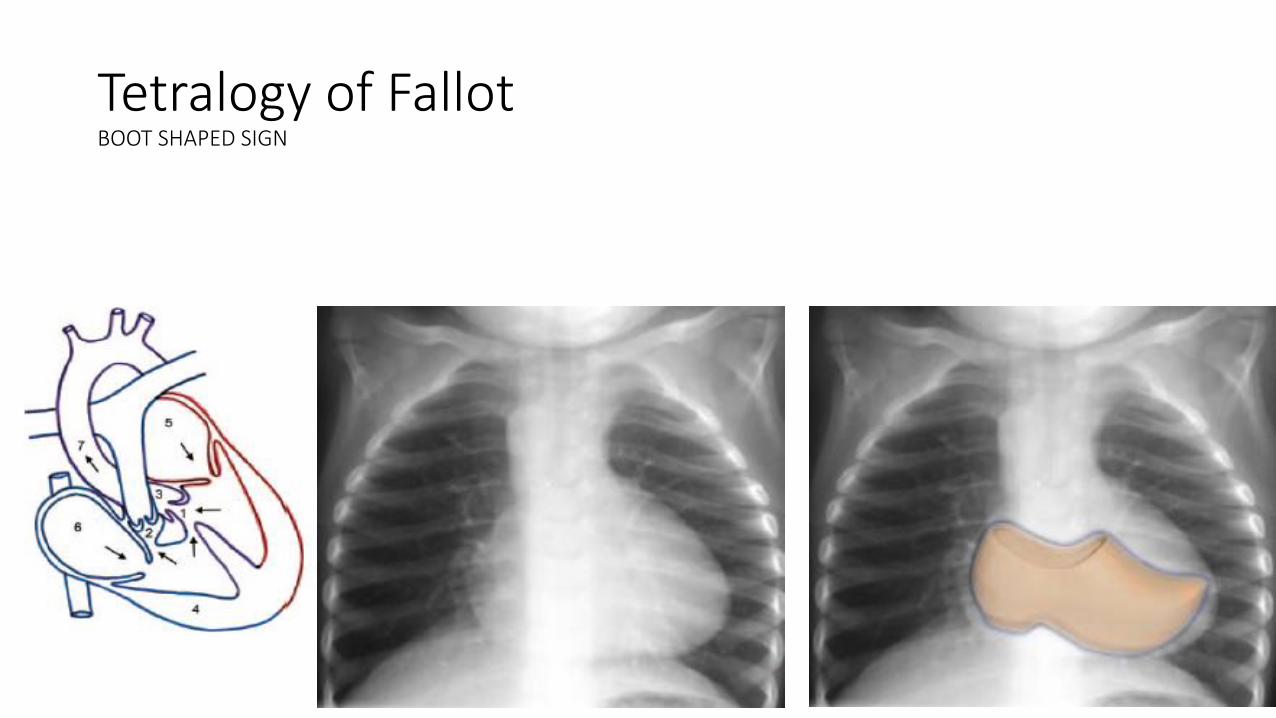
Tetralogy of FallotBOOT SHAPED SIGN
Aortic Coarctation
• 5%–10% of congenital cardiac lesions
• Produced by a deformity of the aortic media and intima, which causes a prominent posterior infolding of the aortic lumen
• Occurs at or near the junction of the aortic arch and the descending thoracic aorta
• Infolding cause eccentric narrowing of the lumen at the level where the ductus or ligamentum arteriosus inserts anteromedially
• Resultant luminal narrowing in turn obstructs the flow of blood from the left ventricle
• Classic radiologic signs• Figure-of-three sign
• Reverse figure-of-three sign
• Rib notching on CXR pathognomonic
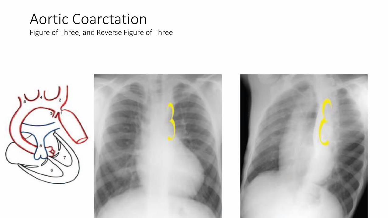
Aortic CoarctationFigure of Three, and Reverse Figure of Three
Ebstein Anomaly
• 0.5%–0.7% of cases of congenital heart disease.
• Characterized by the downward displacement of the septal leaflets and posterior leaflets of the tricuspid valve into the inflow portion of the right ventricle.
• Results in the formation of a common right ventriculoatrial chamber and causes tricuspid regurgitation.
• Insufficiency of the tricuspid valve leads to dilatation of the right ventricular outflow tract and all proximal right heart structures,
• Most consistent imaging feature is right atrial enlargement
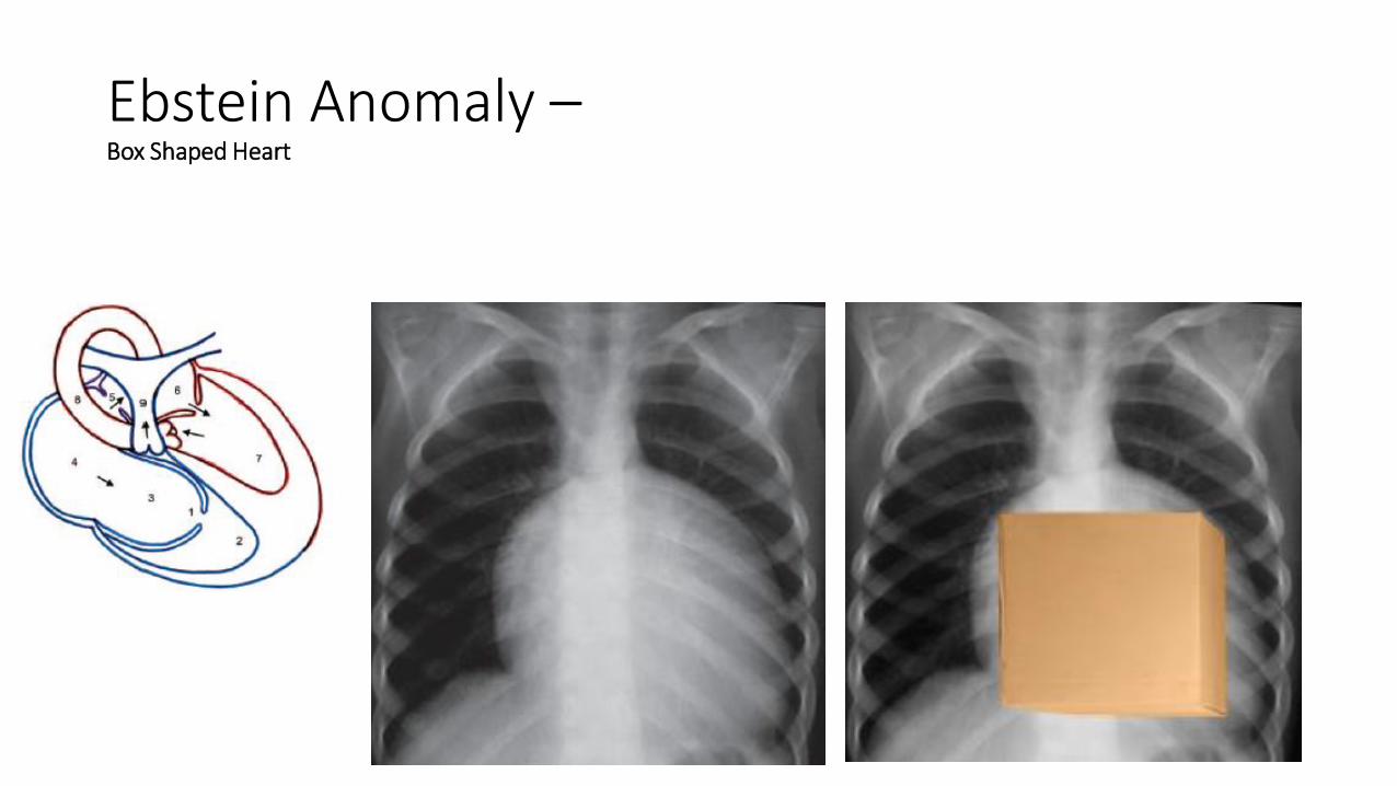
Ebstein Anomaly –Box Shaped Heart
References
• Michael Y. M. Chen, Thomas L. Pope, David J. Ott. Basic Radiology. 2nd ed. Mc. Grow hill. P-86-9.
• Cochard, Larry R.,Netter, Frank H. Netter's Introduction to Imaging. Elseiver. P-54-9.

Thank you