Embed Size (px)
Citation preview

Histology Made Absolutely Easy:
The Cell Membrane:
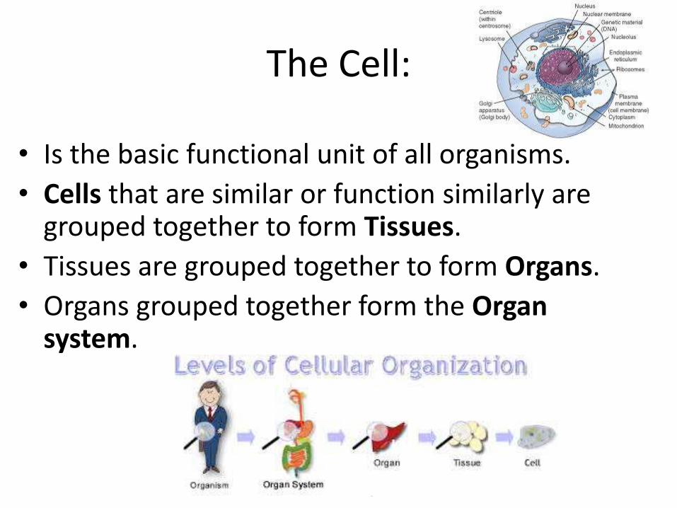
The Cell:
• Is the basic functional unit of all organisms.
• Cells that are similar or function similarly are grouped together to form Tissues.
• Tissues are grouped together to form Organs.
• Organs grouped together form the Organ system.
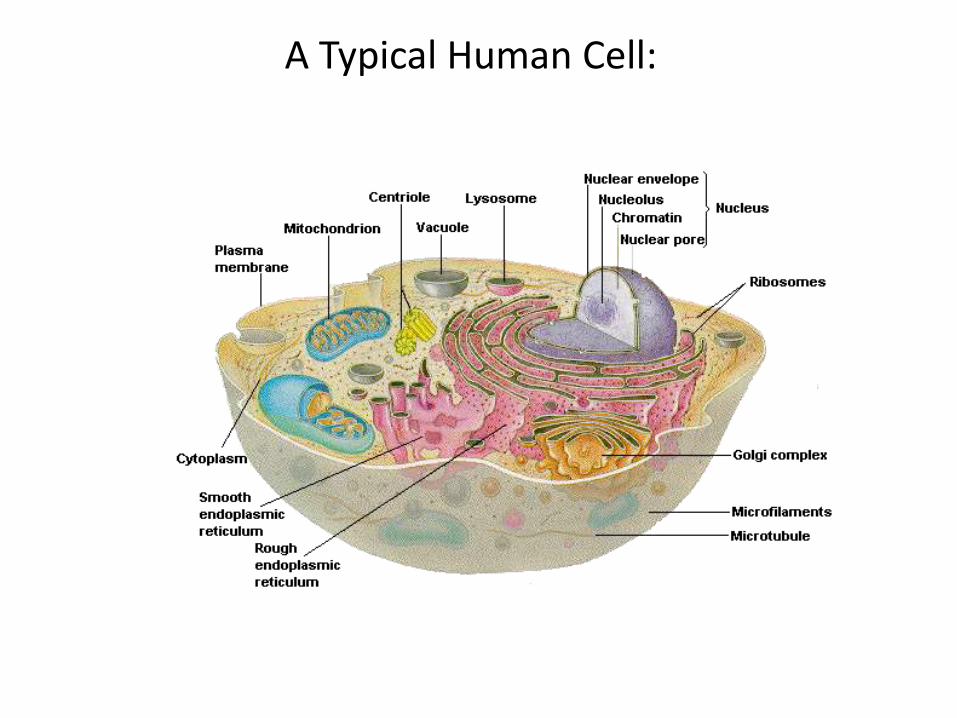
A Typical Human Cell:

Components of a Cell:The cell is a mass of Protoplasm separated from the external environment by a Plasma Membrane.
The Protoplasm is made up of two components:1. Cytoplasm: that contains numerous organelles:
• Mitochondria• Endoplasmic Reticulum• Golgi Apparatus• Ribosomes• Lysosomes• Peroxisomes• The cytoskeleton of the Cell: (a) Microfilaments
(b) Intermediate filaments(c) Microtubules
• Centrosome and centrioles• Cytoplasmic Inclusions
2. Nucleus: that houses the genome of the cell.

The Cell Membrane/Plasma Membrane/ Plasmalemma
Functions are:• Separates the contents of the cell from the external environment• Maintains the shape of the cell• Controls the transport of molecules in and out of the cell (selectivepermeability)• Regulates cell–cell interactions• It bears receptors that aid in recognizing antigens and foreign cells• Transduces extracellular physical or chemical signals into intracellularevents.
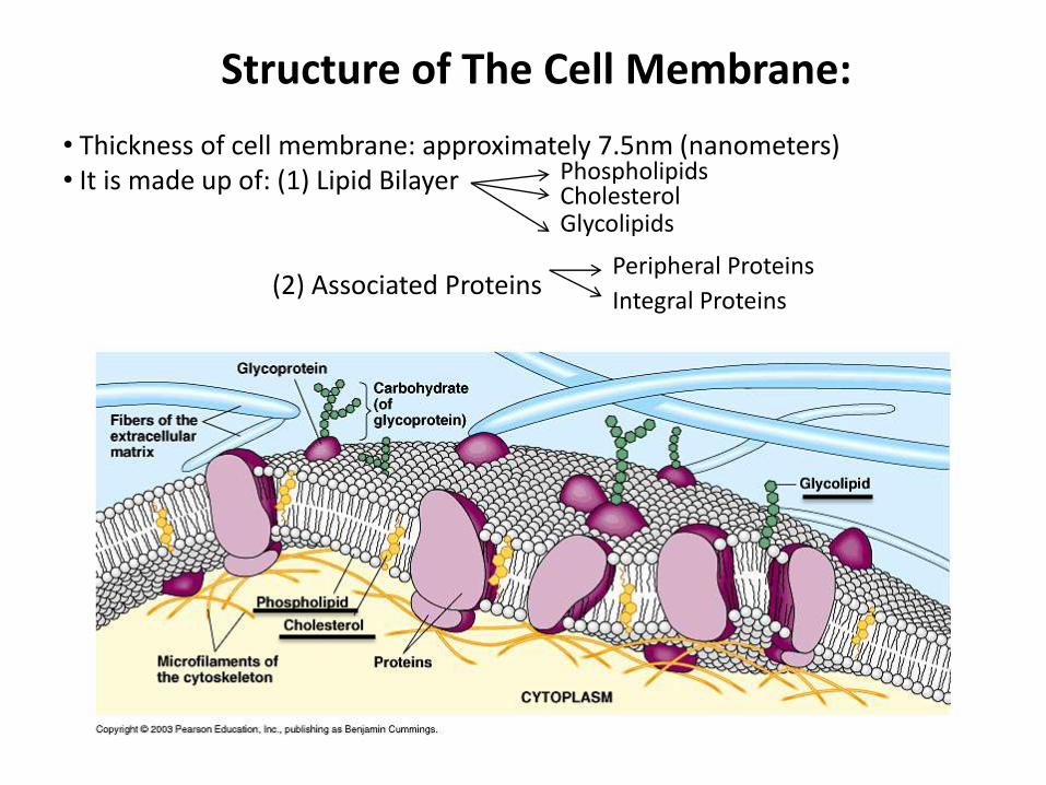
Structure of The Cell Membrane:
• Thickness of cell membrane: approximately 7.5nm (nanometers)• It is made up of: (1) Lipid Bilayer
(2) Associated Proteins
PhospholipidsCholesterolGlycolipids
Integral Proteins
Peripheral Proteins
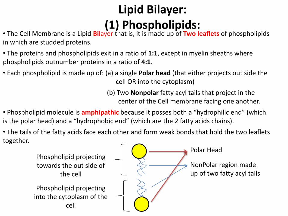
Lipid Bilayer:(1) Phospholipids:
• The Cell Membrane is a Lipid Bilayer that is, it is made up of Two leaflets of phospholipids in which are studded proteins.
• The proteins and phospholipids exit in a ratio of 1:1, except in myelin sheaths where phospholipids outnumber proteins in a ratio of 4:1.
• Each phospholipid is made up of: (a) a single Polar head (that either projects out side the cell OR into the cytoplasm)
(b) Two Nonpolar fatty acyl tails that project in the center of the Cell membrane facing one another.
• Phospholipid molecule is amphipathic because it posses both a “hydrophilic end” (which is the polar head) and a “hydrophobic end” (which are the 2 fatty acids chains).
• The tails of the fatty acids face each other and form weak bonds that hold the two leaflets together.
Polar Head
NonPolar region made up of two fatty acyl tails
Phospholipid projecting towards the out side of
the cell
Phospholipid projecting into the cytoplasm of the
cell

Membrane Faces:Is the part of the outer leaflet facing the external environment
Is the part of the outer leaflet facing the hydrophobic part of cell membrane
Is the part of the inner leafletfacing the hydrophobic part of cell membrane
Is the part of the inner leaflet facing the Cytoplasm
Copyright: Junqueira’s basic histology
Copyright: Histology and cell biology

(2) Cholesterol
• Constitutes 2% of the Plasma membrane lipids.• It is amphipathic in nature.• It is present at both the leaflets of the cell membrane.• Maintains the structural integrity of the cell membrane.• Cell membrane has to maintain its fluidity in order to carry out processes like exocytosis andendocytosis.
Fluidity increases due to: (i) increase in temperature(ii) decreased saturation of fatty acyl tails.
Fluidity decreases due to: (i) Increase in membrane cholesterol content.
(3) Glycolipids:
• Constitute 5% of the Plasma membrane lipids.• Are Amphipathic in nature.• Located on the extracellular part of the outer leaflet.• Carbohydrate side chains from glycolipids and glycoproteins form the “fuzzy” material outsidethe cell membrane called “glycocalyx”
Copyright: Junqueira’s basic histology
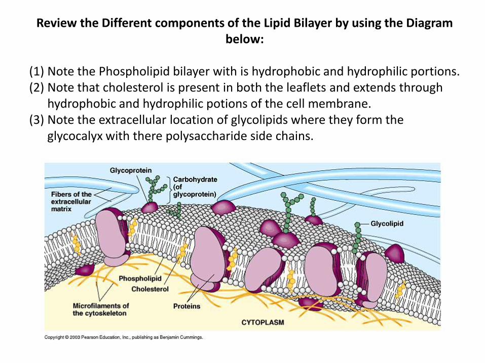
Review the Different components of the Lipid Bilayer by using the Diagram below:
(1) Note the Phospholipid bilayer with is hydrophobic and hydrophilic portions.(2) Note that cholesterol is present in both the leaflets and extends through
hydrophobic and hydrophilic potions of the cell membrane.(3) Note the extracellular location of glycolipids where they form the
glycocalyx with there polysaccharide side chains.

The Glycocalyx:• Is the “sugar coat” located on the outer surface of the outer leaflet of the Cell membrane.• Is responsible for the “fuzziness” seen on Electron Microscope.• Composition: Consists of polar oligosaccharide side chains covalently linked to proteins andsome lipids of the plasma membrane.• Functions:
(a) The glycocalyx aids in attachment of some cells to extracellular matrix components.(b) It binds antigens and enzymes to the cell surface.(c) It facilitates cell-cell recognition and interaction.(d) It protects cells from injury by preventing contact with inappropriate substances.(e) It assists T cells and antigen-presenting cells in aligning with each other.
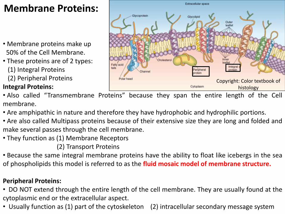
Membrane Proteins:
• Membrane proteins make up50% of the Cell Membrane.• These proteins are of 2 types:
(1) Integral Proteins(2) Peripheral Proteins
Integral Proteins:• Also called “Transmembrane Proteins” because they span the entire length of the Cellmembrane.• Are amphipathic in nature and therefore they have hydrophobic and hydrophilic portions.• Are also called Multipass proteins because of their extensive size they are long and folded andmake several passes through the cell membrane.• They function as (1) Membrane Receptors
(2) Transport Proteins• Because the same integral membrane proteins have the ability to float like icebergs in the seaof phospholipids this model is referred to as the fluid mosaic model of membrane structure.
Peripheral Proteins:• DO NOT extend through the entire length of the cell membrane. They are usually found at thecytoplasmic end or the extracellular aspect.• Usually function as (1) part of the cytoskeleton (2) intracellular secondary message system
Copyright: Color textbook of histology


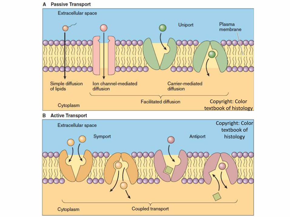
Transport System Across the Cell Membrane:
Copyright: Junqueira’s Basic Histology

Copyright: Color textbook of histology
Copyright: Color textbook of
histology

Active Transport Passive Transport
Definition:
Types of Transport:
Types of Particles
transported:
Movement of molecules DOWN the concentration gradient. It goes from high to low concentration, in order to maintain equilibrium in the cells. Does not require cellular energy.
Active Transport uses ATP to pump molecules AGAINST/UP the concentration gradient. Transport occurs from a low concentration of solute to high concentration of solute. Requires cellular energy.
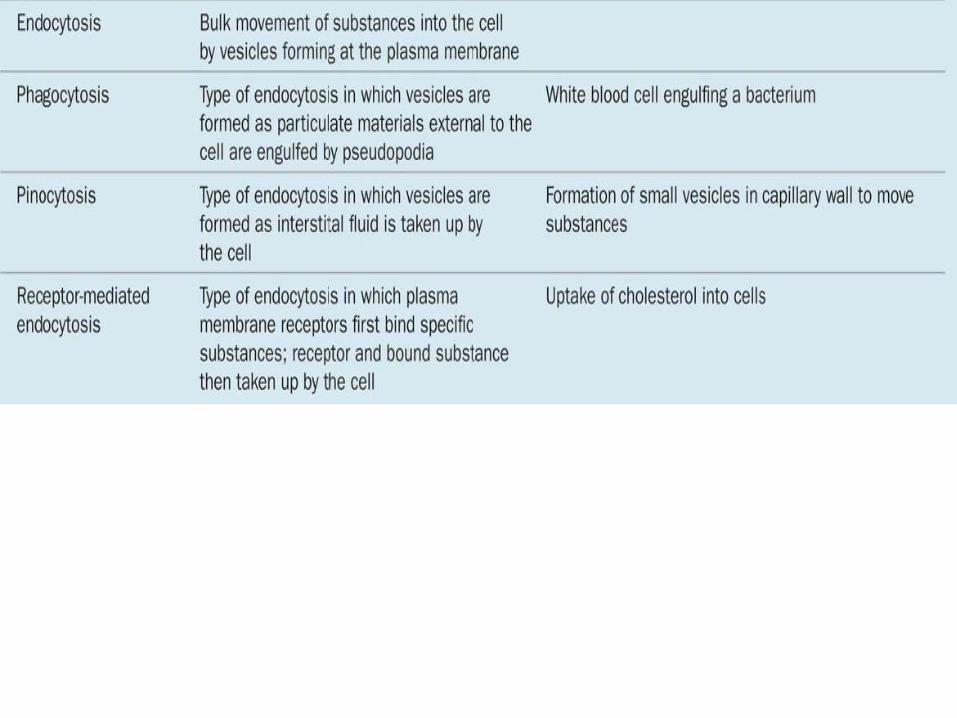
Endocytosis, cell membrane/sodium-potassium pump & exocytosis
Diffusion, facilitated diffusion, and osmosis.
Proteins, ions, large cells, complex sugars.
Anything soluble (meaning able to dissolve) in lipids, small monosaccharides, water, oxygen, carbon dioxide, sex hormones, etc.

Active Transport Passive Transport
Examples:
Importance:
Functions :
Diffusion, osmosis, and facilitated diffusion.
Phagocytosis, pinocytosis, sodium/potassium pump, secretion of a substance into the bloodstream (process is opposite of phagocytosis & pinocytosis)
In eukaryotic cells, amino acids, sugars and lipids need to enter the cell by protein pumps, which require active transport. These items either cannot diffuse or diffuse too slowly for survival.
It maintains equilibrium in the cell. Wastes (carbon dioxide, water, etc.) diffuse out and are excreted; nutrients and oxygen diffuse in to be used by the cell.
Maintains dynamic equilibrium of water, gases, nutrients, wastes, etc. between cells and extracellular fluid; allows for small nutrients and gases to enter/exit. No NET diffusion/osmosis after equilibrium is established.
Transports molecules through the cell membrane against the concentration gradient so more of the substance is inside the cell (i.e. a nutrient) or outside the cell (i.e. a waste) than normal. Disrupts equilibrium established by diffusion.

Types of Endocytosis:

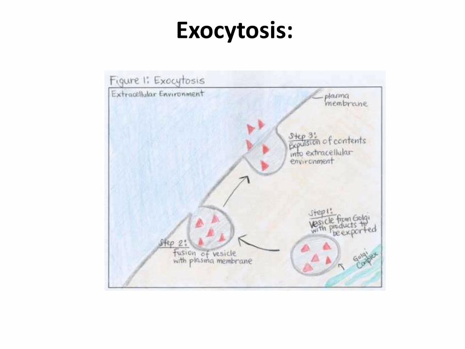
Exocytosis:

Thank you very Much