Embed Size (px)
Citation preview
Hydrocephalus

Introduction
• Hydrocephalus is a problem that neurosurgeons face every day and the onset of hydrocephalus with an increase in intracranial pressure is a permanent sequel to neurosurgery.
• Neurosurgeons need to understand the variable pathophysiology of hydrocephalus and at the same time to search for the best treatment option.

In hydrocephalus
• What happens to a brain when it is compressed by expanding ventricles? It is well understood that when anything enlarges relatively quickly in the closed skull, whether a tumor or growing ventricles, pressure rises and blood flow to the brain is threatened eventually surgical intervention is required to relieve the pressure..
• At the sametime, cerebral ventricles also may enlarge more slowly and the brain may anatomically and functionally accommodate to some extent – chronic hydrocephalus

What happens in chronic Hydrocephalus?

Aspects of CSF and blood flow dynamics andpathology in hydrocephalus
• Vascular components of CSF dynamics – an update- Zofia Czosnyka et al 2008
• Intracranial pressure (ICP) B waves which are helpful in the diagnosis of NPH, are probably caused by similar fluctuations in cerebral blood flow detectable by transcranial Doppler (TCD).
• Waves of the same frequency as ICP B waves can be also seen in cerebral blood oxygenation monitored with near infrared spectroscopy

• In idiopathic intracranial hypertension there is a strong link between ICP and sagittal sinus pressure.
• A rise in ICP provoked by lumbar infusion produces an equivalent rise in sagittal sinus pressure.
• This is probably due to pressure on the dural sinuses by rising ICP and results in obstruction of venous blood outflow. Pressure reactivity, calculated from variations between arterial pressure and ICP is correlated positively with Rout(resistent to outflow).
• Surprisingly, the character of this relationship reverses after shunting.

• There is enough evidence that testing CSF dynamics should be supplemented by testing the cerebrovascular reserve- using for example, non-invasive CO2 reactivity or modern brain imaging techniques.
• In NPH, CBF in white matter decreases as a function of distance from the surface of ventricles. In normal volunteers the distribution of CBF is flat.
• Autoregulation is less efficient closer to surface of ventricles than further away from ventricles. If cerebrovascular defects are severe, no matter how disturbed the CSF circulation, the shunt is unlikely to help.

Modelling and physics of hydrocephalus- Anatomy and Biomechanics
• Anatomical aspects can explain most clinical signs of hydrocephalus. These are the predisposed sites for ventricular enlargement (e.g. frontal lobes), the long periventricular course of lower limb pyramidal fibres and midbrain structures.
• Simple physical laws (LaPlace, Pascal) are fundamental for the understanding of hydrocephalus pathophysiology.

• Recently, traditional pathophysiology has been challenged because elevated resistance to outflow (Rout) and trans- or intra-mantle pressure gradients have been shown to be inessential for disease progression.
• Furthermore, transmantle pressure gradients are also absent in non-communicating hydrocephalus.

• Apparently, the starting point of hydrocephalus evolution is reduced arterial and/or cranio-spinal compliance hindering the physiological Windkessel effect of the basal arteries, which increases capillary pressure and reduces cerebral blood flow. Both together reduce cerebrospinal fluid (CSF) absorption and elevate Rout accordingly.

• Higher capillary pressure results in pronounced brain pulsations which hit incompressible water (CSF) at the inner and outer surfaces.
• The consequences of Pascal's law and variations in local parenchymal compliance, due to differences between intracranial and venous pressures (Starling mechanism) result in pronounced periventricular destruction.

• The outer brain is less affected due to greater damping (compliance). Thus isolated ventricular widening despite free communication between the CSF spaces occurs on the basis of reduced compliance and the Windkessel effect.
• This also explains the close link between hydrocephalus and cerebrovascular diseases.

• Cellular plasticity and physiological compensation play in progressive hydrocephalus, especially in maturing and aging brains, must be incorporated into our knowledge of the pathophysiology and clinical symptoms of this disorder.

CSF dynamics in the macro and micro environment
• The bulk flow concept of CSF dynamics has failed to explain other forms of hydrocephalus than that caused by obstruction of the intraventricular CSF pathways or the ventricular outflow.
• The hydrodynamic hypothesis for hydrocephalus considers in addition to a pure bulk flow problem, the dynamic pulsatile nature of the CSF movement in concert with the venous and arterial pulsatility and the pulsatile movement of the brain itself.

• CSF reabsorption most likely occurs together with the hundred fold amounts of arterially filtered extracellular fluid, into the venous side of the capillary bed.
• The hydrodynamic viewpoint can explain how, apart from the intraventricular interruption of the CSF bulk flow, an obstruction of the intraventricular and extraventricular CSF spaces changes local and global compliance and in consequence, pulsatility patterns, which leads to a hydrocephalic condition.

• The experimental McAllister model of basal cistern obstruction supports this by creating severe hydrocephalus simply by changing the basal cistern properties.
• In clinical practice therefore diagnostic methods like high resolution MRI for anatomical evaluation of the CSF spaces, multidirectional CSF flow studies for functional analysis, and computerised ICP analysis of pulsatility and compliance, need to be combined to develop an overall understanding of the underlying pathology in individual patients.

Assessment of CSF dynamics for diagnosinghydrocephalus
• Shunt insertion explicitly changes the CSF dynamics in patients with hydrocephalus, causing many to improve clinically. However, the relationship between a changed hydrodynamic state and improved clinical performance is not fully known.
• Therefore, further research in this area is an important challenge for the hydrocephalus research community.
• This work involves development of better methods for assessment of CSF dynamic parameters as well as studies to test hypotheses on relationships between CSF dynamics and outcome after shunting.
• The aims are for a better understanding of hydrocephalus pathophysiology and to find new predictive tests.
• The model of the CSF dynamic system includes a pressure independent outflow resistance (Rout), pressure-dependent total craniospinal compliance and a constant CSF formation rate

• The pulsatile properties are further described by the relationship between cardiac-related arterial expansion together with venous, intracranial and spinal compensatory properties.

• The most basic CSF dynamic measurement is intracranial pressure (ICP).
• From this record not only can the mean ICP be determined, but physiological variations such as Bwaves, respiratory waves and ICP pulsations can also be analysed.
• Recently, there has been a number of studies suggesting that a large cardiac pulsatility in the pressure recording is a good predictor for a positive outcome from shunting, and also that the pulse amplitude is reduced by shunting.

• This indicates that hydrocephalus patients have a decreased compliance, possibly due to a slightly increased ICP. After shunting, ICP is reduced giving a better compensatory ability.
• This also creates a link between the bulk flow theory and the pulsatility phenomenon.
• Cardiac-related CSF and blood flow measured with phase contrast MRI are another possibility for assessment of CS dynamics.

• Increased CSF pulsatility in the aqueduct is suggested as an indication for idiopathic normal pressure hydrocephalus (iNPH).
• Another iNPH-related assessment is a shorter arterial venous time delay between blood flow in carotid arteries and sagittal sinus, indicating decreased intracranial compliance.
• A powerful method for estimating CSF dynamic parameters is obtained by mathematical expressions calculated from the model, together with active infusion of artificial CSF.
• The infusion test for assessment of CSF dynamics is used clinically for diagnosing and predicting the response to shunting and for assessment of shunt functioning in-vivo.

Hydrodynamic shunt dysfunction in NPH: known and
unknown risks and how to avoid them
• Lack of clinical collaboration makes it probable that randomized clinical trials are never conducted in the clinical treatment of normal pressure hydrocephalus (NPH).
• However, a clear trend towards better outcome has been shown in studies published in the last decade, due in part to the availability of more sophisticated shunt hardware.
• Because there is no robust evidence proving that any valve is superior, neurosurgeons face important dilemmas in choosing the most adequate shunt
Hydrocephalus in paediatric group
• The distinction between compensated and progressive hydrocephalus can be especially difficult in infants because clinical signs and symptoms can be absent, nonspecific, or unreliable.
• Moreover the size of the ventricles on CT scanning or MR imaging does not always correlate with ICP or neurodevelopmental outcome.

• Hence there is a need for additional, preferably noninvasive, diagnostic techniques like MR angio phase contrast images will measure flow per minute in each vessel
• that can help to decide whether CSF diversion should be performed in infants with hydrocephalus.

Shunt selection
• must be based on the fact that both ICP and CSF dynamics are highly heterogeneous in NPH.
• Patients may have active hydrocephalus (mean ICP above 12 mmHg),
• compensated hydrocephalus (mean ICP 5– 12 mmHg)
• even low-pressure hydrocephalus (< 5 mmHg)..

• In patients with active or compensated hydrocephalus, we recommend using low or very-low pressure opening ball-in-cone valves together with gravitational devices (GD).
• G-valves alone or adjustable valves plus GD are alternative options. However, neurosurgeons must be aware that independent studies show a wide variability in the opening pressure between manufacturers, and even between valves of the same manufacturer.
• In addition, shunt resistance varies greatly, with some valves not suitable for NPH patients.
• Neurosurgeons should base their choice on information from both independent studies and from that provided by manufacturers

Treatment of hydrocephalus and managementof complications
• Shunt surgery, is the most common treatment for both paediatric and adult normal pressure hydrocephalus (NPH). The most frequent complications of shunt surgery are: over- and under drainage and infections.
• Over drainage presents as symptomatic subdural hygromas, haematomas and slit ventricles. Under drainage is either related to obstruction, disconnection, malpositioning, or migration of the shunt system or to a valve with, or set at, excessively high opening pressure, leading to functional drainage.

• During the first year after shunt surgery in paediatric hydrocephalus, 10–20% have a shunt infection and about 17% have shunt malfunction. In adult hydrocephalus, the complication rates for surgery are: infection 5–10%, mechanical malfunction 10–30%, and subdural haematoma, 10–15%.
• In NPH, these complications are rated severe in 20% of patients. Less frequent, but important complications are epilepsy 1–7%, and hearing or visual failure each 2%.
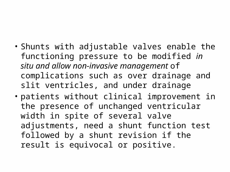
• Shunts with adjustable valves enable the functioning pressure to be modified in situ and allow non-invasive management of complications such as over drainage and slit ventricles, and under drainage
• patients without clinical improvement in the presence of unchanged ventricular width in spite of several valve adjustments, need a shunt function test followed by a shunt revision if the result is equivocal or positive.

• To date, these are either radio- nuclide studies or lumbar infusion tests. The lumbar constant rate infusion test allows assessment of the actual valve opening pressure and comparison with preoperative measures: a decrease in resistance to outflow indicates improved dynamic status and a working shunt.

• One problem with adjustable valves is that they can become re-set accidentally with magnetic fields, as in MRI, cell phones, headphones and home magnets.
• For the Hakim-Medos valve, 140 patients undergoing MRI resulted in a valve setting change in 35 (25%). Questions still to be resolved are whether the following situations affect revision rate in individual patients: using a unishunt (to avoid disconnections), endoscopic or navigated placement of ventricular catheters, peritoneal versus atrial placement of the distal catheter, the site of the ventricular catheter (occipital versus frontal) and the use of adjustable valves.

Endoscopic third ventriculostomy: outcome, controversies andfuture perspectives
• Endoscopic third ventriculostomy (ETV) has been established as a safe treatment for obstructive hydrocephalus in selected patients, with fewer overall complications than shunt insertion. The most common problems for ETV are poor intraoperative vision because of technique (blood leaks), CSF leaks, subdural fluid collection, ventriculitis or meningitis.
• Some patients fail to improve and a range of studies has demonstrated variable success rates of 50– 90

• In NPH a 69% improvement rate has been reported. Clearly success is dependent on the presence of a patent subarachnoid space. Previously shunted patients have been treated with ETV after shunt malfunction, and a proportion (38–84%) become shunt free as a result, with variable complication rates.

• In infants less than 2 years, the success rate has been low, although recently a small study reported 57% success.
• Complications are more frequent than in patients with newly diagnosed hydrocephalus, according to a retrospective review of 131 shunted and un-shunted patients from various age groups and with various etiologies treated with ETV. Shunt-related over drainage can be successfully treated with ETV followed by removal of the shunt.
• Nowadays, the long-held dogma 'once a shunt – always a shunt' is definitely no longer valid.

• After surgery, improvement or restoration of CBF has been observed, mainly in the frontal lobes .
• However, improved CBF has also been reported after shunt surgery in other cerebral regions, such as the temporal, parietal, and occipital cortex, central subcortical region, basal ganglia, hippocampus, and mesencephalon .
THE JOURNAL OF NUCLEAR MEDICINE • Vol. 44 • No. 12 • December 2003

Perfusion scan after shunt surgery for NPH


• specific CBF regions located in frontal and parietal areas that improve after surgery in idiopathic NPH.

Rigidity and topoloy of brain improved partial to near normal in few patients following shunt
• Neuroradiology : 2012 Mar;54(3):189-96. doi: 10.1007/s00234-011-0871-1. Epub 2011 May 3.
• Alteration of brain viscoelasticity after shunt treatment in normal pressure hydrocephalus.
• Freimann FB1, Streitberger KJ, Klatt D, Lin K, McLaughlin J, Braun J, Sprung C, Sack I

• Further evidence of these competing factors can be found in the diffusion-tensor imaging literature. DTI studies investigating microstructural changes in NPH have illustrated region-dependent neuronal changes throughout the brain.
• They have reported that neuronal integrity changes in the periventricular area, including the corticospinal tract, are consistent with changes secondary to mechanical compression and tend to reverse after shunt treatment.

• However, neuronal integrity changes in the frontal lobe white matter, corpus callosum, and deep gray matter are compatible with degenerative changes, which remain unchanged after treatment probably that could be the reason for not recovering memory function in these patients.

How active are patients with idiopathic normal pressure hydrocephalus and does activity improve after shuntsurgery? A
controlled actigraphic study
• Actigraphy allows long-time evaluation of physical activity and resting behaviour in a normal environment. The aim of this study was, by use of actigraphy, to measure motor function, energy expenditure and resting/sleeping time in idiopathic normal pressure hydrocephalus(iNPH) patients before and after surgery, and compare the results with healthy individuals (HI).
• Actigraphy in iNPH patients indicated reduced ambulatory activity and lower energy expenditure compared to HI preoperatively. This did not change postoperatively in spite of improved Time to up and Go(TUG) and gait speed.
• Clin Neurol Neurosurg. 2013 Feb;115(2):192-6. doi: 10.1016/j.clineuro.2012.05.009. Epub 2012 Jun 4.• How active are patients with idiopathic normal pressure hydrocephalus and does activity improve after shuntsurgery? A controlled actigraphic study.• Lundin F1, Ulander M, Svanborg E, Wikkelsø C, Leijon G
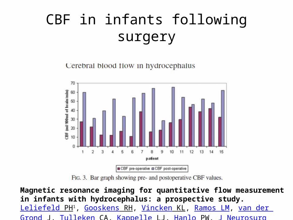
CBF in infants following surgery
Magnetic resonance imaging for quantitative flow measurement in infants with hydrocephalus: a prospective study.Leliefeld PH1, Gooskens RH, Vincken KL, Ramos LM, van der Grond J, Tulleken CA, Kappelle LJ, Hanlo PW. J Neurosurg Pediatr. 2008 Sep;2(3):163-70. doi: 10.3171/PED/2008/2/9/163

References• Cerebrospinal Fluid Research Hazel C Jones*1 and Petra M Klinge Neurosurgical
Department, International Neuroscience Institute.Hannover, Rudolf-Pichlmayr-Str. 4, 30625 Hannover, Germany.
• The Pathophysiology of Idiopathic Normal Pressure Hydrocephalus: Cerebral Ischemia or Altered Venous Hemodynamics? G.A. Bateman AJNR 29 Jan 2008
• J Neurosurg. 2001 Apr;94(4):573-81 Effects of ventriculoperitoneal shunt removal on cerebral oxygenation and brain compliance in chronicobstructive hydrocephalus. Fukuhara T1, Luciano MG, Brant CL, Klauscie J.
• J Cereb Blood Flow Metab. 2006 Oct;26(10):1298-310. Epub 2006 Feb 22. Chronic hydrocephalus-induced changes in cerebral blood flow: mediation through cardiac effects. Dombrowski SM1, Schenk S, Leichliter A, Leibson Z, Fukamachi K, Luciano MG. J Neurosurg Pediatr. 2008 Sep;2(3):163-70. doi: 10.3171/PED/2008/2/9/163.
• Magnetic resonance imaging for quantitative flow measurement in infants with hydrocephalus: a prospective study.
• Leliefeld PH1, Gooskens RH, Vincken KL, Ramos LM, van der Grond J, Tulleken CA, Kappelle LJ, Hanlo PW.