Embed Size (px)
Citation preview
ANATOMY OF JOINTS
By- Dr. Armaan SinghBy- Dr. Armaan Singh
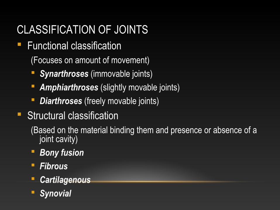
CLASSIFICATION OF JOINTS Functional classification
(Focuses on amount of movement) Synarthroses (immovable joints) Amphiarthroses (slightly movable joints) Diarthroses (freely movable joints)
Structural classification(Based on the material binding them and presence or absence of a
joint cavity) Bony fusion Fibrous Cartilagenous Synovial
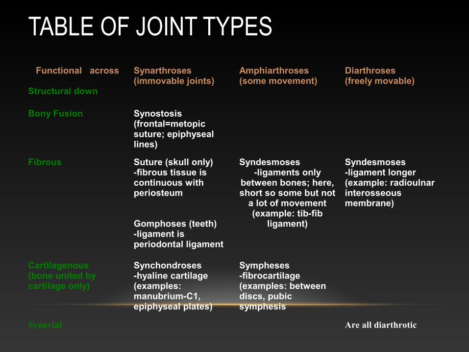
TABLE OF JOINT TYPES
Functional across
Structural down
Synarthroses(immovable joints)
Amphiarthroses(some movement)
Diarthroses(freely movable)
Bony Fusion Synostosis (frontal=metopic suture; epiphyseal lines)
Fibrous Suture (skull only)-fibrous tissue is continuous with periosteum
Gomphoses (teeth)-ligament is periodontal ligament
Syndesmoses-ligaments only
between bones; here, short so some but not
a lot of movement (example: tib-fib
ligament)
Syndesmoses-ligament longer (example: radioulnar interosseous membrane)
Cartilagenous (bone united by cartilage only)
Synchondroses-hyaline cartilage (examples: manubrium-C1, epiphyseal plates)
Sympheses -fibrocartilage (examples: between discs, pubic symphesis
Synovial Are all diarthrotic
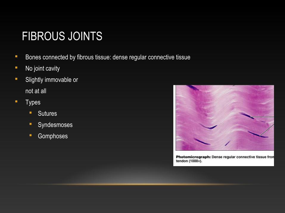
FIBROUS JOINTS
Bones connected by fibrous tissue: dense regular connective tissue
No joint cavity
Slightly immovable or
not at all
Types
Sutures
Syndesmoses
Gomphoses
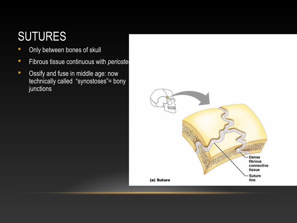
SUTURES Only between bones of skull
Fibrous tissue continuous with periosteum
Ossify and fuse in middle age: now technically called “synostoses”= bony junctions
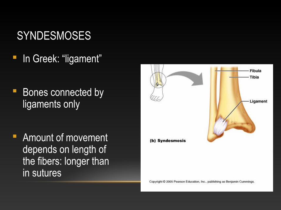
SYNDESMOSES
In Greek: “ligament”
Bones connected by ligaments only
Amount of movement depends on length of the fibers: longer than in sutures
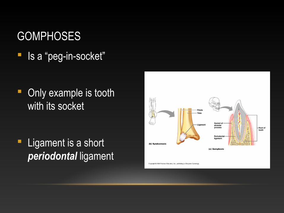
GOMPHOSES
Is a “peg-in-socket”
Only example is tooth with its socket
Ligament is a short periodontal ligament
CARTILAGENOUS JOINTS
Articulating bones united by cartilage
Lack a joint cavity
Not highly movable
Two types
Synchondroses (singular: synchondrosis)
Sympheses (singular: symphesis)
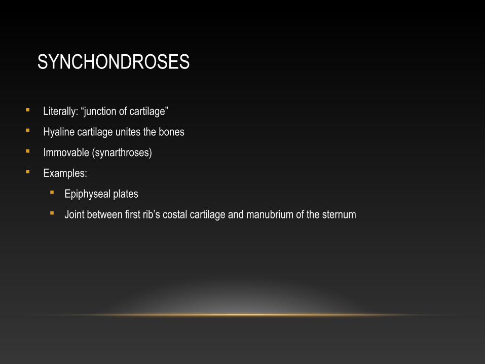
SYNCHONDROSES
Literally: “junction of cartilage”
Hyaline cartilage unites the bones
Immovable (synarthroses)
Examples:
Epiphyseal plates
Joint between first rib’s costal cartilage and manubrium of the sternum
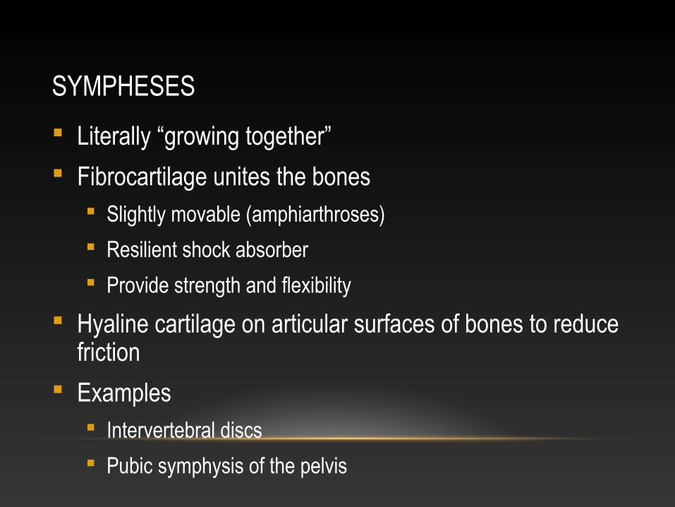
SYMPHESES
Literally “growing together”
Fibrocartilage unites the bones Slightly movable (amphiarthroses)
Resilient shock absorber
Provide strength and flexibility
Hyaline cartilage on articular surfaces of bones to reduce friction
Examples Intervertebral discs
Pubic symphysis of the pelvis
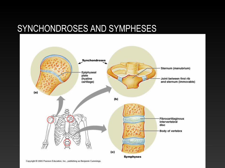
SYNCHONDROSES AND SYMPHESES
Also pubic symphsis
SYNOVIAL JOINTS
Include most of the body’s joints
All are diarthroses (freely movable)
All contain fluid-filled joint cavity
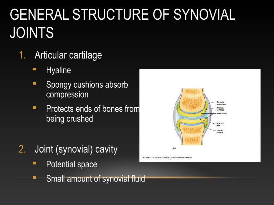
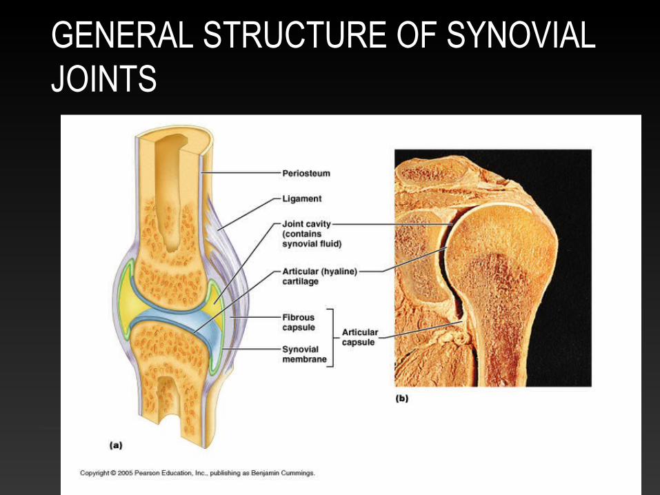
GENERAL STRUCTURE OF SYNOVIAL JOINTS
1. Articular cartilage Hyaline
Spongy cushions absorb compression
Protects ends of bones from being crushed
2. Joint (synovial) cavity Potential space
Small amount of synovial fluid
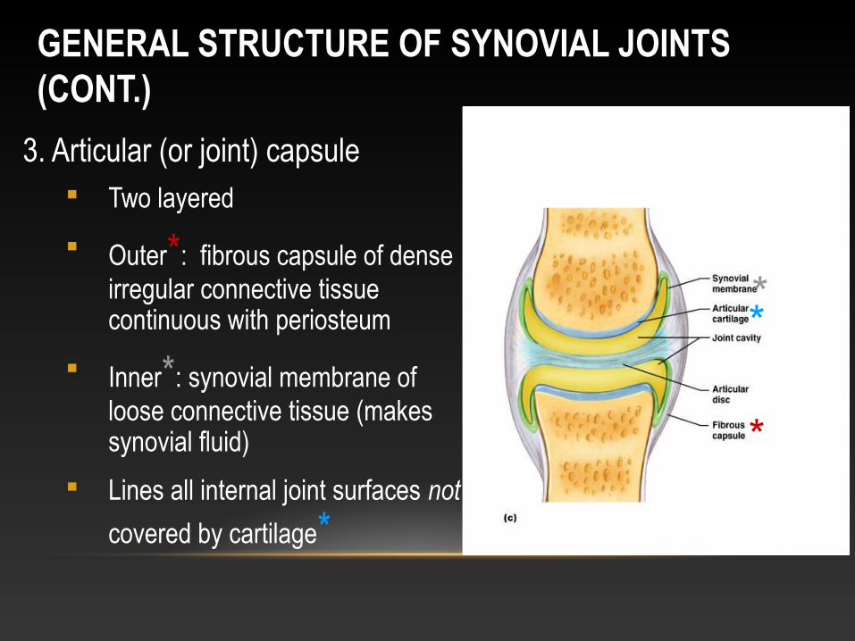
GENERAL STRUCTURE OF SYNOVIAL JOINTS (CONT.)
3. Articular (or joint) capsule Two layered
Outer*: fibrous capsule of dense irregular connective tissue continuous with periosteum
Inner*: synovial membrane of loose connective tissue (makes synovial fluid)
Lines all internal joint surfaces not
covered by cartilage*
*
**
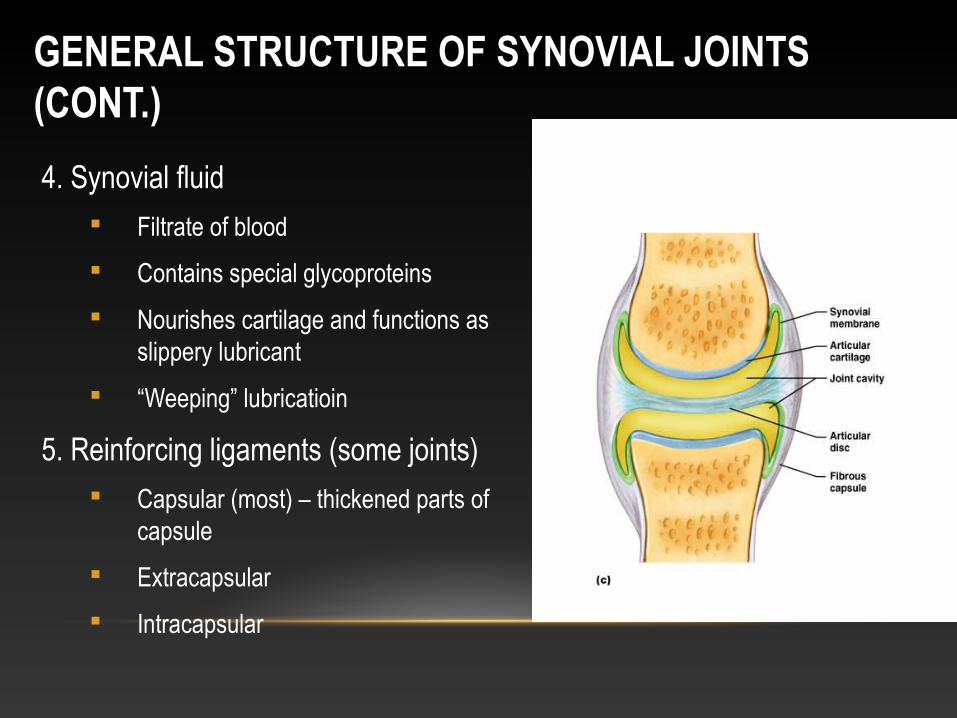
GENERAL STRUCTURE OF SYNOVIAL JOINTS (CONT.)
4. Synovial fluid Filtrate of blood
Contains special glycoproteins
Nourishes cartilage and functions as slippery lubricant
“Weeping” lubricatioin
5. Reinforcing ligaments (some joints) Capsular (most) – thickened parts of
capsule
Extracapsular
Intracapsular
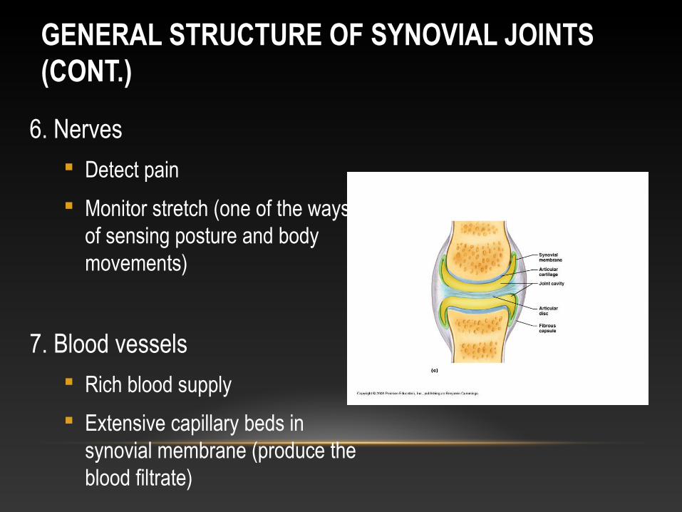
GENERAL STRUCTURE OF SYNOVIAL JOINTS (CONT.)
6. Nerves Detect pain
Monitor stretch (one of the ways of sensing posture and body movements)
7. Blood vessels Rich blood supply
Extensive capillary beds in synovial membrane (produce the blood filtrate)
GENERAL STRUCTURE OF SYNOVIAL JOINTS
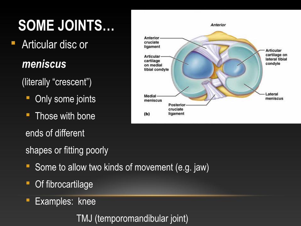
SOME JOINTS… Articular disc or
meniscus
(literally “crescent”)
Only some joints
Those with bone
ends of different
shapes or fitting poorly
Some to allow two kinds of movement (e.g. jaw)
Of fibrocartilage
Examples: knee
TMJ (temporomandibular joint)
sternoclavicular joint
BURSAE AND TENDON SHEATHS Contain synovial fluid
Not joints but often associated with them
Act like ball bearings
Bursa means “purse” in Latin
Flattened sac lined by synovial membrane
Where ligaments, muscles, tendons, or bones overlie each other and rub together
Tendon sheath
Only on tendons subjected to friction
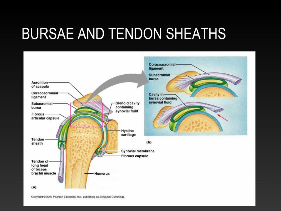
BURSAE AND TENDON SHEATHS
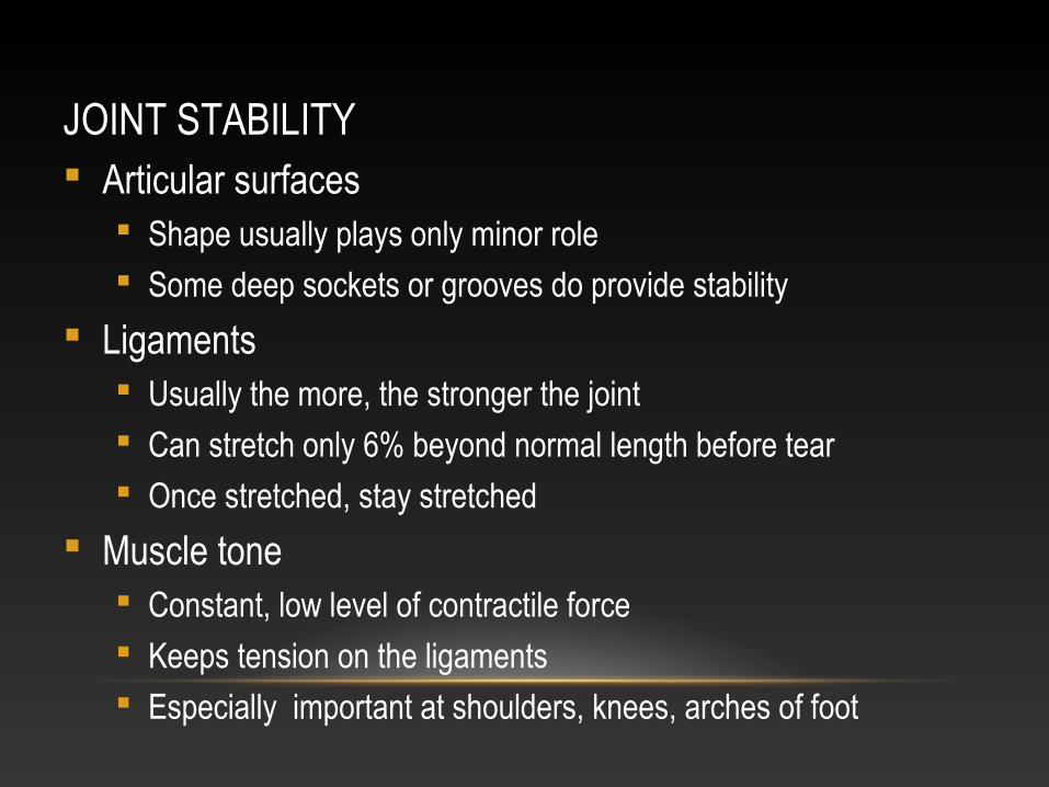
JOINT STABILITY Articular surfaces
Shape usually plays only minor role Some deep sockets or grooves do provide stability
Ligaments Usually the more, the stronger the joint Can stretch only 6% beyond normal length before tear Once stretched, stay stretched
Muscle tone Constant, low level of contractile force Keeps tension on the ligaments Especially important at shoulders, knees, arches of foot
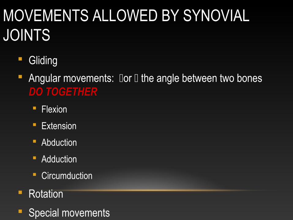
MOVEMENTS ALLOWED BY SYNOVIAL JOINTS
Gliding
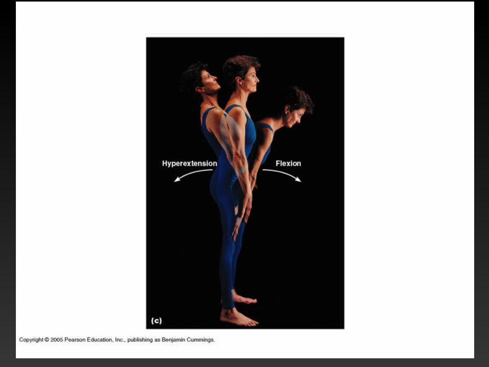
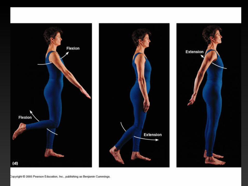
Angular movements: or the angle between two bones DO TOGETHER
Flexion
Extension
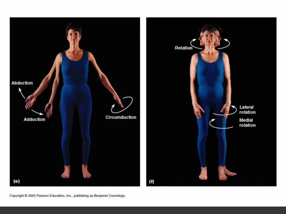
Abduction
Adduction
Circumduction
Rotation
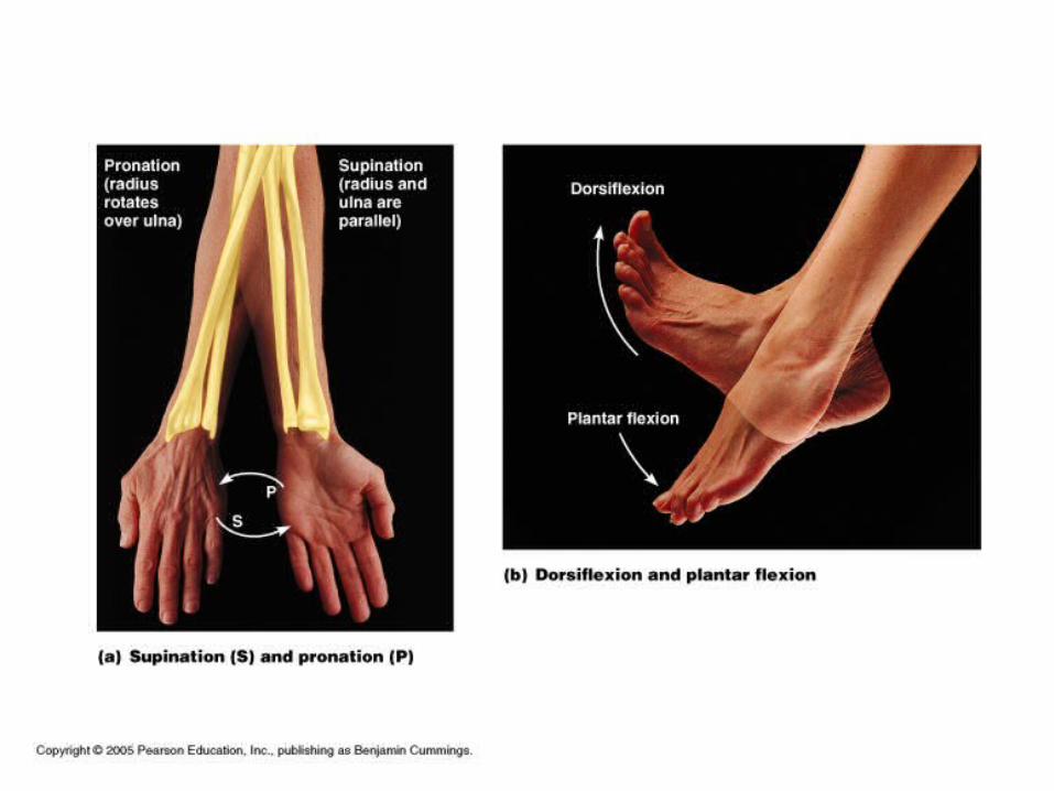
Special movements
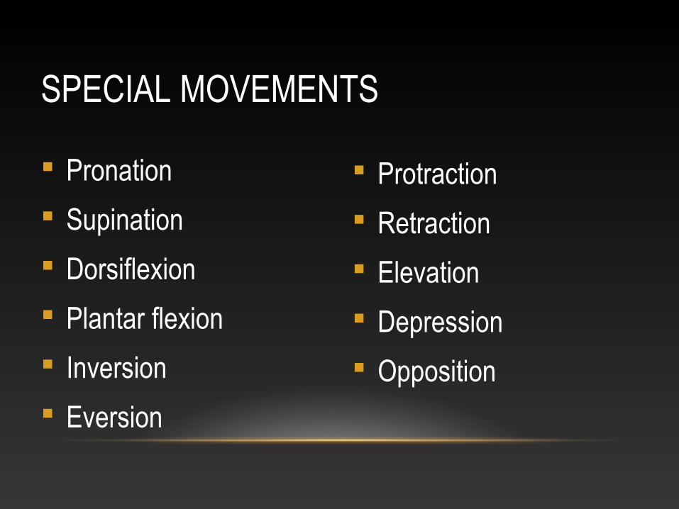
Pronation
Supination
Dorsiflexion
Plantar flexion
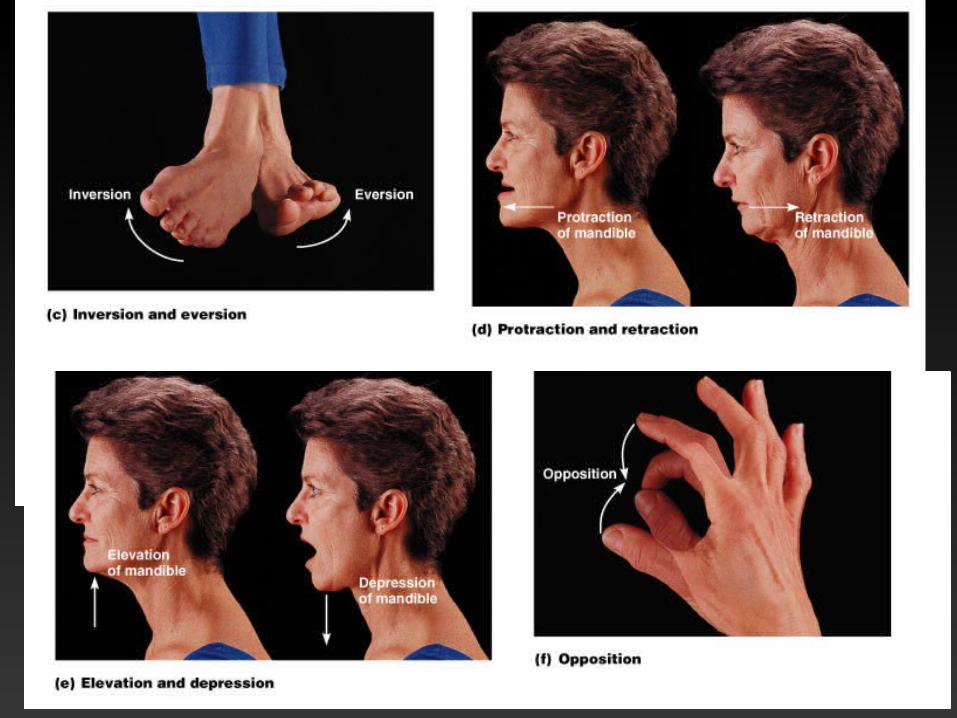
Inversion
Eversion
Protraction
Retraction
Elevation
Depression
Opposition
SPECIAL MOVEMENTS
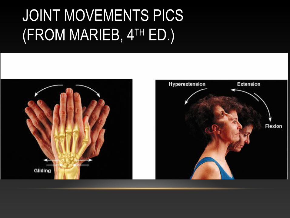
JOINT MOVEMENTS PICS (FROM MARIEB, 4TH ED.)
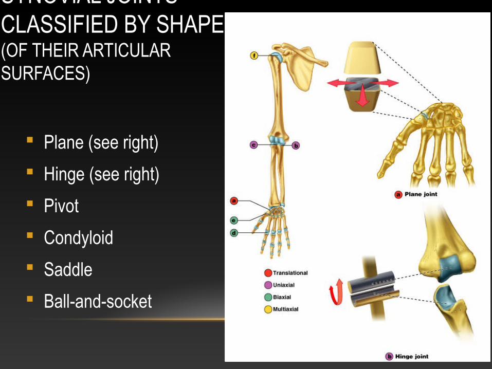
SYNOVIAL JOINTS CLASSIFIED BY SHAPE(OF THEIR ARTICULAR SURFACES)
Plane (see right)
Hinge (see right)
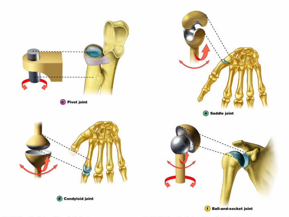
Pivot
Condyloid
Saddle
Ball-and-socket
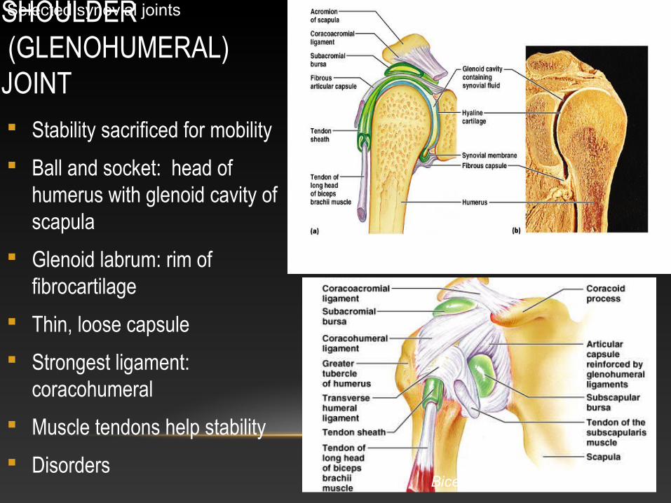
SHOULDER (GLENOHUMERAL) JOINT Stability sacrificed for mobility
Ball and socket: head of humerus with glenoid cavity of scapula
Glenoid labrum: rim of fibrocartilage
Thin, loose capsule
Strongest ligament: coracohumeral
Muscle tendons help stability
Disorders
Selected synovial joints
Rotator cuff muscles add to stability
Biceps tendon is intra-articular
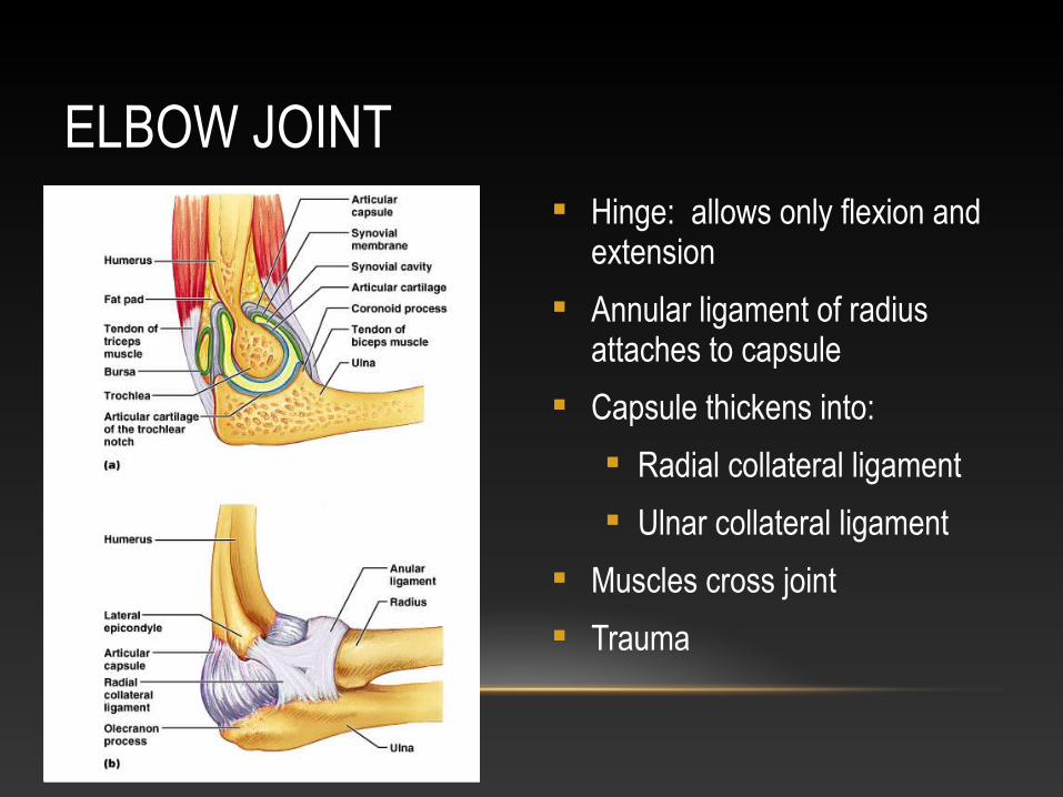
ELBOW JOINT Hinge: allows only flexion and
extension
Annular ligament of radius attaches to capsule
Capsule thickens into:
Radial collateral ligament
Ulnar collateral ligament
Muscles cross joint
Trauma
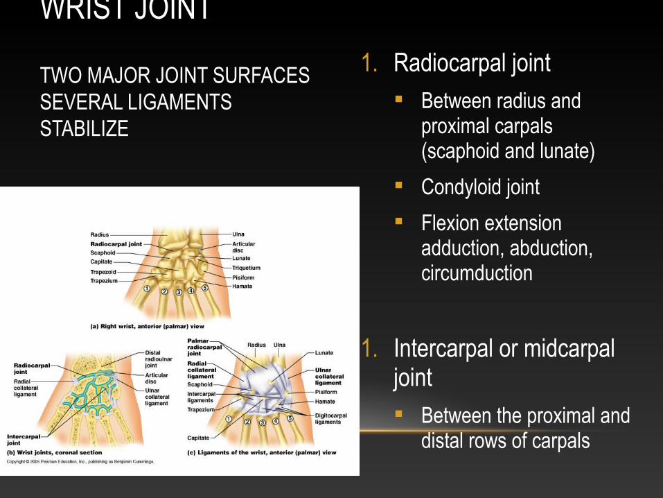
WRIST JOINT TWO MAJOR JOINT SURFACESSEVERAL LIGAMENTS STABILIZE
1. Radiocarpal joint Between radius and
proximal carpals (scaphoid and lunate)
Condyloid joint
Flexion extension adduction, abduction, circumduction
1. Intercarpal or midcarpal joint Between the proximal and
distal rows of carpals
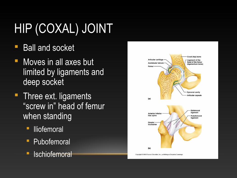
HIP (COXAL) JOINT Ball and socket
Moves in all axes but limited by ligaments and deep socket
Three ext. ligaments “screw in” head of femur when standing Iliofemoral
Pubofemoral
Ischiofemoral
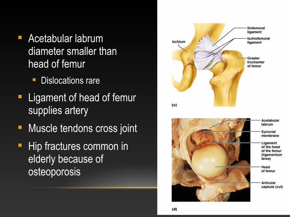
Acetabular labrum diameter smaller than head of femur Dislocations rare
Ligament of head of femur supplies artery
Muscle tendons cross joint
Hip fractures common in elderly because of osteoporosis
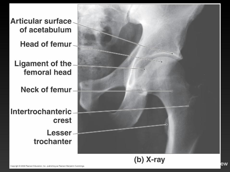
Right hip, AP view
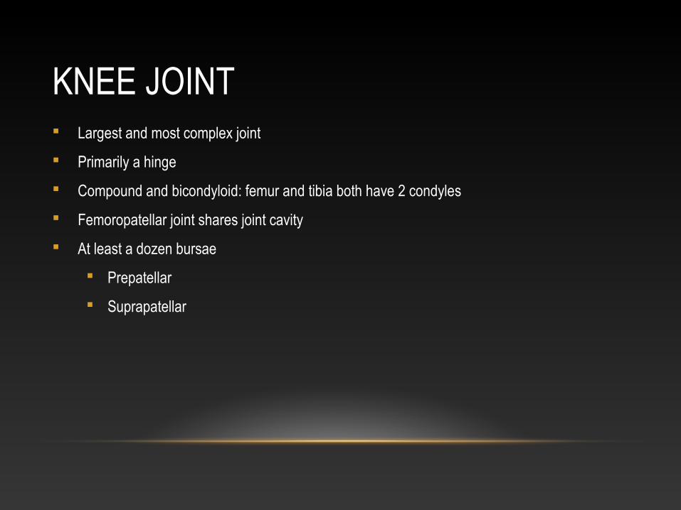
KNEE JOINT Largest and most complex joint
Primarily a hinge
Compound and bicondyloid: femur and tibia both have 2 condyles
Femoropatellar joint shares joint cavity
At least a dozen bursae
Prepatellar
Suprapatellar
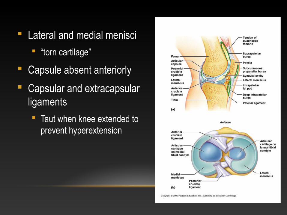
Lateral and medial menisci “torn cartilage”
Capsule absent anteriorly
Capsular and extracapsular ligaments Taut when knee extended to
prevent hyperextension
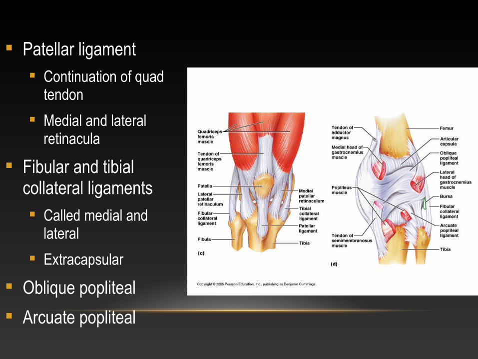
Patellar ligament Continuation of quad
tendon
Medial and lateral retinacula
Fibular and tibial collateral ligaments Called medial and
lateral
Extracapsular
Oblique popliteal
Arcuate popliteal
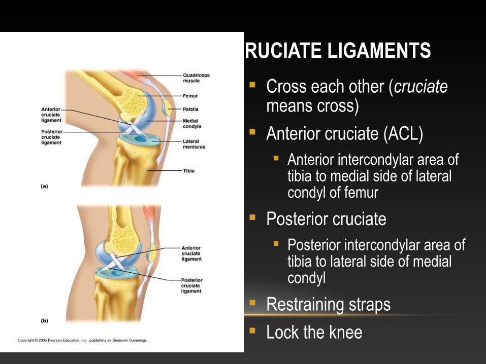
CRUCIATE LIGAMENTS
Cross each other (cruciate means cross)
Anterior cruciate (ACL) Anterior intercondylar area of
tibia to medial side of lateral condyl of femur
Posterior cruciate Posterior intercondylar area of
tibia to lateral side of medial condyl
Restraining straps
Lock the knee
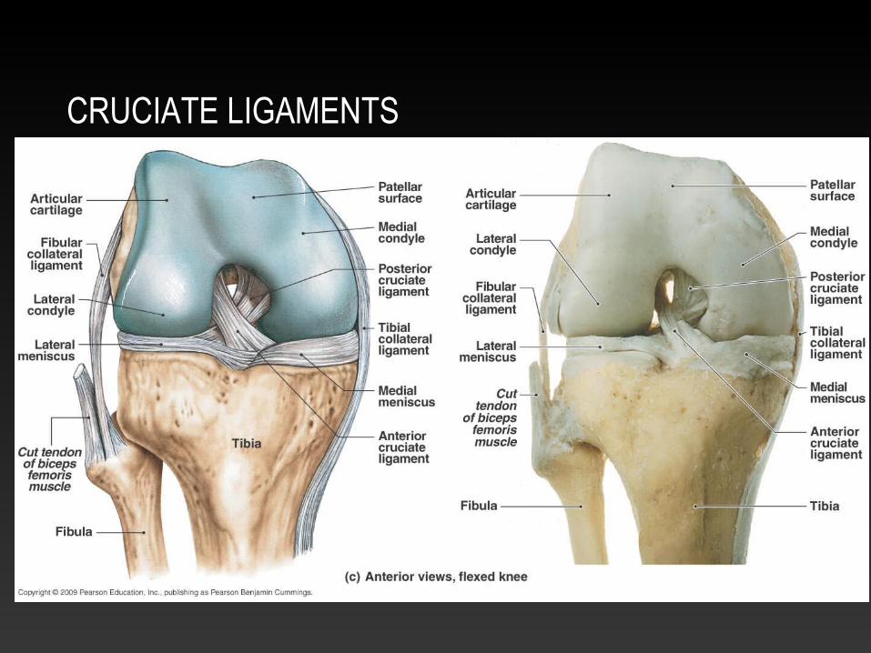
CRUCIATE LIGAMENTS
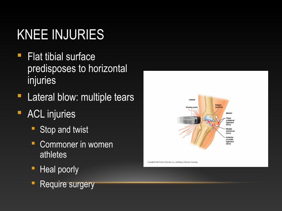
KNEE INJURIES Flat tibial surface
predisposes to horizontal injuries
Lateral blow: multiple tears
ACL injuries Stop and twist
Commoner in women athletes
Heal poorly
Require surgery
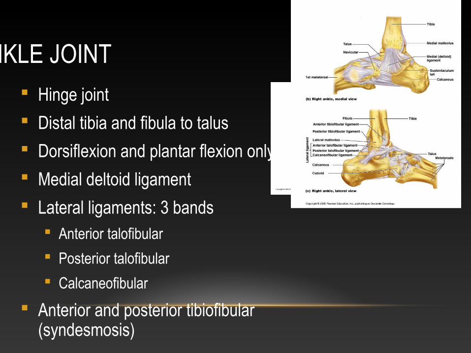
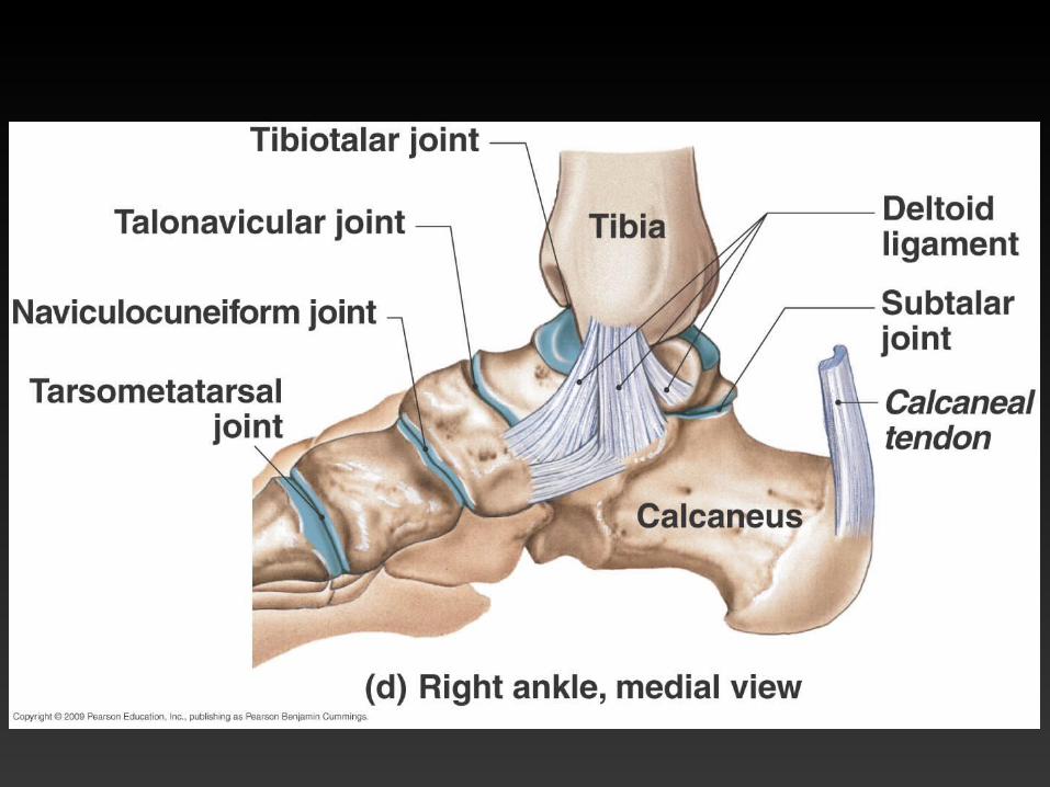
ANKLE JOINT Hinge joint
Distal tibia and fibula to talus
Dorsiflexion and plantar flexion only
Medial deltoid ligament
Lateral ligaments: 3 bands Anterior talofibular
Posterior talofibular
Calcaneofibular
Anterior and posterior tibiofibular (syndesmosis)
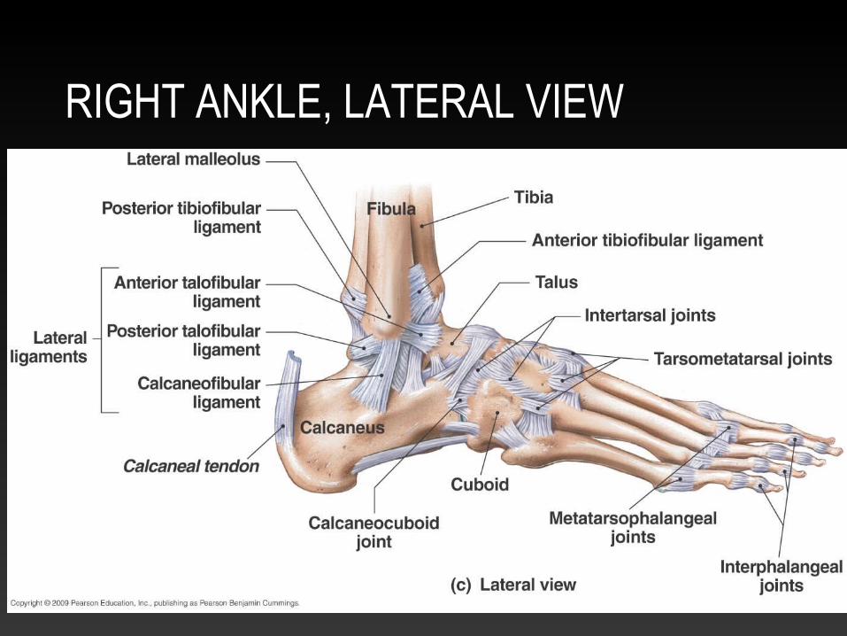
RIGHT ANKLE, LATERAL VIEW
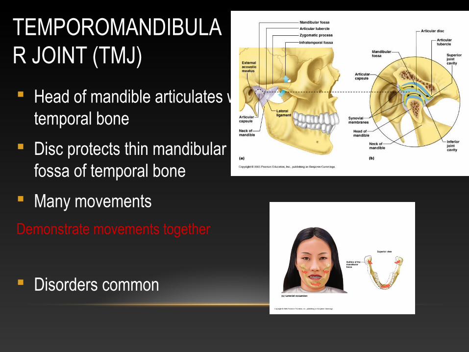
TEMPOROMANDIBULAR JOINT (TMJ)
Head of mandible articulates with temporal bone
Disc protects thin mandibular fossa of temporal bone
Many movements
Demonstrate movements together
Disorders common
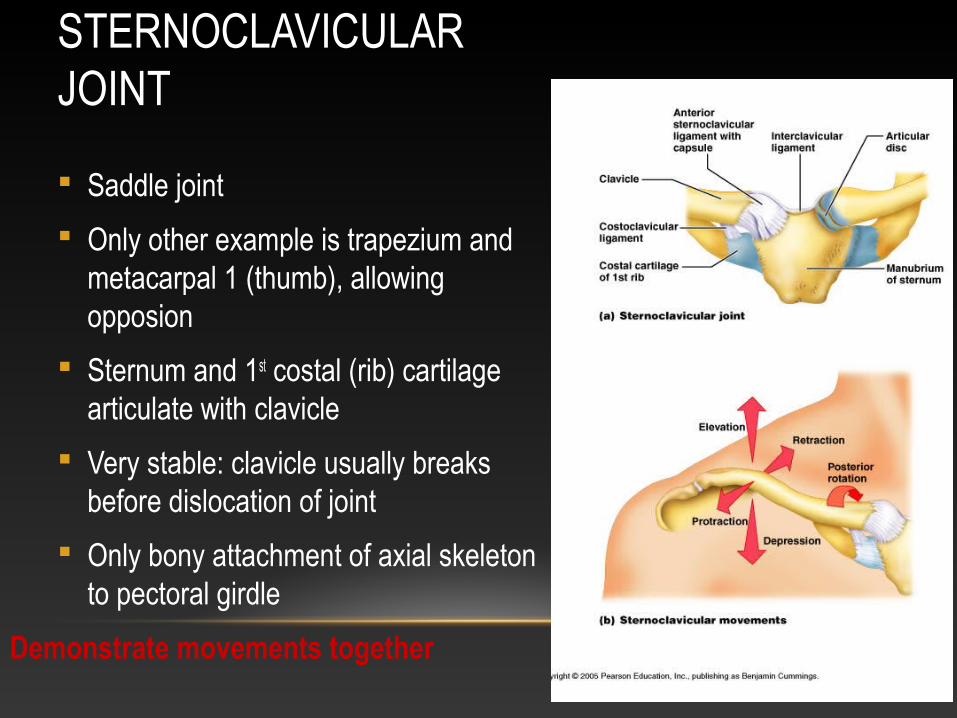
STERNOCLAVICULAR JOINT
Saddle joint
Only other example is trapezium and metacarpal 1 (thumb), allowing opposion
Sternum and 1st costal (rib) cartilage articulate with clavicle
Very stable: clavicle usually breaks before dislocation of joint
Only bony attachment of axial skeleton to pectoral girdle
Demonstrate movements together
DISORDERS OF JOINTS
Injuries Sprains
Dislocatios
Torn cartilage
Inflammatory and degenerative conditions Bursitis
Tendinitis
Arthritis Osteoarthritis (“DJD” – degenerative joint disease)
Rheumatoid arthritis (one of many “autoimmune” arthritites)
Gout (crystal arthropathy)