Embed Size (px)
Citation preview

DefinitionIt is a group of malignant disorder,
affecting the blood and blood –forming tissue of the bone marrow lymph system and spleen.
The word Leukemia comes from the Greek leukos which means "white" and aima which means "blood".
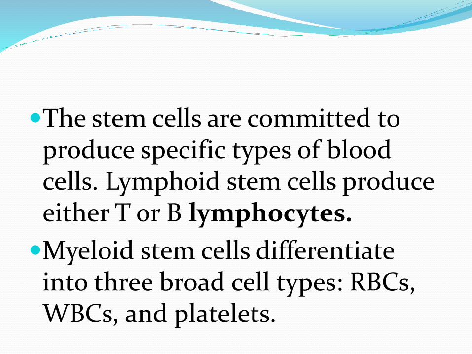
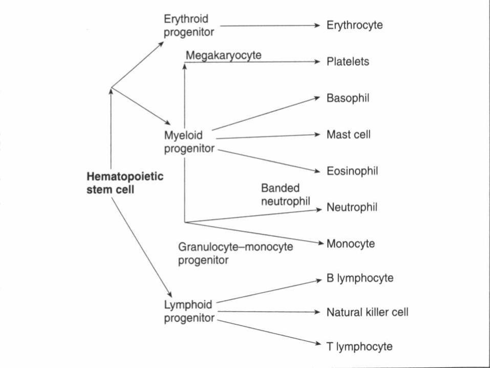
The stem cells are committed to produce specific types of blood cells. Lymphoid stem cells produce either T or B lymphocytes.
Myeloid stem cells differentiate into three broad cell types: RBCs, WBCs, and platelets.
Function of the bone marrowThe bone marrow is found in the inside of
bones. The marrow in the large bones of adults produces blood cells. Approximately 4% of our total bodyweight consists of bone marrow.
There are two types of bone marrow:
1. Red marrow, made up mainly of myeloid tissue.
2. Yellow marrow, made up mostly of fat cells.
Red marrow can be found in the flat bones, such as the breast bone, skull, vertebrae, shoulder blades, hip bone and ribs. Red marrow can also be found at the ends of long bones, such as the humerus and femur.
White blood cells (lymphocytes), red blood cells and platelets are produced in the red marrow. Red blood cells carry oxygen, white blood cells fight diseases. Platelets are essential for blood clotting.
Yellow marrow can be found in the inside of the middle section of long bones.
White blood cells, which help to body fight infection.
Red blood cells, which carry oxygen to all parts of the body.
Platelets, which help in blood clot.If a person loses a lot of blood the body can convert yellow marrow to red marrow in order to raise blood cell production.
Leukemia
DefinitionIt is a group of malignant disorder,
affecting the blood and blood –forming tissue of the bone marrow lymph system and spleen.
etiology
Combination of predisposing factors including genetic and environmental influences.
Chronic exposure to chemical such as benzene
Radiation exposure.
Cytotoxic therapy of breast, lung and testicular cancer.
Congenital anomaly
The presence of primary immunodeficiency and infection with the human T –cell leukemia virus type-1

PATHOPHYSIOLOGY
The lack of control causes –
nomal bone marrow to be replaced by immature and undifferentiated leukocytes or blat cells . –
abnormal immature leukocytes then circulates in the blood and infiltrate the blood forming organs ( liver , spleen, lymph nodes) and other sites throughout the body.
Different types of leukemia
It may be acute or chronic. Acute leukemia gets worse very fast and may make feel sick right away. Chronic leukemia gets worse slowly and may not cause symptoms for years.
Lymphocytic and MyelogenousLeukemias are also subdivided intothe type of affected blood cell. Ifthe cancerous transformationoccurs in the type of marrow thatmakes lymphocytes, the disease iscalled lymphocytic leukemia.
If the cancerous change occurs in the type of marrow cells that produce red blood cells, other types of white cells, and platelets, the disease is called myelogenous leukemia

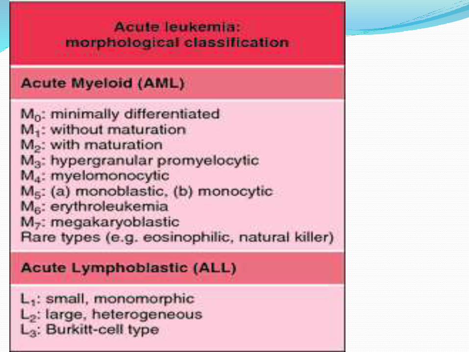
FRENCH- AMERICAN –BRITISH (FAB) CLASSIFICATION OF ACUTE LEUKEMIA
INCIDENCE—
In adults, chronic lymphocytic leukemia (CLL) and acute myelogenous leukemia (AML) are the most common leukemias.
In children, the most common leukemia is acute lymphoblastic leukemia (ALL). Childhood leukemias also include acute myelogenous leukemia (AML) and other myeloid leukemias, such as chronic myelogenous leukemia (CML) and juvenile myelomonocytic leukemia (JMML).
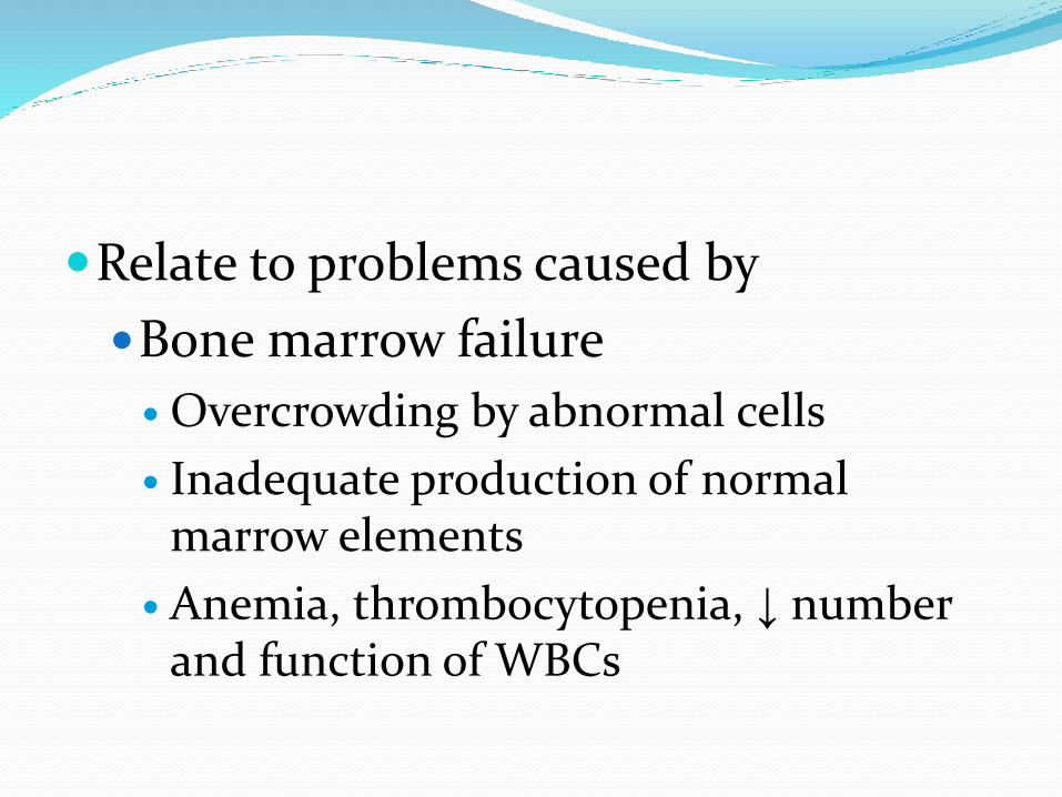
Relate to problems caused by
Bone marrow failure
Overcrowding by abnormal cells
Inadequate production of normal marrow elements
Anemia, thrombocytopenia, ↓ number and function of WBCs
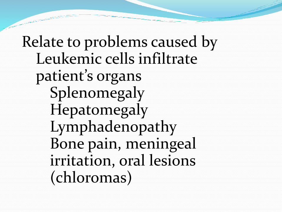
Relate to problems caused byLeukemic cells infiltrate patient’s organs
SplenomegalyHepatomegalyLymphadenopathyBone pain, meningealirritation, oral lesions (chloromas)
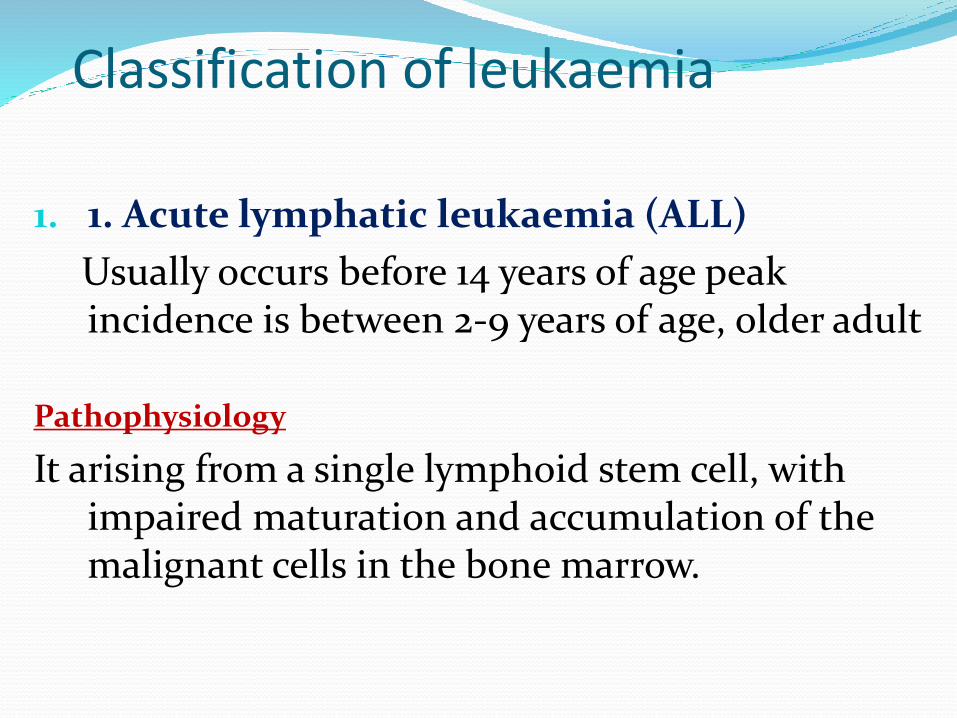
Classification of leukaemia
1. 1. Acute lymphatic leukaemia (ALL)
Usually occurs before 14 years of age peak incidence is between 2-9 years of age, older adult
Pathophysiology
It arising from a single lymphoid stem cell, with impaired maturation and accumulation of the malignant cells in the bone marrow.
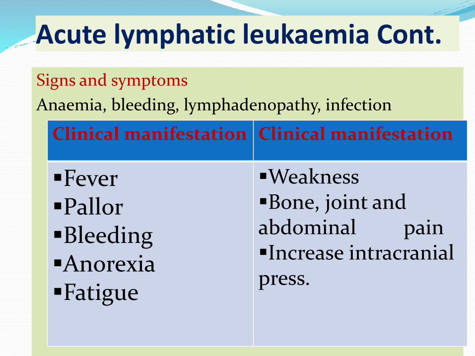
Acute lymphatic leukaemia Cont.
Signs and symptoms
Anaemia, bleeding, lymphadenopathy, infection
Clinical manifestation Clinical manifestation
FeverPallorBleedingAnorexiaFatigue
WeaknessBone, joint and abdominal painIncrease intracranial press.
Generalized lymphadenopathy
Infection of respiratory tract
Anaemia and bleeding of mucus membrane
Weight lossa
Mouth sore
Acute lymphatic leukaemia Cont.Diagnosis
Low RBCs count, Hb, Hct, low platelet count , low normal or high WBC count.
Blood smear show immature lymph blasts.
Treatment
Chemotherapeutic agent, it involve three phases
1. Induction: Using vincristine and prednisone.
2. Consolidation: Using modified course of intensive therapy to eradicate any remaining.
3. Maintenance
Acute lymphatic leukaemia Cont.
Treatment Cont.
Prophylactic treatment of the CNS , intrathecal administration and /or craniospinal radiation with eradicate leukemic cells.
Eat diet that contains high in protein, fibres and fluids.
Acute lymphatic leukaemia Cont.
Treatment Cont.
Avoid infection (hand washing, avoid crowds),injury
Take measure to decrease nausea and to promote appetite, smoking and spicy and hot foods.
Maintain oral hygiene.
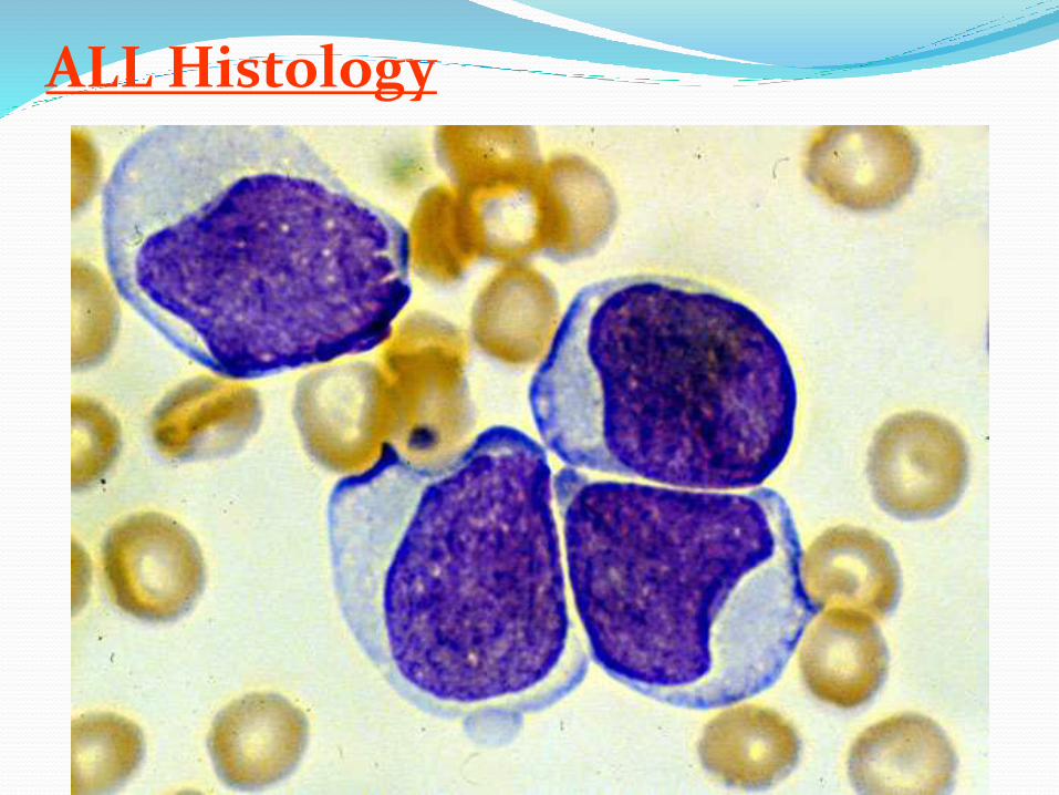
ALL Histology
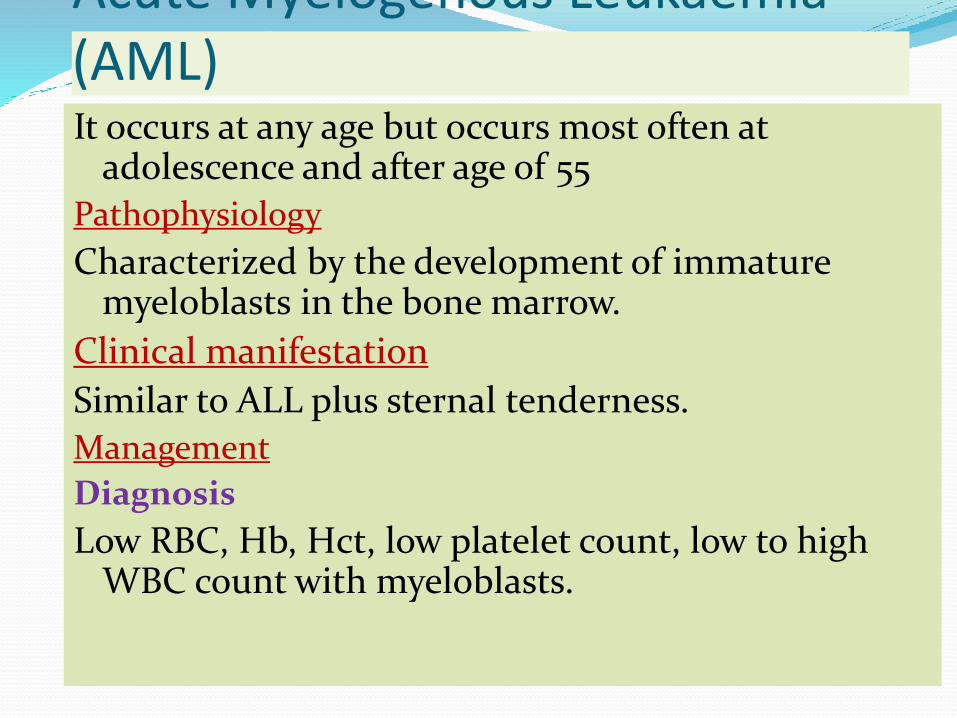
Acute Myelogenous Leukaemia (AML) It occurs at any age but occurs most often at
adolescence and after age of 55Pathophysiology
Characterized by the development of immature myeloblasts in the bone marrow.
Clinical manifestation
Similar to ALL plus sternal tenderness.Management
Diagnosis
Low RBC, Hb, Hct, low platelet count, low to high WBC count with myeloblasts.
Acute Myelogenous Leukaemia (AML) Cont.
Treatment
Use of cytarabine, 6-thioquanine, and doxorubic
The same care of client as All, plus give adequate amounts of fluids(2000 to 3000 ml per day.)
Instruct client about medication, effects, side effects and nursing measures
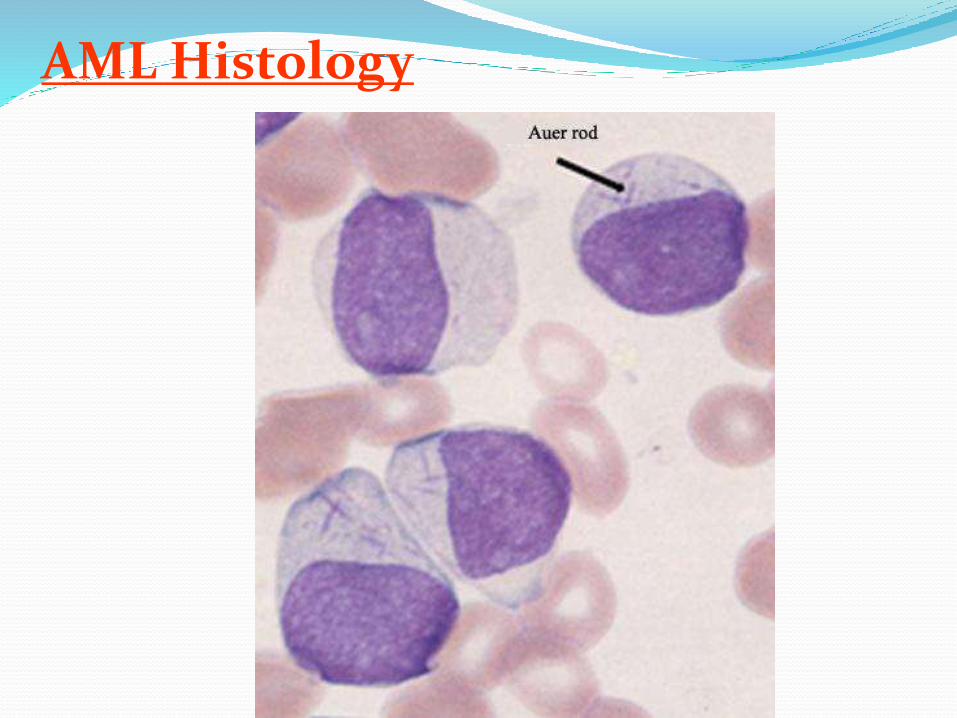
AML Histology
Chronic lymphocytic Leukaemia (CLL)
The incidence of CLl increases with age and is rare under the age of 35.It is common in men.
Pathophysiology
It is characterized by proliferation of small, abnormal , mature B lymphocytes, often leading to decreased synthesis of immunoglobulin and depressed antibody response.
The number of mature lymphocytes in peripheral blood smear and bone marrow are greatly increased
Chronic lymphocytic Leukaemia (CLL) Cont
Clinical Manifestation
Usually there is no symptoms.
Chronic fatigue , weakness , anorexia, splenomegaly , lymphadenopathy, hepatomegaly.
Signs and Symptoms
Pruritic vesicular skin lesions .
Anaemia
Thrombocytopenia.
The WBC count is elevated to a level between 20,000 to 100,000.
Increase blood viscosity and clotting episode.
Chronic lymphocytic Leukaemia (CLL) Cont
Management
I. Persons are treated only when symptoms, particular anaemia , thrombocytopenia , enlarged lymph nodes and spleen appear.
I. Chemotherapy agents such as chlorambucil , and the glucocorticoids.
I. Client and family education is that describe for AML.
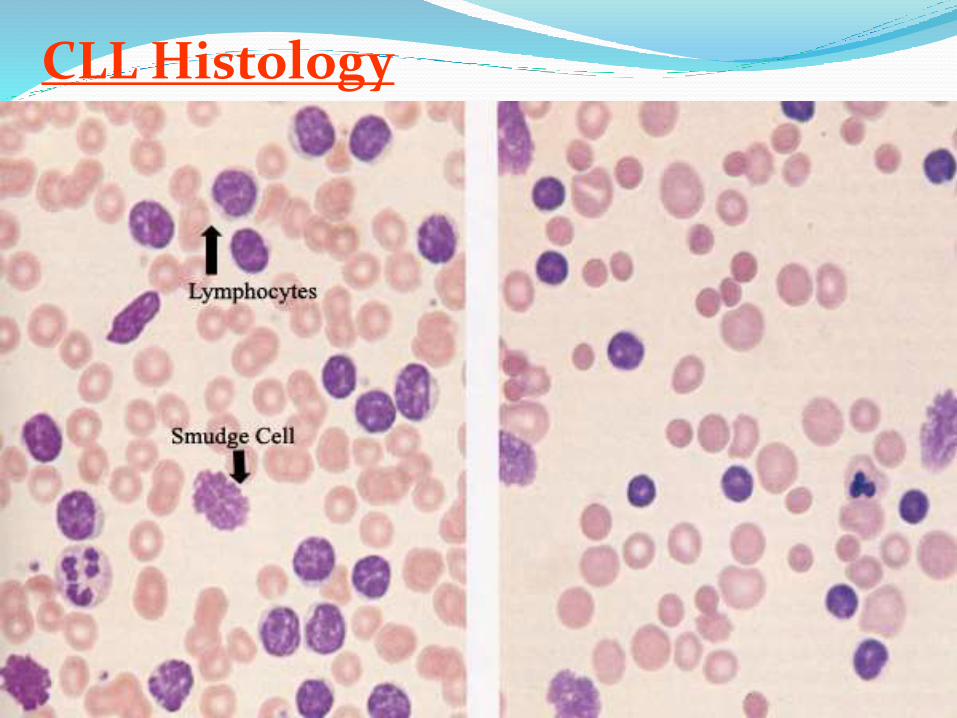
CLL Histology
Chronic MyelogenousLeukaemia(CML)Philadelphia chromosome
The chromosome abnormality that causes chronic myeloid leukemia
Occurs between 25-60 years of age. Peak 45 year
It is caused by benzene exposure and high doses of radiation.
Clinical Manifestation
There is no symptoms in disease. The classic symptoms, include:
Fatigue, weakness, fever.
Weight loss, joint & bone pain.
Chronic Myelogenous Leukaemia(CML) Cont.
Clinical Manifestation Cont.
Massive splenomegaly
The accelerated phase of disease(blosticphase) is characterized by increasing number of granulocytes in the peripheral blood.
There is a corresponding anaemia and thrombocytopenia.
Chronic Myelogenous Leukaemia(CML) Cont.
Diagnosis
Lower RBC count, Hb, Hct, high platelet count early, lower count later.
Normal number of lymphocytes and normal or low number of monocytes in WBC .
Treatment
The commonly drugs are hydroxyurea and busulfan (monitor of WBC count needed with therapy).
The only potential curative therapy of CML is the bone marrow transplant.
Nursing Intervention
Taking measures to prevent infection.
Promoting safety.
Providing oral hygiene.
Preventing fatigue.
Promoting effective coping.
Client and family education.
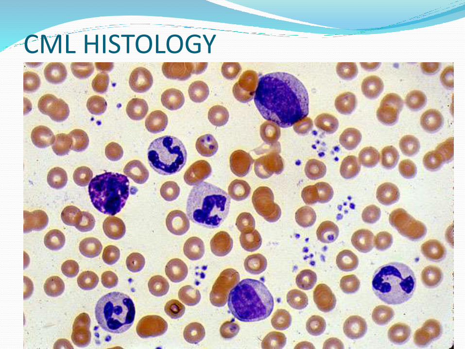
CML HISTOLOGY
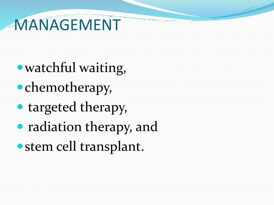
MANAGEMENT
watchful waiting,
chemotherapy,
targeted therapy,
radiation therapy, and
stem cell transplant.
The choice of treatment depends mainly on the following:
The type of leukemia (acute or chronic)
Age
Whether leukemia cells were found in cerebrospinal fluid
WATCHFUL WAITING
chronic leukemia without symptoms, may not need cancer treatment right away.
Watch for health closely so that treatment can start when it begin to have symptoms.
Not getting cancer treatment right away is called watchful waiting.
chemotherapyPeople with acute leukemia need to be
treated right away.
The goal of treatment is to destroy signs of leukemia in the body and make symptoms go away. This is called a remission.
After people go into remission, more therapy may be given to prevent a relapse.
The 3 phases of treatment protocols are;
Induction phase; the usual criteria for complete remission are 5% of the bone marrow cells and normal peripheral blood counts. Once remission completes the consolidation phase begins.
Consolidation phase; modified course of intensive chemotherapy are given to eradicate any remaining disease. Usually a higher dose of 1 or more chemotherapeutic agents are administered.
Maintainance phase; small dose of different combination of chemotheraptic agents are given every 3 to 4 weeks. This phase may continue for a year or longer and is structured to allow the client to live as normal life as possible
Targeted therapy
This affects only tumor cells and spare normal cells. hence decreasing the associated toxicities. Gemtuzumabozofamicin (mylotarg) is an anti D33nmonoclonal antibody linked to calicheamicin, which is potent cytotoxic agent.
STEM CELL TRANSPLANT
Goal;
Totally eliminate leukemic cells from the body using combinations of chemotherapy with or without total body irradiation
Eradicates patient’s hematopoietic stem cells
Replaced with those of an HLA-matched (Human Leukocyte Antigen)
Sibling (is a brother or a sister; that is, any person who shares at least one of the same parents )
Volunteer
Identical twin
Patient’s own stem cells removed before
TYPES OF STEM CELL TRANSPLANTATION
1. Allogeneic Stem Cell Transplant
stem cells are taken from a matching donor. To determine if a donor’s stem cells are the right match, the patient undergoes a human leukocyte antigens (HLA) test. Through this test, we compare the patient’s blood and tissue type against blood samples from the donor.
Donors may include:
HLA-matched relative (most often a sibling)
HLA-matched unrelated donor
HLA miss-matched family member
Unrelated umbilical cord blood
2. Autologous Stem Cell Transplant
In this type of transplant, stem cells are collected from the patient themselves. The stem cells are then harvested, frozen and stored, and then given back to the patient. This type of transplant is rare for leukemiapatients and is typically used in select cases of AML.

Nutrition and Physical Activity
It's important for you to take care of eating well and staying as active.
right amount of calories to maintain a good weight. enough protein. Eating well may help to feel better and have more energy.
Follow-up Care
regular checkups after treatment for leukemia.

NURSING MANAGEMENT
Nursing diagnosis 1. Impaired oral mucous membrane related to
low platelet counts or effect of pathologic conditions and treatment.
2. Ineffective therapeutic management related to lack of knowledge of disease process, activity and medication.
3. imbalanced nutrition less than body requirement reated to anorexia , pain and fatigue.
4. risk for injury related to low platelet counts and treatment

Overall goalsUnderstand and cooperate with the
treatment plan
Experience minimal side effects and complications of disease and treatment
Feel hopeful and supported during the periods of treatment, relapse, and remission
Many physical and psychological needs
Evokes great fear
Goals of rehabilitation
Manage
Physical
Psychosocial
Social
Spiritual
Delayed effects
Support groups
CONCLUSION