Embed Size (px)
Citation preview

Neuromuscular Monitoring
Dr. Mohtasib
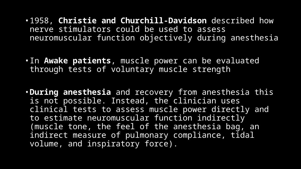
• 1958, Christie and Churchill-Davidson described how nerve stimulators could be used to assess neuromuscular function objectively during anesthesia
• In Awake patients, muscle power can be evaluated through tests of voluntary muscle strength
• During anesthesia and recovery from anesthesia this is not possible. Instead, the clinician uses clinical tests to assess muscle power directly and to estimate neuromuscular function indirectly (muscle tone, the feel of the anesthesia bag, an indirect measure of pulmonary compliance, tidal volume, and inspiratory force).
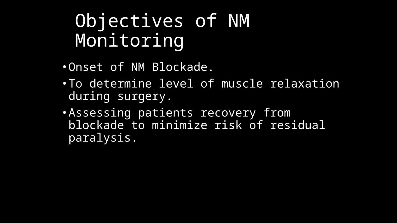
• Onset of NM Blockade.• To determine level of muscle relaxation during surgery.• Assessing patients recovery from blockade to minimize risk
of residual paralysis.
Objectives of NM Monitoring
Why do we Monitor?
Residual post-op NM Blockade • Functional impairment of pharyngeal and upper
esophageal muscles• Impaired ability to maintain the airway • Increased risk for post-op pulmonary complications• Difficult to exclude clinically significant residual
curarization by clinical evaluation
Principles of Peripheral Nerve Stimulation• Each muscle fiber to a stimulus follows an all-or-none pattern• Response of the whole muscle depends on the number of muscle
fibers activated • Response of the muscle decreases in parallel with the numbers of
fibers blocked • Reduction in response during constant stimulation reflects degree of
NM Blockade • For this reason stimulus is supramaximal
Essential features of PNS
• Shape of stimulus should be monophasic and rectangular i.e Square-wave stimulus.• 0.2- 0.3 msec duration so it falls within absolute
refractory period of motor unit in the nerve.• Constant current variable voltage• Battery powered.• Digital display of delivered current.• Audible signal on delivery of stimulus.• Audible alarm for poor electrode contact.• Multiple patterns of stimulation (single twitch,train-of-
four, double-burst, post-tetanic count).
• Current intensity : It is the amperage (mA) of the current delivered by the nerve stimulator(0-80 mA). The intensity reaching the nerve is determined by the voltage generated by the stimulator and resistance and impedance of the electrodes, skin and underlying tissues
• Nerve stimulators are constant current and variable voltage delivery devices• Reduction of temperature increases the tissue resistance (increased impedance) and may
cause reduction in the current delivered to fall below the supramaximal level• Threshold current : It is the lowest current required to depolarize the most sensitive fibres
in a given nerve bundle to elicit a detectable muscle response.• Maximal current: Current which generate response in all muscle fibre• Supramaximal current : It is approximately 25% higher intensity than the current required
to depolarize all fibres in a particular nerve bundle. This is generally attained at current intensity 2-3 times higher than threshold current.
• Submaximal current : A current intensity that induces firing of only a fraction fibres in a given nerve bundle. A potential advantage of submaximal current is that it is less painful than supramaximal current.
• Stimulus frequency : The rate (Hz) at which each impulse is repeated in cycles per second (Hz).
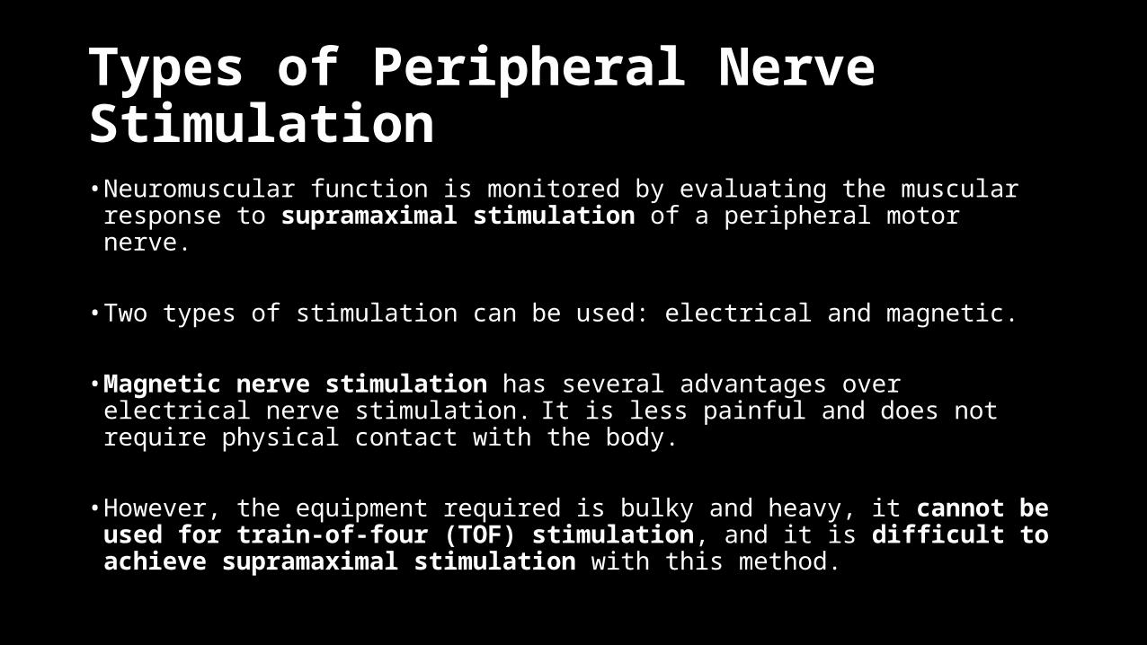
Types of Peripheral Nerve Stimulation• Neuromuscular function is monitored by evaluating the muscular
response to supramaximal stimulation of a peripheral motor nerve.
• Two types of stimulation can be used: electrical and magnetic.
• Magnetic nerve stimulation has several advantages over electrical nerve stimulation. It is less painful and does not require physical contact with the body.
• However, the equipment required is bulky and heavy, it cannot be used for train-of-four (TOF) stimulation, and it is difficult to achieve supramaximal stimulation with this method.
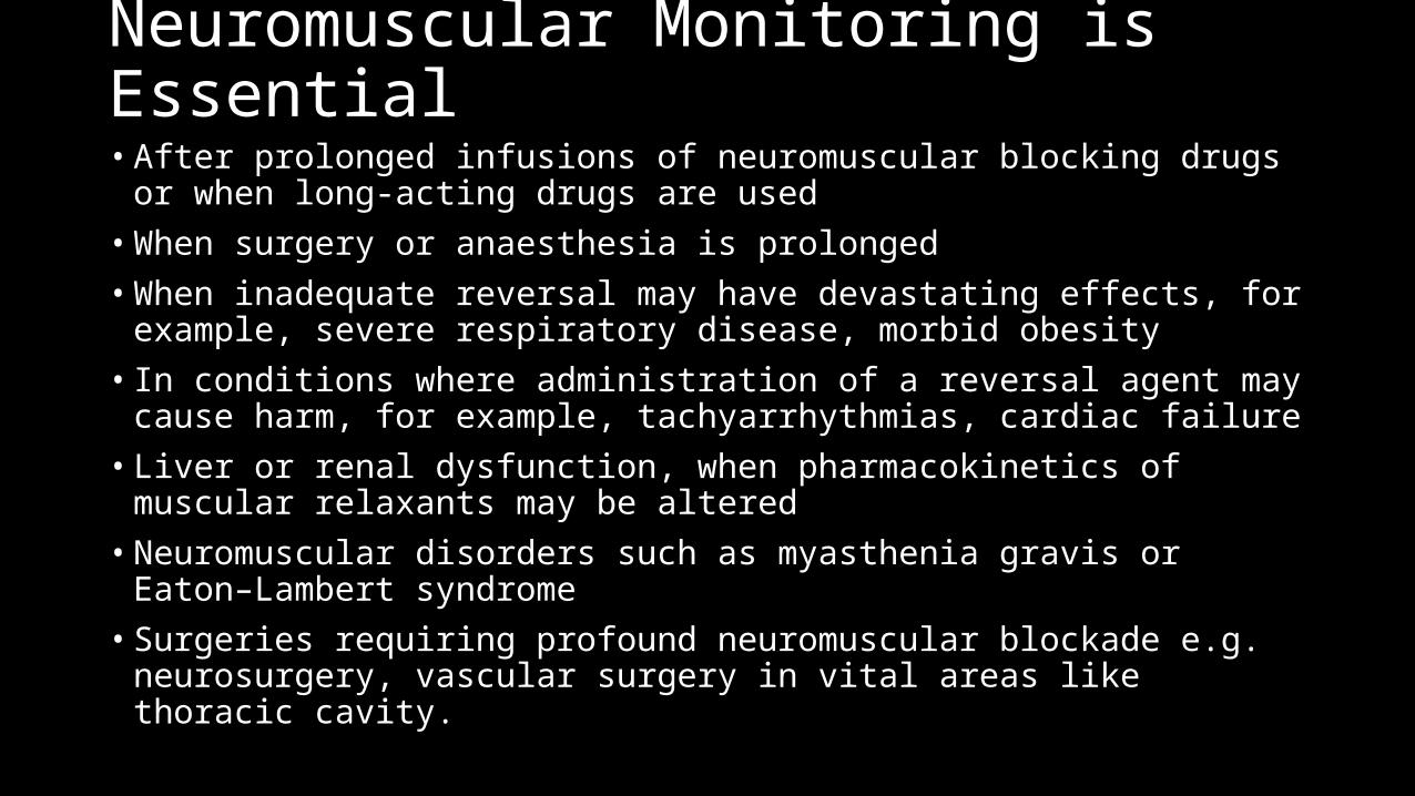
Neuromuscular Monitoring is Essential• After prolonged infusions of neuromuscular blocking drugs or when long-acting
drugs are used• When surgery or anaesthesia is prolonged• When inadequate reversal may have devastating effects, for example, severe
respiratory disease, morbid obesity• In conditions where administration of a reversal agent may cause harm, for
example, tachyarrhythmias, cardiac failure• Liver or renal dysfunction, when pharmacokinetics of muscular relaxants may be
altered• Neuromuscular disorders such as myasthenia gravis or Eaton–Lambert syndrome• Surgeries requiring profound neuromuscular blockade e.g. neurosurgery, vascular
surgery in vital areas like thoracic cavity.
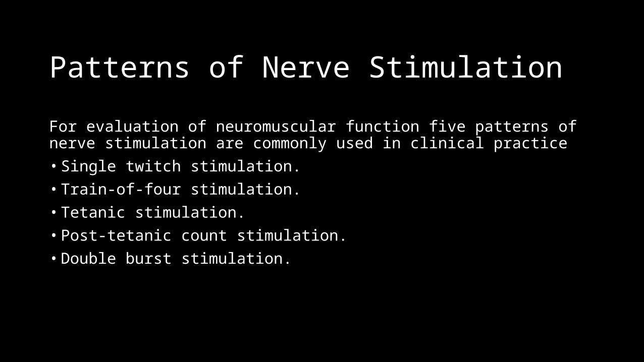
Patterns of Nerve Stimulation
For evaluation of neuromuscular function five patterns of nerve stimulation are commonly used in clinical practice• Single twitch stimulation.• Train-of-four stimulation.• Tetanic stimulation.• Post-tetanic count stimulation.• Double burst stimulation.
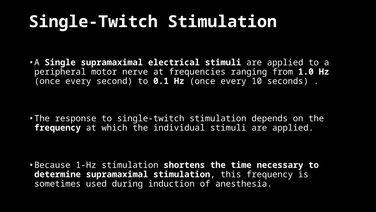
Single-Twitch Stimulation
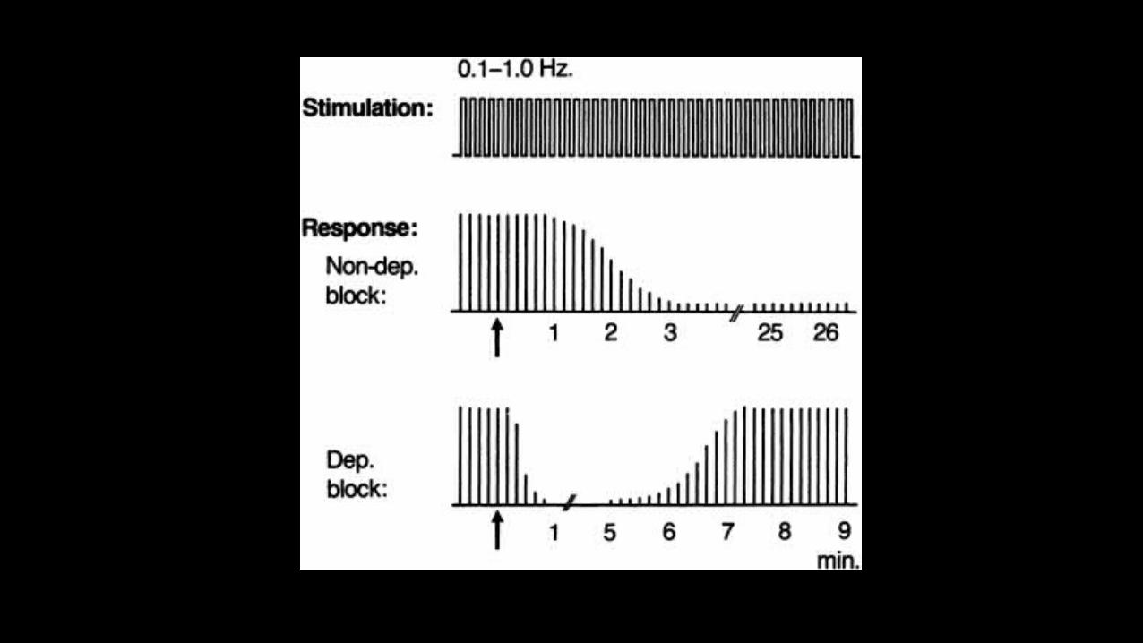
• A Single supramaximal electrical stimuli are applied to a peripheral motor nerve at frequencies ranging from 1.0 Hz (once every second) to 0.1 Hz (once every 10 seconds) .
• The response to single-twitch stimulation depends on the frequency at which the individual stimuli are applied.
• Because 1-Hz stimulation shortens the time necessary to determine supramaximal stimulation, this frequency is sometimes used during induction of anesthesia.
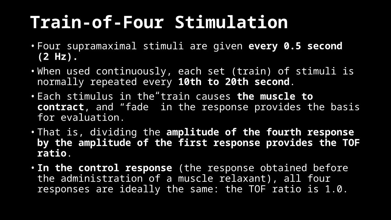
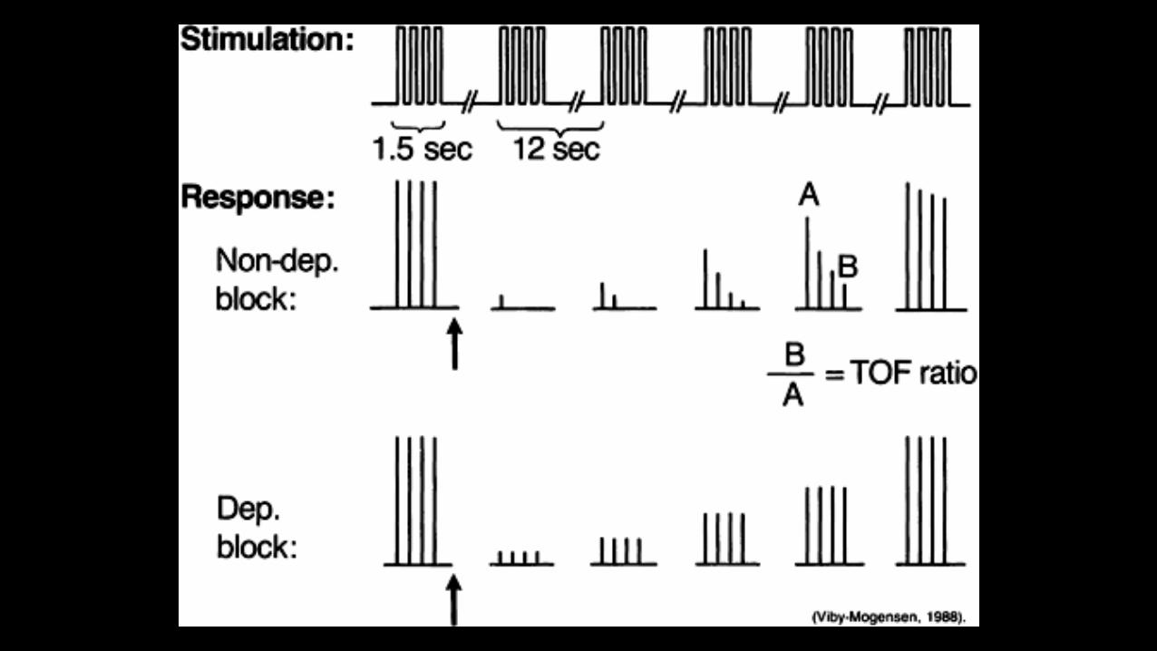
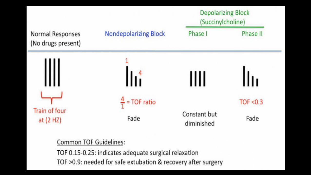
Train-of-Four Stimulation • Four supramaximal stimuli are given every 0.5 second (2 Hz). • When used continuously, each set (train) of stimuli is normally repeated every
10th to 20th second. • Each stimulus in the train causes the muscle to contract, and “fade” in the
response provides the basis for evaluation. • That is, dividing the amplitude of the fourth response by the amplitude of the
first response provides the TOF ratio. • In the control response (the response obtained before the administration of a
muscle relaxant), all four responses are ideally the same: the TOF ratio is 1.0.
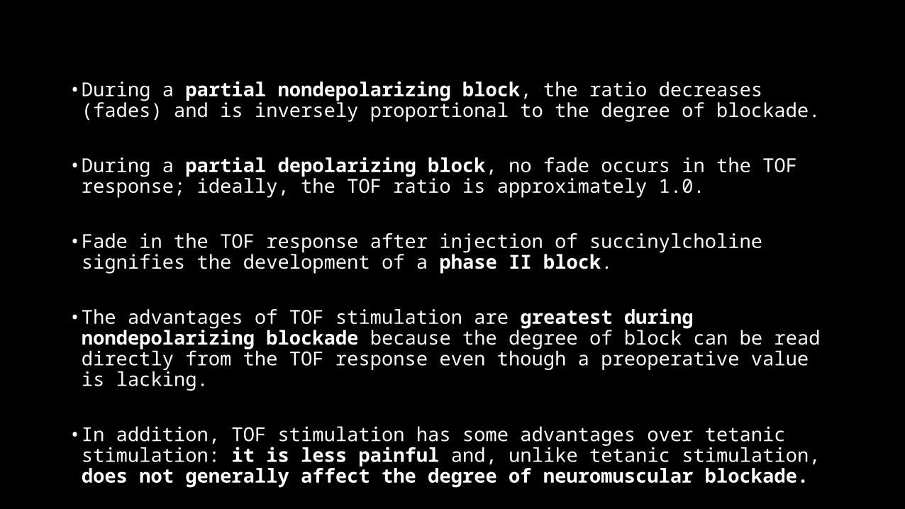
• During a partial nondepolarizing block, the ratio decreases (fades) and is inversely proportional to the degree of blockade.
• During a partial depolarizing block, no fade occurs in the TOF response; ideally, the TOF ratio is approximately 1.0.
• Fade in the TOF response after injection of succinylcholine signifies the development of a phase II block.
• The advantages of TOF stimulation are greatest during nondepolarizing blockade because the degree of block can be read directly from the TOF response even though a preoperative value is lacking.
• In addition, TOF stimulation has some advantages over tetanic stimulation: it is less painful and, unlike tetanic stimulation, does not generally affect the degree of neuromuscular blockade.
Tetanic Stimulation
• Tetanic stimulation consists of very rapid (e.g., 30-, 50-, or 100-Hz) delivery of electrical stimuli. • The most commonly used pattern in clinical practice is 50-Hz stimulation given for
5 seconds, although some investigators have advocated the use of 50-, 100-, and even 200-Hz stimulation for 1 second. • During normal neuromuscular transmission and a pure depolarizing block, the
muscle response to 50-Hz tetanic stimulation for 5 seconds is sustained.• During a nondepolarizing block and a phase II block after the injection of
succinylcholine, the response will not be sustained (i.e., fade occurs)• Fade in response to tetanic stimulation is normally considered a presynaptic
event; the traditional explanation is that at the start of tetanic stimulation, large amounts of acetylcholine are released from immediately available stores in the nerve terminal. • As these stores become depleted, the rate of acetylcholine release decreases until
equilibrium between mobilization and synthesis of acetylcholine is achieved.
• When the “margin of safety” at the postsynaptic membrane (i.e., the number of free cholinergic receptors) is reduced by nondepolarizing neuromuscular blocking drugs, a typical reduction in twitch height is seen with a fade during, for instance, repetitive stimulation. • In addition to this postsynaptic block, nondepolarizing neuromuscular blocking
drugs may also block presynaptic neuronal-type acetylcholine receptors, thereby leading to impaired mobilization of acetylcholine within the nerve terminal.• This effect substantially contributes to fade in the response to tetanic (and TOF)
stimulation. • Although the degree of fade depends primarily on the degree of neuromuscular
blockade, fade also depends on the frequency (Hz) and the length (seconds) of stimulation and on how often tetanic stimuli are applied. • During partial nondepolarizing blockade, tetanic nerve stimulation is followed by
a post-tetanic increase in twitch tension (i.e., post-tetanic facilitation of transmission). • This event occurs because the increase in mobilization and synthesis of
acetylcholine caused by tetanic stimulation continues for some time after discontinuation of stimulation.
• The degree and duration of post-tetanic facilitation depend on the degree of neuromuscular blockade, with post-tetanic facilitation usually disappearing within 60 seconds of tetanic stimulation.
• In contrast, post-tetanic twitch potentiation, which sometimes occurs in mechanical recordings before any neuromuscular blocking drug has been given, is a muscular phenomenon that is not accompanied by an increase in the compound muscle action potential.
• Tetanic stimulation is very painful and therefore not normally acceptable to an unanesthetized patient.
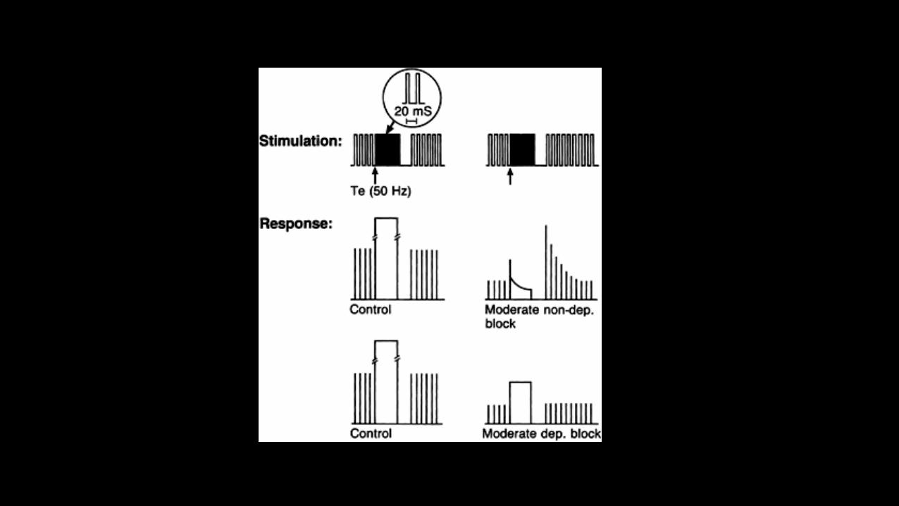
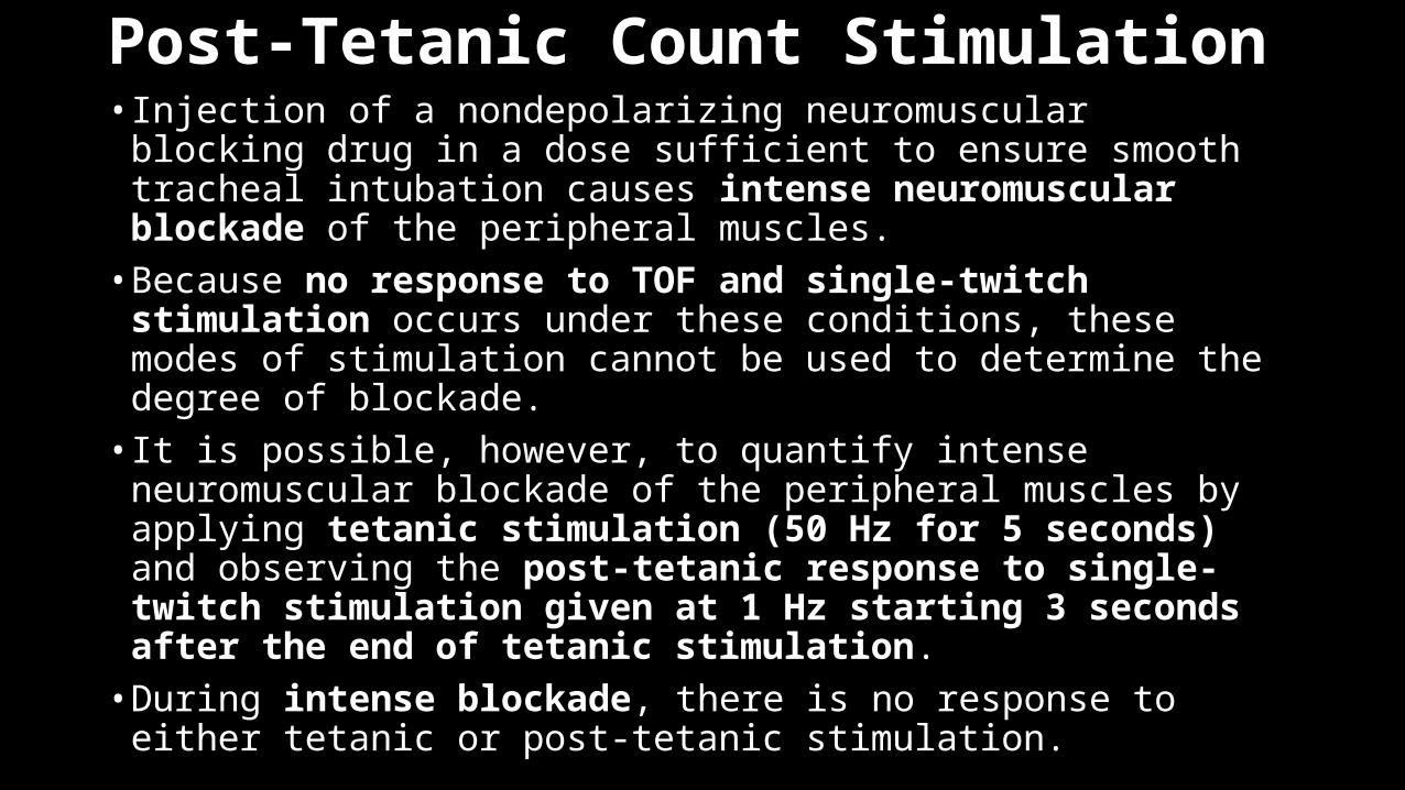
Post-Tetanic Count Stimulation • Injection of a nondepolarizing neuromuscular blocking drug in a
dose sufficient to ensure smooth tracheal intubation causes intense neuromuscular blockade of the peripheral muscles. • Because no response to TOF and single-twitch stimulation occurs
under these conditions, these modes of stimulation cannot be used to determine the degree of blockade. • It is possible, however, to quantify intense neuromuscular blockade
of the peripheral muscles by applying tetanic stimulation (50 Hz for 5 seconds) and observing the post-tetanic response to single-twitch stimulation given at 1 Hz starting 3 seconds after the end of tetanic stimulation.• During intense blockade, there is no response to either tetanic or
post-tetanic stimulation.
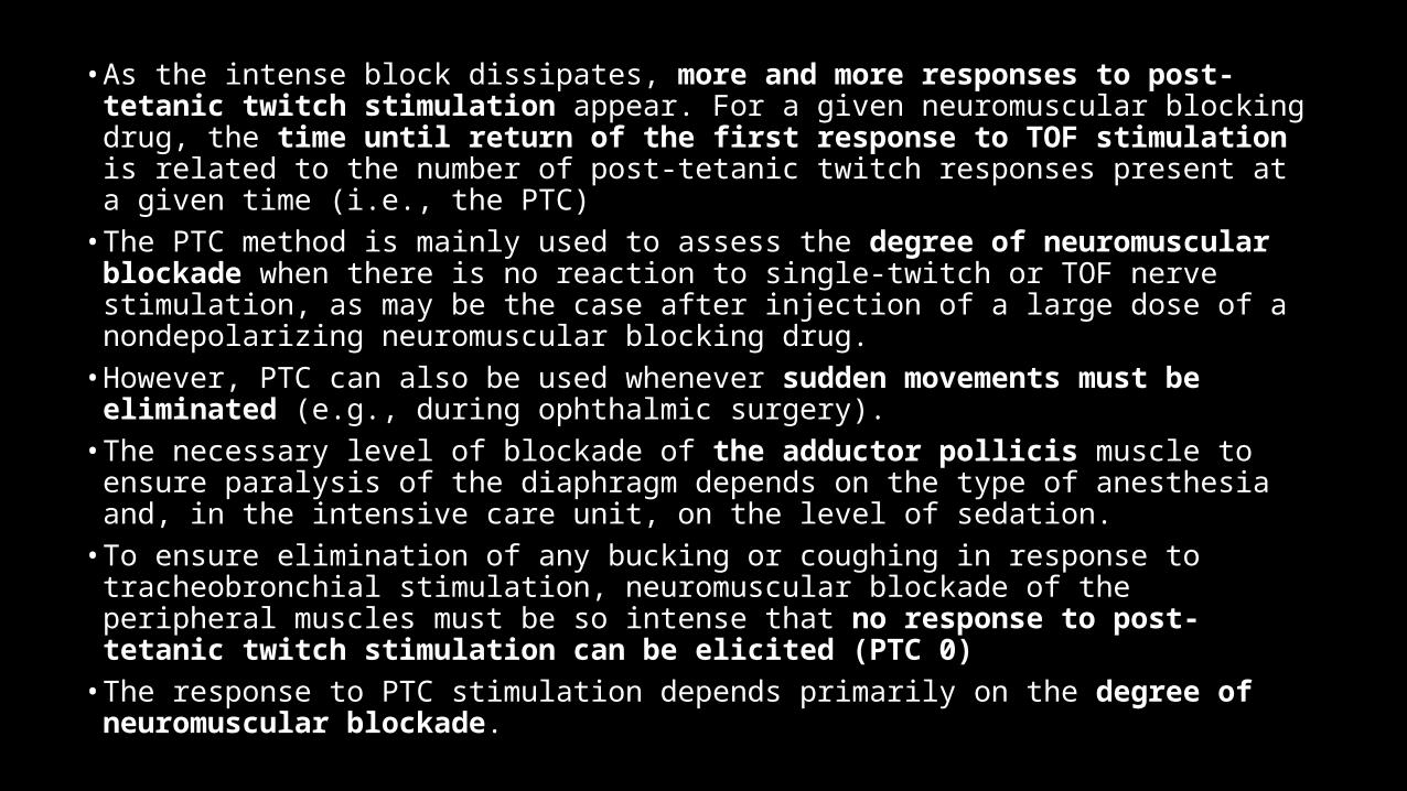
• As the intense block dissipates, more and more responses to post-tetanic twitch stimulation appear. For a given neuromuscular blocking drug, the time until return of the first response to TOF stimulation is related to the number of post-tetanic twitch responses present at a given time (i.e., the PTC)
• The PTC method is mainly used to assess the degree of neuromuscular blockade when there is no reaction to single-twitch or TOF nerve stimulation, as may be the case after injection of a large dose of a nondepolarizing neuromuscular blocking drug.
• However, PTC can also be used whenever sudden movements must be eliminated (e.g., during ophthalmic surgery).
• The necessary level of blockade of the adductor pollicis muscle to ensure paralysis of the diaphragm depends on the type of anesthesia and, in the intensive care unit, on the level of sedation.
• To ensure elimination of any bucking or coughing in response to tracheobronchial stimulation, neuromuscular blockade of the peripheral muscles must be so intense that no response to post-tetanic twitch stimulation can be elicited (PTC 0)
• The response to PTC stimulation depends primarily on the degree of neuromuscular blockade.
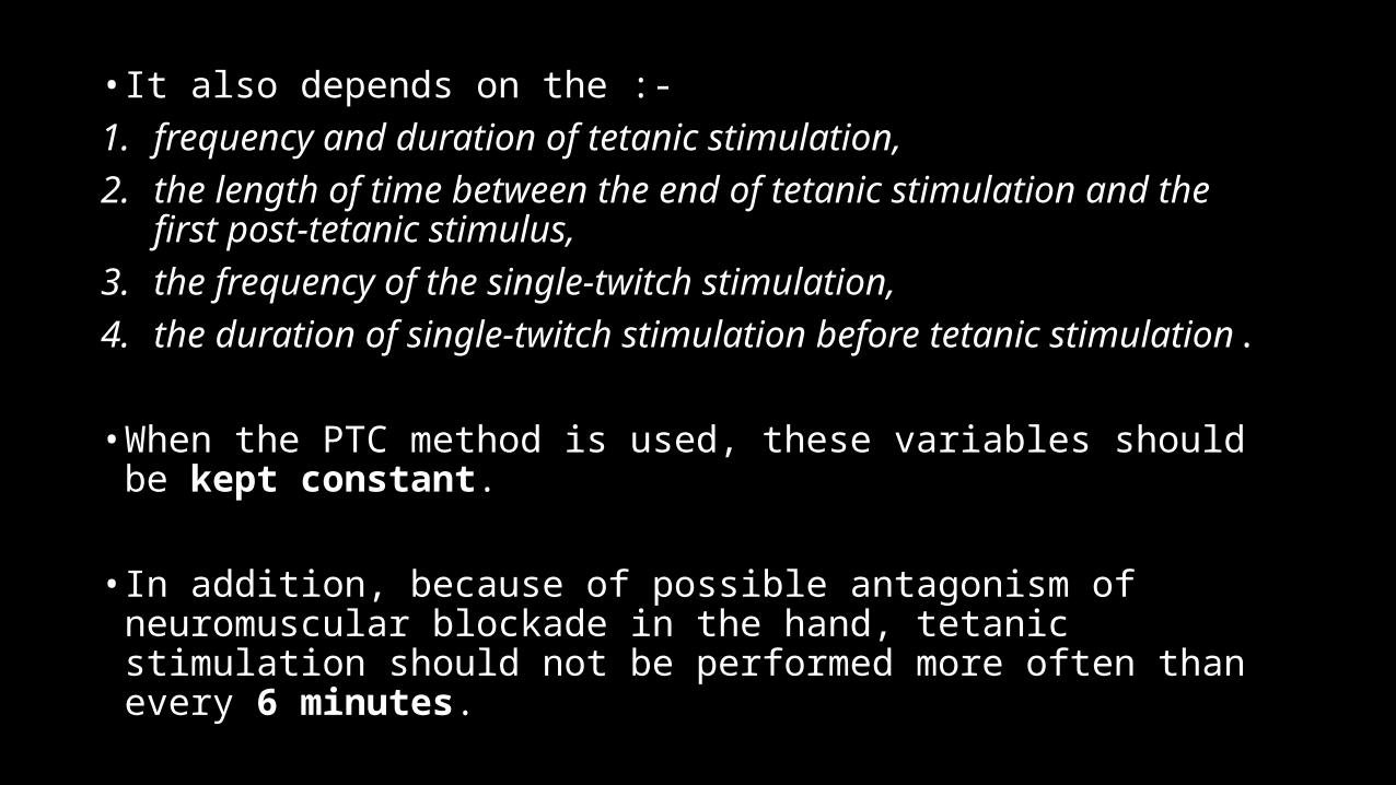
• It also depends on the :- 1. frequency and duration of tetanic stimulation, 2. the length of time between the end of tetanic stimulation and the
first post-tetanic stimulus, 3. the frequency of the single-twitch stimulation, 4. the duration of single-twitch stimulation before tetanic stimulation.
• When the PTC method is used, these variables should be kept constant.
• In addition, because of possible antagonism of neuromuscular blockade in the hand, tetanic stimulation should not be performed more often than every 6 minutes.
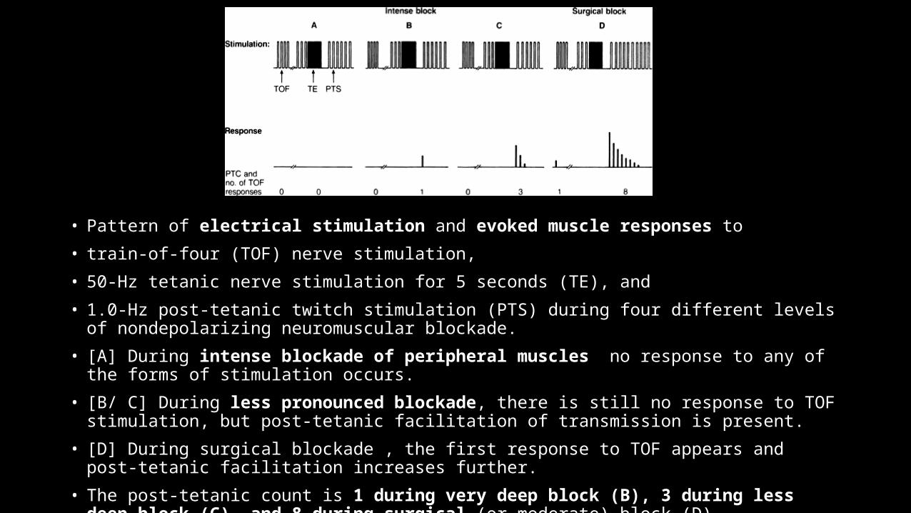
• Pattern of electrical stimulation and evoked muscle responses to
• train-of-four (TOF) nerve stimulation,
• 50-Hz tetanic nerve stimulation for 5 seconds (TE), and
• 1.0-Hz post-tetanic twitch stimulation (PTS) during four different levels of nondepolarizing neuromuscular blockade.
• [A] During intense blockade of peripheral muscles no response to any of the forms of stimulation occurs.
• [B/ C] During less pronounced blockade, there is still no response to TOF stimulation, but post-tetanic facilitation of transmission is present.
• [D] During surgical blockade , the first response to TOF appears and post-tetanic facilitation increases further.
• The post-tetanic count is 1 during very deep block (B), 3 during less deep block (C), and 8 during surgical (or moderate) block (D).
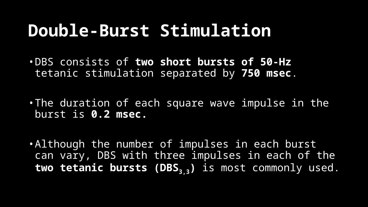
Double-Burst Stimulation
• DBS consists of two short bursts of 50-Hz tetanic stimulation separated by 750 msec.
• The duration of each square wave impulse in the burst is 0.2 msec.
• Although the number of impulses in each burst can vary, DBS with three impulses in each of the two tetanic bursts (DBS3,3) is most commonly used.

• In nonparalyzed muscle, the response to DBS3,3 is two short muscle contractions of equal strength.
• In a partly paralyzed muscle, the second response is weaker than the first (i.e., the response fades).
• DBS was developed with the specific aim of allowing manual (tactile) detection of small amounts of residual blockade under clinical conditions,and during recovery and immediately after surgery, tactile evaluation of the response to DBS3,3 is superior to tactile evaluation of the response to TOF stimulation.
Equipments Required
• The nerve stimulator,• The stimulating electrodes, and• The recording equipment.
The Nerve StimulatorIt delivers the stimulus to the electrodes. Although many nerve stimulators are commercially available, not all meet the basic requirement for clinical use. The real nerve stimulator should have the following properties.• It should deliver monophasic and rectangular waveform pulse of
constant current.• It should deliver a current up to 60-70 mA but not >80mA.• It should be battery operated and should have a battery check.• It should have either built in warning system or current level display
that alerts the user when the current selected is not delivered to the nerve (in case of increased skin resistance)• It should have polarity indicators for electrodes.• It should deliver different modes of stimulation.
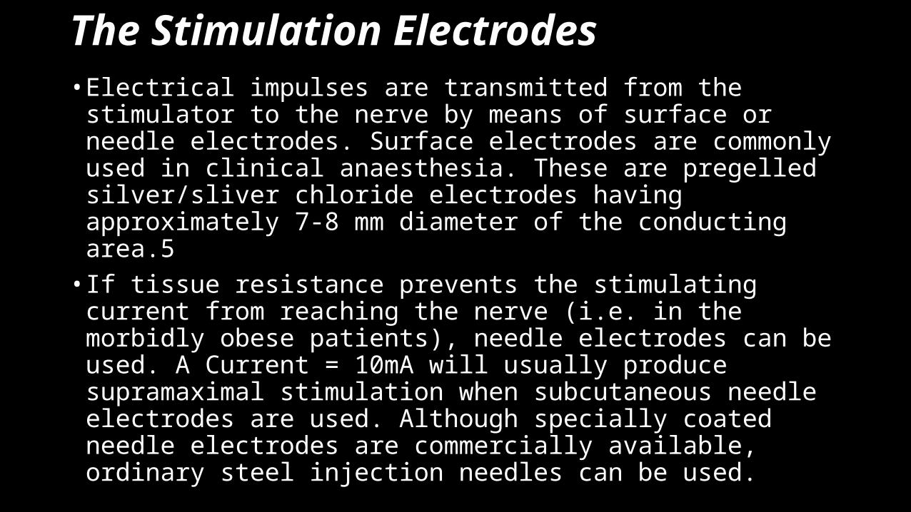
The Stimulation Electrodes• Electrical impulses are transmitted from the stimulator to the nerve
by means of surface or needle electrodes. Surface electrodes are commonly used in clinical anaesthesia. These are pregelled silver/sliver chloride electrodes having approximately 7-8 mm diameter of the conducting area.5• If tissue resistance prevents the stimulating current from reaching the
nerve (i.e. in the morbidly obese patients), needle electrodes can be used. A Current = 10mA will usually produce supramaximal stimulation when subcutaneous needle electrodes are used. Although specially coated needle electrodes are commercially available, ordinary steel injection needles can be used.
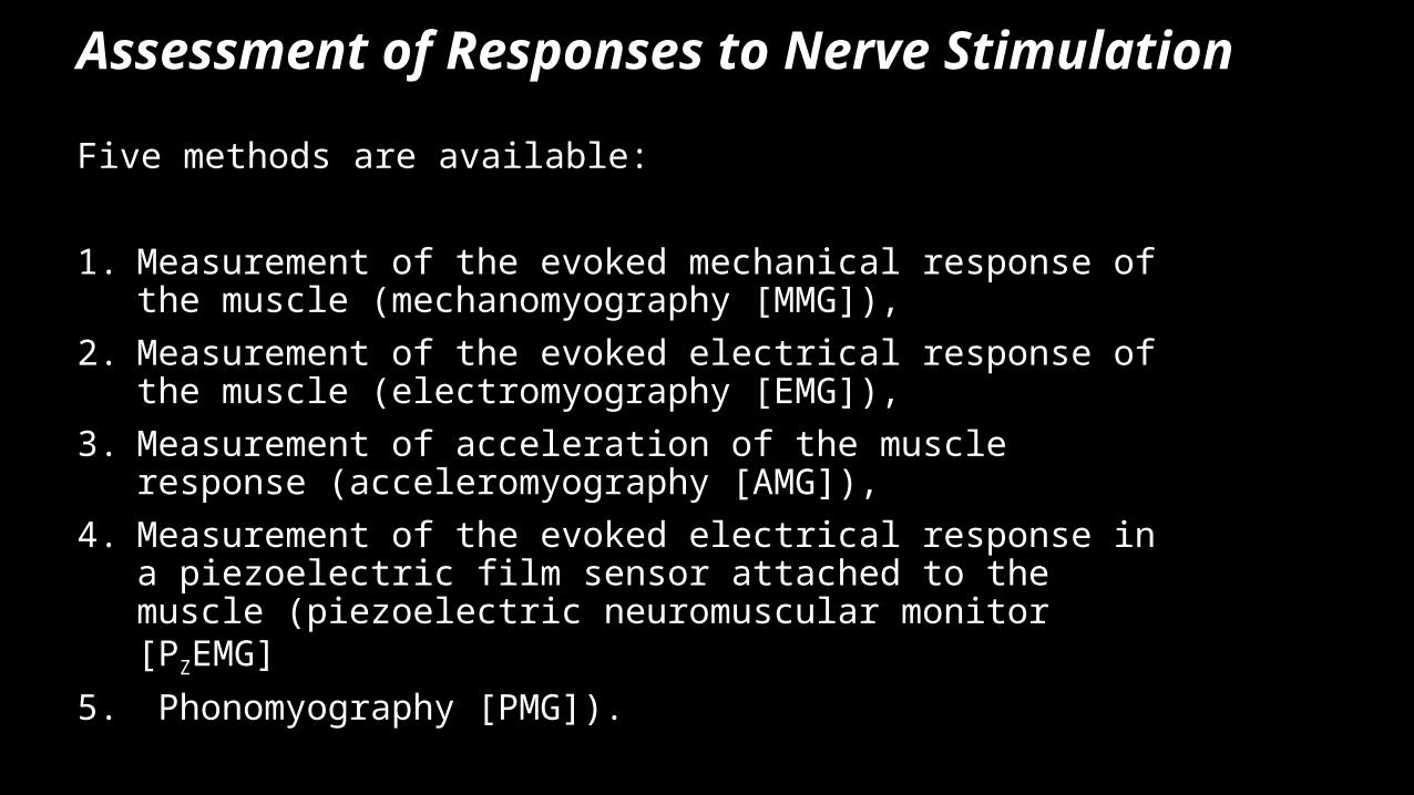
Assessment of Responses to Nerve Stimulation
Five methods are available:
1. Measurement of the evoked mechanical response of the muscle (mechanomyography [MMG]),
2. Measurement of the evoked electrical response of the muscle (electromyography [EMG]),
3. Measurement of acceleration of the muscle response (acceleromyography [AMG]),
4. Measurement of the evoked electrical response in a piezoelectric film sensor attached to the muscle (piezoelectric neuromuscular monitor [PZEMG]
5. Phonomyography [PMG]).
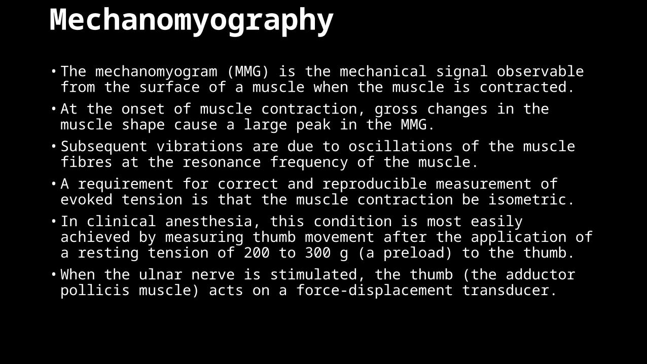
Mechanomyography
• The mechanomyogram (MMG) is the mechanical signal observable from the surface of a muscle when the muscle is contracted. • At the onset of muscle contraction, gross changes in the muscle shape cause a
large peak in the MMG.• Subsequent vibrations are due to oscillations of the muscle fibres at the
resonance frequency of the muscle.• A requirement for correct and reproducible measurement of evoked tension is
that the muscle contraction be isometric.• In clinical anesthesia, this condition is most easily achieved by measuring thumb
movement after the application of a resting tension of 200 to 300 g (a preload) to the thumb. • When the ulnar nerve is stimulated, the thumb (the adductor pollicis muscle) acts
on a force-displacement transducer.
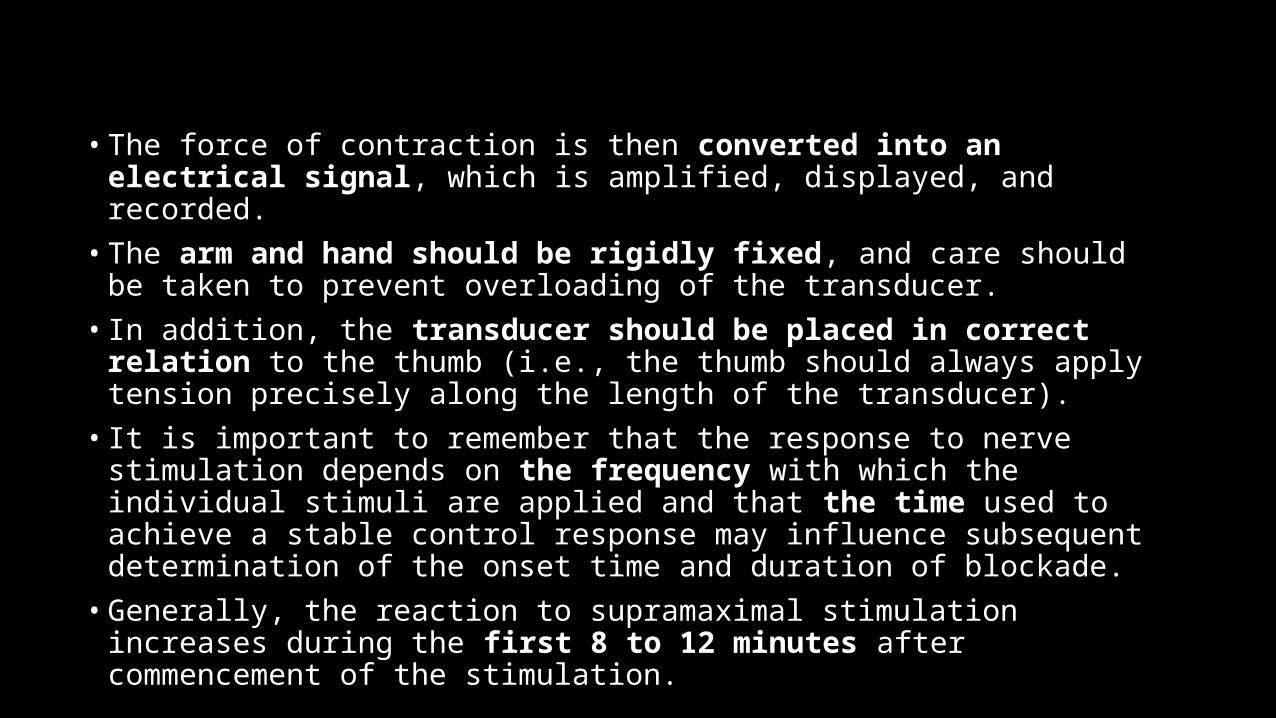
• The force of contraction is then converted into an electrical signal, which is amplified, displayed, and recorded. • The arm and hand should be rigidly fixed, and care should be taken to prevent
overloading of the transducer. • In addition, the transducer should be placed in correct relation to the thumb
(i.e., the thumb should always apply tension precisely along the length of the transducer). • It is important to remember that the response to nerve stimulation depends on
the frequency with which the individual stimuli are applied and that the time used to achieve a stable control response may influence subsequent determination of the onset time and duration of blockade.• Generally, the reaction to supramaximal stimulation increases during the first 8 to
12 minutes after commencement of the stimulation.
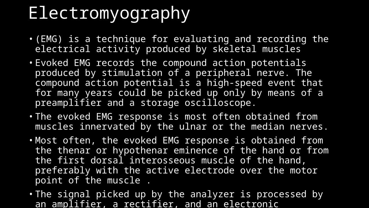
Electromyography • (EMG) is a technique for evaluating and recording the electrical activity
produced by skeletal muscles• Evoked EMG records the compound action potentials produced by stimulation
of a peripheral nerve. The compound action potential is a high-speed event that for many years could be picked up only by means of a preamplifier and a storage oscilloscope.• The evoked EMG response is most often obtained from muscles innervated by
the ulnar or the median nerves. • Most often, the evoked EMG response is obtained from the thenar or
hypothenar eminence of the hand or from the first dorsal interosseous muscle of the hand, preferably with the active electrode over the motor point of the muscle .• The signal picked up by the analyzer is processed by an amplifier, a rectifier, and
an electronic integrator. The results are displayed either as a percentage of control or as a TOF ratio.
• The evoked EMG response is most often obtained from muscles innervated by the ulnar or the median nerves. • Most often, the evoked EMG response is obtained from the thenar or hypothenar
eminence of the hand or from the first dorsal interosseous muscle of the hand, preferably with the active electrode over the motor point of the muscle .• The signal picked up by the analyzer is processed by an amplifier, a rectifier, and
an electronic integrator. The results are displayed either as a percentage of control or as a TOF ratio.
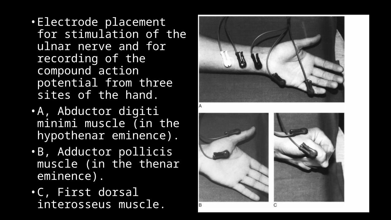
• Electrode placement for stimulation of the ulnar nerve and for recording of the compound action potential from three sites of the hand. • A, Abductor digiti minimi muscle
(in the hypothenar eminence). • B, Adductor pollicis muscle (in
the thenar eminence). • C, First dorsal interosseus
muscle.
• Two new sites for recording the EMG response have been introduced: the larynx and the diaphragm. • By using a noninvasive disposable laryngeal electrode attached to the tracheal
tube and placed between the vocal cords, it is possible to monitor the onset of neuromuscular blockade in the laryngeal muscles.• In paravertebral surface diaphragmatic EMG, the recording electrodes are placed
on the right of vertebrae T12/L1 o r L1/L2 for monitoring the response of the right diaphragmatic crux to transcutaneous stimulation of the right phrenic nerve at the neck. • Evoked electrical and mechanical responses represent different physiologic
events. Evoked EMG records changes in the electrical activity of one or more muscles, whereas evoked MMG records changes associated with excitation-contraction coupling and contraction of the muscle as well.
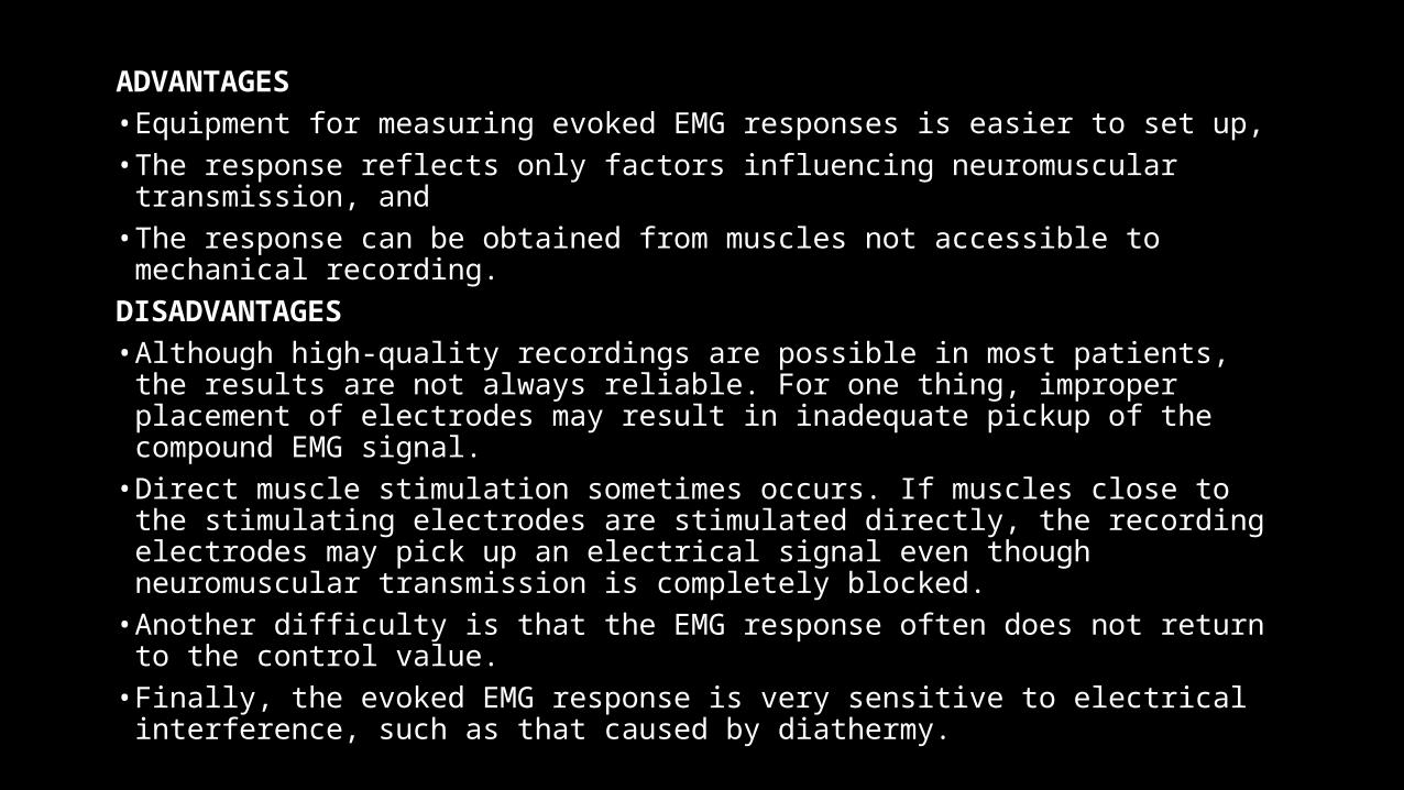
ADVANTAGES • Equipment for measuring evoked EMG responses is easier to set up, • The response reflects only factors influencing neuromuscular transmission, and • The response can be obtained from muscles not accessible to mechanical
recording. DISADVANTAGES• Although high-quality recordings are possible in most patients, the results are
not always reliable. For one thing, improper placement of electrodes may result in inadequate pickup of the compound EMG signal.
• Direct muscle stimulation sometimes occurs. If muscles close to the stimulating electrodes are stimulated directly, the recording electrodes may pick up an electrical signal even though neuromuscular transmission is completely blocked.
• Another difficulty is that the EMG response often does not return to the control value.
• Finally, the evoked EMG response is very sensitive to electrical interference, such as that caused by diathermy.
Acceleromyograph
• Is a piezoelectric myograph, used to measure the force produced by a muscle after it has undergone nerve stimulation.• Acceleromyographs measure muscle activity using a miniature
piezoelectric transducer that is attached to the stimulated muscle.• A voltage is created when the muscle accelerates and that acceleration
is proportion to force of contraction. • Acceleromyographs are more costly than the more common twitch
monitors, but have been shown to better alleviate residual blockade and associated symptoms of muscle weakness, and to improve overall quality of recovery.
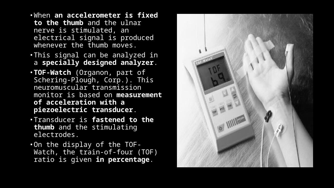
• When an accelerometer is fixed to the thumb and the ulnar nerve is stimulated, an electrical signal is produced whenever the thumb moves.
• This signal can be analyzed in a specially designed analyzer.
• TOF-Watch (Organon, part of Schering-Plough, Corp.). This neuromuscular transmission monitor is based on measurement of acceleration with a piezoelectric transducer.
• Transducer is fastened to the thumb and the stimulating electrodes.
• On the display of the TOF-Watch, the train-of-four (TOF) ratio is given in percentage.
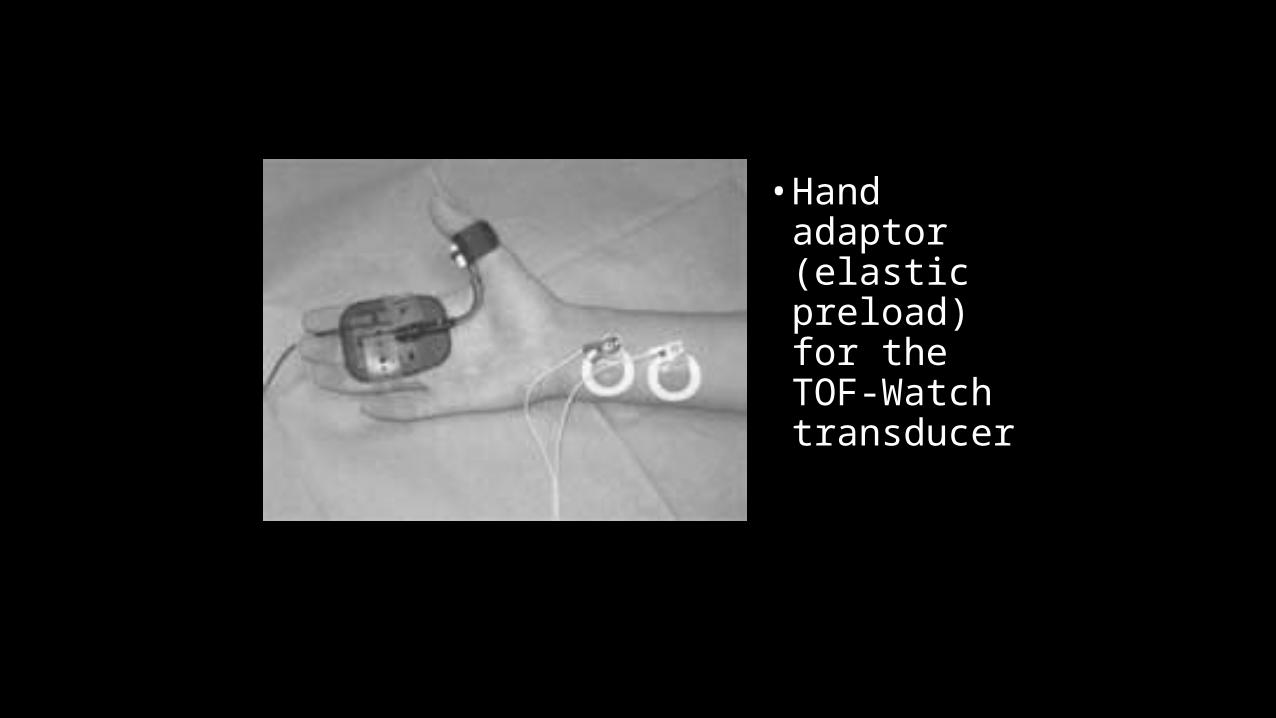
• Hand adaptor (elastic preload) for the TOF-Watch transducer
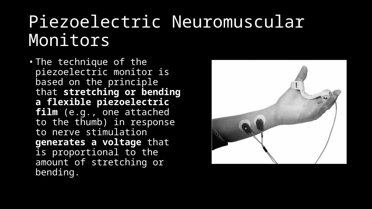
Piezoelectric Neuromuscular Monitors• The technique of the piezoelectric
monitor is based on the principle that stretching or bending a flexible piezoelectric film (e.g., one attached to the thumb) in response to nerve stimulation generates a voltage that is proportional to the amount of stretching or bending.
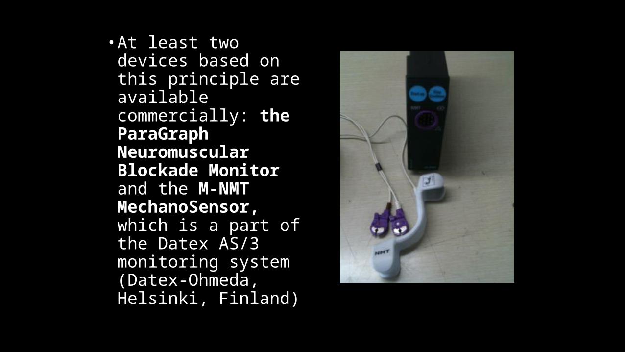
• At least two devices based on this principle are available commercially: the ParaGraph Neuromuscular Blockade Monitor and the M-NMT MechanoSensor, which is a part of the Datex AS/3 monitoring system (Datex-Ohmeda, Helsinki, Finland)
Phonomyography
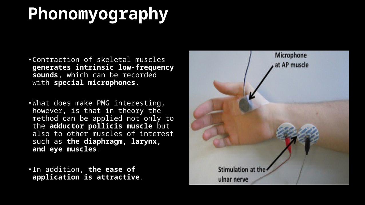
• Contraction of skeletal muscles generates intrinsic low-frequency sounds, which can be recorded with special microphones.
• What does make PMG interesting, however, is that in theory the method can be applied not only to the adductor pollicis muscle but also to other muscles of interest such as the diaphragm, larynx, and eye muscles.
• In addition, the ease of application is
attractive.
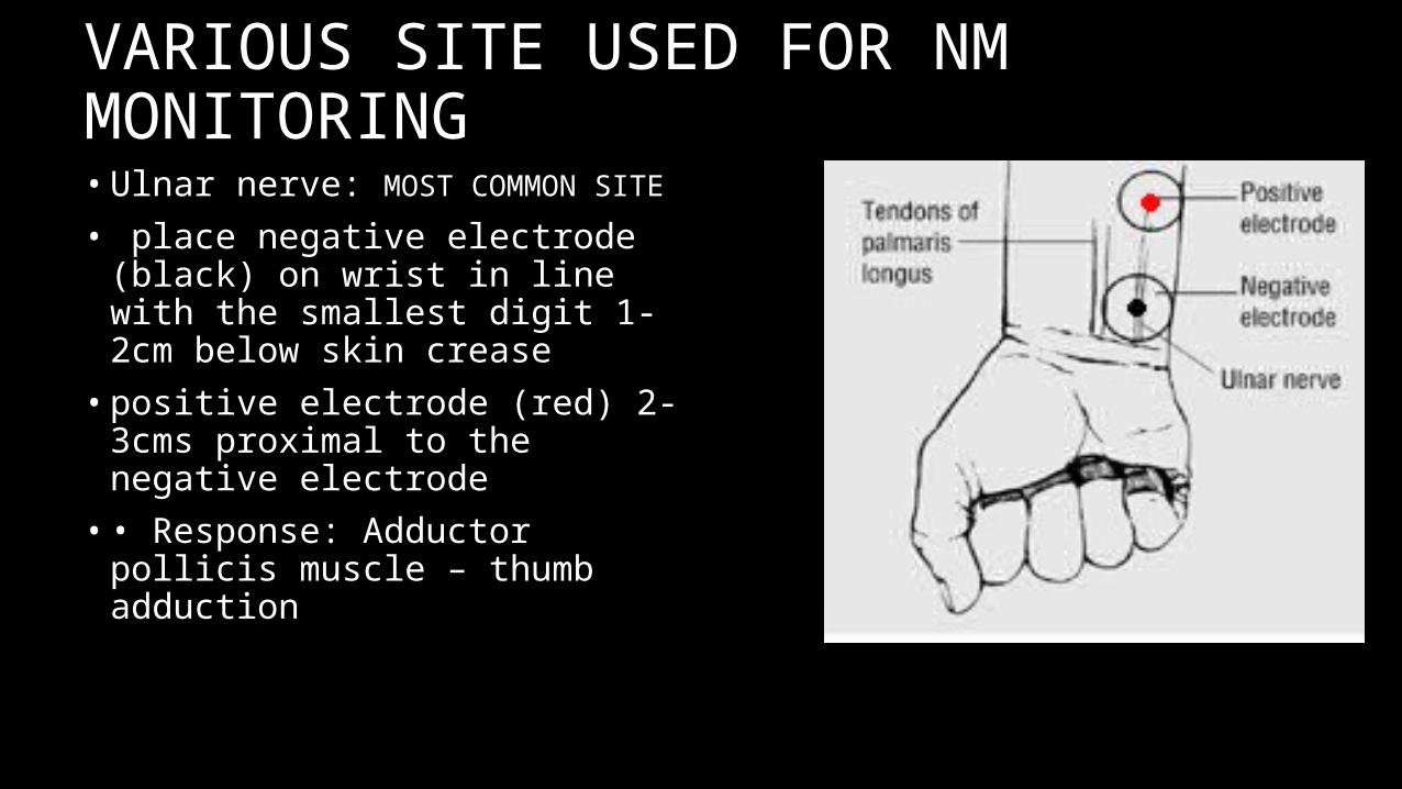
• Ulnar nerve: MOST COMMON SITE
• place negative electrode (black) on wrist in line with the smallest digit 1-2cm below skin crease• positive electrode (red) 2-3cms
proximal to the negative electrode• • Response: Adductor pollicis muscle –
thumb adduction
VARIOUS SITE USED FOR NM MONITORING
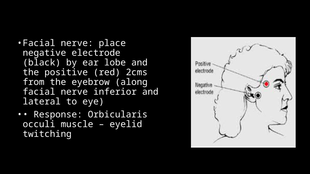
• Facial nerve: place negative electrode (black) by ear lobe and the positive (red) 2cms from the eyebrow (along facial nerve inferior and lateral to eye)• • Response: Orbicularis occuli muscle
– eyelid twitching
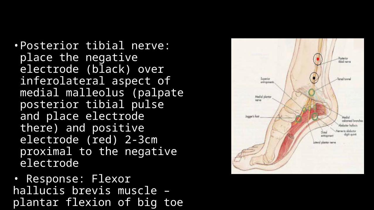
• Posterior tibial nerve: place the
negative electrode (black) over inferolateral aspect of medial malleolus (palpate posterior tibial pulse and place electrode there) and positive electrode (red) 2-3cm proximal to the negative electrode
• Response: Flexor hallucis brevis muscle – plantar flexion of big toe
Recovery
• Return of the fourth response in the TOF heralds the recovery phase.
• During neuromuscular recovery, a reasonably good correlation exists between the actual TOF ratio measured by MMG and clinical observation.
• When the TOF ratio is 0.4 or less, the patient is generally unable to lift the head or arm. Tidal volume may be normal, but vital capacity and inspiratory force will be reduced.
• When the ratio is 0.6, most patients are able to lift their head for 3 seconds, open their eyes widely, and stick out their tongue, but vital capacity and inspiratory force are often still reduced.
• At a TOF ratio of 0.7 to 0.75, the patient can normally cough sufficiently and lift the head for at least 5 seconds, but grip strength may still be as low as about 60% of control.
• When the ratio is 0.8 and higher, vital capacity and inspiratory force are normal. The patient may, however, still have diplopia and facial weakness
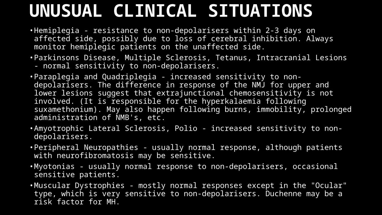
UNUSUAL CLINICAL SITUATIONS• Hemiplegia - resistance to non-depolarisers within 2-3 days on affected side, possibly
due to loss of cerebral inhibition. Always monitor hemiplegic patients on the unaffected side.
• Parkinsons Disease, Multiple Sclerosis, Tetanus, Intracranial Lesions - normal sensitivity to non-depolarisers.
• Paraplegia and Quadriplegia - increased sensitivity to non-depolarisers. The difference in response of the NMJ for upper and lower lesions suggest that extrajunctional chemosensitivity is not involved. (It is responsible for the hyperkalaemia following suxamethonium). May also happen following burns, immobility, prolonged administration of NMB's, etc.
• Amyotrophic Lateral Sclerosis, Polio - increased sensitivity to non-depolarisers.• Peripheral Neuropathies - usually normal response, although patients with
neurofibromatosis may be sensitive.• Myotonias - usually normal response to non-depolarisers, occasional sensitive
patients.• Muscular Dystrophies - mostly normal responses except in the "Ocular" type, which is
very sensitive to non-depolarisers. Duchenne may be a risk factor for MH.
Limitations of Neuromuscular Monitoring
• Despite the important role of NMJ monitoring in anaesthesia practice, it is necessary to use a multifactorial approach for the following reasons:• 1. Neuromuscular responses may appear normal despite persistance of
receptor occupancy by NMBs. T4:T1 ratio is one even when 40-50% of the receptors are occupied.• 2. Because of wide individual variability in evoked responses, some patients
may exhibit weakness at TOF ratio as high as 0.8 to 0.9.• 3. The established cut-off values for adequate recovery do not guarantee
adequate ventilatory function or airway protection.• 4. Increased skin impedence resulting from perioperative hypothermia limits
the appropriate interpretation of evoked responses.
Conclusion
• Many anaesthesiologists do not agree with extensive use of NMJ monitors and argue that patients can be managed satisfactorily without the devices. Although not included under the standards for basic anaesthetic monitoing by the American Society of Anaesthesiologists, the real value of such monitors lies in the fact that they guide the optimal management of patients receiving NMBs.
Sources
• Millers Anesthesia 7th Edition• Understanding Anesthesia Equipment, Dorsch and Dorsch 5th Edition• McGrath CD, Hunter JM. Monitoring of neuromuscular block.
CEACP.2006;6:7-12• Dr. D. Padmaja, Dr. Srinivas Mantha. Monitoring of Neuromuscular
Junction. IJA.2002;46(4) : 279-288