Embed Size (px)
Citation preview

Lesions associated with
abnormal dyskeratosisMade by :
Yousef Abosaif

Definition
Dyskeratosis is an Abnormal keratinization of epithelial
cells

types
1 – benign dyskeratosis
2 – Malignant dyskeratosis

1- Benign dyskeratosis
1- Hereditary Benign Interepithelial
dyskeratosis (HBID)
2- Dyskeratosis congenita
3- keratoacanthoma
4- Darrier’s disease
5- WARTY DYSKERATOMA

2 – Malignant dyskeratosis
• Found in malignant and premalignant conditions
e.g leukoplakia and bowen's disease

1- Dyskeratosis congenita
rare genodermatosis that is usually inherited as an X-
linked recessive trait.
Autosomal dominant and autosomal recessive forms,
although less common, have been reported.
The clinician should be aware of the condition
because the oral lesions may undergo malignant
transformation, and patients are susceptible to aplastic
anemia.

Clinical features
becomes evident during the first 10 years of
life.
Intraorally, the tongue and buccal mucosa
develop bullae; these are followed by erosions
and, eventually, leukoplakic lesions
one third of them become malignant in a 10- to
30- year period
Aplastic anemia develops in approximately
70% of these patients
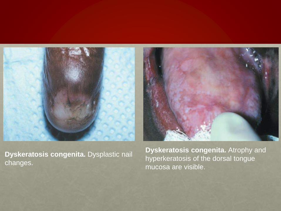
Dyskeratosis congenita. Dysplastic nail
changes.
Dyskeratosis congenita. Atrophy and
hyperkeratosis of the dorsal tongue
mucosa are visible.
HISTOPATHOLOGIC FEATURES
Biopsy specimens of the early oral
mucosal lesions show
hyperorthokeratosis with epithelial
atrophy. As the lesions progress,
epithelial dysplasia develops until frank
squamous cell carcinoma evolves.

TREATMENT AND PROGNOSIS
The discomfort of the oral lesions is managed
symptomatically,and careful periodic oral mucosal
examinations are performed to check for evidence of
malignant transformation.
Routine medical evaluation is warranted to monitor the
patient for the development of aplastic anemia.

2-keratoacanthoma
• self-limiting, epithelial proliferation with a strong
clinical and histopathologic similarity to well-
differentiated squamous cell carcinoma
• The cause of this lesion is unknown, but sun
damage and human papillomavirus (HPV), possibly
subtypes 26 or 37, have been proposed.

CLINICAL FEATURES
• rarely occurs in patients before 45 years of age and
shows a male predilection.
• Keratoacanthoma appears as a firm, nontender,
well-demarcated, sessile, dome-shaped nodule with
a central plug of keratin
• Almost 95% of solitary lesions are found on sun-
exposed skin, and 8% of all cases are found on the
outer edge of the vermilion border of the lips, with
equal frequency on boththe upper and the lower lips

Keratoacanthoma. A nontender,
welldemarcated nodule of the skin of the
nose in an older woman. The nodule
demonstrates a central keratin plug.
Keratoacanthoma. This lesion, which
is located at the outer edge of the
vermilion border of the lip,
demonstrates a prominent core or plug
of keratin.

HISTOPATHOLOGIC FEATURES
• cells appear mature, although considerable
dyskeratosis is typically seen in the form of deeply
located individually keratinizing lesional cells and
keratin pearls similar to those found in well-
differentiated squamous cell carcinoma.
Keratoacanthoma. Low-power
microscopic view showing
extensive epidermal proliferation
with a central keratin plug.
TREATMENT AND PROGNOSIS
• surgical excision of large lesions is indicated for optimal aesthetic appearance because signifi cant scarring may otherwise occur.
• 4% to 8% of treated patients experience recurrence.
• alternative therapies include cryosurgery (reserved for small early lesions), intralesional injection of chemotherapeutic agents and topical imiquimod.
• Systemic chemotherapy, often combined with cryotherapy, may be used to treat patients with multiple lesions

3-HEREDITARY BENIGN
INTRAEPITHELIAL DYSKERATOSIS
• Hereditary benign intraepithelial dyskeratosis
(HBID) is a rare autosomal dominant
genodermatosis
Hereditary benign intraepithelial
dyskeratosis(HBID).
Oral lesions appear as corrugated
white plaques of the buccal
mucosa.
CLINICAL FEATURES
• usually develop during childhood,
• in most instances affecting the oral and conjunctival
mucosa.
• The oral lesions are similar to those of white sponge
nevus, with both conditions showing
thick, corrugated white plaques involving the buccal
and labial mucosa
HISTOPATHOLOGIC FEATURES
• Prominent parakeratin production in addition to
marked acanthosis
• With this dyskeratotic process, an epithelial cell
appears to be surrounded or engulfed by an
adjacent epithelial cell, resulting in the so-called cell-
withina-cell phenomenon
A
b
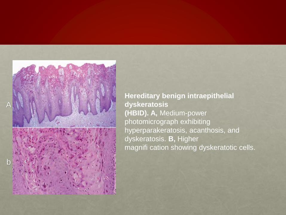
Hereditary benign intraepithelial
dyskeratosis
(HBID). A, Medium-power
photomicrograph exhibiting
hyperparakeratosis, acanthosis, and
dyskeratosis. B, Higher
magnifi cation showing dyskeratotic cells.

TREATMENT AND PROGNOSIS
• Because HBID is a benign condition, no treatment is
generally required or indicated for the oral lesions.
• If superimposed candidiasis develops, then an
antifungal medication can be used
• Patients with symptomatic ocular lesions should be
referred to an ophthalmologist.

4-DARIER’S DISEASE
• is inherited as an autosomal dominant trait, having a
high degree of penetrance and variable expressivity.
• A lack of cohesion among the surface epithelial cells
characterizes this disease, and mutation of a gene
that encodes an intracellular calcium pump has
been identified as the cause for abnormal
desmosomal organization in the affected epithelial
cells.

CLINICAL FEATURES
• Patients with Darier’s disease have numerous
erythematous papules on the skin of the trunk and
the scalp that develop during the first or second
decade of life
• The oral lesions are typically asymptomatic and are
discovered on routine examination. The frequency of
occurrence of oral lesions ranges from 15% to 50%.
• They consist of multiple, normal-colored or white, fl
attopped papules

Darier’s disease. Erythematous
cutaneous
papules on the chest.
Darier’s disease. The oral mucosa
may show multiple white papules

HISTOPATHOLOGIC FEATURES
Darier’s disease. Low-power
photomicrograph
showing a thick keratin plug, intraepithelial
clefting, and elongated rete ridges.
a dyskeratotic process characterized by a central keratin
plug that overlies epithelium exhibiting a intraepithelial
cleft.

TREATMENT AND PROGNOSIS
• Treatment of Darier’s disease depends on the
severity of involvement.
• Photosensitive patients should use a sunscreen,
and all patients should minimize unnecessary
exposure to hot environments.
• For relatively mild cases, keratolytic agents or
emollients may be the only treatment required. For
more severely affected patients,systemic retinoids
are often beneficial,

5-WARTY DYSKERATOMA
• is a distinctly uncommon solitary lesion that can
occur on skin or oral mucosa.
• It is histopathologically identical to Darier’s disease.
• For this reason the lesion has been termed isolated
Darier’s disease.
• The lesion is not otherwise related to Darier’s
disease, however, and its cause remains unknown.

CLINICAL FEATURES
• appears as a solitary, asymptomatic, umbilicated
papule on the skin of the head or neck of an older
adult
• intraoral warty dyskeratoma appears as a pink or
white, umbilicated papule located on the keratinized
mucosa,
especially the hard palate and the alveolar ridge

Warty dyskeratoma. Umbilicated papule
on
the hard palate.
Warty dyskeratoma. Well-circumscribed
invagination filled with a thick parakeratin
plug. There is hyperplasia of the basilar
cells with a suprabasilar cleft
Histopathologically, display dyskeratosis, basilar hyperplasia, and a
suprabasilar cleft

TREATMENT AND PROGNOSIS
• Treatment of the warty dyskeratoma consists of
conservative excision.
• The prognosis is excellent; these lesions have not
been reported to recur, and they have no apparent
malignant potential.

LEUKOPLAKIA
• Is defined by the World Health Organization (WHO) as
“a white patch or plaque that cannot be characterized
clinically or pathologically as any other disease.”
• the clinicalcolor results from a thickened surface
keratin layer
• it is typically consideredto be a precancerous or
premalignant lesion

CAUSE
• The cause of leukoplakia remains unknown, although hypotheses abound.
• TOBACCO
• ALCOHOL
• SANGUINARIA
• ULTRAVIOLET RADIATION
• MICROORGANISMS
• TRAUMA

CLINICAL FEATURES
• usually affects persons older than 40 years of age
• Approximately 70% of oral leukoplakias are found
on the lip vermilion, buccal mucosa, and gingiva.
• Early and mild lesions appear as slightly elevated
gray or gray-white plaques.
• A special high-risk form of leukoplakia, proliferative
verrucous leukoplakia (PVL), is characterized by
the development of multiple keratotic plaques with
roughened surface projections

• Leukoplakia may become dysplastic, even invasive,
with no change in its clinical appearance.
Early or thin leukoplakia.
This early lesion of the
ventral tongue is smooth,
white, and well demarcated
from the surrounding normal
mucosa.
Homogeneous or thick
leukoplakia. A diffuse,
corrugated white patch on the
right ventral surface of the
tongue and fl oor of mouth.
Granular leukoplakia.
Irregular white patch in
the fl oor of the mouth of
a heavy smoker. Early
invasivesquamous cell
carcinoma was found on
biopsy.
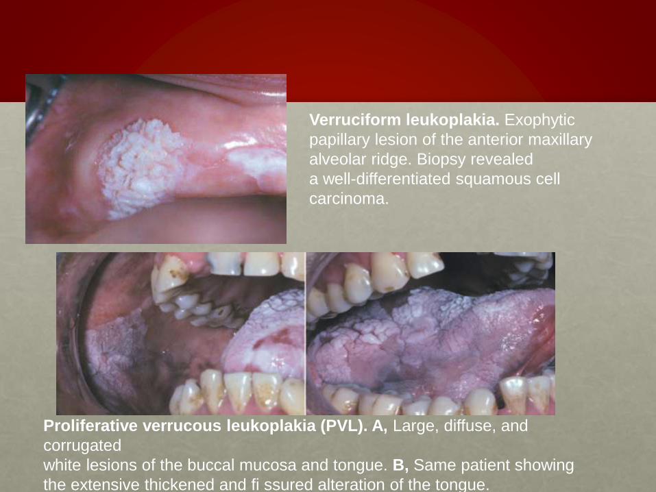
Verruciform leukoplakia. Exophytic
papillary lesion of the anterior maxillary
alveolar ridge. Biopsy revealed
a well-differentiated squamous cell
carcinoma.
Proliferative verrucous leukoplakia (PVL). A, Large, diffuse, and
corrugated
white lesions of the buccal mucosa and tongue. B, Same patient showing
the extensive thickened and fi ssured alteration of the tongue.
HISTOPATHOLOGIC FEATURES
• is characterized by a thickened keratin layer of the
surface epithelium (hyperkeratosis), with or without a
thickened spinous layer (acanthosis).
• The keratin layer may consist of parakeratin,
orthokeratin or a combination of both
• epithelial dysplasia is found in only 5% to 25% of
cases

The histopathologic alterations of dysplastic epithelial cells are similar to those of squamous cell carcinoma and may include the following:
• Enlarged nuclei and cells
• Large and prominent nucleoli
• Hyperchromatic (excessively dark-staining) nuclei
• Pleomorphic (abnormally shaped) nuclei and cells
• Dyskeratosis (premature keratinization of individual cells)
• Increased mitotic activity (excessive numbers of mitoses)
• Abnormal mitotic figures

Mild epithelial dysplasia.
Hyperchromatic and slightly pleomorphic
nuclei are noted in the basal and
parabasal cell layers of this stratifi ed
squamous epithelium.
Moderate epithelial dysplasia.
Dysplastic
changes extend to the midpoint of the
epithelium and are characterized by
nuclear hyperchromatism,
pleomorphism, and cellular crowding.
Severe epithelial dysplasia. Epithelium
exhibiting marked pleomorphism,
hyperchromatism, and scattered mitotic
figures. Atypical cells involve most of the
epithelial thickness.
TREATMENT
• Leukoplakia exhibiting moderate epithelial dysplasia
or worse warrants complete destruction or removal,
if feasible.
• The management of leukoplakia exhibiting less
severe change is guided by the size of the lesion
and the response to more conservative measures,
such as smoking cessation.

Thank You
Reference : Oral and Maxillofacial Pathology - Neville, Brad W.