Embed Size (px)
Citation preview
Journal of Oral and Maxillofacial Pathology Vol. 19 Issue 1 Jan - Apr 2015
INTRODUCTION
Pleomorphic adenoma (PA) is a benign, mixed tumor which most commonly involves the parotid gland. Approximately, 8% of PA involves the minor salivary glands and the palate is the most common site (60–65%).[1] PAs are known to occur in other minor salivary gland sites, including the lip, buccal mucosa and tongue.[2] PA of a buccal minor salivary gland, which lies on the external aspect of buccinators, has not been reported previously. We report a case of a PA apparently arising from such a gland and relevant review of literature. An extensive research has revealed only few well-documented cases of PA of a buccal minor salivary gland. This article is presented to share our experience with a case of very rare PA of a buccal minor salivary gland.
CASE REPORT
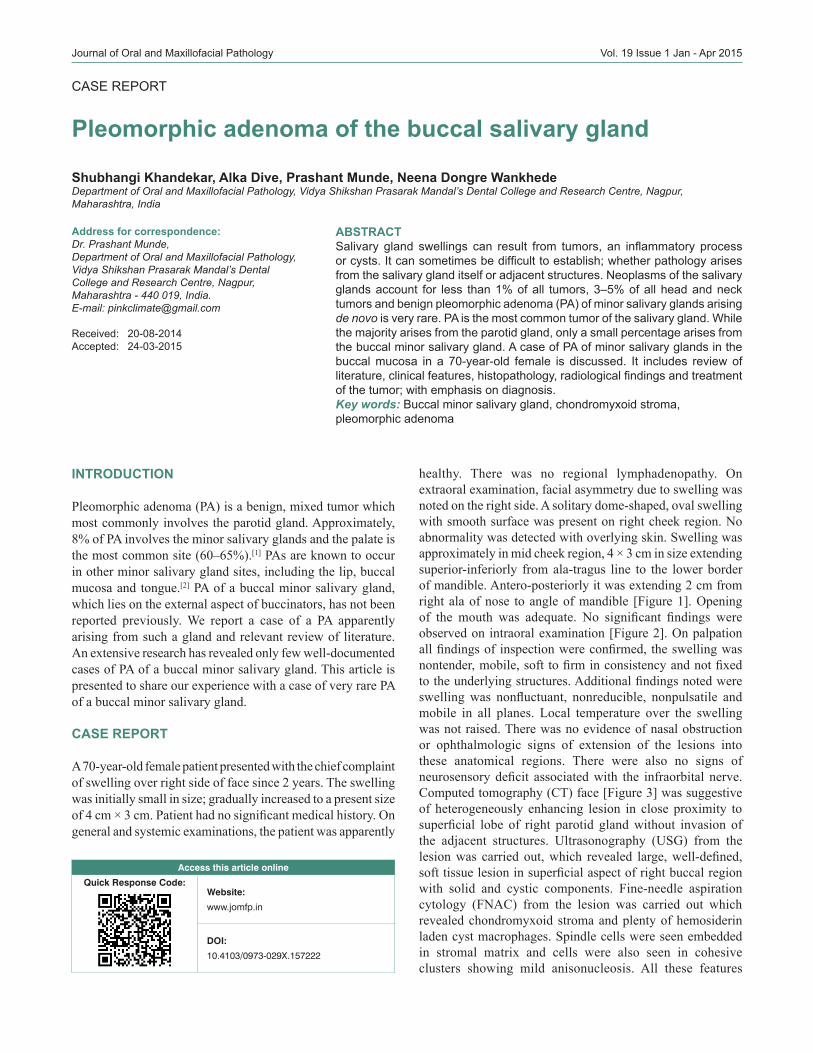
A 70-year-old female patient presented with the chief complaint of swelling over right side of face since 2 years. The swelling was initially small in size; gradually increased to a present size of4cm×3cm.Patienthadnosignificantmedicalhistory.Ongeneral and systemic examinations, the patient was apparently
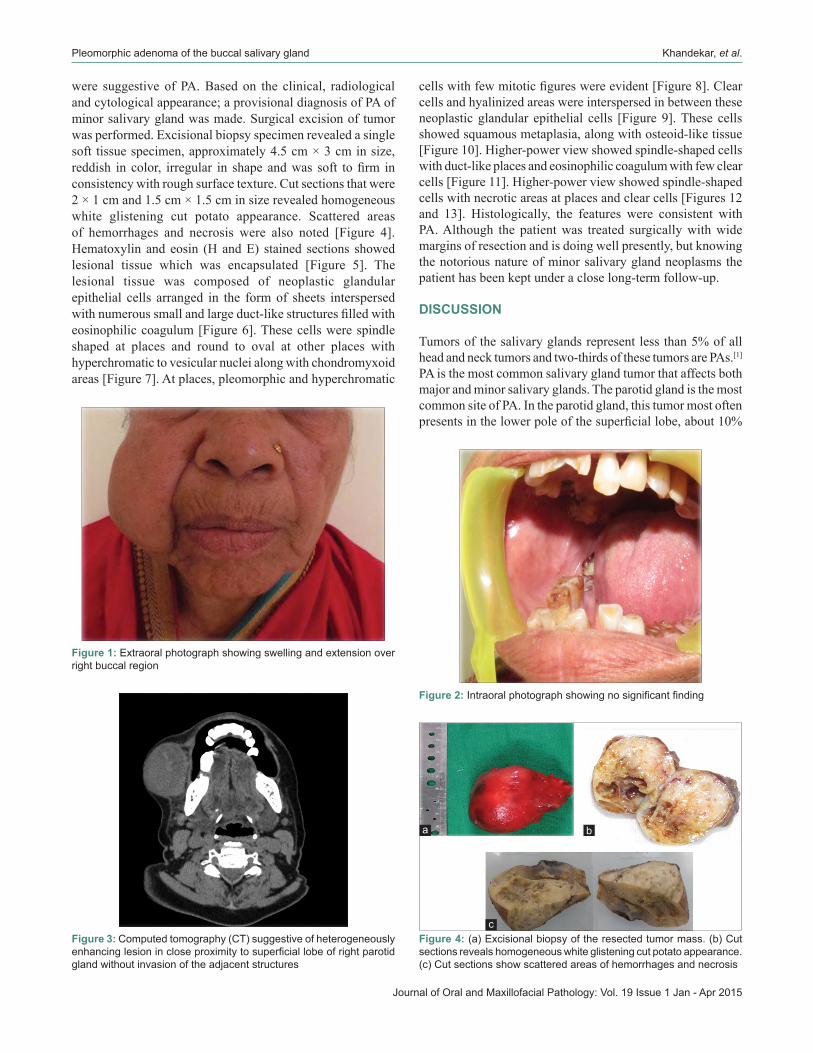
healthy. There was no regional lymphadenopathy. On extraoral examination, facial asymmetry due to swelling was noted on the right side. A solitary dome-shaped, oval swelling with smooth surface was present on right cheek region. No abnormality was detected with overlying skin. Swelling was approximately in mid cheek region, 4 × 3 cm in size extending superior-inferiorly from ala-tragus line to the lower border of mandible. Antero-posteriorly it was extending 2 cm from right ala of nose to angle of mandible [Figure 1]. Opening of the mouth was adequate. No significant findings wereobserved on intraoral examination [Figure 2]. On palpation allfindingsof inspectionwereconfirmed, theswellingwasnontender,mobile,softtofirminconsistencyandnotfixedtotheunderlyingstructures.Additionalfindingsnotedwereswellingwas nonfluctuant, nonreducible, nonpulsatile andmobile in all planes. Local temperature over the swelling was not raised. There was no evidence of nasal obstruction or ophthalmologic signs of extension of the lesions into these anatomical regions. There were also no signs of neurosensorydeficit associatedwith the infraorbital nerve.Computed tomography (CT) face [Figure 3] was suggestive of heterogeneously enhancing lesion in close proximity to superficial lobe of right parotid glandwithout invasion ofthe adjacent structures. Ultrasonography (USG) from the lesionwas carried out,which revealed large,well‑defined,softtissuelesioninsuperficialaspectofrightbuccalregionwith solid and cystic components. Fine-needle aspiration cytology (FNAC) from the lesion was carried out which revealed chondromyxoid stroma and plenty of hemosiderin laden cyst macrophages. Spindle cells were seen embedded in stromal matrix and cells were also seen in cohesive clusters showing mild anisonucleosis. All these features
Pleomorphic adenoma of the buccal salivary gland
Shubhangi Khandekar, Alka Dive, Prashant Munde, Neena Dongre WankhedeDepartment of Oral and Maxillofacial Pathology, Vidya Shikshan Prasarak Mandal’s Dental College and Research Centre, Nagpur, Maharashtra, India
CASE REPORT
Address for correspondence: Dr. Prashant Munde, Department of Oral and Maxillofacial Pathology, Vidya Shikshan Prasarak Mandal’s Dental College and Research Centre, Nagpur, Maharashtra ‑ 440 019, India. E‑mail: [email protected]
Received: 20‑08‑2014 Accepted: 24‑03‑2015
ABSTRACTSalivary gland swellings can result from tumors, an inflammatory process or cysts. It can sometimes be difficult to establish; whether pathology arises from the salivary gland itself or adjacent structures. Neoplasms of the salivary glands account for less than 1% of all tumors, 3–5% of all head and neck tumors and benign pleomorphic adenoma (PA) of minor salivary glands arising de novo is very rare. PA is the most common tumor of the salivary gland. While the majority arises from the parotid gland, only a small percentage arises from the buccal minor salivary gland. A case of PA of minor salivary glands in the buccal mucosa in a 70‑year‑old female is discussed. It includes review of literature, clinical features, histopathology, radiological findings and treatment of the tumor; with emphasis on diagnosis.Key words: Buccal minor salivary gland, chondromyxoid stroma, pleomorphic adenoma
Access this article online
Quick Response Code:Website:
www.jomfp.in
DOI:
10.4103/0973-029X.157222
Journal of Oral and Maxillofacial Pathology: Vol. 19 Issue 1 Jan - Apr 2015
Pleomorphic adenoma of the buccal salivary gland Khandekar, et al.
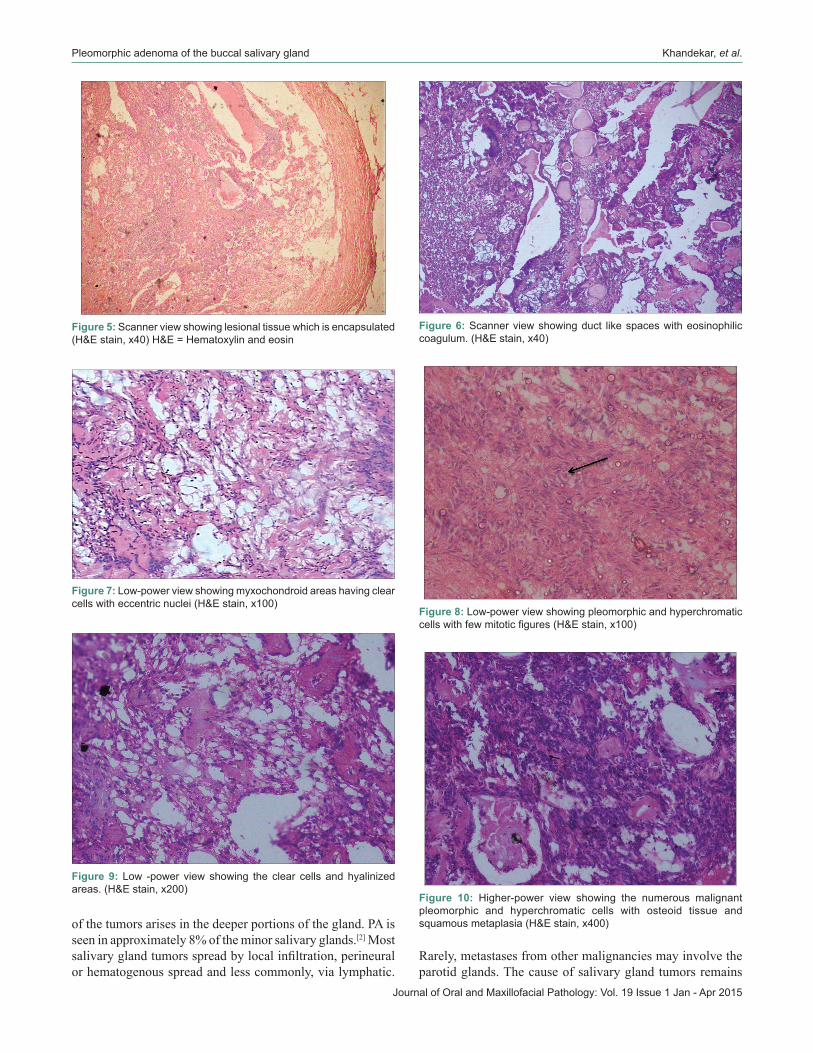
were suggestive of PA. Based on the clinical, radiological and cytological appearance; a provisional diagnosis of PA of minor salivary gland was made. Surgical excision of tumor was performed. Excisional biopsy specimen revealed a single soft tissue specimen, approximately 4.5 cm × 3 cm in size, reddish in color, irregular in shape andwas soft tofirm inconsistency with rough surface texture. Cut sections that were 2 × 1 cm and 1.5 cm × 1.5 cm in size revealed homogeneous white glistening cut potato appearance. Scattered areas of hemorrhages and necrosis were also noted [Figure 4]. Hematoxylin and eosin (H and E) stained sections showed lesional tissue which was encapsulated [Figure 5]. The lesional tissue was composed of neoplastic glandular epithelial cells arranged in the form of sheets interspersed withnumeroussmallandlargeduct‑likestructuresfilledwitheosinophilic coagulum [Figure 6]. These cells were spindle shaped at places and round to oval at other places with hyperchromatic to vesicular nuclei along with chondromyxoid areas [Figure 7]. At places, pleomorphic and hyperchromatic
cellswithfewmitoticfigureswereevident[Figure8].Clearcells and hyalinized areas were interspersed in between these neoplastic glandular epithelial cells [Figure 9]. These cells showed squamous metaplasia, along with osteoid-like tissue [Figure 10]. Higher-power view showed spindle-shaped cells with duct-like places and eosinophilic coagulum with few clear cells [Figure 11]. Higher-power view showed spindle-shaped cells with necrotic areas at places and clear cells [Figures 12 and 13]. Histologically, the features were consistent with PA. Although the patient was treated surgically with wide margins of resection and is doing well presently, but knowing the notorious nature of minor salivary gland neoplasms the patient has been kept under a close long-term follow-up.
DISCUSSION
Tumors of the salivary glands represent less than 5% of all head and neck tumors and two-thirds of these tumors are PAs.[1] PA is the most common salivary gland tumor that affects both major and minor salivary glands. The parotid gland is the most common site of PA. In the parotid gland, this tumor most often presentsinthelowerpoleofthesuperficiallobe,about10%
Figure 1: Extraoral photograph showing swelling and extension over right buccal region
Figure 2: Intraoral photograph showing no significant finding
Figure 3: Computed tomography (CT) suggestive of heterogeneously enhancing lesion in close proximity to superficial lobe of right parotid gland without invasion of the adjacent structures
Figure 4: (a) Excisional biopsy of the resected tumor mass. (b) Cut sections reveals homogeneous white glistening cut potato appearance. (c) Cut sections show scattered areas of hemorrhages and necrosis
c
ba
Journal of Oral and Maxillofacial Pathology: Vol. 19 Issue 1 Jan - Apr 2015
Pleomorphic adenoma of the buccal salivary gland Khandekar, et al.
of the tumors arises in the deeper portions of the gland. PA is seen in approximately 8% of the minor salivary glands.[2] Most salivaryglandtumorsspreadbylocalinfiltration,perineuralor hematogenous spread and less commonly, via lymphatic.
Rarely, metastases from other malignancies may involve the parotid glands. The cause of salivary gland tumors remains
Figure 5: Scanner view showing lesional tissue which is encapsulated (H&E stain, x40) H&E = Hematoxylin and eosin
Figure 6: Scanner view showing duct like spaces with eosinophilic coagulum. (H&E stain, x40)
Figure 7: Low‑power view showing myxochondroid areas having clear cells with eccentric nuclei (H&E stain, x100)
Figure 8: Low‑power view showing pleomorphic and hyperchromatic cells with few mitotic figures (H&E stain, x100)
Figure 9: Low ‑power view showing the clear cells and hyalinized areas. (H&E stain, x200)
Figure 10: Higher‑power view showing the numerous malignant pleomorphic and hyperchromatic cells with osteoid tissue and squamous metaplasia (H&E stain, x400)
Journal of Oral and Maxillofacial Pathology: Vol. 19 Issue 1 Jan - Apr 2015
Pleomorphic adenoma of the buccal salivary gland Khandekar, et al.
obscure,but ionizingradiationhasbeenidentifiedasariskfactor.[1] PAs are known to occur in other minor salivary gland sites, including the lip, buccal mucosa and tongue.[3] There are 800–1,000 minor salivary glands located throughout the oral cavity in the tissue of the buccal, labial and lingual mucosa; the soft palate; the lateral parts of the hard palate; and thefloor of themouth.Unlike themajor glands, theyare not encapsulated by connective tissue, only surrounded by it and usually have a number of acini connected in a tiny lobule.[4] The glandular lobules are 1–5 mm in diameter and are separated by thin connective tissue.[5] A minor salivary gland may have a common excretory duct with another gland, or may have its own excretory duct. Their secretion is mainly mucous in nature (except for Von Ebner glands).[4] The patient usually comes with the chief complain of a small, painless, quiescent nodule which slowly begins to increase in size, sometimes showing intermittent growth. The skin rarely ulcerates even though these tumors may reach a very large size. Pain is not a common symptom, but local discomfort is frequently present. Facial nerve involvement manifested by facial paralysis is rare.[2] PAs of the minor salivary glands usually present as painless, submucosal swellings with size ranging from 2 to 6 cm in greatest diameter, but some tumors are massive.[6] Grossly, they are usually encapsulated, solitary, well‑defined,ovoidorroundmasses.Largerneoplasmsmayhave a characteristic bosselated surface with necrotic or cystic regions. Their consistency varies from hard to rubbery to soft swellingthatmaybefluctuant.Thecutsurfaceofthetumoris characteristically solid and the color varies from gray blue, pale yellow to tan. There may be gritty areas and gelatinous or glistening foci may be present when there is cartilaginous or myxochondroid differentiation.[7] Willis described PA as the lesion with unusual histologic pattern consisting of cells exhibiting the ability to differentiate to epithelial (ductal and nonductal) cells and mesenchymal (chondroid, myxoid and osseous) cells. It demonstrates combinations of glandular epithelium and mesenchyme-like tissue and the proportion of each component varies widely among individual tumors. Foote and Frazell (1954) categorized the tumor into the following types: Principally myxoid, myxoid and cellular components present in equal proportions, predominantly cellular and extremely cellular. The epithelial components form ducts and small cysts that may contain an eosinophilic coagulum, the epithelium may also occur as small cellular nests, sheets of cells anatomizing cords and foci of keratinizing squamous or spindle cells. Myoepithelial cells have variable morphology, sometimes appearing as angular or spindled, rounded with eccentric nuclei and hyalinized eosinophilic cytoplasm resembling plasma cells. Myoepithelial cells are also responsible for the characteristic mesenchyme-like changes, giving a myxoid appearance. Vacuolar degeneration of the myoepithelial cells result in a cartilaginous appearance. Foci of hyalinization, bone and even fat can be noted in the connective tissue stroma of many tumors.[2] FNA biopsy, operated in experienced hands, can determine whether the
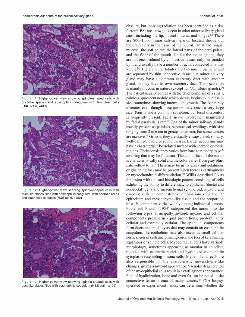
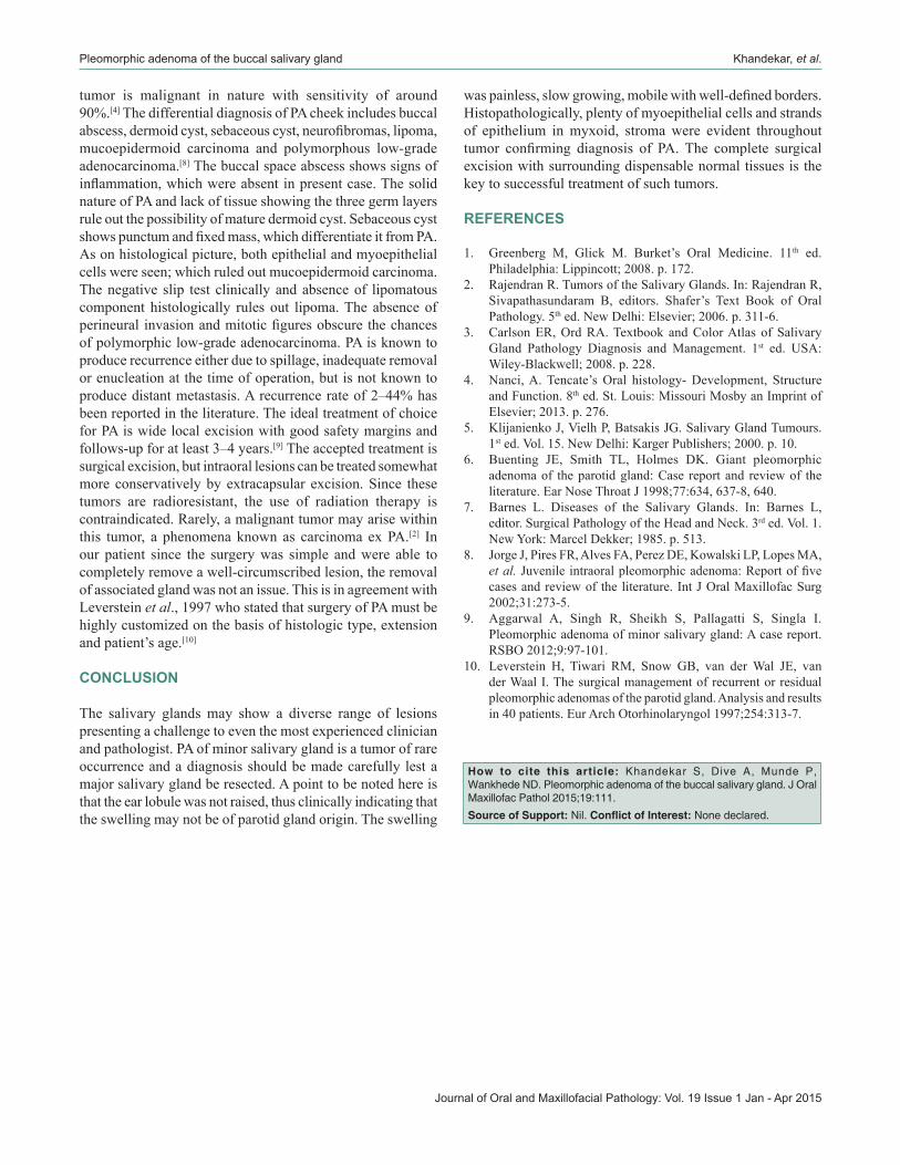
Figure 11: Higher‑power view showing spindle‑shaped cells with duct‑like spaces and eosinophilic coagulum with few clear cells (H&E stain, x400)
Figure 12: Higher‑power view showing spindle‑shaped cells with duct‑like places filled with eosinophilic coagulum, with necrotic areas and clear cells at places (H&E stain, x400)
Figure 13: Higher‑power view showing spindle‑shaped cells with duct‑like places filled with eosinophilic coagulum (H&E stain, x400)
Journal of Oral and Maxillofacial Pathology: Vol. 19 Issue 1 Jan - Apr 2015
Pleomorphic adenoma of the buccal salivary gland Khandekar, et al.
tumor is malignant in nature with sensitivity of around 90%.[4] The differential diagnosis of PA cheek includes buccal abscess,dermoidcyst,sebaceouscyst,neurofibromas,lipoma,mucoepidermoid carcinoma and polymorphous low-grade adenocarcinoma.[8] The buccal space abscess shows signs of inflammation,whichwereabsentinpresentcase.Thesolidnature of PA and lack of tissue showing the three germ layers rule out the possibility of mature dermoid cyst. Sebaceous cyst showspunctumandfixedmass,whichdifferentiateitfromPA.As on histological picture, both epithelial and myoepithelial cells were seen; which ruled out mucoepidermoid carcinoma. The negative slip test clinically and absence of lipomatous component histologically rules out lipoma. The absence of perineural invasionandmitoticfiguresobscurethechancesof polymorphic low-grade adenocarcinoma. PA is known to produce recurrence either due to spillage, inadequate removal or enucleation at the time of operation, but is not known to produce distant metastasis. A recurrence rate of 2–44% has been reported in the literature. The ideal treatment of choice for PA is wide local excision with good safety margins and follows-up for at least 3–4 years.[9] The accepted treatment is surgical excision, but intraoral lesions can be treated somewhat more conservatively by extracapsular excision. Since these tumors are radioresistant, the use of radiation therapy is contraindicated. Rarely, a malignant tumor may arise within this tumor, a phenomena known as carcinoma ex PA.[2] In our patient since the surgery was simple and were able to completely remove a well-circumscribed lesion, the removal of associated gland was not an issue. This is in agreement with Leverstein et al., 1997 who stated that surgery of PA must be highly customized on the basis of histologic type, extension and patient’s age.[10]
CONCLUSION
The salivary glands may show a diverse range of lesions presenting a challenge to even the most experienced clinician and pathologist. PA of minor salivary gland is a tumor of rare occurrence and a diagnosis should be made carefully lest a major salivary gland be resected. A point to be noted here is that the ear lobule was not raised, thus clinically indicating that the swelling may not be of parotid gland origin. The swelling
waspainless,slowgrowing,mobilewithwell‑definedborders.Histopathologically, plenty of myoepithelial cells and strands of epithelium in myxoid, stroma were evident throughout tumor confirming diagnosis of PA.The complete surgicalexcision with surrounding dispensable normal tissues is the key to successful treatment of such tumors.
REFERENCES
1. Greenberg M, Glick M. Burket’s Oral Medicine. 11th ed. Philadelphia: Lippincott; 2008. p. 172.
2. Rajendran R. Tumors of the Salivary Glands. In: Rajendran R, Sivapathasundaram B, editors. Shafer’s Text Book of Oral Pathology. 5th ed. New Delhi: Elsevier; 2006. p. 311-6.
3. Carlson ER, Ord RA. Textbook and Color Atlas of Salivary Gland Pathology Diagnosis and Management. 1st ed. USA: Wiley-Blackwell; 2008. p. 228.
4. Nanci, A. Tencate’s Oral histology- Development, Structure and Function. 8th ed. St. Louis: Missouri Mosby an Imprint of Elsevier; 2013. p. 276.
5. Klijanienko J, Vielh P, Batsakis JG. Salivary Gland Tumours. 1st ed. Vol. 15. New Delhi: Karger Publishers; 2000. p. 10.
6. Buenting JE, Smith TL, Holmes DK. Giant pleomorphic adenoma of the parotid gland: Case report and review of the literature. Ear Nose Throat J 1998;77:634, 637-8, 640.
7. Barnes L. Diseases of the Salivary Glands. In: Barnes L, editor. Surgical Pathology of the Head and Neck. 3rd ed. Vol. 1. New York: Marcel Dekker; 1985. p. 513.
8. Jorge J, Pires FR, Alves FA, Perez DE, Kowalski LP, Lopes MA, et al.Juvenileintraoralpleomorphicadenoma:Reportoffivecases and review of the literature. Int J Oral Maxillofac Surg 2002;31:273-5.
9. Aggarwal A, Singh R, Sheikh S, Pallagatti S, Singla I. Pleomorphic adenoma of minor salivary gland: A case report. RSBO 2012;9:97-101.
10. Leverstein H, Tiwari RM, Snow GB, van der Wal JE, van der Waal I. The surgical management of recurrent or residual pleomorphic adenomas of the parotid gland. Analysis and results in 40 patients. Eur Arch Otorhinolaryngol 1997;254:313-7.
How to cite this article: Khandekar S, Dive A, Munde P, Wankhede ND. Pleomorphic adenoma of the buccal salivary gland. J Oral Maxillofac Pathol 2015;19:111.
Source of Support: Nil. Conflict of Interest: None declared.


![[PAPER] Pleomorphic Adenoma Print.docx](https://img.pdfslide.net/doc/110x75/56d6bd9b1a28ab30168ea546/paper-pleomorphic-adenoma-printdocx.jpg)


![Ductal Adenocarcinoma Ex Pleomorphic Adenoma of the ... · lesions [2, 5]. Carcinoma ex pleomorphic adenoma (Ca ex PA) is a rare transformation of a benign primary PA to a malignant](https://img.pdfslide.net/doc/110x75/60bd399bb7acaf776f026cd1/ductal-adenocarcinoma-ex-pleomorphic-adenoma-of-the-lesions-2-5-carcinoma.jpg)