Embed Size (px)
Citation preview
Scrub typhus in a tertiary care hospital in the eastern part of
Odisha
ww.sciencedirect.com
a p o l l o m e d i c i n e x x x ( 2 0 1 5 ) 1e5
Available online at w
ScienceDirect
journal homepage: www.elsevier .com/locate/apme
Original Article
Scrub typhus in a tertiary care hospital in theeastern part of Odisha
Suneeta Sahu a,*, Sudhi Ranjan Misra b, Prasant Padhan c, Samir Sahu d
a Sr Consultant and HOD, Clinical Microbiologist, Dept of Clinical Microbiology and Immunoserology Apollo
Hospitals, Bhubaneswar, Indiab Sr Consultant, Clinical Microbiologist, Dept of Clinical Microbiology and Immunoserology Apollo Hospitals,
Bhubaneswar, Indiac Sr Consultant, Rheumatologist, Dept of Rheumatology, Apollo Hospitals, Bhubaneswar, Indiad Sr Consultant, Pulmonologist and Intensivist, Dept of Critical Care, Apollo Hospitals, Bhubaneswar, India
a r t i c l e i n f o
Article history:
Received 3 January 2015
Accepted 3 February 2015
Available online xxx
Keywords:
Scrub typhus
Orientia tsutsugamushi
Weil Felix
* Corresponding author.E-mail addresses: drsuneeta_s@apolloho
http://dx.doi.org/10.1016/j.apme.2015.02.0030976-0016/Copyright © 2015, Indraprastha M
Please cite this article in press as: Sahu SMedicine (2015), http://dx.doi.org/10.1016
a b s t r a c t
Aim: Our hospital, tertiary care hospital in the capital of the State of Odisha, had been
witnessing pyrexia of unknown origin, associated with breathlessness, renal and liver
impairment, which did not respond to high antibiotics like Carbapenems but to Doxycy-
cline therefore, the present study was undertaken to identify whether scrub typhus is the
aetiological agent and thereafter their characteristic features were further evaluated as an
effort in supporting its diagnoses and treating patients accordingly.
Methods: 150 Adult patients (age >12 yrs) admitted with pyrexia of unknown origin between
April 2011 and October 2013, were evaluated. Weil Felix test was done in all these patients.
Weil Felix positive samples were tested for Scrub Typhus IgM ELISA.
Results: Of the 150 patients included in the study 50 (33.33%) were found to be positive for
IgM antibodies against Orientia Tsutsugamushi. The cases were seen mainly in the months
between September and November. The common symptoms found were fever, myalgia,
breathlessness, rash and abdominal pain and clouding of memory. The diagnostic features
like eschar were found in 32% patients. Nearly two thirds of patients had fever >30 days
and myalgia (62.5%), breathlessness (64%). Most common complications was ARDS (62.5%)
followed by liver and renal failure (50%).
Conclusion: Our results showed that Scrub typhus should be considered in the differential
diagnosis of POU associated with breathlessness, myalgia, rash, gastrointestinal symp-
toms, hepatorenal syndrome or ARDS. Empirical treatment with Doxycycline may be given
in the cases with strong suspicion of Scrub typhus.
Copyright © 2015, Indraprastha Medical Corporation Ltd. All rights reserved.
spitals.com, [email protected] (S. Sahu).
edical Corporation Ltd. All rights reserved.
, et al., Scrub typhus in a tertiary care hospital in the eastern part of Odisha, Apollo/j.apme.2015.02.003
Table 1 e Signs and symptoms.
Fever <7 days 12.5%
Fever 7e14 days 37.5%
Fever 15e29days 12.5%
Fever >30 days 62.5%
a p o l l o m e d i c i n e x x x ( 2 0 1 5 ) 1e52
1. Introduction
Scrub typhus, caused by Orientia (formerly Rickettsia) tsut-
sugamushi, is an acute infectious disease of variable severity
that is transmitted to humans by an arthropod vector of
the Trombiculidae family. “Tsutsuga” means small and
dangerous and “mushi”means insect or mite. It affects people
of all ages including children. Humans are accidental hosts in
this zoonotic disease. While scrub typhus is confined
geographically to the Asia Pacific region, a billion people are at
risk and nearly a million cases are reported every year.1 Scrub
typhuswas first described from Japan in 1899. It was a dreaded
disease in pre-antibiotic era and amilitarily important disease
that affected thousands of soldiers in the far east during the
second World War.2
The rickettsia is transmitted by bite from an infected mite
to human, after which it grows at the location of the bite and a
characteristic skin lesion known as an eschar is formed. The
rickettsia then spreads systemically via the hematogenous
and lymphatogenous routes. The infected human then de-
velops various systemic symptoms and reactions including
fever, rash, lymphadenopathy, elevations of C-Reacting Pro-
tein (CRP) and liver enzymes.3 In India, scrub typhus broke out
in an epidemic form in Assam and West Bengal during the
Second World War. Later, the presence of this disease was
found throughout India in humans, trombiculid mites and
rodents.4 The term “scrub” is used because of the type of
vegetation (terrain betweenwoods and clearings) that harbors
the vector; however, the name is not entirely correct because
certain endemic areas can also be sandy, semiarid and
mountain deserts. The word “typhus” is derived from the
Greek word “typhus”, which means “fever with stupor” or
smoke.5
Scrub typhus is a diagnostic dilemma because it has non
specific presentations, limited awareness, low index of sus-
picious among clinicians and lack of diagnostic facilities.6 O.
tsutsugamushi is an obligatory intra-cellular gram negative
bacterium, and is a Zoonotic disease. Man is accidentally
infected when he encroaches the mite infected areas, known
as the mite islands. These areas consist of areas with sec-
ondary scrub growth, which grows after the clearance of pri-
mary forest, and hence the term scrub typhus. However the
infection can occur in disease habitats like sea shore, rice-
fields and even semideserts.7 If the diagnosis is delayed or
patient is not treated with appropriate antibiotics, the scrub
typhus can present with serious complications such as renal
failure, mycocarditis, septic shock, meningitis.
Scrub Typhus broke out in an epidemic form in Assam and
West Bengal during world war II. Outbreak of scrub typhus in
southern India has been reported in 2003.8 However cases in
the state of Odisha has not been reported so far.
Myalgia 62.5%
Headache 25%
Cough 28.57%
Breathlessness (64%)
Nausea 37.5%
Vomiting 37.5%
Abd. pain 37.5%
Diarrhea 35.7%
Skin rash 50%
2. Materials & methods
150 Adult patients (age more than 12 yrs) admitted with py-
rexia of unknown origin to our hospital which is a 350 bedded
hospital betweenApril 2011 and October 2013, were evaluated.
Detailed clinical examination including careful search for
Please cite this article in press as: Sahu S, et al., Scrub typhus inMedicine (2015), http://dx.doi.org/10.1016/j.apme.2015.02.003
eschar was made in all patients. Basic laboratory tests were
done in these cases (complete blood count, peripheral smear,
urine analysis, urea, creatinine, glucose, liver function tests).
Additional investigations including blood culture, chest X-ray,
Widal, rapid card test for malarial antigen, serology for
leptospirosis and serology for dengue were also done in the
majority of patients. In additionWeil Felix test was done in all
these patients. Kit Progen, Proteus Antigen suspension for
Weil Felix by Tulip Diagnostics was used. All Weil Felix posi-
tive samples were tested for Scrub Typhus IgM by InBios In-
ternational Inc. Other investigations were done as indicated
(USG abdomen, urine culture) to establish the cause of fever.
Patients diagnosed to have scrub typhus on the basis of eschar
and/or positive Weil Felix test were included in the study.
3. Results
50 patients were diagnosed to have scrub typhus during the
study period of 2 and½ years. The age ranged from 16 to 65 yrs.
Therewere 17 females and 33males.Most of the patientswere
from the nearby districts of Bhubaneswar. Maximum
numbers were seen between April and October.
Table 1 shows the signs and symptoms in these 50 cases,
Breathlessness, being the commonest (64%), other symptoms
were headache (25%), diarrhea (35.7%), skin rash (50%),
abdominal pain, nausea, vomiting was complained by 37.5%
patients. Myalgia was seen in 62.5% patients. 12.5% patients
presented with fever <7 days and same number of patients
were admitted after 15e29 days of fever, whereas fever for
7e14 days was present in 37.5% patients. Common sign seen
were pleural effusion (43%) hepatomegaly (27%) and spleno-
megaly (13%). Eschar was seen in 18 patients. Associated
enteric fever was seen in 4/50 patients. Common sites of
eschar was in lower abdomen and back region. Other sites
involved were cheek, vulva and thigh region.
Table 2 shows the lab parameters in these patients. Total
leucocyte count was raised in majority 50% of patients.
Thrombocytopenia was seen in 19 patients (37.5%). SGOT & or
SGPTwere elevated in 87% patients. Raised bilirubin (�1.2mg/
d) was found in 50% of patients and renal failure (Creatinine
>1.5 mg/dl) was present in 53%. 50% patients had pleural
effusion on admission. Hepatomegaly and splenomegaly was
seen in 27% and 13% respectively. Widal test positive in 1: 360
a tertiary care hospital in the eastern part of Odisha, Apollo
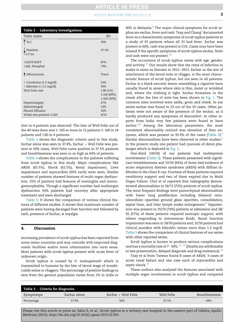
Table 2 e Laboratory investigations.
Tests name SD
TLC 50%
Platelets
<1.0 lac
37.5%
[SGOT/SGPT 87%
[Alk. Phosphal 73%
Albuminuria Trace
[ Creatinine (1.5 mg/dl) 53%
[ Bilirubin (�1.2 mg/dl) 50%
Weil Felix test 1:80 (1%)
1:160 (60%)
1:320 (30%)
Hepatomegaly 27%
Splenomegaly 13%
Pleural Effusion 50%
Widal test positive 1:320 4/50
a p o l l o m e d i c i n e x x x ( 2 0 1 5 ) 1e5 3
titer in 4 patients was observed. The titer of Weil Felix out of
the 40 tests done was 1: 320 or more in 12 patients 1: 160 in 24
patients and 1:80 in 4 patients.
Table 3 shows the diagnostic criteria used in this study.
Eschar alone was seen in 37.8%, Eschar þ Weil Felix was pre-
sent in 50% cases, Weil Felix came positive in 37.5% patients
and breathlessness was seen in as high as 64% of patients.
Table 4 shows the complications in the patients suffering
from scrub typhus in this study. Major complications like
ARDS (62.5%), Shock (62.5%), Renal impairment, Liver
impairment and myocarditis (50% each) were seen, Similar
number of patients showed features of multi organ dysfunc-
tion. 25% of patients had features of meningitis and menin-
goencephalitis. Though a significant number had multiorgan
dysfunction 93% patients had recovery after appropriate
treatment and were discharged.
Table 5: It shows the comparison of various clinical fea-
tures of different studies. It shows that maximum number of
patients were having deranged liver function test followed by
rash, presence of Eschar, & myalgia.
4. Discussion
Increasing prevalence of scrub typhus has been reported from
some Asian countries and may coincide with improved diag-
nostic facilities and/or more urbanization into rural areas.
Most patients with scrub typhus present with acute fever of
unknown origin.
Scrub typhus is caused by O. tsutsugamushi which is
transmitted to humans by the bite of larval stage of trombi-
culidemites or chiggers. The percentage of positive findings in
sera from the general population varies from 2% in India to
Table 3 e Criteria for diagnosis.
Symptomps Eschar alone Eschar þ W
Percentage 37.8% 50%
Please cite this article in press as: Sahu S, et al., Scrub typhus inMedicine (2015), http://dx.doi.org/10.1016/j.apme.2015.02.003
40% in Malaysia.4 The major clinical symptoms for scrub ty-
phus are eschar, fever and rash. Tsay and Chang3 documented
fever as a characteristic symptoms of scrub typhus patients in
a study of 33 patients where all 33 had fever. Eschar was
present in 60%, rash was present is 21%. Cases may have been
missed if the specific symptoms of scrub typhus eschar, fever
and rash were not present.10
The occurrence of scrub typhus varies with age, gender,
and activity.11 Our results show that the rates of infection in
males is same as females in 2012e2013. Eschar at the site of
attachment of the larval mite or chigger, is the most charac-
teristic feature of scrub typhus, but not seen in all patients.
Eschar is a black necrotic lesion resembling a cigarette burn
usually found in areas where skin is thin, moist or wrinkled
and, where the clothing is tight. Eschar formation in the
cheek after the bite of mite has been shown in Fig. 1. The
common sites involved were axilla, groin and cheek. In our
series eschar was found in 23 out of the 50 cases. Often pa-
tients were not aware of the presence of the eschar, as it
hardly produced any symptoms of discomfort. In other re-
ports from India very few patients were found to have
eschar.2,4 Among the laboratory parameters, the most
consistent abnormality noticed was elevation of liver en-
zymes, which was present in 95.9% of the cases (Table 2).6
Similar abnormalities have been observed in other studies.8
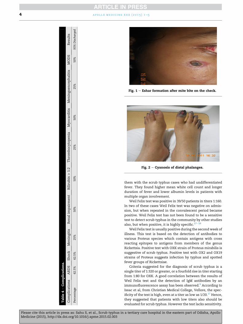
In the present study one patient had cyanosis of distal pha-
langes which is depicted in Fig. 2.
One-third (18/50) of our patients had multisystem
involvement (Table 5). These patients presented with signifi-
cant breathlessness and 32/50 (64%) of these had evidence of
acute respiratory distress syndrome (ARDS) with diffuse in-
filtrates in the chest X-ray. Fourteen of these patients required
ventilatory support and two of them expired due to Multi
Organ Failure. Choi et al reported that radiography demon-
strated abnormalities in 54/72 (72%) patients of scrub typhus.
The most frequent findings were parenchymal abnormalities
with lower lung predilection including bilateral retic-
ulonodular opacities ground glass opacities, consolidation,
septal lines, and hilar lymph nodes enlargement.6 Hypoten-
sion was present in 35/50 (70%) patients at admission and 28/
50 (57%) of these patients required inotropic support, with
others responding to intravenous fluids. Renal function
impairmentwas seen in 28/50 patients and, 32/50 patients had
clinical jaundice with bilirubin values more than 1.2 mg/dl.
Table 5 shows the comparison of clinical features of our series
with other reported series.
Scrub typhus is known to produce serious complications
andhas amortality rate of 7e30%.12e15 (Deaths are attributable
to late presentation, delayed diagnosis and drug resistance).16
Tsay et al from Taiwan found 8 cases of ARDS, 3 cases of
acute renal failure and one case each of myocarditis and
septic shock.10
These authors also analyzed the features associated with
multiple organ involvement in scrub typhus and compared
eil Felix Weil Felix Breathlessness
37.5% 64%
a tertiary care hospital in the eastern part of Odisha, Apollo
Table
4e
Com
plica
tions.
Complica
tions
ARDS
Shock
Meningitis
Renalim
pairment
Bilirubin
>1:2
Thro
mbocy
topenia
Myoca
rditis
Meningoence
phalitis
MODS
Resu
lts
Percentage
62.5%
62.5%
25%
50%
50%
25%
50%
25%
50%
93%
Discharged
Fig. 1 e Eshar formation after mite bite on the check.
Fig. 2 e Cyanosis of distal phalanges.
a p o l l o m e d i c i n e x x x ( 2 0 1 5 ) 1e54
Please cite this article in press as: Sahu S, et al., Scrub typhus inMedicine (2015), http://dx.doi.org/10.1016/j.apme.2015.02.003
them with the scrub typhus cases who had undifferentiated
fever. They found higher mean white cell count and longer
duration of fever and lower albumin levels in patients with
multiple organ involvement.
Weil Felix test was positive in 39/50 patients in titers 1:160.
In two of these cases Weil Felix test was negative on admis-
sion, but when repeated in the convalescent period became
positive. Weil Felix test has not been found to be a sensitive
test to detect scrub typhus in the community by other studies
also, but when positive, it is highly specific.17e19
Weil Felix test is usually positive during the secondweek of
illness. This test is based on the detection of antibodies to
various Proteus species which contain antigens with cross
reacting epitopes to antigens from members of the genus
Rickettsia. Positive test with OXK strain of Proteus mirabilis is
suggestive of scrub typhus. Positive test with OX2 and OX19
strains of Proteus suggests infection by typhus and spotted
fever groups of Rickettsiae.
Criteria suggested for the diagnosis of scrub typhus is a
single titer of 1:320 or greater, or a fourfold rise in titer starting
from 1:80 for OXK. A good correlation between the results of
Weil Felix test and the detection of IgM antibodies by an
immunofluorescence assay has been observed.9 According to
Issac et al, from Christian Medical College, Vellore, the spec-
ificity of the test is high, even at a titer as low as 1/20.17 Hence,
they suggested that patients with low titers also should be
evaluated for scrub typhus. However the test lacks sensitivity.
a tertiary care hospital in the eastern part of Odisha, Apollo
Table 5 e Comparison of various clinical features.
Vellore (Ref7) Shimla (Ref2) South Vietnam (Ref9) Pondicherry (Present series)
No. of cases 27 21 87 50 50
No. of days of fever 5e20 5e25 NA 3e60 3e29
Myalgia 52% 38% 32% 38% 36%
Cough 44% NA 45% 40% 29%
Nausea/vomiting 48% 43% 28% 58% 29%
Lymphadenopathy NA 53% 85% 30% 33%
Hepatomegaly NA 43% 43% 28% 27%
Jaundice 26% 53% NA 10% 50%
Altered sensorium 19% 24% NA 20% 25%
Rash 22% 10% 34% 14% 36%
Eschar 4% 10% 46% 46% 38%
Mortality 11.10% 14.20% NA 2% 7%
Weil Felix positive 77% NA 57% 78% 90%
a p o l l o m e d i c i n e x x x ( 2 0 1 5 ) 1e5 5
In a different study from the same institutionwhich evaluated
various serological tests for scrub typhus, Weil Felix test was
found to have a sensitivity of only 43% but a specificity of 98%
for titers 1:80 or more.19
Several studies have shown that Weil Felix test has high
specificity.17e19 In a ruralMalaysian hospital, the usefulness of
two serological tests for scrub typhus namely, Weil Felix test
and IFA were compared.18 It was found that, at a cut off value
of greater than or equal to 1:400 titer, the IFA test had a
specificity of 96% and at a cut off value of greater than or equal
to 1:320 of OXK had a specificity of 97%. The probability value
for the correct diagnosis for scrub typhus was found to be 78%
for IFA titer of 1:400 or more and 79% for OXK titer 1:320 or
more. When both tests were positive in a single sample, the
probability of correct diagnosis increases to 96%.18
All the reports of scrub typhus from South India have been
from Christian Medical College, Vellore. In one of their studies
referred to earlier, Weil Felix test had a specificity of 98% for
titers 1:80 or more.19 It is noteworthy that the serological tests
for Rickettsial diseases including the specific IgM antibody
tests become positive only in the second week and a second
sample at a later time is often required; serological tests
cannot provide early diagnosis and a specific diagnosis may
not be available until after the patient has died or recovered.10
This study was done in order to have a thorough knowl-
edge of the clinical features of scrub typhus including its
symptoms and signs so that diagnosis of scrub typhus can be
done with this awareness at the earliest and help the patient
get proper treatment in this part of the state.
Conflicts of interest
All authors have none to declare.
r e f e r e n c e s
1. Watt G, Parola P. Scrub typhus and tropical rickettsioses. CurrOpin Infect Dis. 2003;16(5):429e436.
2. Groves MG, Harrington KS. Scrub typhus. In: Beran GW, ed.Handbook of Zoonoses. 2nd ed. Florida: CRC Press; 1994:663e668.
Please cite this article in press as: Sahu S, et al., Scrub typhus inMedicine (2015), http://dx.doi.org/10.1016/j.apme.2015.02.003
3. Allen AC, Spitz S. A Comparative Study of the Pathology of ScrubTyphus (Tsutsugamushi Disease) and Other Rickettsial Diseases.1945.
4. Park K. Epidemiology of communicable diseases. In: Park'sTextbook of Preventive and Social Medicine. 15th ed. Jabalpur,India: Banarsidas Bhanot Publishers; 1998:228e229.
5. Medicine update scrub typhus.6. Vivekanandan M, Mani A, Priya YS, Singh AP, Jayakumar S,
Purty S. Outbreak of scrub typhus in Pondicherry. J AssocPhysicians India. 2010;58:24e28.
7. Mahajan SK. Scrub typhus. J Assoc Physicians India.2005;53:954e958.
8. Mathai E, Rolain JM, Verghese GM, et al. Outbreak of scrubtyphus in southern India during the cooler months. Ann N YAcad Sci. 2003;990:359e364.
9. Amano K, Suzuki N, Fujita M, et al. Serological reactivity ofsera from scrub typhus patients against Weil-Felix testantigen. Microbiol Immunol. 1993;37:927e933.
10. Tsay RW, Chang FY. Serious complications in scrub typhus.J Microbiol Immunol Infect. 1998;31:240e244.
11. Am J Trop Med Hyg. 2002;67(2):162e165. Motohiko Ogawa,ToshikatsuHagiwara,ToshioKishimoto,SadashiShiga,YoshiyaYoshida, Yumiko Furuya, Ikuo Kaiho, Tadahiko Ito, HaruyasuNemoto, Norishige Yamamoto, and Kunihiko Masukawa.
12. Wang CC, Liu SF, Liu JW, et al. Acute respiratory distresssyndrome in scrub typhus. Am J Trop Med Hyg.2007;76:1148e1152.
13. Yen TH, Chang CT, Lin JL, et al. Scrub typhus a frequentlyoverlookedcauseofacuterenal failure.RenFail. 2003;25:397e410.
14. Thap LC, Supanarnond W, Treeprasertsuk S, et al. Septicshock secondary to scrub typhus. Characteristics andcomplications. Southeast Asian J Trop Med Public Health.2002;330:780e786.
15. Cracco G, Delafosse C, Baril L, et al. Multiple organ failurecomplicating probable scrub typhus. Clin Infect Dis.2000;31:191e192.
16. Pandey et al from Himachal Pradesh reported 3 cases of ARDSdue to scrub typhus.
17. Issac R, Varghese GM, Mathai E, et al. Scrub typhus:prevalence and diagnostic issues in rural Southern India. ClinInfect Dis. 2004;39:1395e1396.
18. Brown GW, Shirai A, Rogers C, Groves MG. Diagnostic criteriafor scrub typhus probability values for immunoflourescentantibody and proteus OXK agglutinin titres. Am J Trop MedHyg. 1983;32:1101e1107.
19. Prakash JA, Abraham OC, Mathai E. Evaluation of tests forserological diagnosis of scrub typhus. Trop Doct.2006;36:212e213.
a tertiary care hospital in the eastern part of Odisha, Apollo

Apollo hospitals: http://www.apollohospitals.com/Twitter: https://twitter.com/HospitalsApolloYoutube: http://www.youtube.com/apollohospitalsindiaFacebook: http://www.facebook.com/TheApolloHospitalsSlideshare: http://www.slideshare.net/Apollo_HospitalsLinkedin: http://www.linkedin.com/company/apollo-hospitalsBlog:Blog: http://www.letstalkhealth.in/