Embed Size (px)
Citation preview


Segmental Anatomy of Lungs ,
Anatomy of Mediastinum and
Secondary Lobule
Gamal Rabie Agmy, MD, FCCP
Professor of chest Diseases,
Assiut university

There are approximately 23
generation of dichotomous
branching From trachea to the
alveolar sac
HRCT can identify upto 8th order
central bronchioles

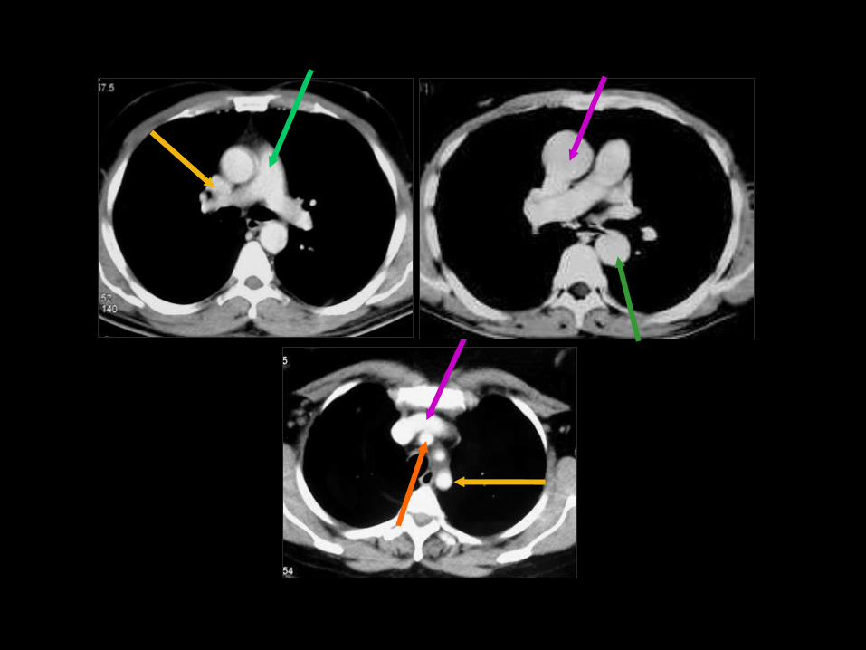
An
App
An
Ap
P App = Apicoposterior
An = Anterior
P = Posterior
1 1
2
3
2
&
3


Upper lobe
Lower lobe

An
App
SL
Ap
P An SL
SL = Superior LL
An = Anterior
App = Apicoposterior
P = Posterior

1 1
4 4


Middle lobe Lower lobe
Lingula Lower lobe

SL
S
I
L
M
SL
AN P L AN
L,M P
SL = Superior LL
AN = Anterior LL
P = Posterior LL
L,M = Lateral, Medial LL


7 6 5
8
9 9
10 10
11 11
12


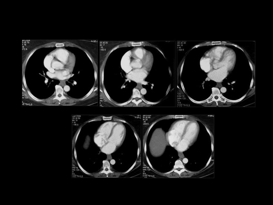
M 44Y with malignant liver F 45Y with post irradiation changes after radical mastectomy
Atelectasis in the middle lobe Post irradiation scarring in
the right upper lobe

35Y male with fever and expectoration
Pneumonic consolidation in the left upper lobe

45Y male with chest pain and hemoptysis
Bronchogenic carcinoma in the left lower lobe
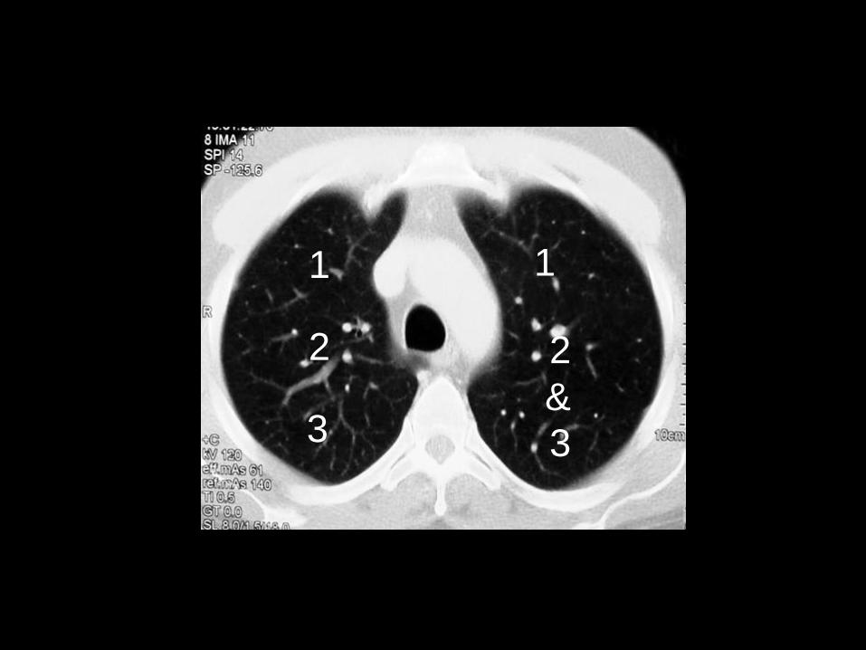
43Y male with acute chest pain and hemoptysis
Multiple infarcts in the lingula as
well as the left lower lobe


Secondary lobule
• The secondary lobule is the basic anatomic
unit of pulmonary structure and function.
Interpretation of interstitial lung diseases is
based on the type of involvement of the
secondary lobule.
It is the smallest lung unit that is surrounded
by connective tissue septa.
It measures about 1-2 cm and is made up of
5-15 pulmonary acini, that contain the alveoli
for gas exchange.
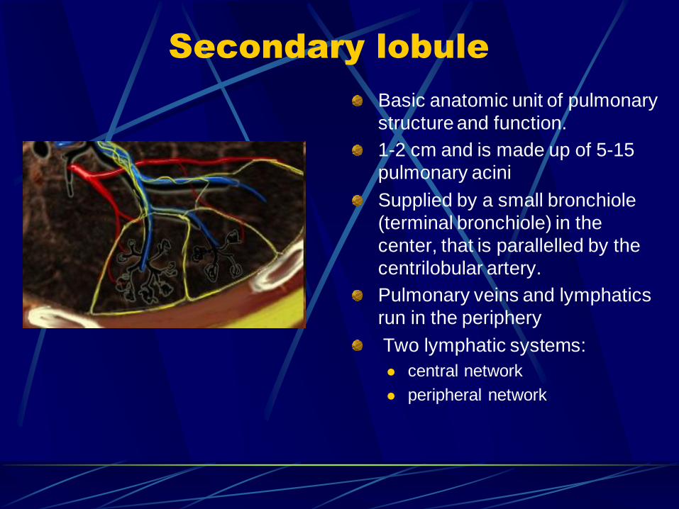
Secondary lobule
Basic anatomic unit of pulmonary
structure and function.
1-2 cm and is made up of 5-15
pulmonary acini
Supplied by a small bronchiole
(terminal bronchiole) in the
center, that is parallelled by the
centrilobular artery.
Pulmonary veins and lymphatics
run in the periphery
Two lymphatic systems:
central network
peripheral network

• The secondary lobule is supplied by a small
bronchiole (terminal bronchiole) in the
center, that is parallelled by the
centrilobular artery.
Pulmonary veins and lymphatics run in the
periphery of the lobule within the
interlobular septa.
Under normal conditions only a few of
these very thin septa will be seen.

There are two lymphatic systems: a
central network, that runs along the
bronchovascular bundle towards the
centre of the lobule and a peripheral
network, that is located within the
interlobular septa and along the pleural
linings.

The terminal bronchiole in the center divides into respiratory bronchioli with acini that contain alveoli. Lymphatics and veins run within the interlobular septa

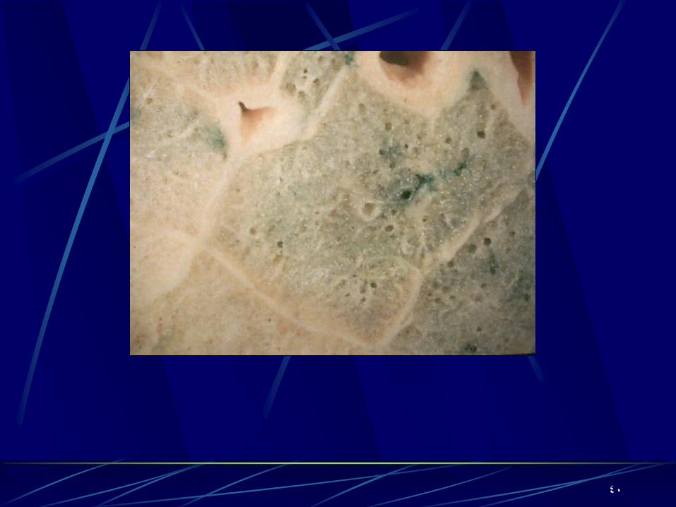
Centrilobular area
It is the central part of the secondary
lobule.
It is usually the site of diseases, that
enter the lung through the airways (
i.e. hypersensitivity pneumonitis,
respiratory bronchiolitis, centrilobular
emphysema ).

Centrilobular area in blue perilymphatic area in yellow

Perilymphatic area
Perilymphatic areais the peripheral part
of the secundary lobule.
It is usually the site of diseases, that are
located in the lymphatics of in the
interlobular septa ( i.e. sarcoid,
lymphangitic carcinomatosis, pulmonary
edema).
These diseases are usually also located in
the central network of lymphatics that
surround the bronchovascular bundle.

Raoof, S. , CHEST 2006; 129:805

40

A group of terminal bronchioles
41

Unit of lung (0.5-3 cm) Irregularly polyhedral Supplied by a group of terminal bronchioles and accompanying pulmonary arterioles surrounded by lymph vessels Demarcated by “interlobular septa”
pulmonary veins pulmonary lymphatics connective tissue stroma

Accompanying pulmonary arterioles
43

Surrounded by lymph vessels
44

Pulmonary veins
45

Pulmonary lymphatics
46

47
Connective Tissue Stroma


HRCT
PATTERN
INCREASED LUNG
ATTENUATION
LINEAR AND RETICULAR OPACITIES
NODULES AND NODULAR OPACITIES
PARENCHYMAL OPACIFICATION
consolidation
Ground glass
DECREASED LUNG
ATTENUATION
CYSTIC LESIONS, EMPHYSEMA, AND BRONCHIEACTASIS
MOSAIC ATTENUATION AND
PERFUSION
AIR TRAPPING ON EXPIRATORY
SCANS
49