Embed Size (px)
Citation preview

Management of choroidal hemangioma
ByHany EL-Defrawy, MVR FellowBEH

Diffuse choroidal hemangioma
Rare Sporodic Neuro-Oculocutaneous disorder Facial nevus flammeus Buphthalmos Diffuse choroidal hemangioma (Tomato-Catsup) Leptomeningeal hemangiomatosis Epilepsy MR Hemiplegia


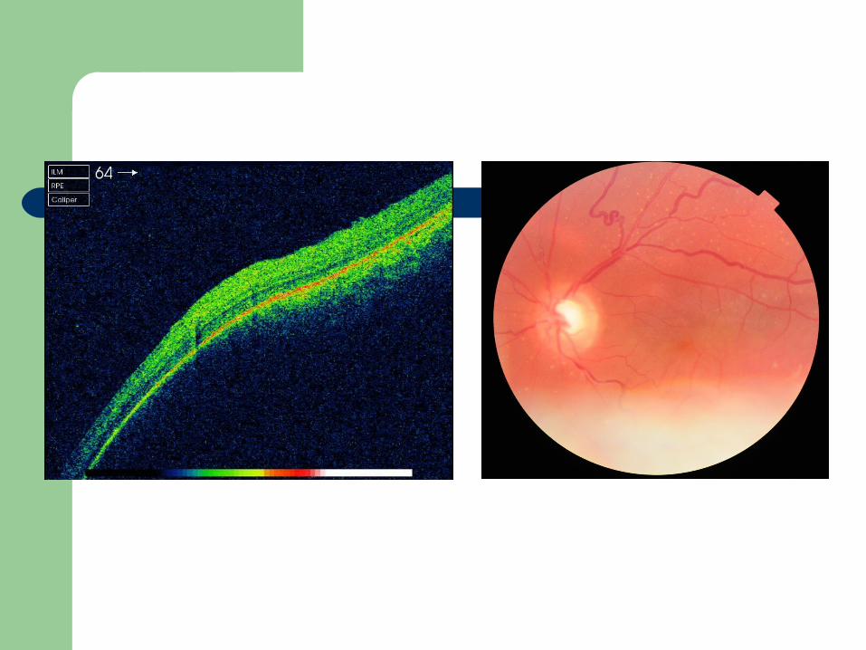

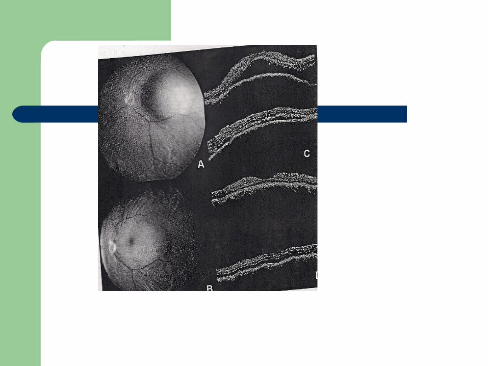
Circumscribed choroidal hemangioma
Uncommon, benign vascular tumour Discrete, smooth round orange red mass Macular and peripapillary region

Causes of visual loss
1. Refractive error
2. Foveal distortion
3. Transudative leakage and CMO
4. Serous Retinal detachment with secondary photoreceptor damage

Diagnosis
Ultrasongraphy FFA OCT MRI


When to treat?
Visual acuity potential Extent of detachment

Why challenging?
Limited therapeutic options (Why?) Surgery (Dangerous)
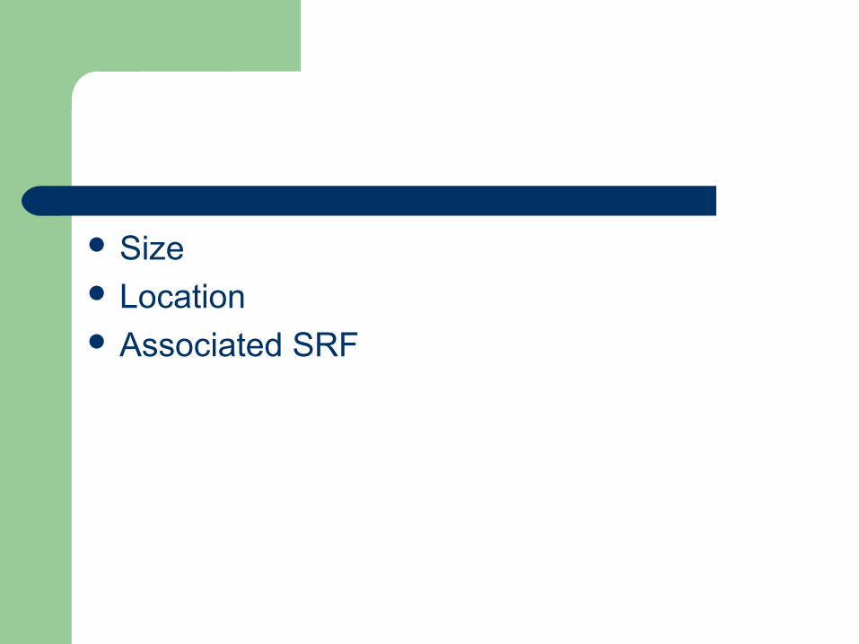
Size Location Associated SRF

Armamentarium
External beam radiation therapy (5 observational studies)
Proton beam therapy Brachytherapy Photodynamic therapy Cryotherapy Transpupillary thermotherapy Anti VEGF IVTA Combined therapy


Radiation
Cataract Radiation optic neuropathy Retinopathy Increase incidence of osteosarcoma Chorioretinal atrophy

Other modalities
Plaque Brachytherapy
1. Murthy 2005
2. Zografos 1996 Proton Beam Stereotactic therapy (Gamma Knife) Anti VEGF PDT

PDT
Anand 2003 Bains et al 2004 Singh et al 2005 Huiskamp 2005 Tsipursky

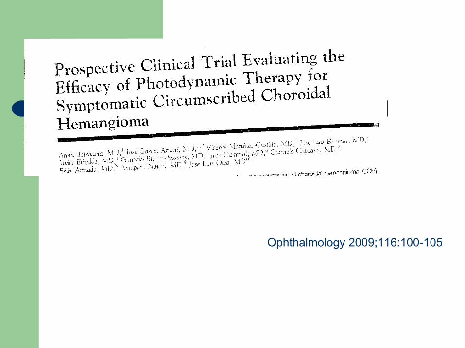
Ophthalmology 2009;116:100-105
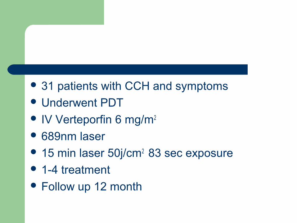
31 patients with CCH and symptoms Underwent PDT IV Verteporfin 6 mg/m2
689nm laser 15 min laser 50j/cm2 83 sec exposure 1-4 treatment Follow up 12 month

Primary end point
1. Absence of exudative RD at 12 month follow up (FFA,OCT, Ophthalmoscopy)
Secondary endpoint
1. Visual acuity
2. Tumour thickness decrease
3. Adverse events

Inclusion criteria1. 18 with CCH height 5 mm and diameter 12 mm2. Exudative RD affecting the fovea3. No cataract within last 2 months Exclusion criteria1. NYHA Class III-IV2. Porphyria3. Liver disease4. Active hepatitis
Results
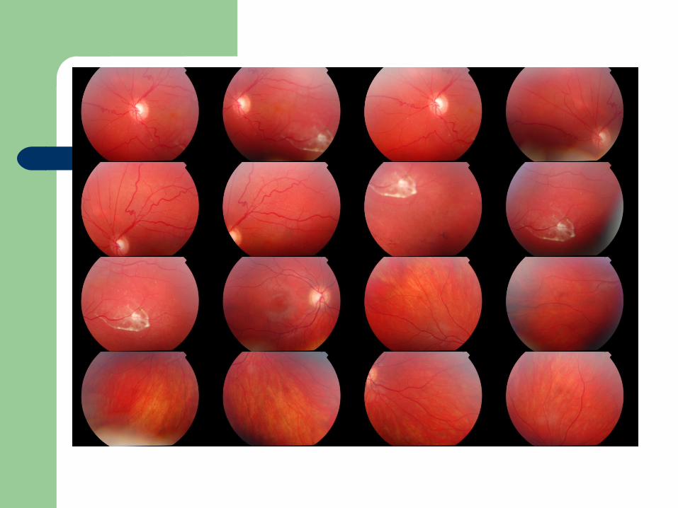
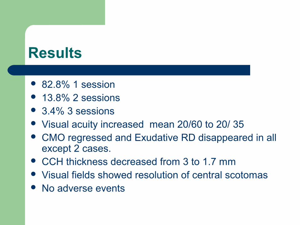
82.8% 1 session 13.8% 2 sessions 3.4% 3 sessions Visual acuity increased mean 20/60 to 20/ 35 CMO regressed and Exudative RD disappeared in all
except 2 cases. CCH thickness decreased from 3 to 1.7 mm Visual fields showed resolution of central scotomas No adverse events

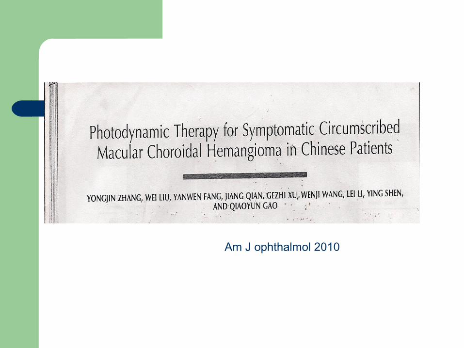
Am J ophthalmol 2010

Retrospective study of 14 patients with symptommatic circumscribed choroidal hemangioma who underwent PDT± IVTA
Shao Huang, James Fabian etal. Optometery and visual science , 2009.

Symptomatic visual loss Exudative Detachment Macular edema Subfoveal fluid Tumour encroaching to within 2 mm of the
fovea.

7.5 mm spot size Overlapping spots 83 sec IVTA (0.1ml of 40 mg/ml) Baseline VA IOP Macular thickness 10 eyes PDT 4 PDT and IVTA
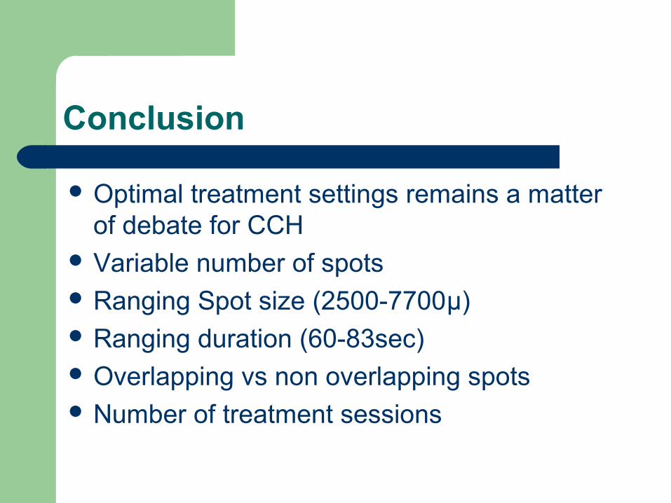
Conclusion
Optimal treatment settings remains a matter of debate for CCH
Variable number of spots Ranging Spot size (2500-7700µ) Ranging duration (60-83sec) Overlapping vs non overlapping spots Number of treatment sessions


![Hobas GRP pipe systems PN 1 - Amiblu · 2020. 6. 10. · Jacking Pipe PN 1 de [mm] SN SN SN SN SN SN SN SN SN SN SN SN Coupling Type 32000 40000 50000 64000 80000 100000 128000 160000](https://img.pdfslide.net/doc/110x75/61236c822e9bd427c4013216/hobas-grp-pipe-systems-pn-1-amiblu-2020-6-10-jacking-pipe-pn-1-de-mm-sn.jpg)