Embed Size (px)
Citation preview

DR. HARSHAD WANKHADE
EMERGENCY MEDICINE AND CRITICAL CARE.

TACHYCARDIA-RHYTHM THAT PRODUCES VENTRICULAR RATE MORE THAN 100 BEATS PER MINUTE.
TACHARRHYTHMIAS ARE ISOLATED PREMATURE COMPLEXES OR NONSUSTAINED OR SUSTAINED FORM OF TACHYCARDIA ORIGINATING FROM MYOCARDIAL FOCI OR REENTRANT CIRCUIT.

Physiological Pathological:
Valvular heart disease. Electrolyte disturbances. Structural heart disease. Ischemic heart disease. Hypertensive heart diseases. Congenital heart disease. Cardiomyopathies. Carditis. RV dysplasia. Drug related. Pericarditis. Pulmonary diseases. Others.

Palpitation.
Dizziness.
Chest Pain.
Syncope.
Irregularity in pulse.
Dyspnea.
Fainting.
Hemodynamic collaps.
Sudden cardiac death.

ECG
24h Holter monitor
Echocardiogram
Stress test
Coronary angiography
Electrophysiology study


ECG CHANGES—
Regular rhythm at a rate of 60-100 bpm (or age-appropriate rate in children).
Each QRS complex is preceded by a normal P wave.
Normal P wave axis: P waves should be upright in leads I and II, inverted in aVR.
The PR interval remains constant.
QRS complexes are < 100 ms wide (unless a co-existent interventricular conduction delay is present).


TREATMENT
USUALLY NOT REQUIRED
UNDERLYING CAUSE- INCREASE HYDRATION
SALT LOADING 10-15 GMS OF SODIUM/DAY
DRUG THERAPY- PROPRANOLOL- 20 mg TDS
CCB-
MINERALOCORTICOIDS
FLUDROCORTISONE 0.1-0.3mg PO
ADRENOCORTICO RECEPTOR ANTAGONIST
MICLODRINE 2.5-10 mg TDS
SSRI
CATHETER ABLATION
ATRIAL PACING THERAPY


An abnormal (non-sinus) P wave is followed by a QRS complex. The P wave typically has a different morphology and axis to the
sinus P waves. The abnormal P wave may be hidden in the preceding T wave,
producing a “peaked” or “camel hump” appearance — if this is not appreciated the PAC may be mistaken for a PJC.
PACS arising close to the AV node (“low atrial” ectopics) activate the atria retrogradely, producing an inverted P wave with a relatively short PR interval ≥ 120 ms (PR interval < 120 ms is classified as a PJC).
PACs that reach the SA node may depolarise it, causing the SA node to “reset” — this results in a longer-than-normal interval before the next sinus beat arrives (“post-extrasystolic pause”). Unlike with PVCs, this pause is not equal to double the preceding RR interval (i.e. not a “full compensatory pause”).
Similarly, PACs arriving very early in the cycle may not be conducted to the ventricles at all. In this case, you will see an abnormal P wave that is not followed by a QRS complex (“blocked PAC”). It is usually followed by a compensatory pause as the sinus node resets.
TREATMENT – BETA BOCKERS.


Narrow QRS complex, either (1) without a preceding P wave or (2) preceded by an abnormal P wave with a PR interval of < 120 ms (these “retrograde” P wave are usually inverted in leads II, III and aVF).
Occurs sooner than would be expected for the next sinus impulse.
Followed by a compensatory pause.
PJCs that arrive early in the cycle may be conducted aberrantly, most commonly with a RBBB morphology.


Irregularly irregular rhythm.
No P waves.
Absence of an isoelectric baseline.
Variable ventricular rate.
QRS complexes usually < 120 ms unless pre-existing bundle branch block, accessory pathway, or rate related aberrant conduction.
Fibrillatory waves may be present and can be either fine (amplitude < 0.5mm) or coarse (amplitude >0.5mm).
Fibrillatory waves may mimic P waves leading to misdiagnosis

Ashman’s Phenomenon – presences of aberrantly conducted beats, usually of RBBB morpholo, due a long refractory period as determined by the preceding R-R interval.
Commonly AF is associated with a ventricular rate ~ 110 – 160.
AF is often described as having ‘rapid ventricular response’ once the ventricular rate is > 100 bpm.
‘Slow’ AF is a term often used to describe AF with a ventricular rate < 60 bpm.
Causes of ‘slow’ AF include hypothermia, digoxin toxicity, medications, and sinus node dysfunction.


TREATMENT
RATE CONTROL= NORMAL SYSTOLIC FUNCTION-
CCB- DILTIAZEM 15mg then 20 mg over 2 min f/b infusion 5-15 mg/hrfor 24 hr shift to oral 120-360 mg daily
Beta blockers- Metoprolol 5-5-5 mg over 2min f/b oral 50 mg bd Propranolol-0.15mg/kgIV UPTO 10mg over 1 min f/b oral
80-240 mg daily.
Digoxin
amiodarone
LV Dysfunction- Digoxin – 0.25mg iv over 2min max 1.5mg in 24hrs f/b
oral 0.125-0.375 mg daily (other drug dose half reduction)
Amiodarone- 150 mg stat over 10 minf/b 360 mg infusion over6 hrs f/b 540 mg over 18 hrs.
NON-PHARMACOLGICAL- AV node ablation

RHYTHM CONTROL- AF <7 DAYS
SYSTOLIC FUNCTION PRESERVED-
PILL IN POCKET FLECAINIDE 200-300 mg OD
PROPAFENONE- 600 mg OD
IBUTILIDE 1mg IV over 10 min
AMIODARONE- 200-400 mg daily
LV DYSFUNCTION- AMIODARONE-
DOFETILIDE
AF >7 DAYS AMOIDARONE
DOFETILIDE
Digoxin and sotalol are NOT preffered for rhythm control.
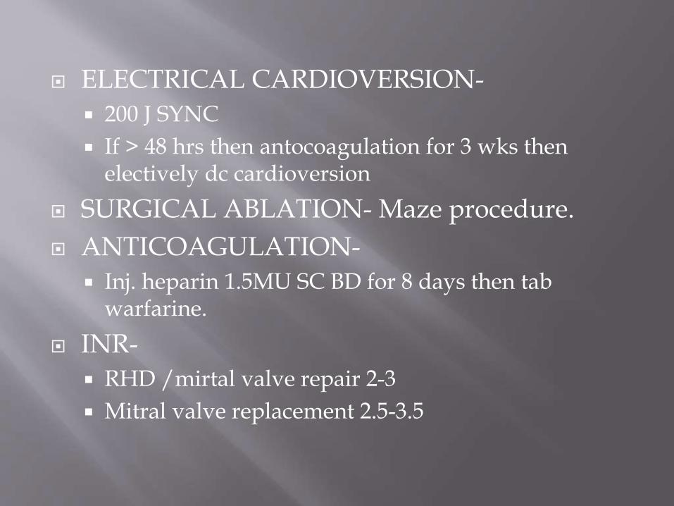
ELECTRICAL CARDIOVERSION-
200 J SYNC
If > 48 hrs then antocoagulation for 3 wks then electively dc cardioversion
SURGICAL ABLATION- Maze procedure.
ANTICOAGULATION-
Inj. heparin 1.5MU SC BD for 8 days then tab warfarine.
INR-
RHD /mirtal valve repair 2-3
Mitral valve replacement 2.5-3.5


Narrow complex tachycardia
Regular atrial activity at ~300 bpm
Flutter waves (“saw-tooth” pattern) best seen in leads II, III, aVF — may be more easily spotted by turning the ECG upside down!
Flutter waves in V1 may resemble P waves
Loss of the isoelectric baseline
Venrticular rate is constant.

TREATMENT:-
Similar to AF.
DC cardio version( 50 J) preferred as it causes more hemodynamic compromised.


TREATMENT:-
CCB- preferred b/o MAT is associated with COPD
PROPAFENONE
FLECAININDE
AMIODARONE
NO ANTICOAGULATION
NO CARDIOVERSION, beta blockers,
Digoxin narrow margin of safety.


ECG- Regular tachycardia ~140-280 bpm. QRS complexes usually narrow (< 120 ms) unless pre-existing
bundle branch block, accessory pathway, or rate related aberrant conduction.
ST-segment depression may be seen with or without underlying coronary artery disease.
QRS alternans – phasic variation in QRS amplitude associated with AVNRT and AVRT, distinguished from electrical alternans by a normal QRS amplitude.
P waves if visible exhibit retrograde conduction with P-wave inversion in leads II, III, aVF.
P waves may be buried in the QRS complex, visible after the QRS complex, or very rarely visible before the QRS complex.
P waves are often hidden – being embedded in the QRS complexes.
Pseudo R’ wave may be seen in V1 or V2. Pseudo S waves may be seen in leads II, III or aVF. In most cases this results in a ‘typical’ SVT appearance with absent
P waves and tachycardia


LONG TERM THERAPY: DIGOXIN
BETA BLOCKER
CCB
CLASS IA OR IC AGENTS
CATHETER ABLATION


IN ORTHODROMIC CONDUCTION
ECG-
Rate usually 200 – 300 bpm
P waves and QRS complex seperated and inverted in lead II, III AVF
RP interval >0.08 sec
QRS Alternans – phasic variation in QRS amplitude associated with AVNRT and AVRT, distinguished from electrical alternans.
AV block not possible
IN ANTIDROMIC CONDUCTION-
ECG changes looks similar to VT.

TREATMENT-
IN ORTHODROMIC CONDUCTION-SIMILAR TO AVNRT.
IN ANTIDROMIC CONDUCTION- TRAET SIMILAR AVNRT OTHERWISE VT PROTOCOL.


ECG-Narrow complex rhythm; QRS duration < 120ms (unless pre-existing bundle branch block or rate-related aberrant conduction).
Ventricular rate usually 60 – 100 bpm. Retrograde P waves may be present and can
appear before, during or after the QRS complex. Retrograde P waves are usually inverted in the
inferior leads (II, III, aVF), upright in aVR + V1. AV dissociation may be present with the
ventricular rate usually greater than the atrial rate. There may be associated ECG features of digoxin
effect or digoxin toxicity.
TREATMENT- REMOVE DIGOXIN


ECG -Atrial rate > 100 bpm. P wave morphology is abnormal when compared with sinus
P wave due to ectopic origin. There is usually an abnormal P-wave axis (e.g. inverted in
the inferior leads II, III and aVF) At least three consecutive identical ectopic p waves. QRS complexes usually normal morphology unless pre-
existing bundle branch block, accessory pathway, or rate related aberrant conduction.
Isoelectric baseline (unlike atrial flutter). AV block may be present — this is generally a physiological
response to the rapid atrial rate, except in the case of digoxin toxicity where there is actually AV node suppression due to the vagotonic effects of digoxin, resulting in a slow ventricular rate (“PAT with block”)
TRATMENT- DIGITALIS TOXICITY- discontinue , DIGIBIND ELECTROLYE IMBALANCE – CORRECT
hypokalemia hypocalcemia
REST SIMILAR TO AVNRT


ECG- Broad QRS complex (≥ 120 ms) with abnormal
morphology. Premature — i.e. occurs earlier than would be expected
for the next sinus impulse. Discordant ST segment and T wave changes. Usually followed by a full compensatory pause. Retrograde capture of the atria may or may not occur.
Sinus rhythm with PVCs of two different morphologies (arrows).
Note the appropriately discordant ST segments / T waves.
The pause surrounding the PVC is equal to double the preceding R-R interval (= a full compensatory pause
TREATMENT- REMOVE STIMULENT


ECG- Regular rhythm. Rate 50-110 bpm. Three or more ventricular complexes. QRS complexes >120ms. Fusion and capture beats TREATMENT- AIVR is a benign rhythm in most settings and does not
usually require treatment. Usually self limiting and resolves when sinus rate exceeds
that of the ventricular foci. Administration of anti-arrhythmics may cause precipitous
haemodynamic deterioration and should be avoided. Treat the underlying cause: e.g. correct electrolytes, restore
myocardial perfusion. Patients with low-cardiac-output states (e.g. severe
biventricular failure) may benefit from restoration of AV synchrony to restore atrial kick – in this case atropine may be trialled in an attempt to increase sinus rate and AV conduction.


ECG Very broad complexes (>160ms). Absence of typical RBBB or LBBB morphology. Extreme axis deviation (“northwest axis”) — QRS is positive
in aVR and negative in I + aVF. AV dissociation (P and QRS complexes at different rates). Capture beats — occur when the sinoatrial node transiently
‘captures’ the ventricles, in the midst of AV dissociation, to produce a QRS complex of normal duration.
Fusion beats — occur when a sinus and ventricular beat coincide to produce a hybrid complex of intermediate morphology.
Positive or negative concordance throughout the chest leads, i.e. leads V1-6 show entirely positive (R) or entirely negative (QS) complexes, with no RS complexes seen.
Brugada’s sign – The distance from the onset of the QRS complex to the nadir of the S-wave is > 100ms.
Josephson’s sign – Notching near the nadir of the S-wave. RSR’ complexes with a taller “left rabbit ear”. This is the
most specific finding in favour of VT. This is in contrast to RBBB, where the right rabbit ear is taller.

JOSEPHSON SIGN BLUE
BRUGADAQ SIGN RED


ECG
Chaotic irregular deflections of varying amplitude
No identifiable P waves, QRS complexes, or T waves
Rate 150 to 500 per minute
Amplitude decreases with duration (coarse VF -> fine VF)


ECG-
A premature atrial complex (beat #9 of the rhythm strip) lands on the end of the T wave, causing ‘R on T’ phenomenon and initiating a paroxysm of polymorphic VT. QT interval slightly prolong
QT interval slightly prolonged.


TREATMENT-
SUSTAINED MONOMORPHIC VT-
UNSTABLE – Monophasic shock 200-300-360 J
STABLE- Good LV Function-
Procainamide
Amiodarone
Lidocaine – 1mg/kgbolus – 0.5mg/kg -0.5- 0.5
Other –
Electrical cardioversion
Temprary pacing
Impaired LV function- Amiodarone
Lignocaine-
SUSTAINED POLYMORPHIC VT- Mostly unstable pt. Electrical cardioversion 200-200-360 J
REGULAR PVT (QT< 0.44sec) Cause MI
Beta blocker. IABCounterpusation Coronary angiography Emergency myocardial revascularization.
TORSADES DE POINTES (QT>0.44 sec) MI- beta blockers Electrolytes- hypokalemia hypomagnesemia Antiarrythmics-
Mg- 2 gm diluted 100 mi of 5%dextrose over 1 hr. Lignocaine Isoprenaline – 2-10 mcg/ min drip.
Temprary pacemaker.

V FIB. 360 J cardioversion 5 cycle CPR 2ND shock 5 cycle CPR 3RD shock
Precordial thump CPR.
Vasopresin 40 U IV.
LONG TERM THERAPY- V. FIB AND SUSTAINED VT- Automatic d fib.
NONSUSTAINED Beta blockers. ACE inhibitors NO ANTIARRTHMIC THERAPY.


CAUSES-
drugs: amiodarone, TCA’s, many antibiotics, fluconazole, erythromycin, metoclopramide, quinidine, haloperidol, droperidol, methadone, ondansetron, SSRI’s
genetic: cardiac ion channel mutation (Na+, K+)
myocardial disease: MI, RF, 3rd degree HB, cardiomyopathy
electrolytes: low Ca2+, low K+, low Mg2+
Congenital long QT syndrome.

TREATMENT-
beta-blockade
avoidance of increased sympathetic tone
cardiac pacing
treat cause
ICD


ECG-ST segment elevation >2mm in >1 of V1-V3 followed by a negative T wave
This ECG abnormality must be associated with one of the following clinical criteria to make the diagnosis: Documented ventricular fibrillation (VF) or
polymorphic ventricular tachycardia (VT).
Family history of sudden cardiac death at <45 years old .
Coved-type ECGs in family members.
Inducibility of VT with programmed electrical stimulation .
Syncope.
Nocturnal agonal respiration.
TREATMENT- ICD


HOCM ECG CHANGES
Left atrial enlargement (P MITRALE)
Left ventricular hypertrophy with associated ST segment / T-wave abnormalities
Deep, narrow (“dagger-like”) Q waves in the lateral > inferior leads
Giant precordial T-wave inversions in apical HCM
Signs of WPW (short PR, delta wave).
Dysrhythmias: atrial fibrillation, supraventricular tachycardias, PACs, PVCs, VT


ECG-
Sinus rhythm with a very short PR interval (< 120 ms).
Broad QRS complexes with a slurred upstroke to the QRS complex — the delta wave.
Dominant R wave in V1 — this pattern is known as “Type A” WPW and is associated with a left-sided accessory pathway.
Tall R waves and inverted T waves in V1-3 mimicking right ventricular hypertrophy — these changes are due to WPW and do not indicate underlying RVH.
Negative delta wave in aVL simulating the Q waves of lateral infarction — this is referred to as the “pseudo-infarction” pattern.


Rapid, irregular, broad complex tachycardia (overall rate ~ 200 bpm) with a LBBB morphology (dominant S wave in V1).
This could easily be mistaken for AF with LBBB.
However, the morphology is not typical of LBBB, the rate is too rapid (up to 300 bpm in places, i.e. too rapid to be conducted via the AV node) and there is a subtle beat-to-beat variation in the QRS width which is more typical of WPW (LBBB usually has fixed width QRS complexes)

TREATMENT-
WITH AVRT PROCAINAMIDE 10-12 mg/kg max 1000 mg in 30
min
Ibutilide – 1mg over 10 min in 10 ml dilution of dextrose.
Adenosine- not effective.
ELECTRICAL CARDIOVERSION.
INTRACARDIAC PACING.
RADIOFREQUENCY ABLATION
WITH AF- STABLE-Procainamide
UNSTABLE- DC cardioversion
CCB, BETABLOCKERS, DIGOXIN contraindicated.


ECG AVRMCM/D
Epsilon wave (most specific finding, seen in 30% of patients)
T wave inversions in V1-3 (85% of patients)
Prolonged S-wave upstroke of 55ms in V1-3 (95% of patients)
Localised QRS widening of 110ms in V1-3
Paroxysmal episodes of ventricular tachycardia with a LBBB morphology
TREATMENT-
ICD treatment of choice.
Catheter ablation
Beta blockers with other antiarrhythmic drugs.