Embed Size (px)
Citation preview
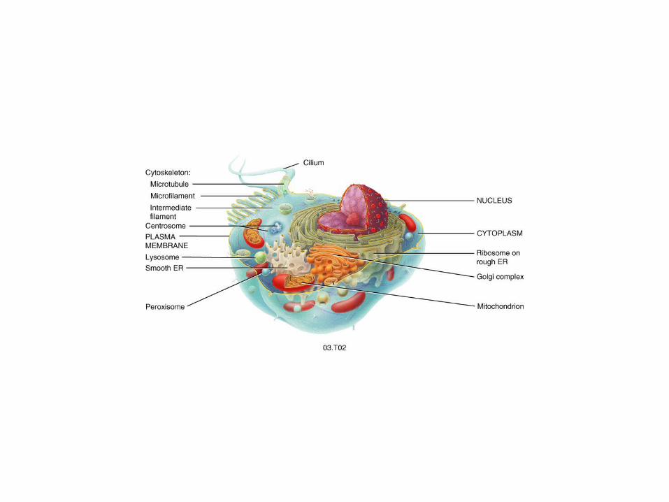
THE CELL
V.S.RAVIKIRAN, MSc.
V.S.RAVIKIRAN, MSc., Department of Biochemistry,
ASRAM Medical college, Eluru-534005.AP, [email protected]
om
V.S.RAVIKIRAN
CELLS
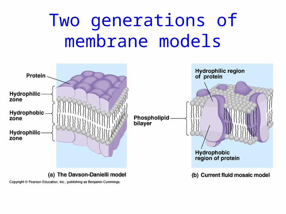
Two generations of membrane models
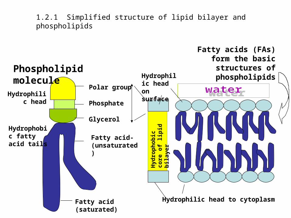
Fatty acids (FAs) form the basic structures of
phospholipids
1.2.1 Simplified structure of lipid bilayer and phospholipids
Polar group
PhosphateGlycerol
Fatty acid-(unsaturated)
Phospholipid molecule
Fatty acid (saturated)
Hydrophilic head
Hydrophobic fatty acid tails
Hyd
rop
hob
ic
core
of
lip
id
bilayer
Hydrophilic head to cytoplasm
Hydrophilic head on surface
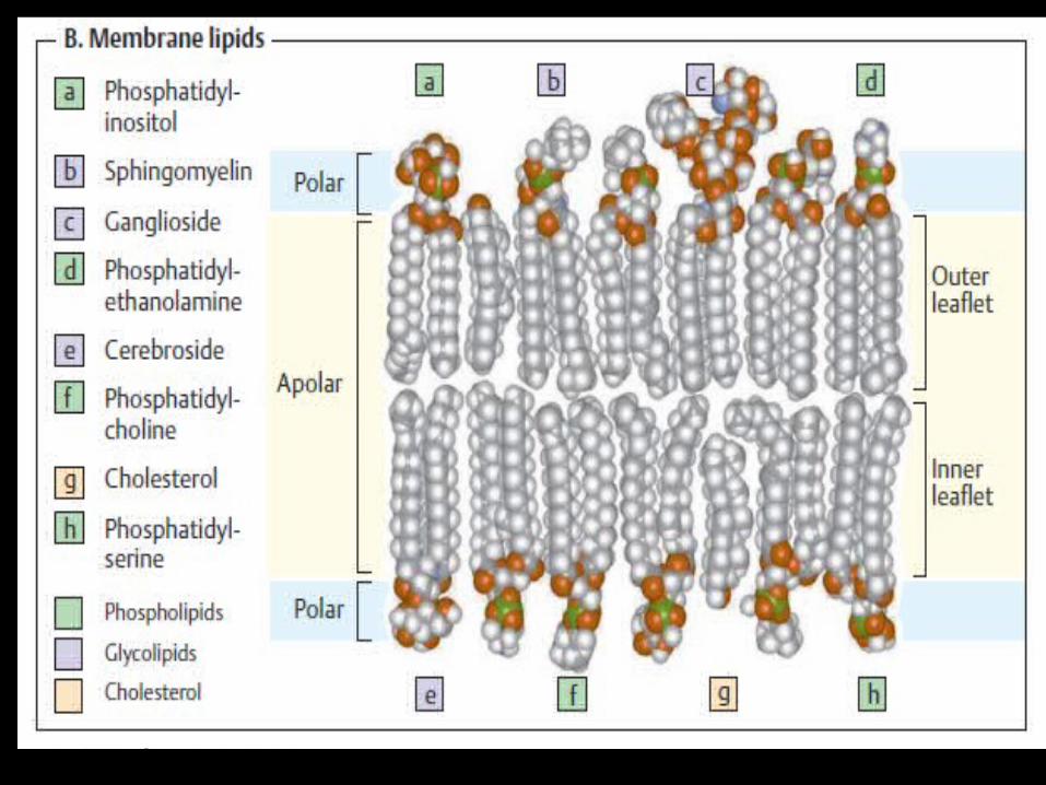
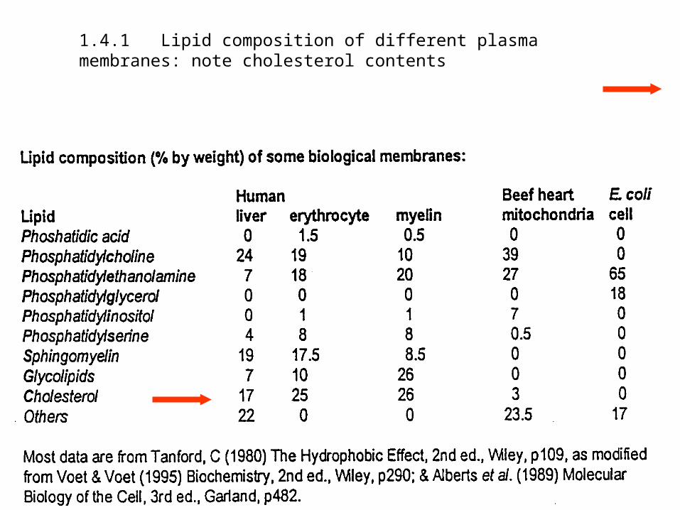
1.4.1 Lipid composition of different plasma membranes: note cholesterol contents
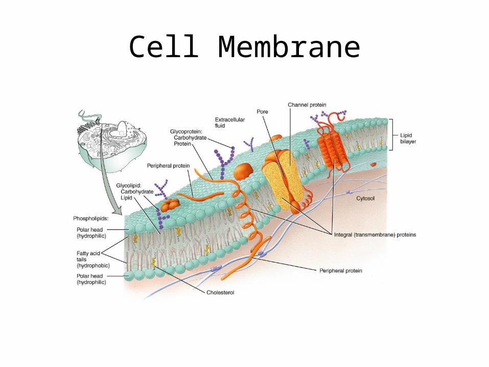
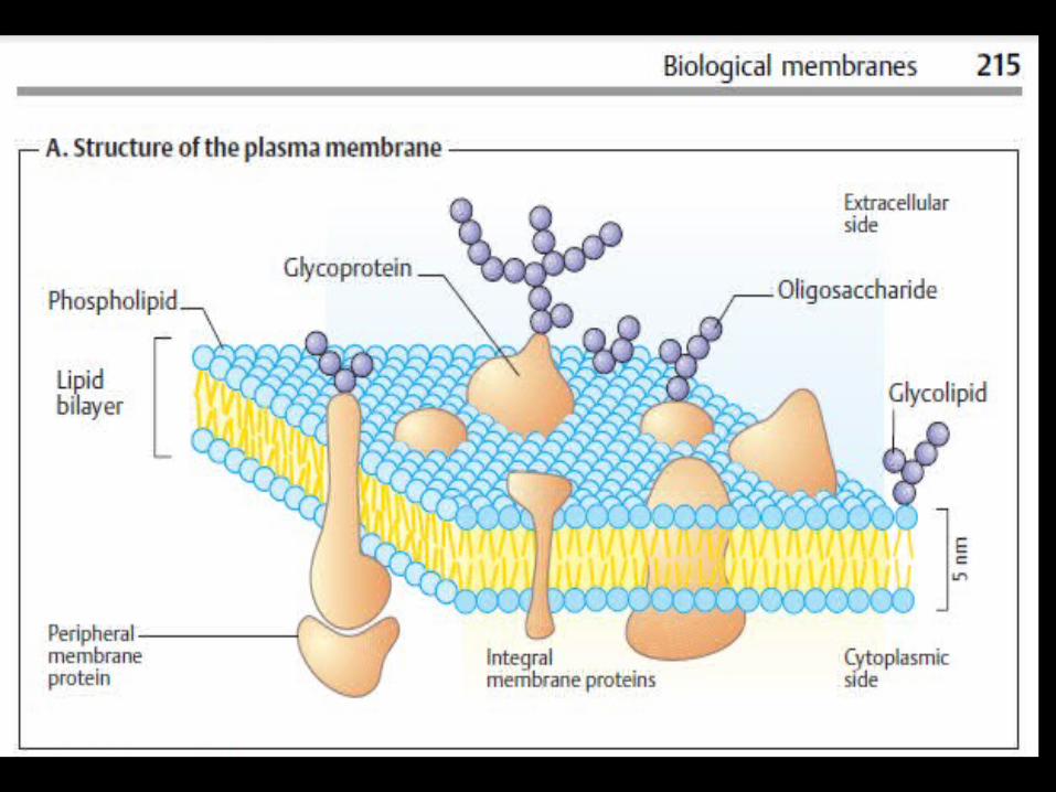
Cell Membrane
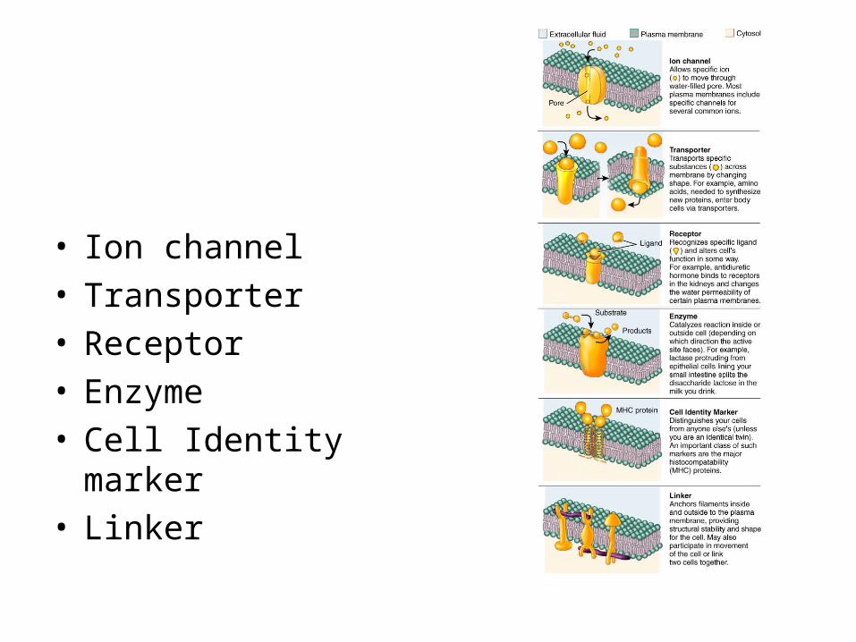
• Ion channel• Transporter• Receptor• Enzyme• Cell Identity marker• Linker
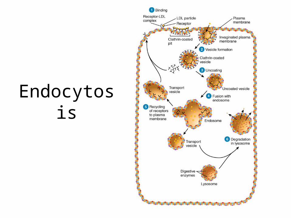
Endocytosis
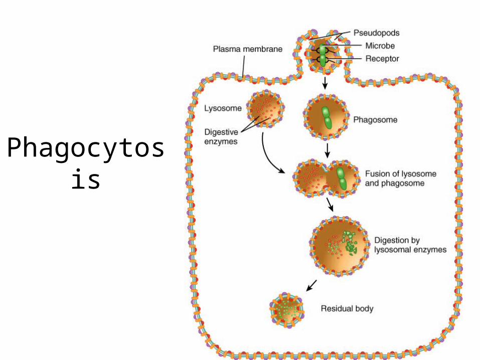
Phagocytosis
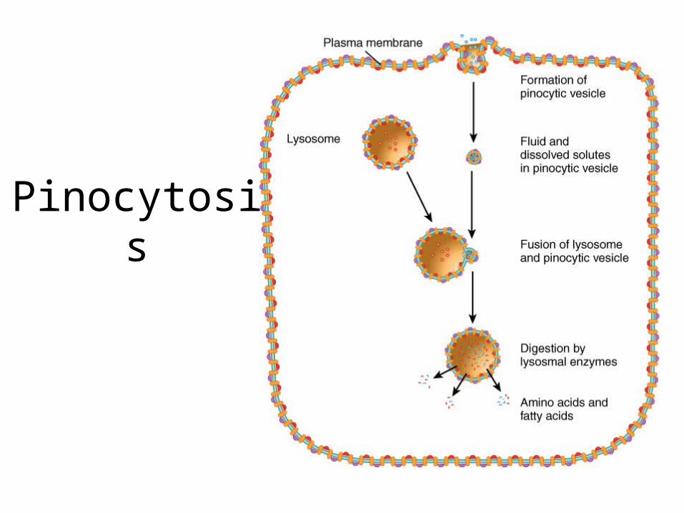
Pinocytosis
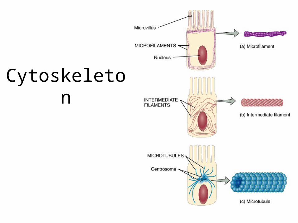
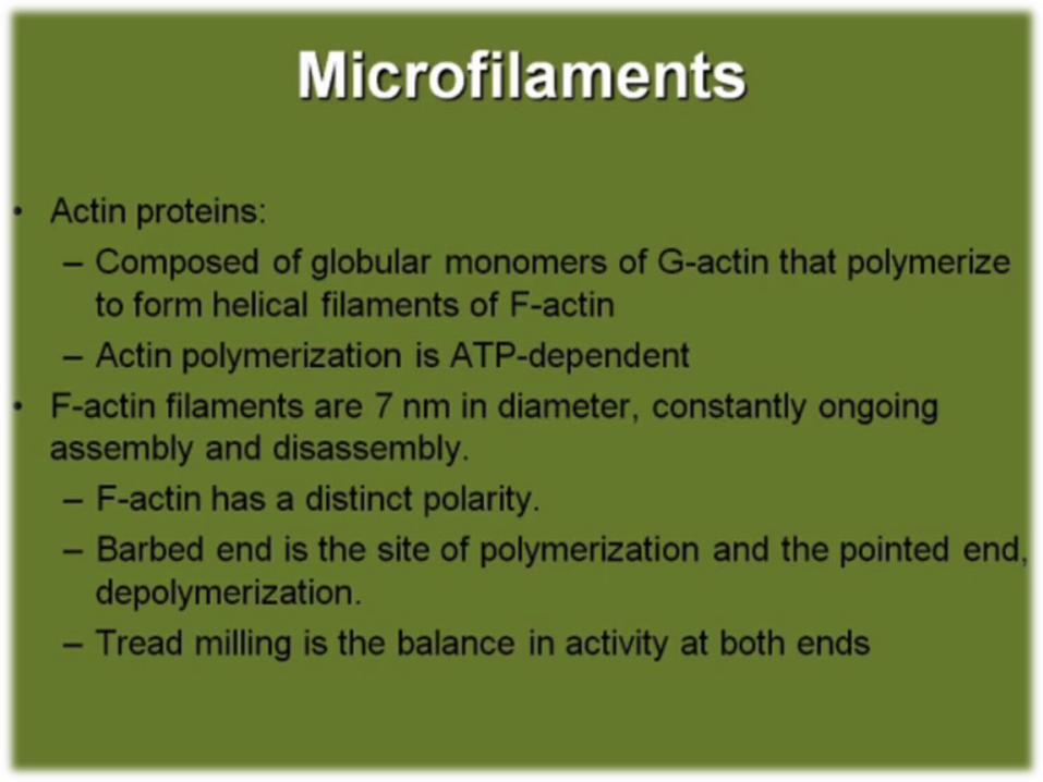
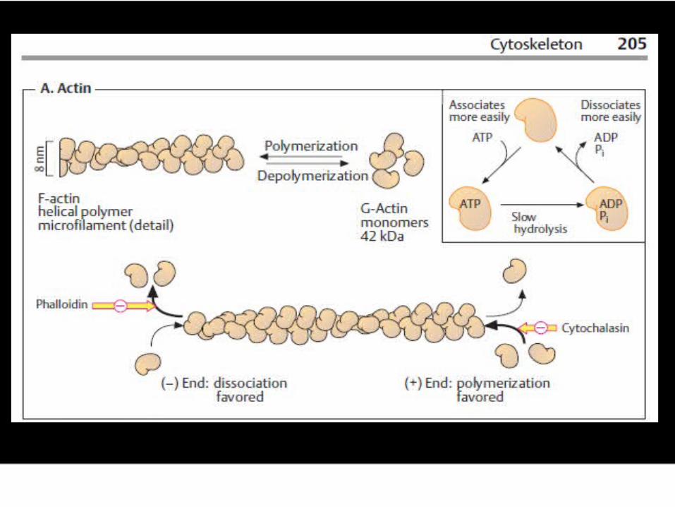
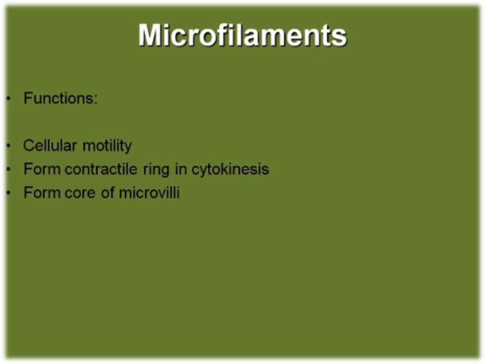
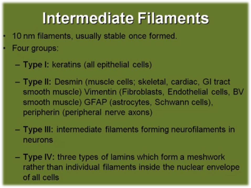
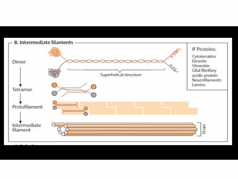
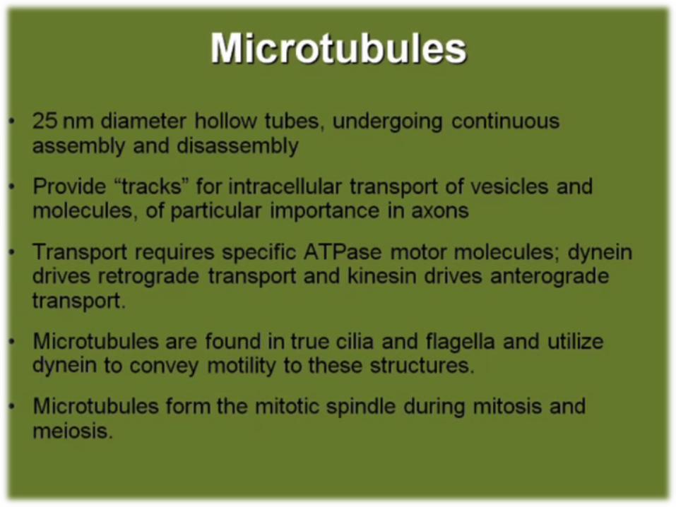
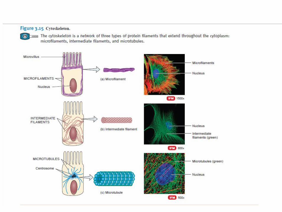
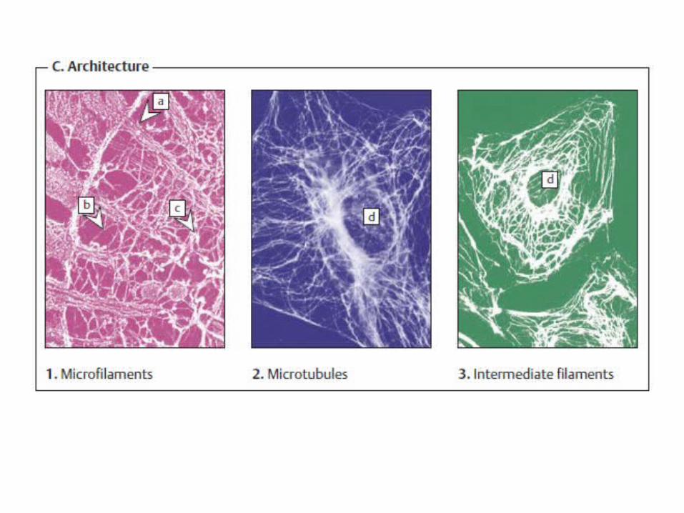
Cytoskeleton
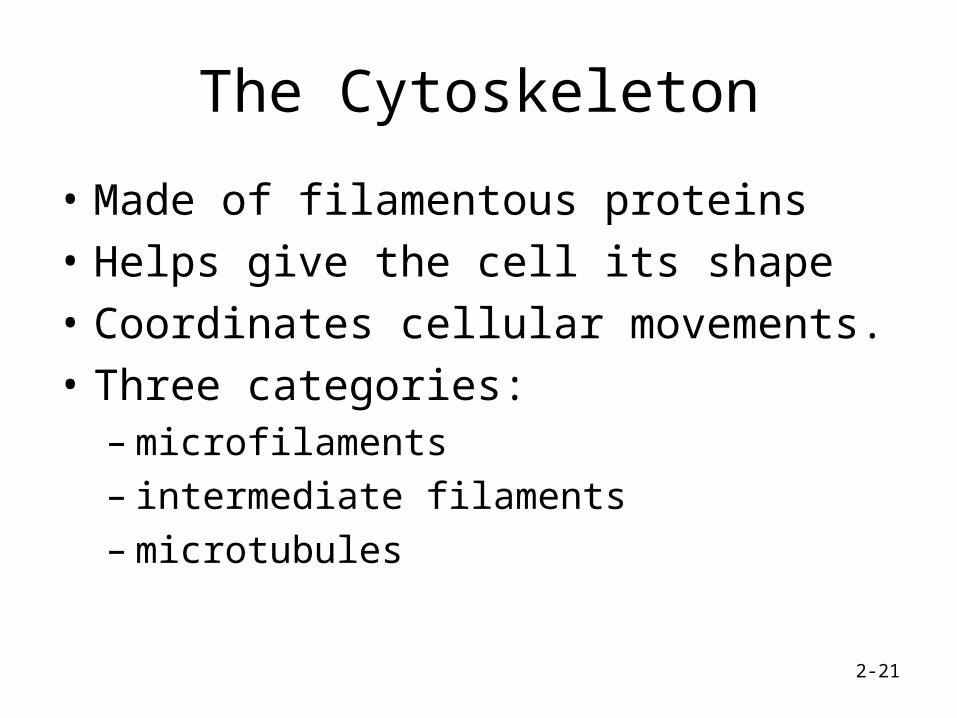
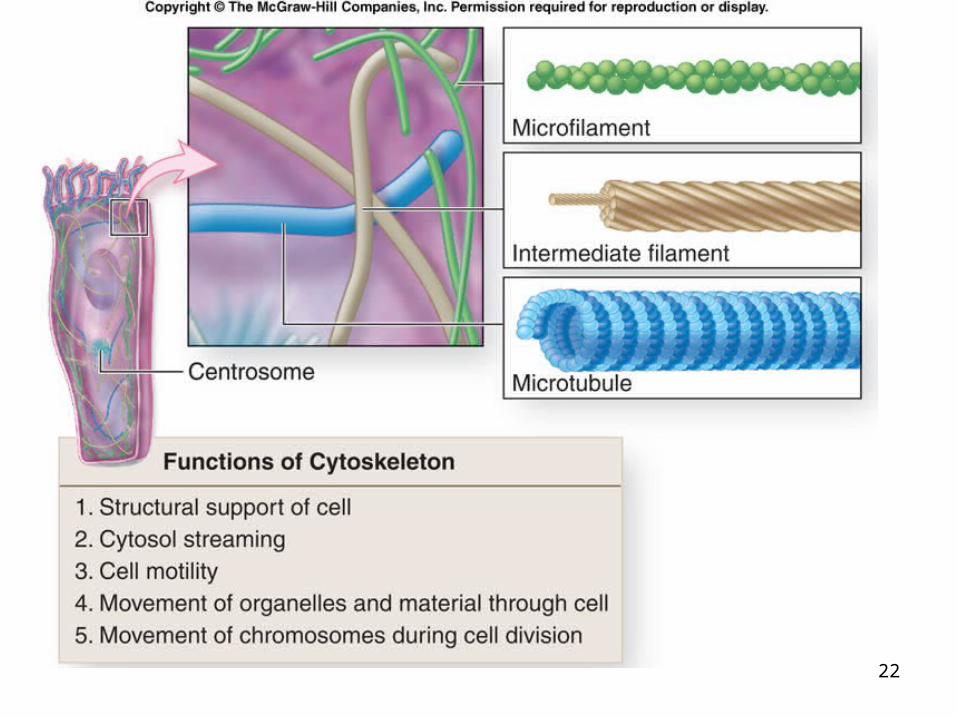
The Cytoskeleton
• Made of filamentous proteins
• Helps give the cell its shape
• Coordinates cellular movements.
• Three categories:– microfilaments– intermediate filaments – microtubules
2-21
22
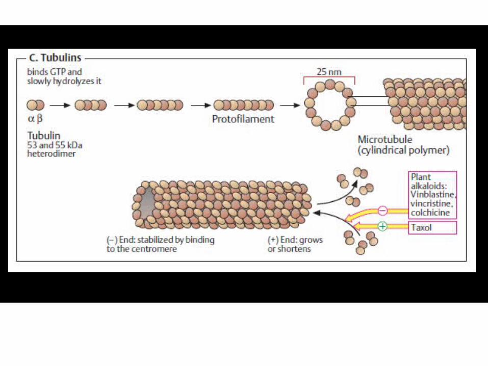
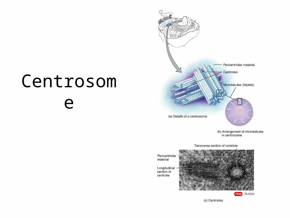
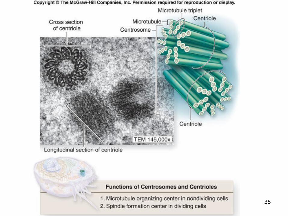
Centrosome
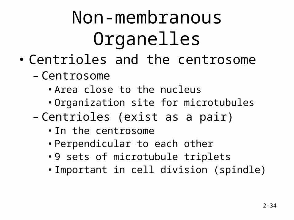
Non-membranous Organelles
• Centrioles and the centrosome– Centrosome
• Area close to the nucleus• Organization site for microtubules
– Centrioles (exist as a pair)• In the centrosome• Perpendicular to each other• 9 sets of microtubule triplets• Important in cell division (spindle)
2-34
35
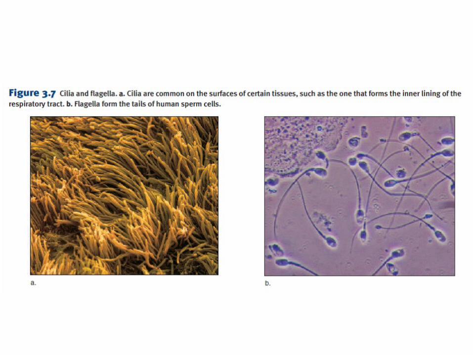
Microvilli, Cilia and Flagella
• Appendages extending from the surface of some cells. – Microvilli:
• short, cytoplasmic extensions• For absorption
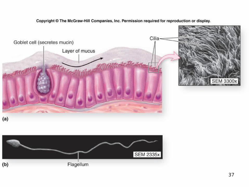
– Cilia:• usually occur in large numbers• work together to move materials or fluids along the surface of a cell.
– Flagella:• longer than cilia, and usually occur as single appendages.• Move the cell
2-36
37
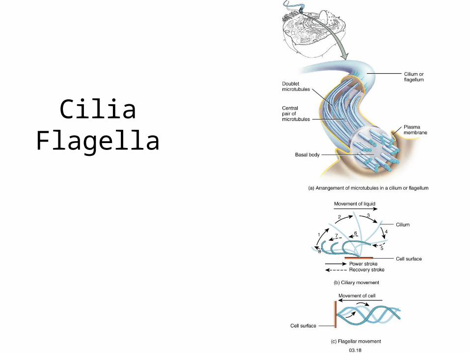
CiliaFlagella
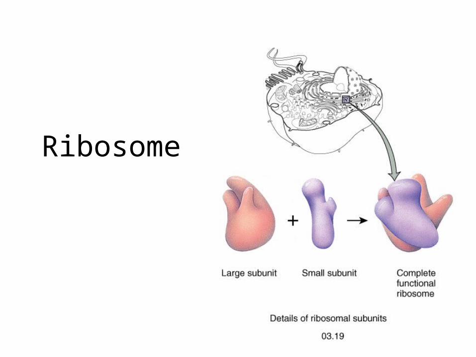
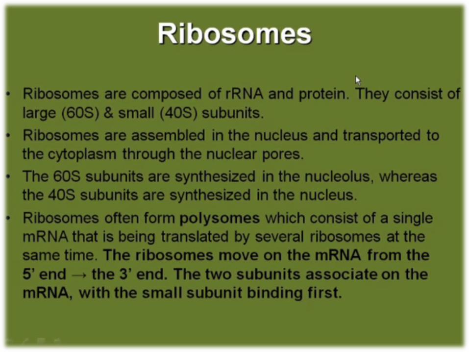
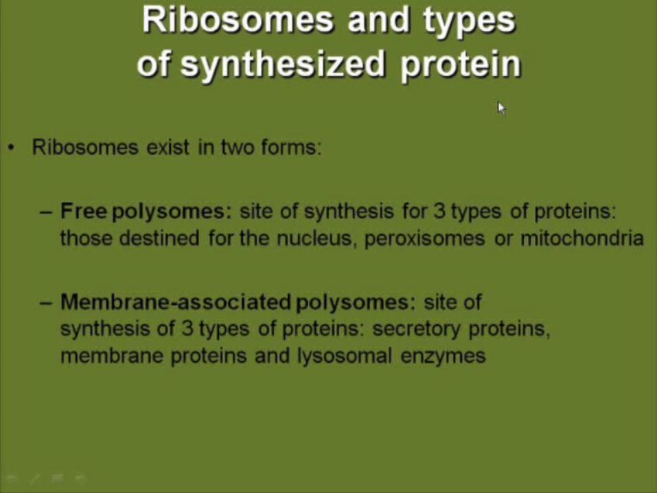
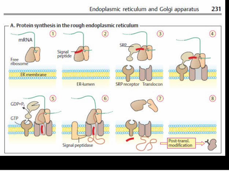
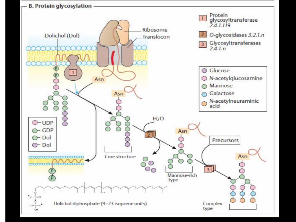
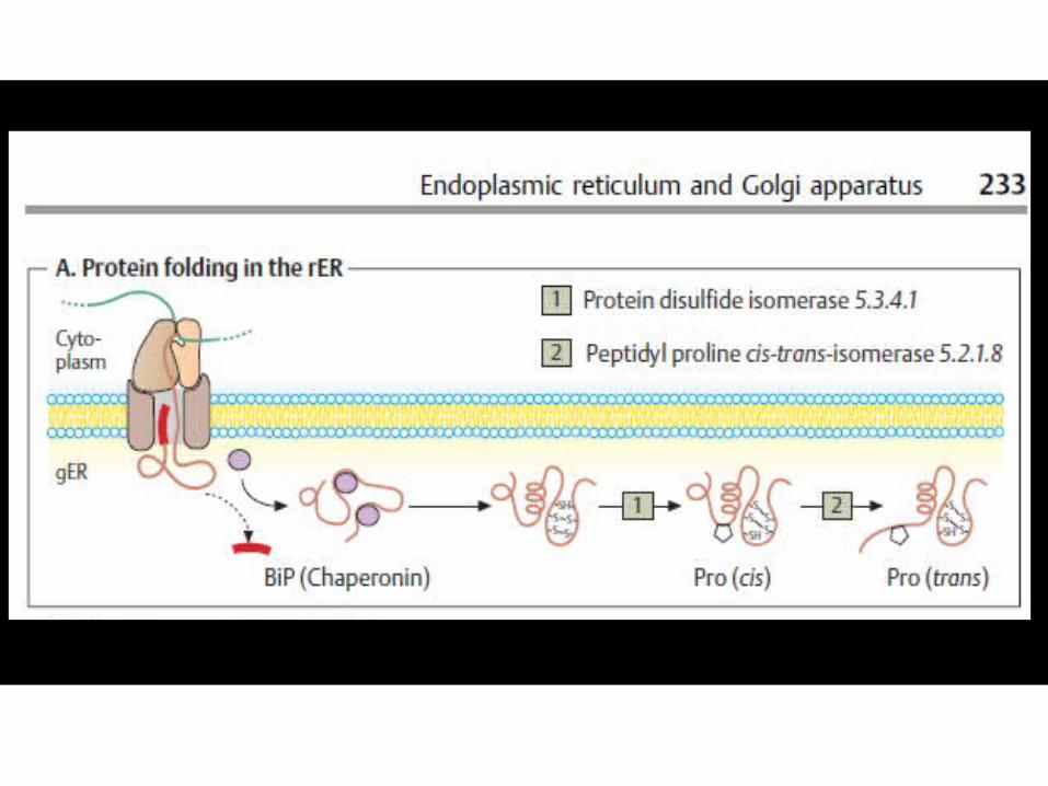
Ribosome
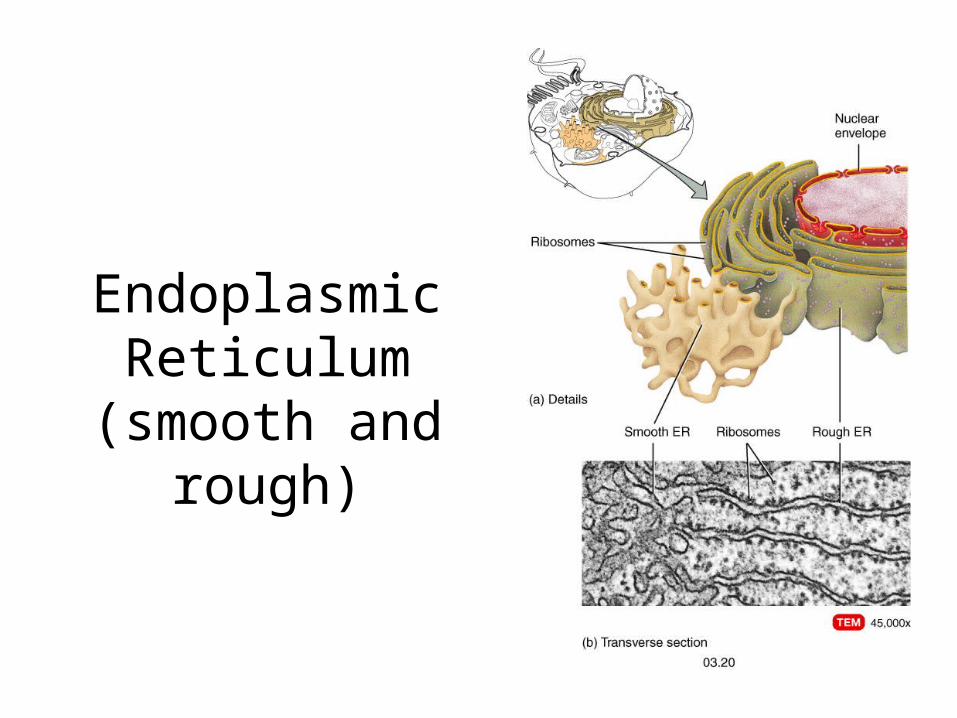
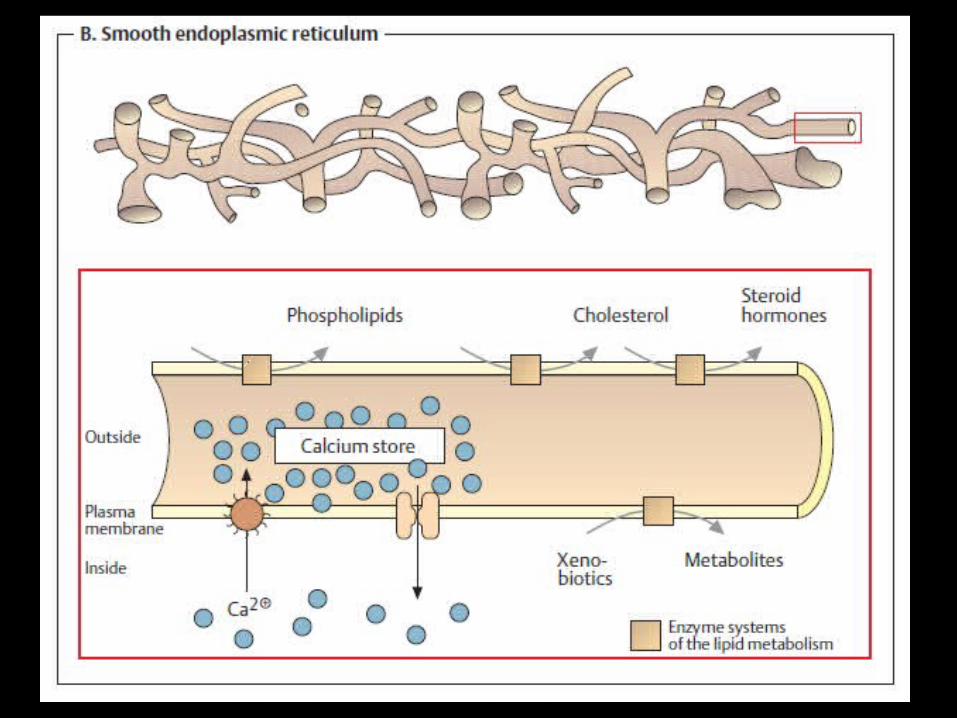
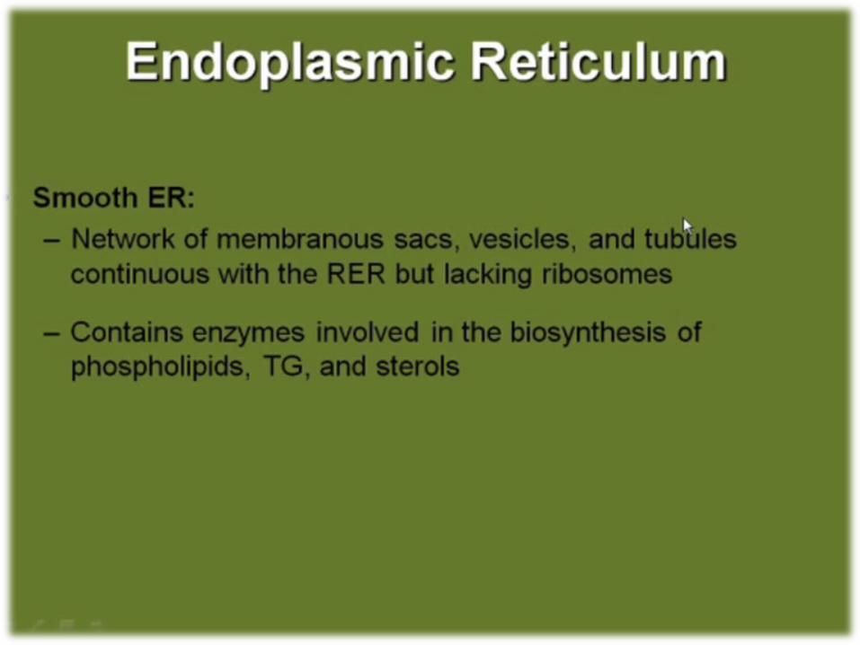
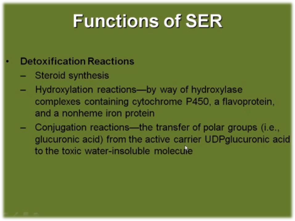
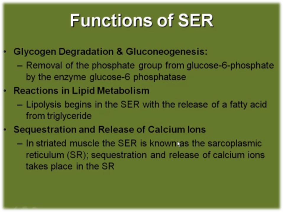
EndoplasmicReticulum
(smooth and rough)
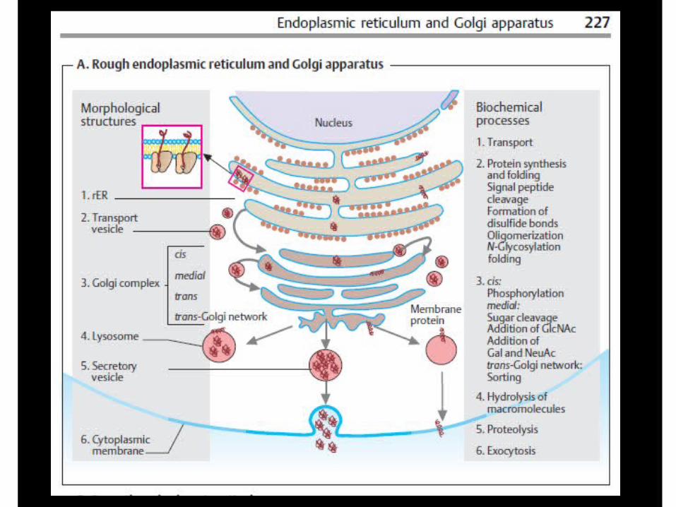
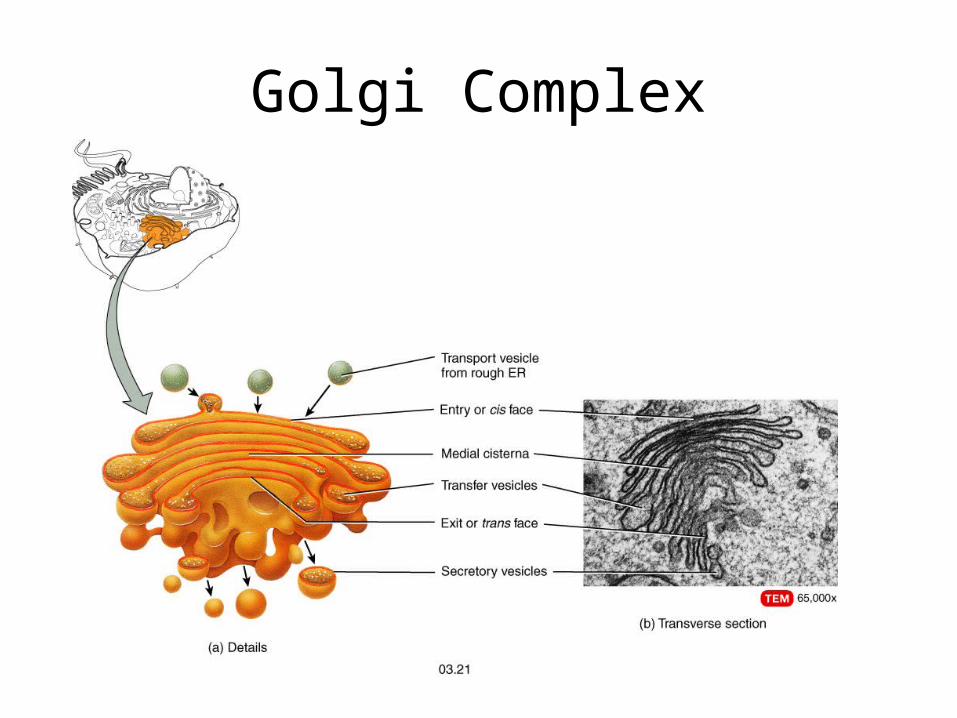
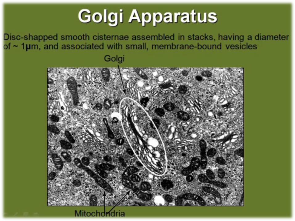
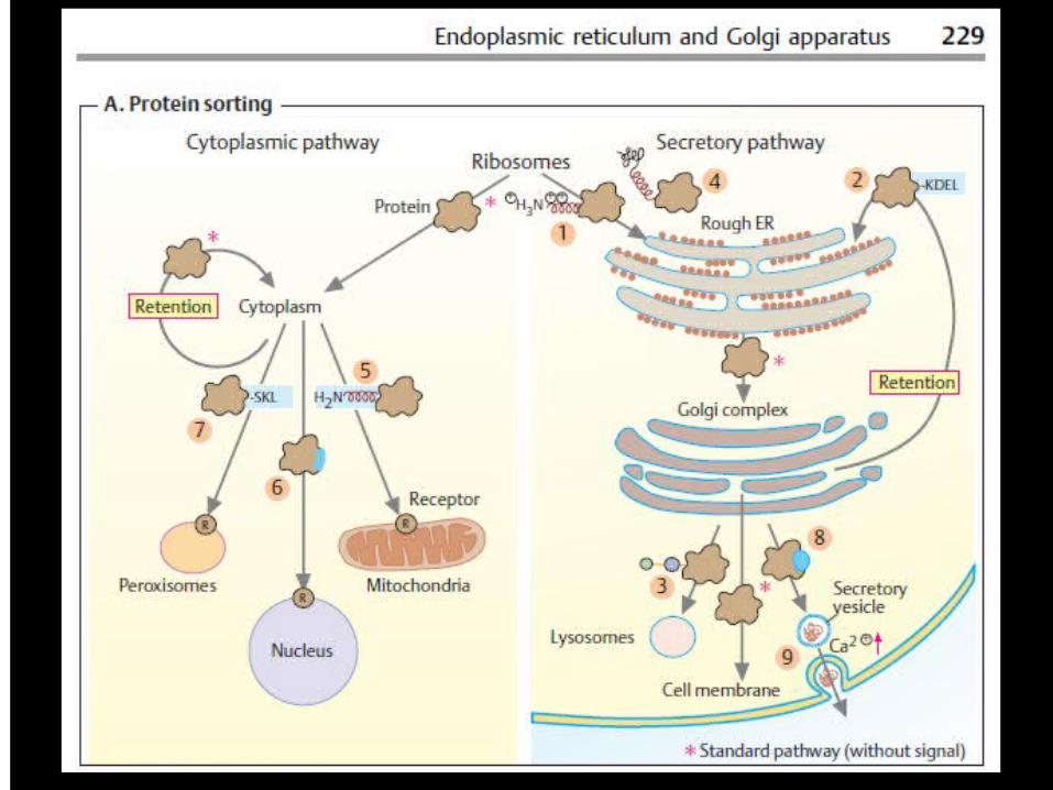
Golgi Complex
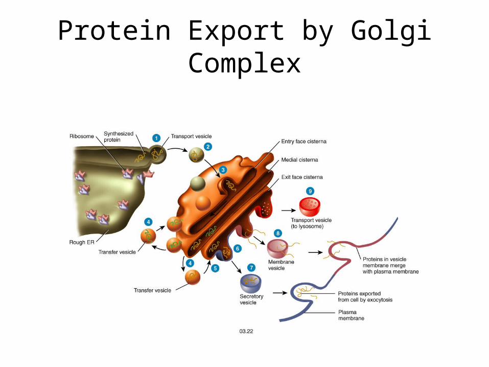
Protein Export by Golgi Complex
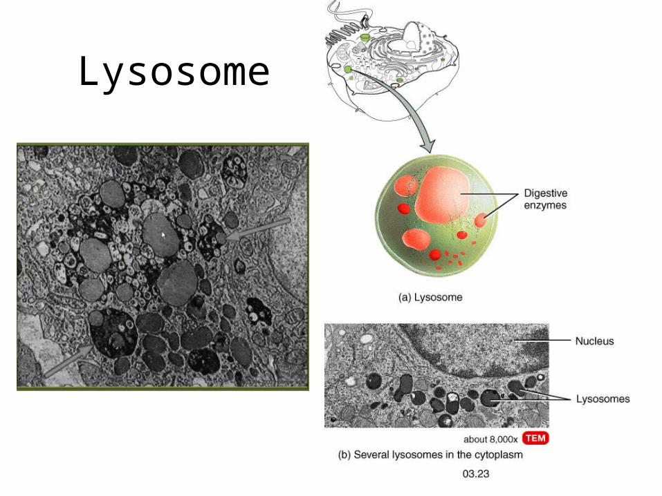
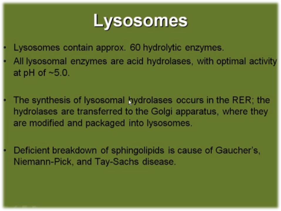
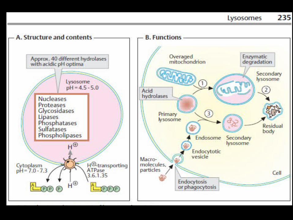
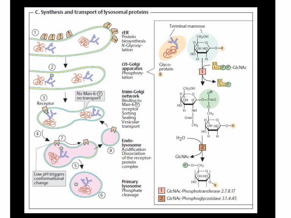
Lysosome
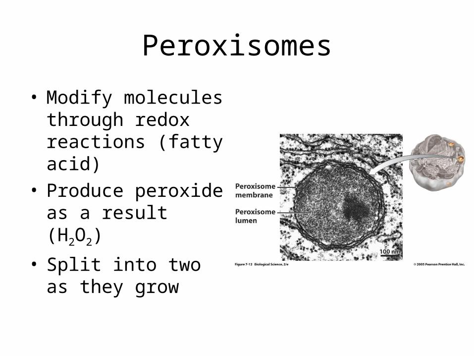
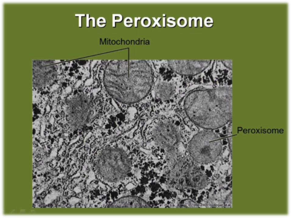
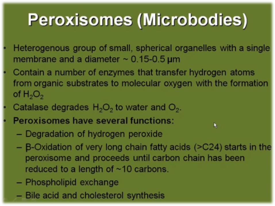
Peroxisomes
• Modify molecules through redox reactions (fatty acid)
• Produce peroxide as a result (H2O2)
• Split into two as they grow
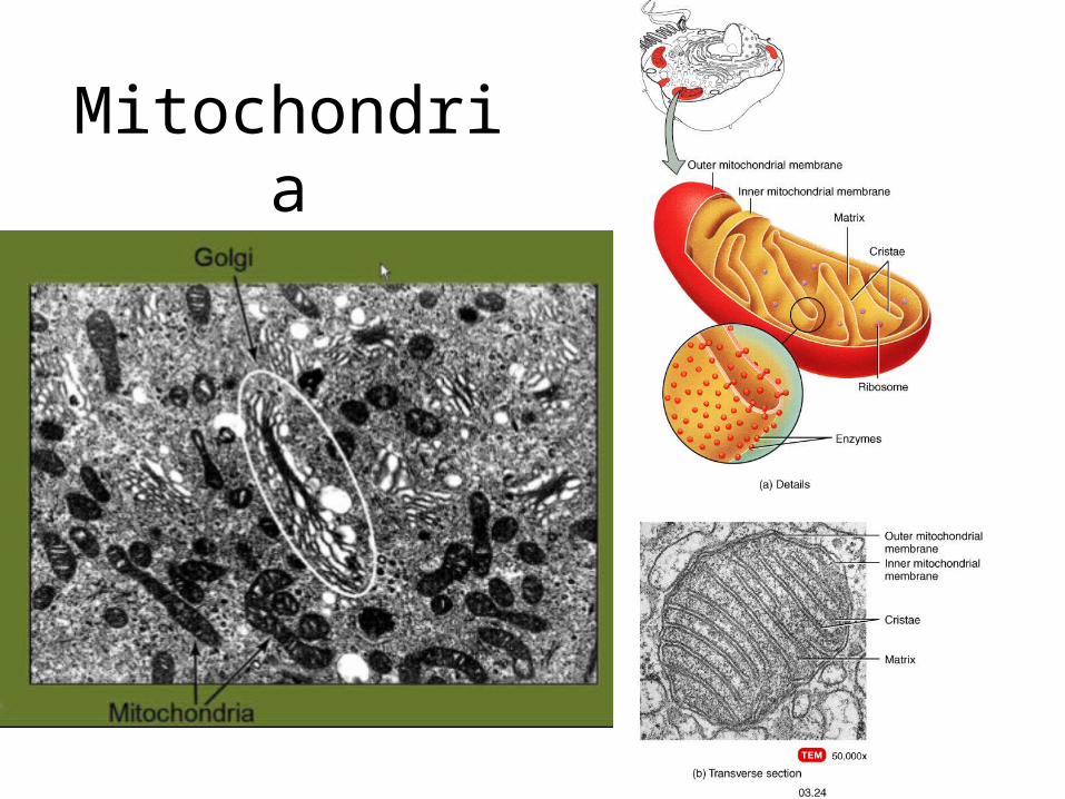
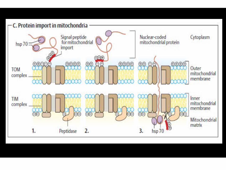
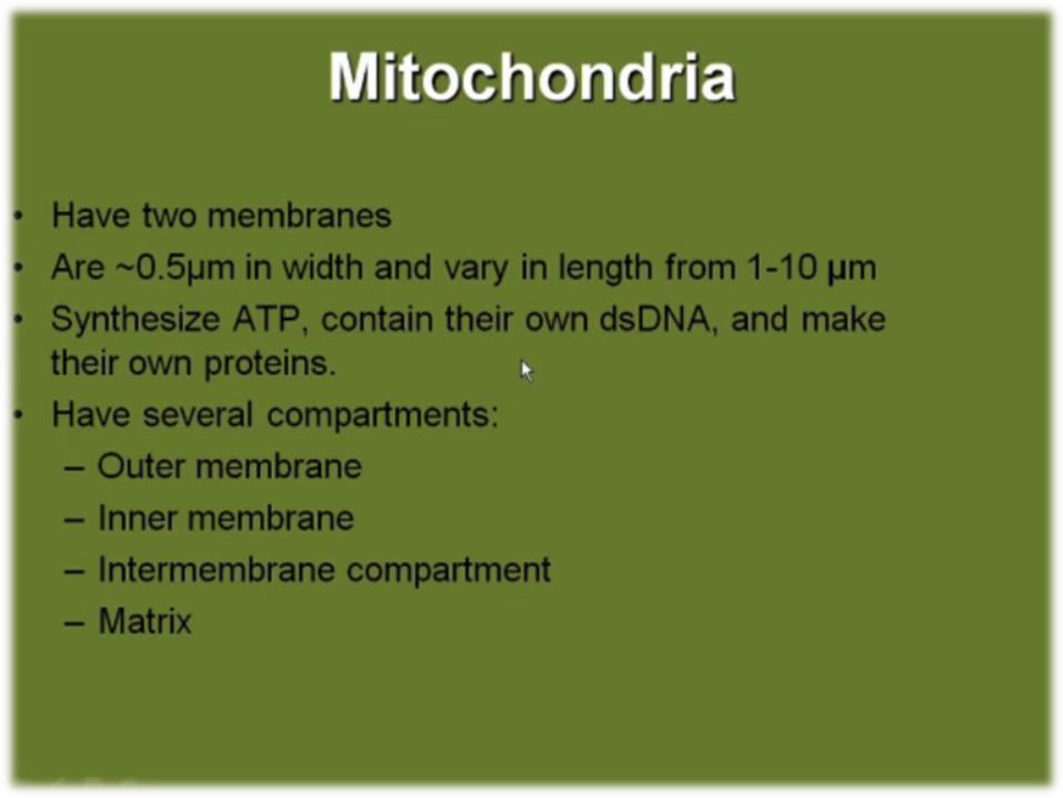
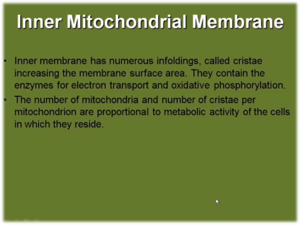
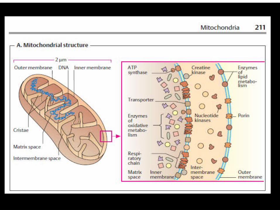
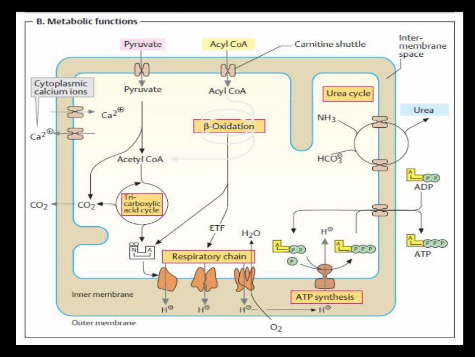
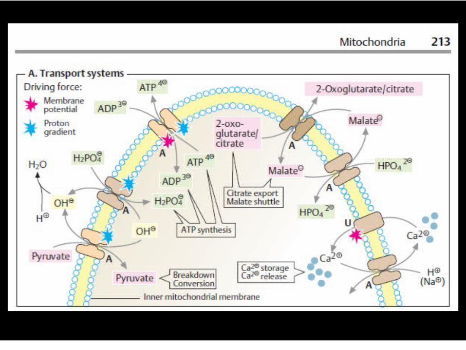
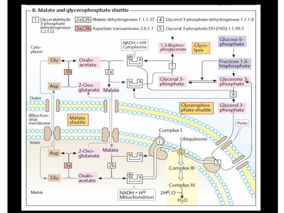
Mitochondria
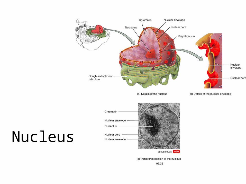
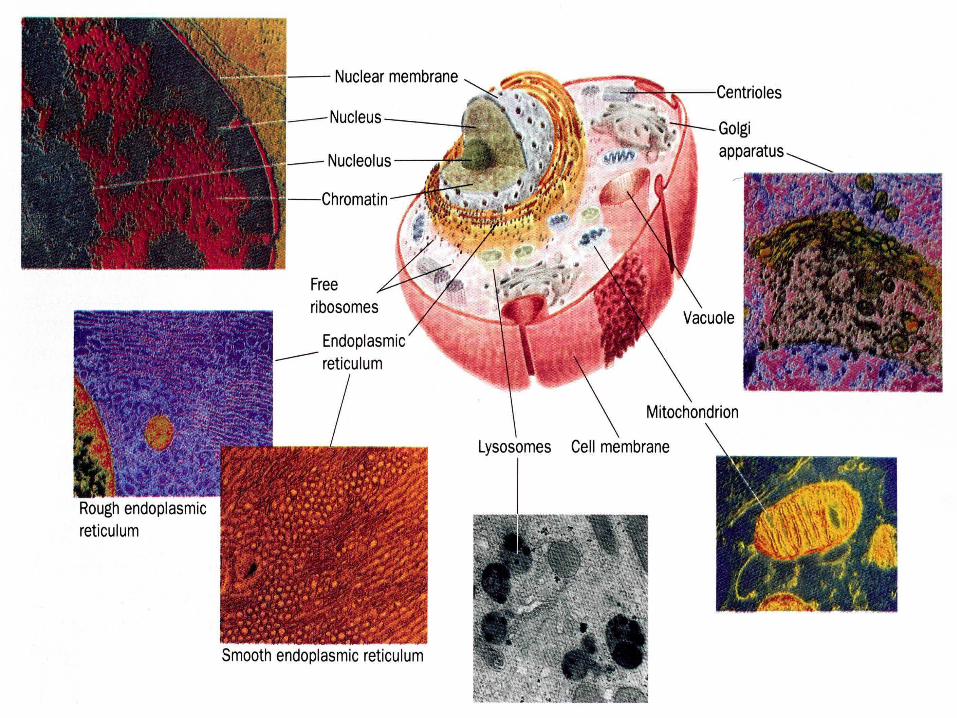
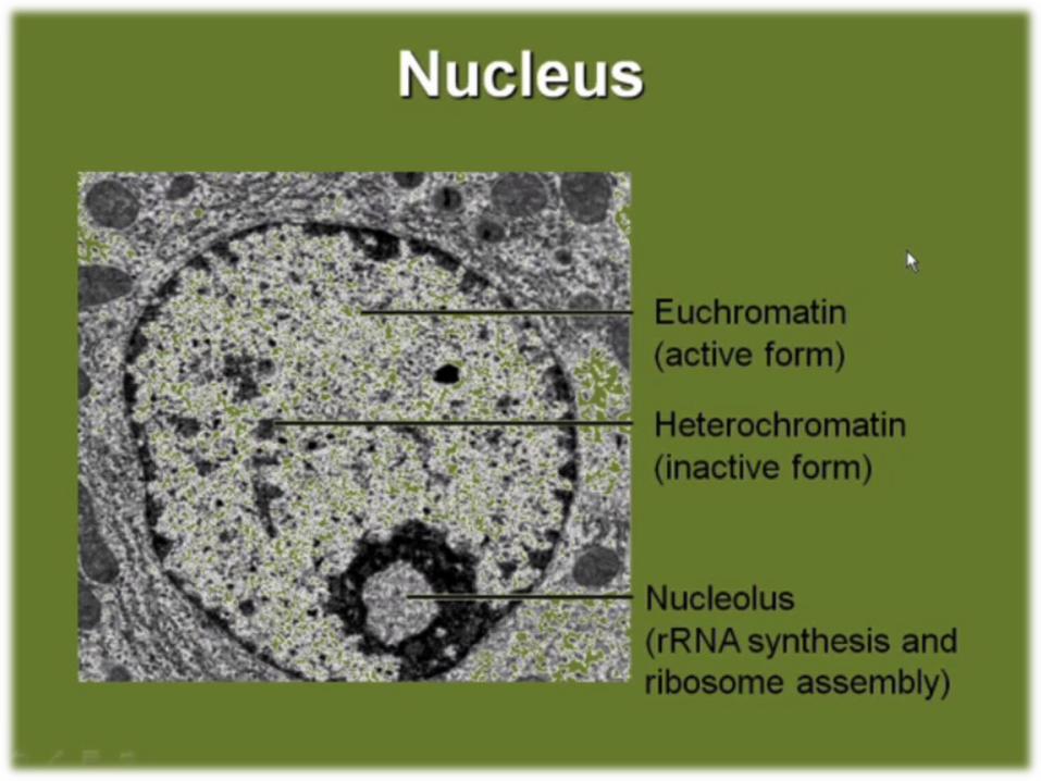
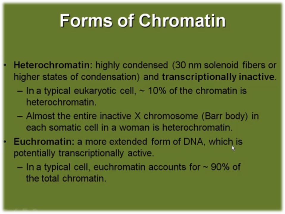
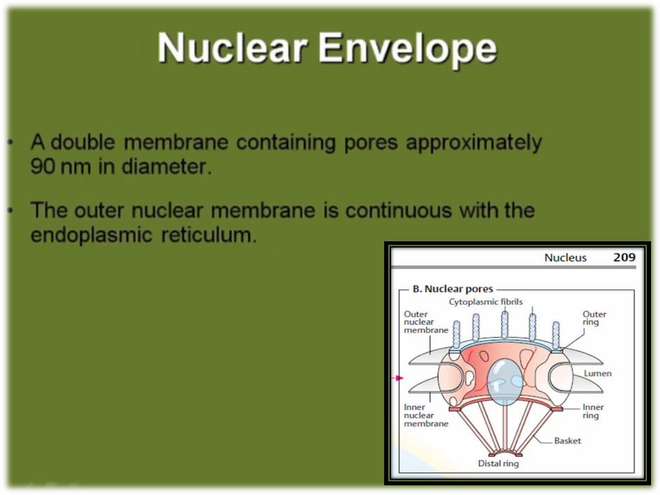
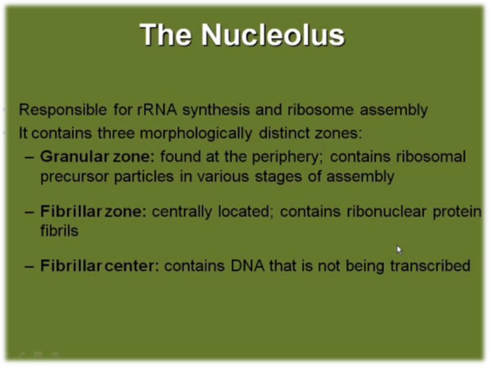
Nucleus
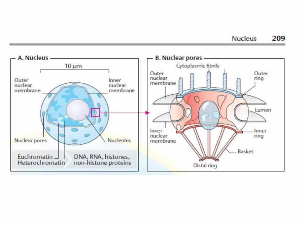
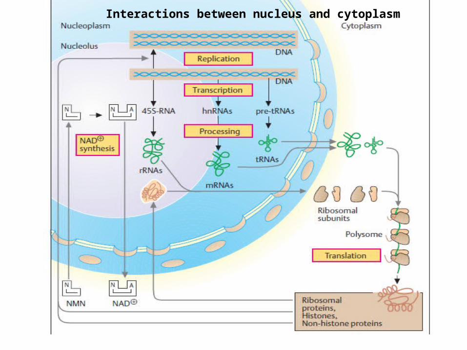
Interactions between nucleus and cytoplasm

86 Copyright Cmassengale
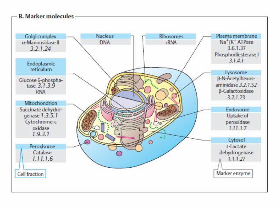
Isolating Organelles by Cell Fractionation
• Cell fractionation– Takes cells apart and separates the major
organelles from one another
• The centrifuge– Is used to fractionate cells into their component
parts
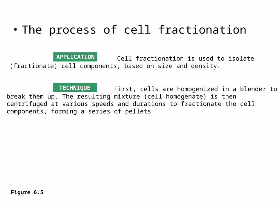
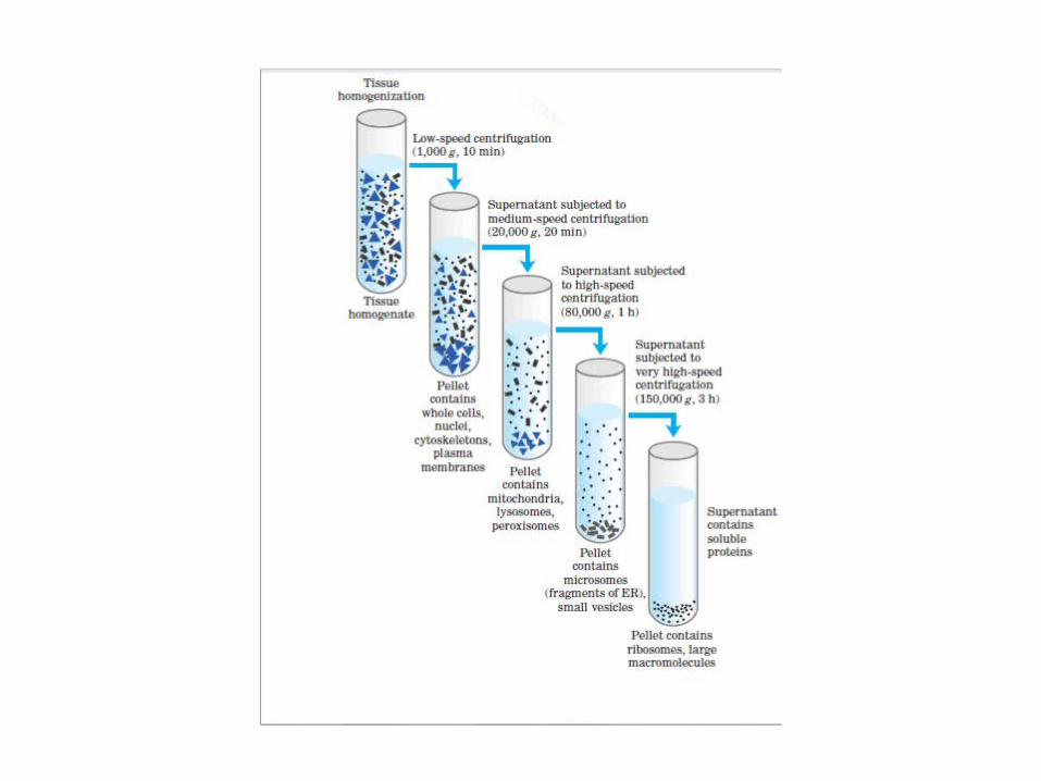
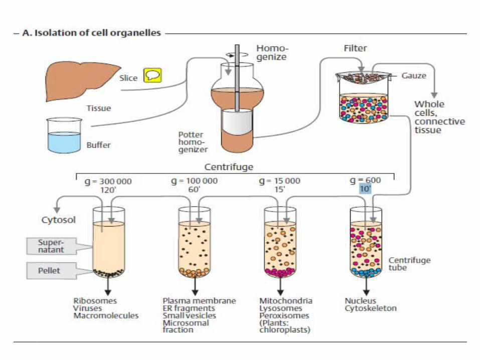
Cell fractionation is used to isolate(fractionate) cell components, based on size and density.
First, cells are homogenized in a blender tobreak them up. The resulting mixture (cell homogenate) is thencentrifuged at various speeds and durations to fractionate the cellcomponents, forming a series of pellets.
• The process of cell fractionation
APPLICATION
TECHNIQUE
Figure 6.5
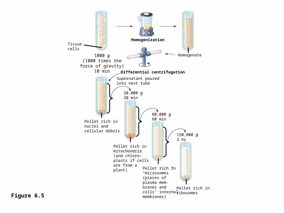
Tissuecells
Homogenization
Homogenate1000 g(1000 times theforce of gravity)
10 min Differential centrifugation
Supernatant pouredinto next tube
20,000 g20 min
Pellet rich innuclei andcellular debris
Pellet rich inmitochondria(and chloro-plasts if cellsare from a plant) Pellet rich in
“microsomes”(pieces of plasma mem-branes andcells’ internalmembranes)
Pellet rich inribosomes
150,000 g3 hr
80,000 g60 min
Figure 6.5
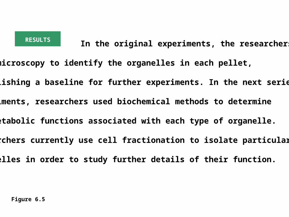
In the original experiments, the researchers
used microscopy to identify the organelles in each pellet,
establishing a baseline for further experiments. In the next series of
experiments, researchers used biochemical methods to determine
the metabolic functions associated with each type of organelle.
Researchers currently use cell fractionation to isolate particular
organelles in order to study further details of their function.
RESULTS
Figure 6.5
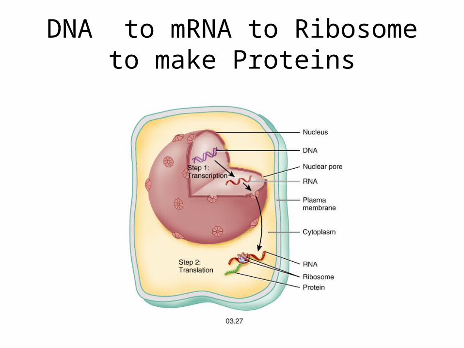
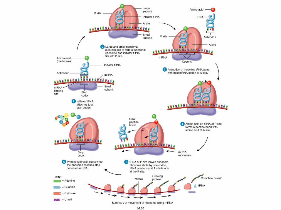
DNA to mRNA to Ribosometo make Proteins
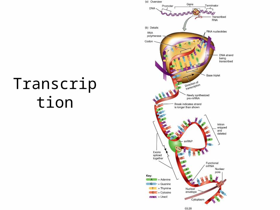
Transcription
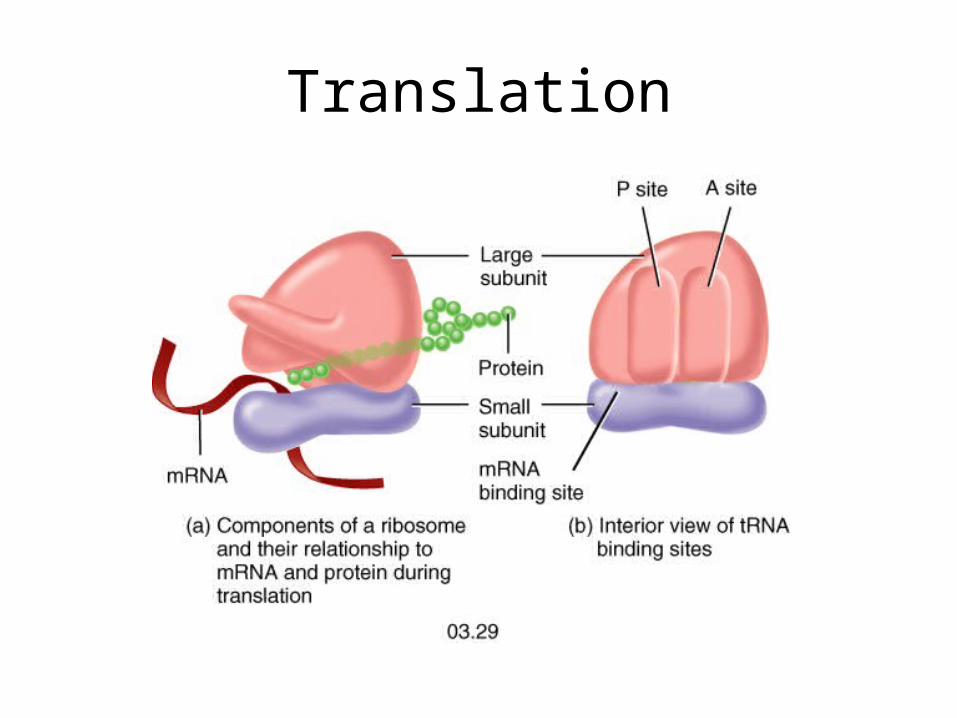
Translation
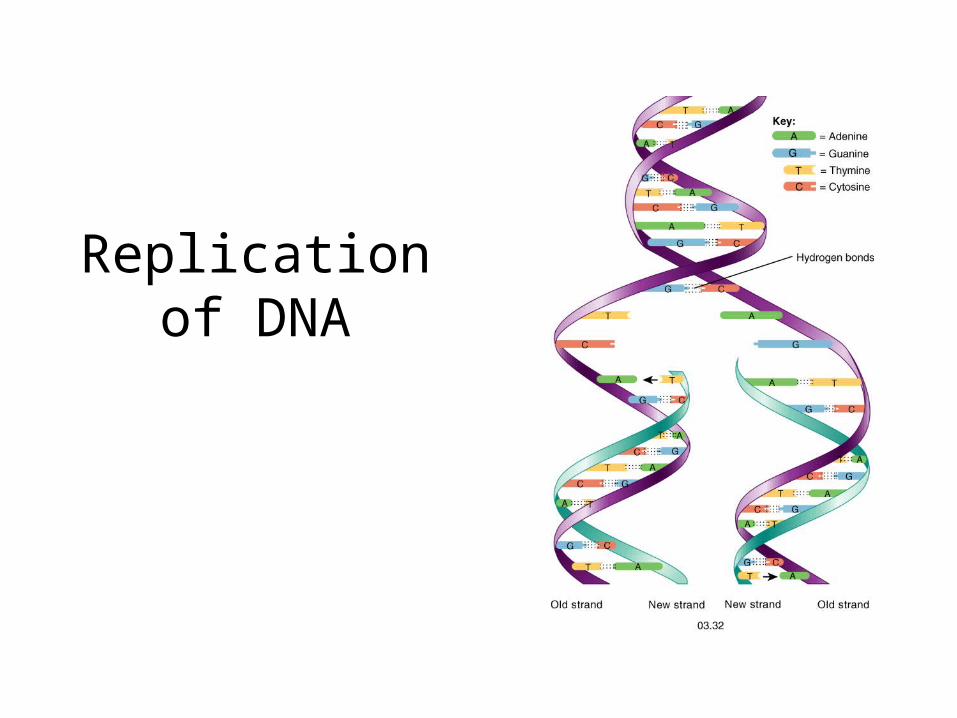
Replicationof DNA
Mitosis and Meiosis
Aging and Cells
Geriatrics
Telomeres
Free radicals
Cancer Terms
• Neoplasia• Benign• Malignant• Metastases• Carcinoma• Sarcoma• Angiogenesis• Mutation
Terms to Know• Anaplasia
• Atrophy
• Dysplasia
• Hyperplasia
• Hypertrophy
• Progeny
• Progeria

THANKS FOR YOUR ATTENTION