Embed Size (px)
Citation preview
TIRADS-ACR 2017
Bs. Đỗ Bình Minh.
Bs.Nguyễn Thị Kiều Trang.
Bs.Nguyễn Vũ Quỳnh Anh.
Medic 13/07/2017 1
Khoa Nội soi –Siêu Âm Bệnh Viện Ung Bướu Tp HCM
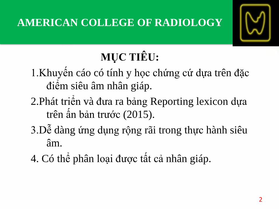
MỤC TIÊU:
1.Khuyến cáo có tính y học chứng cứ dựa trên đặc
điểm siêu âm nhân giáp.
2.Phát triển và đưa ra bảng Reporting lexicon dựa
trên ấn bản trước (2015).
3.Dễ dàng ứng dụng rộng rãi trong thực hành siêu
âm.
4. Có thể phân loại được tất cả nhân giáp.
2
AMERICAN COLLEGE OF RADIOLOGY
BI-RADS
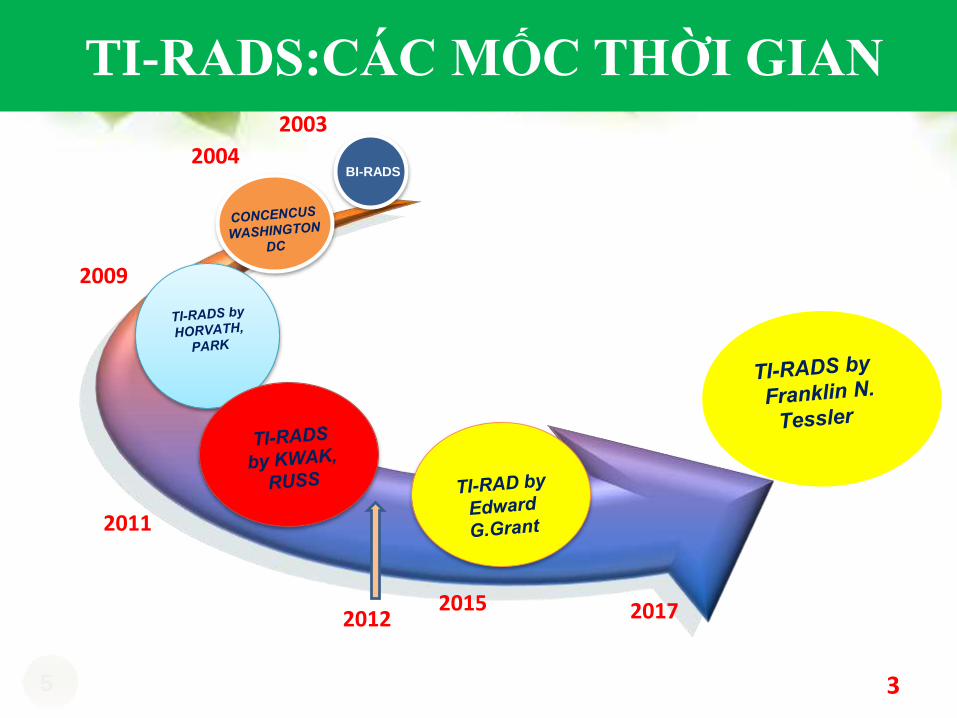
TI-RADS:CÁC MỐC THỜI GIAN
5
2003
2004
2009
2011
2015 2017
3
2012
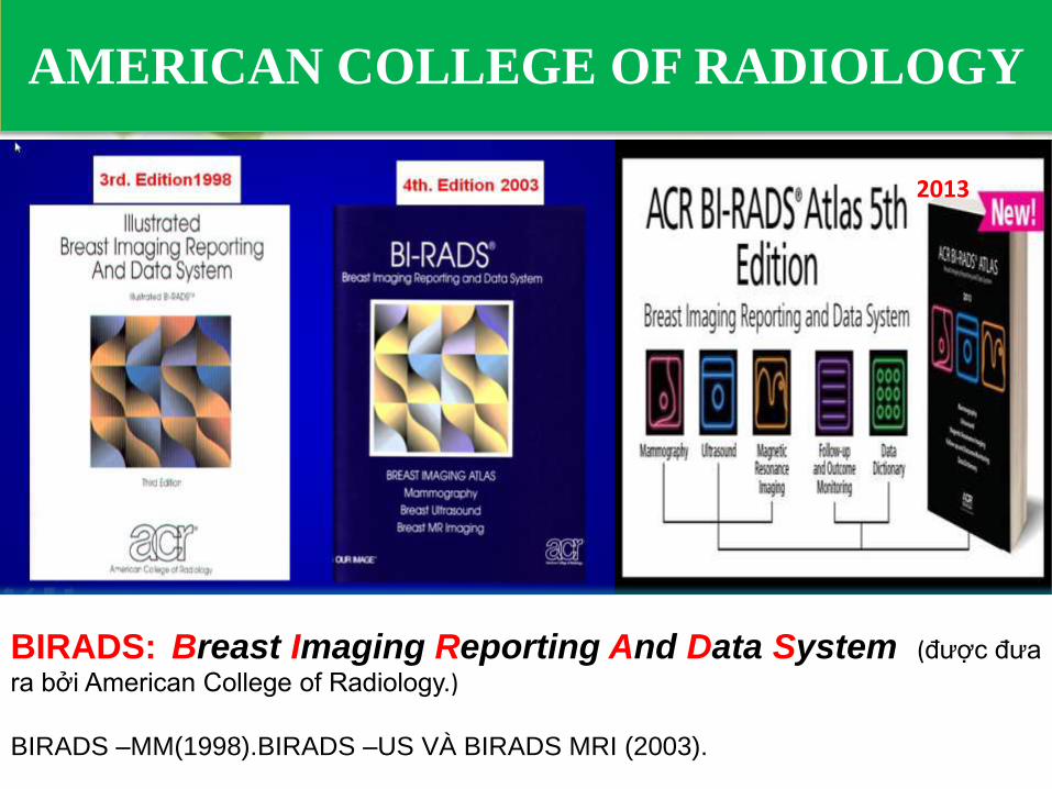
AMERICAN COLLEGE OF RADIOLOGY
4
BIRADS: Breast Imaging Reporting And Data System (được đưa
ra bởi American College of Radiology.)
BIRADS –MM(1998).BIRADS –US VÀ BIRADS MRI (2003).
2013
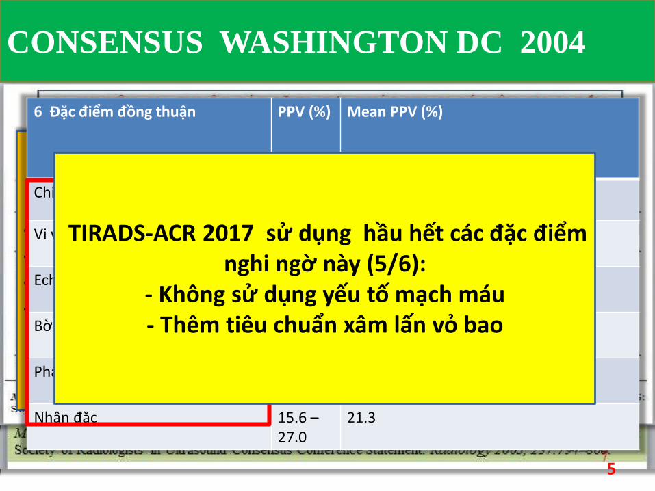
• Consensus WASHINGTON DC năm 2004• 21 chuyên gia tuyến giáp• 14 reports• 6 triệu chứng ác tính đồng thuận:
6 Đặc điểm đồng thuận PPV (%) Mean PPV (%)
Chiều cao > rộng 66.7 66.7
Vi vôi hóa 24.3 –70.7
47.5
Echo kém 11.4 –68.4
39.9
Bờ không đều hoặc halo (-) 9.3 –60.0
34.65
Phân bố mạch máu trong nhân 24 –41.9
32.95
Nhân đặc 15.6 –27.0
21.3
5
TIRADS-ACR 2017 sử dụng hầu hết các đặc điểmnghi ngờ này (5/6):
- Không sử dụng yếu tố mạch máu- Thêm tiêu chuẩn xâm lấn vỏ bao
CONSENSUS WASHINGTON DC 2004
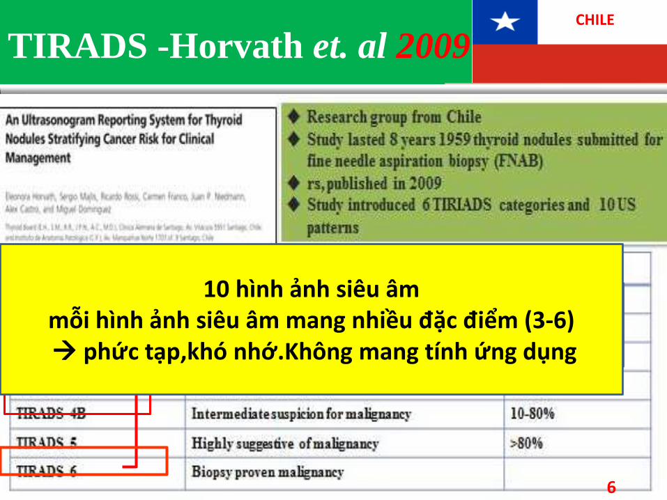
TIRADS -Horvath et. al 2009CHILE
Research group from Chile Study lasted 8 years 1959 thyroid nodules
submitted for fine needle aspiration biopsy (FNAB)
rs, published in 2009 Study introduced 6 TIRIADS categories and 10
US patterns
1-
2-
3-
4-
5-
7-
6-
Giống BIRADS: • 6 nhóm• Nhóm 6 = K đã biết Không có 4C
10 hình ảnh siêu âmmỗi hình ảnh siêu âm mang nhiều đặc điểm (3-6) phức tạp,khó nhớ.Không mang tính ứng dụng
6
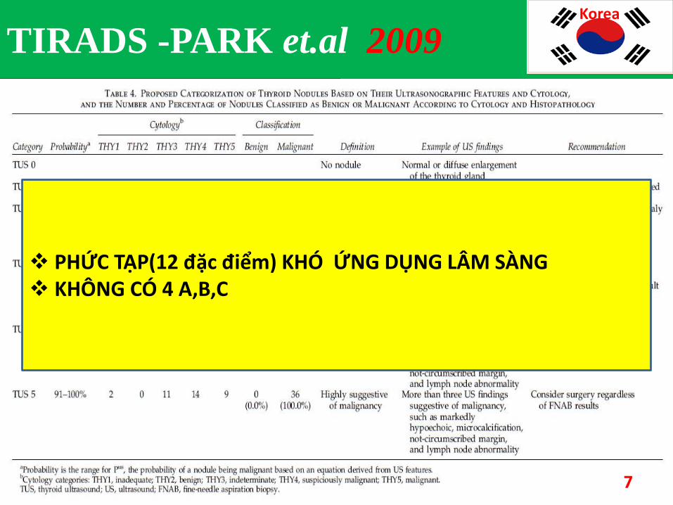
Nghiên cứu 5 năm (T7/2001 -T12/2006)Nghiên cứu tiền cứu kết quả FNA của 1694 BNĐưa ra 5 nhóm TUS và 12 đặc điểm siêu âm
Trong đó:e: la hăng sô = 2.71828
X: la biến số độc lập.
PHỨC TẠP(12 đặc điểm) KHÓ ỨNG DỤNG LÂM SÀNG KHÔNG CÓ 4 A,B,C
7
TIRADS -PARK et.al 2009Korea
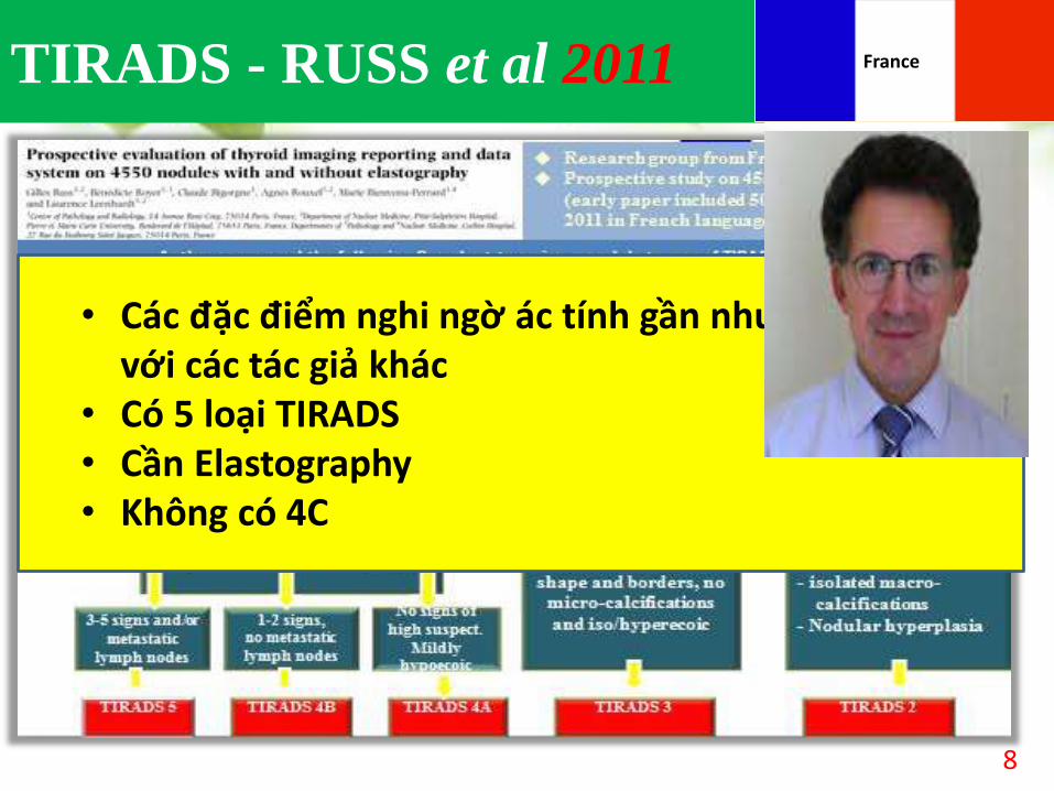
TIRADS - RUSS et al 2011
8
• Các đặc điểm nghi ngờ ác tính gần như tương đồngvới các tác giả khác
• Có 5 loại TIRADS• Cần Elastography• Không có 4C
France
Giống BIRADSTừ nhóm 4 trở lên K↑
9
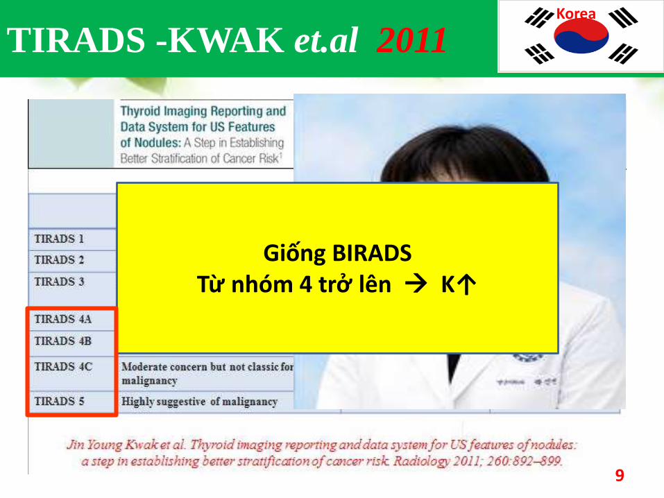
TIRADS -KWAK et.al 2011Korea
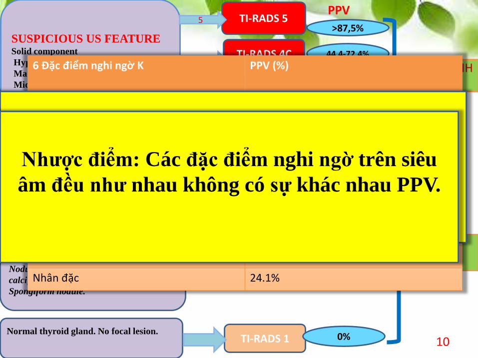
SUSPICIOUS US FEATURESolid component
Hypo-echogenicity
Marked hypoechogenicity
Microlobulated or irregular margins
Micro-calcifications
Taller-than-wide shape
No sign of high suspicious ( Probably
benign )
Simple thyroid cyst
Solid nodule with central cyst
Nodule with homogeneous peripheral
calcification
Spongiform nodule.
Normal thyroid gland. No focal lesion.
TI-RADS 5
TI-RADS 4C
TI-RADS 4B
TI-RADS 4A
TI-RADS 3
TI-RADS 2
TI-RADS 1
CÓ CHỈ ĐỊNH FNA
KHÔNG FNA
29,2%
3-4
1
5
44,4-72,4%
0%
3,3%
1,7%
0%
>87,5%
PPV
10
6 Đặc điểm nghi ngờ K PPV (%)
Bờ đa cung nhỏ hoặc bờ không đều 86%
Echo rất kém 77.8%
Chiều cao> rộng 71, 2 %
Vi vôi hóa 68,5%
Echo kém 25%
Nhân đặc 24.1%
Ưu điểm: Các đặc điểm nghi ngờ có PPV khác
nhau nhưng khi ứng dụng chỉ đếm từng đặc điểm
không cần nhớ những PPV này. Nhược điểm: Các đặc điểm nghi ngờ trên siêu
âm đều như nhau không có sự khác nhau PPV.
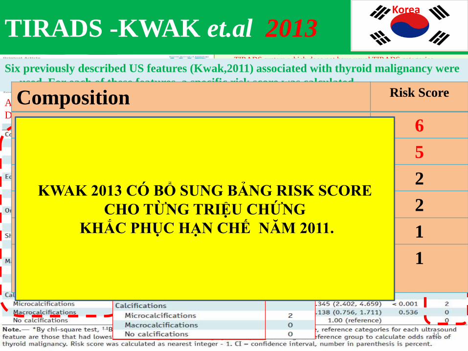
Proposed Image Reporting and Characterization System is a modified
TIRADS system which does not have usual TIRADS categories.
Based on the study of 2000 tumors from 20 different institutions (1796 patients, 1268 were benign and 732 were malignant) authors developed diagnostic prediction model by using ultrasound (US) features of thyroid
nodules to stratify the risk of malignancy.
Six previously described US features (Kwak,2011) associated with thyroid malignancy were
used. For each of these features a specific risk score was calculated.
Association Between Thyroid Malignancy and Various Sonographic Features at Thyroid Nodulesof Training
Data Set on Multiple Logistic Regression and Risk Score Analysis
Composition Risk Score
Marked hypoechogenicity 6
Spiculated (microlobulated) margins 5
Microcalcifications 2
Hypoechogenicity 2
Taller than wider (non-parallel orientation) 1
Ill-defined borders 1
KWAK 2013 CÓ BỔ SUNG BẢNG RISK SCORE
CHO TỪNG TRIỆU CHỨNG
KHẮC PHỤC HẠN CHẾ NĂM 2011.
TIRADS -KWAK et.al 2013Korea
11
12
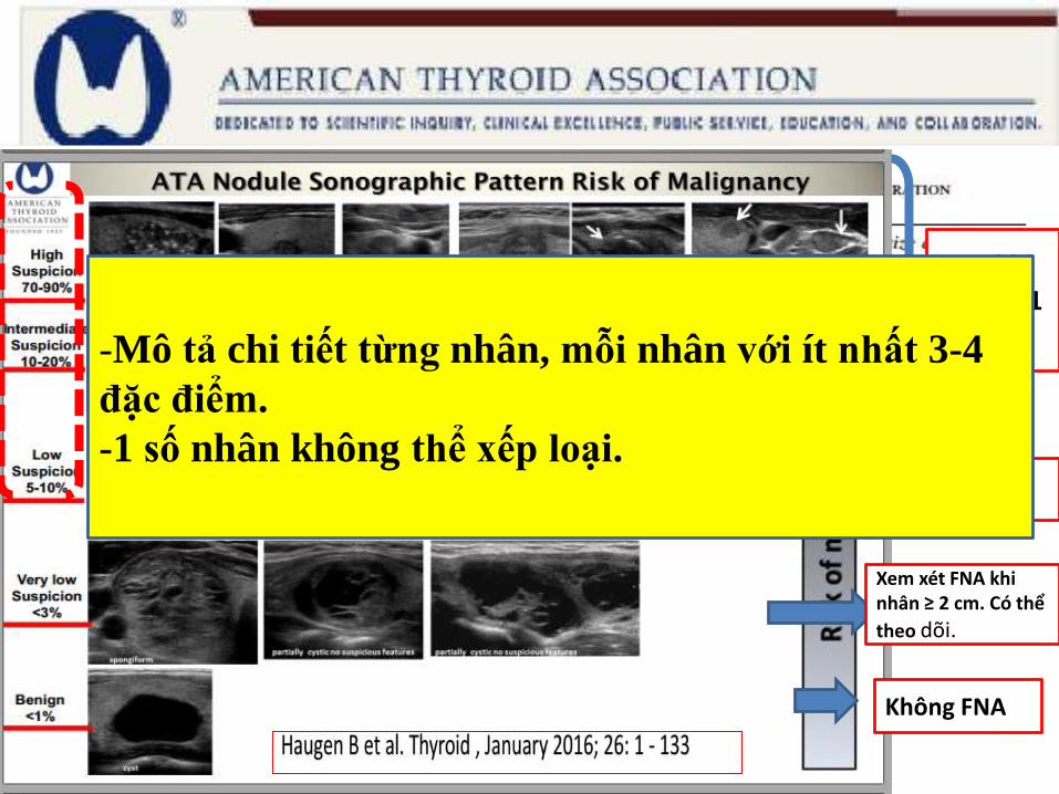
FNA khinhân ≥ 1 cm
FNA khinhân ≥ 1.5 cm
Xem xét FNA khinhân ≥ 2 cm. Có thể
theo dõi.
Không FNA
-Mô tả chi tiết từng nhân, mỗi nhân với ít nhất 3-4
đặc điểm.
-1 số nhân không thể xếp loại.
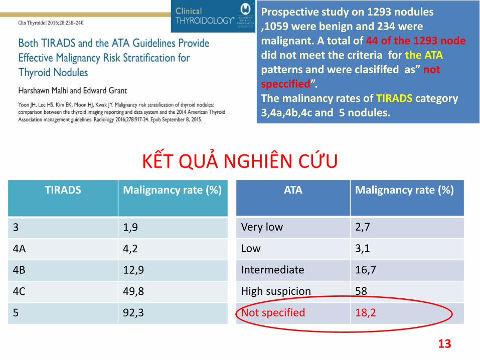
• KẾT QUẢ NGHIÊN CỨU
KẾT QUẢ NGHIÊN CỨU
Prospective study on 1293 nodules,1059 were benign and 234 were malignant. A total of 44 of the 1293 node did not meet the criteria for the ATA patterns and were clasififed as” not speccified”.The malinancy rates of TIRADS category 3,4a,4b,4c and 5 nodules.
TIRADS Malignancy rate (%)
3 1,9
4A 4,2
4B 12,9
4C 49,8
5 92,3
ATA Malignancy rate (%)
Very low 2,7
Low 3,1
Intermediate 16,7
High suspicion 58
Not specified 18,2
13
AMERICAN COLLEGE OF RADIOLOGY
14
Mục tiêu:1.Chuẩn hóa thuật ngữ có thể áp dụng cho tất cả tổn thương
2.Đưa ra khuyến cáo xử lý hạt giáp.
ACR tiến hành 3 Phase:
Phase 1: propose recommendation for nodules discovered incidentally on imaging
by Lincoln Berland, MD, and Jenny Hoang, MBBS.
Phase 2: Establish standardized lexicon by EdwardG.Grant,MD(2015).
Phase 3: Define a risk stratificition by Franklin.N.Tessler,MD(2017).
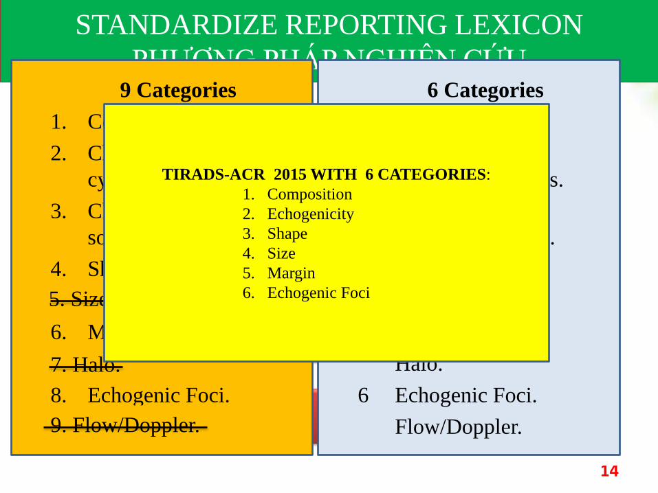
STANDARDIZE REPORTING LEXICON
PHƯƠNG PHÁP NGHIÊN CỨU9 Categories
1. Composition.
2. Characteristic of
cystic/components.
3. Characteristic of
solid/components.
4. Shape.
5. Size/dimensions.
6. Margin.
7. Halo.
8. Echogenic Foci.
9. Flow/Doppler.
6 Categories
1. Composition.
2. Characteristic of
cystic/components.
3. Characteristic of
solid/components.
4. Shape.
Size/dimensions.
5. Margin.
Halo.
6 Echogenic Foci.
Flow/Doppler.
5. Size/dimensions.
7. Halo.
9. Flow/Doppler.
TIRADS-ACR 2015 WITH 6 CATEGORIES:
1. Composition
2. Echogenicity
3. Shape
4. Size
5. Margin
6. Echogenic Foci
ACR TIRADS 2015
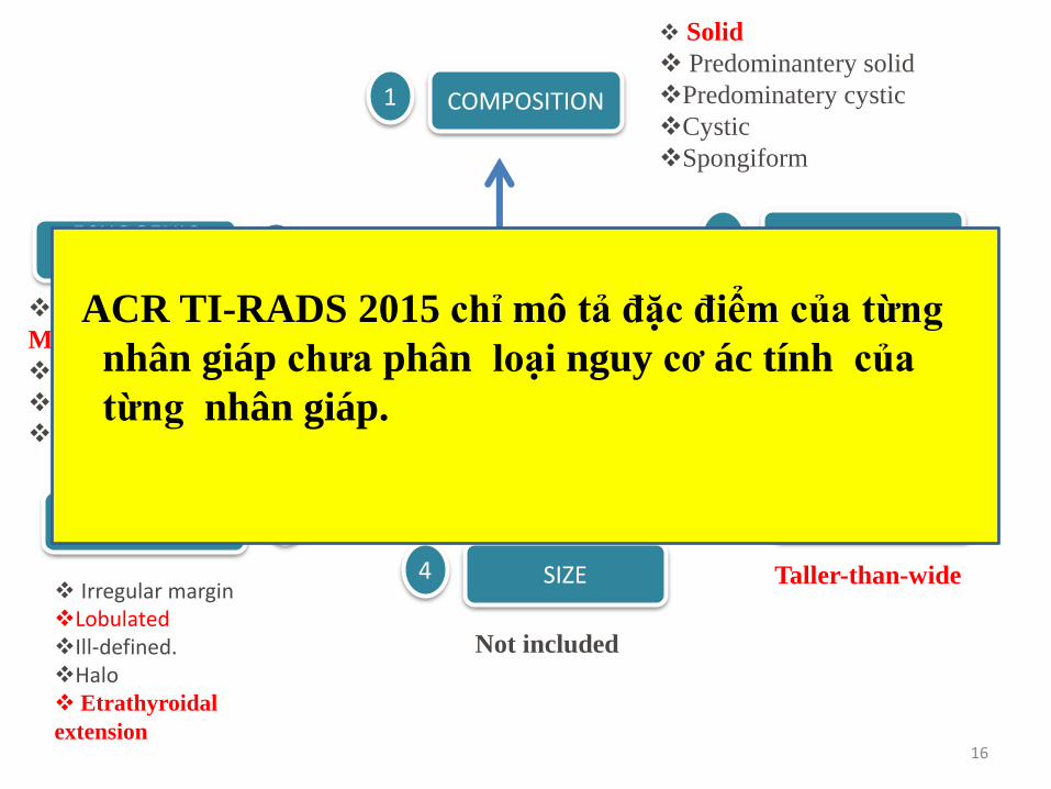
COMPOSITION1
2 ECHOGENICITY
3 SHAPE
4 SIZE
5 MARGIN
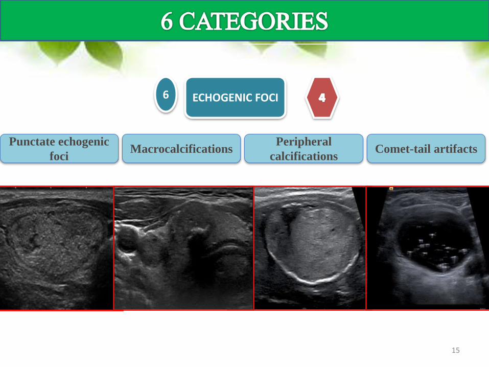
6 ECHOGENIC FOCI
SolidPredominately
solidPredominately
cysticCystic Spongiform
COMPOSITION1
Hyperechoic Isoechoic HypoechoicVery
hypoechoic
2 ECHOGENICITY
Taller-than-
wideNot included
3 SHAPE 4 SIZE
Smooth Irregular Lobulated Ill-defined HaloEtrathyroidal
extension
5 MARGIN
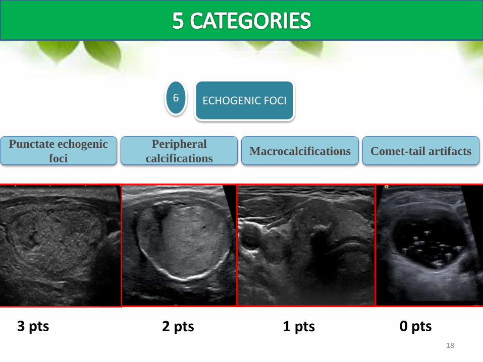
Punctate echogenic
fociMacrocalcifications
Peripheral
calcificationsComet-tail artifacts
6 ECHOGENIC FOCI
15
COMPOSITION1
2 ECHOGENICITY
3 SHAPE
4 SIZE
5MARGIN
6ECHOGENIC
FOCI
ACR TI-RADS 2015
Solid
Predominantery solid
Predominatery cystic
Cystic
Spongiform
Punctate echogenic foci
Microcalcification<1mm.
Macrocalcifications
Peripheral calcifications.
Comet-tail artifacts.
HyperechoicIsoechoicHypoechoicVery hypoechoic
Taller-than-wide
Not included
Irregular marginLobulatedIll-defined.Halo Etrathyroidal
extension16
ACR TI-RADS 2015 chỉ mô tả đặc điểm của từng
nhân giáp chưa phân loại nguy cơ ác tính của
từng nhân giáp.
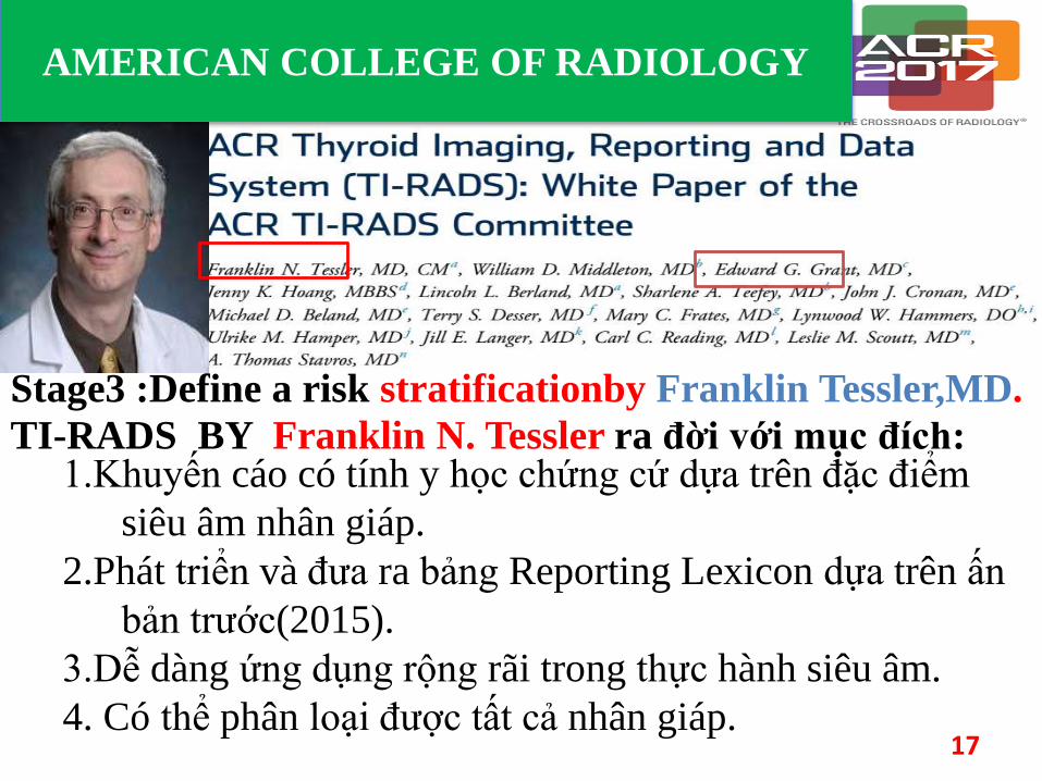
AMERICAN COLLEGE OF RADIOLOGY
.
TI-RADS BY Franklin N. Tessler ra đời với mục đích:
17
1.Khuyến cáo có tính y học chứng cứ dựa trên đặc điểm
siêu âm nhân giáp.
2.Phát triển và đưa ra bảng Reporting Lexicon dựa trên ấn
bản trước(2015).
3.Dễ dàng ứng dụng rộng rãi trong thực hành siêu âm.
4. Có thể phân loại được tất cả nhân giáp.
Stage3 :Define a risk stratificationby Franklin Tessler,MD.
SolidPredominately
solidPredominately
cysticCystic Spongiform
COMPOSITION1
2 pts 1 pts 0 pts 0 pts
Hyperechoic Isoechoic HypoechoicVery
hypoechoic
2 ECHOGENICITY
Anechoic
0 pts 1 pts 2 pts 3 pts
Taller-than-
wide
3 SHAPE
3 pts
Smooth Ill-defined
5 MARGIN
0 pts 2 pts
Irregular LobulatedEtrathyroidal
extension
3 pts0 pts
Punctate echogenic
fociMacrocalcifications
Peripheral
calcificationsComet-tail artifacts
6 ECHOGENIC FOCI
3 pts 1 pts2 pts 0 pts18
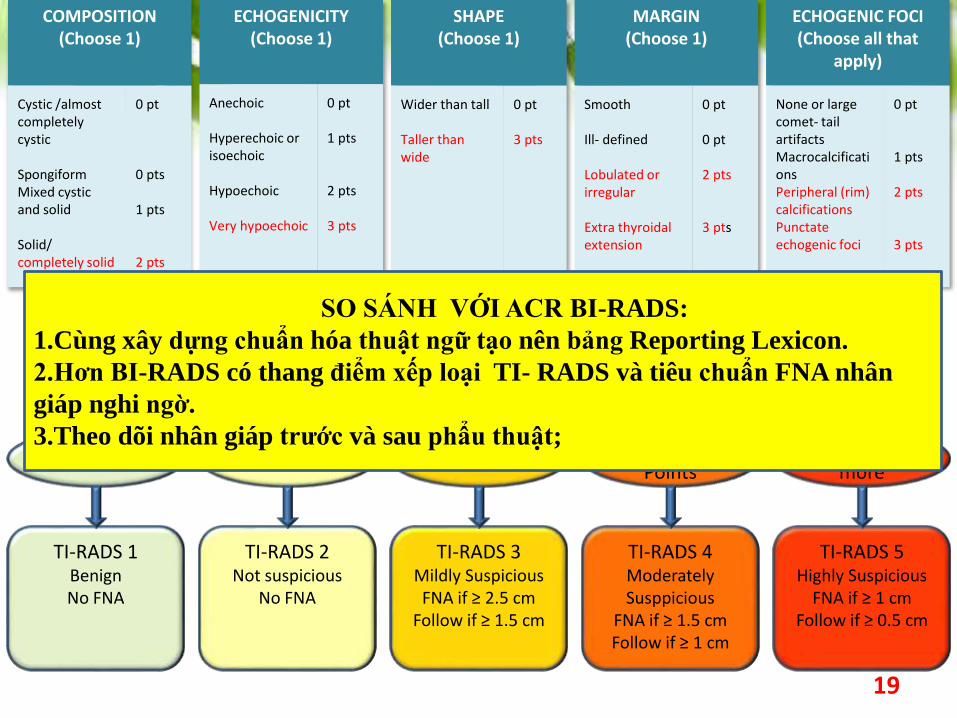
Add Points From All Categories to Determine TI-RADS Level
0 Point 2 Points 3 Points4 to 6 Points
7 Points or more
TI-RADS 1BenignNo FNA
TI-RADS 2Not suspicious
No FNA
TI-RADS 3Mildly SuspiciousFNA if ≥ 2.5 cm
Follow if ≥ 1.5 cm
TI-RADS 4Moderately Susppicious
FNA if ≥ 1.5 cmFollow if ≥ 1 cm
TI-RADS 5Highly Suspicious
FNA if ≥ 1 cmFollow if ≥ 0.5 cm
19
ECHOGENICITY(Choose 1)
Anechoic
Hyperechoic or isoechoic
Hypoechoic
Very hypoechoic
0 pt
1 pts
2 pts
3 pts
SHAPE(Choose 1)
Wider than tall
Taller than wide
0 pt
3 pts
MARGIN(Choose 1)
Smooth
Ill- defined
Lobulated or irregular
Extra thyroidal extension
0 pt
0 pt
2 pts
3 pts
ECHOGENIC FOCI(Choose all that
apply)
None or large comet- tail artifactsMacrocalcificationsPeripheral (rim) calcificationsPunctateechogenic foci
0 pt
1 pts
2 pts
3 pts
COMPOSITION(Choose 1)
Cystic /almost completely cystic
SpongiformMixed cystic and solid
Solid/ completely solid
0 pt
0 pts
1 pts
2 pts
SO SÁNH VỚI ACR BI-RADS:
1.Cùng xây dựng chuẩn hóa thuật ngữ tạo nên bảng Reporting Lexicon.
2.Hơn BI-RADS có thang điểm xếp loại TI- RADS và tiêu chuẩn FNA nhân
giáp nghi ngờ.
3.Theo dõi nhân giáp trước và sau phẩu thuật;
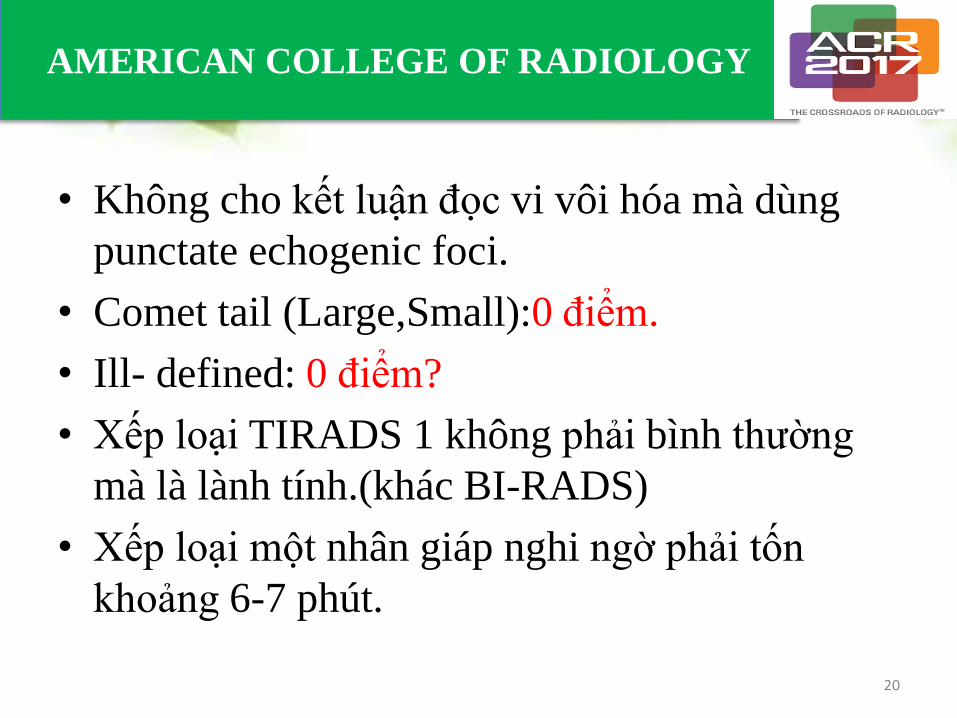
• Không cho kết luận đọc vi vôi hóa mà dùng
punctate echogenic foci.
• Comet tail (Large,Small):0 điểm.
• Ill- defined: 0 điểm?
• Xếp loại TIRADS 1 không phải bình thường
mà là lành tính.(khác BI-RADS)
• Xếp loại một nhân giáp nghi ngờ phải tốn
khoảng 6-7 phút.
20
AMERICAN COLLEGE OF RADIOLOGY
21
BẢNG TỰ LƯỢNG GIÁ
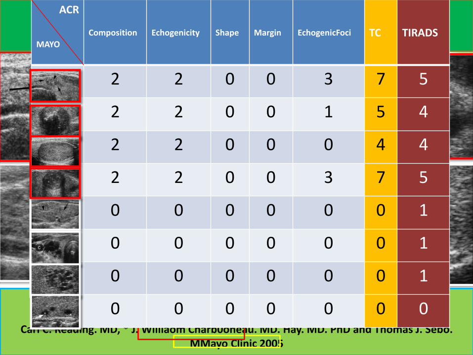
Sonography of Thyroid NodulesA “Classic Pattern” Diagnostic Approach
Carl C. Reading. MD, * J. Williaom Charb0oneau. MD. Hay. MD. PhD and Thomas J. Sebo. MMayo Clinic 2005
ACR
MAYO
Composition Echogenicity Shape Margin EchogenicFoci TC TIRADS
2 2 0 0 3 7 5
2 2 0 0 1 5 4
2 2 0 0 0 4 4
2 2 0 0 3 7 5
0 0 0 0 0 0 1
0 0 0 0 0 0 1
0 0 0 0 0 0 1
0 0 0 0 0 0 0
REFERENCES
1. College of Radiology. Breast imaging reporting and data system: BI-RADS Atlas, 4th edn. Reston, VA, 2003.
2. Mary c Frates ,Carol B.Benson eltal Management of Thyroid Nudules Detected at US society of Radiologist in
Ultrasound Consensus Conference Statement Radiology 2005;237;794-800
3. Horvath E, Majilis S, Rossi R, Franco C, Niedmann J, Castro A & Dominguez M. An ultrasonogram reporting
system for thyroid nodules stratifying cancer risk for clinical management. Journal of Clinical Endocrinology and
Metabolism 2009 90 1748–1751
4. ParkSH,KimSJ,KimEK,KimMJ,SonEJ,KwakJYInterobserveragreementinassessingthesonographicandelasphi
aeaturesofmalignantthyroidnodules.AJRAmJRoentgenol2009;193:W416-W423
5. Kwak JY, Han KH, Yoon JH, Moon HJ, Son EJ, Park SH, et al. Thyroid imaging reporting and data system for US
features of nodules: a step in establishing better stratification of cancer risk. Radiology. 2011;260:892–899
6. Jin Young Kwak et al. imaging reporting and characterization system for US features of nodules:aMulticentric
Korean Retrospective Study . 2013.
7. Russ B, Royer B, Bigorgne C, et al. Prospective evaluation of thyroid imaging reporting and data system on 4550
nodules with and without elastography. Eur J Endocrinol. 2011;16
8. TI-RADS (Thyroid Imaging Reporting and Data System): Are We There Yet? Sergiy V. Kushchayev,Aliaksei L.
SaleiOleg M. Teytelboym, Department of Radiology, Mercy Catholic Medical Center, Darby, PA.
9. ATA Guidelines 2015:Using Untrasound for FNA decision making.Susan.Jmandel,MD MDH.
10. Joon JH et al. Both TIRADS and the ATA guidelines provide effective malignancy risk stratification for
thyroid nodules. Radiology 2016: 278:917-24. Epub Steptember 8, 2015
11. Edward G .Grant, ACR Thyroid Imaging,Reporting and Data Systerm (TI-RADS):White Paper of the ACR
TI-RADS Committee. 2015
12. FrankinN.Tessler ACR Thyroid Imaging,Reporting and Data Systerm (TI-RADS):White Paper of the ACR TI-
RADS Committee. 2017 22
CHÂN THÀNH CẢM ƠN
23