Embed Size (px)
Citation preview


Visual Diagnosis in Emergency and Critical Care MedicineEDITED BY
Christopher P. Holstege | MD, FACEP, FAAEM, FACMT
Director, Division of Medical ToxicologyMedical Director, Blue Ridge Poison CenterAssociate Professor, Departments of Emergency Medicine and PediatricsUniversity of VirginiaCharlottesvilleVirginia
Alexander B. Baer | MD
Associate Medical Director, Blue Ridge Poison CenterClinical Assistant Professor, Department of Emergency MedicineUniversity of VirginiaCharlottesvilleVirginia
Jesse M. Pines | MD, MBA, FAAEM
Lecturer, Department of Emergency MedicineCenter for Clinical Epidemiology and BiostatisticsUniversity of Pennsylvania School of MedicinePhiladelphiaPennsylvania
William J. Brady | MD, FACEP, FAAEM
Vice Chair, Department of Emergency MedicineProfessor, Department of Emergency Medicine and Internal MedicineUniversity of VirginiaCharlottesvilleVirginia


Visual Diagnosis in Emergency and Critical Care Medicine

“To my wife, Angela, for her enlightening encouragement and endless humor.”C.P.H.
“To my wife and child who have always supported and inspired me.”A.B.B.
“To my loving wife Lori for all her support.”J.M.P.
“My thanks and love to my family—King, Lauren, Anne, Chip, and Katherine.”W.J.B.

Visual Diagnosis in Emergency and Critical Care MedicineEDITED BY
Christopher P. Holstege | MD, FACEP, FAAEM, FACMT
Director, Division of Medical ToxicologyMedical Director, Blue Ridge Poison CenterAssociate Professor, Departments of Emergency Medicine and PediatricsUniversity of VirginiaCharlottesvilleVirginia
Alexander B. Baer | MD
Associate Medical Director, Blue Ridge Poison CenterClinical Assistant Professor, Department of Emergency MedicineUniversity of VirginiaCharlottesvilleVirginia
Jesse M. Pines | MD, MBA, FAAEM
Lecturer, Department of Emergency MedicineCenter for Clinical Epidemiology and BiostatisticsUniversity of Pennsylvania School of MedicinePhiladelphiaPennsylvania
William J. Brady | MD, FACEP, FAAEM
Vice Chair, Department of Emergency MedicineProfessor, Department of Emergency Medicine and Internal MedicineUniversity of VirginiaCharlottesvilleVirginia

© 2006 by Blackwell Publishing LtdBMJ Books is an imprint of the BMJ Publishing Group Limited, used under licence
Blackwell Publishing, Inc., 350 Main Street, Malden, Massachusetts 02148-5020, USABlackwell Publishing Ltd, 9600 Garsington Road, Oxford OX4 2DQ, UKBlackwell Publishing Asia Pty Ltd, 550 Swanston Street, Carlton, Victoria 3053, Australia
The right of the author to be identified as the author of this work has been asserted in accordance with the Copyright, Designs and Patents Act 1988.
All rights reserved. No part of this publication may be reproduced, stored in a retrieval system, or transmitted, in any form or by any means, electronic, mechanical, photocopying, recording or otherwise, except as permitted by the UK Copyright, Designs and Patents Act 1988, without the prior permission of the publisher.
First published 2006
1 2006
Library of Congress Cataloging-in-Publication DataVisual diagnosis in emergency and critical care medicine/edited by Christopher P. Holstege . . . [et al.].
p. ; cm.Includes bibliographical references and index.ISBN-13: 978-1-4051-3491-0 (alk. paper)ISBN-10: 1-4051-3491-7 (alk. paper)
1. Emergency medical services—Case studies. 2. Critical care medicine—Case studies.3. Diagnosis. I. Holstege, Christopher P.
[DNLM: 1. Emergency Medicine—Case Reports. 2. Emergency Medicine—ExaminationQuestions. 3. Critical Care—Case Reports. 4. Critical Care—Examination Questions. 5. Diagnosis—Case Reports. 6. Diagnosis—Examination Questions. WB 18.2 V834 2007]
RA645.5.V57 2007362.18—dc22 2006022486
ISBN-13: 978-1-4051-3491-0ISBN-10: 1-4051-3491-7
A catalogue record for this title is available from the British Library
Set in 9/12 pt Meridien by Charon Tec Ltd (A Macmillan Company), Chennai, Indiawww.charontec.comPrinted and bound in Singapore by COS Printers Pte Ltd
Commissioning Editor: Mary BanksEditorial Assistant: Victoria PittmanDevelopment Editor: Nick MorganProduction Controller: Debbie Wyer
For further information on Blackwell Publishing, visit our website:http://www.blackwellpublishing.com
The publisher’s policy is to use permanent paper from mills that operate a sustainable forestry policy, and which has been manufactured from pulp processed using acid-free and elementary chlorine-free practices. Furthermore, the publisher ensures that the text paper and cover boardused have met acceptable environmental accreditation standards.
Blackwell Publishing makes no representation, express or implied, that the drug dosages in this book are correct. Readers must therefore always check that any product mentioned in this publication is used in accordance with the prescribing information prepared by the manufacturers. The author and the publishers do not accept responsibility or legal liability for any errors in the text or for the misuse or misapplication of material in this book.

Contents
v
List of contributors, ix
Foreword, xi
Preface, xii
Illustration credits, xiii
Part 1 | Case Presentations and Questions, 1
1 Rash following brush fire, 3Christopher P. Holstege, MD
2 Herbalist with bradycardia and vision changes, 3William J. Brady, MD
3 Acute eye pain and blurred vision in an elderly female, 4Chris S. Bergstrom, MD & Alexander B. Baer, MD
4 Suspicious hand pain, 4Rex Mathew, MD
5 An elderly man with flank pain, 5Daniel K. Vining, MD & Anthony J. Dean, MD
6 An immigrant child with skin lesions, 6Roger A. Band, MD
7 Wrist pain following a fall, 6Rex Mathew, MD
8 Rash in a child with epilepsy, 7Alexander B. Baer, MD & Christopher P. Holstege, MD
9 Dark urine in an immigrant, 8Suzanne M. Shepherd, MD, Susan A. O’Malley, MD & William H. Shoff, MD
10 Fever and drooling in a child, 8Sarah G. Winters, MD & Brendan E. Carr, MD
11 Altered mental status with an abnormal electrocardiogram, 9William J. Brady, MD
12 Purulent eye discharge in an adult, 9Chris S. Bergstrom, MD & Alexander B. Baer, MD
13 Wrist pain in a young child, 10Craig S. Brummer, MD
14 Postprandial abdominal pain, 10Anthony J. Dean, MD
15 An elderly man from a house fire, 11Christopher P. Holstege, MD
16 Back pain following a fall, 11Andrew D. Perron, MD & Christopher T. Bowe, MD
17 A bite to the leg in tall grass, 12Christopher P. Holstege, MD
18 Facial swelling in a patient with poor dentition, 13Alexander B. Baer, MD & Christopher P. Holstege, MD
19 Elbow pain in a child after a fall, 13Alexander B. Baer, MD
20 A man with diffuse facial edema, 14Kevin S. Barlotta, MD & Alexander B. Baer, MD
21 Chest pain and hypotension in an adult male patient, 14William J. Brady, MD
22 Eye pain after tree branch strike, 15Chris S. Bergstrom, MD & Alexander B. Baer, MD
23 A missing button battery, 15Brendan E. Carr, MD & Sarah G. Winters, MD
24 Acute abdominal pain in pregnancy, 16Anthony J. Dean, MD
25 Painless penile ulcer, 17Andrea L. Neimann, MD
26 Low back pain in car accident victim, 17Edward G. Walsh, MD & William J. Brady, MD
27 A gardener with a non-healing rash, 18Roger A. Band, MD
28 An immigrant with neck swelling, 18Suzanne M. Shepherd, MD, Anthony J. Dean, MD & William H. Shoff, MD
29 Fall on an outstretched hand in a young adolescent, 19William J. Brady, MD & Kevin S. Barlotta, MD

30 Raccoon eyes, 19Angela M. Mills, MD
31 Chest pain and a confounding electrocardiogram pattern, 20William J. Brady, MD
32 Eye pain in a contact lens wearer, 21Chris S. Bergstrom, MD & Alexander B. Baer, MD
33 Heel pain following a fall, 21David F. Gaieski, MD
34 FAST evaluation following trauma, 22Bon S. Ku, MD & Anthony J. Dean, MD
35 Skin lesion in a heroin addict, 22Christopher P. Holstege, MD & Alexander B. Baer, MD
36 Young athlete with back pain, 23Edward G. Walsh, MD & William J. Brady, MD
37 Skin lesions in a comatose patient, 24Christopher P. Holstege, MD
38 Chest pain with sudden cardiac death, 24William J. Brady, MD
39 Fall on an outstretched hand with wrist pain, 25William J. Brady, MD & Kevin S. Barlotta, MD
40 Necrotic skin lesion, 26David A. Kasper, MBA, Aradhna Saxena, MD & Kenneth A. Katz, MD
41 Chest pain with electrocardiographic ST-segment/T-wave abnormalities, 26William J. Brady, MD
42 Chemical eye exposure, 27Chris S. Bergstrom, MD & Alexander B. Baer, MD
43 Hand pain after striking a wall, 27William J. Brady, MD & Kevin S. Barlotta, MD
44 Dyspnea in an alcoholic, 28Anthony J. Dean, MD
45 Slash wound to the neck, 29Kevin S. Barlotta, MD & Alexander B. Baer, MD
46 Foot pain following breaking, 30Munish Goyal, MD
47 Confluent rash in a child, 30Sarah G. Winters, MD & Brendan E. Carr, MD
48 Lost in the cold, 31Adam K. Rowden, DO & Christopher P. Holstege, MD
49 Bradycardia following an herbal ingestion, 32Alexander B. Baer, MD
50 Abdominal pain in an alcoholic, 32Angela M. Mills, MD
51 Pain out of proportion to examination, 33Adam K. Rowden, DO
52 Pleuritic chest pain in a young adult male, 33William J. Brady, MD
53 Eye pain following a bar fight, 34Chris S. Bergstrom, MD & Alexander B. Baer, MD
54 Forearm fracture after falling, 35Alexander B. Baer, MD
55 An elderly woman with groin pain, 36Brendan E. Carr, MD
56 Painful facial rash, 36Chris S. Bergstrom, MD & Alexander B. Baer, MD
57 Confusion, anemia, and abdominal pain in a toddler, 37Alexander B. Baer, MD & Christopher P. Holstege, MD
58 Cardiotoxic effects following caustic ingestion, 37Alexander B. Baer, MD & Christopher P. Holstege, MD
59 Rash and joint pain in a child, 38Mara Becker, MD
60 X-ray findings after laparoscopy, 39Munish Goyal, MD
61 Injector injury to the hand, 40Tracy H. Reilly, MD
62 Chest pain in a middle-aged male patient with ST segment elevation, 40William J. Brady, MD
63 Deformed globe following trauma, 41Joseph Robson, MD & Worth W. Everett, MD
64 Adult male with atraumatic lower back pain and legweakness, 41William J. Brady, MD
65 Fever and rash in a child, 42David L. Eldridge, MD
66 Yellow eyes and skin, 43Jane M. Prosser, MD
67 Fishing in the stomach, 43Christopher P. Holstege, MD
68 Agitation in a botanist, 44Alexander B. Baer, MD & Christopher P. Holstege, MD
69 Skin target lesion, 45Mara Becker, MD
70 Adult male with a sudden, severe headache, 45Andrew L. Homer & William J. Brady, MD
71 Get them undressed!, 46Munish Goyal, MD
vi | Contents

72 Chest pain and subtle ST segment elevation, 46William J. Brady, MD
73 Fluid in my eye, 47Chris S. Bergstrom, MD & Alexander B. Baer, MD
74 Coma following head trauma, 48Andrew L. Homer & William J. Brady, MD
75 Blue hue following endoscopy, 48Andrew L. Homer & Christopher P. Holstege, MD
76 Shoulder pain following direct blow, 49David F. Gaieski, MD
77 An overdose of prenatal vitamins, 49Christopher P. Holstege, MD & Adam K. Rowden, DO
78 Fever and rash in a child, 50David L. Eldridge, MD
79 Lamp oil ingestion, 51David L. Eldridge, MD
80 Diffuse ankle pain following a fall, 51Andrew D. Perron, MD & Christopher T. Bowe, MD
81 Emergency department drop-off, 52Tracy H. Reilly, MD
82 Weakness and bradycardia in an elderly female patient, 52William J. Brady, MD
83 Blurred vision following yard work, 53Allyson Kreshak, MD
84 A gagging child, 53Maureen Chase, MD & Worth W. Everett, MD
85 A child with bruises of different ages, 54David L. Eldridge, MD
86 Traumatic eye pain and proptosis, 54Chris S. Bergstrom, MD & Alexander B. Baer, MD
87 Post-prandial abdominal pain in an elderly woman, 55Hoi Lee, MD
88 Hyperthermia, tachycardia, and confusion in ateenager, 56Alexander B. Baer, MD
89 Acute onset double vision, 56Chris S. Bergstrom, MD & Alexander B. Baer, MD
90 Ankle pain and inability to walk, 57Christopher T. Bowe, MD
91 Tongue swelling in a hypertensive female, 58Kevin S. Barlotta, MD & Alexander B. Baer, MD
92 Wide complex tachycardia in an older male patient, 58William J. Brady, MD
93 Acute onset double vision, 59Chris S. Bergstrom, MD & Alexander B. Baer, MD
94 Foot pain in a gymnast, 60Hoi Lee, MD
95 New facial droop, 60Andrew D. Perron, MD & Christopher T, Bowe, MD
96 Eye pain and swelling, 61Adam K. Rowden, DO & Chris S. Bergstrom, MD
97 Shortening and rotation of the leg following trauma, 61Jane M. Prosser, MD
98 Spider bite in the night, 62Adam K. Rowden, DO & Christopher P. Holstege, MD
99 Wide complex tachycardia in a young adult, 62William J. Brady, MD
100 Abdominal pain in a trauma victim, 63Esther H. Chen, MD
Part 2 | Answers, Diagnoses, and Discussion, 65
Index, 153
Contents | vii


List of contributors
ix
Alexander B. Baer, MDAssociate Medical Director, Blue Ridge Poison Center, and ClinicalAssistant Professor, Department of Emergency Medicine, University ofVirginia, PO Box 800774, Charlottesville, VA 22908-0774
Roger A. Band, MDAssistant Professor, Department of Emergency Medicine, University of Pennsylvania School of Medicine, 3400 Spruce Street, Philadelphia,PA 19104
Kevin L. Barlotta, MDEmergency Medicine Resident, Department of Emergency Medicine,University of Virginia, PO Box 800774, Charlottesville, VA 22908-0774
Mara Becker, MDFellow, Pediatric Rheumatology, Alfred I. duPont Hospital for Children,1600 Rockland Road, Wilmington, DE 19803
Chris S. Bergstrom, MDFellow, Vitreoretinal Surgery, Department of Ophthalmology, EmoryUniversity, 201 Dowman Drive, Atlanta, GA 30322
Christopher T. Bowe, MDAssociate Residency Director, Emergency Medicine Department, MaineMedical Center, 22 Bramhall Street, Portland, ME 04102
William J. Brady, MD, FACEP, FAAEMVice Chair, Department of Emergency Medicine, and Professor,Department of Emergency Medicine and Internal Medicine, Universityof Virginia, PO Box 800669, Charlottesville, VA 22908-0699
Craig S. Brummer, MDAttending Physician, Kennestone Hospital, 4016 Benell Court, Smyrna, GA 30082
Brendan E. Carr, MD, MAInstructor, Department of Emergency Medicine, and Fellow, Division ofTrauma and Surgical Critical Care, Department of Surgery, University of Pennsylvania School of Medicine, 3400 Spruce Street, Philadelphia,PA 19104
Maureen Chase, MDInstructor and Research Fellow, Department of Emergency Medicine,University of Pennsylvania School of Medicine, 3400 Spruce Street,Philadelphia, PA 19104
Esther H. Chen, MDAssistant Professor, Department of Emergency Medicine, University of Pennsylvania School of Medicine, 3400 Spruce Street, Philadelphia,PA 19104
Anthony J. Dean, MDDirector, Emergency Ultrasound, and Assistant Professor, Department of Emergency Medicine, University of Pennsylvania School ofMedicine, 3400 Spruce Street, Philadelphia, PA 19104
David L. Eldridge, MDAssistant Professor, Department of Pediatrics, Brody School ofMedicine, East Carolina University, 600 Moye Boulevard, Greenville, NC 27834
Worth W. Everett, MDAssistant Professor, Department of Emergency Medicine, University of Pennsylvania School of Medicine, 3400 Spruce Street, Philadelphia,PA 19104
David F. Gaieski, MDCo-Director, Early Goal Directed Therapy Program, and AssistantProfessor, Department of Emergency Medicine, University of PennsylvaniaSchool of Medicine, 3400 Spruce Street, Philadelphia, PA 19104
Munish Goyal, MDCo-Director, Early Goal Directed Therapy Program, and AssistantProfessor, Department of Emergency Medicine, University of PennsylvaniaSchool of Medicine, 3400 Spruce Street, Philadelphia, PA 19104
Christopher P. Holstege, MD, FACEP, FAAEM, FACMTDirector, Division of Medical Toxicology, Medical Director, Blue RidgePoison Center, and Associate Professor, Departments of EmergencyMedicine and Pediatrics, University of Virginia, PO Box 800774,Charlottesville, VA 22908-0774
Andrew L. HomerMedical Student, University of Virginia School of Medicine, PO Box800774, Charlottesville, VA 22908-0774
David A. Kasper, MBAPhiladelphia College of Osteopathic Medicine, c/o Kenneth Katz, MD,Department of Dermatology, University of Pennsylvania School ofMedicine, 3600 Spruce Street, Philadelphia, PA 19104
Kenneth A. Katz, MD, MScInstructor and NRSA Postdoctoral Fellow, Department of Dermatology,University of Pennsylvania School of Medicine, 3600 Spruce Street,Philadelphia, PA 19104
Allyson Kreshak, MDResident Physician, Department of Emergency Medicine, University ofPennsylvania School of Medicine, c/o Jesse M. Pines, 3400 SpruceStreet, Philadelphia, PA 19104

Bon S. Ku, MDInstructor and Ultrasound Fellow, Department of Emergency Medicine,University of Pennsylvania School of Medicine, 3400 Spruce Street,Philadelphia, PA 19104
Hoi Lee, MDResident Physician, Department of Emergency Medicine, University ofPennsylvania School of Medicine, 3400 Spruce Street, Philadelphia, PA19104
Rex Mathew, MDAssistant Professor, Department of Emergency Medicine, ThomasJefferson University, 1020 Walnut Street, Philadelphia, PA 19107
Angela M. Mills, MDAssistant Professor, Department of Emergency Medicine, University ofPennsylvania School of Medicine, 3400 Spruce Street, Philadelphia, PA19104
Andrea L. Neimann, MDDepartment of Dermatology, University of Pennsylvania School ofMedicine, c/o Jesse M. Pines, 3400 Spruce Street, Philadelphia, PA19104
Susan A. O’Malley, MDAssociate Attending, Brookhaven Memorial Hospital Medical Center,101 Hospital Road, Patchogue, NY 11772
Andrew D. Perron, MDResidency Director, Department of Emergency Medicine, 22 BramhallStreet, Main Medical Center, Portland, ME 04102
Jesse M. Pines, MD, MBA, FAAEMLecturer, Department of Emergency Medicine, Center for ClinicalEpidemiology and Biostatistics, University of Pennsylvania School ofMedicine, 3400 Spruce Street, Philadelphia, PA 19104
Jane M. Prosser, MDResident Physician, Department of Emergency Medicine, University ofPennsylvania School of Medicine, 3400 Spruce Street, Philadelphia, PA19104
Tracy H. Reilly, MDFellow, Medical Toxicology, Department of Emergency Medicine,University of Virginia, PO Box 800774, Charlottesville, VA 22908-0774
Joseph Robson, MDChief Resident Physician, Department of Emergency Medicine,University of Pennsylvania School of Medicine, 3400 Spruce Street,Philadelphia, PA 19104
Adam K. Rowden, DOFellow, Medical Toxicology, Department of Emergency Medicine,University of Virginia, PO Box 800774, Charlottesville, VA 22908-0774
Aradhna Saxena, MDResident, Department of Dermatology and Cutaneous Biology, ThomasJefferson University Hospital, 833 Chestnut Street, Philadelphia, PA19107
Suzanne M. Shepherd, MD, DTM&HAssociate Professor, Department of Emergency Medicine, and Directorof Education and Research, PENN Travel Medicine, University ofPennsylvania School of Medicine, 3400 Spruce Street, Philadelphia, PA19104
William H. Shoff, MD, DTM&HAssociate Professor, Department of Emergency Medicine, and Director,PENN Travel Medicine, University of Pennsylvania School of Medicine,3400 Spruce Street, Philadelphia, PA 19104
Daniel K. Vining, MDResident Physician, Department of Emergency Medicine, University ofPennsylvania School of Medicine, 3400 Spruce Street, Philadelphia, PA19104
Edward G. Walsh, MDEmergency Medicine Resident, Department of Emergency Medicine,University of Virginia, PO Box 800774, Charlottesville, VA 22908-0774
Sarah G. Winters, MDFellow, Division of General Pediatrics, Children’s Hospital ofPhiladelphia, c/o Brendan Carr, MD, 3400 Spruce Street, Philadelphia,PA 19104
x | List of contributors

Emergency physicians often have a knack for making rapid diagnoses and initiating needed treatments quickly.Developing one’s ability to blend visual, historical and phys-ical clues is generally based upon a combination of under-standing basic pathophysiological mechanisms, identifyingimportant historical and physical findings known to be asso-ciated with these pathophysiological mechanisms, and re-experiencing imprinted visual cues from prior cases. Thisvisual diagnosis book, written by Holstege, Baer, Pines andBrady, takes advantage of all these features in an effort toserve as a valuable teaching tool for the physician or medicalstudent seeking to improve their ability to rapidly diagnoseimportant clinical conditions that may present to virtuallyany emergency department.
The cases are not arranged by body system or by patho-physiological mechanisms (e.g., trauma, infectious disease,etc). Rather, they are presented similar to cases appearing inthe emergency department. Thus, they appear in no particu-lar order or sequence. Children are interspersed with adultsand injuries with medical conditions.
Each case is presented as an unknown along with a graphicillustration, brief case presentation, and management deci-sion. The photos and line drawings are crisp and generallywell demonstrate the patient’s abnormality, although somepresentations may be subtle to the uninitiated. The answersprovided later in the book are much richer than would beanticipated for a case presentation text. The discussions andsupplemental illustrations create the impression that youhave a seasoned emergency physician in the room who iswalking you through the basic pathophysiological mecha-nisms and then clarifying the visual clues and their clinicalsignificance. This detail gives you the confidence that you can
recognize and respond appropriately were you to re-experiencethese case presentations in the future.
As a seasoned clinician, I eagerly read the case presenta-tions first to verify my own rapid diagnostic skills and sec-ondarily to determine if the authors would approach theproblem as I would or if they would make different interven-tional recommendations. Although I am pleased that myinterpretations were highly correlated with those of theauthors, I found the richness of their discussion valuable forrefreshing my understanding of the mechanisms behind theillness associated with each image.
I anticipate that learners (whether novices or seasonedcontinued learners like myself) will appreciate what thisbook has to offer. It is fun and easy to pick up and begin thelearning. Unfortunately, it is sometimes hard to put downfor these same reasons. I anticipate keeping this text handyto reinforce key teaching points with medical students andresidents. Others may find the text extremely valuable forpreparing for the visual image portion of the emergencymedicine written board examination. Those of us who havebeen away from our residency for some time will enjoy thetext as a stimulus leading us to re-experience visual clues onconditions we simply don’t want to miss as clinicians.
I anticipate that this text will be popular and eventuallylead to subsequent "Visual Diagnosis" volumes. Our studentsand practitioners will be pleased to see these as well.
Jerris R. Hedges, MD, MSProfessor of Emergency Medicine
Vice Dean, School of MedicineOregon Health & Sciences University
xi
Foreword

The acute care practitioner faces numerous challenges in theapproach to the critically ill or injured patient. Clearly, the his-tory of the event is a vital portion of the evaluation, providingthe “answer” to the clinical situation in many instances. Thephysical examination and the results of various diagnosticinvestigations, however, are also essential components of themedical evaluation. In fact, the examination, the electrocar-diogram, and the radiograph provide the clinician with eitherthe diagnosis or important information which will lead to thediagnosis. The rash of erythema multiforme, the electrocar-diogram in pronounced hyperkalemia, the radiograph in car-pometacarpal dislocation are all presentations where a single“clinical image” provides the diagnosis or a substantial clue tothe diagnosis – with appropriate therapy soon to follow.Bedside clinical diagnosis, based upon specific clinical images,is a vital skill for the acute care practitioner.
The purpose of this book is to provide some of thosevisual diagnostic clues that might be encountered in acutecare scenarios. In Part 1, each visual cue is associated with an
actual case and a multiple choice question. The correctanswer and a focused discussion then follow in Part 2. Inacademic practice, utilizing a visual cue with an associatedcase presentation and a multiple choice question is a highlyeffective teaching method. In clinical practice, the use ofcase-based scenarios is a popular, effective means of self-education. This enables the teacher or the student to discussthe disease, and importantly the diagnosis and management.We have attempted to capture this teaching style within thecontext of this book. Whether you are an experienced clini-cian in private practice, an academician engaged in teaching,a resident or student in training looking to prepare for tests,we hope this book will provide you with further experienceto excel as a practitioner in the field of medicine.
Christopher P. HolstegeAlexander B. Baer
Jesse M. PinesWilliam J. Brady
xii
Preface

1 Poison Ivy. Case: Christopher P. Holstege; Figs 1 and 2:
Christopher P. Holstege
2 Digoxin Toxicity. Case: Christopher P. Holstege
3 Angle Closure Glaucoma. Case: Chris S. Bergstrom
4 Boxer’s Fracture. Case: Alexander B. Baer; Fig. 1: Alexander
B. Baer
5 Abdominal Aortic Aneurysm. Case: Anthony J. Dean; Figs
1 and 2: Anthony J. Dean
6 Coining. Case: Edward T. Dickinson
7 Perilunate dislocation. Case: Alexander B. Baer; Fig. 1:
Alexander B. Baer
8 Drug Hypersensitivity Reaction. Case: Christopher P.
Holstege; Figs 1 and 2: Christopher P. Holstege
9 Egyptian with Hemolysis. Case: William H. Shoff
10 Retropharyngeal Abscess. Case: Alexander B. Baer
11 Hyperkalemia. Case: William J. Brady; Figs 1 and 2: William
J. Brady
12 Conjunctivitis. Case: Chris S. Bergstrom
13 Buckle Fracture. Case: Alexander B. Baer; Fig. 1: Alexander
B. Baer
14 Cholecystitis. Case: Anthony J. Dean; Fig. 1: Anthony J. Dean
15 Smoke Inhalation. Case: Christopher P. Holstege; Fig. 1:
Christopher P. Holstege
16 Lumbar Wedge Fracture. Case: Alexander B. Baer; Fig. 1:
Alexander B. Baer
17 Snakebite. Case: Alexander B. Baer; Fig. 1: Christopher P.
Holstege
18 Submandibular Abscess. Case: Alexander B. Baer
19 Sail Sign. Case: Alexander B. Baer; Fig. 1: Alexander B. Baer
20 SVC Syndrome. Case: Alexander B. Baer
21 Inferoposterior RV AMI. Case: William J. Brady; Fig. 1:
William J. Brady
22 Corneal Abrasion. Case: Chris S. Bergstrom
23 Button Battery. Case: Sarah G. Winters; Figs 1–4: Sarah G.
Winters
24 Ectopic. Case: Anthony J. Dean; Figs 1 and 2: Anthony J.
Dean
25 Syphilis. Case: William D. James
26 Lumbar Teardrop Fracture. Case: Alexander B. Baer; Fig. 1:
Alexander B. Baer
27 Sporotrichosis. Case: Steve Larson
28 Tuberculosis Adenitis. Case: William H. Shoff
29 Salter–Harris Fracture. Case: William J. Brady; Figs 1 and 2:
William J. Brady
30 Raccoon Eyes. Case: Alexander B. Baer
31 LBBB AMI. Case: William J. Brady; Fig. 1: William J. Brady
32 Corneal Ulcer. Case: Chris S. Bergstrom
33 Calcaneus Fracture. Case: Alexander B. Baer; Fig. 1:
Alexander B. Baer
34 Fast Exam. Case: Anthony J. Dean; Figs 1 and 2: Anthony J.
Dean
35 Skin Popping. Case: Alexander B. Baer; Figs 1–3: Alexander
B. Baer; Fig. 4: Christopher P. Holstege
36 Lumbar Pars Defect. Case: Christopher P. Holstege; Fig. 1:
Christopher P. Holstege
37 Rhabdomyolysis. Case: Christopher P. Holstege; Fig. 1:
Christopher P. Holstege
38 Long QT Syndrome. Case: William J. Brady
39 Scaphoid Fracture. Case: Alexander B. Baer; Figs 1 and 2:
Alexander B. Baer
40 Pyoderma Gangrenosum. Case: Kenneth A. Katz
41 LVH. Case: William J. Brady; Fig. 1: William J. Brady
42 Eye Chemical Injury. Case: Chris S. Bergstrom
43 Carpometacarpal Dislocation. Case: Alexander B. Baer; Fig.
1: Alexander B. Baer
44 Pericardial Tamponade. Case: Anthony J. Dean; Figs 1–4:
Anthony J. Dean
45 Penetrating Neck Injury. Case: Alexander B. Baer
46 Lisfranc Fracture. Case: Alexander B. Baer
47 Erythema Multiforme. Case: Brendan E. Carr
48 Frostbite. Case: Alexander B. Baer
49 Herbal Aconitine. Case: Christopher P. Holstege
50 Sentinel Loop. Case: Alexander B. Baer
51 Gas Gangrene. Case: Alexander B. Baer
52 Pericarditis. Case: William J. Brady
53 Eyelid Laceration. Case: Chris S. Bergstrom
54 Monteggia. Case: Alexander B. Baer; Fig. 1: Alexander B.
Baer
55 Avascular Necrosis. Case: Alexander B. Baer
56 Herpes Zoster. Case: Alexander B. Baer
57 Lead Paint Chips. Case: Christopher P. Holstege; Fig. 1:
Christopher P. Holstege
58 Hydrofluoric Acid. Case: Christopher P. Holstege
59 HSP. Case: Mara Becker
60 Pneumoperitoneum. Case: Munish Goyal
61 Injection Injury. Case: Alexander B. Baer; Fig. 1: Alexander
B. Baer
62 ST Elevation BER. Case: William J. Brady; Fig. 1: William J.
Brady
63 Globe Rupture. Case: Chris S. Bergstrom
64 Cauda Equina Syndrome. Case: Alexander B. Baer; Fig. 1:
Alexander B. Baer
xiii
Illustration credits

65 Kawasaki Disease. Case: Alexander B. Baer
66 Jaundice. Case: Alexander B. Baer
67 Lead Fishing Weight Case: Christopher P. Holstege; Fig. 1:
Christopher P. Holstege
68 Jimson Weed Anticholinergic. Case: Christopher P. Holstege
& Alexander B. Baer
69 Lyme. Case: Carlos Rosé
70 Subarachnoid Hemorrhage. Case: Alexander B. Baer
71 Meningococcemia. Case: Christopher P. Holstege
72 STEMI. Case: William J. Brady
73 Hyphema. Case: Chris S. Bergstrom
74 Epidural Hematoma. Case: Alexander B. Baer
75 Methemoglobinemia. Case: Christopher P. Holstege
76 AC Separation. Case: Alexander B. Baer; Fig. 1: Alexander B.
Baer
77 Iron. Case: Christopher P. Holstege; Figs 1 and 2: Christopher P.
Holstege
78 Neonatal Herpes. Case: Alexander B. Baer
79 Lamp Oil. Case: Christopher P. Holstege
80 Talus Fracture. Case: Alexander B. Baer; Fig. 1: Alexander B.
Baer
81 Opioid. Case: Alexander B. Baer
82 Third Degree AVB. Case: William J. Brady
83 Jimson Weed Eye. Case: Alexander B. Baer
84 Esophageal Foreign Body. Case: Christopher P. Holstege
85 Child Abuse. Case: Christopher P. Holstege
86 Retrobulbar Hemorrhage. Case: Chris S. Bergstrom
87 Gallstone Ileus. Case: Alexander B. Baer
88 Body Packer. Case: Christopher P. Holstege; Figs 1 and 2:
Christopher P. Holstege
89 Third Nerve Palsy. Case: Chris S. Bergstrom
90 Trimalleolar. Case: Alexander B. Baer; Fig. 1: Alexander B.
Baer
91 Angioedema. Case: Christopher P. Holstege; Fig. 1:
Christopher P. Holstege
92 Wide Complex Tachycardia. Case: William J. Brady; Figs 1
and 2: William J. Brady
93 Ocular Foreign Bodies. Case: Chris S. Bergstrom; Figs 1–4:
Chris S. Bergstrom
94 Fifth Metatarsal Fracture. Case: Alexander B. Baer; Fig. 1:
Alexander B. Baer
95 Bell’s Palsy. Case: Alexander B. Baer
96 Periorbital Cellulitis. Case: Alexander B. Baer; Figs 1 and 2:
Chris S. Bergstrom
97 Hip Fracture. Case: Alexander B. Baer; Fig. 1: Alexander B.
Baer
98 Black Widow. Case: Christopher P. Holstege
99 WPW WCT. Case: William J. Brady; Fig. 1: William J.
Brady
100 Splenic Rupture. Case: Alexander B. Baer
xiv | Illustration credits

Part 1 | Case Presentationsand Questions


Case Presentations and Questions | 3
Case presentation: A 9-year-old male presents to theemergency department with facial pain, erythema, andswelling. He was in his normal state of health until this morn-ing when he awoke from sleep and noted a diffuse rash over
his face with a marked burning sensation in the region of therash. It has progressed through the day and involves only hisface and neck and stops at his shirt neckline. He has had nofevers and his immunizations are up to date. He denies anyother complaints. His best friend also awoke with the samerash. The previous day, while playing on the school play-ground, they watched a neighboring farm burning brush.Smoke from the fire blew over the area where they werewatching. His facial examination is pictured as shown.
Question: The rash is due to:A Type 1 allergic reactionB Type 4 allergic reactionC RoseolaD Rubella (German measles)E Rubeola (measles)
See page 67 for Answer, Diagnosis, and Discussion.
Case 1 | Rash following brush fire
Christopher P. Holstege, MD
Case 2 | Herbalist with bradycardia and vision changes
William J. Brady, MD
Case presentation: A 33-year-old female herbalist presentsto the emergency department with a complaint of weaknessand yellow discoloration of her vision. She recently grew theplant pictured at right and is ingesting it as an herbal remedyin an attempt to alleviate menstrual cramps.
On arrival, the patient is alert and oriented. Her initial vitalsigns are blood pressure 110/60 mmHg, pulse 88 beats/minute,respirations 28 breaths/minute. The rest of her examination is unremarkable. An initial 12-lead electrocardiogram (ECG)demonstrates a normal sinus rhythm with multiple prematureventricular contractions (PVC).
Her electrolyte results returned from the laboratory demon-strating marked hyperkalemia. She subsequently developedprogressive bradycardia (see rhythm strip on the next page)over the ensuing 60 minutes.

4 | Visual Diagnosis in Emergency and Critical Care Medicine
Case presentation: A 38-year-old man presents to theemergency department on a Saturday night with a right
hand injury. He states that he simply bumped his hand on a bar stool. On exam, the skin is intact over the fist; there is
Question: Which of the following would be appropriatein the management of this patient?A Digoxin-specific Fab fragmentsB PhysostigmineC Naloxone
D FlumazenilE Amiodarone
See page 67 for Answer, Diagnosis, and Discussion.
II
Case 3 | Acute eye pain and blurred vision in an elderly female
Chris S. Bergstrom, MD & Alexander B. Baer, MD
Case presentation: A 68-year-old female with no significantpast medical history presents to the emergency departmentcomplaining of pain, blurred vision, and colored halos aroundlights in her left eye. She states that her visual symptomsstarted acutely along with associated nausea, vomiting, and afrontal headache.
On physical examination the visual acuity is 20/30 in theright eye and 20/100 in the left. Pupillary exam reveals asluggish, mid-dilated pupil in the left eye as noted in the pic-ture. Slit lamp examination of the left eye shows conjuncti-val injection with a cloudy cornea. The anterior chamber isshallow and the iris detail is blurred. Palpation of the globesthrough closed lids demonstrates a normal tension in theright eye and a firm, tense left eye. Intraocular pressures aremeasured and reveal 15 mmHg in the right eye and58 mmHg in the left.
Question: Which of the following agents would beappropriate to administer to this patient?A Subcutaneous epinephrineB Topical atropineC Topical timolol
D Intravenous atropineE Topical phenylephrine
See page 68 for Answer, Diagnosis, and Discussion.
Case 4 | Suspicious hand pain
Rex Mathew, MD
Case Presentations and Questions | 5
Case 5 | An elderly man with flank pain
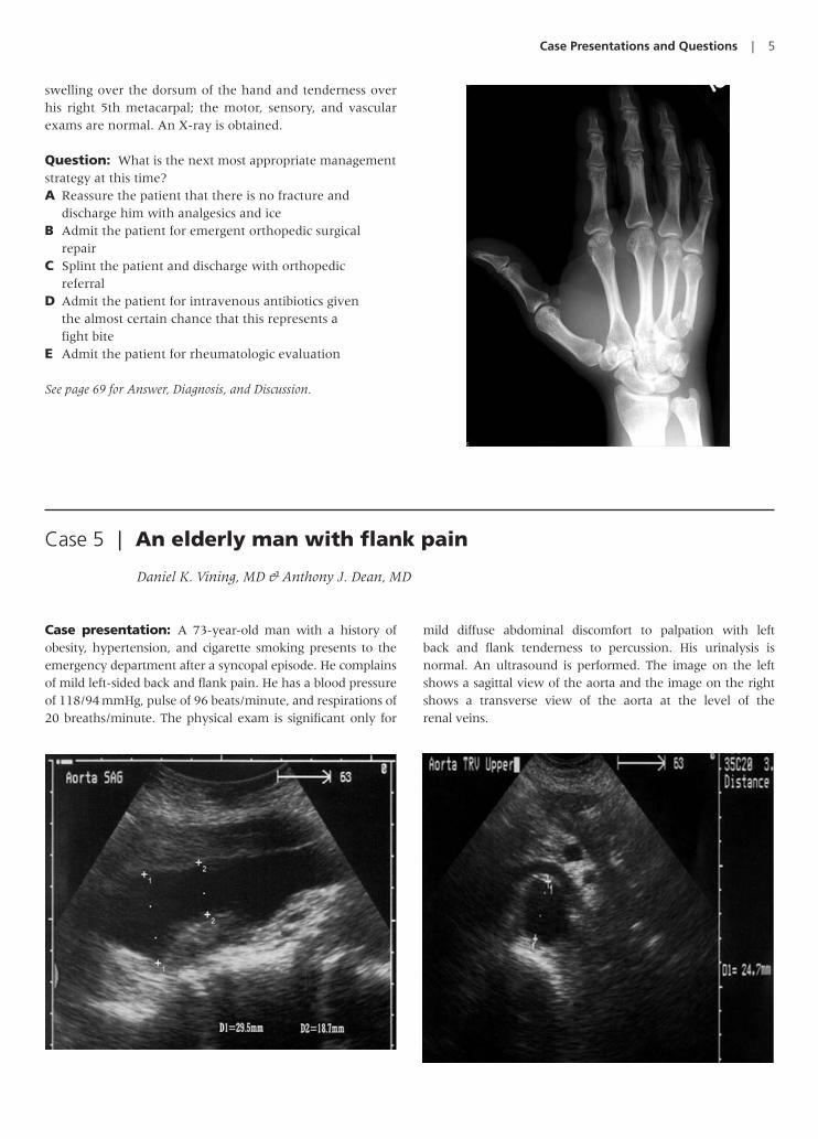
Daniel K. Vining, MD & Anthony J. Dean, MD
Case presentation: A 73-year-old man with a history ofobesity, hypertension, and cigarette smoking presents to theemergency department after a syncopal episode. He complainsof mild left-sided back and flank pain. He has a blood pressureof 118/94 mmHg, pulse of 96 beats/minute, and respirations of20 breaths/minute. The physical exam is significant only for
mild diffuse abdominal discomfort to palpation with left back and flank tenderness to percussion. His urinalysis is normal. An ultrasound is performed. The image on the leftshows a sagittal view of the aorta and the image on the rightshows a transverse view of the aorta at the level of the renal veins.
swelling over the dorsum of the hand and tenderness overhis right 5th metacarpal; the motor, sensory, and vascularexams are normal. An X-ray is obtained.
Question: What is the next most appropriate managementstrategy at this time?A Reassure the patient that there is no fracture and
discharge him with analgesics and iceB Admit the patient for emergent orthopedic surgical
repairC Splint the patient and discharge with orthopedic
referralD Admit the patient for intravenous antibiotics given
the almost certain chance that this represents a fight bite
E Admit the patient for rheumatologic evaluation
See page 69 for Answer, Diagnosis, and Discussion.
6 | Visual Diagnosis in Emergency and Critical Care Medicine
Case presentation: A 21-year-old man complains of leftwrist pain after falling from a 5-foot ladder onto his left
hand. On exam, there is swelling and tenderness over hiswrist and exam is limited due to pain over the dorsum of his
Question: Which of the following is true?A The aorta imaged here has a normal diameter. This
reduces the likelihood of abdominal aortic aneurysm(AAA) to less than 90%.
B The patient has an AAA. If no intra-abdominal free fluidis found in the abdomen, acute aortic aneurysm ruptureis excluded from the differential diagnosis.
C The patient has an AAA. Immediate surgical consul-tation and operative intervention are needed.
D The patient has an AAA. Since the patient is comfortableand hemodynamically stable, he should be admitted for in-patient observation and further evaluation and imaging.
E The aorta imaged here shows significant stenosis. Dopplerflow analysis or angiography is needed to identifywhether this is the cause of the patient’s acute symptoms.
See page 69 for Answer, Diagnosis, and Discussion.
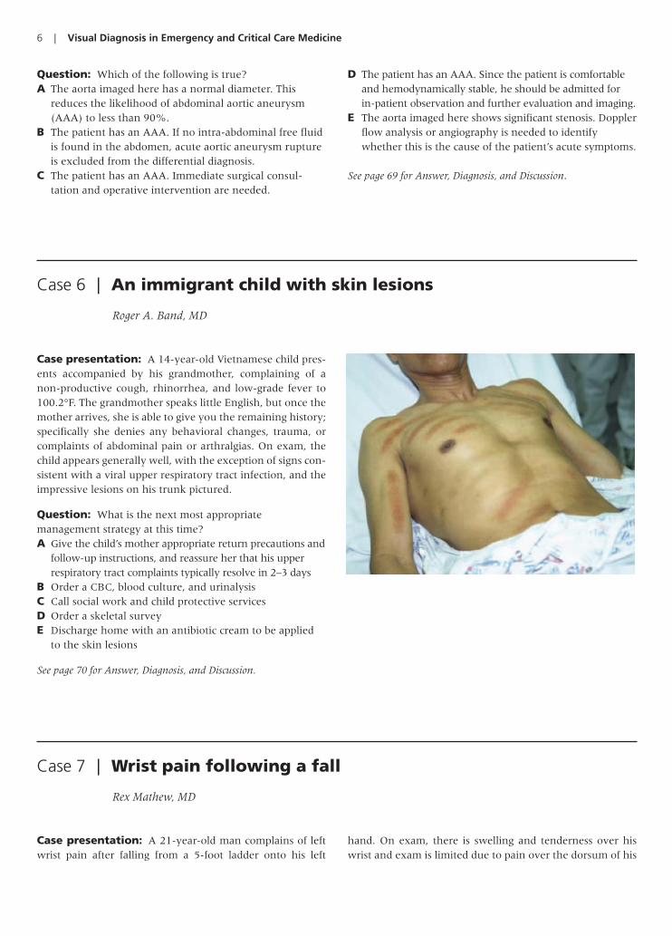
Case 6 | An immigrant child with skin lesions
Roger A. Band, MD
Case presentation: A 14-year-old Vietnamese child pres-ents accompanied by his grandmother, complaining of anon-productive cough, rhinorrhea, and low-grade fever to100.2°F. The grandmother speaks little English, but once themother arrives, she is able to give you the remaining history;specifically she denies any behavioral changes, trauma, orcomplaints of abdominal pain or arthralgias. On exam, thechild appears generally well, with the exception of signs con-sistent with a viral upper respiratory tract infection, and theimpressive lesions on his trunk pictured.
Question: What is the next most appropriatemanagement strategy at this time?A Give the child’s mother appropriate return precautions and
follow-up instructions, and reassure her that his upperrespiratory tract complaints typically resolve in 2–3 days
B Order a CBC, blood culture, and urinalysisC Call social work and child protective servicesD Order a skeletal surveyE Discharge home with an antibiotic cream to be applied
to the skin lesions
See page 70 for Answer, Diagnosis, and Discussion.
Case 7 | Wrist pain following a fall
Rex Mathew, MD

Case Presentations and Questions | 7
Case 8 | Rash in a child with epilepsy
Alexander B. Baer, MD & Christopher P. Holstege, MD
Case presentation: A 3-year-old female with a history ofepilepsy and taking phenytoin presents to the emergencydepartment with a rapidly progressing, painful rash, inabilityto swallow secretions, and difficulty breathing. The rash began5 days previously, at which time her mother noted mild skintenderness, low-grade fever, anorexia, and malaise. The childalso complained of headache and developed diarrhea. She hadbeen seen previously by three different health care providersover the past 5 days; all diagnosed her with a viral exanthema.When the child’s mother awoke on the morning of arrival tothe emergency department, she found her child with markedprogression of the rash pictured at right that now involves hermucus membranes diffusely. In the emergency department,her vitals are as follows: blood pressure 67/34 mmHg, pulse160 beats/minute, respirations 38 breaths/minute, and tem-perature 38.5°C. She is intubated and pictured at right. Her laboratory studies demonstrate that she has both renal andhepatic dysfunction.
Question: Which of the following is correct regarding thischild’s condition?A This child would be expected to have a positive
Nikolsky’s signB If this child had been started on antibiotics earlier, she
would never have required intubationC With proper treatment, this child’s chances of survival
are greater than 90%D This is a primary dermatologic condition, with other
organ involvement not expected
E She should be restarted on her phenytoin to avoidseizure complications
See page 72 for Answer, Diagnosis, and Discussion.
hand; the motor function is strong, and the sensory andvascular exams are otherwise normal. The wrist radi-ographs are noted as shown in the picture.
Question: What is the most likely injury?A Scaphoid fractureB Lunate dislocationC Perilunate dislocationD No fracture or dislocation; normal X-rayE 5th metacarpal fracture
See page 71 for Answer, Diagnosis, and Discussion.

8 | Visual Diagnosis in Emergency and Critical Care Medicine
Case 9 | Dark urine in an immigrant
Suzanne M. Shepherd, MD, Susan A. O’Malley, MD & William H. Shoff, MD
Case presentation: A 38-year-old Egyptian male com-plains of progressive fatigue, skin and eye yellowing, crampyabdominal and low back pain, and darker urine over the past 2days. He denies similar previous episodes. He notes no recenttravel and no use of prescriptions, illicit drugs or herbal medi-cines. He denies alcohol ingestion, raw or tainted foods. He atebaked fish and lightly boiled broad beans approximately 16hours prior to the onset of illness. Vital signs are remarkableonly for a pulse of 108 beats/minute. Conjunctiva are picturedbelow; abdominal exam reveals mild diffuse tenderness. Thespun urine is pictured below.
Question: Which of the following tests would be the mostappropriate next step in his evaluation?A Non-contrast computerized tomography (CT) scan to
evaluate for nephrolithiasisB Urine microscopy and peripheral blood smearC Emergent endoscopic retrograde
cholangiopancreatographyD Ultrasound of the gallbladderE Chest and abdominal flat plate X-ray
See page 73 for Answer, Diagnosis, and Discussion.
Case 10 | Fever and drooling in a child
Sarah G. Winters, MD & Brendan E. Carr, MD
Case presentation: An 18-month-old female presentswith 2 days of fever and irritability. On the day of arrival tothe emergency department, her mother reports the child hasdeveloped new onset of drooling and decreased oral intake.Her past medical history is unremarkable. On physical exam-ination, she is febrile, ill appearing, and has neck stiffness.The following soft tissue lateral of the neck X-ray is obtained.
Question: What is the next most appropriatemanagement strategy at this time?A Discharge to home with supportive careB Discharge to home with oral antibioticsC Admit for intravenous antibiotics and surgical consultationD Emergent intubationE Cricothyroidotomy
See page 73 for Answer, Diagnosis, and Discussion.
Case Presentations and Questions | 9
aVR
aVL
aVF
I
II
III
V1
V2
V3
V4
V5
V6
Case 11 | Altered mental status with an abnormal electrocardiogram
William J. Brady, MD
Case presentation: A 36-year-old female presented to theemergency department via private automobile with lethargyand weakness. Her past medical history and any further detailsregarding the current history of present illness were unavail-able. On examination, the patient was lethargic but arousable;vitals signs were: blood pressure 100/70 mmHg, pulse 75beats/minute, and respirations 16/minute. The remainder ofthe examination was unremarkable except for an apparentdialysis shunt in the left upper extremity. The electrocardio-gram (ECG) is seen below.
Question: In this patient, the most likely diagnosis of thisrhythm disturbance is:A Ventricular tachycardiaB Idoventricular rhythmC Junctional rhythm with bundle branch blockD Sinoventricular rhythmE Normal sinus rhythm
See page 74 for Answer, Diagnosis, and Discussion.
Case 12 | Purulent eye discharge in an adult
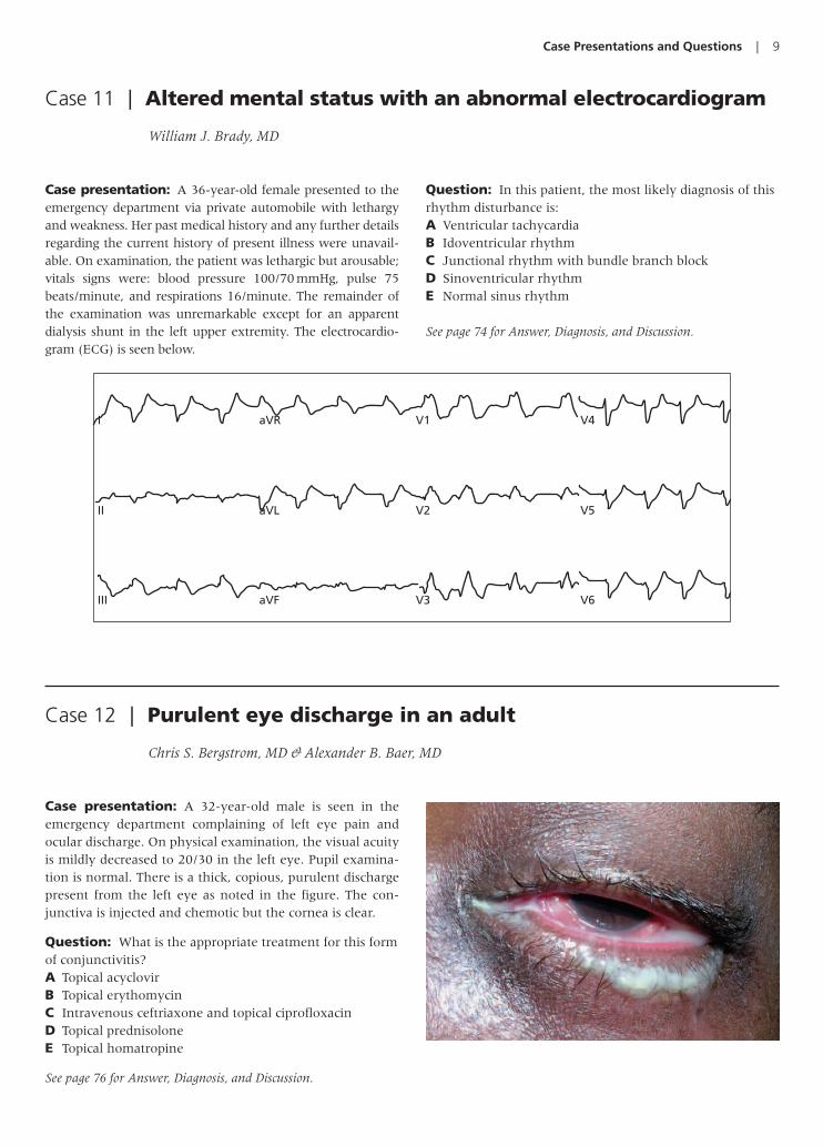
Chris S. Bergstrom, MD & Alexander B. Baer, MD
Case presentation: A 32-year-old male is seen in theemergency department complaining of left eye pain andocular discharge. On physical examination, the visual acuityis mildly decreased to 20/30 in the left eye. Pupil examina-tion is normal. There is a thick, copious, purulent dischargepresent from the left eye as noted in the figure. The con-junctiva is injected and chemotic but the cornea is clear.
Question: What is the appropriate treatment for this formof conjunctivitis?A Topical acyclovirB Topical erythomycinC Intravenous ceftriaxone and topical ciprofloxacinD Topical prednisoloneE Topical homatropine
See page 76 for Answer, Diagnosis, and Discussion.

10 | Visual Diagnosis in Emergency and Critical Care Medicine
Case 13 | Wrist pain in a young child
Craig S. Brummer, MD
Case presentation: An 8-year-old boy fell onto his leftoutstretched arm after tripping on a sidewalk 2 days prior toarrival. He complains of ipsilateral wrist pain that worsenswith movement. On exam, the child is holding a painful,minimally swollen left wrist. His motor, sensory, and vascu-lar exams are normal, but he does have mild tenderness to palpation over his left lateral wrist. An X-ray is obtained andnoted (see figure).
Question: Which management strategy is the mostappropriate for this patient?A Discharge home with no outpatient follow-up necessaryB Hematoma block with closed reductionC Hospital admission with tractionD Emergent orthopedic surgical interventionE Wrist immobilization and analgesic therapy
See page 76 for Answer, Diagnosis, and Discussion.
Case 14 | Postprandial abdominal pain
Anthony J. Dean, MD
Case presentation: A 32-year-old female presents withseveral hours of poorly defined abdominal pain. The pain
has been intermittent for the previous 3 days and most pronounced postprandial. Her abdominal examination is

Case Presentations and Questions | 11
significant for prominent tenderness in both the right upperand lower quadrant. Mild right adnexal tenderness is notedon pelvic examination. The remainder of the examination isunremarkable. A pelvic ultrasound is normal. The patient’sabdominal pain progresses while waiting for an abdominalcomputerized tomography (CT) scan with contrast. Laboratorytests reveal a white blood cell count of 9600/mm3; the serumchemistry, alanine aminotransferase (ALT), aspartate amino-transferase (AST), and serum bilirubin are all normal. A bed-side ultrasound of the right upper quadrant is performed,revealing the images noted.
Question: Which of the following statements is mostaccurate?A These images reveal a normal gallbladder
B These images reveal gallstones with an otherwise normalgallbladder suggesting the diagnosis of either biliary colicor asymptomatic stones unrelated to the patient’spresenting complaint
C These images show no gallstones, but demonstrateevidence of cholecystitis (i.e. acalculous cholecystitis)
D These images reveal gallstones with evidence ofcholecystitis
E These images reveal gallstones and an abnormalgallbladder, but decisions regarding cholecystitis need tobe made clinically, rendering it unlikely given thepatient’s history, exam, and laboratory findings
See page 77 for Answer, Diagnosis, and Discussion.
Case 15 | An elderly man from a house fire
Christopher P. Holstege, MD
Case presentation: A 74-year-old man presents to theemergency department after being transported by emergency medical services from a house fire. He reportedly had beenasleep when the fire broke out. He initially tried to put the fireout, but the fire flashed resulting in extensive burns to hisneck, face, arms, and legs. His facial examination is pictured atright. He is complaining of pain at his burn sites and shortnessof breath. He is expectorating soot-filled sputum. His vital signsreveal pulse 124, blood pressure 98/54 mmHg, respiratory rate38 breaths/minute, and temperature 37.1°C. He is orientatedto person, place, and time. He has a hoarse voice, audible stri-dor, diffuse rales on lung examination, and 40% of his bodysurface area has second and third degree burns.
Question: The next correct step in his managementincludes which of the following?A Administer intravenous fluids at a maintenance rate of
100 mL/hourB Administer an intravenous bolus and then maintenance
infusion of heparinC Avoid administration of opioid analgesics to prevent
sedation
D Initiate bilevel positive airway pressure (BiPap) tomaximize the patient’s oxygenation
E Emergent rapid sequence intubation
See page 78 for Answer, Diagnosis, and Discussion.
Case 16 | Back pain following a fall
Andrew D. Perron, MD & Christopher T. Bowe, MD
Case presentation: A 78-year-old male patient presentsto the emergency department after falling 7 feet from a
ladder onto the ground. He noted immediate pain in thelower back. Examination revealed high lumbar tenderness.

12 | Visual Diagnosis in Emergency and Critical Care Medicine
Case 17 | A bite to the leg in tall grass
Christopher P. Holstege, MD
Case presentation: A 22-year-old male presents to theemergency department with a complaint that a copperheadhad bitten his leg 1 hour ago. He denies any significant painother than mild discomfort where the fangs broke the skin.He has no nausea or vomiting. The examination of theenvenomation site is noted in the figure, with the fang punc-ture wounds circled. He has no tenderness on palpation ofhis leg and his leg compartments are soft. He ambulateswithout difficulty and demonstrates full range of motion ofhis leg.
Question: Which of the following would be appropriatein his management?A Emergent antivenom administrationB Immediate application of a suction deviseC Tourniquet application to his legD Discharge to home if no further development of
symptoms over the next 4 hoursE Incise the bite site and irrigate the wound
See page 80 for Answer, Diagnosis, and Discussion.
Neurologic examination is unremarkable. A radiograph isobtained (see figure).
Question: Which of the following statements is trueregarding the management of this patient?A No further tests are necessary because the patient’s
neurological exam is normalB A computerized axial tomography (CT) scan should be
performedC The patient should be placed in a plaster cast and
discharged with adequate analgesicsD Ultrasound should be performed of the area to rule-out a
significant hematomaE An epidural nerve block should be performed in the
emergency department for pain control
See page 79 for Answer, Diagnosis, and Discussion.

Case Presentations and Questions | 13
Case 18 | Facial swelling in a patient with poor dentition
Alexander B. Baer, MD & Christopher P. Holstege, MD
Case discussion: A 35-year-old male presents to the emer-gency department with a complaint of facial discomfort. Hedeveloped progressive left molar odontalgia over the precedingweek. Over the past 2 days he has been having intermittentfevers and chills. Facial swelling (see figure) began 4 hoursprior to arrival. He denies having any dysphagia, dysphonia, ordyspnea.
Question: What would be the most appropriatemanagement of this patient?A Oral penicillin, opioid analgesics, and outpatient follow-up
with his dentistB Subcutaneous epinephrine, intravenous
methylprednisolone, and diphenhydramine withadmission to a monitored medical floor
C Emergent ultrasound evaluation of the superior vena cavaD Intravenous broad spectrum antibiotics and emergent
neck surgery consultationE Dental alveolar block with lidocaine and outpatient
follow-up with dentistry
See page 81 for Answer, Diagnosis, and Discussion.
Case 19 | Elbow pain in a child after a fall
Alexander B. Baer, MD
Case presentation: An 8-year-old child fell from a treeonto his right outstretched arm. He presents to the emer-gency department 2 hours after the accident with a com-plaint of elbow pain. The patient describes the pain as aconstant ache that is exacerbated with any movement of thearm. On examination, there is moderate swelling localizedto the elbow. Elbow pain occurs with passive movement. Hehas good strength of the hand, and his sensory and vascularexams are normal.
Question: What does this lateral radiograph of the elbowreveal?A Radial head dislocationB Capitellum fractureC Monteggia fractureD Galeazzi fractureE Anterior and posterior fat pads
See page 82 for Answer, Diagnosis, and Discussion.

14 | Visual Diagnosis in Emergency and Critical Care Medicine
Case 20 | A man with diffuse facial edema
Kevin S. Barlotta, MD & Alexander Baer, MD
Case presentation: A 63-year-old male with a history ofneck cancer and subsequent tracheostomy presents with acomplaint of facial “fullness” that rapidly developed over theprevious 24 hours. For the past week, he noted mild facialswelling in the morning, but it rapidly resolved as the day pro-gressed. He denies trauma, fever, or dental discomfort. He is taking no new medications and is not on any angiotensin-converting enzyme inhibitors. His facial examination is pictured.
Question: What is the most likely clinical diagnosis?A AngioedemaB Ludwig’s anginaC Bollous pemphigoidD Superior vena cava syndromeE Stevens–Johnson syndrome
See page 83 for Answer, Diagnosis, and Discussion.
Case 21 | Chest pain and hypotension in an adult male patient
William J. Brady, MD
Case presentation: A 48-year-old man with a history ofdiabetes mellitus complained of substernal chest pain. Onarrival to the emergency department he was pale,diaphoretic, and hypotensive with a blood pressure of70 mmHg by palpation; the pulmonary examination was
unremarkable. An electrocardiogram (ECG) was performedand noted below. Fluid resuscitation was initiated which cor-rected the hypoperfusion. Laboratory studies and a chestradiograph were obtained while appropriate medicationswere administered.
aVR
aVL
aVF
I
II
III
V1
V2
V3
V4
V5
V6

Case Presentations and Questions | 15
Question: The electrocardiogram reveals:A Sinus bradycardia with nonspecific ST-segment–T-wave
abnormalitiesB Sinus bradycardia with inferoposterior RV infarctionC Sinus bradycardia with right ventricular acute
myocardial infarction
D Sinus bradycardia with benign early repolarizationE Sinus bradycardia with anterior myocardial infarction
See page 83 for Answer, Diagnosis, and Discussion.
Case presentation: A 57-year-old patient presents to theemergency department complaining of sharp eye pain, tear-ing, photophobia, and foreign body sensation after beingstruck in the left eye with a tree branch.
Physical examination (pictured below left) reveals mildlyreduced visual acuity to 20/60 in the left eye. Pupillary exam-ination is normal. The left upper eyelid is slightly edematousand the conjunctiva is injected. The cornea is clear withoutevidence of an infiltrate. The anterior chamber is deep andclear. The iris and lens are normal.
Instillation of fluorescein reveals an epithelial stainingdefect with sharp borders measuring 8 mm � 7 mm in size(pictured below right).
Question: Which of the following management plans isthe next appropriate step?A Perform magnetic resonance imaging of the orbitB Administer adequate analgesics and topical antibioticsC Perform an emergent lateral canthotomyD Have the patient insert his contact lensE Administer oral prednisone
See page 85 for Answer, Diagnosis, and Discussion.
Case 22 | Eye pain after tree branch strike
Chris S. Bergstrom, MD & Alexander B. Baer, MD
Case 23 | A missing button battery
Brendan E. Carr, MD & Sarah G. Winters, MD
Case presentation: A 2-year-old boy presents to the emer-gency department with a complaint of food intolerance of
abrupt onset. His mom reports that he was seen playing with asmall calculator just before lunch. She has subsequently

16 | Visual Diagnosis in Emergency and Critical Care Medicine
Case 24 | Acute abdominal pain in pregnancy
Anthony J. Dean, MD
Case presentation: A 17-year-old female presents to theemergency department with pelvic cramping and vaginalspotting for the past 15 hours. Vital signs are stable. Onabdominal exam she has mild lower abdominal tendernesswithout peritoneal signs. Pelvic exam reveals a closed os andmild bilateral adnexal tenderness. A urine pregnancy test ispositive. Blood is obtained for type and screen, and sent forquantitative beta human choriogonadotropin (beta-HCG). A pelvic ultrasound picture is noted below.
Question: Which of the following is true regarding thismidline sagittal transabdominal pelvic sonogram?A With a quantitative beta-HCG of 452 mIU/mL, the
findings of the ultrasound are uninterpretable, since thisis below the discriminatory zone.
B This image shows an early gestational sac consistent with aquantitative beta-HCG of 600 mIU/mL. The patient shouldbe told she has an intrauterine pregnancy and shouldfollow-up with her obstetrician for routine prenatal care.
C This image shows an early gestational sac consistent witha quantitative beta-HCG of 321 mIU/mL. The patientshould be told she probably has an intrauterinepregnancy. Since this is not definite, it is essential thatshe follow-up with her obstetrician for a repeatevaluation and quantitative beta-HCG in 2–3 days.
D With a quantitative beta-HCG of 250 mIU/mL, the imageshows a probable ectopic pregnancy and the obstetricianshould be called for an immediate evaluation in theemergency department.
E With a quantitative beta-HCG of 7430 mIU/mL, thisimage shows an abnormal intrauterine pregnancy andthe patient needs to be referred to obstetrician forevaluation of incomplete abortion.
See page 87 for Answer, Diagnosis, and Discussion.
noticed that the calculator is missing the back and she is con-cerned that he has swallowed the battery. On exam, he is in noapparent distress and is tolerating his secretions. His vital signsare normal. An X-ray is obtained (see figure).
Question: What is the next most appropriatemanagement strategy at this time?A Discharge home and follow with serial outpatient
abdominal X-raysB Administer 25 g of activated charcoal orallyC Admit the patient for intravenous hydration, serial
abdominal X-rays, and stool checks to confirm passageD Infuse 1 mg of glucagon intravenous to decrease lower
esophageal sphincter pressure and monitor over thefollowing 6 hours
E Emergent gastroenterology consultation for endoscopicremoval of foreign body
See page 85 for Answer, Diagnosis, and Discussion.

Case Presentations and Questions | 17
Case 25 | Painless penile ulcer
Andrea L. Neimann, MD
Case presentation: A 35-year-old male presents to theemergency department complaining of a sore that he recentlynoticed on his penis. He describes the sore as painless and hedenies any associated penile discharge. He has never had thisproblem before. He is currently in a stable relationship with aman and recently had an HIV test which was negative. Onexam, there is a single ulcer on the distal end of the penis. Theulcer is 2 cm in diameter and has a slightly raised induratedmargin with a clean base. His inguinal lymph nodes are mildlyenlarged bilaterally. They feel slightly rubbery, but are discreetand non-tender.
Question: What is the next most appropriatemanagement strategy at this time?A Discharge the patient to home without treatmentB Treat the patient empirically for herpesC Be reassured by the patient’s reported negative HIV test
result and suggest no further testing for HIV in the futureD Treat empirically for syphilis based on the history,
physical exam, and knowledge of the epidemiology ofgenital ulcers in your geographic area
E Perform urinalysis and treat based on the results
See page 88 for Answer, Diagnosis, and Discussion.
Case 26 | Low back pain in car accident victim
Edward G. Walsh, MD & William J. Brady, MD
Case presentation: A 48-year-old female was therestrained rear seat passenger in a high-speed motor vehiclecrash. The car was traveling approximately 70 miles/hourand hit an oncoming truck that had crossed the median.There was extensive damage to the vehicle and an on-scenefatality was reported by paramedics. During her prolongedextrication the patient complained of immediate lower backpain and lower extremity paresthesia. She was immobilizedon-scene and transported to the emergency department.Upon arrival, the patient complained of severe lower backpain and bilateral lower extremity paresthesia. She had anotable “seat-belt sign” across her lower abdomen. Herneurological exam was significant for decreased sensationin both lower extremities and decreased dorsiflexion inboth feet. A full trauma evaluation was initiated and multi-ple radiographs including the one shown were obtained.

18 | Visual Diagnosis in Emergency and Critical Care Medicine
Case 27 | A gardener with a non-healing rash
Roger A. Band, MD
Case presentation: This 46-year-old gardener presents to aremote clinic in a Guatemalan village. He complains of nodu-lar, erythematous lesions on his right upper extremity. The
initial lesion on his distal arm eventually ulcerated and otherlesions have been draining non-purulent material. The patientis otherwise without complaints and specifically denies nausea,vomiting, fevers, weight loss, or diarrhea. On exam he appearsgenerally well, with the exception of the impressive lesionsseen in the pictures on his right upper extremity.
Question: What is the most likely cause for these lesions?A Methicillin-resistant Staphylococcus aureusB Scalding injuryC ScabiesD SporotrichosisE Tuberculosis
See page 90 for Answer, Diagnosis, and Discussion.
Question: This presentation is consistent with which type of injury?A T12–L1 dislocationB L5 teardrop fractureC L1 transverse process fracture
D L2 pedicle fractureE L3 burst fracture
See page 89 for Answer, Diagnosis, and Discussion.
Case 28 | An immigrant with neck swelling
Suzanne M. Shepherd, MD, Anthony J. Dean, MD & William H. Shoff, MD
Case presentation: A 45-year-old male immigrant fromMorocco presents with neck discomfort and swelling. Threemonths prior the area was initially swollen with only a
single mass noted. Over the last month, the area began todrain and he has noted an increase in the number of masses.He also has had an unexpected 30-pound weight loss over

Case Presentations and Questions | 19
the last year with associated night sweats. He denies otherillnesses. He lives alone and is employed as a taxi driver.
His examination is unremarkable except for the neckexam depicted in the picture. There are also multiple 2–6 cm,matted, soft, and – in one case – fluctuant, mildly tendermasses along the posterior cervical chain.
Question: Which of the following illnesses would be themost likely cause of this patient’s illness?A MononucleosisB Streptococcal pharyngitisC CytomegalovirusD TuberculosisE Toxoplasmosis
See page 90 for Answer, Diagnosis, and Discussion.
Case 29 | Fall on an outstretched hand in a young adolescent
William J. Brady, MD & Kevin S. Barlotta, MD
Case presentation: A 13-year-old male was playing soccerwhen, at full sprint, he tripped and fell. Both upper extremitieswere extended fully when he impacted the ground on his out-stretched hands. Immediately upon falling, he noted pain in
the right wrist. On examination, he had tenderness in theregion of the distal radius with overlying soft tissue swelling.Radiographs were performed and one is noted here.
Question: The patient can expect the following outcomefrom this injury assuming adequate care is provided:A Complete growth arrest at this injury site with arm
length shorteningB Complete recovery with normal growthC Chronic pain with osteo-necrosis at the injury siteD Chronic disability as a consequence of this injuryE High risk for the development of compartment syndrome
See page 91 for Answer, Diagnosis, and Discussion.
Case 30 | Raccoon eyes
Angela M. Mills, MD
Case presentation: A 25-year-old male was involved in a motorcycle crash without a helmet. The patient had a
documented loss of consciousness at the scene and presentswith a complaint of head pain, dizziness, and nausea. On

20 | Visual Diagnosis in Emergency and Critical Care Medicine
aVR
aVL
aVF
V1
V2
V3
V4
V5
V6
I
II
III
exam there is mild confusion, the findings as shown in thefigure, and a non-focal neurological exam.
Question: What is the next most appropriatemanagement strategy at this time?A Recommend analgesics, ice, and follow-up careB Emergent lateral canthotomyC Order a skull radiographD Order a head computed axial tomography (CT) scan for
possible intracranial injuryE Administer mannitol and dexamethasone emergently
See page 92 for Answer, Diagnosis, and Discussion.
Case 31 | Chest pain and a confounding electrocardiogram pattern
William J. Brady, MD
Case presentation: A 67-year-old male with a past historyof coronary artery disease, diabetes mellitus, and hyperten-sion presented to the emergency department with substernalchest pain. The pain had appeared approximately 2 hours agoand was associated with diaphoresis and nausea. The patientwas treated for acute cardiac ischemia. A 12-lead electro-cardiogram (ECG) is performed and noted below.
Question: Which of the following is the correct statementregarding the ECG presentation of left bundle branch block(LBBB) and possible acute myocardial infarction (AMI)?A LBBB pattern does not confound the clinician’s ability to
use the ECG to diagnose AMI
B Discordant ST segments are most often normal in theLBBB pattern
C Concordant ST segment elevation is a normal finding inthe LBBB pattern
D LBBB pattern is not associated with an increased risk ofpoor outcome in acute coronary syndrome (ACS)patients
E The longer the QRS duration, the greater the risk forACS
See page 93 for Answer, Diagnosis, and Discussion.

Case Presentations and Questions | 21
Case 32 | Eye pain in a contact lens wearer
Chris S. Bergstrom, MD & Alexander B. Baer, MD
Case presentation: A 27-year-old soft contact lens wearerpresents with a complaint of redness, blurred vision, pain,and photophobia in the right eye. The patient admits to “overwearing” her contact lenses, and sleeps in them on a regularbasis. She also states that her eyes are matted together in themorning.
On physical examination, the visual acuity is reduced to20/100 in the right eye. The pupil examination is normal.The conjunctiva is injected and there is a 4 mm oval, white,fluffy opacity on the cornea as noted in the figure.
Slit lamp examination reveals cell and flare in the anteriorchamber. The iris and lens detail are obscured by the opacitybut are otherwise normal.
Question: What is the appropriate management for thiscondition?A Topical steroidB Topical antiviral agentC Topical antibiotics
D Burr removal of this foreign bodyE Oral antiviral agent
See page 94 for Answer, Diagnosis, and Discussion.
Case 33 | Heel pain following a fall
David F. Gaieski, MD
Case presentation: A 26-year-old roofer fell off a 12-foot-high roof and landed on the pavement below. Hewas wearing steel-toed construction boots and landeddirectly on his left heel in a standing position. He feltextreme pain in his heel after the fall and was unable toambulate at the scene. His co-workers called emergencymedical services and he was placed in spinal immobiliza-tion and transported to the emergency department. Onarrival, he complained of left heel pain and midline low back pain. His examination revealed a markedly swollenheel with ecchymosis and tenderness to palpation over theplantar surface of his hind-foot. Examination of his backrevealed bilateral paraspinous muscle tenderness to palpa-tion without midline bony tenderness. An intravenous line was placed, he was given morphine for pain control, anda foot radiograph series was significant for the finding pictured.
22 | Visual Diagnosis in Emergency and Critical Care Medicine
Question: The next most appropriate step in themanagement of this man’s injury is:A Plaster cast immobilization, pain control, and orthopedic
outpatient follow-upB Radiographs of the lumbar spine to rule out
accompanying fracture; orthopedic consultation forpossible operative intervention
C Crutches, non-weight-bearing, pain control, follow-upwith his family physician
D Posterior splint, non-weight-bearing, pain control,orthopedic follow-up for rigid cast in 3–5 days
E Fracture closed reduction and posterior splint
See page 94 for Answer, Diagnosis, and Discussion.
Case 34 | FAST evaluation following trauma
Bon S. Ku, MD & Anthony J. Dean, MD
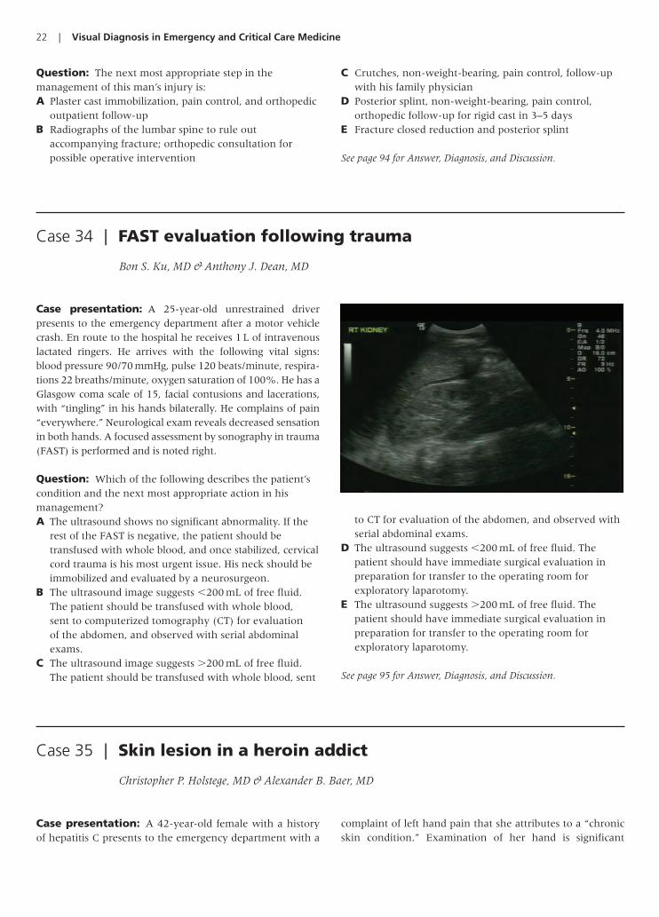
Case presentation: A 25-year-old unrestrained driverpresents to the emergency department after a motor vehiclecrash. En route to the hospital he receives 1 L of intravenouslactated ringers. He arrives with the following vital signs:blood pressure 90/70 mmHg, pulse 120 beats/minute, respira-tions 22 breaths/minute, oxygen saturation of 100%. He has aGlasgow coma scale of 15, facial contusions and lacerations,with “tingling” in his hands bilaterally. He complains of pain“everywhere.” Neurological exam reveals decreased sensationin both hands. A focused assessment by sonography in trauma(FAST) is performed and is noted right.
Question: Which of the following describes the patient’scondition and the next most appropriate action in hismanagement?A The ultrasound shows no significant abnormality. If the
rest of the FAST is negative, the patient should betransfused with whole blood, and once stabilized, cervicalcord trauma is his most urgent issue. His neck should beimmobilized and evaluated by a neurosurgeon.
B The ultrasound image suggests �200 mL of free fluid.The patient should be transfused with whole blood, sent to computerized tomography (CT) for evaluation of the abdomen, and observed with serial abdominalexams.
C The ultrasound image suggests �200 mL of free fluid.The patient should be transfused with whole blood, sent
to CT for evaluation of the abdomen, and observed withserial abdominal exams.
D The ultrasound suggests �200 mL of free fluid. Thepatient should have immediate surgical evaluation inpreparation for transfer to the operating room forexploratory laparotomy.
E The ultrasound suggests �200 mL of free fluid. Thepatient should have immediate surgical evaluation inpreparation for transfer to the operating room forexploratory laparotomy.
See page 95 for Answer, Diagnosis, and Discussion.
Case 35 | Skin lesion in a heroin addict
Christopher P. Holstege, MD & Alexander B. Baer, MD
Case presentation: A 42-year-old female with a historyof hepatitis C presents to the emergency department with a
complaint of left hand pain that she attributes to a “chronicskin condition.” Examination of her hand is significant
Case Presentations and Questions | 23
for the findings shown in the figure. Peripheral intravenousaccess was attempted without success. The patient was dis-covered to have extensive scarring along her bilateral femoraland external jugular veins.
Question: Which of the following best describes her skincondition?A Grand central stationB Skin poppingC Acne vulgarisD PocketingE Acne cystica
See page 96 for Answers, Diagnosis, and Discussion.
Case 36 | Young athlete with back pain
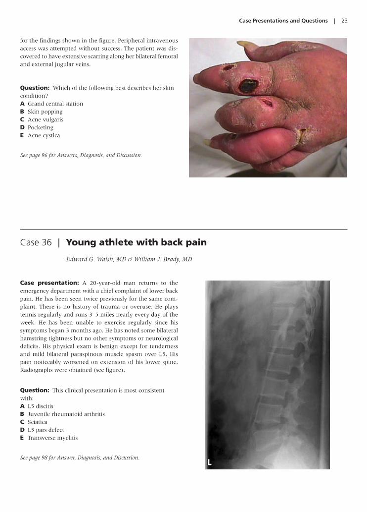
Edward G. Walsh, MD & William J. Brady, MD
Case presentation: A 20-year-old man returns to theemergency department with a chief complaint of lower backpain. He has been seen twice previously for the same com-plaint. There is no history of trauma or overuse. He playstennis regularly and runs 3–5 miles nearly every day of theweek. He has been unable to exercise regularly since hissymptoms began 3 months ago. He has noted some bilateralhamstring tightness but no other symptoms or neurologicaldeficits. His physical exam is benign except for tendernessand mild bilateral paraspinous muscle spasm over L5. Hispain noticeably worsened on extension of his lower spine.Radiographs were obtained (see figure).
Question: This clinical presentation is most consistentwith:A L5 discitisB Juvenile rheumatoid arthritisC SciaticaD L5 pars defectE Transverse myelitis
See page 98 for Answer, Diagnosis, and Discussion.

24 | Visual Diagnosis in Emergency and Critical Care Medicine
Case 37 | Skin lesions in a comatose patient
Christopher P. Holstege, MD
Case presentation: A 24-year-old male presents to theemergency department following an overdose of multiplepills, including sedatives. He was intubated on the scene by
paramedics and transported to the hospital. On examinationof the patient, the skin lesions pictured left are discovered.
A Foley catheter is placed with return of the urine pictured above.
Question: Which of the following is the most appropriateinitial therapy?A Apply silver sulfadiazine cream to the woundsB Administer intravenous broad spectrum antibioticsC Administer adequate intravenous fluids to assure an
adequate urine outputD Perform emergent fasciotomy in the emergency departmentE Administer intravenous potassium
See page 99 for Answer, Diagnosis, and Discussion.
Case 38 | Chest pain with sudden cardiac death
William J. Brady, MD
Case presentation: A 65-year-old man presented to theemergency department with chest pain and syncope. Theexamination demonstrated an alert man in mild distress with normal vital signs; diaphoresis was present on the examination. The patient suddenly slumped over; he was
unresponsive without pulse. The cardiac rhythm strip belowwas obtained.
While attempts were made for electrical therapy, therhythm changed spontaneously to sinus rhythm with apulse. A 12-lead electrocardiogram was obtained and is

Case Presentations and Questions | 25
I
II
III
aVR
aVL
aVF
V1
V2
V3
V4
V5
V6
noted above. Additional diagnostic studies were performedwhile therapy was initiated.
Question: Which of the following would be consistent withthe rhythm strip and electrocardiogram noted in this case?A HypercalcemiaB Hyperchloremia
C HypernatremiaD HypomagnesemiaE Hypophosphatemia
See page 100 for Answer, Diagnosis, and Discussion.
Case 39 | Fall on an outstretched hand with wrist pain
William J. Brady, MD & Kevin S. Barlotta, MD
Case presentation: A 28-year-old male patient presentsto the emergency department after sustaining a fall on hisout-stretched hand (FOOSH). He noted immediate pain butdenied other complaints. Examination revealed snuffboxtenderness. The radiograph as shown was obtained.
Question: In the patient with a FOOSH mechanism withpalpable snuffbox tenderness and a non-diagnosticradiograph, which management course should be pursued?A Discharge home with non-steroidal anti-inflammatory
therapy and no scheduled outpatient physician follow-up.
B Discharge home, prescribe appropriate analgesic therapy,and splint the wrist with instructions to leave in place for 1 week at which time the patient can resume normalactivity.
C Inform the patient that he may have an occult wristfracture, splint the wrist, prescribe appropriate analgesictherapy, and arrange close physician outpatient follow-up.
D Perform magnetic resonance imaging (MRI) at initialevaluation with emergent orthopedic surgeryconsultation.
E Perform fracture reduction under conscious sedation andcast the wrist.
See page 101 for Answer, Diagnosis, and Discussion.
26 | Visual Diagnosis in Emergency and Critical Care Medicine
aVR
aVL
aVF
I
II
III
V1
V2
V3
V4
V5
V6
Case 41 | Chest pain with electrocardiographic ST-segment/T-waveabnormalities
William J. Brady, MD
Case presentation: A 55-year-old female with a historyof hypertension presented to the emergency department
with chest pain. The physical examination was unremark-able. The initial 12-lead electrocardiogram (ECG) is noted
Case 40 | Necrotic skin lesion
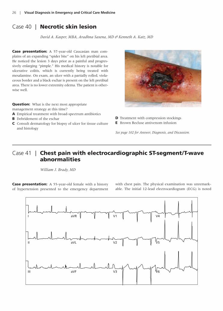
David A. Kasper, MBA, Aradhna Saxena, MD & Kenneth A. Katz, MD
Case presentation: A 57-year-old Caucasian man com-plains of an expanding “spider bite” on his left pretibial area.He noticed the lesion 3 days prior as a painful and progres-sively enlarging “pimple.” His medical history is notable forulcerative colitis, which is currently being treated withmesalamine. On exam, an ulcer with a partially rolled, viola-ceous border and a black eschar is present on the left pretibialarea. There is no lower extremity edema. The patient is other-wise well.
Question: What is the next most appropriatemanagement strategy at this time?A Empirical treatment with broad-spectrum antibioticsB Debridement of the escharC Consult dermatology for biopsy of ulcer for tissue culture
and histology
D Treatment with compression stockingsE Brown Recluse antivenom infusion
See page 102 for Answer, Diagnosis, and Discussion.

Case Presentations and Questions | 27
here. The patient received nitroglycerin and aspirin with resolution of the pain. Serial ECGs did not reveal intervalchange; laboratory studies were negative. The patient ruledout for myocardial infarction.
Question: ECG left ventricular hypertrophy:A Confounds the ECG diagnosis of acute myocardial
infarction
B Does not reduce the ECG’s ability to diagnose acutecoronary syndrome
C Is always associated with ST-segment/T-wave changesD Is an indication for fibrinolysisE Is always associated with hypertension
See page 103 for Answer, Diagnosis, and Discussion.
Case 42 | Chemical eye exposure
Chris S. Bergstrom, MD & Alexander B. Baer, MD
Case presentation: A 34-year-old female is seen in theemergency department after having an unknown chemicalsplashed in her face and eyes. She is complaining of burning,tearing, decreased vision, and light sensitivity. Gross inspec-tion reveals first degree burns to the periorbital skin and lids.The globes are intact.
On physical examination (see figure) the visual acuity is20/200 in each eye. The bulbar and palpebral conjunctiva ismarkedly injected with a watery mucous discharge. Thecorneas are hazy with blurred iris detail. There is a 6 mm ovalarea of blanched bulbar conjunctiva inferiorly near the limbus.The anterior chambers are deep and the pupils are round.
Question: What emergent action should be initiated priorto completing the ophthalmic examination?A Emergent ophthalmology consultationB Litmus testC Irrigation of the eye with copious fluids such as saline or
lactated ringersD Tetanus prophylaxis
E Neutralization with a weak acid or base for a base- oracid-offending agent, respectively
See page 104 for Answer, Diagnosis, and Discussion.
Case 43 | Hand pain after striking a wall
William J. Brady, MD & Kevin S. Barlotta, MD
Case presentation: A 24-year-old intoxicated male pres-ents to the emergency department after striking a wall at anoblique angle. He noted significant pain in his hand anddenied any other complaints. On examination, the hand
demonstrated significant tenderness and soft tissue swellingin the medial-dorsal area. Radiographs, an anteroposteriorview of the hand/wrist and lateral view of the hand/wrist,were obtained as shown in the figures.

28 | Visual Diagnosis in Emergency and Critical Care Medicine
Case 44 | Dyspnea in an alcoholic
Anthony J. Dean, MD
Case presentation: A 50-year-old alcohol abuser presentscomplaining of shortness of breath and abdominal and backpain “like my pancreas”. The patient is agitated and refuses to lie down. She is a heavy smoker and noncompliant with medications for hypertension. The patient denies trauma but has contusions and abrasions on her hands and arms invarious stages of healing. Her vitals are blood pressure
106/85 mmHg, heart rate 124 beats/minute, respiratory rate32/minute. Pulse oximetry is 90% on 2 L nasal cannula. Withexamination limited by the patient’s inability to cooperate, thefollowing are noted: dry mucous membranes, no jugularvenous distension, distant heart sounds, and grossly symmet-ric breath sounds. The following bedside ultrasound imagesare obtained by the examining physician.
Question: The most likely injury in this type ofpresentation is:A Closed fist injury with a “fight bite”B Scaphoid fractureC Carpometacarpal dislocationD Scapholunate dissociationE Radial head fracture
See page 104 for Answer, Diagnosis, and Discussion.

Case Presentations and Questions | 29
Question: Which of the following is true?A With these images, the patient should be sent for
emergent head computerized tomography (CT) toexclude intracranial vascular catastrophe as a cause forher abnormal mental status.
B These images demonstrate intraperitoneal fluidsuggestive of hemorrhage. Whether this is due to acuteaortic aneurysm or occult trauma, the patient requiresimmediate surgical evaluation for possible emergentoperative management.
C These images, combined with the absence of muffled heart sounds, jugular venous distension,
and hypotension, make tamponade exceedinglyunlikely.
D These images show pericardial effusion with evidence of dissection in the abdominal aorta. This suggests anacute proximal dissection, mandating immediateconsultation with a cardiothoracic surgeon for operative repair.
E These images show large pericardial effusion. Thediagnosis of tamponade will be made based on adynamic assessment of cardiac filling during diastole.
See page 105 for Answer, Diagnosis, and Discussion.
Case 45 | Slash wound to the neck
Kevin S. Barlotta, MD & Alexander B. Baer, MD
Case presentation: A 35-year-old female presents to theemergency department after an altercation. She states thatshe was attacked with a hunting knife. She complains of
only pain at the wound site. She denies voice changes or dif-ficulty swallowing. Her injury is depicted left.
Question: What zone of injury is represented in theimage?A Zone IB Zone IIC Zone IIID Zone IVE Zone 0
See page 107 for Answer, Diagnosis, and Discussion.

30 | Visual Diagnosis in Emergency and Critical Care Medicine
Case 46 | Foot pain following breaking
Munish Goyal, MD
Case presentation: A 22-year-old female presents to theemergency department with left foot pain. She was the
restrained driver of a car which struck another car head-on.She used her left foot to compress the brake pedal just prior toimpact. She complains of decreased movement of her toes andsays she can’t walk on her left foot. She denies loss of con-sciousness or any other complaints. Her foot appears mildlyswollen, is exquisitely tender along the midfoot, and has nor-mal distal pulses and capillary refill. The following foot radio-graphs obtained are ordered from triage.
Question: What is the most appropriate managementstrategy at this time?A Place the patient in a posterior splint, give her crutches,
and refer her to an orthopedic specialist as an outpatientB Obtain an orthopedic consult, send pre-operative labs,
and offer the patient analgesicsC Obtain an emergent angiogram to rule out a vascular
injuryD Obtain an emergent orthopedic consult, administer
conscious sedation, and perform an emergent reductionE Administer a hematoma block and perform an emergent
reduction
See page 108 for Answer, Diagnosis, and Discussion.
Case 47 | Confluent rash in a child
Sarah G. Winters, MD & Brendan E. Carr, MD
Case presentation: A 2-year-old boy with no medicalhistory presents to the emergency department with complaints
of a diffuse rash over bilateral lower extremities for the past2 days that is now progressing to his trunk and upper

Case Presentations and Questions | 31
extremities. He is otherwise playful and well appearing withno complaint of itching or fever. His parents deny new
detergents, creams, or drug exposures. His parents report mildupper respiratory symptoms 1 week ago. On physical examhe has multiple confluent lesions with central clearing dif-fusely. The lesions are present on palms and soles but aremost prominent on his bilateral lower extremities. There isno conjunctival injection and there are no sores in or aroundhis mouth or genital area.
Question: What is the next most appropriatemanagement strategy at this time?A Obtain complete blood count (CBC) and blood culture,
administer ceftriaxone, and admit for observationB Obtain CBC and blood culture, but do not treat with
antibioticsC Discharge to home with diphenlhydramine as needed for
itchingD Consult dermatology emergentlyE Administer subcutaneous epinephrine immediately
See page 109 for Answer, Diagnosis, and Discussion.
Case 48 | Lost in the cold
Adam K. Rowden, DO & Christopher P. Holstege, MD
Case presentation: A 38-year-old male presents to theemergency department after being lost in the wilderness. Hisfeet and legs (pictured in the figure) are ice cold with poorpulses. He complains of marked pain and numbness of hislower extremities. His vital signs are normal, including acore body temperature of 37.1°C.
Question: Which of the following is the most appropriateinitial management?A Gradual rewarming with infrared warming lightsB Rapid rewarming of the afflicted extremities in a warm
water (40–42°C) bath for 30 minutesC Urgent surgical consultation for early debridement and
fasciotomyD Vigorous massage in addition to rewarmingE Heparin therapy and administration of warm
intravenous fluids
See page 109 for Answer, Diagnosis, and Discussion.

32 | Visual Diagnosis in Emergency and Critical Care Medicine
Case 49 | Bradycardia following an herbal ingestion
Alexander B. Baer, MD
Case presentation: A 23-year-old male native of HongKong presents with near syncope after a prodrome thatincluded nausea, vomiting, extremity paresthesias, and dys-pnea. The initial vital signs are pulse 32 beats/minute, bloodpressure 75/32 mmHg, respirations 22 breaths/minute. Theremainder of his physical examination is unremarkable. Thefamily brings in an herbal product with a picture of this planton the label.
Question: What would be the next most appropriate stepin his management?A Administer intravenous epinephrineB Administer intravenous physostigmineC Administer intravenous adenosineD Administer intravenous diltiazemE Administer intravenous atropine
See page 110 for Answer, Diagnosis, and Discussion.
Case 50 | Abdominal pain in an alcoholic
Angela M. Mills, MD
Case presentation: A 45-year-old male with a history ofalcohol abuse presents to the emergency department withmidepigastric abdominal pain radiating straight through tohis back and associated with nausea and vomiting. His lab-oratory values are consistent with the diagnosis of acutepancreatitis. A plain film of the abdomen is obtained.
Question: What is the name of this radiologic finding?A Double bubble signB Target signC Sentinel loopD String of pearls signE Coffee-bean sign
See page 111 for Answer, Diagnosis, and Discussion.

Case Presentations and Questions | 33
Case 51 | Pain out of proportion to examination
Adam K. Rowden, DO
Case presentation: A 47-year-old female presents withsevere leg pain and swelling over the past 12 hours. She wasworking in her yard 2 days ago and sustained a small punc-ture wound to the area. She describes the initial wound astrivial. One day following the original injury, the pain beganand steadily increased; she now cannot bear weight orambulate. She has had low-grade fevers at home along with generalized malaise. Her vitals are significant for pulse 125 beats/minute, a blood pressure 88/43 mmHg, respira-tions 28/minute, temperature 38.1°C. Physical examinationof her leg is pictured below. Her exam is hampered due tothe extreme pain induced with even light touch and passivemotion her leg.
An X-ray is obtained and is noted below.
Question: Which of the following is most appropriate inthe management of this patient’s wound?
A Thorough wound irrigation, tetanus prophylaxis, paincontrol, oral antibiotics, and outpatient follow-up in 1 week.
B Pain control, tetanus prophylaxis, one dose ofintravenous antibiotics in the emergency departmentand outpatient follow-up in 48 hours.
C Tetanus prophylaxis and pain control. Follow-upoutpatient in 5 days.
D Tetanus prophylaxis, thorough wound irrigation, paincontrol, and inpatient admission to general medicine forintravenous antibiotics.
E Intravenous antibiotics, fluid resuscitation, and emergent surgical consultation for wound debridement.
See page 111 for Answer, Diagnosis, and Discussion.
Case 52 | Pleuritic chest pain in a young adult male
William J. Brady, MD
Case presentation: A 32-year-old male without medicalhistory presented via ambulance to the emergency depart-ment with chest pain. The pain was left-sided in location andworsened upon both inspiration and reclining. The exami-nation revealed a young patient in moderate distress due to
chest pain. A rhythm strip and a 12-lead electrocardiogram(ECG) were noted (see figures). Laboratory studies were nor-mal and a chest radiograph revealed a normal heart size andlung fields. The patient received intravenous morphine sul-fate and ketorolac, which reduced the pain.

34 | Visual Diagnosis in Emergency and Critical Care Medicine
aVR
aVL
aVF
I
II
III
V1
V2
V3
V4
V5
V6
A
B
Question: The ECG in a patient with the diseaserepresented in this case can show all of the following except:A Diffuse ST segment elevationB Electrical alternansC PR segment changes
D Prominent Q wavesE T-wave inversion
See page 112 for Answer, Diagnosis, and Discussion.
Case 53 | Eye pain following a bar fight
Chris S. Bergstrom, MD & Alexander B. Baer, MD
Case presentation: A 35-year-old male is seen in theemergency department after a bar fight. He has several faciallacerations from a broken bottle. He is complaining of mildperiorbital pain and epiphora. On physical examination hisvisual acuity is 20/30 in the left eye. His pupils are equallyround and responsive to direct and consensual stimulation.There is a superficial laceration of the upper lid that does not
involve the lid margin and without evidence of prolapsedorbital fat. The lower lid has two full thickness lacerationsthrough the lid margin, one of which is medial to the lowerpunctum. The conjunctiva, sclera, and cornea are normal.The anterior chamber is deep and clear. The iris is normaland the pupil is round.

Case Presentations and Questions | 35
Case 54 | Forearm fracture after falling
Alexander B. Baer, MD
Question: Periorbital laceration with prolapsed orbital fatimplies damage to what structure?A Optic nerveB Lacrimal drainage systemC CorneaD Orbital septumE Anterior maxillary wall
See page 112 for Answer, Diagnosis, and Discussion.
Case presentation: A 20-year-old male fell on an out-stretched upper extremity while snowboarding. He presentswith obvious arm deformity. Radiographs of the elbow areobtained and are pictured below.
Question: What is the name of the fracture pictured inthe radiographs?A Boxer’s fractureB Tear drop fracture
C Tillaux fractureD Galeazzi fractureE Monteggia fracture
See page 113 for Answer, Diagnosis, and Discussion.

36 | Visual Diagnosis in Emergency and Critical Care Medicine
Case 55 | An elderly woman with groin pain
Brendan E. Carr, MD
Case presentation: A 65-year-old woman presents to theemergency department with the complaint of atraumaticprogressive groin and buttock pain over the last severalmonths. She has curtailed her activities and is now unable toambulate. Her past medical history is significant only for sar-coidosis and end stage renal disease. She is currently takingprednisone. On exam, there is pain with range of motion andaxial load of the left hip. The X-ray shown right is obtained.
Question: All of the following are associated with thiscondition EXCEPT:A CorticosteroidsB SarcoidosisC Sickle cell diseaseD Previous hip fracture or dislocationE Renal disease
See page 114 for Answer, Diagnosis, and Discussion.
Case 56 | Painful facial rash
Chris S. Bergstrom, MD & Alexander B. Baer, MD
Case presentation: A 63-year-old male is seen in theemergency department complaining of a painful rash overthe left side of his forehead along with blurred vision, red-ness, and light sensitivity in the left eye. He states that hissymptoms started yesterday. Ocular examination reveals a vesicular rash on the left side of the forehead that respectsthe midline and involves the upper lid but does not affect the tip of the nose. There is a mucous discharge present. Slit lamp examination shows conjunctival injection. Thecornea is clear without evidence of staining or infiltrates.The anterior chamber is deep and quiet. The iris and lens are normal.
Question: What is Hutchinson’s sign?A Scrapings of the rash demonstrate hyphae when exposed
to potassium hydroxideB Rash involvement of the tympanic membraneC Fluorescence of the rash when placed under a Wood’s
lamp
D Rash involvement of the corneaE Rash involvement of the nose
See page 114 for Answer, Diagnosis, and Discussion.

Case Presentations and Questions | 37
Case 57 | Confusion, anemia, and abdominal pain in a toddler
Alexander B. Baer, MD & Christopher P. Holstege, MD
Case presentation: A 4-year-old child was seen 3 weeksago by his pediatrician for abdominal cramping and constipa-tion. The child is an immigrant from Africa and has no signifi-cant past medical history. The laboratory evaluation performedduring the last office visit demonstrated a hypochromic, micro-cytic anemia which was thought to be secondary to iron defi-ciency. The child now presents to the emergency departmentwith anorexia, increasing lethargy, and ataxia. The radiographshown right is obtained.
Question: What is the treatment of choice for this child?A D-penicillamineB 2,3-dimercaptosuccinic acid (DMSA or succimer)C Calcium disodium ethylene diamine tetraacetate
(CaNa2EDTA)D Dimercaprol (British Antilewisite or BAL)E Deferoxamine
See page 115 for Answer, Diagnosis, and Discussion.
Case 58 | Cardiotoxic effects following caustic ingestion
Alexander B. Baer, MD & Christopher P. Holstege, MD
Case presentation: A previously healthy 47-year-old maleaccidentally ingested a blue liquid he thought was a sport drink.He immediately noted throat irritation and within 5 minutes of the ingestion, he developed nausea and vomiting. He pre-sented to the emergency department within 1 hour of theingestion with a complaint of nausea, weakness, and intensepleuritic chest pain. His initial vital signs revealed temperature34.5°C, pulse 130 beats/minute, blood pressure 102/66 mmHg,and respirations 20 breaths/minute. His voice was hoarse and he had difficulty swallowing his secretions. His exam was significant for an oropharynx with inflamed mucosa and anabdomen that was soft with mild tenderness diffusely.
His initial electrocardiogram (ECG 1, see figure on nextpage) 1 hour after ingestion was pictured on the next page.His initial arterial blood gas revealed pH 7.28, pCO2 29, pO2 of
209, and HCO3 13. Within one and half hour of his arrival, hebecame increasingly agitated and his systolic blood pressuredropped to 80 mmHg. A repeat ECG (ECG 2) was noted (seefigure on next page). Initial bedside evaluation of the ingestedfluid by litmus paper revealed a pH less than 4.0.
Question: What electrolyte abnormality is most likelypresent on laboratory analysis?A HypermagnesemiaB HypocalcemiaC HypochloremiaD HypernatremiaE Hypophosphatemia
See page 115 for Answer, Diagnosis, and Discussion.

38 | Visual Diagnosis in Emergency and Critical Care Medicine
aVR
aVL
aVF
I
II
III
V1
V2
V3
V4
V5
V6
Case 59 | Rash and joint pain in a child
Mara Becker, MD
Case presentation: An 8-year-old boy presents to theemergency department with a rash for 2 days and an inabil-ity to ambulate due to bilateral ankle pain. The familyreports that the rash began on his legs and is now more gen-eralized. It is not painful nor pruritic. The child is wellappearing with normal vital signs. The skin lesions (see fig-ures on the next page) are palpable and purple in hue and do
not blanch with pressure. The ankles are warm to the touchand have minimal periarticular swelling, but no intra-articu-lar fluid. On exam the right wrist is also painful and warmwith periarticular swelling. The rest of the exam is normal.Laboratory tests reveal a normal complete blood count,coagulation studies, electrolytes, and urinalysis. A blood cul-ture is pending.
ECG 1.
ECG 2.

Case Presentations and Questions | 39
Case presentation: A 65-year-old male presents to theemergency department with mild abdominal pain. He is 2 days post-laparoscopic cholecystectomy and notes pain at one of his incision sites, worse when he ambulates. He istolerating a normal diet without nausea or vomiting, had anormal bowel movement today, and is urinating withoutdifficulty. His abdominal incisions are well healing withouterythema, fluctuance, or drainage. His abdomen is soft, withmild incision tenderness, normal bowel sounds, and withoutdistension. The plain film (see figure) is ordered from triage.
Question: The X-ray findings are consistent with which ofthe following?A PneumoniaB Post-operative pneumoperitoneumC Large bowel obstructionD Biliary airE Kidney stone
See page 117 for Answer, Diagnosis, and Discussion.
Case 60 | X-ray findings after laparoscopy
Munish Goyal, MD
Question: What would be the next step in managing thispatient?A Discharge home with close follow-up by the primary
care doctor and anti-inflammatory medications for thejoint pain
B Admit for observationC Admit for intravenous antibiotic therapyD Consult orthopedics for an ankle arthrocentesisE Administer subcutaneous epinephrine
See page 116 for Answer, Diagnosis, and Discussion.

40 | Visual Diagnosis in Emergency and Critical Care Medicine
Case 62 | Chest pain in a middle-aged male patient with ST segment elevation
William J. Brady, MD
Case presentation: A 42-year-old male presented to the emergency department with chest pain. The examina-tion revealed no distress with normal vital signs. A 12-leadelectrocardiogram (ECG) is noted below. The patientreceived appropriate medications while laboratory studies
and chest radiograph were performed; all such studies werewithin normal limits. Serial ECGs did not demonstrate achange in the ST segment elevation nor any further ST-segment or T-wave abnormality.
Case 61 | Injector injury to the hand
Tracy H. Reilly, MD
Case presentation: A 30-year-old automobile technicianpresents to the emergency department with a complaint ofpain and swelling in the dorsum of his left hand near themetacarpal–phalangeal (MCP) joint of the index finger afterinjury with a grease injector. On exam there is slight swellingof the dorsum of the hand and a small pin-point puncturewound just proximal to the MCP joint of the index finger asnoted in the picture. There is pain with passive movement,good capillary refill of the index finger and thumb, and noneurosensory deficits distal to the injury are appreciated.
Question: What is the most appropriate management?A Check tetanus status, prescribe analgesics, and discharge
homeB Prescribe antibiotics and analgesics, check tetanus status,
and discharge homeC Obtain an X-ray, check tetanus status, immobilize with a
splint, and discharge home with a prescription forantibiotics and analgesics and instructions to follow-upwith an orthopedist in 3–5 days
D Obtain an X-ray, check tetanus status, and arrange animmediate surgical consultation for exploration anddecompression debridement
E Insert a 14-gauge angiocatheter and aspirate the injectedmaterial
See page 117 for Answer, Diagnosis, and Discussion.
I aVR
aVL
aVF
V1 V4
V5
V6V3
V2II
III

Case Presentations and Questions | 41
Question: The ECG pattern benign early repolarization is:A Most often seen in elderly patientsB Indicative of underlying cardiac pathologyC A non-infarction cause of ST segment elevation
D Associated with an increased risk of sudden cardiac deathE Associated with valvular heart disease
See page 118 for Answer, Diagnosis, and Discussion.
Case 63 | Deformed globe following trauma
Joseph Robson, MD & Worth W. Everett, MD
Case presentation: A 79-year-old female was punched inthe face during an assault and complains of pain and decreasedvision in the left eye. On physical exam, there is minimal lightperception on the left, and extraocular movement is decreased.Exam of the pupil is limited by blood in the anterior chamber.The deformity of her globe is shown right.
Question: Which of the following is the appropriate nextstep in the management of this patient?A Access intraocular pressureB Administer ocular steroidsC Discharge the patient with outpatient ophthalmology
follow-upD Administer anti-emetics, opioids, antibiotics, and call
ophthalmologist for emergent consultationE Perform emergent lateral canthotomy
See page 119 for Answer, Diagnosis, and Discussion.
Case 64 | Adult male with atraumatic lower back pain and leg weakness
William J. Brady, MD
Case presentation: A 46-year-old male presented to theemergency department with lower back pain of 3 days dura-tion. The patient denied trauma but noted previous back painwith a radicular distribution in the right lower extremity. The
patient noted the recurrence of this pain with the onset ofweakness in the legs. On examination, lower extremityweakness was noted in symmetric fashion, reduced sensationwas found in the perineum, and the rectal examination

42 | Visual Diagnosis in Emergency and Critical Care Medicine
Case 65 | Fever and rash in a child
David L. Eldridge, MD
Case presentation: A 2-year-old male presents with a 6-day history of fever (temperatures up to 101°C and greatereach day) and irritability. Accompanying this fever is the rashpictured. This rash is distributed along his face, trunk, andflexural surfaces of his extremities. He also has nonexudativeconjunctivitis and dry, cracked, erythematous lips. With pal-pation of his neck, discover that he has a large (2.0 cm), firm,mobile, tender, left-sided anterior cervical lymph node. Hishands and feet appear edematous.
Question: Which of the following is associated with thisclinical syndrome?A Hemorrhagic gastritisB Acute renal failureC Intracranial abscessD Coronary artery aneurysmsE Pancytopenia
See page 121 for Answer, Diagnosis, and Discussion.
demonstrated diminished tone. A post-void residual revealed500 mL of urine remained in the bladder. Plain film radi-ographs were performed and demonstrated minimal degener-ative change. Magnetic resonance imaging (MRI) of the lowerspine was performed and is shown in the figure.
Question: What is the likely neurologic outcome of thisclinical entity if appropriate medical care is offeredimmediately?A Complete loss of motor and sensory function in the
lower extremitiesB Complete normalization of bowel and bladder
functionC Normalization of motor function with progression of
bladder dysfunctionD Stabilization of the current neurologic status without
worseningE Paraplegia
See page 120 for Answer, Diagnosis, and Discussion.

Case Presentations and Questions | 43
Case 66 | Yellow eyes and skin
Jane M. Prosser, MD
Case presentation: A 48-year-old female presents to theemergency department with the chief complaint “My eyesare yellow.” She denies having any other clinical complaints.Her physical exam is unremarkable except for what is pic-tured at right. No abdominal tenderness is elicited.
Question: Which of the following statements is true?A Her scleral icterus effectively rules out hemolysis as a
cause of her conditionB Scleral icterus is a more sensitive sign of
hyperbilirubinemia than generalized jaundiceC The presence of Courvoisier’s sign suggests an infectious
etiology in this patientD The lack of abdominal pain effectively rules out
pancreatic cancer as a cause of her conditionE Vitamin A toxicity is a potential cause of this patient’s
condition
See page 122 for Answer, Diagnosis, and Discussion.
Case 67 | Fishing in the stomach
Christopher P. Holstege, MD
Case presentation: A 3-year-old male was playing with alead fishing sinker. His mother was about to attempt to takeit away from him when he suddenly began choking. By thetime she reached her son, his choking had ceased and he wasactively crying. He subsequently arrives at the emergencydepartment asymptomatic. His physical exam is unremark-able, and an X-ray is obtained (see figure).
Question: Which of the following is the next mostappropriate management step for this patient?A Gastrointestinal lavageB Endoscopic removalC Whole bowel irrigationD Activated charcoal administrationE. Syrup of ipecac administration
See page 122 for Answer, Diagnosis, and Discussion.

44 | Visual Diagnosis in Emergency and Critical Care Medicine
Case 68 | Agitation in a botanist
Alexander B. Baer, MD & Christopher P. Holstege, MD
Case presentation: A 32-year-old male botanist presentsto the emergency department after paramedics received a call from his spouse that he was becoming markedly con-fused. According to his wife, this occurred after he drank atea made from one of his plants, with the plant utilized notedbelow.
The patient is notably agitated, following no commands,constantly moving in the stretcher, picking relentlessly at thebed sheets, and talking with incomprehensible, mumbling
speech. The remainder of his examination is significant forthe following: warm and dry to touch, no bowel sounds,pulse of 140 beats/minute, blood pressure of 160/90 mmHg,temperature of 38.4°C. An examination of his lips and pupils is noted (see figures above and left). A Foley is placedwith return of 1.5 L of urine.
Question: Which of the following could produce thisclinical syndrome?A Jimson weed (Datura stramonium)B Oleander (Nerium oleander)C Lilly of the valley (Convallaria majalis)D Monkshood (Aconitum Napellus)E Death cap mushroom (Amanita phalloides)
See page 123 for Answer, Diagnosis, and Discussion.

Case Presentations and Questions | 45
Case 69 | Skin target lesion
Mara Becker, MD
Case presentation: A medical student rotating in emer-gency department comes for advice. She has developed a newrash on her calf over the past 24 hours. It is erythematous withcentral clearing and it has been expanding as the day progressesand is noted right. It is non-painful and non-pruritic. This is theonly lesion she knows about thus far. She has had no fever andadditionally only complains of some mild fatigue and muscleaches after hiking at Valley Forge in Pennsylvania on her lastweekend off. She asks you if she should call out sick for heremergency department shift tonight.
Question: Which of the following is the next correct stepin her management?A Send her home with instructions to remain in isolation
until the rash resolvesB Initiate therapy with doxycyclineC Perform incision and drainage at the center of the target
lesionD Administer brown recluse spider antivenomE Infuse intravenous gammaglobulin
See page 124 for Answer, Diagnosis, and Discussion.
Case 70 | Adult male with a sudden, severe headache
Andrew L. Homer & William J. Brady, MD
Case presentation: A 55-year-old male presents to theemergency department complaining of the acute onset of asevere global headache while at rest 2 hours prior to presenta-tion. The patient also complains of neck stiffness and nausea.Further history reveals the presence of milder headaches thathave occurred intermittently over the past 2 weeks. His vitalsare significant for a blood pressure of 165/95 mmHg and apulse of 90 beats/minute; he is afebrile. The patient is in signif-icant distress due to pain; the remainder of the examination isnormal. While awaiting further evaluation, he suddenlybecomes unresponsive. A head computerized tomography(CT) scan is performed emergently (see figure).
Question: Which of the following statements is trueregarding this condition?A Pressors should be administered to keep the mean
systolic blood pressure above 160 mmHg

46 | Visual Diagnosis in Emergency and Critical Care Medicine
Case 72 | Chest pain and subtle ST segment elevation
William J. Brady, MD
Case presentation: A 46-year-old male presented to theemergency department with chest pain of 2 hours duration;the pain was accompanied by nausea and diaphoresis. Thepatient appeared pale and diaphoretic; otherwise, the exami-nation was unremarkable. His electrocardiogram (ECG) wasnoted on the next page. The serum troponin was elevated,confirming the diagnosis of acute myocardial infarction (AMI).He was transferred to the catheterization laboratory for percu-taneous coronary intervention. The patient had an uneventfulhospital course.
Question: Choose the incorrect statement regardingreciprocal ST segment depression:A Reciprocal ST segment depression is associated with
an increased chance of poor outcome in ST-elevationmyocardial infarction (STEMI)
B Reciprocal ST segment depression increases theprobability that ST segment elevates results from AMI
C Reciprocal ST segment depression is defined as ST segment depression occurring in the STEMI patient
B Barbiturates are the antiseizure treatment of choice forthis condition
C A classic “lucid interval” is present in one-third ofpatients who present early with this condition
D Lumbar puncture is the definitive treatment of choicefor this patient
E A head CT may be falsely negative in as many as10–15% of cases presenting early with headache
See page 125 for Answer, Diagnosis, and Discussion.
Case 71 | Get them undressed!
Munish Goyal, MD
Case presentation: A 19-year-old male is brought to theemergency department by his friends for fever and confu-sion. The patient is lethargic and warm to touch. His friendsstate he was complaining of a headache this morning andbecame increasingly confused today. He has no medical his-tory and was asymptomatic yesterday. On exam, you notethat he is hypotensive, tachycardic and has the followingskin findings upon fully undressing him (see figure).
Question: What is the next most appropriate managementstrategy at this time?A Administer acetaminophen and order a routine head
computerized tomography scan to evaluate his headacheB Obtain a dermatology consultationC Place the patient in isolation, administer immediate
antibiotics, and draw appropriate culturesD Order an electrocardiogram, alcohol level, electrolytes,
and ammonia level to look for a source of his confusionE Biopsy the skin lesion and send it to pathology
See page 125 for Answer, Diagnosis, and Discussion.

Case Presentations and Questions | 47
D Reciprocal ST segment depression includes the STsegment depression encountered in patients withconfounding patterns such as left bundle branch block
E Reciprocal ST segment depression confers an improvedprognosis
See page 126 for Answer, Diagnosis, and Discussion.
I aVR
aVL
aVF
V1
V2
V3
V4
V5
V6
II
III
Case 73 | Fluid in my eye
Chris S. Bergstrom, MD & Alexander B. Baer, MD
Case presentation: A 22-year-old Caucasian male is eval-uated in the emergency department with a complaint ofmarked left eye pain and blurred vision after being struck inthe eye with a lead fishing weight. On physical exam, hisvisual acuity is 20/60 in the left eye. Pupillary examination isnormal. Slit lamp exam shows a clear cornea. The anteriorchamber is deep with suspended red blood cells in the aque-ous humor (see figure). The iris detail is slightly obscured butotherwise normal with a central, round pupil.
Question: What is the next best step in this patient’smanagement?A Administration of oral aspirinB Infusion of intravenous heparinC Emergent lateral canthotomyD Administration of oral lisinoprilE Administration of atropine 1% ophthalmic drops
See page 126 for Answer, Diagnosis, and Discussion.

48 | Visual Diagnosis in Emergency and Critical Care Medicine
Case presentation: A 19-year-male presents to the emer-gency department 30 minutes after a motor vehicle collisionin which he sustained a head injury. Emergency medical serv-ices reports that he has no recollection of the accident and wit-nesses report that he was unconscious for approximately 1 minute. The patient is now alert and oriented to person,place, and time. He is complaining of only a headache at thesite of impact. On exam, there is a 3 cm laceration on the leftlateral forehead with an underlying bony step-off. Theremainder of his physical exam is benign. Five minutes later,the patient becomes increasingly lethargic which progresses toa complete loss of consciousness. After stabilization, an emer-gent head computed tomography (CT) scan is ordered whichis shown right.
Question: Which of the following statements is true?A A lucid interval is seen in over 90% of patients with this
conditionB This condition is more common in the elderlyC Extravagated blood crosses suture lines in this
conditionD Deaths are rare in patients with this condition if they are
not in coma preoperatively
E Venous blood is most common source of the hematomain this condition
See page 127 for Answer, Diagnosis, and Discussion.
Case 74 | Coma following head trauma
Andrew L. Homer & William J. Brady, MD
Case 75 | Blue hue following endoscopy
Andrew L. Homer & Christopher P. Holstege, MD
Case presentation: A 39-year-old previously healthyfemale presents to the emergency department as a transferfrom an outpatient endoscopy clinic. She had been in her usualstate of health until 45 minutes into her esophagogastroduo-denoscopy when she was noted to have a gradual decrease inher oxygen saturations and dyspnea. On arrival, her vital signswere as follows: pulse 125 beats/minute, blood pressure96/43 mmHg, respiratory rate 36 breaths/minute, temperature37.3°C. Her examination is unremarkable except for the skinfindings shown in the figures (the patient’s hand is on theright, the nurses hand is on the left). Her lungs are clear andher pulses are strong.

Case Presentations and Questions | 49
Question: Which of the following is the appropriateantidote for this toxicity?A Prussian blueB PhysostigmineC Deferoxamine
D Methylene blueE Naloxone
See page 128 for Answer, Diagnosis, and Discussion.
Case 76 | Shoulder pain following direct blow
David F. Gaieski, MD
Case presentation: A 41-year-old male with no signifi-cant past medical history was playing touch football withfriends at his college reunion. He was running with the ball,twisted sideways to try to avoid being tagged, and fell on thelateral aspect of his left shoulder with the right arm bentacross the body. He experienced sudden onset of pain in hisleft shoulder and decreased range of motion. He deniesnumbness, tingling, or weakness in the arm. On examina-tion, he is tender to palpation over his acromioclavicularjoint, there are no breaks in his skin, and he has pain withforced adduction of the arm. He was given ibuprofen forpain control and an X-ray was obtained (see figure).
Question: The next most appropriate step in themanagement of this man’s injury is:A Orthopedic consultation for urgent surgical repairB Figure eight wrap, pain control, and orthopedic
follow-upC Hematoma block and reduction
D Sling for immobilization and comfort, pain control,follow up with orthopedics
E Emergent computerized tomography (CT) scan of theshoulder
See page 129 for Answer, Diagnosis, and Discussion.
Case presentation: A 16-year-old female intentionallyoverdosed on an unknown quantity of “vitamins.” She arrives
to the emergency department 4 hours after overdose com-plaining of nausea, vomiting, and epigastric abdominal pain.
Case 77 | An overdose of prenatal vitamins
Christopher P. Holstege, MD & Adam K. Rowden, DO
50 | Visual Diagnosis in Emergency and Critical Care Medicine
Case presentation: A 10-day-old male is brought to theemergency department by his mother. He has not eaten wellfor the past 24 hours and has reportedly been “very sleepy.”Yesterday he began to develop a rash that now appears redat the base and is progressively “blistering” with clear fluidpictured below on his legs and face. He also has a swollenfinger (right). Tonight he has had two episodes of uncontrol-lable shaking movements of his arms and legs – each lastingfor a “few minutes.” He was born 3 weeks premature and hismother claims no problems or issues with the pregnancy. Heis afebrile but lethargic on exam currently.
Question: Which of the following tests would be leasthelpful in the clinical management of this patient given the likely diagnosis in this case?A Computer tomography (CT) of the headB Viral cultures of eyes, rectum, and skin lesionsC Polymerase chain reaction (PCR) testing of cerebrospinal
fluid (CSF)D Liver transaminase levelsE Chest radiograph
See page 131 for Answer, Diagnosis, and Discussion.
Case 78 | Fever and rash in a child
David L. Eldridge, MD
Her initial vital signs reveal pulse 123 beats/minute, bloodpressure 85/34 mmHg, respirations 24/minute, and tempera-ture 37.2°C. Her examination is significant only for epigastrictenderness on palpation of her abdomen. Her laboratory stud-ies are significant for the following: iron 567 mg/dL, serumbicarbonate 15 mEq/L, glucose 256 mg/dL, white blood countof 13.2 � 109/L. Her X-ray is pictured right.
Question: Which of the following is the next mostappropriate management step for this patient?A Begin an intravenous infusion of deferoxamineB Administer dimercaprol (British Antilewisite, BAL)
intramuscularlyC Infuse calcium disodium ethylenediaminetetraacetate
(EDTA)D Administer succimer (2,3-dimercaptosuccinic acid,
DMSA) orallyE Administer D-penicillamine orally
See page 129 for Answer, Diagnosis, and Discussion.
Case Presentations and Questions | 51
Case presentation: A 21-year-old male patient presentsto the emergency department with ankle and foot pain afterjumping to the ground from a height of approximately 10feet. He is unable to ambulate due to pain. Examinationrevealed medial ankle pain over the deltoid ligament on the medial ankle. Neurovascular examination was normal.A radiograph was performed (see figure).
Question: In this patient with an axial load mechanism,palpable medial ankle tenderness, and the radiograph thatis shown, what emergency department management courseshould be pursued?A Ace wrap, ice, elevation, and discharge homeB Aircast, weight bearing as tolerated, and discharge homeC Splint, crutches, and primary care outpatient follow-upD Potential further imaging (i.e. computerized axial
tomography scan) and orthopedic consultationE Hematoma block and fracture reduction
See page 132 for Answer, Diagnosis, and Discussion.
Case 80 | Diffuse ankle pain following a fall
Andrew D. Perron, MD & Christopher T. Bowe, MD
Case 79 | Lamp oil ingestion
David L. Eldridge, MD
Case presentation: A 2-year-old male was brought byambulance to the emergency department. About 30 minutesbefore presentation, his mother had found him next to asoda bottle that contained lamp oil. The bottle was spilled allover the floor and she smelled lamp oil on his breath. Thechild was also coughing and choking. His mother called foran ambulance. Upon arrival, the emergency personnelfound the child appearing sleepy, but with continued cough.On 2 L/minute of oxygen delivered by nasal cannula, hisoxygen saturation was 89% with a respiratory rate of 55breaths/minute. He had prominent subcostal retractions.Auscultation of his lungs revealed crackles bilaterally anddecreased lung sounds at both bases.
The chest radiograph was taken one hour after the inges-tion (see figure).
Question: Which of the following is most appropriate atthis point in the management of this patient?A Immediate gastric lavage as presentation is still within
one hour of ingestion
B Intravenous broad spectrum antibiotics givenprophylactically
C Intravenous high-dose corticosteroids givenprophylactically
D Continued close monitoring and supportive careE Place nasogastric tube and give activated charcoal
See page 131 for Answer, Diagnosis, and Discussion.

52 | Visual Diagnosis in Emergency and Critical Care Medicine
Case presentation: A 78-year-old woman with a history ofcoronary artery disease experienced progressive weaknessassociated with chest pain. On arrival in the emergency depart-ment, the patient was without distress. The vital signs were sig-nificant only for bradycardia with a rate of 55 beats/minute(bpm). The 12-lead electrocardiogram (ECG) demonstratedsinus bradycardia at a rate of 62 bpm with inferior ST segmentdepression; a bedside qualitative test was positive for troponin.The patient was diagnosed with a non-ST elevation myocardialinfarction (STEMI) while appropriate therapy was initiated.Approximately 45 minutes after arrival, the patient developedhypotension with profound weakness and recurrent chest
pain. The blood pressure was 85 mmHg by palpation and thepulse was approximately 25 bpm. The ECG monitor demon-strated the rhythm below.
Question: The most frequent etiology of this type ofrhythm is:A Acute myocardial infarctionB Cardiotoxic medication ingestionC Acute myocarditisD Primary conduction system diseaseE Pericarditis
See page 133 for Answer, Diagnosis, and Discussion.
Case 82 | Weakness and bradycardia in an elderly female patient
William J. Brady, MD
Case presentation: A 22-year-old college student isdropped off by her friends in the triage area of the emergencydepartment after the friends told the triage nurse that thepatient would not wake up. The patient is lethargic, is arous-able only to painful stimuli and is without obvious signs oftrauma. The presenting vital signs are: pulse 52 beats/minute,blood pressure of 85/70 mmHg, respirations 8/minute andshallow, oxygen saturation 90% on room air, and tempera-ture 36°C. The remainder of the patient’s examination isunremarkable except for the eye exam and the skin examnoted under the axilla (see figures).
Question: Which of the following signs or symptomswould least likely be observed in a patient suspected of anacute opioid overdose?A Respiratory depressionB MiosisC SomnolenceD BradycardiaE Hyperthermia
See page 133 for Answer, Diagnosis, and Discussion.
Case 81 | Emergency department drop-off
Tracy H. Reilly, MD
II

Case Presentations and Questions | 53
Case 83 | Blurred vision following yard work
Allyson Kreshak, MD
Case presentation: A 46-year-old male presented with achief complaint of blurry vision. The patient had been outsidecleaning up his yard in Central Virginia prior to developing thesymptoms. He denied headache, nausea, or vomiting and hadno other neurologic complaints. He denied a history of trauma.He was otherwise healthy. His vital signs were as follows: heartrate 88 beats/minute, blood pressure 142/76 mmHg, oral tem-perature 37.1°C, respirations 16/minute, pulse oximetry 99%on room air. His exam was remarkable for only the findingnoted in the figure.
Question: Which of the following is a likely cause of thispatient’s condition?A StrokeB Tentorial herniationC Cataracts
D Tonic pupilE Jimson weed exposure (Datura stramonium)
See page 134 for Answer, Diagnosis, and Discussion.
Case 84 | A gagging child
Maureen Chase, MD & Worth W. Everett, MD
Case presentation: The mother of a 3-year-old boy pres-ents to the emergency department with her son who refusesto eat today and gagged on the few bites of dinner he triedlast night. She noted some mild drooling today but reports
that he has otherwise been active and playful. She expressesconcern that he may have swallowed something while playing unsupervised for a brief period yesterday afternoon.The child is well-appearing with normal phonation and no

54 | Visual Diagnosis in Emergency and Critical Care Medicine
stridor. The remainder of his physical exam is unremarkable.Obtain the radiographs pictured on page 53.
Question: What is the next most appropriatemanagement strategy?A Order a computerized tomography (CT) scan of the neckB Consult an ear-nose-throat (ENT) specialist for an
emergent nasopharyngeal laryngoscopy
C Attempt to see if the child can drink and, if so, dischargehome with repeat X-ray in 24 hours to determine if theobject has passed
D Consult gastroenterology for emergent esophagoscopyE Emergently intubate the child for airway protection
See page 135 for Answer, Diagnosis, and Discussion.
Case presentation: The 3-year-old male pictured right isbrought in by his parents because of altered mental status fol-lowing a fall. His parents report that about an hour ago, hetripped on some of his own toys and fell down the stairs athome. He did not lose consciousness but he has been “verysleepy” since. On exam he is lethargic but arousable and obeyscommands. He also has gross deformity, point tenderness, anderthyema at the middle anterior aspect of his right humerus.He also has facial contusions and back contusions as picturedthat appear to be of varying ages.
Question: Which of the following bruising patterns isleast concerning for child abuse?A Bruising on the right knee of a 2-month-old infantB Multiple bruising on the knees and shins of a
developmentally appropriate 3 year oldC A developmentally normal 5-year-old child with
multiple bruising along his cheeks and foreheadD A developmentally normal 2 year old with bruising along
the arms that appears to have a consistent loop patternE Multiple bruises along the buttocks of a developmentally
normal 2-year-old child
See page 136 for Answer, Diagnosis, and Discussion.
Case 85 | A child with bruises of different ages
David L. Eldridge, MD
Case presentation: A 32-year-old female is seen in theemergency department after a domestic altercation. She is
complaining of left eye pain, swelling, and decreased vision.Ocular examination reveals visual acuity of 20/400 in the left
Case 86 | Traumatic eye pain and proptosis
Chris S. Bergstrom, MD & Alexander B. Baer, MD

Case Presentations and Questions | 55
eye with limited extraocular motility. An afferent pupillarydefect is present in the left eye. There is marked proptosis of theleft eye with periorbital edema and bullous subconjunctivalhemorrhage as noted in the picture. The globe is hard andthere is resistance to retropulsion. The intraocular pressure is65 mmHg in the left eye.
Question: Which of the following is not indicated in thetreatment of this condition?A TimololB BrimonidineC AcetazolamideD Lateral canthotomyE Lisinopril
See page 136 for Answer, Diagnosis, and Discussion.
Case presentation: A 70-year-old woman presents to theemergency department with a one-week history of progres-sive abdominal pain and intermittent vomiting. She had a previous history of bloating and intermittent post-prandialabdominal pain. She has never had surgery. On examination,the abdomen is mildly distended and tympanitic, has normalbowel sounds, and is moderately tender to palpation in rightupper quadrant. Abdominal X-rays show distended loops ofsmall bowel and gas within the liver in a branching pattern.Abdominal computed tomography (CT) scan with intravenousand oral contrast reveals the image shown right.
Question: Which of the following is true regarding thiscondition?A Less than one-third of patients with this condition have
a previous history of biliary symptoms, and overallmortality is less than 1%
B This condition accounts for less than 5% of all non-strangulated small bowel obstructions seen in thegeneral population
C Its diagnosis prompts outpatient follow-up with asurgical consultation
D Plain abdominal X-ray is usually sufficient to make thediagnosis
E Rigler’s Triad consists of large bowel obstruction,pneumobilia, and adhesions of the large bowel
See page 137 for Answer, Diagnosis, and Discussion.
Case 87 | Post-prandial abdominal pain in an elderly woman
Hoi Lee, MD

56 | Visual Diagnosis in Emergency and Critical Care Medicine
Case presentation: A 47-year-old female presents to theemergency department with a complaint of acute retro-bulbar pain and left lid droop. She also states that when she holds her eyelid open she has double vision. Her pastmedical and ocular history is negative for hypertension and diabetes. She takes no medications. Physical examina-tion reveals a complete ptosis of the left upper lid as noted inthe figure.
Case 89 | Acute onset double vision
Chris S. Bergstrom, MD & Alexander B. Baer, MD
Case presentation: An 18-year-old South Americanmale was traveling from Miami to New York City by Amtraktrain. While at a train station along the way, he was noted bythe conductor to be acting oddly and the police were called.On search of his possessions, the packets pictured belowwere discovered.
He was subsequently transported by paramedics with policeescort to the emergency department. His initial vital signs onpresentation are as follows: blood pressure 195/113 mmHg,pulse 143 beats/minute, respirations 36/minute, temperature38.7°C. His examination is significant for confusion, agitation,
mydriasis, diaphoresis, and tremor. A radiograph of hisabdomen is noted below.
Question: Which of the following would be indicated inhis management?A PropranololB LorazepamC SotalolD ProcainamideE Gastric lavage
See page 137 for Answer, Diagnosis, and Discussion.
Case 88 | Hyperthermia, tachycardia, and confusion in a teenager
Alexander B. Baer, MD
Case Presentations and Questions | 57
Manually lifting the eyelid reveals the left eye with anoutward deviation. Evaluation pictured of the extraocularmuscles shows marked reduction in adduction, elevation,and depression of the left globe.
Visual acuity is 20/20 in each eye. Pupillary testing showsmarked anisocoria with the right pupil 3 mm and the leftpupil 8 mm. The left pupil does not constrict to light stimula-tion. The remaining ocular and neurological examination isnormal.
Question: What is the most likely cause of this patient’sphysical exam?A Bell’s palsyB Forth cranial nerve palsyC Third cranial nerve palsyD Benign strabismus of childhoodE Fifth cranial nerve palsy
See page 138 for Answer, Diagnosis, and Discussion.
Case presentation: A 32-year-old male patient presentsto the emergency department after sustaining a fall whilewalking down an incline. He reported a twisting motion of
his left ankle and an inability to stand on his left leg since thefall. He denies other injury. Radiographs were preformedand are noted below.
Case 90 | Ankle pain and inability to walk
Christopher T. Bowe, MD

58 | Visual Diagnosis in Emergency and Critical Care Medicine
Question: In the patient with the injury noted on theradiographs, how can the injury be best described andinitially managed?A Nondisplaced fracture – splint with outpatient
orthopedic follow-upB Fracture/dislocation – reduction and splint with
outpatient orthopedic follow-upC Unstable bimalleolar fracture – urgent orthopedic
consultation
D Unstable trimalleolar fracture – urgent orthopedicconsultation
E Stable bimalleolar fracture – splint with outpatientorthopedic follow-up
See page 139 for Answer, Diagnosis, and Discussion.
Case presentation: A 60-year-old female with a history ofhypertension presents to the emergency department with acomplaint of progressive tongue swelling over the past 8 hours.She denies having eaten anything new or atypical, there is nohistory of an insect envenomation, and she has had no newmedication changes. She denies having difficulty breathingand is able to swallow her secretions. She is only taking 1 pre-scription medication for her hypertension: lisinopril. Shereports one previous episode one week ago of less severity thatspontaneously resolved. Her examination is significant formarked tongue edema noted in the picture with the inability tofully retract the tongue back into the mouth. The rest of herexamination is unremarkable.
Question: What is the next most appropriatemanagement strategy at this time?A Reassurance and discharge to home with a prescription
for a first generation cephalosporinB Emergent dentistry consultation, obtain blood cultures,
and administer a third generation cephalosporinC Admission to a monitored unit for observation, cessation
of her lisinopril, antihistamines, and corticosteroids.D Computerized tomography (CT) scan of the neck with
intravenous contrast to evaluate for abscess and
consultation with ear-nose-throat service for emergentincision and drainage
E Chest CT to evaluate for a potential lesion obstructingvenous drainage from the head through the superiorvena cava
See page 140 for Answer, Diagnosis, and Discussion.
Case 91 | Tongue swelling in a hypertensive female
Kevin S. Barlotta, MD & Alexander B. Baer, MD
Case 92 | Wide complex tachycardia in an older male patient
William J. Brady, MD
Case presentation: A 57-year-old male with a history ofangina and coronary artery disease experienced a suddensyncopal event. The patient regained consciousness minutes
later and noted only palpitations and weakness. He wastransported to the emergency department via private vehicle. On arrival, he was pale and diaphoretic with

Case Presentations and Questions | 59
a blood pressure of 80 mmHg by palpation and a pulse of 190 beats/minute. The cardiac monitor below demonstrateda wide complex tachycardia as showed in leads II and V. Thepatient was sedated and cardioverted with the return ofsinus rhythm and an adequate blood pressure.
Question: In the setting of a wide complex tachycardia,select the correct statement:A Urgent therapy is dependent upon a precise rhythm
diagnosis
B Ventricular tachycardia and supraventricular tachycardiawith aberrant conduction are easily distinguished
C Certain electrocardiographic features suggest thediagnosis of ventricular tachycardia
D Patient age is an absolute indicator of rhythm diagnosisin a wide complex tachycardia
E Wide complex tachycardia due to drugs is easilydistinguished from other causes
See page 141 for Answer, Diagnosis, and Discussion.
II
V
Case presentation: A 37-year-old male is seen in the emer-gency department with a complaint of blurred vision, redness,and foreign body sensation in the right eye. His symptomsstarted 3 days prior to presentation when he felt something hithis eye while working under his car. His visual acuity is 20/60in the right eye and the pupillary examination is normal. Theconjunctiva is injected and there is a central, metallic cornealforeign body embedded 30% into the corneal stroma with an associated rust ring and infiltrate (as pictured). There is1� cellular reaction in the anterior chamber. The iris and lensare normal.
Question: What is the next most appropriate step in themanagement of this patient?A Magnetic resonance imaging of the eye to rule out
intraorbital foreign bodyB Intraorbital injection of lidocaine prior to manipulation
of the foreign bodyC Eye irrigation and then discharge to home with
outpatient ophthalmology reevaluation in 1 weekD Eye patch, antibiotics, and follow-up with the patient’s
primary physician in 3–5 days
E Removal of the ocular foreign body under topicalanesthesia using a 25 g needle
See page 142 for Answer, Diagnosis, and Discussion.
Case 93 | Acute onset double vision
Chris S. Bergstrom, MD & Alexander B. Baer, MD

60 | Visual Diagnosis in Emergency and Critical Care Medicine
Case presentation: A 19-year-old female gymnast pres-ents with worsening pain in her right foot that developedover the previous 3 days. The pain is located over the lateralaspect of her foot. Physical examination reveals mild tender-ness with palpation over the fifth metatarsal with associatedswelling in the area. Patient is able to ambulate with a slightlimp. Her foot radiographs are shown.
Question: Which of the following is true regarding thisinjury?A A computed axial tomography (CT) scan should be
obtained to rule out possible metatarsophalangeal jointinvolvement
B Nonunion due to lack of vascular supply is a commoncomplication for this fracture
C Treatment includes application of a short-leg walkingcast for up to 4–6 weeks, and outpatient follow-up withan orthopedist
D Emergent orthopedic consultation is required for thistype of injury
E Hematoma block with fracture reduction should beperformed
See page 144 for Answer, Diagnosis, and Discussion.
Case 94 | Foot pain in a gymnast
Hoi Lee, MD
Case presentation: A 43-year-old female presents to theemergency department with a concern that she is having astroke. She notes left facial weakness, pain in her left ear,and that whenever she drinks water it spills out the left sideof her mouth. The symptoms have progressed over the past24 hours. The photograph demonstrates the patient whenshe is asked to “smile and look up”.
Question: In a patient with this condition, which of thefollowing medical treatments should be considered?A PrednisoneB Gamma globulinC ThrombolyticsD HeparinE Edrophonium
See page 145 for Answer, Diagnosis, and Discussion.
Case 95 | New facial droop
Andrew D. Perron, MD & Christopher T. Bowe, MD

Case Presentations and Questions | 61
Case presentation: A 25-year-old, previously healthyfemale presents with a complaint of eye pain for 3 days. Shedenies associated fever or chills. She has no change in hervision. She denies nasal discharge or sinus pressure. She ini-tially noted redness of her eyelid which then progressivelyswelled and became more painful. Her physical exam (notedin the picture) reveals her eyelid and surrounding soft tissueto be warm and swollen. Her extra-ocular movements areintact; her visual acuity is normal; her pupillary reflex isintact; the conjunctiva is non-inflamed. Her vitals are normaland she is afebrile.
Question: Which of the following is the next correct stepin her management?A Administer oral antibiotics to cover S. aureus and
discharge her home with close outpatient follow-up with her primary care physician
B Administer intravenous antibiotics to cover H. influenzaeand admit to the hospital
C Administer intravenous antibiotics to cover H.influenzae and S. aureus, and obtain an enhanced orbital CT
D Obtain an urgent ear-nose-throat (ENT) consultation for debridement and start intravenous antibiotics
E Initiate high-dose prednisone and send an erythrocytesedimentation rate (ESR) while arranging rheumatologyfollow-up
See page 146 for Answer, Diagnosis, and Discussion.
Case 96 | Eye pain and swelling
Adam K. Rowden, DO & Chris S. Bergstrom, MD
Case 97 | Shortening and rotation of the leg following trauma
Jane M. Prosser, MD
Case presentation: A 34-year-old male is brought by helicopter to the emergency department after involvement ina motor vehicle crash. The medics report significant damage tothe car requiring extrication of the patient. The patient isawake and alert. He complains of severe pain in his left lowerextremity but denies other complaints. His external examina-tion is noted in the figure and his pulses and sensation areintact in both lower extremities.
Question: Which of the following statements is true?A This patient likely has an isolated lower extremity
injuryB An emergent orthopedics consult should be obtained
prior to completion of a trauma evaluation

62 | Visual Diagnosis in Emergency and Critical Care Medicine
C Lower extremity angiography should be performedimmediately
D This patient could have a femoral shaft fracture inaddition to a hip fracture
E Immediate hematoma block and fracture reductionshould be performed
See page 147 for Answer, Diagnosis, and Discussion.
Case 98 | Spider bite in the night
Adam K. Rowden, DO & Christopher P. Holstege, MD
Case presentation: A 25-year-old female presents to theemergency department with severe back pain and spasm. Shealso complains of nausea and chest tightness. She was awokenfrom sleep by a “pin-prick” sensation to her right thigh. Asearch of her bed sheets revealed the creature pictured. Overthe next 60 minutes, her pain intensified first in her right leg, and then into her groin and into her back. She tookibuprofen without relief. Her physical exam is significant for a red target lesion approximately 1 cm in circumference onher right thigh, hypertension (180/100 mmHg), tachycardia(145 beats/minute), and marked spasm of her lumbar andthoracic paraspinal muscles.
Question: Which of the following is most appropriateinitial therapy?A Start an infusion of intravenous calcium chloride
through her hand lineB Immediately push antivenin intravenouslyC Administer intravenous nitrates and beta blockers for
her marked tachycardia and hypertension
D Administer intravenous opioids for pain control andbenzodiazepines for muscle relaxation
E Apply a lymphatic tourniquet to prevent furthersystemic absorption of the venom
See page 148 for Answer, Diagnosis, and Discussion.
Case 99 | Wide complex tachycardia in a young adult
William J. Brady, MD
Case presentation: A 24-year-old female patient with nomedical history of significance was transported to the emer-gency department via paramedics complained of suddenweakness and palpitations; all symptoms had resolved prior
to the paramedic’s arrival at the scene. In the emergencydepartment, the patient noted recurrence of the symptoms;examination at that time demonstrated an alert patient withminimal distress. The vital signs were blood pressure

Case Presentations and Questions | 63
aVR
aVL
aVF
I
II
III
V1
V2
V3
V4
V5
V6
Case 100 | Abdominal pain in a trauma victim
Esther H. Chen, MD
100/70 mmHg, pulse 240 beats/minute, and respiration38/minute. The monitor revealed a rapid, wide complexrhythm noted in the figure on page 62.
The patient received amiodarone intravenously; duringthe infusion, she became lethargic with a sudden reductionin blood pressure. Immediate electrical cardioversion wasundertaken with return of a normal mental status and theelectrocardiogram noted above; the remainder of the exam-ination normalized as well.
Question: Of the listed interventions, the mostappropriate initial intervention is:A Intravenous diltiazemB Oral metoprololC Intravenous procainamideD Oral amiodaroneE Intravenous potassium
See page 149 for Answer, Diagnosis, and Discussion.
Case presentation: A 25-year-old male unrestraineddriver involved in a head-on collision with another vehiclepresents with a complaint of chest pain and abdominal pain.
There was no airbag deployment and the car was moderatelydamaged. Vital signs are blood pressure 95/60 mmHg,pulse120 beats/minute, respiratory rate 20/minute, oxygen

64 | Visual Diagnosis in Emergency and Critical Care Medicine
saturation 100% on room air. The exam is significant for theskin finding noted in the figure, crepitus over the left lateralchest wall, and left upper quadrant abdominal tenderness.
Question: Which statement is true regarding this injury?A These injuries always require operative repairB The liver, not the spleen, is more commonly injured
after blunt abdominal traumaC Patients with an initial negative computerized axial
tomography (CT) scan can still develop delayed splenicrupture 1 week after the initial injury
D A focused assessment by sonography in trauma (FAST)ultrasound exam is the most accurate for identifyingabdominal solid organ injury
E Greater than 90% of splenic injuries are associated withrib fractures
See page 150 for Answer, Diagnosis, and Discussion.

Part 2 | Answers, Diagnoses,and Discussion

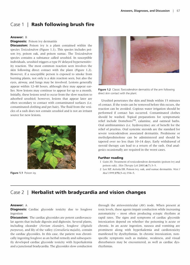
Answers, Diagnoses, and Discussion | 67
Answer: BDiagnosis: Poison ivy dermatitisDiscussion: Poison ivy is a plant contained within thespecies Toxicodendron (Figure 1.1). This species includes poi-son ivy, poison oak, and poison sumac. The Toxicodendronspecies contains a substance called urushiol. In susceptibleindividuals, urushiol triggers a type IV delayed hypersensitiv-ity reaction. The most common reaction seen involves theskin following direct contact with the plant (Figure 1.2).However, if a susceptible person is exposed to smoke fromburning plants, not only is a skin reaction seen, but also theeyes, airway, and lungs may be involved. Lesions generallyappear within 12–48 hours, although they may appear ear-lier. New lesions may continue to appear for up to a month.Initially, these lesions tend to occur from the slow reaction toadsorbed urushiol; however, lesions that appear later areoften secondary to contact with contaminated surfaces (i.e.contaminated clothing and pet hair). The fluid from the vesi-cles of a rash does not contain urushiol and is not an irritantsource for new lesions.
Urushiol penetrates the skin and binds within 15 minutesof contact. If the toxin can be removed before this occurs, thereaction can be avoided. Copious water irrigation should beperformed if contact has occurred. Contaminated clothesshould be washed. Topical preparations for symptomaticrelief include DomeboroTM, calamine, and oatmeal baths.Oral antihistamines (i.e. hydroxyzine) are of benefit for therelief of pruritus. Oral systemic steroids are the standard forsevere toxicodendron associated dermatitis. Prednisone ormethylprednisolone can be administered and should betapered over no less than 10–14 days. Early withdrawal ofsteroid therapy can lead to a return of the rash. Oral anal-gesics occasionally are required in the worst cases.
Further reading1 Guin JD. Treatment of toxicodendron dermatitis (poison ivy and
poison oak). Skin Therapy Lett 2001;6(7):3–5.
2 Lee NP, Arriola ER. Poison ivy, oak, and sumac dermatitis. West J
Med 1999;171(5–6):354–5.
Case 1 | Rash following brush fire
Figure 1.1 Poison ivy.
Figure 1.2 Classic Toxicodendron dermatitis of the arm followingdirect skin contact with the plant.
Case 2 | Herbalist with bradycardia and vision changes
Answer: ADiagnosis: Cardiac glycoside toxicity due to foxglove ingestionDiscussion: The cardiac glycosides are potent cardiovascu-lar agents that include digoxin and digitoxin. Several plants,including oleander (Nerium oleander), foxglove (Digitalispurpurea), and lily of the valley (Convallaria majalis), containthe cardiac glycosides. In this case, the patient was chroni-cally ingesting foxglove as an herbal remedy and subsequen-tly developed cardiac glycoside toxicity with hyperkalemiaand a junctional bradycardia. The glycosides slow conduction
through the atrioventricular (AV) node. When present attoxic levels, these agents impair conduction while increasingautomaticity – most often producing ectopic rhythms atrapid rates. The signs and symptoms of cardiac glycosideintoxication depend on whether the poisoning is acute orchronic. In an acute ingestion, nausea and vomiting areprominent along with hyperkalemia and cardiotoxicitymanifested by dysrhythmias. In chronic intoxication, non-specific symptoms such as malaise, weakness, and visual disturbances may be encountered, as well as cardiac dys-rhythmias.

68 | Visual Diagnosis in Emergency and Critical Care Medicine
A broad range of cardiac dysrhythmias is seen in the patient with cardiac glycoside toxicity, including bradycardia,AV block, and tachycardia. The three classically described dys-rhythmias which suggest the diagnosis include paroxysmalatrial tachycardia with block, junctional tachycardia, and ven-tricular tachycardia. Paroxysmal atrial tachycardia with blockoccurs progressively rather than suddenly; in fact, the term“paroxysmal” is a misnomer. The atrial rate is usually between150 and 250 beats per minute; the degree of AV block varies,with second degree and Wenckebach being the most commonforms.
Junctional rhythms, including junctional tachycardia,result from suppression of impulse formation at the sinoatrialnode to the degree that the inherent AV pacemaker cells out-pace the sinoatrial nodal cells. The escape rhythms result in aregular ventricular rate of 40–60 beats per minute, but accel-erated junctional rhythms and junctional bradycardias arecommon. Ventricular tachycardia is a common manifestationof severe cardiac glycoside poisoning. As a result of increasedautomaticity, premature ventricular beats are often the earli-est dysrhythmia associated with cardiac glycoside intoxication
and account for half of the dysrhythmias associated with dig-italis. Bidirectional ventricular tachycardia is a rare dysrhyth-mia which is most commonly caused by cardiac glycosidetoxicity.
Potential treatment for symptomatic patients with cardiacglycoside toxicity includes the administration of digoxin-specific Fab fragments. For coexisting hyperkalemia, theearly administration of intravenous crystalloids, albuterol,sodium bicarbonate, and insulin with glucose should be per-formed to help decrease the hyperkalemia.
Further reading1 Ma G, Brady WJ, Pollack M, Chan TC. Electrocardiographic mani-
festations: digitalis toxicity. J Emerg Med 2001;20: 145–52.
2 Rich SA, Libera JM, Locke RJ. Treatment of foxglove extract poi-
soning with digoxin-specific Fab fragments. Ann Emerg Med
1993;22(12):1904–7.
3 Holstege CP, Eldridge DL, Rowden A. Electrocardiographic changes
associated with poisoning. Emerg Med Clin North Am 2006;24(1):
159–77.
Case 3 | Acute eye pain and blurred vision in an elderly female
Answer: CDiagnosis: Acute angle closure glaucomaDiscussion: Acute angle closure glaucoma is much rarerthan open angle glaucoma. It generally affects patients intheir sixth to seventh decade of life and is slightly more com-mon in females. Acute angle closure glaucoma is character-ized by a rapid rise in intraocular pressure. The rapid increasein pressure causes conjunctival injection and corneal edemaas shown in the photo on page 4. The major factor respon-sible for precipitating an angle closure attack is prolonged periods of mid-pupillary dilation. Acute angle closure attackstend to occur after prolonged periods in dim illumination.Attacks also occur after periods of severe stress due to adren-ergic stimulation and dilation of the pupil. Both adrenergicagonists and anticholinergic agents, either topical or sys-temic, are also capable of precipitating an acute attack. Theintraocular pressure rises when the iris occludes the trabecu-lar meshwork preventing aqueous outflow. Severe pressureelevations can occur within 1 hour resulting in permanentdamage.
Initial management of acute angle closure glaucomainvolves pharmacologic lowering of the intraocular pressure
by inhibiting aqueous production. A careful medical historyshould be taken, specifically for asthma, chronic obstructivepulmonary disease, congestive heart failure, renal failure,and drug allergies. If not contraindicated, a topical betablocker such as timolol or levobunolol, 0.5%, should beinstilled along with topical brimonidine 0.15%. A carbonicanhydrase inhibitor such as acetazolamide, two 250 mgtablets, should be given orally or 500 mg given intra-venously. Severe cases may require intravenous mannitol, indoses of 1–2 g/kg infused over 45 minutes. Vital signs shouldbe monitored closely when giving mannitol. The intraocularpressure should be rechecked within 1 hour. Ophthal-mologic consultation is necessary for patients with acuteangle closure glaucoma since the definitive treatment is aperipheral laser iridectomy.
Further reading1 Saw SM, Gazzard G, Friedman DS. Interventions for angle-clo-
sure glaucoma: an evidence-based update. Ophthalmology
2003;110(10):1869–78.
2 Tripathi RC, Tripathi BJ, Haggerty C. Drug-induced glaucomas:
mechanism and management. Drug Saf 2003;26(11):749–67.

Answers, Diagnoses, and Discussion | 69
Case 4 | Suspicious hand pain
Answer: CDiagnosis: Boxer’s fractureDiscussion: Fractures of the 5th metacarpal neck areamong the most common fractures in the hand, accountingfor 10% of all hand fractures. These fractures are oftencaused by striking a solid object with a closed fist and areknown as a “boxer’s fracture.” However, this is a misnomersince these fractures rarely occur during boxing. Rather, askilled fighter may fracture the index metacarpal becausethe blow comes straight from the body along the line ofgreatest force transmission, while an inexperienced fightermay use a “roundhouse” type motion to cause this injury.
Typically, metacarpal neck fractures will have an apex dor-sal angulation (palmar angulation of the distal fragment).Maintaining the fracture in reduction can be difficult due todeforming forces of the surrounding muscles. Metacarpalneck fractures rarely require surgery. Minimally angulatedor displaced fractures can be managed with simple immobi-lization for 3–4 weeks. The degree of acceptable angulationis controversial. Most surgeons agree that no more than 45°of angulation is allowed in the 5th metacarpal. Someauthors argue that 5th metacarpal neck fractures with anydegree of angulation do not need reduction and may simplybe immobilized for 4 weeks. The reasoning behind this isthat simple immobilization will not maintain satisfactoryreduction. However, rotational injury does need to beaddressed as this can impair function and result in a defor-mity with an overlap of the affected and adjacent fingers. Ifthe decision is to reduce, the most common method ofreduction is the 90–90 method. After anesthesia (a hematomablock), traction is applied to the metacarpal distal to theinjury. The metacarpal-phalangeal and interphalangealjoints are then flexed at 90° and pressure is applied volarlyover the metacarpal shaft and dorsally over the flexed prox-imal interphalangeal (PIP) joint.
The decision to reduce or simply immobilize should be madeby an experienced emergency physician or in conjunction
with a hand surgeon. Ice, elevation, and analgesia are indi-cated acutely, particularly in the first 48–72 hours. Initial ther-apy typically consists of an ulnar gutter splint with the arm inposition of function (swelling often prevents optimal castingacutely) extending from below the elbow to the PIP joint (butnot including the PIP joint). Fifth metacarpal fractures shouldbe seen by an orthopedic surgeon within 1 week of injury.
Further reading1 Ashkenaze DM, Ruby LK. Metacarpal fractures and dislocations.
Orthop Clin North Am 1992;23:19–33.
2 Poolman RW, Goslings JC, Lee JB, et al. Conservative treatment for
closed fifth (small finger) metacarpal neck fractures. Cochrane
Database Syst Rev 2005;Jul 20 (3):CD003210.
Figure 4.1 The case X-ray with an arrow pointing to the Boxer’sfracture.
Case 5 | An elderly man with flank pain
Answer: CDiagnosis: Abdominal aortic aneurysm leakDiscussion: Sonographic evaluation of the abdominal aortarequires the real-time visualization of the aorta from thediaphragmatic hiatus to the bifurcation, externally from thexiphoid process to the umbilicus. About 90% of all abdominal
aortic aneurysms (AAAs) are found inferior to the renal arter-ies, therefore, a normal aorta at or above the renal arteries cer-tainly does not exclude aneurysm. Effective aortic scanningrequires differentiation of several retroperitoneal structures.First, identify the vertebral bodies, which are seen as curvilin-ear hyperechoic structures casting a dense shadow. The aorta

70 | Visual Diagnosis in Emergency and Critical Care Medicine
lies just anterior and to the left of these. The aorta should notbe confused with the inferior vena cava (IVC), which is fur-ther to the patient’s right compared to the aorta, is thinwalled, does not have any branches from its anterior surface,and collapses with deep inspiration and probe pressure. Ofnote, the IVC, like the aorta, does have pulsations. Bowel gasand patient obesity may interfere with the complete real-timevisualization of the aorta, resulting in failure to identify a sac-cular or a localized fusiform AAA. A limited sonographic studymandates alternative imaging if haemodynamically stable.Bedside ultrasound performed by appropriately trained emer-gency physicians is 95% accurate in measuring the infrarenalaorta.
The aortic diameter is measured from outer wall to outerwall, and is most accurately measured in the transverse planein the antero-posterior direction. The normal aorta is less than3 cm in diameter and tapers from diaphragm to its bifurcation.Any dimension greater than this is technically an aneurysm,although problems rarely occur at diameters less than 4 cm.The 5 year rupture rate for aneurysms less than and greaterthan 5 cm are �4% and �25%, respectively. Many AAAsdevelop echogenic intraluminal thrombus which can be mis-taken for the true aortic walls, as in the present case. Figure5.1 shows the image from the case with arrows delineatingthe true extent of the aneurysm. Figure 5.2 shows a lowertransverse view than that shown in the case, with theaneurysm and the pseudolumen more readily apparent.
AAAs rupture into the peritoneal cavity only 10–20% of thetime. Most of these patients die before reaching the emergencydepartment. More commonly, symptoms are due to acute tearsof the vessel wall with varying amounts of acute extravasation.Leaks which are nonlethal tend to occur into the left retroperi-toneum. Based on this natural history, the syncope frequentlyseen in patients with acute AAA is most likely due to pain andensuing vasovagal response rather than massive hemorrhage.For this reason, it is more useful to speak of “acute” or “symp-tomatic” rather than “ruptured” AAA. Any aneurysm causingacute symptoms should be treated with equal emergency,regardless of the patient’s current vital signs, hemodynamicstatus, or evidence of hemorrhage. Overall mortality for ruptured AAA is 50%. Early recognition, preparation forpotentially rapid and fatal hemodynamic decompensation,emergent surgical consultation, and operative intervention arenecessary for optimal management.
Further reading1 Barkin AZ, Rosen CL. Ultrasound detection of abdominal aortic
aneurysm. Emerg Med Clin North Am 2004;22(3):675–82.
2 Aburahma AF, Woodruff BA, Stuart SP, et al. Early diagnosis and
survival of ruptured abdominal aortic aneurysms. Am J Emerg
Med 1991;9(2):118–21.
Figure 5.1 Sagittal view of the aorta from the case described witharrows delineating the true extent of the aneurysm (C, clot).
Figure 5.2 A lower transverse view of the aorta than that viewshown in Figure 5.1, with the aneurysm and the pseudolumen morereadily apparent (Ps, pseudolumen).
Case 6 | An immigrant child with skin lesions
Answer: ADiagnosis: CoiningDiscussion: Often mistaken for abuse, coining is a commonpractice throughout Asian cultures. Referred to as “cao gio”
(scratch the wind) in Vietnamese, this practice is a folk rem-edy intended to treat fevers, headache, and other maladies byenabling the release of “bad winds” from the body. Coiningcan mimic physical abuse and is of great concern to many
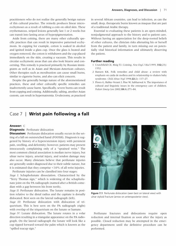
Answers, Diagnoses, and Discussion | 71
practitioners who do not realize the generally benign natureof this cultural practice. The remedy produces linear micro-ecchymoses as a result of rubbing a coin on oiled skin. Theseerythematous, striped lesions generally last 1 or 2 weeks butcan transit into lasting areas of hyperpigmentation.
Aside from coining, there are many other culturally spe-cific practices that can result in impressive patterned ecchy-mosis. In cupping for example, cotton is soaked in alcoholand ignited inside a glass cup. Once the glass is heated andoxygen removed, the cotton is withdrawn and the cup placedimmediately on the skin, creating a vacuum. This results incircular ecchymotic areas that can also look bizarre and con-cerning. This remedy is practiced primarily by Russian immi-grants as well as in Asian and Mexican-American cultures.Other therapies such as moxibustion can cause small burns,similar to cigarette burns, and also can elicit concern.
Despite the generally benign nature of the aforementionedpractices, these and other culturally specific remedies caninadvertently cause harm. Specifically, severe burns can resultfrom cupping and coining. Additionally, salting, another Asiancustom, can result in hypernatremia. Uvulectomy, as practiced
in several African countries, can lead to infection, as can thesmall, deep, therapeutic burns known as maquas that are partof a traditional Arabic therapy.
Essential to evaluating these patients is an open-minded,nonjudgmental approach to the history and to patient care.Without having an appreciation for the deep-rooted beliefsof other cultures, the clinician risks alienating his or herselffrom the patient and family, in turn missing out on poten-tially vital historical information and ultimately disservingthe patient.
Further reading1 Crutchfield CE, Bisig TJ. Coining. New Engl J Med 1995; 332(23):
1552.
2 Hansen KK. Folk remedies and child abuse: a review with
emphasis on caida de mollera and its relationship to shaken baby
syndrome. Child Abuse Negl 1998;22(2): 117–27.
3 Flores G, Rabke-Verani J, Pine W, Sabharwal A. The importance of
cultural and linguistic issues in the emergency care of children.
Pediatr Emerg Care 2002;18(4):271–84.
Case 7 | Wrist pain following a fall
Answer: CDiagnosis: Perilunate dislocationDiscussion: Perilunate dislocation usually occurs in the set-ting of a fall on outstretched hand (FOOSH). Diagnosis is sug-gested by history of a hyperextension injury with persistentpain, swelling, and deformity; however, patients may presentinnocuously complaining only of a “sprained wrist.” Themost common clinical association is median nerve injury, butulnar nerve injury, arterial injury, and tendon damage mayalso occur. Many clinicians believe that perilunate injuriesare generally under-diagnosed due to their subtle nature, butit is estimated that they comprise �10% of all wrist injuries.
Perilunate injuries can be classified into four stages:Stage I: Schapholunate dissociation. Characterized by the“Terry Thomas sign,” which is widening of the schapholu-nate joint on the PA radiograph (named after a British come-dian with a gap between his front teeth).Stage II: Perilunate dislocation. The lunate remains in posi-tion relative to the distal radius and the capitate is dorsallydislocated. Best seen on the lateral radiograph (left).Stage III: Perilunate dislocation with dislocation of tri-quetrium. This is best seen on the PA radiograph (right)with overlap of the triquetrium on the lunate or hamate.Stage IV: Lunate dislocation. The lunate rotates in a volardirection resulting in a triangular appearance on the PA radio-graph. On the lateral radiograph the lunate will look like acup tipped forward toward the palm which is known as the“spilled teacup sign.”
Figure 7.1 Perilunate dislocation (seen best on lateral view) withulnar styloid fracture (arrow on anteroposterior view).
Perilunate fractures and dislocations require open reduction and internal fixation as soon after the injury aspossible. Closed reduction may be attempted in the emer-gency department until the definitive procedure can be performed.
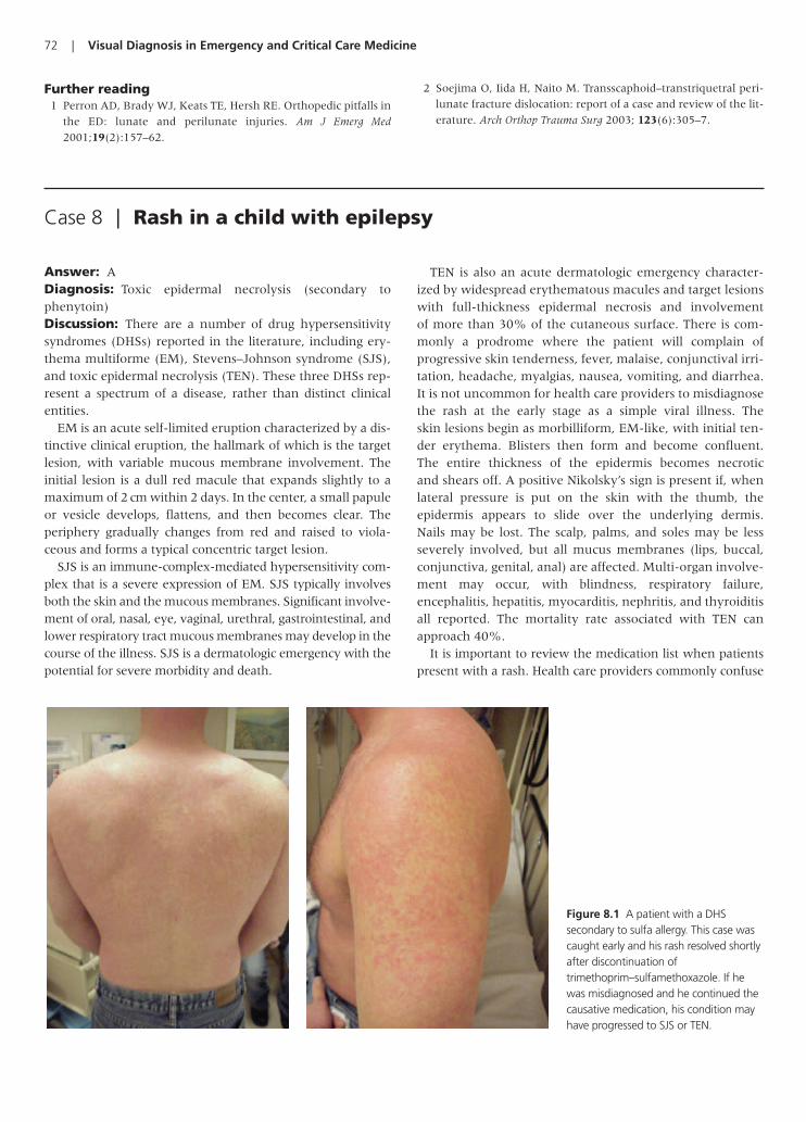
72 | Visual Diagnosis in Emergency and Critical Care Medicine
Further reading1 Perron AD, Brady WJ, Keats TE, Hersh RE. Orthopedic pitfalls in
the ED: lunate and perilunate injuries. Am J Emerg Med
2001;19(2):157–62.
2 Soejima O, Iida H, Naito M. Transscaphoid–transtriquetral peri-
lunate fracture dislocation: report of a case and review of the lit-
erature. Arch Orthop Trauma Surg 2003; 123(6):305–7.
Case 8 | Rash in a child with epilepsy
Answer: ADiagnosis: Toxic epidermal necrolysis (secondary to phenytoin)Discussion: There are a number of drug hypersensitivitysyndromes (DHSs) reported in the literature, including ery-thema multiforme (EM), Stevens–Johnson syndrome (SJS),and toxic epidermal necrolysis (TEN). These three DHSs rep-resent a spectrum of a disease, rather than distinct clinicalentities.
EM is an acute self-limited eruption characterized by a dis-tinctive clinical eruption, the hallmark of which is the targetlesion, with variable mucous membrane involvement. Theinitial lesion is a dull red macule that expands slightly to amaximum of 2 cm within 2 days. In the center, a small papuleor vesicle develops, flattens, and then becomes clear. Theperiphery gradually changes from red and raised to viola-ceous and forms a typical concentric target lesion.
SJS is an immune-complex-mediated hypersensitivity com-plex that is a severe expression of EM. SJS typically involvesboth the skin and the mucous membranes. Significant involve-ment of oral, nasal, eye, vaginal, urethral, gastrointestinal, andlower respiratory tract mucous membranes may develop in thecourse of the illness. SJS is a dermatologic emergency with thepotential for severe morbidity and death.
TEN is also an acute dermatologic emergency character-ized by widespread erythematous macules and target lesionswith full-thickness epidermal necrosis and involvement of more than 30% of the cutaneous surface. There is com-monly a prodrome where the patient will complain of progressive skin tenderness, fever, malaise, conjunctival irri-tation, headache, myalgias, nausea, vomiting, and diarrhea.It is not uncommon for health care providers to misdiagnosethe rash at the early stage as a simple viral illness. The skin lesions begin as morbilliform, EM-like, with initial ten-der erythema. Blisters then form and become confluent. The entire thickness of the epidermis becomes necrotic and shears off. A positive Nikolsky’s sign is present if, whenlateral pressure is put on the skin with the thumb, the epidermis appears to slide over the underlying dermis. Nails may be lost. The scalp, palms, and soles may be lessseverely involved, but all mucus membranes (lips, buccal,conjunctiva, genital, anal) are affected. Multi-organ involve-ment may occur, with blindness, respiratory failure,encephalitis, hepatitis, myocarditis, nephritis, and thyroiditisall reported. The mortality rate associated with TEN canapproach 40%.
It is important to review the medication list when patientspresent with a rash. Health care providers commonly confuse
Figure 8.1 A patient with a DHSsecondary to sulfa allergy. This case wascaught early and his rash resolved shortlyafter discontinuation oftrimethoprim–sulfamethoxazole. If hewas misdiagnosed and he continued thecausative medication, his condition mayhave progressed to SJS or TEN.

Answers, Diagnoses, and Discussion | 73
drug hypersensitivity reactions with “viral exanthems” becausethere is often preceding fever and malaise.
For all DHSs, prompt withdrawal of the suspected drug(s),good supportive care, and referral to a burn unit in cases ofSJS or TEN are the mainstays of therapy. Beyond these sim-ple measures, there is a lack of strong supportive evidence ofthe benefit of other specific therapies. Most authors reserveantibiotics for proven infections rather than prophylacticadministration. Temporary semi-synthetic skin substituteapplication in areas of denuding has been shown to decreasepain, reduce fluid loss, reduce sepsis, and increase woundhealing. Corticosteroids have long been used in the
management of SJS or TEN. However, there are multiplestudies documenting no demonstrable benefit to steroids forSJS or TEN. Further, there is literature that suggestsincreased mortality with steroid use.
Further reading1 Chave TA, Mortimer NJ, Sladden MJ, et al. Toxic epidermal
necrolysis: current evidence, practical management and future
directions. Br J Dermatol 2005;153:241–53.
2 Letko E, Papaliodis DN, Papaliodis GN, et al. Stevens–Johnson
syndrome and toxic epidermal necrolysis: a review of the litera-
ture. Ann Allerg Asthma Immunol 2005;94(4):419–36.
Case 9 | Dark urine in an immigrant
Answer: BDiagnosis: Acute hemolysis secondary to glucose-6-phos-phate dehydrogenase (G6PD) deficiency and favismDiscussion: G6PD deficiency, the most common humanenzymatic disease of red blood cells, is an inherited X-linkeddisorder with a high prevalence in individuals of African,Middle Eastern, Southern European, Southeast Asian, andOceanian descent. G6PD is involved in the production ofNADPH, which maintains glutathione and other proteins, inthe reduced state when erythrocytes are subjected to oxidantstress. Conditions of oxidant stress, including infection, drugswith a high redox potential such as the antimalarial pri-maquine and sulfa drugs, and favism (broad bean ingestion),produce hemolysis of deficient erythrocytes in susceptibleindividuals. Favism has also been reported in breast-fedinfants of mothers who ate fava beans. Most individuals whoare G6PD remain clinically asymptomatic. Sequestration ofdamaged erythrocytes occurs in the liver and spleen.Symptom severity depends on the number of erythrocytesdestroyed. Anemia stimulates erythropoiesis, with an increasein more oxidant-tolerant reticulocytes in 5 days.
Favism results from the presence of two beta-glycosides infava beans, vicine, and convicine. These are cleaved by intes-tinal beta-glucosidase to produce the pyrimidine aglyconesdivicine and isouramil, which reduce the activity of catalase
and lower the concentration of reduced glutathione. This, forunclear reasons, does not produce favism every time affectedindividuals eat broad beans, but the amount eaten and thedegree to which the beans are cooked are important factors.
The fall in hemoglobin is acute and often severe, and maybe fatal without transfusion. With favism, the spun urine clas-sically shows hemoglobinuria, with 0–1 RBC and a clear redsupernatant. The blood smear will show anisocytosis, poly-chromasia, poikilocytosis, and “blister cells.” Serum hemoglo-bin (3–4 g fall) and haptoglobin will be low, while lactatedehydrogenase and unconjugated bilirubin will be elevated.
In this patient, G6PD testing showed decreased enzymeactivity. He responded well to transfusion. He was educatedregarding oxidant stressors to avoid.
Further reading1 Corchia C, Balata A, Meloni GF, Meloni T. Favism in a female
newborn infant whose mother ingested fava beans before deliv-
ery. J Pediatr 1995;127:807–8.
2 Galiano S, Gaetani GF, Barabino A, et al. Favism in the African
type of glucose 6-phosphate dehydrogenase deficiency. Br Med J
1990;300:236–9.
3 Meloni T, Forteleoni G, Meloni GF. Marked decline of favism
after neonatal glucose-6-phosphate dehydrogenase screening
and health education: the Northern Sardinian experience. Acta
Haematol 1992;87:29–31.
Case 10 | Fever and drooling in a child
Answer: CDiagnosis: Retropharyngeal abscessDiscussion: Retropharyngeal abscess (RPA) is primarily adisease of childhood, and most frequently occurs in children
between ages of 2 and 4 years. Infections are most commonin this age group because the retropharyngeal lymph nodesthat drain the nasopharynx, middle ear, and posteriorparanasal sinuses occupy the prevertebral space. These

74 | Visual Diagnosis in Emergency and Critical Care Medicine
Case 11 | Altered mental status with an abnormal electrocardiogram
Answer: DDiagnosis: HyperkalemiaDiscussion: The electrocardiogram (ECG) (Figure 11.1)reveals a wide complex rhythm with an irregular rate. Uponreview of the medical record, a history of chronic renal fail-ure with hemodialysis was noted; laboratory analysis con-firmed the suspicion for hyperkalemia with a serumpotassium of 8.3 mEq/dL. With this history and laboratory
result, the ECG diagnosis of the wide complex rhythm – witha sine-wave configuration – is sinoventricular rhythm due tohyperkalemia. While the laboratory results do, in fact, con-firm the diagnosis, the clinician should consider this diag-nostic possibility based upon the initial ECG rhythm.
Hyperkalemia, a common life-threatening metabolic emer-gency, is most often diagnosed in patients with renal failure.Hyperkalemia may present with lethargy and weakness as
aVR
aVL
aVF
I
II
III
V1
V2
V3
V4
V5
V6
Figure 11.1 Wide QRS complex rhythm with sine-wave configuration.
nodes begin to involute by the age of 5 years. Clinical symp-toms of RPA include fever, irritability, neck stiffness/pain(worse with extension), and drooling. In severe cases respi-ratory distress and stridor may be present as a result of air-way compromise. On physical examination, one might seebulging of the posterior pharyngeal wall, although given theage of the involved patients and the likely difficulty of theexamination, this may not be obvious. Involved organismsare most commonly group A Streptococcus, Staphylococcusaureus, and the anaerobes that occupy the oral cavity.
Radiographic evaluation of children with the above com-plaints mandates differentiation of RPA from other etiologiesincluding epiglottitis, foreign body aspiration, and meningi-tis. Lateral neck images are often obtained in an effort toevaluate the width of the prevertebral space and anteriordisplacement of the airway. To avoid artificially widenedspaces, the child’s neck should be kept in full extension dur-ing inspiration. Pathology should be suspected if theretropharyngeal space is greater than 7 mm at C2 or 14 mmat C6, such as is noted in this case.
Computerized tomography (CT) scanning with contrast isfrequently used to further characterize the nature of the
swelling and is more sensitive in evaluating the differencebetween cellulitis and true abscess. Furthermore, CT can helpto determine the extension of the infection into neighboringregions and the extent of airway compromise. The manage-ment of RPA is appropriate antibiotic coverage with or without surgical drainage. Patients with obvious respiratorydistress are the most likely candidates for drainage and CT scan can help to guide the surgical approach. In moresubtle cases, CT findings can be useful in determiningwhether or not surgical drainage is required. Radiologic procedures should only be performed in those who are medically stable, appropriately monitored, and with emer-gent airway equipment at bedside. The immediate threat to life is loss of the airway followed by overwhelming infection.
Further reading1 Craig F, Schunk J. Retropharyngeal abscess in children: clinical
presentation, utility of imaging and current management.
Pediatrics 2003;111(6):1394–8.
2 Al-Sabah B, Bin Salleen H, Hagr A, et al. Retropharyngeal abscess
in children: 10-year study. J Otolaryngol 2004; 33(6):352–5.

Answers, Diagnoses, and Discussion | 75
the sole manifestation; alternatively, the patient may demon-strate significant ECG abnormality in addition to the mentalstatus and constitutional signs. Both ECG morphologic andrhythm findings are seen in these patients; a subset of thesepatients, those with widening of the QRS complex, are at sig-nificant risk of decompensation and death. While serumchemistry analysis is the primary diagnostic investigation, thesyndrome is suggested with certain ECG findings, includingprominent T-waves and widened QRS complex. In fact, becauseof the potential delay in laboratory analysis, prompt diagnosis oflife-threatening hyperkalemia can be made by the ECG.
From a pathophysiologic perspective, increasing potassiumlevels are associated with depressed electrical conduction.Furthermore, significant variation is noted with respect toserum potassium levels and clinical manifestation; in general,sudden or rapid increases in the potassium concentration inserum are associated with earlier development of clinical ill-ness, including ECG abnormalities. It is important to note,however, that the relationship between potassium levels andECG changes may vary between different patients. It is alsoimportant to realize that the ECG is not a reliable test for mildto moderate hyperkalemia. Mild levels of hyperkalemia areassociated with acceleration of terminal repolarization, result-ing in T-wave changes (Figure 11.2B and Table 11.1) – namelyan increase in amplitude of the T-wave. The T-wave isdescribed as prominent with descriptors such as “tenting” or“peaking” of the T-wave; the T-wave is tall, narrow, and sym-metric. The “peaked” T-wave is generally considered the earli-est sign of hyperkalemia. Mild to moderate hyperkalemiacauses depression of conduction, resulting in progressive pro-longation of the PR and QRS intervals (Figures 11.1 and11.2A; Table 11.1). The atrial myocardium is particularly sensitive to hyperkalemia. P-wave amplitude lessens withprogressive increase in the serum potassium; eventually, theP-wave will disappear, even in the presence of continuedsinus node activity, producing the “sinoventricular” rhythm of significant hyperkalemia. With minimal QRS complex
A B
V5
V4
V6
Figure 11.2 A Wide QRS complex sine-wave configuration in pronouncedhyperkalemia. B Prominent T-waves ofhyperkalemia – tall, narrow, peaked,and symmetric.
Table 11.1 Electrocardiographic manifestations of serumhyperkalemia relative to serum potassium level
Serum potassium Electrocardiographic manifestations
5.5–6.5 mEq/L Prominent T-wave– Tall, narrow, symmetric– Most prominent in precordial leads
6.5–8.0 mEq/L Decreased P-wave amplitudeProlonged PR intervalProminent T-waveQRS complex widening (minimal to sine-wave configuration)Dysrhythmia– Atrioventricular block– Intraventricular block– Bradycardia– Ventricular ectopy
�8.0 mEq/L Sinoventricular rhythm– Absence of P-wave– QRS complex widening, progressing
to sine-wave QRS complexVentricular tachycardiaVentricular fibrillationAsystole
widening, the ECG can mimic a bundle branch block appear-ance. The QRS complex continues to widen and may eventually blend with the T-wave by creating a “sine-wave”appearance (Figures 11.1 and 11.2A). Continued increases inthe potassium level eventually produce ventricular fibrillation.
The management of the patient with hyperkalemia includestherapies aimed at the stabilization of the myocardium (calcium), temporary shifting of the excess potassium intra-cellularly (dextrose, insulin, beta-adrenergic agonists, magne-sium, and sodium bicarbonate), and ultimate removal of thepotassium from the body (gastrointestinal binding resins and

76 | Visual Diagnosis in Emergency and Critical Care Medicine
hemodialysis). The goals of therapy are a reduction of theserum potassium level coupled with a stabilization of themyocardial cell membrane. Response to therapy is oftenprompt with visualization noted on the ECG monitor; cautionis advised, however, that improvement is often transient untildefinitive therapy (i.e. hemodialysis) is provided. The mostappropriate initial medication is calcium, delivered in the formof either calcium chloride or calcium gluconate. Calcium worksby restoring a more appropriate electrical gradient across thecell membrane. Caution is advised in the setting of hyper-kalemia related to digoxin toxicity; anecdotal reports suggest atendency toward asystole in this clinical setting. Several agents
are capable of transiently moving the potassium from theextracellular to intracellular space, including glucose, insulin,beta-adrenergic agonists (i.e. albuterol), magnesium, sodiumbicarbonate, and intravenous saline. Ultimate lowering of theserum potassium is accomplished with binding resins (e.g.sodium polystyrene) and/or hemodialysis.
Further reading1 Amal Mattu A, Brady WJ, Robinson D. Electrocardiographic
manifestations of hyperkalemia. Am J Emerg Med 2000;18:721–9.
2 Schaefer TJ, Wolford RW. Disorders of potassium. Emerg Med Clin
North Am 2005;23(3):723–47.
Case 12 | Purulent eye discharge in an adult
Answer: CDiagnosis: Bacterial conjunctivitis due to NeisseriagonorrheaDiscussion: Bacterial conjunctivitis can affect patients of allages. The causative organisms are numerous and differ in fre-quency between adults and children. Symptoms of bacterialconjunctivitis include hyperemia, ocular discharge, eyelidsmatted together in the morning, and foreign body sensation.
Hyperacute, purulent conjunctivitis is the most severe formof bacterial conjunctivitis and is classically associated withNeisseria gonorrhea. This infection may progress rapidly toinvolve the cornea, resulting in scarring or perforation. Theabrupt onset of symptoms and hyperpurulent discharge willoften bring these patients to the emergency department forevaluation and treatment.
Broad spectrum, topical antibiotic therapy is sufficient formost acute conjunctival infections. However, any suspicionof gonococcal infection requires laboratory evaluation. Gram
staining, looking for Gram negative, intracellular diplococci,along with culture and sensitivity of conjunctival scrapingsshould be done prior to treatment. Cultures should be placedon chocolate agar, blood agar, or Thayer–Martin media.
Treatment for gonococcal conjunctivitis requires topicaland systemic therapy. Ceftriaxone, 1 g intravenous or intra-muscular, and topical ciprofloxacin ophthalmic drops every2 hours are usually effective along with irrigation with sterile saline four times a day. Treatment for concomitantchlamydial coinfection and evaluation of patient’s sexualpartners are also recommended.
Further reading1 Deschenes J, Seamone C, Baines M. The ocular manifestations
of sexually transmitted diseases. Can J Ophthalmol 1990;
25(4):177–85.
2 Harkins T. Sexually transmitted diseases. Optom Clin 1994;
3(4):129–56.
Case 13 | Wrist pain in a young child
Answer: EDiagnosis: Buckle fractureDiscussion: This patient has a buckle fracture (also knownas a “Torus” fracture) of the distal radius. A cortical bulge or“buckling” occurring at the metaphysis of a long bone isdiagnostic of this type of fracture. Lateral films often showsigns of buckling better than on the anterior–posterior view.Both cortical margins are affected, but a discreet fracture lineor trabecular disruption is absent. On occasion, radiographic
signs of a fracture are not evident on the initial X-ray and aclinical diagnosis must be pursued. A second set of X-rays2–3 weeks after the trauma should be obtained. It is impor-tant to remember that sprains are rare in 9-year-old childrenand they do not typically complain about 3 days of pain following minor injuries (Figure 13.1).
The etiology of this type of fracture is typically a fall on outstretched arm injury. It is a compression fracture of thelong bone typically occurring in children. Because of the
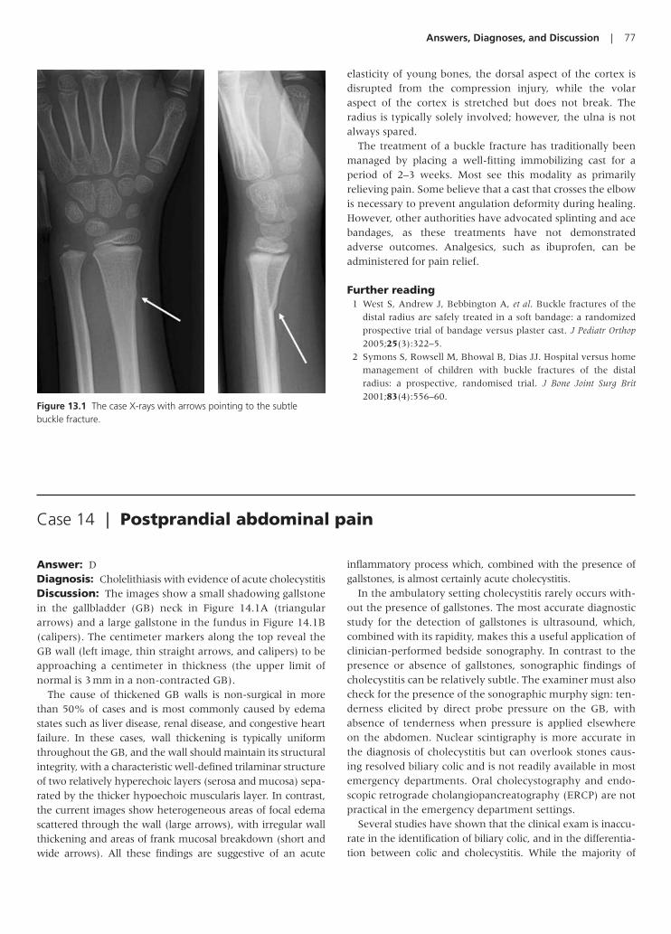
Answers, Diagnoses, and Discussion | 77
elasticity of young bones, the dorsal aspect of the cortex isdisrupted from the compression injury, while the volaraspect of the cortex is stretched but does not break. Theradius is typically solely involved; however, the ulna is notalways spared.
The treatment of a buckle fracture has traditionally beenmanaged by placing a well-fitting immobilizing cast for aperiod of 2–3 weeks. Most see this modality as primarilyrelieving pain. Some believe that a cast that crosses the elbowis necessary to prevent angulation deformity during healing.However, other authorities have advocated splinting and acebandages, as these treatments have not demonstratedadverse outcomes. Analgesics, such as ibuprofen, can beadministered for pain relief.
Further reading1 West S, Andrew J, Bebbington A, et al. Buckle fractures of the
distal radius are safely treated in a soft bandage: a randomized
prospective trial of bandage versus plaster cast. J Pediatr Orthop
2005;25(3):322–5.
2 Symons S, Rowsell M, Bhowal B, Dias JJ. Hospital versus home
management of children with buckle fractures of the distal
radius: a prospective, randomised trial. J Bone Joint Surg Brit
2001;83(4):556–60.Figure 13.1 The case X-rays with arrows pointing to the subtlebuckle fracture.
Case 14 | Postprandial abdominal pain
Answer: DDiagnosis: Cholelithiasis with evidence of acute cholecystitisDiscussion: The images show a small shadowing gallstonein the gallbladder (GB) neck in Figure 14.1A (triangulararrows) and a large gallstone in the fundus in Figure 14.1B(calipers). The centimeter markers along the top reveal theGB wall (left image, thin straight arrows, and calipers) to beapproaching a centimeter in thickness (the upper limit ofnormal is 3 mm in a non-contracted GB).
The cause of thickened GB walls is non-surgical in morethan 50% of cases and is most commonly caused by edemastates such as liver disease, renal disease, and congestive heartfailure. In these cases, wall thickening is typically uniformthroughout the GB, and the wall should maintain its structuralintegrity, with a characteristic well-defined trilaminar structureof two relatively hyperechoic layers (serosa and mucosa) sepa-rated by the thicker hypoechoic muscularis layer. In contrast,the current images show heterogeneous areas of focal edemascattered through the wall (large arrows), with irregular wallthickening and areas of frank mucosal breakdown (short andwide arrows). All these findings are suggestive of an acute
inflammatory process which, combined with the presence ofgallstones, is almost certainly acute cholecystitis.
In the ambulatory setting cholecystitis rarely occurs with-out the presence of gallstones. The most accurate diagnosticstudy for the detection of gallstones is ultrasound, which,combined with its rapidity, makes this a useful application ofclinician-performed bedside sonography. In contrast to thepresence or absence of gallstones, sonographic findings ofcholecystitis can be relatively subtle. The examiner must alsocheck for the presence of the sonographic murphy sign: ten-derness elicited by direct probe pressure on the GB, withabsence of tenderness when pressure is applied elsewhereon the abdomen. Nuclear scintigraphy is more accurate inthe diagnosis of cholecystitis but can overlook stones caus-ing resolved biliary colic and is not readily available in mostemergency departments. Oral cholecystography and endo-scopic retrograde cholangiopancreatography (ERCP) are notpractical in the emergency department settings.
Several studies have shown that the clinical exam is inaccu-rate in the identification of biliary colic, and in the differentia-tion between colic and cholecystitis. While the majority of

78 | Visual Diagnosis in Emergency and Critical Care Medicine
patients with biliary disease have some combination of upperabdominal pain, nausea, vomiting, fever, right upper quadranttenderness, and leukocytosis, many patients with these symp-toms do not have biliary disease. Biliary pain is most com-monly in the epigastrium (not the right upper quadrant)lasting 5–24 hours (average is 16). Patients most commonlydescribe biliary pain as “steady” in character, not “colicky”.Pain that occurs “in waves” is much more likely to be non-biliary in origin. It reaches peak intensity soon after onset, andgradually subsides thereafter. Many patients with provencholecystitis (based on pathology or nuclear scintigraphy) donot have fever or leukocytosis.
Further reading1 Shea JA, Berlin JA, Escarce JJ. Revised estimates of diagnostic
test sensitivity and specificity in suspected biliary tract disease.
Arch Int Med 1994;154:2573–81.
2 Trowbridge RL, Rutkowski NK, Shojania KG. Does this patient
have cholecystitis? J Am Med Assoc 2003;289(1):80–6.
3 Gruber PJ, Silverman RA, Gottesfeld S. Presence of fever and
leukocytosis in acute cholecystitis. Ann Emerg Med 1996;
28(3):273–7.
4 Singer AJ, McCracken G, Henry MC. Correlation among clinical,
laboratory, and hepatobiliary scanning findings in patients with
acute cholecystitis. Ann Emerg Med 1996;28(3):267–72.
A B
Figure 14.1 A A small shadowing gallstone in the gallbladder (GB) neck (triangular arrows). B A large gallstone in the fundus (calipers).
Case 15 | An elderly man from a house fire
Answer: EDiagnosis: Smoke inhalation with associated burnsDiscussion: One of the primary clinical problems in thesmoke inhalation victim is respiratory compromise. Thepatient may have voice changes and his speech may progres-sively worsen as the airway becomes increasingly edematous.Stridor and acute respiratory arrest may develop rapidly if thepatient is not managed appropriately. The patient may havedifficulty managing airway secretions with copious quantitiesof soot containing sputum being expectorated. Visualization of
the vocal cords by direct laryngoscopy may be difficult due tosoot accumulation, secretions, or edema. Auscultation of thechest may demonstrate rhonchi, rales, and wheezing sugges-tive of acute lung injury (ALI). ALI is defined as diffuse alveo-lar filling of acute onset with hypoxemia but without left atrialhypertension. The most severe manifestation of ALI is theacute respiratory distress syndrome and is defined based on thepatient’s ability to oxygenate. Bronchospasm may occur, par-ticularly in patients with underlying reactive airway disease. Inpatients with severe bronchospasm, breath sounds, including

Answers, Diagnoses, and Discussion | 79
Figure 15.1 Bronchoscopy of the patient in this case. Note the edema of the airways and the black soot deposition on the mucosa.
wheezing, may be virtually inaudible. Tachycardia and tachy-pnea may be pronounced, and hypotension may occur withfaint or no peripheral pulses noted.
Critical airway compromise may be present upon arrival atthe hospital or it may develop soon after arrival. A major pitfallin managing a patient with smoke inhalation is failing toappreciate that rapid deterioration is possible. The history andphysical findings help to determine significant smoke exposureand the potential for clinical deterioration. Upper airwaypatency must be evaluated and rapidly established. Whenobvious oropharyngeal burns are observed, upper airwayinjury is almost certain even if overt injuries are not present.Distal airway injury may be present and underestimated.When evidence of upper airway injury exists, early endotra-cheal intubation should be performed under controlled cir-cumstances. Indications for early intubation include coma,stridor, or full-thickness circumferential neck burns. Othersigns suggestive of an increased risk for airway compromiseinclude facial burns, carbonaceous sputum, edema of the posterior pharynx, and singed nasal hairs. Aggressive fluid resuscitation of the burned patient is necessary, but may con-tribute to upper airway edema. Therefore, early intubationmay be necessary in the patient with dermal burns undergoingaggressive fluid management. Treatment of progressive respi-ratory failure includes mechanical ventilation, continuous pos-itive airway pressure, positive end expiratory pressure, andvigorous clearing of pulmonary secretions. Carbon monoxidepoisoning may also be present in the burned patient. It istreated with supplemental oxygen therapy, administered byhigh flow, tight-fitting mask, endotracheal tube, or hyperbaricoxygen therapy depending on the circumstances.
Further reading1 Miller K, Chang A. Acute inhalation injury. Emerg Med Clin North
Am 2003;21(2):533–57.
2 Kuo DC, Jerrard DA. Environmental insults: smoke inhalation,
submersion, diving, and high altitude. Emerg Med Clin North Am
2003;21(2):475–97.
3 Lee-Chiong TL. Smoke inhalation injury. Postgrad Med J 1999;
105(2):55–62.
Case 16 | Back pain following a fall
Answer: BDiagnosis: Lumbar wedge fractureDiscussion: Spinal fractures resulting from axial compres-sion form a continuum from minor wedge compression andend plate fractures to severe burst fractures with spinal canalcompromise. More than 150,000 persons in North Americasustain fractures of the vertebral column each year, and11,000 of these patients sustain spinal cord injuries. The tho-racolumbar spine and lumbar spine are the most commonsites for fractures due to the high mobility of the lumbar spinecompared to the more rigid thoracic spine.
When the clinician encounters an axial compression injuryto the spine, a determination must be made as to whether it isa simple (and stable) wedge fracture versus a more severeinjury, such as a burst fracture with potential spinal instability.The most benign injuries, such as end plate fractures or wedge
fractures involving a 50% or less loss of vertebral height, arerelatively easy to care for and the emphasis is placed on pro-viding the patient with adequate analgesia. Wedge fractureswhere there is more than 50% loss of height, multiple adja-cent wedge fractures, significant kyphotic angulation, or anyconcern for posterior vertebral involvement or canal compro-mise are considered high risk.
Radiographically, the clinician should look for certainfindings on the lateral radiograph to help make the determi-nation whether an injury represents a wedge fracture versusa burst fracture. Besides the 50% body height cut-off, the cli-nician needs to examine for posterior cortical disruption ofthe vertebral body, loss of posterior vertebral height, or acompression angle �20%.
A common question with these injuries is “which patientsneed advanced imaging in the emergency department” for

80 | Visual Diagnosis in Emergency and Critical Care Medicine
clarification of these injuries. Axial imaging (with computedaxial tomography or CT) is the gold standard for inspectingthe posterior elements of the vertebra, as well as assuring the
integrity of the spinal canal. Two studies (Campbell 1995,Ballock 1992) have specifically looked at the clinician’s abil-ity to determine wedge versus burst morphology on plainfilm as opposed to CT. In 17–20% of cases, burst fractureswere misinterpreted on the plain films and thought to repre-sent simple wedge injuries.
In summary, while the wedge fracture is a relatively benigncondition, the clinician must be sure that it does not, in fact,represent a burst injury with a markedly increased potentialfor morbidity. In wedge fractures with 50% or greater loss ofheight, evidence of posterior cortical disruption, or a compres-sion angle �20 degrees, it is wise to utilize CT to determinewhether there is more extensive involvement.
Further reading1 Ballock RT, Mackersie R, Abitol JJ, Cervilla V, Resnick D, Garfin
SR. Can burst fractures be predicted from plain radiographs?
J Bone Joint Surg Br 1992;74(1):147–50.
2 Campbell SE, Phillips CD, Dubovsky E, Cail WS, Omary RA.
The value of CT in determining potential instability of simple
wedge-compression fractures of the lumbar spine. Am J Neuroradiol
1995;16(7):1385–92.
3 Terregino CA, Ross SE, Lipinski MF, Foreman J, Hughes R.
Selective indications for thoracic and lumbar radiography in
blunt trauma. Ann Emerg Med 1995;26(2):126–9.
4 Wintermark M, Mouhsine E, Theumann N, Mordasini P,
van Melle G, Leyvraz PF, Schnyder P. Thoracolumbar spine frac-
tures in patients who have sustained severe trauma: depiction
with multi-detector row CT. Radiology 2003;227(3):681–9.
Figure 16.1 Enlarged lateral lumbar spine film of the describedpatient. The L2 wedge fracture is plainly evident. There is loss ofapproximately 50% of the original vertebral height (compare dashedlines to solid lines). The posterior vertebral appears to be intact.
Case 17 | A bite to the leg in tall grass
Answer: DDiagnosis: Crotalid envenomationDiscussion: Thousands of crotalid snakebites occur each yearthroughout the world. Crotalids may be distinguished fromother species by their triangular shaped heads, elliptical shapedpupils, and a single row of subcaudal scales. They also possessinfrared heat-sensing pits, thus the name “pit viper”, whichenable them to locate prey and guide the direction of strike.
The spectrum of clinical presentation ranges from asympto-matic to cardiovascular collapse and death. Bites fromCrotalidae species which do not introduce venom (“dry bites”)have been estimated to occur in up to 20% of exposures, suchas occurred in this case. Skin lesions may appear as distinctpuncture marks or as faint scratches. Pain is frequently theinitial complaint. When envenomation occurs, swelling usu-ally begins within minutes of the bite. Tissue necrosis may fol-low cyanosis, and bleb formation may occur over the affectedareas. In addition to direct tissue damage, rhabdomyolysis,
nausea, vomiting and diaphoresis may be seen. Three distinctsnakebite-induced coagulopathies have been reported:venom-induced thrombocytopenia, fibrinolysis, and dissemi-nated intravascular coagulation. Coagulopathy may be mani-fest as petechia, gastrointestinal bleeding, epistaxis, hemoptysis,and bleeding from wounds or phlebotomy sites.
Tourniquets, wound incisions, suction, extraction devises,cryotherapy, heat application, electric shock therapy, andwound excision should not be performed. The bite area shouldbe gently cleansed. Circumferential measurement at severalpoints along the affected limb should be started shortly afterthe patient’s arrival and repeated with neurovascular checks athourly intervals until swelling subsides.
In those cases where envenomation has occurred, hypoten-sion may be seen secondary to fluid loss through third spacing,vomiting, hemorrhage, or vasovagal effects. Crystalloid admin-istration should begin immediately in these patients and priorto antivenom administration. Pain control usually requires

Answers, Diagnoses, and Discussion | 81
intravenous opioid agents. Prophylactic antibiotics and steroidadministration are not recommended.
Currently, two snake antivenoms are approved for treatingNorth American crotaline envenomations: equine derivedpolyvalent IgG (Wyeth–Ayerst Laboratories) and ovine poly-valent Fab immunoglobulin fragments (CroFabTM fromProtherics Inc.). Dosing and mixing of these products arefound on the product insert. Pediatric patients should betreated with the same amount of antivenom as adults, regard-less of their weight. The most critical decision facing the clini-cian treating snakebite victims is when antivenom therapy isappropriate. Because anaphylaxis and serum sickness havebeen associated with the use of antivenom (less commonlywith ovine polyvalent Fab), potential risk to the patient must be weighed against benefits. The major indications for antivenom therapy include rapid progression of swelling,
significant coagulation defect, and cardiovascular collapse.Patients who are asymptomatic or have minimal symptomsdo not require antivenom.
Patients who remain asymptomatic for 4 hours after thesnakebite and who have normal coagulation studies may bereleased. Symptomatic patients should be considered candi-dates for hospitalization and the need for antivenom therapydetermined.
Further reading1 Singletary E, Rochman AS, Bodmer JCA, Holstege CP.
Envenomations. Med Clin North Am 2005;89(6):1195–224.
2 Holstege CP, Miller MB, Wermuth M, Furbee B, Curry SC.
Crotalid snake envenomation. Crit Care Clin 1997;13(4):889–921.
Figure 17.1 A snakebite victim who was envenomated by a copperhead on his left ring finger. Note the marked tissue edema and necrosis that developed around the bite site.
Case 18 | Facial swelling in a patient with poor dentition
Answer: DDiagnosis: Submandibular abscess (Ludwig’s angina)Discussion: Infection of the submandibular space occurswhen a periapical abscess of the second or third molar pene-trates the inner cortex of the mandible and gains access to thearea inferior to the insertion of the mylohyoid muscles. As theinfection tracks posterior, the sublingual space becomesinvolved. Ludwig’s angina refers to the resultant cellulitis ofthe submandibular space, beginning in the submaxillary space
and spreading to the sublingual space via the fascial planes. Asthe submandibular space begins to expand as a result of thecellulitis or abscess, the floor of the mouth becomes induratedand the tongue is forced upward and backward. This can sub-sequently cause airway obstruction.
Ludwig’s angina typically manifests with fever, pain,drooling, trismus, dysphagia, submandibular mass (as visual-ized in this case), and dyspnea. Airway compromise caused bydisplacement of the tongue can occur abruptly. Obvious signs

82 | Visual Diagnosis in Emergency and Critical Care Medicine
Case 19 | Elbow pain in a child after a fall
Answer: EDiagnosis: Fat pad signDiscussion: The “sail sign” or the “fat pad sign,” depicted bythe presence of anterior and posterior fat lucencies, is seen onthis lateral elbow (Figure 19.1). This sign suggests the poten-tial presence of a supracondylar fracture in children and aradial head or proximal ulna fracture in adults. The anteriorfat pad may be seen in a normal radiograph along the ante-rior, distal humerus, but becomes displaced superior andanterior in the presence of a hemarthrosis from a likely intra-articular fracture. In contrast, the posterior fat pad is not seenin normal radiographs because it lies within the olecranonfossa. The presence of a posterior fat pad is always consideredabnormal and a sign of significant distention of the joint cap-sule from a hemarthrosis. If the posterior fat pad is present inthe setting of trauma, there is greater than a 90% incidenceof an intra-articular fracture. In the absence of trauma,inflammatory etiologies must be considered, including septicjoint, gout, and bursitis.
Another diagnostic technique used to detect less obvioussupracondylar fractures is the “anterior humeral line” whichinvolves tracing a line along the anterior surface of the distalhumerus through the capitellum. The capitellum shouldthen be divided into three equal sections horizontally. Theanterior humeral line should pass through the middle sec-tion of the capitellum. If this line transects the anteriorrather than the middle section or passes completely anteriorto the capitellum, there is likely a supracondylar fracture or physis disruption of the capitellum.
of airway obstruction necessitating immediate artificial airwayinclude stridor, dysphonia, and dyspnea. If airway compromiseis not imminent and time allows, fiberoptic nasotracheal intu-bation or tracheostomy, under local anesthesia, should be per-formed in a multidisciplinary fashion (ideally in the operatingroom). Some authors recommend early definitive airwayintervention even if there are no signs of airway compromise.Abrupt airway closure has occurred in patients with Ludwig’sangina despite careful observation. Published reports docu-ment that active airway management in the form of endotra-cheal intubation or tracheotomy was eventually required in35% of cases of Ludwig’s angina. Computerized tomographyscans are utilized in select cases to determine the extent of
infection if the patient is medically stable, appropriately moni-tored, and emergent airway equipment is at bedside. Beforeantibiotics, nearly half of Ludwig’s angina cases died. Withbroad spectrum antibiotics and prompt surgical care, the mor-tality rate now is less than 5%.
Further reading1 Quinn FB. Ludwig angina. Arch Otolaryngol Head Neck Surg 1999;
125(5):599.
2 Saifeldeen K, Evans R. Ludwig’s angina. Emerg Med J 2004;
21(2):242–3.
3 Marple BF. Ludwig angina: a review of current airway manage-
ment. Arch Otolaryngol Head Neck Surg 1999;125(5):596–9.
Figure 19.1 Sail sign. The long arrow is pointing at the anterior sailsign; the short arrow is pointing at the posterior sail sign.
The radiocapitellar line is helpful for determining a radialhead dislocation or a radial neck fracture. This involvesdrawing a line through the central portion of the radius. Theaxis of this line should intersect the capitellum. If the linedoes not intersect the capitellum, there is likely a dislocationof the radial head or a fracture of the radial neck.
Further reading1 Goswami G. The fat pad sign. Radiology 2002;222(2): 419–20.
2 Skaggs DL, Mirzayan R. The posterior fat pad sign in association
with occult fracture of the elbow in children. J Bone Joint Surg Am
1999;81(10):1429–33.

Answers, Diagnoses, and Discussion | 83
Case 20 | A man with diffuse facial edema
Answer: DDiagnosis: Superior vena cava syndromeDiscussion: The superior vena cava (SVC) is the majorconduit for venous blood return from the head, neck, upperextremities, and upper thorax. Obstruction of this blood flow(i.e., compression, infiltration, and thrombosis) results inSVC syndrome (SVCS), which is associated with elevatedvenous pressure in those structures drained by the SVC.Early signs include periorbital edema and facial swelling thatis more prominent in the morning upon awaking. Latersigns include thoracic and neck vein distension, plethora ofthe face, as well as edema and cyanosis of the face and upperextremities. Headache, dyspnea, orthopnea, and cough arealso common complaints.
The azygos system of veins receives venous blood from theposterior intercostal veins and therefore drains blood fromthe chest wall (and esophagus). The azygos veins originate atabdominal levels where they establish important anasto-moses with the inferior vena cava (IVC). Obstruction of theSVC above the level of azygos vein allows blood to bypass theSVC obstruction through chest wall collaterals and re-enterthe SVC through the azygos venous system. In contrast,when the SVC is obstructed below the level of the azygosvein, blood must travel through the chest wall collateralveins to enter the azygos venous system and subsequentlymust continue to flow through the azygos system to emptyinto the IVC. The longer and more convoluted course retardsvenous return and accounts for the development of moreprominent symptoms. Additionally, the severity of symptomsis related to the rate at which the SVC obstruction occurs; theslower the onset of obstruction, the more time for collateralblood flow to develop and the less severe the symptoms.
Historically, SVCS was frequently associated with complica-tions of syphilis and tuberculosis. Today, compression of theSVC by malignant mediastinal tumors accounts for approxi-mately 90% of cases. The most common mediastinal tumorsassociated with SVCS are bronchogenic carcinoma (70%) and non-Hodgkin’s lymphomas (10–15%). The most commonhistological diagnosis is small cell lung carcinoma. Thrombosisof the SVC is a potential cause of SVCS in patients with a his-tory of intravenous drug abuse and central venous catheterplacement.
Immediate management in the emergency department isaimed at alleviating patient symptoms and determining air-way patency. Treatment options include supplemental oxy-gen, elevation of the patient’s head, steroids, and diuretics.Depending on the etiology, both radiation therapy andchemotherapy are utilized as primary treatment options forlong-term management of SVC compression due to tumors.
Prognosis of patients treated for SVCS is dependent uponthe underlying etiology. Histological diagnosis is essential aspatients with SVCS resulting from bronchogenic carcinomahave a worse prognosis when compared with lymphomapatients. Overall survival is estimated at 25% at 1 year and10% at 30 months. The prognosis for SVCS not associatedwith malignancy is excellent; infectious or thromboticcauses respond to appropriate antibiotics or anticoagulation,respectively.
Further reading1 Baker GL, Barnes HJ. Superior vena cava syndrome: etiology,
diagnosis, and treatment. Am J Crit Care 1992;1(1):54–64.
2 Wudel LJ, Nesbitt JC. Superior vena cava syndrome. Curr Treat
Options Oncol 2001;2(1):77–91.
Case 21 | Chest pain and hypotension in an adult male patient
Answer: BDiagnosis: Inferoposterior RV AMIDiscussion: The electrocardiogram (ECG) of the case (see page 14) reveals sinus bradycardia at approximately50 beats/minute with ST segment elevation in leads III and aVf consistent with inferior wall ST-elevation myocardialinfarction (STEMI); additionally, ST segment depression is seenin leads V1–V3, consistent with either posterior wall acutemyocardial infarction (AMI) or anterior wall ischemia; poste-rior ECG leads V8 and V9 (Figure 21.1A(iii)) demonstrated STsegment elevation, confirming posterior wall AMI. Lastly, STsegment depression with T-wave inversion (case ECG) is notedin leads I and AVL, consistent with reciprocal change.
Posterior wall myocardial infarction refers to AMI of the pos-terior wall of the left ventricle. This region of the heart is usuallyperfused by branches of the right coronary artery (prominentposterolateral or posterior descending arteries) or the left cir-cumflex artery. As such, the 12-lead ECG will usually demon-strate an inferior or lateral wall STEMI as well as ST segmentdepression, prominent R wave, and upright T-wave in the rightprecordial leads (Figure 21.1A(i) and (ii)). Myocardial infarc-tion involving the posterior wall usually occurs in conjunctionwith inferior or lateral AMIs; isolated posterior wall myocardialinfarction, however, is encountered even less frequently.
The use of posterior ECG leads is more helpful in the eval-uation of posterior wall AMI when compared to the standard

84 | Visual Diagnosis in Emergency and Critical Care Medicine
A B
i
i
ii
ii
iii
V1 V1
RV3
RV4
RV5
V2
iii
V8 V9II III aVf
Figure 21.1 A Posterior wall AMI. (i) Lead V1 with ST segment depression, upright T wave, and prominent R wave consistent with posterior wallAMI. (ii) Leads V1 and V2 with ST segment depression and upright T wave, consistent with posterior wall AMI. (iii) Posterior leads V8 and V9 withST segment elevation, consistent with posterior wall AMI. B Right ventricular AMI. (i) Minimal ST segment elevation in lead V1 as seen in RV AMI.(ii) Minimal ST segment elevation in leads RV3–RV5 as seen in RV AMI. (iii) Inferior leads II, III, and aVf in a patient with inferior wall AMI with RVinfarction; note the relatively greater magnitude of ST segment elevation in lead III compared to the other inferior leads. This relative imbalance oflead III ST segment elevation results from the axis of imaging of lead III, which most closely observes the right ventricle.
12-lead ECG. ST segment elevation greater than 1 mm inleads V8 and V9 confirms the diagnosis of posterior myocar-dial infarction (Figure 21.1A(iii)). In fact, the presence of STsegment elevation in the posterior ECG leads is more indica-tive of posterior AMI than the findings observed in leadsV1–V3. The sensitivity of the posterior ECG leads may be ashigh as 90% for identifying posterior AMI. The magnitude ofST segment elevation is less pronounced in the posteriorECG leads – the posterior leads are located distant from themyocardium, allowing for more resistance to current flowand less pronounced ST segment elevation.
Right ventricular myocardial infarction presents withhypotension, elevated jugular venous pressure, and clear lungfields; the ECG demonstrates ST segment elevation in theinferior (case ECG) and right ventricular leads (Figures21.1B(i) and (ii)). Right ventricular myocardial infarctionoccurs in approximately one-third of inferior wall AMIs. The
12-lead ECG reveals ST segment elevation in the inferior leadswith the greatest magnitude of elevation in lead III comparedto the other leads (Figure 21.1B(iii)); furthermore, lead V1may also demonstrate ST segment elevation in that this lead(Figure 21.1B(i)), of all the standard leads, most closelyimages the right ventricle. The use of additional leads greatlyincreases the ability to diagnose right ventricular infarction.
The addition of lead RV4 provides objective evidence ofRV involvement – more so than that noted on the 12-leadECG. RV infarction is diagnosed with 80–100% sensitivity byST segment elevation greater than 1 mm in lead RV4.Alternatively, the clinician can use an entire reversal of theprecordial leads, namely RV1–RV6 (Figure 21.1B(ii)); in acomparison to the use of single lead RV4, the entire array didnot increase the diagnostic ability of the additional leadapproach. As with the posterior leads, the magnitude of theST segment elevation is less pronounced than is usually seen

Answers, Diagnoses, and Discussion | 85
in the standard 12-leads of the ECG; this relatively less pro-nounced magnitude results from the fact that the right ventri-cle is composed of considerably less muscle when compared tothe left ventricle.
Further reading1 Zalenski RG, Rydman RJ, Sloan EP, et al. Value of posterior
and right ventricular leads in comparison to the standard 12-lead
electrocardiogram in evaluation of ST-segment elevation in
suspected acute myocardial infarction. Am J Cardiol 1997;79:
1585–97.
2 Brady WJ, Hwang V, Sullivan R, et al. A comparison of
12- and 15-lead ECGs in ED chest pain patients: impact on diag-
nosis, therapy, and disposition. Am J Emerg Med 2000;18:239–43.
3 Haji SA, Movahed A. Right ventricular infarction – diagnosis and
treatment. Clin Cardiol 2000;23:473–82.
Case 22 | Eye pain after tree branch strike
Answer: BDiagnosis: Corneal abrasionDiscussion: The photographs on page 15 demonstrate acorneal epithelium defect before and after staining with flu-orescein. Patients with corneal abrasions usually complain ofsharp pain, foreign body sensation, and tearing. A history ofantecedent trauma can usually be elicited. Patients withouta history of trauma may be suffering from recurrent cornealerosion syndrome; however, herpes simplex keratitis mustalso be ruled out in these cases.
Slit lamp examination is critical in the workup of cornealabrasions to determine the size, depth, and location of theabrasion. This also allows the clinician to rule out an anteriorchamber reaction and infiltrate indicating a true cornealulcer. Finally, a careful slit lamp exam with eyelid eversion isnecessary to determine the presence of a laceration, pene-trating trauma, or foreign body.
Treatment for corneal abrasions should include a topicalantibiotic. Erythromycin ointment or trimethoprim/ polymyxinB drops given four times a day are usually sufficient. Contactlens wearers should be given an antibiotic with pseudomonal
coverage such as tobramycin or ciprofloxacin. Steroid useshould be avoided. Cycloplegic agents such as cyclopentolate orhomatropine may be given twice a day for comfort from trau-matic iritis. Tetanus immunization status should be assessedand a booster immunization given if necessary. Contact lenswearers must discontinue lens wear until the abrasion has beencompletely healed for a week.
The primary indication for patching is patient comfort byimmobilizing the eyelid from rubbing up and down over thedenuded surface. Small or superficial abrasions do not requirepatching. Larger abrasions may be patched if it makes thepatient more comfortable. Abrasions involving vegetativematter or abrasions (or ‘those’) in contact lens wearers shouldnot be patched. Patients should be reevaluated in 24 hours.
Further reading1 Shields SR. Managing eye disease in primary care. How to rec-
ognize and treat common eye problems. Postgrad Med
2000;108(5):83–86, 91–6.
2 Mukherjee P, Sivakumar A. Tetanus prophylaxis in superficial
corneal abrasions. Emerg Med J 2003;20(1):62–4.
Case 23 | A missing button battery
Answer: EDiagnosis: Button battery ingestion with esophagealimpactionDiscussion: Ingestions of foreign bodies by toddlers are arelatively common presenting complaint in pediatric emer-gency departments. Button battery ingestions are manageddifferently from other foreign body ingestions, such as coin
ingestions. Most (about 90%) button battery ingestions areasymptomatic and are based on parental history. Button bat-teries can cause mucosal injury by leaking battery contents,through electrical discharge, and from pressure necrosis.
Symptoms are not a reliable predictor of the presence of abutton battery retained in the gastrointestinal (GI) tract.Radiographic localization of the battery is an important first

86 | Visual Diagnosis in Emergency and Critical Care Medicine
Figure 23.3 Mucosal ulceration is clearly seen through endoscopyfollowing disc battery removal.
Figure 23.4 Radiographs of an ingested coin. Note both sides areuniformly the same size and the object is thin. Button batteries ofsimilar diameter to a coin are thicker and have a step off; the anodeside has a smaller diameter than the cathode side.
step in determining the management strategy, as batteriesretained within the esophagus are managed differently frombatteries found further along the GI tract. Anteroposterior andlateral films are required to evaluate for an esophageal battery.
Button batteries have an anode and a cathode. This oftengives them the characteristic double density shadow causedby the step off between the anode and the cathode. Thisfinding can help to differentiate the battery from an ingestedcoin (Figure 23.4).
Batteries retained within the esophagus are typicallyentrapped at one of three levels of anatomic narrowing: thelevel of the cricopharyngeus muscle, the level of the aortic
Figure 23.2 The button battery removed.
Figure 23.1 Disc battery impaction site as seen through endoscopy.

Answers, Diagnoses, and Discussion | 87
arch, and the level of the lower esophageal sphincter.Mucosal damage may develop less than 4 hours followingingestion with retention of a button battery within theesophagus. If a button battery is retained in the esophagus,then ulceration, perforation, tracheoesophageal fistula, andesophageal stricture may occur (see Figures 23.1–23.3).However, greater than 90% of all batteries ingested passthrough the GI tract without development of complications.As a result of the typically benign course of battery inges-tions, it is recommended that batteries distal to the esopha-gus be followed with outpatient serial abdominal X-rays andstool examination to prove passage. Batteries retained
within the esophagus are more likely to have associatedcomplications and should be removed emergently underdirect visualization.
Further reading1 Yardeni D, Yardeni H, Coran AG, Golladay ES. Severe
esophageal damage due to button battery ingestion: can it be
prevented? Pediatr Surg Int 2004;20(7):496–501.
2 Samad L, Ali M, Ramzi H. Button battery ingestion: hazards of
esophageal impaction. J Pediatr Surg 1999;34(10): 1527–31.
3 Maves MD, Lloyd TB, Carithers JS. Radiographic identification
of ingested disc batteries. Pediatr Radiol 1986;16(2):154–6.
Case 24 | Acute abdominal pain in pregnancy
Answer: DDiagnosis: Ruptured ectopic pregnancyDiscussion: In evaluating first trimester pain and bleeding,since no clinical sign or symptom is sufficiently accurate toexclude ectopic, all patients need pelvic ultrasonography.The quantitative beta-HCG level should never be used as abasis for determining the need for ultrasound. In fact, a beta-HCG �1000 mIU/mL has been shown to put a patient withfirst trimester pain or bleeding presenting to the emergencydepartment at a 4-fold increased risk of ectopic comparedwith patients whose beta-HCG is �1000 mIU/mL.
The primary focus of the emergency physician performingpelvic ultrasonography for first trimester pain or bleeding isthe identification of an intrauterine pregnancy (IUP) to effec-tively rule out ectopic pregnancy (barring clinical conditionsthat increase the risk of heterotopic pregnancy, especially useof progestational agents and/or in vitro fertilization). The firststep is the correct identification of the uterus (see Figure 24.1,arrows), which in this view might be mistaken to include the large retrouterine mass, which is probably clotted blood(arrow heads), and free fluid (FF). The calipers indicate a small intrauterine collection of fluid (dark region) sur-rounded by a single layer of endometrium (relatively hyper-echoic, i.e. whiter on the image).
The earliest sign of IUP is a double decidual sac (DDS, alsoknown as the “double decidual sign”), comprised of two layersof endometrium surrounding an anechoic sac. This sac usuallyhas smooth walls, is eccentrically located, and is rounded inshape (see Figure 24.2), unlike the image of the present case,which shows a single layer of endometrium with a centrallylocated fluid collection with a “pointy” shape. As can be seenin Figure 24.2, the two layers of the DDS are relatively hyper-echoic (white) compared to the surrounding myometrium.Characteristically the outer layer (arrows) usually does not
form a complete ring around the inner layer (arrow heads).The double decidual sign is used by some as definitive evidenceof IUP but considered by others to be too subject to the inter-pretation of the sonographer to be used to rule out ectopic.If this more conservative approach is taken, the earliest defin-itive sign of IUP is a yolk sac, followed by a fetal pole and/orintrauterine fetal heart tones.
In the present case, the intrauterine findings would neverbe sufficient to rule in IUP, regardless of the quantitative beta-HCG. If the patient had not had the large adnexal mass andfluid collection and a beta-HCG �1500 mIU/mL the ultra-sound image would have been interpreted as “possible earlyIUP (viable or non-viable), cannot rule out ectopic.” This,assuming the patient is clinically stable and reliable, wouldallow for discharge with strict instructions to return for repeatevaluation with quantitative beta-HCG assessment in 2–3
Figure 24.1 Image with markers (FF: free fluid; BL: bladder; see text for details).

88 | Visual Diagnosis in Emergency and Critical Care Medicine
days. This strategy allows for serial quantitative beta-HCGevaluations. If the patient does indeed have a viable early IUP,the beta-HCG will double every 2–3 days. If the beta-HCG fallsby half every 2–3 days, the patient most likely has an
intrauterine fetal demise, with abortion either complete orinevitable. She will be followed as an out-patient until herbeta-HCG falls to zero. If the beta-HCG does not follow eitherof these courses, the presence of ectopic is more likely,although abnormal IUP is still a possibility. Gynecologicalmanagement with continued serial observation, laparoscopy,or dilation and curettage will be determined by clinical factorsin combination with patient and physician preference.
With the large adnexal mass seen in the current case, thediagnosis is presumptive ruptured ectopic, regardless of thepatient’s beta-HCG and clinical appearance. This mandatesurgent obstetrical evaluation in the emergency department,with preparations made for the patient to be taken to surgery.
Further reading1 Reardon RF, Martel ML. First trimester pregnancy. In: Emergency
Ultrasound. Ma OJ and Mateer JR, Eds. New York: McGraw-Hill,
2003:239–76.
2 Kaplan BC, Dart RG, Varaklis K. Predictive value of history and
physical examination in patients with suspected ectopic preg-
nancy. Ann Emerg Med 1999;33(3): 283–90.
Figure 24.2 Double decidual sac: transverse view of the uterus.
Case 25 | Painless penile ulcer
Answer: DDiagnosis: Chancre of primary syphilisDiscussion: Genital ulcers occur in sexually active individ-uals throughout the world. Physicians encountering patientswith ulcers tend to rely heavily on history and physicalexams in order to make a diagnosis, but this approach maybe inappropriate. There is considerable variation and overlapin presentation, and generally additional diagnostic testsneed to be performed. Also, concomitant infection with HIVcan subtly alter the clinical presentation and compound thedifficulty in diagnosing the cause of genital ulcers. Physiciansneed to use the opportunity of having the patient physicallypresent to administer appropriate therapy under theassumption that follow-up of patients, although ideal, maynot occur.
The Centers for Disease Control and Prevention (CDC)currently recommend an approach to the diagnosis andtreatment of genital ulcers that relies heavily on clinicalpresentation and the knowledge of local epidemiologicaldata on the prevalence of causes of genital ulcers in a specificgeographic area. In the United States, the three most com-mon causes of genital ulcers in sexually active young adultsare herpes simplex virus (HSV), syphilis, and chancroid. Thetypical clinical presentation of syphilis is a single painless,indurated ulcer with firm, nontender inguinal adenopathy.HSV tends to present with multiple vesicles or a cluster of
painful ulcers preceded by vesiculopustular lesions. Tenderinguinal lymph nodes are commonly associated. Chancroidulcers tend to be multiple, painful, and purulent and areoften associated with inguinal lymphadenopathy with fluctuance or overlying erythema. The lymphadenopathy isoften unilateral and painful. Lymphogranuloma venereum(LGV) and Granuloma Inguinale rarely cause genital ulcersin the United States.
Diagnostic tests should be performed whenever possibleand should be directed toward ascertaining the cause of thegenital ulcer as well as screening for commonly occurringcoinfections with other sexually transmitted diseases (suchas Chlamydia trachomatis, Neisseria gonorrheae, HIV, hepatitisB, and hepatitis C). For syphilis, options to assist in making acorrect diagnosis include serologic tests (i.e. VDRL and RPR),darkfield microscopy and tissue biopsy. For HSV one can dotzank smears, direct fluorescence antibody tests, viral cul-tures, or polymerase chain reaction (PCR). In the case of H. ducreyi (chancroid), Gram stain and culture on selectivemedia are suggested.
Treatment should ideally be directed toward the identifiedcause. Since diagnostic tests are often not available at thetime of presentation or may not always yield a specific cause,or if patient compliance is in question, empiric therapyshould be based on the clinical presentation and the epidemi-ology of the etiologic agents in a given area. If necessary,

Answers, Diagnoses, and Discussion | 89
patients may require treatment for HSV, syphilis, and chan-croid (in areas of high incidence) on the day of their initialvisit. Also, all patients should be offered HIV counseling andtesting on the day of presentation, and they should be counseled about safe-sex practices. Follow-up should beencouraged to discuss laboratory results, ensure treatmentwas appropriate, and ascertain if healing of the ulcer hasoccurred. Lastly, patients should be advised to encouragetheir partners to seek care for potential coexistent sexuallytransmitted disease.
Further reading1 Mertz KJ, Tress D, Levine WC, et al. Etiology of genital ulcers and
prevalence of human immunodeficiency virus coinfection in 10
US cities: the Genital Ulcer Disease Surveillance Group. J Infect
Dis 1998;178(6):1795–8.
2 Morse SA, Trees DL, Htun Y, et al. Comparison of clinical diagno-
sis and standard laboratory and molecular methods for the diag-
nosis of genital ulcer disease in Lesotho: association with human
immunodeficiency virus infection. J Infect Dis 1997;175(3):583–9.
3 Lynn WA, Lightman S. Syphilis and HIV: a dangerous combina-
tion. Lancet Infect Dis 2004;4(7):456–66.
Case 26 | Low back pain in car accident victim
Answer: BDiagnosis: Anterior teardrop fracture at L5Discussion: Teardrop fractures result from extensive flex-ion and compression forces. Common mechanisms produc-ing these forces are seen in motor vehicle crashes, sportsinjuries, diving accidents, and falls. Teardrop fractures areusually seen in the cervical spine, especially at C2 or C5/C6.They can occur at any level, however, and forced flexion/compression at the waist can cause a lumbar teardrop frac-ture (see arrow in Figure 26.1 pertaining to X-ray of caseabove).
All teardrop fractures should be treated as unstable regard-less of where they occur. Instability results because there isoften complete disruption of ligamentous and bony elements.The anteroinferior fracture segment remains attached to theanterior longitudinal ligament with retropulsion of the verte-bral body into the spinal canal. Patients with this type ofinjury will often present with neurological deficits secondaryto cord impingement. Paraplegia, loss of pain, touch, andtemperature sensation are common findings.
All trauma patients complaining of back pain or distractinginjuries should receive prompt spine radiographs. Teardropfractures are best seen on a standard lateral view of the spine.Fracture lines are typically seen along the anteroinferior bor-der of the vertebral body, and prevertebral swelling of the softtissues at the level of trauma combined with subluxation ordislocation of the interfacetal joints characterize this injury.The posterior ligament complex, the posterior longitudinalligament, the anterior longitudinal ligament, and the inter-vertebral disk are all disrupted. Severe kyphosis at the injurysite can lead to extensive cord damage.
Patients with radiographic and clinical evidence of ateardrop fracture should remain immobilized in spine precau-tions and be urgently evaluated by a spinal cord injury team.
Further reading1 Woolard A, Oussedik S. Injuries to the lumbar spine: identification
and management. Hosp Med 2005;66(7): 384–8.
2 Savitsky E, Votey S. Emergency department approach to acute
thoracolumbar spine injury. J Emerg Med 1997;15(1):49–60.
Figure 26.1 A lumbar teardrop fracture.

90 | Visual Diagnosis in Emergency and Critical Care Medicine
Case 27 | A gardener with a non-healing rash
Answer: DDiagnosis: SporotrichosisDiscussion: Classically known as rose gardener’s disease,sporotrichosis is caused by Sporothrix schenckii, a dimorphic fun-gus that is indigenous to many parts of the world. Althoughthis yeast can cause systemic illness (generally in the immune-suppressed host), it most commonly causes localized lympho-cutaneous disease. This organism preferentially grows in moss,hay, and soil, and therefore has a predilection for affecting gar-deners, farmers, and those sustaining soil contaminatedtrauma or animal bite wounds.
Lymphocutaneous disease, the most common manifesta-tion of S. schenckii exposure, occurs after a precipitating trau-matic inoculation of the fungal spores. A papular primarylesion follows, developing weeks to months later. Theselesions often become nodular and eventually can ulcerate.With ongoing infection new lesions continue to develop,advancing along the lymphatics proximal to the site of ini-tial inoculation. Occasionally, however, patients will develop
only a solitary, plaque-like lesion that usually does not ulcer-ate. Infection can mimic that of some species of Mycobacteriaas well as Leishmaniasis, Nocardia, and Tularemia. The vastmajority of cases involving S. schenckii are lymphocutaneous;however, systemic illness (pulmonary and disseminated disease) does occur after inhalation of S. schenckii conidia.
Definitive diagnosis of sporotrichosis can be made by aspi-rating some of the non-grossly purulent fluid from the indi-vidual lesions. Itraconazole is the drug of choice forcombating lymphocutaneous disease, with amphotericin Bbeing the therapy of choice for disseminated disease.
Further reading1 Kauffman CA. Sporotrichosis. Clin Infect Dis 1999; 29(2):
231–6.
2 Kauffman CA, Hajjeh R, Chapman SW. Practice guidelines for
the management of patients with sporotrichosis. For the
Mycoses Study Group. Infectious Diseases Society of America.
Clin Infect Dis 2000;30(4):684–7.
Case 28 | An immigrant with neck swelling
Answer: DDiagnosis: Tuberculosis adenitisDiscussion: Tuberculosis (TB), an infection caused by bacilliof the Mycobacterium tuberculosis complex (M. tuberculosis,M. bovis, M. africanum, and M. microti), usually involves the lungsand causes more deaths worldwide than any other infectiousdisease. Infection is typically caused by inhalation of infectedaerosolized respiratory droplet nuclei from an individual withactive pulmonary TB and deposition of these droplets in theterminal alveoli. It has been estimated that one-third of theworld’s population harbors this infection. Each year TB claimsan estimated 3 million lives, the vast majority of which occurin developing countries. TB saw a global resurgence in the1990s, attributed largely to the HIV epidemic, although inad-equate control programs, immigration from developing coun-tries, and other social changes are also important factors. TBwas declared a global health emergency in 1993 by the WHO.
The clinical manifestations of TB can be quite varied andmay reflect involvement of any system or organ. In immune-competent hosts, a vigorous granulomatous response usuallysucceeds in stopping progression of infection. In 10–20% ofindividuals, infection results in clinical disease as a primaryinfection or after reactivation later in life. Five percent ofimmuno-competent patients progress to active TB within 2 years of infection, and another 5% do so during the
remainder of their lives. The likelihood of progression is increased 2–3-fold in persons with minor immuno-compromise. In significantly immuno-compromised individ-uals with HIV, progression to disease can occur within 3 months in one-third of patients. In most individuals, thelung serves as both the primary site of infection and also thesite of disease manifestation.
Extrapulmonary TB was seen in approximately 15% ofpatients in the United States before HIV. Extrapulmonary TB,alone or co-existent with pulmonary TB, affects up to two-thirds of HIV patients. Extrapulmonary TB is also morecommon at the extremes of age and in immigrants fromdeveloping countries. Tuberculosis adenitis is the most common form of extrapulmonary TB, occurring in more than 40% of those with extrapulmonary involvement. In developed countries, most cases of lymphadenitis occur inthe second and third decade of life. More than 90% of palpa-ble tuberculous lymph nodes occur in the head and neckarea. Commonly, several nodes within a chain are involved,with bilateral involvement not uncommon. Generalized lym-phadenopathy and hepatosplenomegaly are uncommon.Involvement usually is painless and slowly grows over weeksto months. Most untreated individuals will develop a chroni-cally draining sinus tract. Systemic symptoms, if present, areusually not prominent.

Answers, Diagnoses, and Discussion | 91
The definitive diagnosis of lymphadenitis requires isolationof the TB in culture. Fine needle aspiration is the diagnosticmodality of choice, as incisional biopsies can lead to sinus tractformation. Histopathology shows epithelioid cell granulomawith or without giant cells and caseation necrosis. MTB may beseen within the granuloma, but is often rare. The treatment ofchoice is 2 months of intensive chemotherapy with daily isoni-azid (INH), rifampin (RIF), and pyrazinamide (PZA), supple-mented with ethambutol (EMB) or streptomycin (STM) if theprevalence of INH resistance in the community is 4% orhigher. If the organisms are susceptible, INH and RIF are continued three times weekly for an additional 4 months.
Further reading1 Anonymous. Control of tuberculosis in the United States. Joint
Statement of the American Thoracic Society, the Centers for
Disease Control, and the Infectious Disease Society of America.
Respir Care 1993;38(8):929–39.
2 Ferrer R. Lymphadenopathy: differential diagnosis and evalua-
tion. Am Fam Physician 1998;58(6):1313–20.
3 Anonymous. Update: adverse event data and revised American
Thoracic Society/CDC recommendations against the use of rifampin
and pyrazinamide for treatment of latent tuberculosis infection –
United States, 2003. Morb Mortal Wkly Rep 2003;52(31):735–9.
4 Schneider E, Castro KG. Epidemiology of tuberculosis in the
United States. Clin Chest Med 2005;26(2):183–95.
Case 29 | Fall on an outstretched hand in a young adolescent
Answer: BDiagnosis: Distal radius fracture, Salter–Harris type IDiscussion: Musculoskeletal injuries are a common causefor pediatric emergency department (ED) visits. One type ofinjury involves fracture to the physeal plate (also known asthe physis or growth plate) of the growing skeleton. The pri-mary function of the physis is longitudinal bone growth,where bone cells are laid down and subsequently ossified. Thenutrient blood supply for this zone of bone growth comesfrom the epiphysis (the distal or end portion of the bone); nor-mal growth is dependant on an intact vascular pathway. Inskeletally immature children, the ligamentous structures willhave more relative strength than bone. This relative imbal-ance in strength means that with trauma, the physis is morelikely to suffer a disruptive injury than the adjacent strong,flexible ligament. The Salter–Harris classification system ismost commonly used to describe these injuries, which aregraded I–V based on the extent of involvement of the physis,epiphysis, and metaphysis. In general, the higher the grade
assigned, the greater the chance of injury to the vascular supply to the physis and subsequent growth abnormalities(Figure 29.1).
The classic patterns described by Salter and Harris aredepicted in this case. Type I fractures are seen most fre-quently in infants and toddlers, representing approximately5% of physeal injuries. The epiphysis separates from themetaphysis; in fact, there is no osseous fracture to the epi-physis or metaphysis. Salter–Harris type II injuries are themost common type encountered, accounting for 75% ofphyseal injuries. The line of fracture runs through the hyper-trophic cell zone of the physis and then out through a seg-ment of metaphyseal bone. Salter–Harris type III injury is anintra-articular fracture of the epiphysis with extension throughthe hypertrophic cell layer of the physis. Type III fracturesaccount for approximately 10% of physeal injuries. A Salter–Harris type IV fracture line originates at the articular surface,crosses the epiphysis, extends through the full thickness ofthe physis, and exits through a segment of the metaphysis.
I II III IV V
Figure 29.1 Salter–Harris classification system with fractures I–V. Note that increasing Salter–Harris classification system number carries a greaterrisk of growth arrest due to physeal injury.

92 | Visual Diagnosis in Emergency and Critical Care Medicine
Type IV injuries are seen most commonly at the lower end ofthe humerus. Type IV injuries represent approximately 10%of all physeal fractures. Salter–Harris type V injuries are theleast frequent of physeal fractures, accounting for less than1% of growth plate injuries; this type of fracture is by the farthe most likely injury to result in focal bone growth arrest.These injuries occur most frequently at the knee or ankle,and are the result of a severe abduction or adduction injurythat transmits profound compressive forces across the physis.This resultant axial compression crushes the physis, and
specifically injures the cells of the reserve and proliferativezones (Figure 29.2).
A Salter–Harris type I injury is most appropriately managedwith splint immobilization, intermittent icing, elevation, andreferral to an orthopedic surgeon for re-evaluation. Type IIinjuries, if there is no angulation or significant displacement ofthe fracture fragment, can be similarly managed. Type III andIV fractures usually require orthopedic consultation withnear-perfect re-alignment of the epiphyseal fracture fragmentfor both blood supply maintenance and joint congruity. TypeV injuries similarly require orthopedic consultation with cast-ing followed by limited use of the involved extremity. In cer-tain instances, any type Salter–Harris fracture may requirereduction and/or realignment of the fracture fragment; infact,the patient noted in this case required reduction of the epi-physis prior to discharge.
Further reading1 Della-Giustina K, Della-Giustina DA. Emergency
department evaluation and treatment of pediatric orthopedic
injuries. Emerg Med Clin North Am 1999;17: 895–922.
2 England SP, Sundberg S. Management of common pediatric frac-
tures. Pediatr Clin North Am 1996;43:991–1012.
3 Kaeding CC, Whitehead R. Musculoskeletal injuries
in adolescents. Primary Care Clin Office Pract 1998;25: 211–23.
Figure 29.2 Lateral forearm and wrist with a Salter–Harris type Ifracture of the distal radius. Note that the epiphysis (outlined in black)of the distal radius has been displaced into a dorsal position relative tothe physis (arrows) and metaphysis of the bone. The clinician canconfuse the epiphyseal in this radiograph with a carpal bone, entirelymissing the growth plate fracture.
Case 30 | Raccoon eyes
Answer: DDiagnosis: Periorbital hematomaDiscussion: “Raccoon eyes,” depicted in the picture onpage 20, are also known as periorbital hematoma and isoften a sign of basilar skull fracture. These injuries are asso-ciated with a high morbidity and mortality, and should bediagnosed in a timely fashion. Basilar skull fractures are lin-ear fractures at the base of the skull and often occur throughthe temporal bone leading to bleeding into the middle earand hemotympanum. These fractures are also often associ-ated with a torn dura, and patients are at risk for developingmeningitis. Patients with basilar fractures may complain ofvertigo, tinnitus, dizziness, and decreased hearing. In addi-tion to periorbital hematoma, other signs of basilar skullfracture include mastoid ecchymoses (Battle’s sign orretroauricular hematoma), hemotympanum, cerebral spinalfluid rhinorrhea or otorrhea, and seventh nerve palsy.Periorbital and mastoid ecchymoses may take a few hours toappear and may be absent during the initial evaluation.
A study examining the positive predictive values of theabove clinical signs found unilateral periorbital hematoma tohave a positive predictive value for skull base fractures of90%, bilateral periorbital hematoma of 70%, and Battle’s
sign of 100%. The positive predictive values for acuteintracranial lesions (including subdural hematoma, braincontusion, pneumocephalus, epidural hematoma, and brainswelling) were 85% for unilateral periorbital hematoma,68% for bilateral periorbital hematoma, and 66% for Battle’ssign. This suggests the clinical signs of raccoon eyes andBattle sign have very high positive predictive values for bothskull fractures and intracranial injury.
Those patients who are suspected of having a skull frac-ture should undergo emergent computed tomography (CT)scan of the head to evaluate for intracranial injury and todefine the fracture. Skull radiographs do not detect basilarskull fractures well. Patients with a basilar fracture should beadmitted for observation. Antibiotics may be considered forthe prevention of meningitis.
Further reading1 Pretto Flores L, De Almeida CS, Casulari LA. Positive predictive
values of selected clinical signs associated with skull base frac-
tures. J Neurosurg Sci 2000;44(2):77–82.
2 Herbella FA, Mudo M, Delmonti C, et al. “Raccoon eyes” (peri-
orbital haematoma) as a sign of skull base fracture. Injury 2001;
32(10):745–7.

Answers, Diagnoses, and Discussion | 93
B C DA
E G
H
I
F
Figure 31.1 Rule of appropriate discordance in the LBBB pattern. A Discordant ST segment depression (normal finding). B Discordant STsegment depression (normal finding). C and D Discordant ST segment elevation less than 5 mm (normal finding). E and F Concordant STsegment elevation (abnormal finding, consistent with AMI). G Concordant ST segment depression (abnormal finding, consistent with AMI).H and I Excessive, discordant ST segment elevation greater than 5 mm (abnormal finding, consistent with ACS).
Case 31 | Chest pain and a confounding electrocardiogram pattern
Answer: BDiagnosis: Left bundle branch block and acute myocardialinfarctionDiscussion: The electrocardiograph (ECG) in this casedemonstrates a normal sinus rhythm and a left bundle branchblock (LBBB) with excessive discordant ST segment elevationin leads V2, V3, and V4, as well as concordant ST segment ele-vation in leads V5 and V6 – the findings are consistent withanterolateral ST-elevation myocardial infarction (STEMI) inan LBBB pattern.
Patients with LBBB and acute myocardial infarction (AMI)are at an increased risk of experiencing a poor outcome; thesepatients should be rapidly and aggressively managed. Inpatients with AMI, both the pre-existing and the new-onsetLBBB are clinical markers for a significantly worsened prog-nosis in terms of higher mortality, lower left ventricular ejec-tion fraction, and increased incidence of cardiovascular
complications. The new development of LBBB in the setting of AMI suggests proximal occlusion of the left anteriordescending artery. A prior history of LBBB in patients withAMI places the patient at risk of cardiogenic shock due to theoften associated depressed left ventricular function. Despitethis increased risk of poor outcome, patients with LBBB lessoften receive fibrinolytic therapy. These same patients showsignificant benefit when treated with thrombolytic therapy;the presence of a new LBBB in the setting of AMI is consid-ered an indication for pharmacologic thrombolysis.
LBBB is a confounding pattern which reduces the ECG’sability to detect acute coronary syndrome (ACS). A new, orpresumably new, LBBB is strongly suggestive of ACS whennoted in the appropriate clinical presentation. Pre-existingLBBB, however, shares many ECG similarities to various ECGfindings of ACS. In the normal LBBB presentation, the right-sided precordial leads demonstrate ST segment elevation and

94 | Visual Diagnosis in Emergency and Critical Care Medicine
Case 32 | Eye pain in a contact lens wearer
Answer: CDiagnosis: Corneal ulcer secondary to PseudomonasDiscussion: The photograph on page 21 shows a largecorneal ulcer depicted by a fluffy stromal infiltrate and anoverlying epithelial defect. There are several etiologies ofulcerative keratitis, but infectious causes should be pre-sumed until proven otherwise. Bacterial corneal ulcerationis the most common infectious etiology and often sight-threatening. Virulent pathogens can have a rapid onset andprogression that, left untreated, may progress to perforation. Less frequently, infectious causes may be fungal, viral, andprotozoan.
Case history is important in determining the cause ofcorneal ulceration. The most frequent risk factor for cornealulcer in the United States is contact lens wear. This riskincreases significantly in patients who sleep in their lensesovernight. Traumatic ocular injuries, particularly from
vegetable matter, increase the suspicion of a fungal etiology.Corneal scrapings for Gram stain and culture are paramountin the management of corneal ulcers.
Initial therapy with a broad-spectrum antibiotic is recom-mended until the offending organism is identified in culture.Severe cases may require fortified antibiotics. Contact lenswearers must discontinue their lens wear. Ophthalmologicconsultation is required for corneal ulcers since thesepatients require close outpatient follow-up.
Further reading1 Schein OD, Glynn RJ, Poggio EC, et al. The relative risk of ulcer-
ative keratitis among users of daily-wear and extended-wear soft
contact lenses. A case–control study. Microbial Keratitis Study
Group. New Engl J Med 1989;321(12):773–8.
2 Ma JJ, Dohlman CH. Mechanisms of corneal ulceration.
Ophthalmol Clin North Am 2002;15(1):27–33.
Case 33 | Heel pain following a fall
Answer: BDiagnosis: Displaced calcaneus fractureDiscussion: Calcaneus fractures are the most commontarsal bone fractures. To induce a calcaneus fracture, a signifi-cant amount of force must be transmitted. Calcaneus fracturesare usually a result of an axial load, commonly occurring aftera fall from a significant height when landing in a standing
position. They are part of the spectrum of axial loadinginjuries to the lower extremity, which can include calcaneusfractures, pylon (plafond) fractures, tibial plateau fractures,femur fractures, and fracture–dislocation of the hip. Greaterthan 20% of calcaneus fractures have accompanying lumbarspine compression fractures, so all patients with calcaneusfractures should have screening lumbar spine radiographs.
tall, upright T waves; these T waves mimic prominent Twaves seen in STEMI. The QS pattern of LBBB in these leadsresembles the Q waves seen in infarction. Depressed ST seg-ments with T wave inversions are seen in some or all of thelateral leads (V5, V6, I, and aVL) in LBBB; both of theseresemble ischemic changes seen in ACS. Yet these findings inLBBB are merely expressions of the “rule of appropriate dis-cordance.”
The “rule of appropriate discordance” describes the appro-priate ST segment deflections relative to the major portion ofthe QRS complex. The ST segment and T wave vectors areexpectedly discordant, or opposite in direction, to the majorvector of the QRS complex in those leads. Sgarbossa and col-leagues reported three ECG predictors of AMI in the presenceof LBBB, including: (1) ST segment elevation of at least 1 mmthat is concordant with the QRS complex; (2) ST segmentdepression of at least 1 mm in leads V1, V2, or V3; and (3) ST
segment elevation of at least 5 mm that is discordant with theQRS complex. Ultimately, the approach to the patient withLBBB and possible myocardial infarction remains compli-cated; diagnostic adjuncts to the history and physical exami-nation (e.g., serial ECGs, comparison with prior ECGs,echocardiography, serum cardiac marker measurement, etc.)should be liberally employed when the ECG does not showobvious evidence of AMI as noted by the Sgarbossa criteria.
Further reading1 Sgarbossa EB, Pinski SL, Barbagelata A, et al. Electrocardio-
graphic diagnosis of evolving acute myocardial infarction in the
presence of left bundle branch block. New Engl J Med 1996;
334:481–7.
2 Rosner MH, Brady WJ. The ECG diagnosis of acute myocardial
infarction in the presence of left bundle branch block. Am J
Emerg Med 1998;16(7):697–700.

Answers, Diagnoses, and Discussion | 95
Figure 33.1 Boehler’s angle measurement from the clinical case. Two lines are drawn, one from the posterior calcaneal tubercle (A) to themost superior point of the calcaneus (B), the other from the anteriorcalcaneal tubercle (C) to the most superior part of the calcaneus (B).Boehler’s angle (BA) represents the acute angle of intersection betweenthese two lines and is normally between 20 and 40 degrees. An angleof less than 20 degrees, such as in this case, suggests a calcaneal fracture.
Foot X-rays to evaluate for calcaneus fractures can be diffi-cult to interpret. Subtle, non-displaced calcaneus fractures areoften missed on a routine foot series. Close examination of thelateral view for changes in the cortical and trabecular boneoften reveals subtle fractures. Increased density of markings inthe trabecular architecture may be the only sign of a compres-sion fracture. Dedicated axial views of the calcaneus, obtainedwith the X-ray beam projected perpendicular to the long axis of the calcaneus, can improve sensitivity. Boehler’s angledetermined by the angle formed by two straight lines: onedrawn along the superior surface of the posterior tuberosity ofthe calcaneus to the superior tip of the subtalar articular sur-face and the other connecting the superior tip of the subtalararticular surface with the apex of the anterior surface of thecalcaneus is normally between 20 and 40 degrees. The major-ity of calcaneus fractures have a decrease in Boehler’s angle toless than 20 degrees as shown in Figure 33.1. Computerizedtomography (CT) scanning helps in pre-operative manage-ment of complex calcaneus fractures to differentiate extra-articular from intra-articular fractures and should also beconsidered in the patient with ongoing pain, inability to ambu-late, and normal plain radiographs.
Calcaneus fractures are classified similarly to as all otherfractures, and the type of fracture dictates subsequent man-agement. A careful search for evidence of open fracture mustbe done. Non-displaced fractures can be treated with immobi-lization and orthopedic follow-up. Fractures displaced morethan 3 mm, comminuted fractures, and intra-articular frac-tures (extending into the subtalar joint) need operative man-agement for open reduction and internal fixation of thefracture fragments. The post-injury course can be complicatedby compartment syndrome (suggested by extreme swelling atthe plantar arch), nonunion, chronic pain, and arthritis of thesubtalar joint.
Further reading1 Germann CA, Perron AD, Miller MD, et al. Orthopedic pitfalls in
the ED: calcaneal fractures. Am J Emerg Med 2004;22(7):607–11.
2 Chen MY, Bohrer SP, Kelley TF. Boehler’s angle: a reappraisal.
Ann Emerg Med 1991;20(2):122–4.
Case 34 | FAST evaluation following trauma
Answer: EDiagnosis: Intraperitoneal free fluid (i.e. blood)Discussion: The focused assessment by sonography intrauma (FAST) evaluates for free fluid in the potential spacesof the trunk. Varying views and degrees of complexity havebeen advocated in performing this examination. These canbe simplified and unified if it is remembered that the exam istrying to detect abnormal collections of fluid as they will befound in a supine patient. The potential spaces that are eval-uated in the right upper (RUQ) and left upper quadrants(LUQ) are the pleural, subphrenic and perirenal. The peri-renal space is usually referred to as the hepatorenal orMorison’s pouch in the RUQ, and the splenorenal in theLUQ. The colic gutters, sometimes included in the FAST, are
anatomical extensions of the perirenal spaces; therefore, acomplete exam of the inferior extent of the perirenal spaceswill effectively include these spaces. The pericardial space isevaluated from a subxiphoid (also known as “subcostal”)window, or, if necessary (i.e. due to abdominal wall trauma,abdominal pain, or protuberance) via parasternal views. Thefinal potential spaces to be evaluated are in the pelvis: theretrovesicular space in the male and the rectouterine spacein the female (“pouch of Douglas”). These are the mostdependent spaces in the entire abdominal cavity. However,in most cases of blunt trauma, the sources of peritonealblood are the solid organs and vascular structures of theupper abdomen, which will fill the dependent spaces of theupper abdomen before reaching the level of the pelvic brim

96 | Visual Diagnosis in Emergency and Critical Care Medicine
and draining into the pelvis. For this reason, blood is muchmore frequently found in upper abdominal locations than inthe pelvis in the supine patient.
Figure 34.1 shows the RUQ view from the case. Figure 34.2shows a normal RUQ demonstrating all three of the spacesdiscussed above. The diaphragm (long arrows) divides thepleural space from the subphrenic space. Morison’s pouch(short arrows) is seen between the liver and kidney. In theabsence of abnormal collections of free fluid, Morison’spouch and the diaphragm are strongly echogenic, thereforeappearing white on the image. In Figure 34.1, the dark ane-choic stripe (arrowheads) is caused by fluid in this potentialspace. In more than 50% of cases of positive FAST exams,free fluid is seen in Morison’s pouch. Some authorities havemade the a priori estimate that a small stripe in Morison’spouch represents about 250 mL of intraperitoneal fluid.However, accurate operative assessment of intraperitonealfree fluid volumes is not possible. Studies using diagnosticperitoneal lavage as a model suggest that the volume neededto be reliably detected in Morison’s pouch is closer to 650 mLof free fluid. Placing the patient in Trendelenburg reduces thisvolume by 33% to about 450 mL.
In the case of this trauma patient, the sonographic evi-dence of hemoperitoneum with hemodynamic instabilitymandates immediate transfer to the operating room forexploratory laparotomy. Although computed tomography
(CT) scanning is superior to sonography in identifying solidorgan injury, it has several disadvantages. These include theremoval of the patient from the resuscitation area, the use ofintravenous and oral contrast agents, commitment of signif-icant manpower resources, and expense. In this case itwould also delay the definitive care of a patient already inhemorrhagic shock. The transfusion of blood products is acritical action, but allowing the patient to remain in theemergency department for serial exams is not a viable optionbecause of the strong evidence for a life-threatening injury.Finally, diagnostic peritoneal lavage in this case is not war-ranted since the FAST exam has already demonstrated thepresence of intraperitoneal fluid.
Further reading1 Rose J. Ultrasound in abdominal trauma. Emerg Med Clin North
Am 2004;22:581–99.
2 Jehle D, Guarino J, Karamanoukian H. Emergency department
ultrasound in the evaluation of blunt abdominal trauma. Am J
Em Med 1993;11(4):342–6.
3 Rozycki GS, Ochsner MG, Feliciano DV, Thomas B, et al. Early
detection of hemoperitoneum by ultrasound examination of
the right upper quadrant: a multicenter study. J Trauma 1998;
45(5):878–83.
4 Abrams BJ, Sukumvanich P, Seibel R, Moscati R, et al. Ultra-
sound for the detection of intraperitoneal fluid: the role of tren-
delenburg positioning. Am J Emerg Med 1999;17(2):117–20.
Figure 34.2 Normal right upper quadrant view (Kid: kidney; PI Sp: pleural space).
Case 35 | Skin lesion in a heroin addict
Answer: BDiagnosis: Skin poppingDiscussion: This case demonstrates skin findings consistentwith cellulitis and multiple ulcerations suggestive of skinpopping. Upon further questioning, the patient admitted to
heroin use through subcutaneous injections. Figure 35.1shows the fifth proximal phalanx projecting through the skinat a site of one of her skin popping ulcerations. The patientrecalled that she had fallen on her hand approximately 1 month prior.
Figure 34.1 Right upper quadrant view (K: kidney; L: liver).

Answers, Diagnoses, and Discussion | 97
Skin popping is performed by drug abusers usually afterintravenous access is no longer available secondary toscelerosis, thrombosis, dissection, or overlying infection oftheir veins shown in Figure 35.2. The techniques employedare often not sterile and involve the deposition of not onlythe abused drug but also contaminates, such as particulatematter and bacteria, subcutaneously. Skin popping oftenresults in abscess formation and cellulitis. Skin popping is arisk factor for developing deadly clostridial infections includ-ing tetanus, botulism, and necrotizing fasciitis. Staphylococcus,Streptococcus, and Bacillus species have also been isolated fromthese wounds.
Parenteral drug abusers are known to use many differentroutes of injection to obtain their fix. Careful physical exam-ination should be performed on all suspected drug abusers.Grand central station is a rare practice that involves theinjection of illicit drugs directly into the heart using the
sub-xiphoid approach. Pocketing involves the injection ofdrugs into the subclavian vein using a supraclavicularapproach through the pocket palpated between the clavicleand the neck.
Further reading1 Binswanger IA, Kral AH, Bluthenthal RN, et al. High prevalence
of abscesses and cellulitis among community-recruited injection
drug users in San Francisco. Clin Infect Dis 2000;30(3):579–81.
2 Brown PD, Ebright JR. Skin and soft tissue infections in injection
drug users. Curr Infect Dis Rep 2002;4(5): 415–19.
Figure 35.1 The skin popper of the chapter case with a differentview of her hand revealing the proximal phalanx of her fifth digitprotruding through the abscess.
Figure 35.2 Characteristic “track” marks of an intravenous drugabuser.
Figure 35.3 Injection marks within the axilla of an intravenous drugabuser. The axilla was utilized to avoid detection.
Figure 35.4 Characteristic multiple abscesses on the leg of a skinpopper.

98 | Visual Diagnosis in Emergency and Critical Care Medicine
A B
Figure 36.1 A Oblique projection radiograph (same patient as noted in case) demonstrates the presence of a pars defect. B This resembles a Scotty dog with a collar (drawn with arrow).
Case 36 | Young athlete with back pain
Answer: DDiagnosis: Pars defect at L5Discussion: Lower back pain in a young athletic patient isa common phenomenon. Often, these complaints are non-specific. Substantial decrease in performance and recurrenceof symptoms can be important historical elements.
Pars defect (spondylolysis) is a defect of the posterior bonyspine consisting of an interruption in the vertebral archbetween the superior and inferior articular processes (parsinterarticularis). The majority (90%) of cases occur at L5although other lumbar vertebrae may be involved. It is mostcommonly bilateral but unilateral defects are observed. Thepathophysiology of spondylolysis appears to center aroundrepeated trauma to the pars interarticularis giving rise tostress fractures of the arch. Preexisting spinal abnormalitiessuch as spina bifida occulta are associated with a higher riskof spondylolysis. Some believe that there is a genetic compo-nent to spondylolysis but this remains controversial.
Spondylolysis is more common in men and young athleteswho participate in sports such as football, soccer, gymnastics,wrestling, and tennis. The disorder is often asymptomatic butcan be associated with significant morbidity. Patients withbilateral spondylolysis can progress to spondylolisthesis – aforward slippage of the adjacent vertebrae.
Evaluation in the emergency department should include athorough neurological exam to assess for any significantdeficits, which might signify a more serious lesion. Carefulattention should be paid to range of motion abnormalities.Often pain on extension or rotation of the lumbar spine may bethe only finding on exam. Plain radiographs of the lumbarspine, consisting of anteroposterior (AP), lateral, and obliqueviews should be obtained. On a normal oblique view, the poste-rior spinal elements form a “Scotty dog” appearance (see Figure36.1). A fracture line through the neck of the Scotty dog signi-fies a disruption of the pars interarticularis. Magnetic resonanceimaging (MRI), computerized axial tomography (CT) scanningand single photon emission computed tomography (SPECT)can be useful adjuncts for diagnosis. Management in the emer-gency department includes analgesia and activity limitation.Consultation with a spine specialist may be necessary if symp-toms persist despite activity limitation. Bracing and surgery maybe needed if there is no response to conservative management.
Further reading1 Bono CM. Low-back pain in athletes. J Bone Joint Surg Am
2004;86-A(2):382–96.
2 Greenan TJ. Diagnostic imaging of sports-related spinal disor-
ders. Clin Sports Med 1993;12(3):487–505.

Answers, Diagnoses, and Discussion | 99
Case 37 | Skin lesions in a comatose patient
Answer: CDiagnosis: RhabdomyolysisDiscussion: Rhabdomyolysis is defined as the breakdown ofskeletal muscle due to injury. Muscle injury can occur frommultiple causes. This muscle damage may result in potentiallife-threatening complications including myoglobinuric acuterenal failure, hyperkalemia, disseminated intravascular coag-ulation, and compartment syndrome.
The primary laboratory test that indicates the presence ofrhabdomyolysis is an elevated serum creatine phosphokinase(CPK). The CPK-MM isoenzyme predominates in rhabdo-myolysis, comprising at least 98% of the total value. The otherlaboratory indicator frequently seen in rhabdomyolysis is myo-globinuria. Myoglobin functions as an oxygen store in skeletalmuscle fibers. As myoglobin is released into the circulation fromdamaged muscle cells it produces visible pigmenturia (classi-cally a “coca-cola” colored urine) that can be misidentified ashematuria. If patient has what appears to be marked hematuria,but the microscopic examination of the urine fails to demon-strate any red blood cells, then myoglobinuria should be sus-pected. Other important laboratory findings in rhabdomyolysisinclude hyperkalemia, hypocalcemia, hyperphosphatemia,hyperuricemia, and elevated levels of other muscle enzymesincluding lactate dehydrogenase and aminotransferases.
There is an extensive list of causes of rhabdomyolysis. Rarehereditary causes of rhabdomyolysis consist primarily of vari-ous enzyme defects. There are numerous acquired causes, andinclude traumatic, ischemic, metabolic, infectious, inflamma-tory, and toxic causes. Although the causes of rhabdomyolysisare diverse, the pathogenesis follows a final common path-way, ultimately leading to muscle necrosis and release of muscle components into the circulation. The common patho-genesis of all disease processes causing rhabdomyolysis is anacute rise in the cytosolic and mitochondrial calcium concen-tration in affected muscle cells, which sets off a chain of eventsthat ultimately results in muscle cell necrosis.
The clinical features of rhabdomyolysis are quite variabledepending on the cause of the rhabdomyolysis. Muscle painand weakness may be the presenting complaints in thosepatients who are alert. Muscle tenderness may be noted onexamination. Patients who are comatose may have only subtlefindings on examination. The case presented demonstrated apatient whose only physical finding was skin breakdown overpressure points noted in the pictures on page 24 (the patientwas found lying on his side). Figure 37.1, for example, demon-strates a comatose phenobarbital overdose patient who hadsubtle skin changes in the area of her buttocks associated withpressure necrosis and underlying rhabdomyolysis.
The complications of rhabdomyolysis are due to the localeffects of muscle injury and the systemic effects of released
muscle components. Hyperkalemia can precipitate dysrhyth-mias and cardiac arrest. Hyperkalemia may be potentiated by associated hypocalcemia. Hyperkalemia should be recog-nized early and treated with appropriate pharmacologic ther-apies (i.e. intravenous fluids, albuterol, sodium bicarbonate,insulin with glucose, calcium, and ion exchange resins) andpotentially emergent dialysis. Compartment syndrome candevelop in acute rhabdomyolysis as muscle swelling occurswithin a tight fascial compartment, leading to compression of vessels and nerves. Prolonged ischemia and infarction ofmuscle tissue can result in replacement of muscle by inelasticfibrous tissue and severe contractures (i.e. Volkmann’s con-tracture). The treatment of suspected compartment syn-drome is urgent decompression by open fasciotomy. Acuterenal failure is the most common complication of rhab-domyolysis, occurring in approximately 30% of patients.Prevention of acute renal failure involves maintenance of circulating blood volume by adequate fluid replacement to assure a urine output of approximately 1.0 mL/kg/hour.Mannitol may be considered to maintain a diuresis. Alkalin-ization of the urine by administering an intravenous infusionof sodium bicarbonate has been suggested since acidic urinefavors myoglobin nephrotoxicity. However, this therapy iscontroversial since no strong evidence exists that this therapyalters the clinical outcome and it may in fact induce furtherhypocalcemia.
Further reading1 Coco TJ, Klasner AE. Drug-induced rhabdomyolysis. Curr Opin
Pediatr 2004;16(2):206–10.
2 Malinoski DJ, Slater MS, Mullins RJ. Crush injury and rhab-
domyolysis. Crit Care Clin 2004;20(1):171–92.
Figure 37.1 Subtle skin changes in the buttocks region of acomatose phenobarbital overdose associated with pressure necrosis.The patient was subsequently found to have marked underlyingrhabdomyolysis. This case demonstrates the importance of exposingand completely examining the skin of all comatose patients presentingto the emergency department.

100 | Visual Diagnosis in Emergency and Critical Care Medicine
Case 38 | Chest pain with sudden cardiac death
Answer: DDiagnosis: Polymorphic ventricular tachycardia and QTprolongationDiscussion: The rhythm strip demonstrates polymorphicventricular tachycardia (PVT) with a polymorphous QRScomplex; note the variation in the morphology and amplitudeof the QRS complex – it appears to twist around a fixed point.This rhythm is an example of a specific type of PVT, torsade depointes (TdP). The ECG demonstrates normal sinus rhythmwith prolonged QT-interval and T-wave inversions in leads II,III, aVf, V2, V3, V4, V5, and V6 – consistent with an acutecoronary syndrome.
Long QT syndrome (LQTS) is an electrophysiologic cardiacdisorder in which the repolarization phase of the ventricularaction potential is lengthened. It is manifested as a prolonga-tion of the QT interval on the surface ECG and is clinicallysignificant in that it can precipitate the development of PVTand sudden cardiac death. Congenital and acquired forms ofLQTS are encountered, the former due to abnormalities intransmembrane ion channels and the latter due to multiplecauses including medications and toxins, electrolyte imbal-ance, bradycardia, central nervous system event, acute coro-nary syndrome, autonomic neuropathy, and the humanimmunodeficiency virus.
Patients with LQTS may present with a variety of symp-toms, ranging from mild dizziness to syncope or, in theextreme, sudden cardiac death – likely resulting from TdP, aform of PVT that occurs in the setting of QT-interval prolon-gation. TdP is recognized on a surface ECG as progressive,complete 180 degree twisting of the QRS complexes aroundan imaginary baseline. Because TdP is usually limited to10–12 beats and terminates spontaneously, patients maycomplain of only mild dizziness or may even be asympto-matic. Syncope and sudden cardiac death can result fromrapidly recurring or sustained episodes of TdP which ulti-mately degenerate into ventricular fibrillation. Clinical pres-entation: patients with LQTS may present to the emergencydepartment (ED) with a variety of symptoms, ranging frommild dizziness to syncope or, in the extreme, sudden cardiacdeath. These symptoms are all due to TdP, a form of PVT thatoccurs in the setting of QT-interval prolongation. It is recog-nized on a surface ECG as progressive, complete 180 degreetwisting of the QRS complexes around an imaginary base-line. Because torsades is usually limited to 10–12 beats andterminates spontaneously, patients may complain of onlymild dizziness or may even be asymptomatic. Syncope andsudden cardiac death can result from rapidly recurring orsustained episodes of torsades which ultimately degenerateinto ventricular fibrillation.
The diagnosis of LQTS is made via the combination ofsymptoms, family history, and ECG abnormality, T-wave
alternans, and U-waves. A QT interval which is prolonged,usually greater than 450 m/second, is considered abnormaland potentially suggestive of the syndrome. For example, aQTc interval greater than 440 m/second places a patient at a2–3 higher risk for sudden death than does a QTc interval ofless than 440 m/second. The QTc interval may be calculatedusing the Bazett formula (Table 38.1) or via comparativemeasurements to the accompanying R–R interval – a QTinterval which is less than one-half the accompanying R–Rinterval is considered normal.
The management of the patient with LQTS involvesimmediate therapy of active dysrhythmia followed by recog-nition of the syndrome; further management of LQTS theninvolves a correction of any precipitating issues, such as elec-trolyte abnormality, bradycardia, medication adverse effect,acute coronary ischemia, etc. Emergent treatment involveselectrical cardioversion or defibrillation for the patient pre-senting in sustained TdP. Treatment is then needed to pre-vent the recurrence of TdP, which may involve correctingany electrolyte abnormality, removing any offending agentpossibly prolonging the QT interval, and instituting tempo-rary transvenous overdrive cardiac pacing if necessary. Theacquired forms of LQTS generally do not require any long-term treatment as correction of the offending issue is usuallyadequate. In patients with acquired LQTS secondary tobradycardia, implantation of a permanent pacemaker is usu-ally effective. Long-term treatment of congenital LQTS,however, is essential to prevent recurrences of TdP, includ-ing beta-adrenergic blocking agents and implantable pace-maker-cardioverter-defibrillator.
Further reading1 Schwartz PJ. Idiopathic long QT syndrome: progress and ques-
tions. Am Heart J 1985;2:399–411.
2 Schwartz PJ, Moss AJ, Vincent GM, et al. Diagnostic criteria
for the long QT syndrome: an update. Circulation 1993;
88:782–4.
Table 38.1 Determination of the corrected QT interval
Calculated determinationBazett formula – calculates the QT interval corrected for heart rate by dividing the QT interval by the square root of the R–R intervalQTc � QT/[R–R]0.5
Rapid bedside determinationFor rates between 60 and 100 bpm, the QT interval may be rapidlyand reliably corrected for the particular rate. If the QT interval is lessthan one-half the accompanying R–R interval, the QT interval isappropriate for that rate.

Answers, Diagnoses, and Discussion | 101
Case 39 | Fall on an outstretched hand with wrist pain
Answer: CDiagnosis: Scaphoid fractureDiscussion: One of the most common, and yet dangerous,diagnoses made in the injured extremity is the wrist sprain.Wrist sprains certainly do occur but the diagnosis should onlybe entertained after careful physical and radiographic exami-nations have ruled out fracture and dislocation in this region.In fact, the diagnosis wrist sprain is one of exclusion. Properdiagnosis of the wrist injury is dependent on a thoroughknowledge of the topographical anatomy of the wrist andcareful, systematic evaluation of the extremity and appropriateradiographs. Fractures of the scaphoid account for 60–70% ofall diagnosed carpal injuries. Unfortunately, fracture of thescaphoid bone is a frequently missed diagnosis. Radiographicfindings are either subtle or absent making the diagnosis diffi-cult in the absence of a thorough, well-performed clinicalexamination. Accurate early diagnosis is critical as the mor-bidity associated with a missed or late diagnosis is significant,including chronic pain, reduced functional ability, and osteo-necrosis.
The classic history for scaphoid fracture is the fall on theoutstretched hand – the so-called FOOSH mechanism. Thepatient generally has immediate pain yet has little or no softtissue swelling; it is not uncommon for the patient to continuehis or her activity, only seeking medical care at a later time.
In that the demonstration of specific point tendernesswithin the carpus is the most important diagnostic test inassessing injuries to the wrist, it is critical that clinicians becomfortable with the anatomy of this area. The anatomicsnuffbox is the area between the tendons of the first andthird dorsal compartments on the radial side of the wrist.This area is best palpated by bringing the thumb into radialabduction, defining the hollow situated between the exten-sor pollicus longus, the abductor pollicus longus, and exten-sor pollicus brevis. The radial styloid is palpable at the base ofthe snuffbox and the body of the scaphoid is palpable in thedepths of this area. On the palmar aspect, the distal wristcrease is the visible landmark which defines the underlyinganatomy. At the intersection of the flexor carpi radialis ten-don and the radial wrist crease, the scaphoid tuberosity ispalpable as a bony prominence at the base of the thenar mus-cles. Proper palpation during physical examination, however,will exacerbate pain, prompting the physician to obtain theproper films. The patient’s degree of swelling, discomfort, andmotion loss will be variable. Palpation of the scaphoid bone inthe anatomic snuffbox is most reliable diagnostic maneuver;direct palpation of the scaphoid tuberosity from the palmaraspect should also demonstrate tenderness. An additionalmaneuver involves an axial load placed upon the first digit inextension; with scaphoid fracture, such a load transfers forceonto the injured bone, producing pain.
Although the diagnosis of scaphoid fracture is suggested bythe history and physical examination, it is confirmed only by
Figure 39.1 AP wrist view demonstrates a normal appearing scaphoid bone (black oval circle) with normal relationships to adjacentcarpal structures.
Figure 39.2 Navicular view of the patient in Figure 39.1 demonstratesa fracture in the mid portion (arrow), or waist, of the scaphoid. Notethat this fracture was not apparent on the standard AP wrist view.

102 | Visual Diagnosis in Emergency and Critical Care Medicine
radiographic evaluation – in a majority of cases. As with mostorthopedic radiographic evaluations, a minimum of twoviews oriented perpendicular to one another are appropriate(the anteroposterior and lateral views); additionally, anoblique view provides yet another important perspective.Supplemental views, such as the navicular view (a focusedimage of the scaphoid oriented in an anteroposterior (AP)fashion with the long axis of the bone parallel to the film),provides additional radiographic assistance. Even with appro-priate films and interpretation, these fractures can be subtleand difficult to see. A significant percentage of these fractureswill not be visible on any view at initial presentation.
Most suspected scaphoid injuries should be treated initiallyas though a fracture exists, that is splint and refer to a clinicianfor repeat evaluation. Since a number of these fractures arenot radiographically detectable on initial presentation, mostpatients with an appropriate injury mechanism and examina-tion (i.e. snuffbox tenderness) should be treated with a thumbspica splint and evaluated by a clinician in 10–14 days. Strictadherence to this policy will ensure that any fracture withsubtle or absent radiographic initial findings will still be appro-priately treated without delay. Undiagnosed, and thereforeimproperly treated, scaphoid fracture is complicated by chronicpain, reduced functional ability, and early arthritis due to theischemic necrosis and malunion.
Further reading1 Brooks S, Wluka AE, Stuckey S, Cicuttini F. The management of
scaphoid fractures. J Sci Med Sport 2005;8:181–9.
2 Foes B, Speake, P, Body R. Best evidence topic report. Magnetic
resonance imaging or bone scintigraphy in the diagnosis of plain
X ray occult scaphoid fractures. Emerg Med J 2005;22:434–5.
3 Barnaby W. Fractures and dislocations of the wrist. Emerg Med
Clin North Am 1992;10:133–49.
Figure 39.3 Scaphoid fracture (arrow) in a child. The patientpresented with wrist pain after a FOOSH mechanism. Examinationdemonstrated tenderness with no soft tissue swelling in the anatomicsnuffbox; importantly, no tenderness was noted at the growth plateof the distal radius which is the more likely fracture location in thisparticular patient population.
Case 40 | Necrotic skin lesion
Answer: CDiagnosis: Pyoderma gangrenosumDiscussion: Pyoderma gangrenosum (PG) is an uncommoninflammatory skin disease of unknown etiology. It typicallypresents on the trunk or lower extremities of adults 40–60years old. Women may be slightly more often affected thanmen. Approximately 50% of cases of PG are associated withone of the following four main categories of systemic disease:
(1) inflammatory bowel disease (i.e. Crohn’s disease andulcerative colitis)
(2) hematological disorders (i.e. acute lymphoid andmyeloid leukemias, myeloma)
(3) rheumatological conditions (i.e. rheumatoid arthritisand lupus erythematosus)
(4) hepatic disease (i.e. chronic active hepatitis, primary biliary cirrhosis).
PG usually presents initially as a small pustule or nodulethat patients often attribute to a spider bite. The lesion thentypically enlarges and ulcerates. The resulting ulcer classicallyhas a distinct violaceous “rolled-up” margin that is elevated,undermined, and may be covered with necrotic debris andsmall abscesses. There may be an associated purulent andhemorrhagic exudate. The ulcer may be a few centimeterswide or may span the length of an entire limb. PG lesions

Answers, Diagnoses, and Discussion | 103
typically heal with a characteristic slightly depressed, “punchedout” scar.
Patients with PG may also complain of fever, malaise, gas-trointestinal symptoms, and arthralgias. The work-up of apatient with an ulcer should be guided by the physical findingsas well as comorbidities. Before a diagnosis of PG can be made,other causes of ulcers must be ruled out. Tissue biopsy and tis-sue culture, rather than simply a swab culture of the ulcerbase, are the most definitive ways to rule out other causes ofulcers, which include (but are not limited to) vascular, trau-matic, infectious (i.e. bacterial, fungal, and atypical mycobacte-rial), inflammatory (i.e. vasculitic), and neoplastic (i.e. basal orsquamous cell carcinoma). Performing a wedge-shaped inci-sional biopsy from peri-lesional skin toward the center of theulcer, traversing the ulcer edge, is recommended; if this is not
possible, several punch biopsies of various areas of the ulcerscan be performed.
PG is treated with immunosuppressive agents, most com-monly intralesional or oral steroids. Debridement is contraindi-cated, as lesions typically worsen following trauma in aphenomenon known as “pathergy.” The prognosis for patientswith PG is generally favorable, though ulcers may recur.
Further reading1 Weenig RH, Davis MDP, Dahl PR, Su WPD. Skin ulcers misdiag-
nosed as pyoderma gangrenosum. New Engl J Med 2002;347(18):
1412–18.
2 Su WP, Davis MD, Weenig RH, Powell FC, Penrry HO. Pyoderma
gangrenosum: clinicopathologic correlation and proposed diag-
nostic criteria. Int J Dermatol 2004;43(11):790–800.
Case 41 | Chest pain with electrocardiographic ST-segment/T-waveabnormalities
Answer: ADiagnosis: Left ventricular hypertrophy (LVH)Discussion: The patient’s ECG noted in the case demon-strates a normal sinus rhythm with voltage criteria for leftventricular hypertrophy (S-wave in lead V1 plus the R-wavein lead V6 greater than 35 mm) and ST segment changes (ST segment elevation in leads V1–V3 and ST-segment
depression/T-wave inversion in leads I, aVL, V5, and V6).The ST-segment/T-wave changes in this LVH pattern aretermed the “strain pattern” (Figure 41.1).
In patients with LVH, ST-segment/T-wave changes areencountered in approximately 70% of cases; these changesresult from altered repolarization of the ventricularmyocardium due to LVH, representing the “new norm.” These
I aVR V1 V4
aVL V2 V5
aVF V3 V6
II
III
Figure 41.1 Normal sinus rhythm with voltage criteria for LVH (S-wave in lead V1 plus the R-wave in lead V6 greater than 35 mm) and anabsence of ST-segment/T-wave abnormalities of significance.
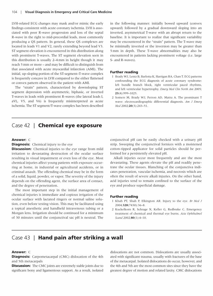
104 | Visual Diagnosis in Emergency and Critical Care Medicine
LVH-related ECG changes may mask and/or mimic the earlyfindings consistent with acute coronary ischemia. LVH is asso-ciated with poor R-wave progression and loss of the septal R-wave in the right to mid-precordial leads, most commonlyproducing a QS pattern. In general, these QS complexes arelocated in leads V1 and V2, rarely extending beyond lead V3.ST segment elevation is encountered in this distribution alongwith prominent T-waves. The ST segment elevation seen inthis distribution is usually 2–4 mm in height though it mayreach 5 mm or more – and may be difficult to distinguish fromthat associated with acute myocardial infarction (AMI). Theinitial, up-sloping portion of the ST-segment–T-wave complexis frequently concave in LVH compared to the either flattenedor convex pattern observed in the patient with AMI.
The “strain” pattern, characterized by downsloping ST segment depression with asymmetric, biphasic, or inverted T-waves in leads with prominent R-waves (the lateral leads I,aVL, V5, and V6) is frequently misinterpreted as acuteischemia. The ST segment/T-wave complex has been described
in the following manner: initially bowed upward (convexupward) followed by a gradual downward sloping into aninverted, asymmetrical T-wave with an abrupt return to thebaseline. It is important to realize that significant variabilitymay be encountered in the “strain” pattern. The T-wave maybe minimally inverted or the inversion may be greater than5 mm in depth. These T-wave abnormalities may also beencountered in patients lacking prominent voltage (i.e. largeS- and R-waves).
Further reading1 Brady WJ, Lentz B, Barlotta K, Harrigan RA, Chan T. ECG patterns
confounding the ECG diagnosis of acute coronary syndrome:
left bundle branch block, right ventricular paced rhythms,
and left ventricular hypertrophy. Emerg Med Clin North Am 2005;
23(4):999–1025.
2 Somers M, Brady WJ, Perron AD, Mattu A. The prominant T
wave: electrocardiographic differential diagnosis. Am J Emerg
Med 2002;20(3):243–51.
Case 42 | Chemical eye exposure
Answer: CDiagnosis: Chemical injury to the eyeDiscussion: Chemical injuries to the eye range from mildirritation to devastating destruction of the ocular surfaceresulting in visual impairment or even loss of the eye. Mostchemical injuries affect young patients with exposure occur-ring at home, in industrial or agricultural accidents, or incriminal assault. The offending chemical may be in the formof a solid, liquid, powder, or vapor. The severity of the injurydepends on the offending agent, the surface area of contact,and the degree of penetration.
The most important step in the initial management ofchemical injuries is immediate and copious irrigation of theocular surface with lactated ringers or normal saline solu-tion, even before testing vision. This may be facilitated usinga topical anesthetic and handheld intravenous tubing or aMorgan lens. Irrigation should be continued for a minimumof 30 minutes until the conjunctival sac pH is neutral. The
conjunctival pH can be easily checked with a urinary pHstrip. Sweeping the conjunctival fornices with a moistenedcotton-tipped applicator for solid particles should be per-formed for a persistently elevated pH.
Alkali injuries occur most frequently and are the mostdevastating. These agents elevate the pH and readily pene-trate the ocular tissues. Blanching of the conjunctiva indi-cates penetration, vascular ischemia, and necrosis which areoften the result of severe alkali injuries. On the other hand,acid injuries tend to remain confined to the surface of theeye and produce superficial damage.
Further reading1 Khah PT, Shah P, Elkington AR. Injury to the eye. Br Med J
2004;328(7430):36–8.
2 Kuckelkorn R, Schrage N, Keller G, Redbrake C. Emergency
treatment of chemical and thermal eye burns. Acta Ophthalmol
Scand 2002;80(1):4–10.
Case 43 | Hand pain after striking a wall
Answer: CDiagnosis: Carpometacarpal (CMC) dislocation of the 4thand 5th metacarpalsDiscussion: The CMC joints are extremely stable joints due tosignificant bony and ligamentous support. As a result, isolated
dislocations are not common. Dislocations are usually associ-ated with significant trauma, usually with fractures of the baseof the metacarpal. Isolated dislocations do occur, however, andthe 4th and 5th are the most common sites since they have thegreatest degree of motion and related laxity. CMC dislocations

Answers, Diagnoses, and Discussion | 105
usually result from extreme forces such as motor vehicle acci-dent, direct blow to the hand, or oblique impact of the closedfist with an immovable object.
The deformity of the dislocated joint is often obscured bythe significant soft tissue swelling present on the dorsum ofthe hand. Tenderness is usually significant. An obvious step-off deformity may be observed and/or palpated at the level ofthe dislocation (the proximal end of the metacarpal as it over-rides the distal carpus), particularly if the patient presentsearly prior to the onset of significant swelling. A critical factorin the evaluation is found in the recognition of associatedmetacarpal fracture. A careful assessment should be made ofneurovascular status of the hand. Specific attention should bedirected to the status of the deep branch of the ulnar nerve inthat it lies immediately volar to the 5th CMC joint; themedian nerve may also be injured. Vascular compromise,particularly in patients with injury to the 3rd metacarpal,may involve the deep palmar arterial arch which lies directlybeneath the 3rd CMC joint. Integrity of the wrist extensortendons must be assessed in these dislocation injuries as dis-ruption may occur. Additionally, those patients who havesuffered a direct blow are at risk for compartment syndromein the hand. The anteroposterior radiograph view may revealan overlap of the carpal bones over the proximal metacarpals(Figure 43.1A, arrow). The lateral radiograph often is diag-nostic with obvious dislocation of the CMC joint visualizedwithout problem (Figure 43.1B, arrow).
Treatment in the emergency department consists of closedreduction, which is accomplished with longitudinal traction.The hand should then be splinted; digital motion should beencouraged to prevent stiffness associated with swelling.Orthopedic consultation is necessary as these dislocationsoften require percutaneous pinning or open reduction withinternal fixation.
Further reading1 Griffiths MA, Moloney DM, Pickford MA. Multiple carpometa-
carpal dislocations after a low-impact injury: a missed diagnosis.
J Trauma 2005;58:391–2.
2 Kumar S, Arora A, Jain AK, Agarwal A. Volar dislocation of mul-
tiple carpometacarpal joints: report of four cases. J Orthop
Trauma 1998;12:523–6.
3 Mabee JR, Lee TJ, Halus S. Dorsal dislocation of the four ulnar
metacarpals. Am J Emerg Med 1997;15:408–11.
A
B
Figure 43.1 A Anteroposterior view of the hand and wrist. B Lateral view of the hand and wrist.
Case 44 | Dyspnea in an alcoholic
Answer: EDiagnosis: Pericardial tamponadeDiscussion: Table 44.1 is an overview of the conditionswhich can be rapidly diagnosed and/or excluded by bedsideultrasound of the thorax and abdomen when confronted by a
patient with undifferentiated dyspnea, hypotension, or pulse-less electrical activity. Several of these conditions are consider-ations in the case presented.
Figures 44.1 and 44.2 are images of the heart showing the leftventricle (LV), right ventricle (RV), and a large circumferential

106 | Visual Diagnosis in Emergency and Critical Care Medicine
Figure 44.1 Apical four chamber view. Figure 44.2 Subxiphoid view.
circulation. Beck’s triad is a late finding in tamponade, sothat its absence does not exclude the possibility of this diag-nosis; which should be made prior to the onset of hypoten-sion. While potentially very serious, intracranial causes ofaltered mental status are less rapidly lethal than hemody-namic ones, so the latter should be sought and addressedfirst, especially in a patient with abnormal vital signs such asthis one.
With evolving training and experience in bedside sonogra-phy, the specialty of emergency medicine has appropriated
pericardial effusion (Figures 44.1 and 44.2; arrows). Effusions are often described as “small/physiological” when they arenon-circumferential, “moderate” when they are circumferen-tial but �10 mm in thickness in diastole, large (10–20 mm in thickness), and very large (�20 mm). Although no calipermeasurements have been performed, the centimeter markersalong the upper margins of the images indicate that this effu-sion is as large as 40 mm in thickness. Cardiac tamponade isdemonstrated in real-time by diastolic collapse of any cham-ber in the presence of moderate or large effusion. Figure 44.3shows a right upper quadrant view similar to that used to iden-tify free fluid (FF) in the focused assessment by sonography intrauma (FAST). A very large volume of FF can be seen betweenthe liver and kidney (Morison’s pouch). While this could con-ceivably be blood, its uniformity, complete lack of echogenicity,and marked posterior acoustic enhancement (causing artifac-tually increased echoes from the underlying tissues marked *),argue against this fluid being intraperitoneal hemorrhage.With the patient’s history of alcohol abuse, and the small,hyperechoic liver seen in Figure 44.3, this fluid is much morelikely to be due to ascites. In Figure 44.4, a longitudinal view ofthe upper aorta can be seen showing the celiac and superiormesenteric (SMA) trunks. Probably due to compression, FF isnot apparent in this view. Since 90% of abdominal aorticaneurysms (AAA) occur between the renal arteries (1 cm infe-rior to the SMA) and the bifurcation, aneurysm cannot beexcluded based on this view alone. However, there is also noevidence to indicate the presence of AAA.
To summarize, the ultrasound images suggest a variety ofabnormalities, which, typical of the resuscitation setting,need to be prioritized according to both their overall likeli-hood, and potential lethality. The diagnosis that poses thegreatest risk of precipitous deterioration is that of cardiactamponade. It is also the one which would take precedencein management according to the standard emergency medi-cine priorities of the “ABC’s”: airway, breathing, and
Table 44.1 Organs to be investigated and potential diagnoses thatmay be identified by emergency medicine bedside ultrasound (EMBU) in the evaluation of unexplained hypotension, PEA, orcardiopulmonary arrest.
EMBU of the thoraxPericardium and heartEffusion � tamponade, severe hypovolemia, massive pulmonaryembolus, cardiogenic shock, myocardial infarction*, gross valvulardysfunction*
Proximal aortaProximal dissection*
Pleural spacesPneumothorax, massive pleural effusion, massive pulmonaryconsolidation
EMBU of the abdomenInferior vena cavaSevere hypovolemia, massive pulmonary embolusAbdominal aortaAbdominal aortic aneurysm, distal dissection*
Peritoneal cavityFree fluid, pneumoperitoneum*
*Indicates conditions which can be identified, but cannot be reliablyexcluded with EMBU.
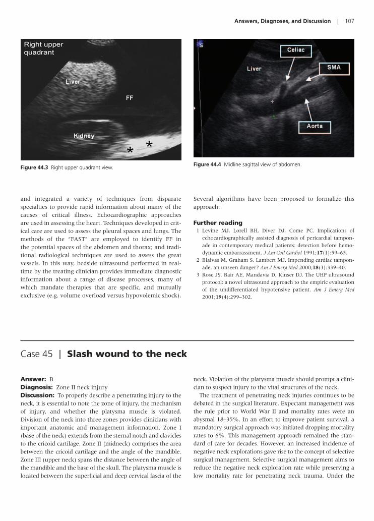
Answers, Diagnoses, and Discussion | 107
Figure 44.3 Right upper quadrant view. Figure 44.4 Midline sagittal view of abdomen.
and integrated a variety of techniques from disparate specialties to provide rapid information about many of thecauses of critical illness. Echocardiographic approaches are used in assessing the heart. Techniques developed in crit-ical care are used to assess the pleural spaces and lungs. Themethods of the “FAST” are employed to identify FF in the potential spaces of the abdomen and thorax; and tradi-tional radiological techniques are used to assess the greatvessels. In this way, bedside ultrasound performed in real-time by the treating clinician provides immediate diagnosticinformation about a range of disease processes, many ofwhich mandate therapies that are specific, and mutuallyexclusive (e.g. volume overload versus hypovolemic shock).
Several algorithms have been proposed to formalize thisapproach.
Further reading1 Levine MJ, Lorell BH, Diver DJ, Come PC. Implications of
echocardiographically assisted diagnosis of pericardial tampon-
ade in contemporary medical patients: detection before hemo-
dynamic embarrassment. J Am Coll Cardiol 1991;17(1):59–65.
2 Blaivas M, Graham S, Lambert MJ. Impending cardiac tampon-
ade, an unseen danger? Am J Emerg Med 2000;18(3):339–40.
3 Rose JS, Bair AE, Mandavia D, Kinser DJ. The UHP ultrasound
protocol: a novel ultrasound approach to the empiric evaluation
of the undifferentiated hypotensive patient. Am J Emerg Med
2001;19(4):299–302.
Case 45 | Slash wound to the neck
Answer: BDiagnosis: Zone II neck injuryDiscussion: To properly describe a penetrating injury to theneck, it is essential to note the zone of injury, the mechanismof injury, and whether the platysma muscle is violated.Division of the neck into three zones provides clinicians withimportant anatomic and management information. Zone I(base of the neck) extends from the sternal notch and claviclesto the cricoid cartilage. Zone II (midneck) comprises the areabetween the cricoid cartilage and the angle of the mandible.Zone III (upper neck) spans the distance between the angle ofthe mandible and the base of the skull. The platysma muscle islocated between the superficial and deep cervical fascia of the
neck. Violation of the platysma muscle should prompt a clini-cian to suspect injury to the vital structures of the neck.
The treatment of penetrating neck injuries continues to bedebated in the surgical literature. Expectant management wasthe rule prior to World War II and mortality rates were anabysmal 18–35%. In an effort to improve patient survival, amandatory surgical approach was initiated dropping mortalityrates to 6%. This management approach remained the stan-dard of care for decades. However, an increased incidence ofnegative neck explorations gave rise to the concept of selectivesurgical management. Selective surgical management aims toreduce the negative neck exploration rate while preserving alow mortality rate for penetrating neck trauma. Under the
108 | Visual Diagnosis in Emergency and Critical Care Medicine
Case 46 | Foot pain following breaking
Answer: BDiagnosis: Lisfranc fracture (medial cuneiform and 2ndmetatarsal base fracture with lateral displacement of the2nd–5th metatarsals)Discussion: The tarsometatarsal joint is named afterJacques Lisfranc, a French physician in Napoleon’s armywho first described amputations through this joint. Anyinjury to this joint, from sprain to fracture, is termed aLisfranc injury. Lisfranc injuries are uncommon because ofthe high force that is required to disrupt this joint. Lisfrancinjuries occur as a result of rotation of the body on a fixedforefoot, axial loading, or crush injuries. Although motorvehicle collisions are usually the mechanism, up to one thirdof Lisfranc injuries arise from stumbles or falls.
The clinical presentation of a Lisfranc fracture includessevere midfoot pain, inability to bear weight, and paresthesias.Examination often reveals edema, ecchymosis, and midfoottenderness. Patients with severe injuries may have obviousdeformity of the midfoot with forefoot displacement. Vascularinjury may occur because a branch of the dorsalis pedis arterydives between the 1st and 2nd metatarsal, forming the plantararch. Injury to this vessel is uncommon, however, and can
lead to hemorrhage, vascular compromise, and compartmentsyndrome. Radiographic findings suggestive of a Lisfrancinjury include widening between the 1st and 2nd or 2nd and3rd metatarsal base. In addition, any fracture involving theLisfranc joint (base of the metatarsals, cuboid, and cuneiforms)should raise suspicion for joint disruption. A fracture involv-ing the 2nd metatarsal base is virtually pathognomonic forLisfranc joint injury.
Management of a Lisfranc fracture is operative andrequires an orthopedic consultation. The patient requiresserial vascular checks, however, doesn’t need an angiogramwithout evidence of vascular compromise. Although rapidreduction of the displaced metatarsals is desirable, thepatient does not require emergent reduction, as the fracturedoes not compromise skin or vascular structures.
Further reading1 Perron AD, Brady WJ. Evaluation and management of the high-
risk orthopedic emergency. Emerg Med Clin North Am 2003;
21(1):159–204.
2 Perron AD, Brady WJ, Keats TE. Orthopedic pitfalls in the ED:
Lisfranc fracture-dislocation. Am J Emerg Med 2001;19(1):71–5.
management scheme of selective surgical exploration, Zone Iand Zone III injuries are initially imaged to clearly define apatient’s vascular anatomy. Angiography remains the goldstandard for diagnosing vascular injuries. The desire for pre-operative imaging stems from the tendency of Zone I injuriesto remain clinically concealed and the difficulty associatedwith surgically approaching Zone III injuries. In contrast, the anatomy of the neck in Zone II is easily approached and explored. Currently, most surgeons choose to surgicallyexplore Zone II injuries without preoperative imaging.
Although the necessity of surgical neck exploration and thereliability of physical examination continue to be debated,serial examinations remain an essential part of the evaluationof a patient with a penetrating neck injury. While unstablepatients require immediate airway management (with cervi-cal spine immobilization) and more immediate surgical inter-vention, stable patients necessitate serial examinations for“soft” and “hard” signs of penetrating neck trauma. Hard signsinclude expanding hematoma, severe active bleeding, shocknot responsive to fluids, decreased or absent radial pulse, vas-cular bruit or thrill, cerebral ischemia, and airway obstruction.Patients who develop hard signs will likely require surgicalintervention. Soft signs include hemoptysis, hematemesis,
dyspnea, dysphonia, dysphagia, subcutaneous or mediasti-nal air, nonexpanding hematoma, and the presence of focalneurologic deficits.
Patients who are in profound shock or who have uncon-trollable hemorrhage from a penetrating neck injury shouldgo directly to the operating room for hemorrhage control.Definitive airway management, large-bore intravenous accessand blood samples sent for cross-match are essential in theimmediate encounter. Bedside vessel ligation can result in sig-nificant neurologic impairment and is not the treatment ofchoice. Temporary hemorrhage control with direct pressurecan be utilized prior to transport to the operating room. Analternative to immediate operative intervention of patientswith zone I and zone III injuries depends on the availability ofinterventional radiology. Angiography and embolization canbe utilized in these patients. However, involvement of an inter-ventional radiologist should not delay definitive intervention.
Further reading1 Thal ER, Meyer DM. Penetrating neck trauma. Curr Prob Surg
1992;29(1):1–56.
2 McConnell DB, Trunkey DD. Management of penetrating
trauma to the neck. Adv Surg 1994;27:97–127.

Answers, Diagnoses, and Discussion | 109
Case 47 | Confluent rash in a child
Answer: CDiagnosis: Erythema multiforme (minor)Discussion: Erythema multiforme (EM) is a hypersensitiv-ity reaction which manifests as a diffuse eruption with very characteristic lesions. These lesions are usually sym-metric, involve palms of hands and soles of feet, and pre-dominate on the extensor surfaces of the upper and lowerextremities. Although these are characteristic locations they can be found anywhere on the body. The rash of EMcan look macular, urticarial, or vesicobullous, but the proto-typical lesion is a target lesion with a dusky center. Often therash changes from one form of lesion to another as the dis-ease progresses. The rash itself generally lasts at least 1 week,but can last up to 6 weeks. Patients are often otherwiseasymptomatic although they can also have itching associatedwith the lesions or involvement of the oral mucosa.
The causes of EM are often unknown, but in children aremost commonly related to infectious causes, whereas inadults it is much more frequently related to drug reaction ormalignancy. The most common infectious agent attributedto EM is herpes simplex virus. The differential diagnosis ofEM includes pemphigus, bullous pemphigoid, urticaria, orother viral exanthema. Treatment for EM minor may involvecessation of inciting agents, but is mainly supportive includ-ing antihistamines and/or NSAIDS. Systemic glucocorticoids
are sometimes used although there are no randomized trialsshowing a clear benefit.
It is important to note that EM is thought to be part ofcontinuum of more serious illness such as Stevens–Johnson(EM major) and toxic epidermal necrolysis (TEN). It isimportant on physical exam to evaluate mucosal surfaces todifferentiate between EM minor and major. EM minorinvolves the skin and only one other mucosal surface, usu-ally the mouth. In contrast, Stevens–Johnson involves theeye, oral cavity, genital mucosa, upper airway, or esophagus.Stevens–Johnson and TEN are much more serious condi-tions with significantly higher mortality rates. Treatment forthese conditions is frequently compared to burn care andhospital admission is required. It is important to keep theseother entities in mind even in cases of EM minor becausepatients and their families should be discharged with clearinstructions about signs to look for that may indicate pro-gression to more serious disease.
Further reading1 Carder KR. Hypersensitivity reactions in neonates and infants.
Dermatol Ther 2005;18(2):160–75.
2 William PM, Konklin RJ. Erythema multiforme: a review and con-
trast from Stevens-Johnson syndrome/toxic epidermal necrolysis.
Dent Clin North Am 2005;49(1): 67–76.
Case 48 | Lost in the cold
Answer: BDiagnosis: FrostbiteDiscussion: Frostbite is a cold-related injury that is due tothe freezing of tissues when exposed to temperatures at orbelow the freezing point of intact skin. As the temperature to the skin drops, blood flow diminishes, ice crystals form,osmotic disequilibrium occurs, cellular dehydration devel-ops, and cellular wall integrity becomes compromised. Theend result is cellular death and capillary damage.
Initial management should first be directed at assessingcore body temperature and hydration. Both active and passiverewarming may be considered, depending on the patient’score body temperature. Most patients will also benefit fromintravenous fluid administration as some degree of dehydra-tion is typically seen.
After initial stabilization, rapid rewarming of the afflictedextremity is of paramount importance. A warm water bath
at temperatures between 40°C and 42°C is the recommendedfor approximately 30 minutes. Utilizing water at lower temperatures results in slower and potentially incompleterewarming which worsens outcome; utilizing water at highertemperatures risks the development of thermal burns. Asextremity reperfusion begins, severe pain can be expected andopioid analgesics are typically required. Dry air rewarming,especially over a campfire, is discouraged because of the possi-bility of thermal injury. Massaging the frostbitten extremityshould be avoided as it may result in further tissue damage.
After rewarming, attention is directed toward wound careand pain control. Wounds should be wrapped in sterile gauzeafter application of topical Aloe vera. Aloe vera is an inhibitor of thromboxane which is thought to play a large role in tissueloss. Ibuprofen should also be given because of its ability toinhibit thromboxane. Tetanus immunization status should beassessed and the patient should avoid smoking.

110 | Visual Diagnosis in Emergency and Critical Care Medicine
Case 49 | Bradycardia following an herbal ingestion
Answer: EDiagnosis: Aconitum dephinifolium (monkshood) poisoningDiscussion: The use of herbal products has dramaticallyincreased over the past decade, driving physicians to becomeeducated in regards to potential herbal complications anddrug interactions. Studies have documented that approxi-mately one in every five patients presenting to the emer-gency department uses some type of herbal preparation.
Plants that contain aconitine include Aconitum napellus(monkshood), A. vulparia (wolfsbane), A. carmichaeli(chuanwu), and A. kusnezoffii (caowu). Members of thisplant genus grow throughout the world. Aconitine expo-sures are commonly associated with the overzealous con-sumption of certain herbal preparations. Although a numberof fatal poisonings have been reported, aconitine is still read-ily available at many nutrition or herbal medicine stores.
These flowers of the plant, which are commonly purple,form a hood-like structure, hence the name “monkshood.” Allparts of this plant are potentially poisonous. Aconitine appearsto increase sodium entry into muscle, nerve, baroreceptors,and Purkinje fibers to produce a positive inotropic effect,enhanced vagal tone, neurotoxicity, increased automaticity,and torsade de pointe. Bifascicular ventricular tachycardia, a
dysrhythmia most frequently associated with digitalis toxicity,has also been reported in patients poisoned with aconitine.
Following exposure, symptoms have been reported to occurbetween 3 minutes and 2 hours, with a median of 30 minutes.Symptoms may persist for up to 30 hours. Neurologic com-plaints include initial visual impairment, dizziness, limb pares-thesias, weakness, ataxia, and coma. Chest discomfort, dyspnea,tachycardia, bradycardia, ectopic beats, supraventricular tachy-cardia, bundle branch block, intermittent bigeminy, ventriculartachycardia, ventricular fibrillation, and asystole have all beendescribed.
Treatment is primarily supportive. The paramount con-cern is management of lethal arrhythmias. Symptomaticbradydysrhythmias may respond to atropine administration.Ventricular tachycardia has been reported to be refractory topharmacologic therapy.
Further reading1 Guha S, Dawn B, Dutta G, et al. Bradycardia, reversible pancon-
duction defect and syncope following self-medication with a
homeopathic medicine. Cardiology 1999;91(4):268–71.
2 Furbee B, Wermuth M. Life-threatening plant poisoning. Crit
Care Clin 1997;13(4):849–88.
Considerable controversy exists in regard to managementof blisters. Clear vesicles and bullae are rich in thromboxane,which is thought to increase tissue loss. Aspiration of thesebullae has been advocated as a way to remove the immunemodulators while preserving the barrier to infections. Othershave advocated debridement as a means to remove throm-boxane and allow for direct application of Aloe vera.
Surgical management of frostbite injuries should be delayed.There are three zones of injury: (1) the distal zone which pro-gresses to dry gangrene and eventually requires amputation;(2) the proximal zone which heals without intervention; (3)the intermediate zone which has the potential for salvage.Treatment and wound care are directed toward this zone inhope of salvaging as much tissue as possible. Complicationsmay occur that require surgical intervention (i.e. compart-ment syndrome or gas gangrene), but the available evidencedoes not suggest that early debridement of afflicted tissueimproves outcome.
Heparin therapy has been shown to be of benefit in some animal models. Thrombosis of the microvasculature is afrequent finding after rewarming and may contribute to tissueloss. Heparin therapy was directed at reversing this thrombo-sis. Currently available human studies, however, have failedto show any benefit of heparin therapy over rapid rewarmingand general wound care.
Further reading1 Biem J, Koehncke N, Classen D, Dosman J. Out of the cold:
management of hypothermia and frostbite. Can Med Assoc J
2003;168(3):305–11.
2 Murphy JV, Banwell PE, Roberts AH, McGrouther DA. Frostbite:
pathogenesis and treatment. J Trauma 2000;48(1):171–8.
3 Reamy BV. Frostbite: review and current concepts. J Am Board
Fam Pract 1998;11(1):34–40.

Answers, Diagnoses, and Discussion | 111
Case 50 | Abdominal pain in an alcoholic
Answer: CDiagnosis: Sentinel loopDiscussion: The term sentinel loop was first used in 1946 byLevitin. On plain film, he described it as an isolated loop of dis-tended adynamic bowel. A sentinel loop may be seen on radio-graph when there is localized inflammation in the peritonealcavity. A segmental paralytic ileus affecting one or two loopsof small bowel may subsequently occur adjacent to this local-ized inflammation. Gas and fluid accumulate in this isolatedarea of small bowel as the localized inflammatory processleads to a focal decrease in peristalsis.
There are various disease processes that may lead to a sentinel loop on radiograph. When found in the upperabdomen, a sentinel loop may be due to pancreatitis, chole-cystitis, or less commonly pyelonephritis or splenic injury. Inacute pancreatitis, while many various radiographic findingsmay appear on plain film, the sentinel loop is the most spe-cific. Appendicitis may lead to a sentinel loop on plain film inthe right lower quadrant. The sentinel loop is a nonspecificradiographic finding and sometimes may mimic small oreven large bowel obstruction.
Further reading1 Davis S, Parbhoo SP, Gibson MJ. The plain abdominal radiograph
in acute pancreatitis. Clin Radiol 1980;31(1): 87–93.
2 Ranson JH. Diagnostic standards for acute pancreatitis. World J
Surg 1997;21(2):136–42.
Figure 50.1 Sentinel loop (arrow pointing to area).
Answer: EDiagnosis: Clostridial myonecrosis (gas gangrene)Discussion: Clostridial myonecrosis, which is more com-monly known as gas gangrene, is a surgical emergency.Clostridium perfringens is the most common pathogen isolated,but many clostridial species are capable of producing the dis-ease. Exotoxins, which are produced under anerobic condi-tions, are responsible for morbidity and mortality associatedwith clostridial myonecrosis. These toxins result in numerousdetrimental effects, including lysis of muscle cells, destructionof blood vessels, hemolysis, and cardiac suppression.
The clinical picture can initially appear benign, necessitatingvigilance in making this clinical diagnosis. Pain is rapidly pro-gressive over hours to days and is typically out of proportion tophysical findings. Low-grade fever is common and is lowerthan would be expected given the severity of the infection. Thewound itself is usually unimpressive with surrounding pale tis-sue. In later stages the wound can appear dusky and progress
to dark discoloration. Wound discharge is commonly foul-smelling and brown, with a blood tinged appearance. Thiswound discharge should be sent for culture and Gram stain,and typically reveals a preponderance of Gram-positive rodsand a lack of neutrophils. Gas in the soft tissues can clinicallymanifest as crepitus. Radiographic evidence of air in the softtissues suggests the diagnosis (as noted in the X-ray of thiscase). Some authors advocate sonography as a tool to identifygas in the wound before it is apparent clinically or on plainradiographs.
Once the diagnosis is entertained, empiric therapy shouldbe initiated immediately. Intravenous penicillin should begiven in doses of 12–16 million international units per daydivided in four daily doses. Emergent surgical evaluation is essential. Usually multiple surgeries are needed to ensureadequate debridement. Radical excision and amputation arecommonly necessary to prevent mortality (as occurred inthis case). Aggressive fluid resuscitation should be initiated
Case 51 | Pain out of proportion to examination

112 | Visual Diagnosis in Emergency and Critical Care Medicine
Answer: DDiagnosis: Acute pericarditisDiscussion: The ECG is typical of acute myopericarditiswith pericardial effusion manifested by electrical alternans.The rhythm strip demonstrates electrical alternans with alter-nating QRS complex amplitudes. The ECG demonstrates sig-nificant, diffuse ST segment elevation with a concavemorphology; additionally, PR segment depression is seen inmost leads with ST segment elevation. Lead aVr demon-strates reciprocal PR segment elevation.
Acute pericarditis produces diffuse inflammation of the peri-cardium and superficial epicardium. Numerous etiologies areencountered in the patient with acute myopericarditis, includ-ing infectious, inflammatory, toxicologic, and rheumatologiccauses. The ECG can demonstrate significant findings inpatients with acute pericarditis, including ST segment eleva-tion, T-wave inversion, and PR segment abnormalities. Thesechanges result from endocardial inflammation; it is importantto realize that the pericardium is electrically silent. Because ofinvolvement of the pericardium and endocardium, pericarditisis most appropriately termed myopericarditis.
The ECG changes caused by pericarditis evolve throughfour classic stages. The first stage is characterized by ST seg-ment elevation which is followed by resolution of the eleva-tion in the second stage. The third stage occurs with T-waveinversion followed by normalization of all such changes anda return to the baseline ECG in stage four. The temporal evo-lution of these electrocardiographic stages occurs in a very
unpredictable fashion – stages one through three are seenover hours to days while stage four may not be reached forseveral weeks. Further, patients may not manifest all charac-teristic ECG features.
ST segment elevation seen in patients with stage one pericarditis is usually less than 5 mm in height, observed innumerous leads, and characterized by a concavity to its initialupsloping portion. The ST segment elevation is most oftenseen in multiple leads though it may be limited to a specificanatomic segment. PR segment depression associated withpericarditis perhaps is the most helpful feature in arriving atthe correct electrocardiographic diagnosis; such a finding hasbeen described as “almost diagnostic” for acute pericarditis.Reciprocal PR segment elevation is seen in lead aVr and is usually very helpful in the diagnosis.
Pericardial effusion may also be seen in patients with peri-carditis; the presence of the effusion may affect the electrocar-diographic signal noted on the ECG. These findings includewidespread low voltage (resulting from increased resistance toinjury current flow with the accumulated fluid) and electricalalternans (a beat-to-beat alteration in QRS complex size dueto shifting fluid in the pericardium).
Further reading1 Chan T, Brady WJ, Pollack M. Electrocardiographic manifestations:
acute myopericarditis. J Emerg Med 1999;17:865–72.
2 Lange RA, Hillis LD. Clinical practice: acute pericarditis. New Engl J
Med 2004;351(21):2195–202.
Case 52 | Pleuritic chest pain in a young adult male
in the emergency department as hypotension and septicshock are commonly encountered. Hyperbaric oxygen ther-apy has been advocated as an adjunctive modality, but mustbe used in concert with intravenous antibiotics and surgicaldebridement.
Further reading1 Gonzalez MH. Necrotizing fasciitis and gangrene of the upper
extremity. Hand Clin 1998;14(4):635–45.
2 Headley AJ. Necrotizing soft tissue infections: a primary care
review. Am Fam Physician 2003;68(2):323–8.
Answer: DDiagnosis: Full thickness lid lacerationDiscussion: This patient has two full thickness lid lacerationsto the left lower lid. When a patient presents with an apparentisolated lid laceration, it is necessary to first exclude globeinjury or rupture. Determining the mechanism of injury with athorough history will help direct management. Complete
ocular examination is necessary to rule out globe injury.Examination should include visual acuity, pupils, extraocularmotility, external adnexa, slit lamp examination, tonometry,and dilated fundus exam. Computed tomography scan of thebrain and orbit (axial and coronal views), using 1–3 mm cuts,should be obtained when a foreign body or globe rupture is sus-pected. Tetanus prophylaxis should be given when indicated.
Case 53 | Eye pain following a bar fight

Answers, Diagnoses, and Discussion | 113
The presence of fat in a periocular wound indicates that theorbital septum has been violated. In these cases it is necessaryto determine if damage to the levator muscle has occurred.Lacerations occurring in the medial canthal area require eval-uation of the lacrimal drainage system.
Many eyelid lacerations can be repaired in the emergencyroom. Lid lacerations requiring repair in the operating roominclude those involving the lacrimal drainage apparatus,
involvement of the levator muscle, extensive tissue loss, orassociated globe trauma requiring surgery.
Further reading1 Chang EL, Rubin PA. Management of complex eyelid lacera-
tions. Int Ophthalmol Clin 2002;42(3):187–201.
2 Larian B, Wong B, Crumley RL, et al. Facial trauma and ocu-
lar/orbital injury. J Craniomaxillofac Trauma 1999;5(4):15–24.
Answer: EDiagnosis: Monteggia fractureDiscussion: A Monteggia fracture is an eponym for anulnar fracture associated with a dislocation of the radial head.The dislocation of the radial head (note how the line alongthe axis of the radius does not intersect the capitellum inFigure 54.1) can be easily overlooked if the radiographs fail toinclude the elbow. If the radial head dislocation is missed,prolonged disarticulation and subsequent disability may occur.Monteggia fractures can result from falling on an outstretchedhand with forced pronation. The energy exerted in fracturingthe ulna travels along the interosseus membrane pulling theradial head from the annular ligament.
Reduction of the radial head must be achieved expedi-tiously followed by splinting of the forearm in a position offunction with wrist extended. Monteggia fractures are oftenmanaged with internal fixation because of the increased riskof non-union.
Radial motor nerve injury can occur from stretching orcompressing the nerve as it passes across the elbow. The radialnerve becomes the posterior interosseous nerve which sup-plies the extensor muscles of the wrist, fingers, and thumb.Motor neuropathies are usually treated with conservative man-agement with surgical exploration being reserved for thosewho do not regain function within 2–3 months.
A Galeazzi fracture is an eponym for a radial fracture associ-ated with the dislocation of the distal radioulnar joint. Galeazzifractures are typically treated with open reduction and internalfixation.
Further reading1 Wilkins KE. Changes in the management of Monteggia frac-
tures. J Pediatr Orthop 2002;22(4):548–54.
2 Perron AD, Hersh RE, Brady WJ, Keats TE. Orthopedic pitfalls in
the ED: Galeazzi and Monteggia fracture–dislocation. Am J Emerg
Med 2001;19(3):225–8.
Case 54 | Forearm fracture after falling
Figure 54.1 Monteggia fracture with long arrow pointing to proximal ulnar fracture and short arrow pointing to radial head dislocation.

114 | Visual Diagnosis in Emergency and Critical Care Medicine
Answer: BDiagnosis: Avascular necrosis (AVN) of the left femoral headDiscussion: Patients presenting with progressive groinand/or thigh pain radiating to the buttock must be evaluatedfor AVN of the femoral head. AVN is a progressive disease ofjoint destruction with many causes. Steroid exposure, alco-holism, hemoglobinopathies, renal disease, and a history of hipfracture or hip dislocation all put patients at risk for ischemia ofthe femoral head. Independent of the precipitating event, allpatients with ischemic bone soon develop abnormal bonegrowth, edema, and fibrosis. Males are at higher risk for AVNand it typically develops in patients after the age of 40 years.Groin pain that is worse with weight bearing and limited rangeof motion of the affected joint are common initial complaints.Plain radiographic findings can lag behind clinical symptoms ofhip pain and difficulty walking.
Diagnosis is dependent upon clinical suspicion and con-firmed by radiographic studies. The plain radiographic studiesto obtain include anteroposterior and frog leg lateral views(frog leg views allow for evaluation of the superior portion ofthe femoral head). A staging system exists for AVN. In stageone, plain radiographs are normal but if the clinician has ahigh index of suspicion diagnosis can be achieved using mag-netic resonance imaging. Stage two is characterized by diffuse
osteoporosis of the femoral head; therefore subchondral lyticlesions can be seen on plain radiography but the shape of thefemoral head is preserved. In the third stage, many of thepathognomonic changes become evident on radiography;there is frank collapse of bone, the crescent sign may be seen(subchondral radiolucency), and there is flattening of thefemoral head. The fourth stage is progression of subchondralbone collapse including marked deformity of the femoralhead with or without acetabular involvement. In comparisonto osteoarthritis, the joint space is preserved until late in thedisease process when the entire joint has collapsed.
A clinical syndrome of AVN also has a staging system, but ithas not been demonstrated to correlate well with the X-rayfindings or disease progression. Treatment for AVN initiallyincludes avoidance of weight bearing if caught early, but thisdisease typically progresses to the need for joint replacement.
Further reading1 Musso ES, Mitchell SN, Schink-Ascani M, Bassett CA. Results
of conservative management of osteonecrosis of the femoral
head: a retrospective review. Clin Orthop Relat Res 1986;(207):
209–15.
2 Mont MA, Payman RK, Laporte DM, et al. Atraumatic osteonecro-
sis of the humeral head. J Rheumatol 2000;27(7):1766–73.
Case 55 | An elderly woman with groin pain
Answer: EDiagnosis: Herpes zoster ophthalmicusDiscussion: Herpes zoster ophthalmicus is a reactivation ofthe varicella virus affecting the ophthalmic division of thetrigeminal nerve. It is important to perform a careful medicalhistory on patients presenting with herpes zoster ophthalmi-cus to determine whether the patient may be immunocom-promised. Patients younger than 40 years require a completesystemic evaluation. Older patients do not require evaluationunless immunodeficiency is suspected from the case history.
Ocular manifestations of herpes zoster ophthalmicus areimmense and often require long-term follow-up. When herpeszoster ophthalmicus affects the tip of the nose this is known asHutchinson’s sign, indicating involvement of the nasociliarybranch of the ophthalmic division. There is a higher risk ofocular involvement in patients with a positive Hutchinson’ssign. Ocular evaluation for patients with herpes zoster oph-thalmicus includes visual acuity, intraocular pressures, carefulslit lamp examination, and a dilated fundus exam.
Patients with active skin lesions for less than 72 hoursshould be started on an oral antiviral agent such as acyclovir,800 mg five times a day or famciclovir, 500 mg three times aday. Patients that are systemically ill or immunocompromisedshould be admitted to the hospital and given intravenous acy-clovir. Bacitracin or erythromycin ointment should be appliedto the skin lesions three times a day. Patients with ocularinvolvement need ophthalmologic consultation since theymay require corticosteroid therapy and regular outpatient follow-up. A primary physician should also manage thesepatients since pain from herpes zoster ophthalmicus is oftensevere and post-herpetic neuralgia can linger for months.
Further reading1 Baran R, Kechijian P. Hutchinson’s sign: a reappraisal. J Am Acad
Dermatol 1996;34(1):87–90.
2 Yoshida M, Hayasaka S, Yamada T, et al. Ocular findings in Japanese
patients with varicella-zoster virus infection. Ophthalmologica 2005;
219(5):272–5.
Case 56 | Painful facial rash

Answers, Diagnoses, and Discussion | 115
Answer: DDiagnosis: Lead encephalopathy due to ingestion of paintchipsDiscussion: There are multiple radioopacities noted in the plain film consistent with ingestion of paint chips (Figure 57.1).
A list of the more common radioopacities is summarized by the mnemonic CHIPES – calcium/chloral hydrate, heavymetals/halogenated hydrocarbons, iron, phenothiazines/potassium, enteric-coated, and salicylates. However, multiplestudies regarding the radioopacity of ingested pharmaceuticalshave not consistently supported this and other mnemonics.
A federal mandate banned the use of lead in house and fur-niture paint since 1978. Any child who lives or has contactwith houses built before this time is at risk for elevated lead levels through ingestion of chips or by frequent hand-to-mouth activity in the dust-laden dilapidated environment.Children have fairly efficient absorption of lead, averaging40–50%. Lead affects multiple organ systems by disruptingvarious enzymatic activities. In children, subtle clinical effectsmay be seen even at low lead levels (�10 �g/dL). Obviousacute neurotoxicity at much higher levels is attributable tocerebral edema from fluid egress across the blood–brain bar-rier. Lead disrupts many of the enzymes in heme biosynthesisresulting in a microcytic hypochromic anemia that mimicsiron deficiency anemia. Vomiting, anorexia, constipation, and“lead colic” have all been described in children.
Lead encephalopathy, as demonstrated in this case, must betreated emergently with chelation therapy. British antilewisite(BAL) is the chelator of choice because it readily crosses theblood–brain barrier. BAL is the only chelator that is bothrenally cleared and biliarly cleared. The disadvantages to BALinclude the following: (1) it is administered only by intramus-cular injection; (2) it is exceedingly painful and needs to bemixed with an anesthetic such as procaine; (3) it is mixed in peanut oil and cannot be given to children with known peanut allergies. Calcium disodium ethylenediaminetetraac-etate (CaNa2EDTA), the only other parenteral chelator, does
not cross the blood–brain barrier. Administration ofCaNa2EDTA solely without BAL could place the patient at riskfor progressive cerebral edema by mobilizing lead from theother tissues and causing it to redistribute into the brain.
Asymptomatic children should be chelated with oral 2,3-dimercaptosuccinic acid (DMSA) at levels only exceed-ing 44 �g/dL. Health Department notification should bemade at blood lead levels at or exceeding 10 �g/dL.
Further reading1 Gordon RA, Roberts G, Amim Z, Williams RH, Paloucek FP.
Aggressive approach in the treatment of acute lead encephalopathy
with an extraordinarily high concentration of lead. Arch Pediatr
Adolesc Med 1998;152(11):1100–04.
2 Feldman RG, White RF. Lead neurotoxicity and disorders of
learning. J Child Neurol 1992;7(4):354–9.
Case 57 | Confusion, anemia, and abdominal pain in a toddler
Figure 57.1 Ingestion of paint chips.
Answer: BDiagnosis: Fluoride toxicityDiscussion: In this case, the patient ingested an unknownliquid substance. His initial symptoms of throat burning, eme-sis, difficulty with phonation, inability to manage secretions,and chest and abdominal pain were consistent with a caustic
ingestion. A rapid bedside litmus paper test revealed that thesubstance had an acidic pH. An initial arterial blood gasrevealed a metabolic acidosis with concomitant respiratoryalkalosis. The patient subsequently progressed to hypotensionwith electrocardiographic (ECG) changes of both QRS com-plex widening and QT interval prolongation. For the astute
Case 58 | Cardiotoxic effects following caustic ingestion
clinician who considers the differential diagnosis for an acidicagent that produces metabolic acidosis, hypotension, and ECGabnormalities (QRS complex widening and QT interval pro-longation), the diagnosis is readily determined to be hydroflu-oric acid ingestion.
Hydrofluoric acid is utilized in many industrial settings for the production of integrated circuits, fluorides, plastics,germicides, insecticides and for the etching and cleaning of sil-icone, glass, metal, stone, and porcelain. Hydrofluoric acidcontaining products are sold as automotive cleaning productsin local stores. Hydrofluoric acid rapidly corrodes and pene-trates skin and mucous membranes. Ingestion of hydrofluoricacid may result in local mucosal caustic effects, nausea, vom-iting, abdominal pain, and hemorrhagic gastritis. Systemicelectrolyte abnormalities may occur. The absorbed fluorideions rapidly bind to available calcium and magnesium ions,decreasing the body’s levels of these divalent cations. Hyper-kalemia often follows due to an efflux of potassium out of cellsinto the extracellular space.
This patient manifested progressive QT interval prolonga-tion. Hydrofluoric acid causes QT interval prolongation byinducing hypomagnesemia and hypocalcemia. These eventsmay place the patient at risk for polymorphic ventriculartachycardia or torsades de pointes.
All patients presenting with signs and symptoms consis-tent with hydrofluoric acid ingestion should be aggressivelymanaged. The patient’s airway should be patent and ade-quate ventilation assured. If necessary, endotracheal tubeintubation should be performed early before edema leads toairway obstruction. The patient should be placed on contin-uous cardiac monitoring with pulse oximetry and frequentneurological checks should be made. The initial treatment ofhypotension consists of intravenous fluids, followed by pres-sors as needed. The patient’s pulmonary status should bemonitored closely for clinical signs consistent with pul-monary aspiration. Activated charcoal, syrup of ipecac, and
gastric lavage are absolutely contraindicated in patients who have ingested caustics. Serum electrolytes should beobtained hourly and include serial calcium, magnesium, andpotassium levels. The clinician should obtain serial ECGslooking for signs of hypocalcemia (prolonged QTc interval)and hyperkalemia (peaked T-waves). Large amounts of cal-cium and magnesium may be needed to normalize serumlevels. Fluoride-induced hyperkalemia has been reported tobe difficult to reverse. Early aggressive therapy with glucose,insulin and/or sodium bicarbonate may be effective.
Dermal hydrofluoric acid exposures are more commonlyseen than ingestions and occur when household rust removersor tire cleaners are used (concentrations range from 6% to12%). These agents often cause delayed finger pain after liq-uid drips from the spray bottle onto unprotected hands duringapplication of the product. The exposed dermal site may ini-tially look quite innocuous and still cause significant local andpotentially systemic toxicity. Inhalational exposures can causelaryngeal, pharyngeal, or pulmonary edema. Ocular exposurecan lead to extensive damage to the eye similar to other acidswith copious irrigation as the mainstay of treatment.
Symptomatic hand exposures can be treated with topicalgels (25 mL of 10% calcium gluconate in 75 mL of a water-soluble lubricant such a KY Jelly). In cases with refractorypain, some authors have advised arterial catheter placementproximal (radial artery) in the exposed hand and infusion ofcalcium gluconate. A typical infusion regimen is as follows: a10 mL solution of calcium gluconate mixed in 50 mL of 5%dextrose in water and infused over 4 hours.
Further reading1 Holstege CP, Baer AB, Brady WJ. The electrocardiographic toxi-
drome: the ECG presentation of hydrofluoric acid ingestion.
Am J Emerg Med 2005;23:171–6.
2 Bertolini JC. Hydrofluoric acid: a review of toxicity. J Emerg Med
1992;10(2):163–8.
116 | Visual Diagnosis in Emergency and Critical Care Medicine
Answer: ADiagnosis: Henoch–Schönlein purpura (HSP)Discussion: HSP is the most common systemic vasculitis in children, with rapid onset over the course of days toweeks. The disease is an example of a leukocytoclastic vas-culitis, characterized by immune complex deposition leadingto necrosis and inflammation of small blood vessels, mostcommonly the postcapillary venules. The disease course isusually over 6 weeks, with the majority of patients resolvingtheir disease in 1 month. Relapses can occur in 16–40% ofpatients, up to 2 years after the initial symptoms. Clinical
manifestations of HSP are seen in several organ systemsincluding the skin, joints, gastrointestinal tract, and kidney.
Cutaneous involvement is seen in 100% of patients and ischaracterized by palpable purpura, most commonly on thelower extremities and in dependent areas, such as the poste-rior thighs and buttocks. However, the rash can involve upperextremities as well. When the rash is more widespread, it is imperative to differentiate HSP from more serious condi-tions such as meningococcemia, idiopathic thrombocytopenicpurpura (ITP), subacute bacterial endocarditis (SBE) andhemolytic uremic syndrome (HUS).
Case 59 | Rash and joint pain in a child

Answers, Diagnoses, and Discussion | 117
Joint involvement occurs in 50–80% of patients with HSP,and commonly involves large lower extremity joints, such asknees and ankles. True arthritis is rare, but pain, periarticularswelling and limitation of range of motion can be significant.Usually anti-inflammatory medications such as naproxen orIbuprofen are helpful for these symptoms. Arthrocentesis isnot indicated.
Gastrointestinal involvement is seen in 65–70% of patientswith HSP, usually within 1–4 weeks of the onset of rash. Rarely(14–36%) patients may have abdominal pain before the rash isevident. The symptoms usually involve colicky periumbilicalpain and vomiting, with or without abdominal distention. Thisresults from submucosal and subserosal edema and hemor-rhage. Rarely, emergent gastrointestinal complications such assmall bowel intussusception, bowel ischemia and infarction,and bowel perforation may occur. Although no evidence in theliterature supports the use of glucocorticoids for treatment ofabdominal pain, traditionally they are used in hope of shorten-ing the duration of the gastrointestinal symptoms. Steroidshave no effect on the overall course of the disease.
The most significant morbidity associated with HSP is sec-ondary to renal involvement. It is the risk of renal involve-ment which will dictate the close follow-up these patientsrequire. Renal disease can occur in 20–34% of patients withHSP, and the spectrum ranges from microscopic hematuria,with or without proteinuria, to fulminant renal failure whichcan occur in 1–5% of patients. Due to this small yet significantrisk, patients with HSP need weekly blood pressure checksand weekly urinalyses throughout the course of disease.
Overall, HSP is a benign vasculitic disease of childhood,requiring mostly supportive care. Two-thirds of patients will resolve their symptoms within 1 month of onset. Closefollow-up by a primary care physician is appropriate for themajority of cases.
Further reading1 Lanzkowsky S, Lanzkowsky L, Lanzkowsky P. Henoch–
Schoenlein purpura. Pediatr Rev 1992;13(4):130–7.
2 Saulsbury FT. Henoch–Schonlein purpura in children. Report of 100
patients and review of the literature. Medicine 1999;78(6):395–409.
Answer: BDiagnosis: Post-operative pneumoperitoneumDiscussion: Pneumoperitoneum refers to air within theperitoneal cavity. It characteristically appears as radiolucencybetween the right hemi-diaphragm and the liver, or in a supe-riorly dependent location on abdominal radiograph. It usuallyindicates a perforated abdominal viscus. Tumor, ulcers, ortrauma may cause any portion of the bowel to perforate, lead-ing to an associated peritonitis that typically requires urgentabdominal surgery. Pneumoperitoneum does not require sur-gery in 5–15% cases.
Nonsurgical pneumoperitoneum (NSP) is defined by thepresence of air in the peritoneal space that is detectable byplain film X-rays and either is managed successfully by obser-vation and supportive care alone or results in a nondiagnosticlaparotomy. NSP is most commonly retained post-operativeair. It may occur after peritoneal dialysis catheter placement,gastrointestinal endoscopic procedures, mechanical ventilation,
cardiopulmonary resuscitation, pneumothorax, pneumomedi-astinum, sexual intercourse, or vaginal douching.
Post-operative pneumoperitoneum usually resolves 3–6 daysafter surgery, although it may persist for as long as 24 days.When abdominal pain and distention are minimal, and peri-toneal signs and evidence of a systemic inflammatory response(fever, tachycardia, leukocytosis, and tachypnea) are absent,conservative management is appropriate.
Further reading1 Mularski RA, Sippel JM, Osborne ML. Pneumoperitoneum: a
review of nonsurgical causes. Crit Care Med 2000;28(7):2638–44.
2 Roberts PA, Wrenn K, Lundquist S. Pneumoperitoneum after
percutaneous endoscopic gastrostomy: a case report and review.
J Emerg Med 2005;28(1):45–8.
3 Rosen DM, Lam AM, Chapman M, Carlton M, Cario GM. Methods
of creating pneumoperitoneum: a review of techniques and
complications. Obstet Gynecol Surv 1998;53(3):167–74.
Case 60 | X-ray findings after laparoscopy
Answer: DDiagnosis: High-pressure injection injuryDiscussion: Injection injuries that involve the hand and/orupper extremity are uncommon and most are job related.
The advent of high-pressure industrial equipment has led tothe occurrence of injection of fuel oil, grease, cement, paints,and solvents in the body. The lower the viscosity of theinjected substance the higher the potential for spread through
Case 61 | Injector injury to the hand

118 | Visual Diagnosis in Emergency and Critical Care Medicine
soft tissue. A pressure of 100 pounds per square inch (psi) canbreach the skin. It is not uncommon for airless spray-guns orfuel injectors to generate 3000–5000 psi.
The patient who presents with this type of injury is typicallya young male who has injured his non-dominant hand. Theinjured site commonly appears as a small harmless puncture
with some surrounding soft tissue swelling. Clinicians shouldnot be misled by this innocuous presentation as significantfunctional morbidity and loss of limb are possible.
Emergent management should include tetanus prophy-laxis, analgesia, and broad-spectrum antibiotics. Radiographsof the involved areas can help determine the extent of the softtissue spread of the injected material. Many, but not all, of the injectable materials are radioopaque. Also, subcutaneousemphysema caused by the high-pressure injection may beappreciated. Immediate surgical consultation is required and the emergency physician should recognize this injury aspotentially limb-threatening and the definitive treatment tomaximize recovery and functional outcome is surgical decom-pression and debridement.
Further reading1 Vasilevski D, Noorbergen M, Depierreux M, Lafontaine M. High-
pressure injection injuries to the hand. Am J Emerg Med 2000;
18(7):820–4.
2 Gutowski KA, Chu J, Choi M, Friedman DW. High-pressure hand
injection injuries caused by dry cleaning solvents: case reports,
review of the literature, and treatment guidelines. Plast Reconstr
Surg 2003;111(1):174–7.
3 Barr ST, Wittenborn W, Nguyen D, Beatty E. High-pressure
cement injection injury of the hand: a case report. J Hand Surg
2002;27(2):347–9.
Figure 61.1 Surgical exploration of the case noted above. Note themarked tissue edema and damage induced.
Answer: CDiagnosis: ST segment elevation secondary to benignearly repolarizationDiscussion: The 12-lead electrocardiogram (ECG) in thiscase demonstrates a normal sinus rhythm with ST segmentelevation in the precordial leads. The elevated ST segment isconcave with obvious elevation of the J point. These featuresare suggestive of benign early repolarization (BER).
The syndrome of BER is felt to be a normal variant, notindicative of underlying cardiac disease or increased cardio-vascular risk. BER has been reported in men and women ofall age and ethnic groups; for unknown reasons, BER is seenmore often in young individuals, men, and African-Americanpersons. The mean age of patients with BER was approxi-mately 40 years with the majority of individuals being lessthan 50 years of age.
The ECG definition of BER includes the following character-istics: [1] ST segment elevation; [2] upward concavity of theinitial portion of the ST segment; [3] notching or slurring of theterminal QRS complex; [4] symmetric, concordant T waves oflarge amplitude; [5] widespread or diffuse distribution of ST
segment elevation on the ECG; and [6] relative temporal sta-bility. The ST segment elevation begins at the J point with thedegree of J point elevation usually less than 3.5 mm. This ST segment elevation morphologically appears as if the ST seg-ment has been evenly lifted upwards from the isoelectric base-line at the J point. This elevation results in a preservation of thenormal concavity of the initial, up-sloping portion of the ST-segment–T-wave complex (a very important electrocardio-graphic feature used to distinguish BER-related ST segmentelevation from STE associated with ST-elevation myocardialinfarction or STEMI).
With ST segment elevation, a concave morphology of theelevated segment is more often associated with a non-STEMIcause of the ECG abnormality while a non-concave (obliquelystraight or convex) shape is seen in STEMI patients. This technique uses the morphology of the initial portion of the ST-segment/T-wave, defined as beginning at the J point andending at the apex of the T-wave (Figures 62.1C and 62.1D).Patients with non-infarctional ST segment elevation (i.e., withearly repolarization, Figure 62.1C) tend to have a concavemorphology of the waveform. Conversely, patients with
Case 62 | Chest pain in a middle-aged male patient with ST segment elevation

Answers, Diagnoses, and Discussion | 119
ST segment elevation due to STEMI have either obliquely flator convex waveforms (grouped together as non-concave,Figure 62.1D). The use of this ST segment elevation waveformanalysis in ED chest pain patients is a very specific clinical tool –meaning that it should be used to rule in patients with STEMI.It must be stressed that this technique is not sensitive; itshould not be used as a justification alone for ruling out aSTEMI. This morphologic observation should only be used asa guideline. As with most guidelines, it is not perfect. Atypicalpatterns of STEMI can present with a concave pattern while
non-infarction causes of ST segment elevation can manifest anon-concave morphology of the elevated segment.
Further reading1 Brady WJ. Benign early repolarization: electrocardiographic man-
ifestations and differentiation from other ST segment elevation
syndromes. Am J Emerg Med 1998;16:592–7.
2 Brady WJ, Syverud SA, Beagle C, et al. Electrocardiographic ST seg-
ment elevation: the diagnosis of AMI by morphologic analysis of
the ST segment. Acad Emerg Med 2001;8:961–7.
Answer: DDiagnosis: Globe ruptureDiscussion: A history of blunt trauma to the orbital regionwith an exam showing total hyphema (also called eight-ball orblackball hyphema), severe subconjunctival hemorrhage, andlimited extraocular muscle mobility suggests serious intraocu-lar injury. When the mechanism of injury or exam findings are
concerning for globe rupture, appropriate treatment should beinitiated and continued until the diagnosis is reliably excluded.Globe rupture represents an ophthalmologic emergency andmanagement is surgical in practically all cases.
Any injury that violates the integrity of ocular structuresby perforation, penetration, and/or intraocular foreign bodyconstitutes an open-globe injury. Findings suggestive of globe
Case 63 | Deformed globe following trauma
A B
C D
Apex of T Wave
J Point
Figure 62.1 A ST segment elevation in benign early repolarization. Note the concave nature of the elevated ST segment (arrows). B ST segmentelevation in STEMI. Note the convex or obliquely straight morphology of the elevated ST segment. C Determination of the morphology of theelevated ST segment in benign early repolarization. The J point and apex of the T wave are located; a line is drawn through these two points. If the ST segment is below the line, then a non-STEMI cause of ST segment elevation is likely. D Determination of the morphology of the elevated ST segment in STEMI. A line is drawn through the J point and the apex of the T wave; if the ST segment is above or superimposed on the ST segment, then STEMI is a likely explanation of the elevated ST segment.

120 | Visual Diagnosis in Emergency and Critical Care Medicine
rupture or an open-globe injury include a peaked or irregularpupil, scleral buckling, intraocular contents found outside ofglobe, full thickness scleral or corneal lacerations, prolapse or distortion of the iris, traumatic cataract, traumatic lenssubluxation or dislocation, either a shallow or deep anteriorchamber (compared to the other side), vitreous hemorrhage,and enophthalmos or exophthalmos (with associated retro-bulbar hemorrhage). The appearance of an ooze of fluorescentfluid, under cobalt-blue light, from the corneal surface afterfluorescein staining represents an aqueous humor leak froma full thickness injury (positive Siedel test).
Management of the patient with suspected globe ruptureshould begin with protection of the injured eye with a rigidshield. It is critical to avoid putting any pressure on or aroundthe globe to minimize the chance of extrusion of intraocularcontents. Therefore soft patches, as well as measurements ofintraocular pressure, are contraindicated when considering aglobe rupture. Once the diagnosis is entertained, no furtherpalpation or manipulation of the affected eye should beundertaken until the patient is examined by ophthalmology.Prophylactic antibiotics should be started, ideally within 6 hours of the injury, to prevent the complication of endoph-thalmitis. Antibiotic selection may vary, but coverage forgram-positive organisms, skin flora, pseudomonas, and anaer-obic organisms should be considered. Intravenous cefazolinand a fluoroquinolone have good antimicrobial coverage andintraocular penetration, and are considered reasonable initialtreatment selections. Tetanus updates, analgesia, and anti-emetics should be given to avoid valsalva maneuvers with
associated increased intraocular pressure. Computerizedtomography (CT) scanning of the orbits is the most sensitiveand accessible imaging study to diagnose rupture, detect for-eign bodies, and visualize the relevant anatomy.
Further reading1 Aldave AJ, Gertner GS, Davis GH, et al. Bungee cord-associated
ocular trauma. Ophthalmology 2001;108(4):788–92.
2 Monolopolous J. Emergency primary eye care. Tips for diagnosis
and acute management. Aust Fam Phys 2002;31(3):233–7.
Answer: DDiagnosis: Cauda equina syndromeDiscussion: Cauda equina syndrome (CES) is a neurologicdisorder that is caused by compression of the spinal nerveroots comprising the cauda equina, the terminus of the spinalcord. CES presents classically with lower extremity weakness,perineal numbness, and bladder and/or bowel dysfunction.The usual etiology is lumbar disc herniation; other causesinclude metastasis to the spine, hematoma, abscess, fracturewith fragment compression, and transverse myelitis. The typ-ical patient is an adult male in the fourth and fifth decadeswith disc herniation and a history of recent lower back dis-comfort; less commonly, patients will present with simultane-ous new onset back pain and CES. CES is an uncommonpresentation with one in 2500 lower back pain patientsexhibiting the syndrome.
The history reveals lower back pain associated with lowerextremity weakness, perineal numbness, and bladder and/or
Case 64 | Adult male with atraumatic lower back pain and legweakness
Figure 63.1 Indications of a ruptured globe: a “peaked” pupil withoutan afferent pupillary defect, mild conjunctival chemosis, a corneallaceration with prolapsed iris tissue, and a shallow anterior chamber.
bowel dysfunction. The physical examination demonstratescombined motor and sensory deficits. Neurologic deficitsinclude bilateral leg weakness, positive straight leg raise,decreased deep tendon reflexes, saddle anesthesia, and blad-der retention or incontinence. Three bedside maneuvers willassist the clinician in the consideration of CES, including arectal examination, post-void residual (PVR), and straight legraise. The rectal examination, assessing perineal sensationand anal sphincter tone, is the first important step. Then, aPVR should be measured; values greater than 100 mL ofurine should prompt concern. Urinary retention is both sen-sitive and specific for the diagnosis. Lastly, a straight leg raiseshould be attempted to further evaluate for suspected radicu-lar symptoms.
The suspicion for the diagnosis is raised by an awareness ofthe syndrome coupled with clinical suspicion for CES.Confirmatory evidence is supplied by advanced radiographicimaging. Initially, a plain view radiograph is performed to rule

Answers, Diagnoses, and Discussion | 121
out lumbar fracture and this film will likely be normal or non-specifically abnormal in this presentation. Additional appro-priate imaging includes magnetic resonance imaging (MRI)scanning. Ideally, all patients with suspected CES shouldundergo MRI of the spine for confirmation and localization ofthe lesion. If MRI is not available, then CT-myelography is analternative imaging tool.
In the emergency department, treatment should includeintravenous steroids and surgical consultation. Steroid therapyis recommended early in the course of evaluation; dexametha-sone at a dose of 6–8 mg is a reasonable choice. Surgical consultation, either from a neurosurgeon or orthopedic spinesurgeon, should also be performed. The urgency with whichthe surgeon decides on operative management is clearly not adecision made by the emergency clinician. Controversy existswithin the surgical community regarding the urgency of oper-ation. Most authors agree that early surgical intervention isthe best approach with “early” defined as occurring within thefirst 48 hours after diagnosis. Other surgeons feel that urgentsurgery at the time of diagnosis is the most appropriate. It iswidely believed that patients who have earlier operations havedecreased neurologic disability. Unfortunately, many patientsare left with permanent deficits. In general, the patient is likelyto be discharged from the hospital with the same neurologicstatus present as was noted at the time of surgery.
Further reading1 Shapiro S. Medical realities of cauda equina syndrome secondary
to lumbar disc herniation. Spine 2000;25:348–51.
2 Hussain SA, Gullan RW, Chitnavis BP. Cauda equina syndrome:
outcome and implications for management. Br J Neurosurg 2003;
17:164–7.
Figure 64.1 The MRI of the clinical case. There is herniation of the L4disc with compression (large arrow) of the spinal nerve roots (caudaequina). The normal disc position at the T12 level is shown with thesmall arrow.
Answer: DDiagnosis: Kawasaki diseaseDiscussion: Kawasaki disease (KD) is a systemic vasculitisoccurring most commonly in the pediatric population. Datafrom the United States reveal a male preponderance(1.5–1.7:1), 76% of cases occurring before age 5, and a median age of diagnosis and hospitalization at age 2 years.It is currently the most common cause of acquired heart dis-ease in children in the United States. The most feared com-plication of this disease is coronary artery aneurysms orectasia which develop in about 15–25% of untreated chil-dren. KD’s exact etiology remains unknown.
The challenge in the diagnosis of KD is that it is a clinicaldiagnosis with no specific laboratory testing available. To meetthe classic diagnosis of KD, a patient must have fever for 5days or more and at least four of the following five clinical
symptoms: polymorphous rash (often erythematous andmaculopapular); inflammatory changes of the lips and mouth(strawberry tongue, red lips that are dry and cracked; diffuseerythema of the oral or pharyngeal mucosa that is generallywithout exudates); bilateral conjunctivits that is nonexudative;changes in the extremities including edema, erythema of thepalms and soles, and (2–3 weeks into the course) peeling of thefingers and toes. Some of these symptoms may be transientand require a careful history. Children with prolonged feverand less than four of the above symptoms may still have coro-nary artery abnormalities and are considered “atypical” KD.Children �1 year of age often have an atypical presentationand are also at greater risk for developing cardiac sequelae.
Besides these classic findings, there are other clinical andlaboratory findings that have been reported with KD. Childreninvariably will have an elevated erythrocyte sedimentation
Case 65 | Fever and rash in a child

122 | Visual Diagnosis in Emergency and Critical Care Medicine
Answer: BDiagnosis: Jaundice secondary to hyperbilirubinemiaDiscussion: The first step in the diagnosis of jaundice isdetermining the type of hyperbilirubinemia. Blood should beanalyzed for levels of conjugated and unconjugated bilirubin,alanine aminotransferase, aspartate aminotransferase, alka-line phosphatase, and gamma-glutamyltransferase. If uncon-jugated bilirubin is elevated primarily, then a disorder ofbilirubin metabolism should be suspected. Increased bilirubinproduction occurs from hemolysis, ineffective erythropoiesis,massive transfusion, or resorption of large hematomas.Decreased hepatocelluar uptake can be caused by drugs suchas rifampin. Decreased conjugation is caused by Gilbert’s orCrigler–Najjar syndrome and is the cause of physiologic jaun-dice of the newborn.
If the hyperbilirubinemia is predominantly conjugated, etiology is likely to be secondary to liver disease, cholestasis,extrinsic bile duct compression, Dubin–Johnson, or Rotor’ssyndrome. Alanine aminotransferease and aspartate amino-transferase elevation suggests intrinsic liver disease or acholestatic process. Normal to mildly elevated transaminases,with an elevated alkaline phosphate and gamma-glutamyl-transferase suggests extrinsic bile duct compression. Etiologiesof intrinsic liver disease include hepatitis, cirrhosis, hepatotox-ins, metabolic disorders of the liver such as Wilson’s disease,
HELLP syndrome, and infiltrative diseases like amyloidosis andmetastatic carcinoma. Cholestatic diseases include graft versushost disease, primary biliary cirrhosis and drugs such as eryth-romycin and chlorpromazine.
Extrinsic obstruction of the bile ducts can be caused bycholelithiasis and cholecystitis, primary sclerosing cholangitis,postsurgical strictures, neoplasms of the pancreatic head andbiliary tract, and pancreatitis. The classic diagnosis associatedwith painless jaundice is pancreatic carcinoma. Courvoisier’ssign is painless enlargement of the gallbladder that representsextrinsic compression of the biliary tree, however it is presentin less than a third of patients with biliary obstruction. Initialimaging of the right upper quadrant can be done with eithercomputerized tomography or ultrasound. Further workupshould be directed by the outcome of initial lab values andabdominal imaging. Scleral icterus is more sensitive than jaun-dice for the detection of hyperbilirubinemia because of thehigh concentration of elastin in the sclera which has a highaffinity for bilirubin. Vitamin A toxicity does not cause scleralicterus.
Further reading1 Akobeng A. Neonatal jaundice. Clin Evid 2004;(12):501–7.
2 O’Regan D, Tait P. Imaging of the jaundiced patient. Hosp Med
2005;66(1):17–22.
Case 66 | Yellow eyes and skin
Answer: BDiagnosis: Lead foreign body retained in the stomachDiscussion: Serious lead poisoning has been associated witheither massive lead foreign body ingestions or prolonged
retention of foreign bodies in the gastrointestinal tract. Thereare fatalities from lead foreign bodies retained in the gas-trointestinal tract. There are also reports of markedly ele-vated blood lead levels within 24 hours following ingestion
Case 67 | Fishing in the stomach
rate (ESR). A urinalysis often reveals a sterile pyuria. An elevated platelet count is also common with KD. Hydrops ofthe gallbladder, while not specific to KD, has also beenreported with ultrasound in some of these patients. WhenKD is clinically suspected, an echocardiogram should be per-formed at the time of diagnosis to look for coronary arteryabnormalities.
Prompt treatment with intravenous immunoglobulin (IVIG)combined with higher, anti-inflammatory doses of aspirin inthe acute phase of the illness (generally within 10 days of feveronset) decreases the risk of coronary artery abnormalities
to �5%. Once persistently afebrile, the amount of aspiringiven is lowered to an anti-platelet dose. This dose of aspirin isgenerally maintained until follow-up echocardiograms areperformed. The exact role and dosing schedule of aspirin inthe treatment of these cases is still debated.
Further reading1 Royle J, Burgner D, Curtis N. The diagnosis and management of
Kawasaki disease. J Paediatr Child Health 2005;41(3):87–93.
2 Newburger JW, Fulton DR. Kawasaki disease. Curr Opin Pediatr
2004;16(5):508–14.

Answers, Diagnoses, and Discussion | 123
of large lead foreign bodies or multiple lead foreign bodies.Endoscopic removal should be performed when childrenpresent with lead foreign bodies retained within the stomachto avoid these complications.
There are numerous myths and antiquated practices asso-ciated with gastrointestinal decontamination following toxiningestion. Home use of syrup of ipecac has been abandonedand it is inappropriate to administer it in the emergencydepartment. The use of gastric lavage is rarely indicated inthe management of the poisoned patient. Studies have failedto demonstrate a clinical benefit from the use of gastriclavage in the overdose setting. Gastric lavage is certainly notindicated in this case as the fishing weight is too large toremove by lavage. Whole bowel irrigation (WBI) involvesthe enteral administration of an osmotically balanced poly-ethylene glycol–electrolyte solution in a sufficient amountand rate to physically flush ingested substances through the gastrointestinal tract. It has been theorized, though notdefinitively proven, that toxins ingested may be purged withWBI before absorption can occur. WBI is not indicated in thiscase as the object ingested is too large to pass the pylorus(Figure 67.1). Activated charcoal (AC) is still utilized in theemergency department for the decontamination of the gastrointestinal tract. It acts by adsorbing a wide range of toxins. It also acts by enhancing toxin elimination, if sys-temic absorption has already occurred, by creating a concen-tration gradient between the contents of the bowel and thecirculation. AC also has the potential of interrupting entero-hepatic circulation if the particular toxin is secreted in thebile and enters the gastrointestinal tract prior to reabsorp-tion. Single dose AC is indicated if the clinician estimatesthat a clinically significant fraction of the ingested substanceremains in the gastrointestinal tract, the toxin is adsorbed by
charcoal, and further absorption may result in clinical dete-rioration. AC is most effective within the first 60 minutesafter oral overdose and decreases in effectiveness over time.AC is contraindicated in any patient with a compromisedairway, after an ingestion of a corrosive substance (acid oralkali), and in cases where the toxin does not adsorb to AC.AC is not indicated in this case as lead is not adsorbed by AC.
Further reading1 Heard K. Gastrointestinal decontamination. Med Clin North Am
2005;89(6):1067–78.
2 McKinney PE. Acute elevation of blood lead levels within hours
of ingestion of large quantities of lead shot. J Toxicol Clin Toxicol
2000;38(4):435–40.
Figure 67.1 The lead sinker seen in the case radiograph followingendoscopic recovery.
Answer: ADiagnosis: Anticholinergic syndrome by Jimson weed(Datura stramonium)Discussion: The physical exam is consistent with anti-cholinergic poisoning. In this particular case, the patientingested a tea brewed from Jimson weed (Datura stramo-nium) seeds. Each seed of Jimson weed contains atropineand has been used since colonial times as both an herbalmedicine and a drug of abuse. A key feature that differenti-ates this clinical syndrome from sympathomimetic toxicitywould be the absence of sweating, presence of urinary reten-tion, and loss of bowel sounds. Anticholinergic drugs orplants competitively bind to and block the muscarinic recep-tors. Anticholinergic agent ingestion can cause confusion,
agitation, hallucinations, anhydrosis, mydriasis, cycloplegia,tachycardia, fever, hypertension, urinary retention, and ileus.Anticholinergic toxicity is described by the phrase, “Mad as a hatter, red as a beet, dry as a bone, blind as a bat, and hot as ahare.”
The differential diagnosis for the anticholinergic syndromeis broad and includes multiple plants (i.e. deadly nightshade),mushrooms, and drugs (i.e. diphenhydramine, pheno-thiazines). The death cap mushroom (Amanita phalloides) isnot anticholinergic and contains a potent hepatotoxin.Patients who ingest these mushrooms do not present withan anticholinergic syndrome; instead, they present withdelayed vomiting, dehydration, and potentially progress tofulminant liver failure.
Case 68 | Agitation in a botanist

124 | Visual Diagnosis in Emergency and Critical Care Medicine
Answer: BDiagnosis: Erythema migrans from Lyme diseaseDiscussion: This is an example of the target lesion ery-thema migrans (EM), associated with Lyme disease (LD). LDis the most common vector born disease in the US. It is causedby Borrelia burgdorferi, a spirochete, transmitted by the Ixodesspecies deer tick. LD occurs in areas where there is an abun-dance of deer ticks and the percentage of infected ticks ishigh. The highest incidence of LD in the US is found in northeastern, north-central, and western states. Connecticutfollowed by Rhode Island, New York, New Jersey, Delaware,Pennsylvania, Maryland, Massachusetts, and Wisconsinaccount for the vast majority of cases reported.
The transmission risk is directly related to the duration offeed. Nymphs (responsible for 85% of transmission) require36–48 hours of a blood meal for transmission, whereasadults require 48–72 hours. This would require that the tickbe attached and engorged if found on the skin. Overall, therisk for LD after a tick bite in endemic areas has been shownto be only approximately 3%. These ticks not only carry LD,but also can carry Babesia microti and Ehrlichia equi.
The inoculation of B. burgdorferi into the skin leads to alocal inflammatory reaction leading to the characteristic tar-get lesion, EM. This is the only pathognomonic feature of LD,and it is sufficient to diagnose the disease. It typically appearsas a flat erythematous lesion with reinforced borders, whichexpands with a constant diameter over a period of days. Itcan appear solid, ring shaped with central clearing or as abull’s eye. It appears at the site of attachment of the tick whilethe spirochetes are confined to the skin. Occasionally vesiclesand/or necrotic areas may occur at the center of the lesionleading to pain in the usually asymptomatic lesion. Theappearance of EM usually signifies the “Early localized” phaseof disease, within the first 3–30 days after the tick bite, whichmay also be accompanied by regional lymphadenopathy andconstitutional flu-like symptoms. EM will resolve on its own,even without treatment, over several weeks and the vast
majority go unnoticed. In the emergency department, it isimportant to differentiate EM from other conditions such ascellulitis, nummular eczema, granuloma annulare, ringworm, or an insect bite.
If the host’s immune system does not contain the spirochetelocally, hematogenous spread leads to “Early disseminated” dis-ease. This stage may also present with EM, which may be sin-gle or multiple. Additional symptoms include constitutionalsymptoms, meningitis, carditis, cranial neuritis, and radicu-loneuritis. This phase is seen 3–12 weeks after the tick bite.Finally, “Late disease” is seen greater than 2 months after thetick bite and is usually characterized as arthritis and/or centralnervous system involvement. Lyme arthritis is the most com-mon manifestation of LD that presents to the emergencydepartment. It is usually mono- or oligoarticular and com-monly presents with intermittent swelling of the knee, ankleand/or elbow joints.
The treatment of choice for patients with early localized dis-ease above 8 years of age is doxycycline. Children 8 years ofage or younger should be treated with amoxicillin (50 mg/kg/day) to avoid the potential side effect of staining the enamelof pre-erupted teeth. For patients with LD with any associatedcentral nervous system or cardiac involvement, intravenoustherapy with ceftriaxone is recommended.
Prevention is as important as treatment in controlling thisdisease. A daily skin check in endemic areas is one of the bestmethods of prevention of spread of the disease. Immediatetick removal should be performed with care.
Further reading1 DePietropaolo DL, Powers JH, Gil JM, Foy AJ. Diagnosis of lyme
disease. Am Fam Physician 2005;72(2):297–304.
2 Edlow JA. Lyme disease and related tick-borne illnesses. Ann
Emerg Med 1999;33(6):680–93.
3 Bachman DT, Srivastava G. Emergency department presentations
of Lyme disease in children. Pediatr Emerg Care 1998;14(5):
356–61.
Case 69 | Skin target lesion
The majority of anticholinergic poisonings typically havegood outcomes with simply supportive care along with ben-zodiazepines for agitation. In the extreme cases of poisoningwith anticholinergic agents, patients can develop rhabdo-myolysis, marked hyperthermia, and extreme agitation.
Anticholinergic agents may also interfere with other recep-tors than just muscarinic receptors. For example, cyclic antide-pressants inhibit cardiac sodium channels causing prolongationof the QRS complex, phenothiazines inhibit cardiac potassium
efflux channels causing QT prolongation, and diphenhy-dramine inhibits histamine receptors resulting in seizures.
Further reading1 Vanderhoff BT, Mosser KH. Jimson weed toxicity: management
of anticholinergic plant ingestion. Am Fam Physician 1992;46(2):
526–30.
2 Dyer S. Plant exposures: wilderness medicine. Emerg Med Clin North
Am 2004;22(2):299–313.

Answers, Diagnoses, and Discussion | 125
Answer: EDiagnosis: Subarachnoid hemorrhageDiscussion: A non-traumatic subarachnoid hemorrhage(SAH) most commonly results from a ruptured aneurysm(80% of the time) within the subarachnoid space and leads tothe death or disability of 18,000 people in North Americaannually. Aneurysms are acquired lesions from vascularstresses at bifurcations or bends of arteries; rupture is morelikely in people with hypertension, those who smoke or drinkalcohol, and those with multiple or enlarging aneurysms. Themedian age for a SAH is 50 years of age and there is a 2.1 timesincreased incidence in African-Americans than in whites anda slightly higher incidence in women than in men.
The typical presentation of a SAH is the acute onset of asevere “thunderclap” headache that is classically described as“the worst headache of my life.” As many as 30–50% ofpatients will report sentinel headaches over the proceedingweeks to months that result from the slow leakage of blood.Signs of meningeal irritation are seen in over 75% ofpatients although it may take several hours for these symp-toms to manifest. One-half of patients will experience loss ofconsciousness at the onset of the bleed and between one-tenth and one-quarter will experience a seizure within anhour of onset. Nausea, vomiting and photophobia are alsocommon presenting symptoms. About 50% of patients willhave increased blood pressure at the time of presentationfrom the Cushing response. The physical exam may be nor-mal, but approximately one-quarter will have non-focalneurological abnormalities. Papilledema or subhyloid retinalhemorrhages may be present on fundoscopic exam.
A non-contrast head computerized tomography (CT) is indicated as the diagnostic test of choice in all patients with
suspected SAH and is most sensitive at 24 hours after the onsetbut will be falsely negative in as many as 10–15% of patients.A lumbar puncture should be considered when there is a highclinical suspicion but a negative head CT; lumbar puncture ispositive when there is a consistently elevated number of redblood cells in two consecutive tubes of cerebrospinal fluid(CSF) or if xanthochromia, the resulting CSF color changefrom red blood cell degradation, is present. A lumbar puncturemay be falsely negative less than 2 hours after the onset ofbleeding and is most sensitive 12 hours after the bleed. Oncethe diagnosis is made, neurosurgical consultation with cerebralangiography should be obtained to assess the status of thebleeding. Patients should be considered for non-sedatingseizure prophylaxis and calcium channel blockers that reducethe incidence of cerebral vasospasm. A goal mean blood pres-sure of less than 130 mmHg should be obtained with antihy-pertensive agents. If herniation is suspected, mannitol orfurosemide can be used to reduce intracranial pressure. Theprognosis of patients with SAH is poor; between 10 and 15%will die before reaching the hospital, 40% will die within aweek, and over half will die within 6 months.
Further reading1 Edlow JA. Diagnosis of subarachnoid hemorrhage. Neurocrit Care
2005;2(2):99–109.
2 Le Roux PD, Winn HR. Management of the ruptured aneurysm.
Neurosurg Clin North Am 1998;9(3):525–40.
3 Liebenberg WA, Worth R, Firth GB. Aneurysmal subarachnoid
haemorrhage: guidance in making the correct diagnosis. Postgrad
Med J 2005;81(957):470–3.
Case 70 | Adult male with a sudden, severe headache
Answer: CDiagnosis: MeningococcemiaDiscussion: Neisseria meningitidis is an encapsulated gramnegative diploccus that is a strict human pathogen.Meningococci can cause a clinical presentation ranging frommild illness to fulminant disease. The duration of symptomsranges from less than 12 hours to greater than 14 days.Petechiae or purpura with fever are the classical findings ofmeningococcemia. Other findings include headache, neckstiffness, hyperthermia, hypothermia, hypotension, arthritis,seizures, irritability, lethargy, emesis, diarrhea, cough, andrhinorrhea.
The skin should be carefully examined for the presence oflesions, which may be the only clue of meningococcemia.Petechiae are present in 50–60% of cases, but macular andmaculopapular nodules are also common. Septic shock withpetechiae or purpura indicates endotoxin release and/or dis-seminated intravascular coagulation. Other causes of petechiaeinclude thrombotic thrombocytopenic purpura, idiopathicthrombocytopenic purpura, blunt trauma, cirrhosis, nutri-tional deficiencies, chemotherapy, or thrombocytopenia ofother origin.
Initial therapy includes placing the patient in isolation,administering immediate antibiotics, and drawing appropriate
Case 71 | Get them undressed!

126 | Visual Diagnosis in Emergency and Critical Care Medicine
Answer: BDiagnosis: Subtle STEMI with reciprocal changeDiscussion: The patient’s 12-lead electrocardiogram (ECG)demonstrates subtle ST segment elevation in the inferiorleads. The presence of ST segment depression in leads I andaVl, known as reciprocal ST segment depression or reciprocalchange, confirms that the subtle ST segment elevation in the inferior leads results from ST-elevation myocardial infarc-tion (STEMI).
Reciprocal ST segment depression, also known as recipro-cal change, is defined as STD in leads separate and distinctfrom leads reflecting ST segment elevation. The cause ofreciprocal change remains unknown but may involve: (1)displacement of the injury current vector away from theinfarcting myocardium; (2) co-existing distant ischemia;and/or (3) a manifestation of infarct extension. Regardless ofits cause, reciprocal change in the setting of transmural acutemyocardial infarction (AMI) identifies a patient withincreased chance of poor outcome and, therefore, an individ-ual who may benefit from a more aggressive approach in theemergency department. Furthermore, its presence on theECG supports the diagnosis of AMI with very high sensitivityand positive predictive values greater than 90%. Patientswith inferior wall AMI manifest reciprocal change in approx-imately 75% of cases while anterior wall myocardial infarctsdemonstrate such STD much less often – usually in one-thirdof patients.
In a large prehospital chest pain population undergoing 12-lead ECG analysis, Otto and Aufderheide reported that recip-rocal ST segment depressions supported the diagnosis of AMIwith both a high specificity and a high positive predictivevalue – both greater than 90%. In an emergency departmentbased chest pain population, it was noted that reciprocal STsegment depression was very useful in the ECG diagnosis ofSTEMI with both a specificity and a positive predictive valueof 93%. The presence of reciprocal changes has inadequatesensitivity to exclude STEMI, but its presence should alert theclinician of STEMI when analyzing ECGs with borderline oratypical ST segment elevations. Inferior AMI patients withprecordial ST segment depression (i.e. reciprocal changes indi-cating extension to posterior wall) or elevation in right ven-tricular leads (i.e. extension to right anterior ventricular wall)have a worse prognosis and presumably benefit more fromfibrinolytic agents than patients with isolated inferior ST seg-ment elevation.
Further reading1 Otto LA, Aufderheide TP. Evaluation of ST segment elevation
criteria for the prehospital electrocardiographic diagnosis of
acute myocardial infarction. Ann Emerg Med 1994;23:17–24.
2 Brady WJ, Perron AD, Syverud SA, et al. Reciprocal ST-segment
depression: impact on the electrocardiographic diagnosis of ST-
segment elevation acute myocardial infarction. Am J Emerg Med
2002;20:35–8.
Case 72 | Chest pain and subtle ST segment elevation
Answer: EDiagnosis: Traumatic hyphemaDiscussion: A traumatic hyphema is defined as blood inthe anterior chamber of the eye usually the result of bluntglobe trauma. The blood may be suspended in the aqueous
(as shown in figure on page 47), layered in the anteriorchamber, or filling the entire anterior chamber. It is impor-tant to determine the mechanism of injury causing thehyphema. Complete ocular examination is important to rule out a ruptured globe. A dilated fundus examination or
Case 73 | Fluid in my eye
cultures. Transmission occurs via infected secretions necessitat-ing droplet isolation precautions. Because meningococcus canhave such a rapidly progressive course, antibiotics should beadministered as soon as possible. Our patient shows evidence ofseptic shock and thus should receive antibiotics without stan-dard delays, including head computerized tomography (CT)and lumbar puncture. Cultures, including blood and cere-brospinal fluid should be drawn as soon as possible to help iso-late the pathogenic organism.
Further reading1 Hazelzet JA. Diagnosing meningococcemia as a cause of sepsis.
Pediatr Crit Care Med 2005;6(3):S50–4.
2 Kirsch EA, Barton RP, Kitchen L, Giroir BP. Pathophysiology,
treatment and outcome of meningococcemia: a review and recent
experience. Pediatr Infect Dis J 1996;15(11):967–78.
3 Holstege CP. Petechiae and purpura associated with meningo-
coccemia. Ann Emerg Med 2005;45(5):560.

Answers, Diagnoses, and Discussion | 127
ultrasound should be performed to rule out posterior seg-ment injury. A computerized tomography (CT) scan of thebrain and orbits should be considered when globe rupture orintraocular foreign body is suspected.
The intraocular pressure should be measured in all patientswith a hyphema provided globe rupture has been ruled out.Intraocular pressure elevation greater than 25 mmHg occursin 25% of patients with a hyphema. The pressure elevation isdue to trabecular meshwork obstruction by red blood cells ordamage to the normal aqueous outflow channels. Intraocularpressure can be easily measured in the emergency depart-ment using various instruments, such as a Tono-Pen. Patientsof Mediterranean or African descent patients should bescreened for sickle cell trait or disease.
Treatment for hyphemas is mainly supportive unless theintraocular pressure is uncontrolled. Patients should be con-fined to bedrest or limited activity. Elevate the head 30 degrees.A metal shield should be placed over the injured eye at all times.Atropine 1% ophthalmic drops should be instilled twice a dayto relieve ciliary spasms and enhance patient comfort.Prednisolone acetate 1% ophthalmic suspension instilled fourtimes a day will help suppress inflammation in the injuredeye. Patients should avoid aspirin containing products.
Patients with elevated intraocular pressure can usually bemanaged with topical antiglaucoma therapy. Treatment isaimed at reducing aqueous production. A topical betablocker (timolol or levobunolol 0.5%) twice a day may becombined with an alpha agonist (brimonidine 0.2%) threetimes a day and a topical carbonic anhydrase inhibitor (dor-zolamide 2%) three times a day. If unsuccessful, oral aceta-zolamide 500 mg twice a day may be added. However,carbonic anhydrase inhibitors should be avoided in patientswith sickle cell disease or trait since these medications may reduce the anterior chamber pH and induce sickling.Despite medical therapy, approximately 5% of patients witha hyphema will require surgical intervention to control theirintraocular pressure. Ophthalmology consultation should beobtained for all patients with hyphema since these patientswill require long-term monitoring for development of glaucoma.
Further reading1 Walton W, Von Hagen S, Grigorian R, Zarbin M. Management
of traumatic hyphema. Surv Ophthalmol 2002;47(4):297–334.
2 Sankar PS, Chen TC, Grosskreutz CL, Pasquale LR. Traumatic
hyphema. Int Ophthalmol Clin 2002;42(3):57–68.
Answer: DDiagnosis: Epidural hematomaDiscussion: An epidural hematoma (EDH) is the accumu-lation of blood in the space between the dura and the innersurface of the skull that results from trauma in 85–90% ofcases. The temperoparietal region is the area most com-monly fractured (66–80% of the time) which results in dam-age to the underlying middle meningeal artery or one of itsdural branches. The extravasation of blood is limited by thesuture lines due to the attachment of the dura to the skull atthese points. EDHs are most common in people less than 20years old (60% of cases); EDHs are rare in children less than2 years of age because the skull is soft and less likely to frac-ture and rare in adults older than 50 where the dura is moretightly adherent to the skull.
The classic “lucid interval” between the initial loss of con-sciousness at the time of injury and a delayed decline of levelof consciousness is seen in less than a third of cases. Posteriorfossa EDHs are associated with a delayed but extremely rapiddeterioration of mental status that can quickly progress tocoma or death in only a matter of minutes. The most com-mon presenting signs and symptoms are severe headache,nausea, seizure, and occasionally focal neurological deficits.On examination, the Cushing response from increased
intracranial pressure might result in hypertension and brady-cardia. Lacerations, contusions, or bony step-offs indicative ofunderlying skull fracture are common but not always pres-ent. Dilated, sluggish, or fixed pupils bilaterally or ipsilaterallyto the side of injury are concerning for increased intracranialpressure and impending herniation.
An emergent head computed tomography (CT) withoutcontrast is indicated in all patients with suspicion for anEDH. Acute EDHs may appear as a lens-shaped hypodensitybetween the skull and brain parenchyma – a convex, bulgingoutward contour. Lumbar puncture is not indicated. Oncethe diagnosis is made, immediate neurosurgical consultationis needed. Small EDHs can be treated conservatively, butlarger or unstable EDHs require surgical evacuation. Signs ofincreased intracranial pressure should be managed by elevat-ing the head of the bed to 30 degrees once the C-spine iscleared, assuring an adequate blood pressure, and administrat-ing mannitol at a dose 0.25–1.0 g/kg intravenous. Intubatedpatients should be ventilated at 16–20 breaths/minute witha tidal volume of 10–12 mL/kg to maintain a carbon dioxidepartial pressure of 28–32 mmHg. Prognosis is usually excel-lent with early intervention with mortality rates rangingfrom 0% in preoperatively non-comatosed patients to20–40% in those in a coma before surgery.
Case 74 | Coma following head trauma

128 | Visual Diagnosis in Emergency and Critical Care Medicine
Answer: DDiagnosis: MethemoglobinemiaDiscussion: Methemoglobinemia occurs when the ironatom within the hemoglobin molecule is oxidized from theferrous (Fe2�) to the ferric (Fe3�) form. This results in impairedoxygen and carbon dioxide carrying capacity that can lead toa functional anemia and tissue hypoxia. It most commonlyoccurs as the result of exposure to oxidizing compounds ortheir metabolites (such as in this case, the patient receivedbenzocaine during endoscopy), but can also result fromgenetic, dietary, or idiopathic causes. Methemoglobin (MHb)renders the blood a chocolate color, which is pathognomonicat the bedside. Non-anemic healthy patients can tolerateMHb levels up to 15% without symptoms. Levels between20% and 30% may result in anxiety, tachycardia, changes inmental status and headache. MHb levels above 50% maycause coma, seizures, dysrhythmias, and death. However,cases of patients with levels greater than 70% have beenreported with minimal symptoms.
The most important mechanism for prevention of methe-moglobinemia in humans is the nicotinamide adenine dinu-cleotide (NADH) dependent methemoglobin reductase system(Figure 75.1). This enzyme is responsible for the removal ofthe majority of MHb that is produced under normal circum-stances. The other enzyme, nicotinamide adenine dinucleotidephosphate (NADPH) methemoglobin reductase, is a minorpathway for the removal of MHb under normal conditions.However, when high concentrations of MHb are present, theNADH enzyme pathway becomes saturated and the NADPHenzyme system becomes dominant.
Cyanosis occurs in patients when as little as 1.5 g/dL ofhemoglobin is in the MHb form, whereas 5 g/dL of deoxyhe-moglobin is required to produce cyanosis. Pulse oximetry ismisleading when MHb is present. Pulse oximetry only mea-sures the relative absorbance of two wavelengths of light,thereby differentiating only oxyhemoglobin from deoxy-hemoglobin. At high levels of MHb, the pulse oximeter reads asaturation of approximately 85%, which corresponds to equalabsorbance of both wavelengths. The partial pressure of oxy-gen on the arterial blood gas reflects plasma oxygen contentand does not correspond to the oxygen carrying capacity ofhemoglobin. Therefore, in patients with MHb, their partialpressure of oxygen remains within the normal referencerange. Co-oximetry should be requested to measure the
MHb level. Co-oximetry can measure the relative absorbanceof four different wavelengths of light and can thereby differ-entiate MHb from carboxyhemoglobin, oxyhemoglobin, anddeoxyhemoglobin.
Once the diagnosis has been made, treatment is supportivefor patients with minimal signs or symptoms. For symptomaticpatients, methylene blue is the treatment of choice. Methyleneblue is dosed at 1–2 mg/kg of a 1% solution, infused intra-venously over 3–5 minutes. This dose can be repeated at 30minutes if there is no improvement in symptoms. Methyleneblue is an effective electron donor for NADPH methemoglo-bin reductase enzyme, thereby speeding the conversion ofiron from the ferric (Fe3�) to the ferrous (Fe2�). Methyleneblue when infused can cause burning at the site of injection.The intravenous line should be flushed promptly after injec-tion. Methylene blue may also result in a false lowering ofthe pulse oximetry saturation readings.
Further reading1 Ash-Bernal R, Wise R, Wright SM. Acquired methemoglobine-
mia: a retrospective series of 138 cases at 2 teaching hospitals.
Medicine (Baltimore) 2004;83(5):265–73.
2 Henretig FM, Gribetz B, Kearney T, Lacouture P, Lovejoy FH.
Interpretation of color change in blood with varying degree of
methemoglobinemia. J Toxicol Clin Toxicol 1988;26:293–301.
3 Wright RO, Lewander WJ, Woolf AD. Methemoglobinemia: etiology,
pharmacology, and clinical management. Ann Emerg Med 1999;
34(5):646–56.
Case 75 | Blue hue following endoscopy
NADH
NADHMethemoglobinReductase
NADHMethemoglobinReductase
Leukomethylene
Oxidant
HbFe2�
HbFe2�
HbFe3�
Methylene Blue
(Methemoglobin)
NAD
NADH
NAD
Figure 75.1 The enzymatic pathways to reduce methemoglobin.
Further reading1 Ibanez J, Arikan F, Pedraza S. Reliability of clinical guidelines in
the detection of patients at risk following mild head injury:
results of a prospective study. J Neurosurg 2004;100(5):825–34.
2 Lee EJ, Hung YC, Wang LC. Factors influencing the functional out-
come of patients with acute epidural hematomas: analysis of 200
patients undergoing surgery. J Trauma 1998;45(5):946–52.

Answers, Diagnoses, and Discussion | 129
Answer: DDiagnosis: Type II acromioclavicular (AC) separationDiscussion: The AC joint stabilizes the scapula and gleno-humeral joint in relation to the thorax. It forms the onlydirect, bony connection between the arm and the thoraxand axial skeleton. The AC joint is stabilized by several struc-tures: the AC joint capsule and ligaments; the coracoacro-mial (CA) ligament; the coracoclavicular (CC) ligament,consisting of the conoid and trapezoid portions. AC separa-tion is better thought of as a joint sprain and is the result ofa direct blow to the adducted arm or an indirect result of afall on the outstretched hand (FOOSH) injury. The severityof injury to the AC joint depends upon the number of thesestructures damaged. AC separations are classified by theRockwood classification into Types I to VI. A Type I AC sep-aration (partial or complete disruption of the AC ligament)reveals tenderness to palpation over the AC joint on physicalexamination but the radiographs are normal. X-rays of aType II AC separation (complete disruption of the AC liga-ment and partial disruption of the CC ligaments) reveals dis-placement of the distal clavicle in relation to the acromion.This separation can range from a few percent to 100 percent.When the displacement is greater than 100% the injury is aType III AC separation, with complete disruption of the ACand CC joints. Types IV–VI are uncommon and the lateralclavicle is widely displaced from the acromium due to dis-ruption of either the periosteum of the clavicle or the deltoidor trapezius muscles.
Management of AC separations is usually symptomatic andnon-operative. All orthopedic injuries require careful evalua-tion to rule out accompanying injuries. Type I separations aretreated with analgesia, ice, a sling for comfort, and a briefperiod of immobilization, followed by progressive strength-ening and range-of-motion (ROM) exercises. Type II AC sep-arations in the past were further evaluated with “weightedX-rays,” where the patient held a 10 lb weight in the hand onthe injured side to maximize the degree of displacementbetween the clavicle and the acromium and rule out occultType III separations. Generally, these studies have been aban-doned because they can be extremely painful and do not alter
management. The general consensus is that Type II injuriesshould be managed the same as Type I. Indications for ortho-pedic consultation and/or operative repair include TypesIII–VI AC separations, significant tenting of the skin, andopen AC separations. The latter of these injuries are rare.There is controversy about the management of Type III ACseparations, with some shoulder specialists favoring conser-vative management and others favoring operative repair.Types IV–VI require operative intervention. Figure eightimmobilization is not indicated in the management of ACseparations. In the past, figure eight bandages were routinelyused in the management of clavicle fractures, but their usehas been abandoned because they are difficult for the patientto wear and the fractures frequently become displaced againas soon as the figure eight immobilizer is discontinued.
Further reading1 Bossart PJ, Joyce SM, Manaster BJ, Packer SM. Lack of efficacy
of “weighted” radiographs in diagnosing acute acromioclavicular
separation. Ann Emerg Med 1988;17(1):20–4.
2 Schlegel TF, Burks RT, Marcus RL, Dunn HK. A prospective eval-
uation of untreated acute grade III acromioclavicular separa-
tions. Am J Sport Med 2001;29(6):699–703.
Case 76 | Shoulder pain following direct blow
Figure 76.1 The same X-ray as in the case with an arrow pointing tothe Type II AC separation (approximately 50% separation).
Answer: ADiagnosis: Iron toxicityDiscussion: Iron toxicity occurs from ingestion of ironsupplements. The total elemental iron ingested is dependent
upon the formulation ingested. Common formulations andtheir percent by weight of elemental iron are as follows: fer-rous gluconate (12%), ferrous lactate (19%), ferrous sulfate(20%), ferrous chloride (28%), and ferrous fumurate
Case 77 | An overdose of prenatal vitamins

chloroform, chloral hydrate, metals (i.e. arsenic, bismuth, lead,mercury, and zinc), potassium chloride, phosphorous, phenoth-iazines, paradichlorobenzene mothballs, acetazolamide, sodiumchloride, and spironolactone.
There is limited utility of gastrointestinal decontaminationfollowing iron overdose. Ipecac and gastric lavage are generallynot recommended. Activated charcoal does not adsorb ironand should not be administered. Whole bowel irrigation maybe useful, especially if tablets are visible on X-ray or levels arerising. Adequate fluid resuscitation and supportive care are theprimary initial interventions. Deferoxamine is the antidote thatchelates free iron. Indications include significant clinical signsof toxicity (i.e. protracted vomiting or diarrhea), metabolic aci-dosis, shock, serum iron levels �500 �g/dL, and/or an X-raypositive for multiple pills. Deferoxamine infusions are givenintravenously at a starting rate of 15 mg/kg/hour, not toexceed 1.0 g\hour, over a total of 6 hours. The patient shouldthen be reevaluated. Deferoxamine-induced hypotension mayoccur at fast rates, and adequate hydration should be assuredbefore infusion initiation. As iron is chelated and excreted,urine will develop a characteristic rusty-red (“vin rose”)appearance (Figure 77.2).
Further reading1 Siff JE, Meldon SW, Tomassoni AJ. Usefulness of the total iron
binding capacity in the evaluation and treatment of acute iron
overdose. Ann Emerg Med 1999;33(1):73–6.
2 Mills KC, Curry SC. Acute iron poisoning. Emerg Med Clin North
Am 1994;12(2):397–413.
3 Eldridge DL, Dobson T, Brady W, Holstege CP. Utilizing diagnos-
tic investigations in the poisoned patient. Med Clin North Am
2005;89(6):1079–106.
130 | Visual Diagnosis in Emergency and Critical Care Medicine
(33%). Toxic effects (gastrointestinal symptoms) generallyoccur at �20 mg/kg of elemental iron. Severe toxicity(shock) generally occurs at doses higher than 50 mg/kg. Fiveclassic clinical phases have been described:
Phase 1 (0–12 hours): Gastrointestinal distress predominatesinitially due to iron’s direct corrosive effects. Abdominal pain,diarrhea, and emesis may occur. In severe cases, hematemesis,hematochezia, large fluid losses, and shock occur.Phase 2 (6–24 hours): A “latent” phase may be seen, where gas-trointestinal symptoms subside, but end-organ toxicity contin-ues.Phase 3 (6–48 hours): Multisystem organ failure ensues:hypotension, depressed myocardial output, oliguria, anuria,coagulopathy, lethargy, seizures, and coma.Phase 4 (1–4 days): Fulminant liver failure may develop.Phase 5 (2–8 weeks): Initial gastrointestinal corrosive injury canproduce pyloric and intestinal obstruction from strictures.
An iron level should be obtained and a repeat level drawn to assure iron levels are not increasing. Serum iron levels�500 �g/dL are commonly associated with systemic toxicity.Elevated white blood counts and blood glucose may be seen,but are unreliable in predicting severity of toxicity. Total iron-binding capacity (TIBC) is unreliable and should not be used toestimate free iron levels. The iron tablets may individually bevisible on abdominal X-rays or may be seen as a diffuse radiopac-ity in the stomach (Figure 77.1). A negative X-ray does not rule out iron ingestion (i.e. children’s chewable multivitaminproducts are not radiopaque). Iron is not the only substanceknown to be radiopaque. Other substances that have beenreported to be radiopaque include calcium, carbon tetrachloride,
Figure 77.1 The radiograph of the chapter case with arrows delineatingthe radioopacity of the dissolved iron tablets in the stomach.
Figure 77.2 The “vin rose” urine of a patient who has iron toxicityand has been chelated with deferoxamine.

Answers, Diagnoses, and Discussion | 131
Answer: ADiagnosis: Neonatal herpes simplex virus (HSV) infectionDiscussion: Newborns are at decidedly more risk of obtain-ing HSV infection from a mother who is having her firstepisode of genital herpes near delivery than those whosemothers are having a recurrent infection. Unfortunately, manymothers are suffering from subclinical infections and haveasymptomatic viral shedding that goes undetected. As the virushas a prolonged incubation (5–21 days), a healthy-appearingneonate may be discharged from the nursery and subsequentlybecome acutely ill. Some other risk factors for transmissionfrom mother to neonate include the following: exposure tocervical viral shedding, low-birth-weight infants, deliverybefore 38 weeks gestation, use of fetal scalp electrodes, ruptureof membranes �4 hours, and maternal age �21 years. Cesareansection reduces the transmission risk.
Neonatal HSV infection is classified as either localized toskin, eyes, and mouth (SEM); central nervous system (CNS)diseases; or disseminated. Disseminated disease involvesmultiple organ systems (including CNS, adrenal glands, skin,eyes, and mouth) but shows preference for the lungs andliver. Typically, SEM and disseminated disease presents earlier in life (10–12 days) than CNS disease presents (16–19days). Suggestive symptoms (such as skin lesions, fever, orseizures) are not always present in disseminated and CNSdisease. In fact, studies have shown that no collection ofsymptoms will identify all neonates with HSV infection.Many argue HSV should be particularly considered in the
differential diagnosis of any acutely ill infant who is youngerthan 1 month of age with any of the following: seizures,unexplained acute hepatitis, typical skin lesions, septic pres-entation (especially with negative bacterial cultures), or thepresence of HSV risk factors.
As far as diagnosis, viral culture remains the definitivemodality with possible sites to culture including: skin vesicles,oropharynx, eyes, urine, blood, stool, and CSF. Polymerasechain reaction (PCR) of the CSF and blood is increasingly useddue to its quick turnaround and high sensitivity and specificity.A chest radiograph and liver transaminases are also useful indisseminated disease due to HSV’s affinity for the lungs andliver. Though a head computer tomography (CT) may be help-ful in the setting of acute neurologic changes, in the case ofHSV infection, images can appear normal early in the course.
High-dose intravenous acyclovir (60 mg/kg/day in threedivided doses) is the treatment of choice for neonatal HSVinfection. Mortality from CNS and disseminated HSV infectionis high when untreated; therefore acyclovir should be startedimmediately with any suspicion of neonatal HSV infection.
Further reading1 Colletti JE, Homme JL, Woodridge DP. Unsuspected neonatal
killers in emergency medicine. Emerg Med Clin North Am 2004;
22(4):929–960.
2 Kimberlin DW. Neonatal herpes simplex infection. Clin Microbiol
Rev 2004;17(1):1–13.
3 Lewis P, Glaser CA. Encephalitis. Pediatr Rev 2005; 26(10):353–63.
Case 78 | Fever and rash in a child
Answer: DDiagnosis: Hydrocarbon ingestion/aspirationDiscussion: Accidental ingestion and aspiration of hydro-carbons is a potentially fatal childhood poisoning. The likeli-hood of aspiration and subsequent severe lung injurycorrelates directly with the chemical properties of the agentingested: surface tension, viscosity, and volatility. Low surfacetension permits the hydrocarbon to spread rapidly on contactwith the respiratory tract. A low viscosity leads to deeper, dis-tal penetration of the fluid into the respiratory tract. Highvolatility (or greater tendency to rapidly evaporate) is thoughtto be damaging in two ways. First, by replacing alveolar gaswith its gaseous form, the hydrocarbon can cause hypoxemiaand second, by rapid systemic absorption, entry into the cen-tral nervous system, and subsequent neurological symptoms.
Clinical presentation varies in these cases. Pediatric patientswho are thought to ingest a hydrocarbon should be observed
closely. If no symptoms occur in 6–8 hours, they are unlikely todevelop and discharge can be contemplated. Gastrointestinaleffects occur from mucosal irritation and often include abdom-inal pain, nausea, vomiting, and diarrhea. Neurological symp-toms are also common and can include dizziness, euphoria,visual disturbances, seizures, and coma. Still the respiratorysymptoms are often the most prominent and concerning partof the presentation. Initial, proximal airway irritation can causechoking and coughing. With subsequent lower airway involve-ment, tachypnea, crackles, wheezes (bronchospasm), decreasedbreath sounds, retractions, grunting, and nasal flaring candevelop. Fever resulting from this pulmonary insult is common.Lower respiratory symptoms and fever may develop quickly(within 30 minutes) or may be delayed for several hours.Persistent or worsening respiratory problems should be evi-dent in 6–8 hours. Chest radiograph findings can occur as earlyas 30 minutes after aspiration, with nearly all manifesting
Case 79 | Lamp oil ingestion

132 | Visual Diagnosis in Emergency and Critical Care Medicine
within 12 hours. Initial findings can include alveolar infiltratesextending from perihilar regions to involve any segment, butpredominantly involve the lower lobes. Other findings includepulmonary edema, atelectasis, and consolidation.
Persistent respiratory distress mandates admission andclose observation. Treatment is largely supportive with oxygen.Bronchodilators can be used for suspected bronchospasmbut conclusive proof to efficacy is lacking. Gastric lavage isstrictly contraindicated as the risk of inducing vomiting andcausing further aspiration is prohibitive. Activated charcoalhas not been shown to bind these substances and may also
lead to vomiting. Prophylactic antibiotics and steroids havenot been shown to be useful or effective in the acute phaseof these ingestions.
Further reading1 Victoria MS, Nangia BS. Hydrocarbon poisoning: a review.
Pediatr Emerg Care 1987;3(3):184–6.
2 Truemper E, Reyes de la Rocha S, Atkinson SD. Clinical charac-
teristics, pathophysiology, and management of hydrocarbon
ingestion: case report and review of the literature. Pediatr Emerg
Care 1987;3(3):187–93.
Answer: DDiagnosis: Talar fractureDiscussion: The talus is the second most frequently frac-tured tarsal bone after the calcaneus, and fractures of thisbone are generally divided into injuries of the anatomichead, neck, and body. These injuries comprise 3–5% of allfoot fractures, but are likely underreported, as injuries to thetalar dome frequently go unrecognized until later presenta-tion. While injuries to the medial and lateral processes of thetalus may be relatively minor, fractures through the body,neck, head, or posterior process can carry significant mor-bidity. The driving force in searching out these injuries is thefact that, like the scaphoid in the hand, blood supply to thetalus is tenuous, and fractures that are unrecognized or inad-equately treated can result in avascular necrosis (AVN) to thebone. Even when identified early and cared for properly,fractures of the talus may lead not only to AVN, but alsoarthritis, as well as chronic pain and non-union.
The potential for missing these injuries in the emergencydepartment is real, as osteochondral fractures of the talardome, posterior process fractures, and lateral process frac-tures all may be difficult to detect radiographically and clini-cally can be mistaken for ankle sprain. Talar fractures can beseen at any age, and are usually the result of motor vehiclecollisions or falls from a height.
Plain radiographs of both the foot and ankle are used todiagnose talar fractures. The views obtained depend on theparticular fracture. Fractures of the lateral process are espe-cially difficult as they may be nearly invisible on the antero-posterior (AP) ankle radiograph or lateral view of the foot.Mortise views of the ankle will help to evaluate the body of the talus, as well as detect injury to the talar dome.Computerized axial tomography (CT) scan and magneticresonance imaging (MRI) can be used to detect radiographi-cally occult fractures of the talus as well as to determine theamount of articular surface involvement. While not required
Case 80 | Diffuse ankle pain following a fall
Figure 80.1 The medial talar fracture is indicated with an arrow.
in the emergency department evaluation, the clinicianshould have a low threshold to obtain an advanced imagingstudy if there is concern for occult injury or a need to deter-mine the full extent of the fracture.
The treatment for suspected or confirmed talar fracturesdepends on the fracture morphology, and portion of thebone involved. Non-articular chip fractures can be treated

Answers, Diagnoses, and Discussion | 133
conservatively with non-weight bearing, splinting, and sureorthopedic outpatient follow-up. Significant fractures thatrequire orthopedic consultation in the emergency depart-ment include those through the neck (due to blood supplyissues) or those with significant articular surface involve-ment. If there is any doubt about the diagnosis or extent ofinjury, advanced imaging with CT or MRI is recommended.
Further reading1 Adelaar RS, Madrian JR. Avascular necrosis of the talus.
Orthoped Clin North Am 2004;35(3):383–95.
2 Furlong J, Morrison WB, Carrino JA. Imaging of the talus. Foot
Ankle Clin 2004;9(4):685–701.
3 LeBlanc KE. Ankle problems masquerading as sprains. Prim Care
2004;31(4):1055–67.
Answer: EDiagnosis: Opioid toxicityDiscussion: The first picture on page 52 depicts a disconju-gate gaze and miosis in a woman who was intoxicated withheroin. The second picture on page 52 depicts the site ofwhere the patient injects – the axilla. Opium, a dried extractfrom the poppy plant Papaver somniferum, has been used formedicinal purposes for centuries. Since ancient times, manyopiate alkaloids such as morphine have been derived fromthe poppy plant. In addition, synthetic or semisynthetic opi-oid agents have been derived by altering the morphinechemical structure. There are three major classes of opioidreceptors. Opioids are agents capable of binding opioid recep-tors and include both natural and synthetic substances.
The classic opioid toxidrome includes central nervous sys-tem depression, respiratory depression, and miosis. Other opi-oid toxic effects include: hypotension, flushing, pruritus,bronchospasm, pulmonary edema, nausea, vomiting, reducedgut motility, and seizures.
The diagnosis of opioid poisoning is a clinical diagnosis.Miosis is often an excellent clue but may be absent inpropoxyphene, meperidine, and concurrent co-ingestions.Routine urine drug screening is not required for the diagnosis.Urine drug screening may yield false positives or false negativesas may be the case with synthetic opioids like methadone andpropoxyphene. Patient care should be based on clinical presen-tation and not laboratory diagnostics.
Most deaths related to opioid poisoning are due to respira-tory depression and hypoxia. A decrease in tidal volume aswell as respiratory rate may be observed in patients whohave ingested opioids. Also, there are specific opioid agentsthat may cause other potentially lethal effects. Propoxyphenemay cause cardiac dysrhythmias through effects on myocar-dial sodium channels. Seizures have been associated withmeperidine and tramadol ingestions.
Management of patients with opioid poisoning involvesmaintenance of airway patency and supportive care. A chestradiograph would be helpful if you suspect aspiration ornoncardiogenic pulmonary edema. Naloxone, an opioidreceptor antagonist, may be administered to improve respi-ratory drive and ventilation. Avoid complete reversal of theopioid drug effect to prevent acute withdrawal symptoms inopioid dependent patients. Naloxone can be administered indoses of 0.2 – 0.4 mg intramuscularly, intravenously, or sub-cutaneously and titrated to clinical effect. Patients should beobserved for re-sedation for 4–6 hours after the last dose ofnaloxone is given since the half-life of naloxone is shorterthan most opioid agents. This observation period should beextended in those patients with renal insufficiency.
Further reading1 Warner-Smith M, Darke S, Lynskey M, Hall W. Heroin overdose:
causes and consequences. Addiction 2001;96(8):1113–25.
2 Sporer KA. Acute heroin overdose. Ann Intern Med 1999;
130(7):584–90.
Case 81 | Emergency department drop-off
Answer: ADiagnosis: Third-degree atrioventricular block (AVB)Discussion: The rhythm strip on page 52 demonstratescomplete independent activity of atria and ventricles; fur-thermore, the atrial and ventricular rhythms are both regular
with a wide QRS complex. These findings are consistent withcomplete atrioventricular (AV) dissociation, also known asthird-degree AVB.
Heart block is a descriptive term used to characterize a dis-turbance in the conduction of the electrical impulse in the
Case 82 | Weakness and bradycardia in an elderly female patient
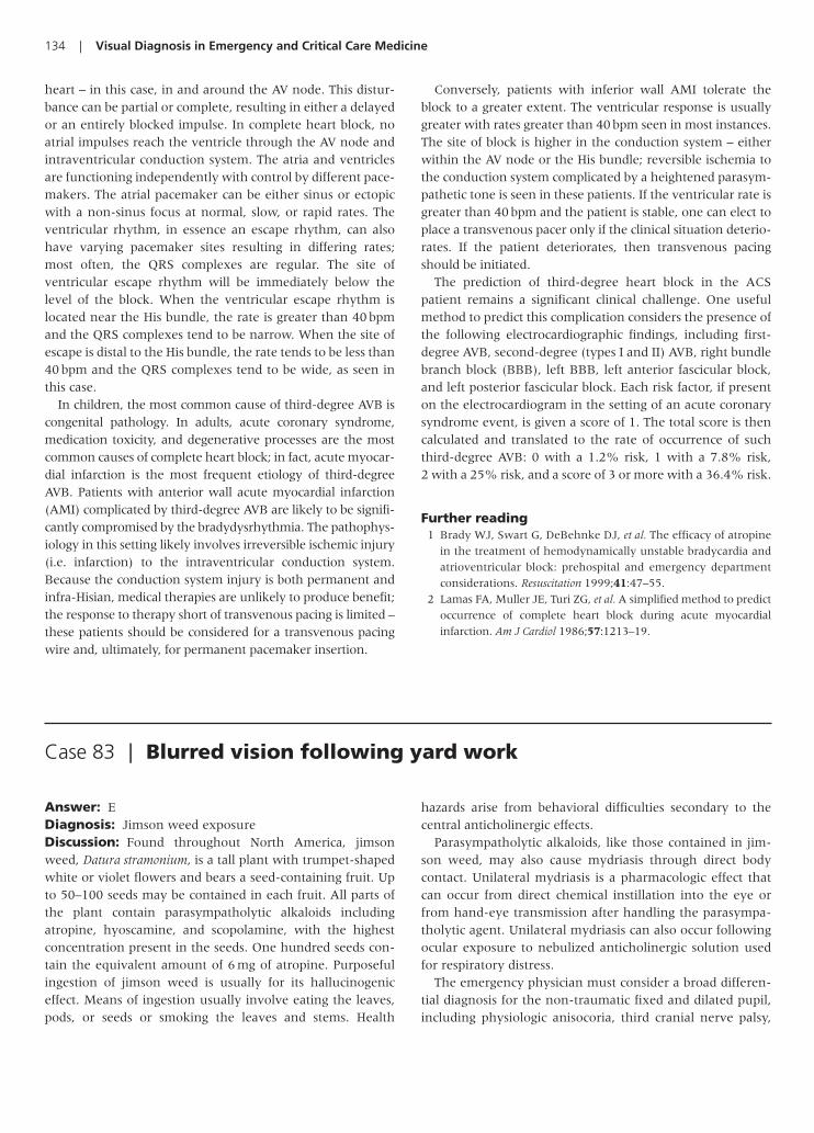
134 | Visual Diagnosis in Emergency and Critical Care Medicine
Answer: EDiagnosis: Jimson weed exposureDiscussion: Found throughout North America, jimsonweed, Datura stramonium, is a tall plant with trumpet-shapedwhite or violet flowers and bears a seed-containing fruit. Upto 50–100 seeds may be contained in each fruit. All parts ofthe plant contain parasympatholytic alkaloids includingatropine, hyoscamine, and scopolamine, with the highestconcentration present in the seeds. One hundred seeds con-tain the equivalent amount of 6 mg of atropine. Purposefulingestion of jimson weed is usually for its hallucinogeniceffect. Means of ingestion usually involve eating the leaves,pods, or seeds or smoking the leaves and stems. Health
hazards arise from behavioral difficulties secondary to thecentral anticholinergic effects.
Parasympatholytic alkaloids, like those contained in jim-son weed, may also cause mydriasis through direct bodycontact. Unilateral mydriasis is a pharmacologic effect thatcan occur from direct chemical instillation into the eye orfrom hand-eye transmission after handling the parasympa-tholytic agent. Unilateral mydriasis can also occur followingocular exposure to nebulized anticholinergic solution usedfor respiratory distress.
The emergency physician must consider a broad differen-tial diagnosis for the non-traumatic fixed and dilated pupil,including physiologic anisocoria, third cranial nerve palsy,
Case 83 | Blurred vision following yard work
heart – in this case, in and around the AV node. This distur-bance can be partial or complete, resulting in either a delayedor an entirely blocked impulse. In complete heart block, noatrial impulses reach the ventricle through the AV node andintraventricular conduction system. The atria and ventriclesare functioning independently with control by different pace-makers. The atrial pacemaker can be either sinus or ectopicwith a non-sinus focus at normal, slow, or rapid rates. Theventricular rhythm, in essence an escape rhythm, can alsohave varying pacemaker sites resulting in differing rates;most often, the QRS complexes are regular. The site of ventricular escape rhythm will be immediately below thelevel of the block. When the ventricular escape rhythm islocated near the His bundle, the rate is greater than 40 bpmand the QRS complexes tend to be narrow. When the site ofescape is distal to the His bundle, the rate tends to be less than40 bpm and the QRS complexes tend to be wide, as seen inthis case.
In children, the most common cause of third-degree AVB iscongenital pathology. In adults, acute coronary syndrome,medication toxicity, and degenerative processes are the mostcommon causes of complete heart block; in fact, acute myocar-dial infarction is the most frequent etiology of third-degreeAVB. Patients with anterior wall acute myocardial infarction(AMI) complicated by third-degree AVB are likely to be signifi-cantly compromised by the bradydysrhythmia. The pathophys-iology in this setting likely involves irreversible ischemic injury(i.e. infarction) to the intraventricular conduction system.Because the conduction system injury is both permanent andinfra-Hisian, medical therapies are unlikely to produce benefit;the response to therapy short of transvenous pacing is limited –these patients should be considered for a transvenous pacingwire and, ultimately, for permanent pacemaker insertion.
Conversely, patients with inferior wall AMI tolerate theblock to a greater extent. The ventricular response is usuallygreater with rates greater than 40 bpm seen in most instances.The site of block is higher in the conduction system – eitherwithin the AV node or the His bundle; reversible ischemia tothe conduction system complicated by a heightened parasym-pathetic tone is seen in these patients. If the ventricular rate isgreater than 40 bpm and the patient is stable, one can elect toplace a transvenous pacer only if the clinical situation deterio-rates. If the patient deteriorates, then transvenous pacingshould be initiated.
The prediction of third-degree heart block in the ACSpatient remains a significant clinical challenge. One usefulmethod to predict this complication considers the presence ofthe following electrocardiographic findings, including first-degree AVB, second-degree (types I and II) AVB, right bundlebranch block (BBB), left BBB, left anterior fascicular block,and left posterior fascicular block. Each risk factor, if presenton the electrocardiogram in the setting of an acute coronarysyndrome event, is given a score of 1. The total score is thencalculated and translated to the rate of occurrence of suchthird-degree AVB: 0 with a 1.2% risk, 1 with a 7.8% risk, 2 with a 25% risk, and a score of 3 or more with a 36.4% risk.
Further reading1 Brady WJ, Swart G, DeBehnke DJ, et al. The efficacy of atropine
in the treatment of hemodynamically unstable bradycardia and
atrioventricular block: prehospital and emergency department
considerations. Resuscitation 1999;41:47–55.
2 Lamas FA, Muller JE, Turi ZG, et al. A simplified method to predict
occurrence of complete heart block during acute myocardial
infarction. Am J Cardiol 1986;57:1213–19.
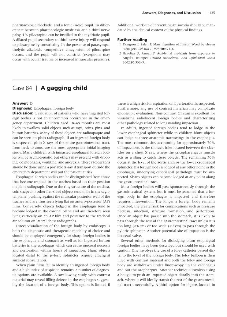
Answers, Diagnoses, and Discussion | 135
pharmacologic blockade, and a tonic (Adie) pupil. To differ-entiate between pharmacologic mydriasis and a third nervepalsy, 1% pilocarpine can be instilled in the mydriatic pupil.A dilated pupil secondary to third nerve injury will respondto pilocarpine by constricting. In the presence of parasympa-tholytic alkaloids, competitive antagonism of pilocarpineoccurs, and the pupil will not constrict (exceptions mayoccur with ocular trauma or increased intraocular pressure).
Additional work-up of presenting anisocoria should be man-dated by the clinical context of the physical findings.
Further reading1 Tiongson J, Salen P. Mass ingestion of Jimson Weed by eleven
teenagers. Del Med J 1998;70:471–6.
2 Havelius U, Asman P. Accidental mydriasis from exposure to
Angel’s Trumpet (Datura suaveolens). Acta Ophthalmol Scand
2002;80:332–5.
Answer: DDiagnosis: Esophageal foreign bodyDiscussion: Evaluation of patients who have ingested for-eign bodies is not an uncommon occurrence in the emer-gency department. Children aged 18–48 months are mostlikely to swallow solid objects such as toys, coins, pins, andbutton batteries. Many of these objects are radioopaque andcan be seen on plain radiograph. If an ingested foreign bodyis suspected, plain X-rays of the entire gastrointestinal tract,from neck to anus, are the most appropriate initial imagingstudy. Many children with impacted esophageal foreign bod-ies will be asymptomatic, but others may present with drool-ing, odynophagia, vomiting, and anorexia. These radiographsshould be done using a portable X-ray if transport outside theemergency department will put the patient at risk.
Esophageal foreign bodies can be distinguished from thosethat become trapped in the trachea based on their positionon plain radiograph. Due to the ring structure of the trachea,coin shaped or other flat-sided objects tend to lie in the sagit-tal plane, pushing against the muscular posterior wall of thetrachea and are thus seen lying flat on antero-posterior (AP)films. Conversely, objects lodged in the esophagus tend tobecome lodged in the coronal plane and are therefore seenlying vertically on an AP film and posterior to the trachealair column on lateral chest radiographs.
Direct visualization of the foreign body by endoscopy isboth the diagnostic and therapeutic modality of choice andshould be employed emergently for sharp foreign bodies inthe esophagus and stomach as well as for ingested buttonbatteries in the esophagus which can cause mucosal necrosisand perforation within hours of impaction. Sharp objectslocated distal to the pyloric sphincter require emergent surgical consultation.
When plain films fail to identify an ingested foreign bodyand a high index of suspicion remains, a number of diagnos-tic options are available. A swallowing study with contrastmaterial may reveal filling defects in the esophagus suggest-ing the location of a foreign body. This option is limited if
there is a high risk for aspiration or if perforation is suspected.Furthermore, any use of contrast materials may complicateendoscopic evaluation. Non-contrast CT scan is excellent forvisualizing radiolucent foreign bodies and characterizingother pathology related to longstanding impaction.
In adults, ingested foreign bodies tend to lodge in thelower esophageal sphincter while in children blunt objectscan lodge at three anatomic narrowings in the esophagus.The most common site, accounting for approximately 70%of impactions, is the thoracic inlet located between the clav-icles on a chest X ray, where the cricopharyngeus muscleacts as a sling to catch these objects. The remaining 30%occur at the level of the aortic arch or the lower esophagealsphincter. If a foreign body is lodged at any other point in theesophagus, underlying esophageal pathology must be sus-pected. Sharp objects can become lodged at any point alongthe gastrointestinal tract.
Most foreign bodies will pass spontaneously through thegastrointestinal system, but it must be assumed that a for-eign body in the esophagus is impacted and thereforerequires intervention. The longer a foreign body remainsimpacted, the greater risk for complications such as pressurenecrosis, infection, stricture formation, and perforation.Once an object has passed into the stomach, it is likely topass through the rest of the gastrointestinal tract unless it istoo long (�6 cm) or too wide (�2 cm) to pass through thepyloric sphincter. Another potential site of impaction is theileocecal valve.
Several other methods for dislodging blunt esophagealforeign bodies have been described but should be used withcaution. One involves the use of a foley catheter passed dis-tal to the level of the foreign body. The foley balloon is thenfilled with contrast material and both the foley and foreignbody are withdrawn under fluoroscopy up the esophagusand out the oropharynx. Another technique involves usinga bougie to push an impacted object distally into the stom-ach, where it will ideally transit the rest of the gastrointesti-nal tract uneventfully. A third option for objects located in
Case 84 | A gagging child
the distal esophagus involves the use of agents such asglucagon and nifedipine that relax the lower esophagealsphincter with expectant passage of the foreign body beyondthat point. Emetics (ipecac) are not recommended becauseof the potential for both aspiration and esophageal injury inthe setting of vomiting against a fixed obstruction.
Further reading1 Kay M, Wyllie R. Pediatric foreign bodies and their manage-
ment. Curr Gastroenterol Rep 2005;7(3):212–18.
2 Cheng W, Tam PK. Foreign body ingestion in children: experi-
ence with 1265 cases. J Ped Surg 1999;34(10):1472–6.
136 | Visual Diagnosis in Emergency and Critical Care Medicine
Answer: BDiagnosis: Child abuseDiscussion: Much effort has been directed toward collect-ing information about patterns of bruising in the physicalexam of a child in order to allay or corroborate the suspicionof child abuse. This remains an area of active research. Anyconcern based on bruising alone should always be examinedin the context of the developmental status of the child, theclinical scenario, and the explanation given by caregivers(and the consistency of that explanation). While each childshould be evaluated in such context, review of the recentmedical literature does support the finding that some pat-terns of bruising are more suggestive of abuse and shouldtherefore alert the physician to consider that possibility.
Some patterns of bruising are more often found with acci-dental mechanisms and may be more reassuring. For exam-ple, bruising of the shins and knees in a child old enough toambulate independently can be expected as developmentallyappropriate. Bruises over bony prominences (e.g., forehead)may also be less suspicious in appropriately mobile children.
In contrast, bruising in a child or infant that is not inde-pendently mobile is worthy of some suspicion as the mecha-nism of falling accidentally is not plausible. There are certainareas of the body, where if bruising is found, abuse may besuggested. These include the face, trunk, arms, hands, and
buttocks. If the bruising being examined is located away frombony prominences, it may also deserve scrutiny. Multiplebruising in clusters or with pathognomonic patterns alsoshould be investigated. Examples of the latter might includebruises in a pediatric patient with distinctive imprints (suchas from implements like an electrical cord or leather belt) oruniformity of size.
Finally, there is also the issue of estimating the age of bruises.Despite statements in some sources that aging of bruises can bedone based on color, a recent review of the existing medicalliterature has questioned this opinion. No convincing scien-tific data could be found by the authors of this review to sug-gest color variation can be used to accurately age a bruise. Thisreview of the literature also found that a clinician’s ability toidentify the age of a bruise within 24 hours of its creation waspoor. Significant intra-observer and inter-observer variabilitywas also reported in these circumstances.
Further reading1 Maguire S, Mann MK, Sibert J, Kemp A. Are there patterns of
bruising in childhood which are diagnostic or suggestive of abuse?
A systematic review. Arch Dis Child 2005;90(2):182–6.
2 Maguire S, Mann MK, Sibert J, Kemp A. Can you age bruises
accurately in children? A systematic review. Arch Dis Child 2005;
90(2):187–9.
Case 85 | A child with bruises of different ages
Answer: EDiagnosis: Acute retrobulbar hemorrhageDiscussion: Acute retrobulbar hemorrhage can causesevere permanent vision loss if not recognized and treated inthe emergency department. Vision loss occurs as the hemor-rhage fills the retrobulbar space and transmits pressure onthe outside of the globe causing dramatic elevation in theintraocular pressure. The acute rise in pressure can compressthe circulation from mechanical tamponade and restrict flowthrough the central retinal artery. Treatment to lower theintraocular pressure should be initiated immediately whenvision is threatened.
Medical treatment for elevated intraocular pressure shouldbe initiated with a topical beta blocker (timolol or levobunolol0.5%) and alpha agonist (brimonidine 0.15%), one drop eachevery 30 minutes for two doses. In addition, a carbonic anhy-drase inhibitor such as acetazolamide (i.e. two 250 mg tabletsorally or 500 mg intravenously) should be given. Mannitol(20%, 1.0 g/kg intravenous) can also decrease intraocular pres-sure by reducing vitreous volume. Caution must be observedwhen giving mannitol to patients with congestive heart fail-ure or renal failure.
Performing a lateral canthotomy and cantholysis immedi-ately lowers the intraocular pressure by expanding the
Case 86 | Traumatic eye pain and proptosis

Answers, Diagnoses, and Discussion | 137
Answer: BDiagnosis: Gallstone ileus with pneumobiliaDiscussion: Gallstone ileus is a condition that occurs whena gallstone erodes through the gallbladder wall into adjacentbowel, creating a cholecystoenteric fistula that provides apathway for gallstones to travel into the duodenum or trans-verse colon. When large gallstones (�2 cm) travel to a nar-row part of the small bowel (distal ileum or jejunum), theycan create a mechanical partial obstruction. Although up toone-third of patients with gallstone ileus do not have a his-tory of biliary colic. It typically presents as a small bowelobstruction and is more prevalent in females and the elderly.Gallstone ileus causes almost 25% of all partial small bowelobstructions in patients over 65. Initial plain radiographs willshow partial small bowel obstruction in 70% of the cases,but pneumobilia or calcified mass in distal small bowel canbe seen in only a third of plain radiographs. Ultrasound canmake a definitive diagnosis with the presence of pneumo-bilia and the detection of the calculus by tracing the dilatedsmall bowel to the site of the obstruction; however, overly-ing small loops of bowel can obscure deep calculi, and gaswithin the gallbladder fossa is frequently not recognized.Due to the multitude of causes for small bowel obstructionand the importance of assessing the level and severity of theobstruction, abdomen CT scan with intravenous and oral
contrast has been utilized recently to investigate causes forsmall bowel obstruction.
The presence of gas within the liver must be differentiated bylocation, either biliary or portal venous system. Portal venousgas tends to be more peripheral and fragmented. The differen-tial diagnosis for portal venous gas includes necrotizing entero-colitis and bowel ischemia. Biliary gas typically outlines thecommon bile duct and its major branches, and it shouldprompt one to look for the few causes of pneumobilia: (1) recent surgery (i.e. endoscopic retrograde cholangiopan-creatography with sphincterotomy); (2) infection (emphyse-matous cholecystitis); (3) passage of a gallstone; and (4) thepresence of a fistula (cholecystoduodenal or cholecystocolic).When an abdominal CT study reveals the criteria for Rigler’sTriad of gallstone ileus (small bowel obstruction, pneumobilia,and calcified gallstone in distal small bowel), surgery should becontacted immediately for emergent surgical intervention.
Further reading1 Reisner RM, Cohen JR. Gallstone ileus: a review of 1001
reported cases. Am Surg 1994;60(6):441–6.
2 Rivadeneira DE, Curry WT. Images of interest – gastrointestinal:
gallstone ileus. J Gastroenterol Hepatol 1994;16:105.
3 Swift SE, Spencer JA. Gallstone Ileus: CT findings. Clin Radiol
1998;53:451–4.
Case 87 | Post-prandial abdominal pain in an elderly woman
intraorbital volume. This is accomplished by first anesthetiz-ing the skin using 2% lidocaine with epinephrine. Next, anincision is made at the lateral canthus exposing the canthaltendons. The lower limb of the canthal tendon is then incisedusing sharp scissors, releasing the lower lid from its lateralattachment. Surgical repair of the canthotomy/cantholysis isperformed several days later when the swelling has subsided.Ophthalmology consultation is required for follow-up.
Further reading1 Ghufoor K, Sandhu G, Sutcliffe J. Delayed onset of retrobulbar
haemorrhage following severe head injury: a case report and
review. Injury 1998;29(2):139–41.
2 Larian B, Wong B, Crumley RL, et al. Facial trauma and
ocular/orbital injury. J Craniomaxillofac Trauma 1999;5(4):15–24.
Case 88 | Hyperthermia, tachycardia, and confusion in a teenager
Answer: BDiagnosis: Body packer with cocaine toxicity secondary topacket ruptureDiscussion: The plastic bag contains multiple condomwrapped packets of cocaine. This patient is a “body packer,”a person who ingests and transports packets of illicit drugsacross country lines in order to evade customs officials. This
should not to be confused with a “body stuffer,” a personwho quickly ingests illicit drugs (typically wrapped) to avoiddetection by law enforcement officials.
This person demonstrates a sympathomimetic syndrome. Onfurther questioning, this patient stated that he was transport-ing packets of cocaine from Columbia to the United States.Beta-adrenergic antagonists, including mixed alpha- and

138 | Visual Diagnosis in Emergency and Critical Care Medicine
Figure 88.1 An X-ray of the bag of packets noted in the case. Takinga radiograph of the packets, if available, may help to demonstratetheir appearance and assist with locating them on a body packer’sabdominal radiograph.
Figure 88.2 The abdominal radiograph from the case with arrowsdelineating a retained packet near the rectum. This film demonstratesthe potential difficulty in localizing drug packets on plain X-ray.
Case 89 | Acute onset double vision
Answer: CDiagnosis: Third cranial nerve palsyDiscussion: This patient has an isolated, pupil-involvingthird nerve palsy of the left eye. The third cranial nerve inner-vates the superior, inferior, and medial recti muscles as well asthe inferior oblique muscle. In addition, parasympathetic fibersto the pupillary sphincter muscle run along the dorsal andperipheral aspect of the third cranial nerve. Patients with third
nerve dysfunction typically present with a ptosis, limited ocu-lar motility, and variable pupil involvement.
Isolated third nerve palsy with pupillary involvement mustbe presumed secondary to an aneurysm until proven other-wise. The most common site is at the junction of the posteriorcommunicating artery and the internal carotid artery. A mag-netic resonance imaging (MRI) with contrast and magnetic res-onance angiography (MRA) should be obtained emergently.
beta-adrenergic antagonists, are contraindicated in cocainetoxicity. Beta-blockers can potentially induce unopposedalpha agonist activity, placing the patients at risk forvasospasm. The patient should be treated with benzodi-azepines, such as lorazepam, in an attempt to decrease hissympathomimetic state. Benzodiazepines should be titrateduntil the patient has calmed and his heart rate has dimin-ished, which may take larger than typical doses. If markedhypertension persists despite benzodiazepines, nitrates, andphentolamine can be utilized.
In the asymptomatic packer, gastrointestinal decontamina-tion with activated charcoal concurrently with polyethyleneglycol is the mainstay of decreasing potential drug absorptionand decreasing enteric transit time until rectal elimination.
If the patient were to develop cardiac dysrhythmias,sodium channel blocking agents such as procainamideshould be avoided in cocaine toxicity. Cocaine is itself a car-diac sodium channel blocker. If QRS prolongation or cardiacdysrhythmias developed, sodium bicarbonate should be thepharmacologic treatment administered first.
Further reading1 Traub SJ, Hoffman RS, Nelson LS. Body packing – the internal con-
cealment of illicit drugs. New Engl J Med 2003;349(26):2519–26.
2 Beno S, Calello D, Baluffi A, Henretig FM. Pediatric body packing:
drug smuggling reaches a new low. Pediatr Emerg Care 2005;
21(11):744–6.

Answers, Diagnoses, and Discussion | 139
Isolated pupil-sparing third nerve palsy implies completeloss of ocular motility with normal pupillary function. Thesepalsies presumably result from microvascular injury and aremost often seen in older patients with hypertension or diabetes. Neuroimaging is not necessary in patients over 50years of age if vasculopathic risk factors are present. Thesetypically resolve spontaneously over 2–3 months.
Pain is not helpful in determining the etiology of a thirdnerve palsy. Even though most patients with aneurysmal thirdnerve palsies complain of pain, many microvascular third
nerve palsies also present with pain. Neuro-ophthalmology orneurology consult is warranted for patients presenting to theemergency department with a third nerve palsy.
Further reading1 Lee AG, Hayman LA, Brazis PW. The evaluation of isolated third
nerve palsy revisited: an update on the evolving role of magnetic
resonance, computed tomography, and catheter angiography.
Surv Ophthalmol 2002;47(2):137–57.
2 Bennett JL, Pelak VS. Palsies of the third, fourth, and sixth cranial
nerves. Ophthalmol Clin North Am 2001;14(1): 169–85.
Figure 90.1 Lateral ankle view demonstrates fracture of the posterior malleolus of the tibia (circle).
Case 90 | Ankle pain and inability to walk
Answer: DDiagnosis: Trimalleolar fractureDiscussion: Ankle fractures and dislocations are amongthe most common orthopedic injuries cared for in the emer-gency department. The majority of ankle injuries involve arotational mechanism. Patients typically report immediateonset of pain following the traumatic event and inability tobear weight.
The initial evaluation of a patient with an acute ankle injury begins with a directed history. The mechanism of theinjury will focus the evaluation toward understood patterns of injury. Information regarding the patient’s ability to ambu-late following the event assists in the decision to obtain radio-graphs. Secondary injuries from the traumatic event and thecause of the event should be identified. A directed physical
exam is performed to identify areas of tenderness. The anklejoint includes the distal fibula, tibia and the talus. Examinationof the knee and foot is necessary to evaluate for secondaryinjuries associated with ankle injury (fractures of the proximalfibula and base of the fifth metatarsal in particular). The bonystructures of the ankle are easily palpable secondary to mini-mal soft-tissue coverage. Careful examination of the medialankle includes assessing for tenderness of the distal tibia andthe deltoid ligament. Examination of the lateral ankle includesassessment of the distal fibula and the anterior talofibular, pos-terior talofibular, and calcaneofibular ligaments. The anteriorankle is palpated to assess for injury to the tibiofibular syn-desmotic ligament and the Achilles tendon is palpated in theposterior ankle. Neurovascular status is examined to excludesecondary injury.
Figure 90.2 AP ankle radiograph demonstrates displaced fracture ofthe distal tibia at the level of the talar dome. An oblique fracture ofthe distal fibula is visualized. Both fractures are also apparent on themortise view. Fractures are noted by a circle annotation.

140 | Visual Diagnosis in Emergency and Critical Care Medicine
The decision to perform radiographs can be guided by theOttawa Ankle rules (OAR). Following the OARs, radiographsare obtained if there is an inability to bear weight immedi-ately after the event or in the emergency department (ED), orif there is tenderness at the tip or posterior aspect of the dis-tal 6 cm of the medial or lateral malleolus. Anteroposterior(AP) and lateral radiographs will reveal the majority of anklefractures. Additionally, the mortise view (AP rotated 15degrees internally) may be of assistance if suspicion for bonyinjury is high and standard views are equivocal.
Fractures of the ankle joint are classified either as stable orunstable. Stable fractures involve one side of the ankle joint,either the medial or lateral malleolus. If the fracture involveseither malleolus with ligamentous disruption of the oppositeside of the ankle, the fracture is identified as unstable. Anexample is a fracture of the lateral malleolus and disruptionof the medial deltoid ligament.
Unstable ankle fractures involve both sides of the anklejoint. They can be further classified as a bimalleolar fracture(involvement of the medial and lateral malleoli) or trimalle-olar fracture (involvement of the posterior, medial and
lateral malleoli). A trimalleolar fracture is apparent in thiscase.
Treatment of an unstable ankle fracture involves openreduction and internal fixation. The articular cartilage of theankle requires proper alignment as small deviations lead tosignificant risk of posttraumatic arthritic changes. Precisealignment is best achieved operatively. The timing of surgeryis dependent on multiple clinical factors including patientstability, secondary injuries, and co-morbidities. Orthopedicconsultation should be obtained after the identification ofthe fracture for operative planning.
Further reading1 Kay RM, Matthys GA. Pediatric ankle fractures: evaluation and
treatment. J Am Acad Orthopedic Surg 2001;9:268.
2 Deasy C, Murphy D, McMahon GC, Kelly IP. Ankle fractures:
emergency department management… is there room for
improvement? Eur J Emerg Med 2005;12(5): 216–19.
3 DeVries JS, Wijgman AJ, Sierevelt IN, Schaap GR. Long-term
results of ankle fractures with a posterior malleolar fragment. J
Foot Ankle Surg 2005;44(3):211–17.
Case 91 | Tongue swelling in a hypertensive female
Answer: CDiagnosis: Angioedema secondary to angiotensin con-verting enzyme inhibitor (ACEI)Discussion: Angioedema is a condition marked by nonde-pendent, asymmetric, non-pitting edema that results from aloss of vascular integrity and extravasation of fluid into theinterstitial tissues of the body with preference for the face,lips, tongue, oropharynx, and larynx. Two distinct mecha-nisms, mast cell-mediated and non-mast cell-mediatedangioedema (MCMA and NMCMA), have been implicated inthe majority of case presentations.
MCMA can be viewed as an extension of the more superfi-cial process of urticaria formation. Whereas the edema associ-ated with urticaria is confined to the superficial dermis,MCMA involves edema formation in the deep dermis andsubdermal tissues. This process is mediated by the release ofhistamine, leukotrienes, and prostaglandins from mast cellscausing increased vasodilation and capillary permeability withresultant edema formation. Typical causes of MCMA includefood and drug exposures. Chronic cases have been linked toan autoimmune process whereby antibodies directly activatemast cells. Treatment of MCMA begins with cessation of anyoffending agent. Additionally, antihistamines and cortico-steroids are useful. Epinephrine remains the treatment ofchoice for any airway compromise or vasomotor instability. Adefining feature of MCMA is its association with the presenceof urticaria and/or pruritus in 90% of cases. The absence of
these symptoms should prompt the clinician to considercauses of NMCMA.
NMCMA occurs in the absence of urticaria and pruritus andis thought to occur independently of mast cell degranulation.Two proposed mechanisms for the development of NMCMA
Figure 91.1 A patient with isolated lower lip edema. The cause wasfound to be secondary to her ACEI (missed on her first presentation to the emergency department 1 week previously). This swellingresolved after 4 hours of observation in the emergency department.No pharmacologic therapy was necessary.

Answers, Diagnoses, and Discussion | 141
include increased activity of bradykinin and abnormalities inthe complement cascade (i.e. C1-esterase inhibitor deficiency).The most common cause of NMCMA presenting to emergencyrooms is exposure to ACEI. NMCMA occurs in 0.1–0.7% ofpatients taking ACEI and is more common in older patientsand African-Americans. NMCMA from ACEI use typicallyoccurs within days to weeks after therapy is started, but candevelop years after uneventful use. ACEI induced NMCMA isnot related to the development of ACEI induced cough and hasequal likelihood of development with all ACEI.
Bradykinin is a potent vasodilator that normally functionsto oppose the vasoconstrictive effects of angiotensin II.Angiotensin converting enzyme functions to increase pro-duction of angiotensin II and decrease the activity ofbradykinin. The antihypertensive effects of ACEI thereforebecome apparent, causing a relative decrease in angiotensinII (decreased vasoconstriction) and a relative increase inbradykinin activity (increased vasodilation). It is this rise inbradykinin activity that is thought to be responsible for the(NMCMA) associated with ACEI use.
Treatment of NMCMA requires particular focus on thepatient’s airway and vasomotor stability. Epinephrine (0.3–0.5 mg intramuscular) and definitive airway management(endotracheal intubation or surgical airway) are required forany airway or circulatory compromise. Antihistamines and
corticosteroids may have some benefit and their administrationshould be considered in patients with NMCMA.
NMCMA related to a deficiency in the C1-esterase inhibitor,a component of the complement cascade, occurs in bothhereditary (hereditary angioedema or HAE) and acquiredforms. HAE typically presents in childhood following trau-matic or stressful situations, while the acquired form is com-monly associated with certain malignancies and presents at anolder age. This form of angioedema is treated in the acute set-ting in identical fashion as ACEI induced NMCMA. In addi-tion, androgens (stanozol 2–4 mg/day and danazol200–400 mg/day) have been used to treat this condition bothprophylactically and in the acute setting. Fresh frozen plasmahas been given to acutely replete the C1-esterase inhibitorprotein.
Further reading1 Baxi S, Dinakar C. Urticaria and angioedema. Immunol Allergy
Clin North Am 2005;25(2):353–67.
2 Hide M, Francis DM, Grattan CE, et al. Autoantibioties against
the high affinity IgE receptor as a cause of histamine release in
chronic urticaria. New Engl J Med 1993; 328:1599–604.
3 Brown NJ, Ray WA, Snowden M, Griffin MR. Black Americans
have an increased rate of angiotensin enzyme inhibitor-associated
angioedema. Cli Pharmacol Ther 1996;60:8–13.
Case 92 | Wide complex tachycardia in an older male patient
Answer: CDiagnosis: Wide complex tachycardia – ventricular tachy-cardiaDiscussion: The two electrocardiographic rhythm strips inthis case (lead II and the V lead) demonstrate a wide QRScomplex tachycardia (WCT). The QRS complex is markedlywide with a very rapid, regular rhythm. There is also thepresence of atrioventricular (AV) dissociation in lead II(Figure 92.1, arrows), which is very strongly suggestive ofventricular tachycardia (VT) as the rhythm diagnosis.
A WCT is defined electrocardiographically as a dysrhyth-mia with a QRS complex greater than 0.12 second in dura-tion and a ventricular rate greater than 120 bpm. In patientspresenting with a WCT, the differentiation of VT fromsupraventricular tachycardia (SVT) with aberrant ventricu-lar conduction is difficult. Aberrant ventricular conductionmay be due to a pre-existing bundle branch block (BBB), afunctional (rate-related) bundle malfunction resulting in awidened QRS complex when the heart rate exceeds a char-acteristic maximum for that patient, or accessory AV con-duction as encountered in pre-excitation syndromes (Wolff–Parkinson–White syndrome).
Certain clinical variables may support a diagnosis of VT inthe WCT presentation. Patient age greater than 50 years sug-gests VT; younger age is associated with SVT yet in a lessrobust fashion. Consideration of the past medical historyalso is helpful; a history of angina, myocardial infarction,coronary artery bypass grafting, valvular heart disease, orcongestive heart failure strongly suggests VT. It must bestressed that the presence or absence of hemodynamic insta-bility does not support either diagnosis; either dysrhythmiacan present with instability or stability.
Electrocardiographic variables suggestive of VT (Figure92.2) include a broad QRS complex, AV dissociation, fusionbeats, capture beats, and QRS complex features. Whennoted, AV dissociation is diagnostic of VT; unfortunately, AVdissociation is uncommon and, if present, difficult to discern.The second useful feature in establishing a diagnosis of VT isthe presence of fusion (a combination of a supraventricularimpulse with a ventricular impulse, producing a QRS com-plex of different morphology) or capture (a supraventricularimpulse passes through the AV node, causing an electricaldepolarization of the ventricles) beats; the finding of either afusion or a capture beat confirms the diagnosis of VT, but

142 | Visual Diagnosis in Emergency and Critical Care Medicine
again its utility is limited. Fusion and/or capture beats areobserved in less than 10% of cases of VT. QRS complex con-cordance (precordial QRS complexes entirely positive ornegative) favors VT.
The limited usefulness of these features has led to the con-struction of several stepwise approaches to the diagnosis ofWCT. For example, the approach suggested by Brugada andBrugada involves a four-step algorithm. This tool, as well asnumerous similar proposals, however, is somewhat cumber-some. Also, the majority of these protocols have a default diag-nosis of SVT with aberration. Clearly, defaulting to a less severediagnosis is problematic, fraught with error and danger for thepatient and clinician. Certainly, the clinician is required to treatthe patient – not the electrocardiogram (ECG). In other words,
with the idea that the specific rhythm diagnosis may not bepossible, the clinician should focus on the patient and his/herhemodynamic status – and base initial therapy on the entireclinical picture; furthermore, the clinician should not default toa less severe illness – SVT with aberration; if in doubt, managethe patient as if VT is the rhythm diagnosis.
Further reading1 Hudson KB, Brady WJ, Chan TC, Pollack M, Harrigan RA.
Electrocardiographic features of ventricular tachycardia. J Emerg
Med 2003;25:303–14.
2 Brugada P, Brugada J, Mont L, Smeets J, Andries EW. A new
approach to the differential diagnosis of a regular tachycardia
with a wide QRS complex. Circulation 1991;83:1649–59.
II
Figure 92.1 The presence of AV dissociation in lead II (arrows) is very strongly suggestive of VT as the rhythm diagnosis.
AtrioventricularDissociation
PositiveQRS Concordancy
NegativeQRS Concordancy
Rapid, Wide, RegularQRS Complex Capture Beat Fusion Beat
III
V1
V2
V3
V4
V5
V6
Figure 92.2 ECG features suggestive of VT.
Case 93 | Acute onset double vision
Answer: EDiagnosis: Ocular foreign body
Discussion: Ocular foreign bodies are frequently encoun-tered in emergency departments. The evaluation of patients

Answers, Diagnoses, and Discussion | 143
presenting with possible ocular foreign bodies is dictated by acareful history. The mechanism of injury and suspected par-ticulate matter is crucial in determining the presence and loca-tion of the foreign body. For instance, a history of metalstriking metal should raise concern for possible intraocularforeign body. Visual acuity must be documented prior toexamining the eye or performing any procedure. Perform acareful pupillary evaluation with attention to the size andshape of each pupil as well as response to stimulation.Thorough slit lamp examination should be conducted for anyirregularities or asymmetry in the conjunctiva, cornea, ante-rior chamber, iris, or lens. The eyelids must be everted, pro-vided there is no evidence of globe perforation, when aforeign body is suspected under the lids. Dilated fundus exam-ination is required for patients with suspected intraocular for-eign body. Ultrasound or CT scan of the orbits may benecessary when an intraocular or intraorbital foreign body issuspected.
The photos at the foot of the page show the corneal foreignbody of the case presented before and after removal. The photoon the right reveals a residual rust ring and corneal infiltrate.Corneal foreign bodies are best evaluated with a slit lamp.Assessment of the foreign body depth should be determinedprior to removal. Foreign bodies that have penetrated into theanterior chamber should be removed in the operating room.Most embedded foreign bodies can be removed at the slit lamp
under topical anesthesia using a 25 g needle or foreign bodyspud. A red-brown rust ring results if a foreign body has beenembedded for more than a few hours. The rust ring is bestremoved using a battery-powered ophthalmic burr. Therapyfollowing a foreign body removal includes a broad-spectrumantibiotic. Cycloplegia should be considered if an anteriorchamber reaction is present. Reexamination in 24 hours isrequired.
The photograph below left demonstrates an everted eyelidwith an insect stuck to the palpebral conjunctiva. Verticallinear scratches on the cornea often indicate a foreign bodyembedded in the palpebral conjunctiva under the eyelid.The linear scratches result from the foreign body rubbing onthe cornea as the patient blinks their eyes. Conjunctival for-eign bodies can usually be removed with a moist cotton-tipapplicator or simple irrigation. A broad-spectrum antibioticointment should be instilled after foreign body removal.
The pictures at the top of the page 144 demonstrate a defectnear the pupillary border (white arrow); dilated exam revealsa posterior subcapsular cataract with an intraocular foreignbody imbedded in the posterior lens (red arrow). The diagnosisof a penetrating ocular injury is often obvious to the observer;however, some may be quite subtle and require a high index ofsuspicion and careful observation. A detailed history andnature of the injury should be obtained. Visual acuity must bedocumented and a careful examination performed. Signs suggestive of intraocular foreign body include deep eyelid laceration, conjunctival laceration/hemorrhage, iris-cornealadhesion, shallow anterior chamber, hypotony, iris defect, andacute cataract. A CT scan of the brain and orbits (1 mm axialand coronal views) should be obtained. Since over 90% ofintraocular foreign bodies are metallic, MRI is contraindicatedin most cases of suspected intraocular foreign body. Patientssuspected of having an intraocular foreign body require oph-thalmologic consultation. The eye should be protected with ashield. Patients should be NPO and placed on bedrest withbathroom privileges. Tetanus should be given when indicatedand antiemetics prescribed to prevent Valsalva.
This patient pictured on page 144 (bottom picture) hassuffered a perforating ocular injury with an entrance wound

144 | Visual Diagnosis in Emergency and Critical Care Medicine
in the cornea and an exit wound somewhere in the posteriorpole of the eye. The white substance in the anterior chamberis hydrated lens material that resulted from the foreign bodypassing through the lens and disrupting the lens capsule. ACT scan of the brain and orbits is indicated for all patientssuspected of having an ocular or orbital foreign body. The CTscan in this case shows an intraorbital foreign body (redarrow) in the posterior orbit lodged between the lateral rec-tus muscle and the optic nerve. The patient also has a posi-tive Seidel test. This is a simple test to determine if afull-thickness wound is present. It is performed using fluo-rescein and a cobalt blue light. Fluorescein is instilled in theeye and the wound is observed using the blue light. A test ispositive when aqueous leaks out of the wound and is seenwashing away the fluorescein.
Management of patients with an intraorbital foreign bodyin the emergency department involves shielding the eye andconsulting ophthalmology. Tetanus toxoid should be givenwhen indicated. Hospitalization is necessary for foreign bod-ies requiring surgical removal. Patients should be treatedwith a 10–14-day course of systemic antibiotics. Patientsshould be monitored closely for evidence of infection.
Further reading1 Mester V, Kuhn F. Intraocular foreign bodies. Ophthalmol Clin
North Am 2002;15(2):235–42.
2 Williams DF, Mieler WF, Abrams GW, Lewis H. Results and prog-
nostic factors in penetrating ocular injuries with retained
intraocular foreign bodies. Ophthalmology 1988;95:911–16.
Case 94 | Foot pain in a gymnast
Answer: CDiagnosis: Dancer’s fracture (fracture in the distal shaft ofthe 5th metatarsal)
Discussion: A fracture of the distal shaft of the 5th metatarsalis commonly referred to as a “dancer’s fracture” because of its association with ballet dancers. Dancer’s fracture is

Answers, Diagnoses, and Discussion | 145
Figure 94.1 Dancer’s fracture (arrow points to fracture of 5thmetatarsal).
characterized by a spiral-oblique fracture in the mid-shaft orneck of the 5th metatarsal. In one specific ballet technique(dem pointe maneuver), a high twisting force is applied in theforefoot and can stress the 5th metatarsal enough to cause afracture. This fracture can also result from direct trauma to thelateral foot. The classic patient presents with a painful limp butcan bear weight on the affected foot. The most common phys-ical exam finding is tenderness over 5th metatarsal andswelling over the lateral aspect of the foot. Another commonfracture that must be excluded is a Jones fracture (transversefracture of the proximal diaphysis of the 5th metatarsal). Jonesfractures have a significant risk of malunion or nonunion.
Sagittal, mediolateral, and anterioposterior planes of theaffected foot must be obtained to assess for angulation anddisplacement of the distal fragment. Conservative treatmentwith a short-leg cast or stiff shoe for 2–6 weeks is recom-mended for most cases. Prolonged nonweight bearing is associated with an increased risk for reflex sympathetic dys-trophy. Closed reduction in the emergency department isrequired for fractures where there is angulation of greaterthan 10 degrees or a displacement of greater than 3 mm. Dueto the conservative management of this injury, special atten-tion must be given to the sagittal plane radiograph to assessfor angulation. A healed angulated 5th metatarsal shaft maybe complicated by plantar keratosis in the future. Indicationsfor open reduction include: compartment syndrome, unsta-ble or open fractures, or a failed closed reduction. Orthopedicconsultation in the emergency department is recommendedfor multiple or displaced fractures.
Further reading1 Ankle and Foot. Rosen’s Emergency Medicine Concepts and Clinical
Practice. 5th Ed. Vol. 1, Marx JA, Ed. St. Louis, MO: Mosby.
2002:728–9.
2 O’Malley MJ, Hamilton WG, Munyak J. Fractures of the distal
shaft of the fifth metatarsal. Dancer’s fracture. Am J Sport Med
1996;24(2):240–3.
Case 95 | New facial droop
Answer: ADiagnosis: Bell’s palsyDiscussion: The patient pictured on page 60 has Bell’spalsy. Note ptosis of the left eye and the weakness of theorbicularis muscles on the left side of the face. Also note theabsence of wrinkling to the forehead on the affected left side.Bell’s palsy is the most common disorder affecting the facialnerve. It is an abrupt, isolated, unilateral, peripheral facialparalysis without detectable causes. While the actual patho-physiology is unknown, the most widely accepted theorypostulates inflammation of the facial nerve causing it to becompressed as it courses through the temporal bone.
The incidence in the United States is approximately 23 cases per 100,000 persons. It occurs equally in men andwomen, most commonly between ages 10 and 40. Clinicalconditions associated with Bell’s palsy include pregnancy(especially third trimester), immunocompromised states,and diabetes. Patients may present with a concern that theyhave suffered a stroke. Other common symptoms includepain in or behind the ear, numbness on the affected side ofthe face, a recent upper respiratory infection, drooling, alter-ation in taste, and hyperacusis.
The classic definition describes a lower motor neuron deficitof the facial nerve, manifesting as weakness of the entire face

146 | Visual Diagnosis in Emergency and Critical Care Medicine
(upper and lower) on the affected side. This is in contradis-tinction to upper motor neuron lesions such as a corticalstroke, where the upper third of the face is spared while thelower two-thirds are paralyzed. While considered an idio-pathic facial paralysis, there is significant evidence to supportan infectious cause. Herpes simplex virus (HSV-1) has beenisolated in many patients and is the most likely infectiousagent, although there are likely other etiologic agents with ashared common pathway leading to facial nerve dysfunction.
No specific laboratory tests exist to diagnose Bell’s palsy.Clinical suspicion helps to direct what tests may be of value,and may include thyroid function studies and Lyme titer.One can also consider obtaining (if clinically suspected) arapid plasma reagin (RPR) or a venereal disease research laboratory (VDRL) test for syphilis, as well as a humanimmunodeficiency virus (HIV) test. There is no evidence tosupport emergent imaging studies with Bell’s palsy.
The primary treatment in the emergency department is withpharmacologic management. The remainder of care focuses onpatient education as to the course of the disease and eye careinstructions. While considered by some to be controversial,treatment with steroids remains common if the patient pres-ents within 7 days of symptom onset. The postulated mecha-nism of action is in reducing facial nerve swelling. Current datasupports using steroids as a means to improve outcomes, andearlier treatment is preferred (i.e. prednisone at 40 –60 mg/dayfor 7–14 days). Recent evidence supports HSV as the presumed
cause in more than 70% of cases; therefore, antiviral agentsare a logical choice for pharmacologic management. Recom-mendations include acyclovir 400–800 mg five times a day for10 days or valcyclovir 1 g three times a day for 10 days.
The eye on the affected side is potentially at risk forcorneal drying and foreign body exposure, as the lid may notclose completely, especially when the patient is asleep. Thisis generally managed with artificial tears and some form ofeye protection (patch or glasses during the day, taping theeye shut at night).
The vast majority (85% or more) of patients recover with-out any cosmetically obvious deformities. Ultimately, 10%will have some residual asymmetry of the facial muscles, and5% will suffer from significant facial nerve deficits. In mostcases, recovery begins 3 weeks after symptom onset, butmay take up to a year for complete resolution. Patients withincomplete facial nerve involvement have a more favorableprognosis than those with a complete deficit.
Further reading1 Gilden DH. Bell’s palsy. New Engl J Med 2004;351(13): 1323–31.
2 Murakami S, Mizobuchi M, Nakashiro Y. Bell palsy and herpes
simplex virus: identification of viral DNA in endoneurial fluid and
muscle. Ann Int Med 1996;124:27–30.
3 Ramsey MJ, DerSimonian R, Holtel MR, Burgess LP.
Corticosteroid treatment for idiopathic facial nerve paralysis: a
meta-analysis. Laryngoscope 2000;110:335–41.
Case 96 | Eye pain and swelling
Answer: ADiagnosis: Periorbital cellulitisDiscussion: Periorbital cellulitis, also called preseptal celluli-tis, is a superficial skin infection which has not penetrated theorbital septum. Its presentation mimics the more seriousorbital cellulitis, also called postseptal cellulitis, which is aninfection of the deeper tissues of the orbit. In the majority ofcases, the distinction can be made clinically without the needfor radiographic studies. In cases where the clinical diagnosisis in question, an enhanced CT of the orbits can aid in makinga definitive diagnosis.
Periorbital cellulitis in adults and children over 5 years isusually the result of a secondary skin infection with S. aureusas the predominant pathogen. In children of 5 years andunder, S. aureus is still the most common pathogen but H.influenzae is responsible for a significant minority, usuallyfrom bacteremic spread. Periorbital cellulitis in those of 5years and older can be treated with oral antibiotics and fol-lowed up as outpatients. Children less than 5 years are more
likely to have a bacteremic source and should be treatedwith intravenous antibiotics to cover S. aureus and H. influen-zae, have a blood culture drawn, and should be consideredcandidates for hospital admission.
Orbital cellulitis is a more serious diagnosis. Figure 96.1demonstrates a child with orbital cellulitis. This photographdemonstrates proptosis, edema, and limited ocular motilityin the right eye as the child looks up to the left.
The majority of orbital cellulitis cases result as an exten-sion from an adjacent bacterial sinusitis. Orbital cellulitis inadults is usually a polymicrobial infection whereas in chil-dren it is often caused by a single organism. The differentialdiagnosis of orbital cellulitis includes preceptal cellulitis,orbital pseudotumor, thyroid eye disease, orbital tumors,metastatic disease, varix, and trauma.
The CT scan in Figure 96.2 demonstrates a subpe-riosteal abscess (red arrow) overlying a nearly opacified sinus.CT scanning is essential in cases of suspected orbital cellulitisto confirm the diagnosis, to evaluate the sinuses, and to rule

Answers, Diagnoses, and Discussion | 147
Figure 96.1 Orbital cellulitis.Figure 96.2 CT of orbital cellulitis.
out orbital foreign body. Mucormycosis is a life-threateningdisease which must be considered in diabetic and immuno-compromised patients presenting with orbital cellulitis.
Treatment for orbital cellulitis includes admission to thehospital for intravenous antibiotics. Blood cultures should beobtained. ENT should be consulted for surgical drainage ifclinically indicated. Patients must be observed closely for opticnerve dysfunction by monitoring visual acuity and pupillaryresponses.
Periorbital and orbital cellulitis can usually be distinguishedon clinical grounds. Periorbital cellulitis presentations typically
lack fever and have normal eye examinations, including fullrange of all extra-ocular eye movements, normal pupillaryreflexes, and normal visual acuity. In equivocal cases, CT ofthe orbits can help make the diagnosis.
Further reading1 Givner LB. Periorbital versus orbital cellulitis. Pediatr Infect Dis J
2002;21(12):1157–8.
2 Tovilla-Canales JL, Nava A, Tovilla y Pomar JL. Orbital and peri-
orbital infections. Curr Opin Ophthalmol 2001; 12(5):335–41.
Case 97 | Shortening and rotation of the leg following trauma
Answer: DDiagnosis: Left hip fractureDiscussion: This patient has a left hip fracture. Hip frac-tures include fractures of both the pelvis at the acetabulumand the proximal femur. Fractures of the femoral neck andintertrochanteric fractures cause the classic physical exami-nation findings of the lower extremity seen in this case con-sisting of foreshortening and external rotation of the lowerextremity. While examining the lower extremity, carefulattention should be focused to the neurovascular exam.
Fractures of the femoral neck are associated with fracturesof the femoral shaft in 20% of patients. A high index of sus-picion must be present as the shaft fracture is missed in asmany as 40% of patients. The patient’s X-ray in this case isnoted below.
The X-ray reveals that this patient has sustained anintertrochanteric fracture of the left femur. This injury in
younger adults typically occurs after a high-speed motorvehicle crash or a fall from a height. In older patients, thisfracture can occur after a fall of any type. A significant forceis required to cause this fracture and it is therefore approxi-mately 50% of cases that have other associated injuries.
A complete trauma evaluation must be performed. Thepatient may be distracted by pain secondary to the hip fractureand unaware of other injuries. Femur fractures can result inthe loss of 2–3 units of blood into the thigh and thereforehemodynamic instability can ensue. The patient should have a type and crossmatch sent for at least 2 units of blood.The physician must rule out other potential sources of hemodynamic instability before attributing blood loss to thehip fracture alone. Complete trauma evaluation must be thefirst priority, then orthopedic consult should be obtained.Internal fixation is usually performed on an urgent, not emer-gent basis.

148 | Visual Diagnosis in Emergency and Critical Care Medicine
Case 98 | Spider bite in the night
Answer: DDiagnosis: Black widow envenomationDiscussion: Black widow spiders (Latrodectus species) arefound over a wide geographic range on every continent butAntarctica. The female spider is responsible for the charac-teristic toxicity and is shiny black in color with a red hourglass of varying size on its ventral abdomen. Its webs are dis-organized, appear abandoned, and are often found nearhuman habitation.
The venom of the black widow contains alpha-latrotoxin.This toxin binds to multiple sites resulting in the unregulatedopening of cation channels and the subsequent influx of cal-cium at presynaptic neurons. Elevated cytosolic calciumcauses unregulated release of neurotransmitters. As a result,neurotransmitters, such as acetylcholine and norepineph-rine, are increased. This results in activation of both the sym-pathomimetic and cholinergic systems, and causes increasedstimulation at the neuromuscular junction.
Initially, a pinprick sensation may be felt at the bite site.However, the initial pain may go unnoticed. Locally, a smallcircle of erythema and/or induration may be seen at the bitesite. Within the ensuing hours, systemic signs and symptomsmay develop, progress, and last for days. Muscle cramping isa characteristic finding associated with black widow enveno-mation. The cramps may be mild and remain localized to thesite or the cramps can involve all muscle groups diffuselyand become severe and unrelenting. The worst cases maydevelop opisthotonus posturing and abdominal rigidity.Nausea, vomiting, headache, palpitations, hypertension,tachycardia, diaphoresis, priapism, facial edema, renal failure,
and anxiety may all be clinically seen. The diagnosis of blackwidow envenomation is made solely on the clinical presen-tation; there are no clinical laboratory tests available to con-firm the diagnosis.
Wounds should be cleansed and if needed tetanus pro-phylaxis administered. Patients should be monitored for atleast 6 hours. In those patients manifesting muscular spasms,opioid analgesics orally or intravenously should be consid-ered. Antispasmodics, such as diazepam, may also provideadditional relief. Methocarbamol and dantrolene have beenutilized for treatment, with mixed results. Intravenous cal-cium was initially touted to be efficacious to alleviate the painassociated with cramps, but subsequent studies have notfound it to be of significant benefit. Tourniquets have no rolein first aid management of black widow envenomations.
Latrodectus mactans antivenom (equine-derived) is rap-idly effective at relieving the clinical effects associated with toxicity. However, its use is limited to those patientsmanifesting significant toxicity not relieved by conven-tional therapy, or those with health problems that place them at increased risk for complications. The antivenom has an associated risk of anaphylaxis and if utilized, should beadministered in a hospitalized setting with full resuscitationcapabilities.
Further reading1 Singletary EM, Rochman AS, Bodmer JC, Holstege CP.
Envenomations. Med Clin North Am 2005;89(6):1195–224.
2 Saucier JR. Arachnid envenomation. Emerg Med Clin North Am
2004;22:405–22.
Hip fractures can have a subtle appearance on X-ray.Occult fractures of the hip not seen on X-ray are possible anddiagnosis may require magnetic resonance imaging (MRI).There are several radiographic methods to help locate frac-tures on plain films. One technique is to follow Shenton’s
line. This is a curved line that extends from the superior bor-der of the obturator foramen down along the medial aspectof the femur. If there is disruption of the smooth C shapethat is normally present, fracture or dislocation should besuspected. Another technique is to look for the S and reverseS curves that are present on the medial and lateral aspects ofa normal femoral head and neck. If disrupted, the presenceof a fracture should be suspected.
Further reading1 Rudman N, McIlmail D. Emergency department evaluation and
treatment of hip and thigh injuries. Emerg Med Clin North Am
2000;18(1):29–66.
2 Perron AD, Miller MD, Brady WJ. Orthopedic pitfalls in the ED:
radiographically occult hip fracture. Am J Emerg Med 2002;20(3):
234–7.
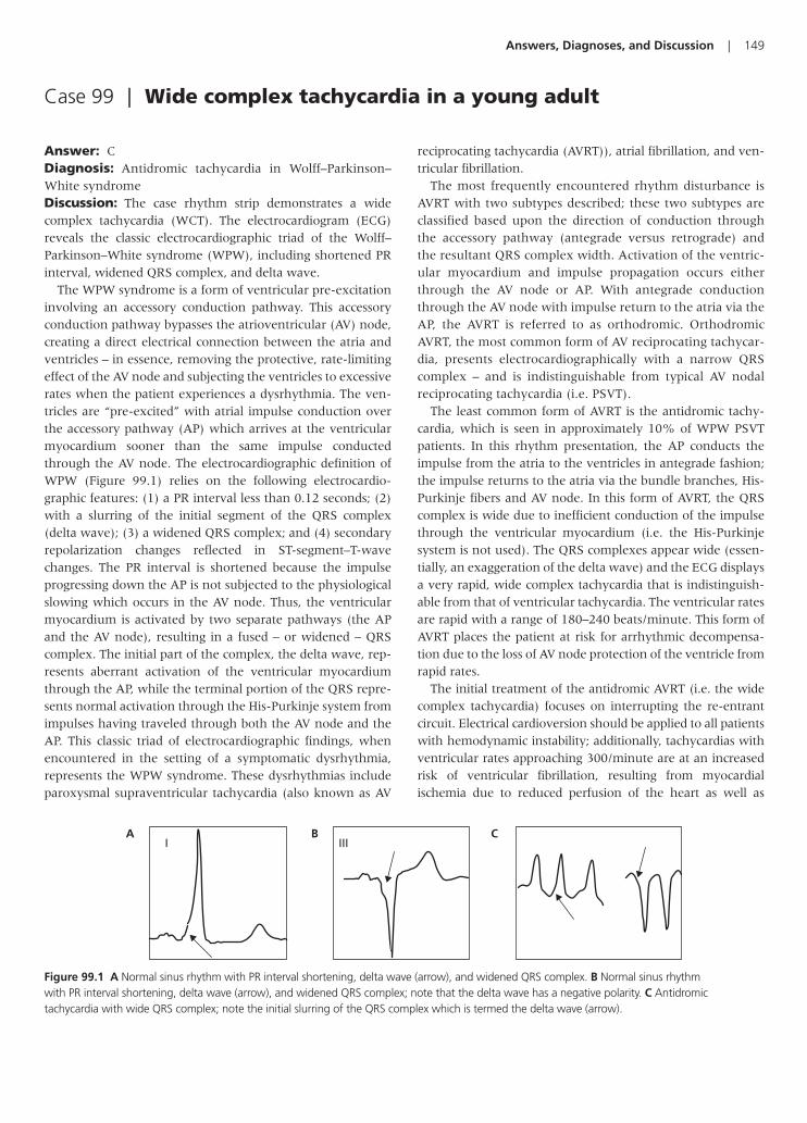
Answers, Diagnoses, and Discussion | 149
Case 99 | Wide complex tachycardia in a young adult
Answer: CDiagnosis: Antidromic tachycardia in Wolff–Parkinson–White syndromeDiscussion: The case rhythm strip demonstrates a widecomplex tachycardia (WCT). The electrocardiogram (ECG)reveals the classic electrocardiographic triad of the Wolff–Parkinson–White syndrome (WPW), including shortened PRinterval, widened QRS complex, and delta wave.
The WPW syndrome is a form of ventricular pre-excitationinvolving an accessory conduction pathway. This accessoryconduction pathway bypasses the atrioventricular (AV) node,creating a direct electrical connection between the atria andventricles – in essence, removing the protective, rate-limitingeffect of the AV node and subjecting the ventricles to excessiverates when the patient experiences a dysrhythmia. The ven-tricles are “pre-excited” with atrial impulse conduction overthe accessory pathway (AP) which arrives at the ventricularmyocardium sooner than the same impulse conductedthrough the AV node. The electrocardiographic definition ofWPW (Figure 99.1) relies on the following electrocardio-graphic features: (1) a PR interval less than 0.12 seconds; (2)with a slurring of the initial segment of the QRS complex(delta wave); (3) a widened QRS complex; and (4) secondaryrepolarization changes reflected in ST-segment–T-wavechanges. The PR interval is shortened because the impulseprogressing down the AP is not subjected to the physiologicalslowing which occurs in the AV node. Thus, the ventricularmyocardium is activated by two separate pathways (the APand the AV node), resulting in a fused – or widened – QRScomplex. The initial part of the complex, the delta wave, rep-resents aberrant activation of the ventricular myocardiumthrough the AP, while the terminal portion of the QRS repre-sents normal activation through the His-Purkinje system fromimpulses having traveled through both the AV node and theAP. This classic triad of electrocardiographic findings, whenencountered in the setting of a symptomatic dysrhythmia,represents the WPW syndrome. These dysrhythmias includeparoxysmal supraventricular tachycardia (also known as AV
reciprocating tachycardia (AVRT)), atrial fibrillation, and ven-tricular fibrillation.
The most frequently encountered rhythm disturbance isAVRT with two subtypes described; these two subtypes areclassified based upon the direction of conduction throughthe accessory pathway (antegrade versus retrograde) andthe resultant QRS complex width. Activation of the ventric-ular myocardium and impulse propagation occurs eitherthrough the AV node or AP. With antegrade conductionthrough the AV node with impulse return to the atria via theAP, the AVRT is referred to as orthodromic. OrthodromicAVRT, the most common form of AV reciprocating tachycar-dia, presents electrocardiographically with a narrow QRScomplex – and is indistinguishable from typical AV nodalreciprocating tachycardia (i.e. PSVT).
The least common form of AVRT is the antidromic tachy-cardia, which is seen in approximately 10% of WPW PSVTpatients. In this rhythm presentation, the AP conducts theimpulse from the atria to the ventricles in antegrade fashion;the impulse returns to the atria via the bundle branches, His-Purkinje fibers and AV node. In this form of AVRT, the QRScomplex is wide due to inefficient conduction of the impulsethrough the ventricular myocardium (i.e. the His-Purkinjesystem is not used). The QRS complexes appear wide (essen-tially, an exaggeration of the delta wave) and the ECG displaysa very rapid, wide complex tachycardia that is indistinguish-able from that of ventricular tachycardia. The ventricular ratesare rapid with a range of 180–240 beats/minute. This form ofAVRT places the patient at risk for arrhythmic decompensa-tion due to the loss of AV node protection of the ventricle fromrapid rates.
The initial treatment of the antidromic AVRT (i.e. the widecomplex tachycardia) focuses on interrupting the re-entrantcircuit. Electrical cardioversion should be applied to all patientswith hemodynamic instability; additionally, tachycardias withventricular rates approaching 300/minute are at an increasedrisk of ventricular fibrillation, resulting from myocardialischemia due to reduced perfusion of the heart as well as
AI III
CB
Figure 99.1 A Normal sinus rhythm with PR interval shortening, delta wave (arrow), and widened QRS complex. B Normal sinus rhythm with PR interval shortening, delta wave (arrow), and widened QRS complex; note that the delta wave has a negative polarity. C Antidromictachycardia with wide QRS complex; note the initial slurring of the QRS complex which is termed the delta wave (arrow).

150 | Visual Diagnosis in Emergency and Critical Care Medicine
Wide QRS complex tachycardia
Regularity?
Regular IrregularVentricular tachycardia
(Monomorphic)Supraventricular tachycardia
with aberrant conductionSinus tachycardiaAtrial tachycardia
Paroxysmal supraventriculartachycardia
Atrial flutterWide complex tachycardiaSodium channel blockade
Hyperkalemia-related
Ventricular tachycardia(Polymorphic)
Atrial fibrillation (WPW)Supraventricular tachycardia
with aberrant conductionAtrial fibrillation (non-WPW)
Atrial flutter with variable blockAtrial tachycardia with variable block
Figure 99.2 Differential diagnosis of wide complex tachycardia.
Grade Type Injury Treatment
I Hematoma Subcapsular, �10% surface area (SA) NOLaceration Capsular tear, �1 cm parenchymal depth
II Hematoma Subcapsular, �50% SA; intraparenchymal, �5 cm diameter NOLaceration 1–3 cm parenchymal depth
III Hematoma Subcapsular, �50% SA; intraparenchymal, �5 cm NO/OLaceration �3 cm parenchymal depth or involving trabecular vessels
IV Laceration Laceration of segmental or hilar vessels (�25% spleen) OVascular Hilar vascular injury that devascularizes the spleen
NO: Nonoperative; O: Operative.
subsequent depolarizations falling on the electrically vulnera-ble repolarization phase. In the hemodynamically stablepatient, the agents of first choice would be either procainamideor amiodarone. Agents such as calcium channel antagonists,beta-adrenergic blocking agents, or digoxin which act prima-rily on the AV node are contraindicated since they will facilitateconduction down the AP and could potentially lead to anincreased ventricular rate with ventricular fibrillation.
Further reading1 Wolff L, Parkinson J, White PD. Bundle-branch block with short
PR interval in healthy young people prone to paroxysmal tachy-
cardia. Am Heart J 1930;5:685–704.
2 Rosner M, Brady WJ, Kefer M, Martin ML. Electrocardiography in
the patient with the Wolff–Parkinson– White syndrome: diagnostic
and initial therapeutic issues. Am J Emerg Med 1999;17:705–14.
Case 100 | Abdominal pain in a trauma victim
Answer: CDiagnosis: Splenic ruptureDiscussion: The spleen is the most commonly injuredintraperitoneal organ after blunt abdominal trauma, fol-lowed by the liver and the intestines. Motor vehicle colli-sions are the most common mechanism of injury (50–75%)in the United States, particularly in children, followed bydirect blows to the abdomen (15%) and falls (6–9%). Themajority of alert patients will present with abdominal painor left shoulder pain (Kehr’s sign) and have localized leftupper quadrant tenderness on exam. Others may exhibitsigns of acute intraperitoneal hemorrhage, such as hypoten-sion or tachycardia. Associated risk factors are splenomegalyand substance abuse; only 10–25% of splenic injuries areassociated with rib fractures.
In hemodynamically stable patients, abdominal and pelviccomputerized axial tomography (CT) imaging can reliablyidentify solid organ and retroperitoneal pathology afterblunt trauma. Using oral contrast rarely increases diagnosticaccuracy and simply delays imaging. Although the focusedassessment by sonography in trauma (FAST) exam can beused to rapidly detect intraperitoneal blood, it cannot reli-ably identify injuries to solid organs, the retroperitoneum, orthe diaphragm.
Advances in diagnostic imaging have revolutionized themanagement of splenic injuries. CT imaging can accuratelydefine the degree of splenic disruption such that more injuriesare treated nonoperatively. Operative management is depend-ent on the extent of the injury (Table 100.1). Most recently,up to 90% of children and 50% of adults are treated success-
Table 100.1 AmericanAssociation for the Surgery ofTrauma – Splenic Injury Scale

Answers, Diagnoses, and Discussion | 151
fully without surgery. After discharge, patients should be onbed rest for 72 hours, limit their physical activity for 6 weeks,and withhold contact sports for 6 months.
Delayed splenic rupture, characterized by abdominal painand/or signs of internal bleeding that develop after anasymptomatic period of at least 48 hours, occurs in 1% ofblunt abdominal trauma. About 50% of patients will presentwithin 1 week of their injury, usually precipitated by minorstress such as twisting or bending. Although uncommon,
delayed diagnosis or misdiagnosis may cause significantmorbidity and mortality.
Further reading1 Allen TL, Greenlee RR, Price RR. Delayed splenic rupture pre-
senting as unstable angina pectoris: case report and review of the
literature. J Emerg Med 2002;23(2):165–9.
2 Harbrecht BG. Is anything new in adult blunt splenic trauma?
Am J Surg 2005;190(2):273–8.


Index
AAbdominal aortic aneurysm (AAA) 69–70,
106
rupture 70
Abdominal pain
in alcoholic 32, 111postprandial 10–11, 55, 77–78, 137in pregnancy 16, 87–88in toddler 37, 115in trauma victim 63–64, 150–151
Acetazolamide 68, 127, 136
Aconitine 110
Aconitum dephinifolium poisoning,
see Monkshood poisoning
Acromioclavicular separation
Types I–VI 129
Activated charcoal 116, 123, 130, 132, 138
Acute angle closure glaucoma, in elderly
female 68
Acute cholecystitis 77
Acute coronary syndrome (ACS) 93–94,
134
Acute lung injury (ALI) 78
Acute myocardial infarction (AMI) 83–84,
93, 104, 126, 134
Acute pain
in abdomen, during pregnancy 16, 87–88in eye 4, 68
Acute pericarditis 112
Acute renal failure 99
Acute retrobulbar hemorrhage 136
Acyclovir 114, 131, 146
Agitation, in botanist 44, 123–124Alkali injury 104
Aloe vera 109, 110
Alpha-latrotoxin 148
Amanita phalloides, see Death cap mushroom
Amiodarone 150
Aneurysms 125
Angioedema, secondary to ACEI 140
Angiotensin converting enzyme inhibitor
(ACEI) 140
Ankle pain
following fall 51, 132–133and instability, to walk 57–58, 139–140
Anterior humeral line 82
Anticholinergic syndrome 123
Antidromic tachycardia
in Wolff–Parkinson–White syndrome 149
Antihistamine 67, 109, 140, 141
Aspirin 122, 127
Atraumatic lower back pain and leg
weakness, in adult male 41–42, 120–121Atrioventricular block, see AVB
Atrioventricular dissociation, see AVB
Atropine 123, 127, 134
Avascular necrosis (AVN) 132
of left femoral head 114
AVB
third-degree 133
AVRT 149
Azygos venous system 83
BBacitracin 114
Back pain
atraumatic lower back pain, in adult male
41–42, 120–121in car accident victim 17–18, 89following fall 11–12, 79–80in young athlete 23, 98
Bacterial conjunctivitis 76
Basilar skull fractures 92
Bell’s palsy 145
Benign early repolarization (BER) 118
Benzodiazepine 124, 138
Bifascicular ventricular tachycardia 110
Bile ducts, extrinsic obstruction 122
Biliary gas 137
Biliary pain 78
Bilirubin 122
Bimalleolar fracture 140
Black widow envenomation 148
Blue hue, following endoscopy 48–49, 128Blurred vision
and eye pain, in elderly female 4, 68following yard work 53, 134–135
Body packer, with cocaine toxicity
secondary to packet rupture 137
Body stuffer 137
Boehler’s angle 95
Boxer’s fracture 69
Bradycardia
following herbal ingestion 32, 110and vision changes 3–4, 67–68and weakness, in elderly female patient
52, 133–134Bradykinin 141
Brimonidine 68, 127, 136
British antilewisite (BAL) 115
Bronchodilators 132
Bronchospasm 78–79, 131, 132
Bruises, in child 54, 136Brush fire victim 3, 67Buckle fracture 76
Button battery ingestion with esophageal
impaction 85
CCalcaneus fractures 94
Calcium 76, 116, 148
Calcium disodium ethylene diamine-
tetraacetate (CaNa2EDTA) 115
Cardiac dysrhythmias 68, 133, 138
Cardiac glycosides 67
Cardiac tamponade 106
Cardiotoxic effects following caustic
ingestion 37–38, 115–116Carpometacarpal (CMC) joints 104
Cauda equina syndrome (CES) 120–121
Cefazolin 120
Ceftriaxone 76, 124
Chancroid ulcers 88
Chemical injuries, to eye 27, 104Chest pain
and abdominal pain, in trauma victim
63–64, 150–151and electrocardiogram pattern 20, 93–94with electrocardiographic ST-segment–
T-wave abnormalities 26–27,
103–104and hypotension, in adult male patient
14–15, 83–85pleuritic chest pain, in young adult male
33–34, 112
Note: Page numbers in italics refer to Case Presentations and Questions, while those in bold refer to Answers, Diagnoses, and Discussion.
153
Chest pain (continued)
with ST segment elevation, in middle-
aged male patient 40–41, 118–119and subtle ST segment elevation 46–47,
126with sudden cardiac death 24–25, 100
Child abuse 136
Chlorpromazine 122
Cholecystitis 77, 122
Cholecystoenteric fistula 137
Cholelithiasis 77, 122
Cholestatic diseases 122
Clostridial myonecrosis 111
Cocaine 137–138
Coining 70
Cold-related injury 31, 109–110Coma, following head trauma 48,
127–128Confluent rash, in child 30–31, 109Confusion, anemia, and abdominal pain,
in toddler 37, 115Contact lens wearer 85
eye pain in 21, 94Corneal abrasions 85
Corneal ulcer 94
Corticosteroid 73, 141
Courvoisier’s sign 122
Crigler–Najjar syndrome 122
Crotalid envenomation 80
Cupping 71
Cyanosis 128
DDancer’s fracture 144
Dantrolene 148
Dark urine, in immigrant 8, 73Datura stramonium, see Jimsonweed
Death cap mushroom 123
Deer tick 124
Deferoxamine 130
Diazepam 148
2,3-Dimercaptosuccinic acid (DMSA) 115
Diphenhydramine 123, 124
Distal radius fracture 91
Dorzolamide 127
Double decidual sign (DDS) 87
Double vision 56–57, 59, 138–139,
142–144Drooling and fever, in child 8, 73–74Drug hypersensitivity syndrome (DHS)
72, 73
Dyspnea, in alcoholic 28–29, 105–107
EEctopic pregnancy 87–88
Elbow pain in child, after fall 13, 82Epidural hematoma (EDH) 127
Epinephrine 137, 140, 141
Erythema migrans 124
Erythema multiforme 72, 109
Erythromycin 85, 114, 122
Esophageal foreign body 135
Esophageal impaction, button battery
ingestion 15–16, 85–87Exotoxins 111
Extrapulmonary TB 90
Eye pain
and blurred vision, in elderly female
4, 68in contact lens wearer 21, 94after lacerations, from broken bottle
34–35, 112–113after object strike 15, 47, 85, 126–127and ocular discharge 9, 76and proptosis 54–55, 136–137and swelling 61, 146–147
Eyelid laceration, see Lid laceration
FFacial droop 60, 145–146Facial edema 14, 83Facial rash 36, 114Facial swelling, with poor dentition
13, 81–82Fall on outstretched hand (FOOSH) 71, 129
with wrist pain 25, 101–102in young adolescent 19, 91–92
FAST evaluation, following trauma
22, 95–96Fat pad sign 82
Favism 73
Femoral head
avascular necrosis of 114
Femoral neck fracture 147
Fever
and confusion 46, 125–126and drooling, in child 8, 73–74and rash, in child 42, 50, 121–122, 131
Fishing, in stomach 43, 122–123Flank pain, in elderly man 5–6, 69–70Fluoride toxicity 115
Fluoroquinolone 120
Focused assessment by sonography in
trauma (FAST) 22, 95–96, 106, 107,
150
FOOSH mechanism 101–102
Foot pain
following breaking 30, 108in gymnast 60, 144–145
Forearm fracture
after falling 35, 113Foxglove ingestion 67
Frostbite injury 109
Furosemide 125
GGagging child 53–54, 135–136Galeazzi fracture 113
Gallbladder neck 77, 78
Gallstone 77, 78
Gallstone ileus and pneumobilia 137
Gas gangrene, see Clostridial myonecrosis
Gastric lavage 116, 123, 132
Genital ulcers 88
Gilbert’s syndrome 122
Globe deformity, following trauma
41, 119–120Globe rupture 119, 127
Glucagons 136
Glucose-6-phosphate dehydrogenase
(G6PD) 73
Groin pain, in elderly woman 36, 114
HHand pain
after striking wall 27–28, 104–105suspicious 4–5, 69
Hard signs 108
Headache, sudden and severe
in adult male 45–46, 125Heart block 133
Heel pain, following fall 21–22, 94–95Henoch–Schönlein purpura (HSP) 117
Heparin therapy 110
Herpes simplex virus (HSV) 88–89, 109,
131, 146
Herpes zoster ophthalmicus 114
House fire
elderly victim 11, 78–79Hutchinson’s sign 114
Hydrocarbon ingestion/aspiration 131
Hydrofluoric acid 116
Hydroxyzine 67
Hyperbilirubinemia 122
Hyperkalemia 67, 68, 74, 99, 116
Hyperthermia, tachycardia, and confusion,
in teenager 56, 137–138Hypotension 79, 80, 106, 115–116, 130
and chest pain, in adult male patient
14–15, 83–85
IIbuprofen 77, 109, 117
Injection injury, high-pressure,
to hand 40, 117–118Intraperitoneal free fluid 95
Intrauterine pregnancy (IUP) 87–88
Intrinsic liver disease, etiologies 122
Ipecac 116, 123, 130, 136
Iron toxicity 129
Itraconazole 90
JJaundice, secondary to hyperbilirubinemia
122
Jimsonweed 123, 134
Joint pain and rash, in child 38–39,
116–117Jones fracture 145
Junctional tachycardia 68
154 | Index

LLamp oil ingestion 51, 131–132Latrodectus species 148
Latrodectus mactans antivenom 148
Lead encephalopathy 115
Lead foreign body, in stomach 122
Left bundle branch block (LBBB) 93
Left hip fracture 147–148
Left ventricular hypertrophy (LVH) 103
Leg shortening and rotation, following
trauma 61–62, 147–148Leg weakness and atraumatic lower back
pain, in adult male 41–42, 120–121Levobunolol 68, 127, 136
Lid laceration 112
Lidocaine 137
Lisfranc injury 108
Long QT syndrome (LQTS) 100
Lorazepam 138
Low back pain, in car accident victim
17–18, 89Ludwig’s angina 81
Lumbar teardrop fracture, see Teardrop
fracture
Lumbar wedge fracture 79
Lunate dislocation 71
Lyme disease (LD) 124
Lymphadenopathy 88, 124
MMagnesium 116
Mannitol 68, 99, 125, 127, 136
Mast cell mediated angioedema (MCMA)
140
Meningococcemia 125
Mental status, altered
with abnormal electrocardiogram 9,
74–76following fall 54, 136
Metacarpal neck fractures 69
Methemoglobinemia 128
Methocarbamol 148
Methylene blue 128
Methylprednisolone 67
Monkshood poisoning 110
Monteggia fracture 113
Morison’s pouch 95, 96, 106
Mucormycosis 147
Musculoskeletal injuries 91–92
Myoglobin 99
Myopericarditis 112
NNaloxone 133
Naproxen 117
Neck swelling, in immigrant 18–19, 90–91Necrotic skin lesion 26, 102–103Neisseria gonorrhea 76, 88
Neisseria meningitidis 125
Neonatal HSV infection 131
Nifedipine 136
Nitrates 138
Non-healing rash, in gardener 18, 90Non-mast cell mediated angioedema
(NMCMA) 140–141
Nonsurgical pneumoperitoneum (NSP) 117
OOcular foreign body 142
Opioid toxicity
drop-off, in emergency department 52,
133Orbital cellulitis 146
Orthodromic AVRT 149
Ottawa Ankle rules (OAR) 140
Out of proportion pain 33, 111–112
PP-wave 75
Paint chips ingestion 115
Papaver somniferum 133
Parasympatholytic alkaloids 134–135
Paroxysmal atrial tachycardia with block
68
Pars defect, see Spondylolysis
Pathergy 103
Penile ulcer, painless 17, 88–89Pericardial tamponade 105
Perilunate dislocation 71
Periorbital cellulitis 146
Periorbital hematoma 19–20, 92Petechiae 125
Phenothiazines 123, 124
Phentolamine 138
Platysma muscle 107
Pleuritic chest pain, in young adult male
33–34, 112Pneumobilia and gallstone ileus 137
Pneumoperitoneum 117
Poison ivy dermatitis 67
Polymorphic ventricular tachycardia (PVT)
100
Portal venous gas 137
Post-operative pneumoperitoneum
X-ray findings, after laparoscopy 39, 117Postprandial abdominal pain 10–11, 77–78
in elderly woman 55, 137Postseptal cellulitis, see Orbital cellulitis
Prednisolone acetate 127
Prednisone 67, 146
Prenatal vitamins overdose 49–50, 129–130Preseptal cellulitis, see Periorbital cellulitis
Procainamide 138, 150
Proximal interphalangeal (PIP) joint 69
Pseudomonas 94
Purulent eye discharge, in adult 9, 76Pyoderma gangrenosum (PG) 102
QQT prolongation 100
RRaccoon eyes 19–20, 92Radial motor nerve injury 113
Radiocapitellar line 82
Rash
following brush fire 3, 67confluent rash, in child 30–31, 109in epilepsy child 7, 72–73facial rash, painful 36, 114and fever, in child 42, 50, 121–122, 131and joint pain, in child 38–39, 116–117non-healing rash, in gardener 18, 90
Reciprocal ST segment depression 126
Retropharyngeal abscess (RPA) 73–74
Rifampin 122
Right ventricular acute myocardial
infarction 15, 83Rose gardener’s disease, see Sporotrichosis
Rule of appropriate discordance 93, 94
SSail sign, see Fat pad sign
Salter–Harris types I–V injuries 91–92
Scaphoid fracture 101
Scleral icterus 122
Sentinel loop 111
Shoulder pain, following direct blow 49, 129Skin lesion
in comatose patient 24, 99in heroin addict 22–23, 96–97in immigrant child 6, 70–71necrosis 26, 102–103
Skin popping 96
Skin target lesion 45, 124Skull fractures, see Basilar skull fractures
Slash wound, to neck 29, 107–108Slit lamp examination 85, 143
Smoke inhalation
with associated burns 78
Snakebite 12, 80–81antivenom therapy 81
Sodium bicarbonate 138
Soft signs 108
Spider bite 26, 62, 102–103, 148Spilled teacup sign 71
Spinal fracture, see Lumbar wedge fracture
Splenic rupture 150
Spondylolysis 98
Sporothrix schenckii 90
Sporotrichosis 90
ST-elevation myocardial infarction (STEMI)
83, 93–94, 118–119, 126
ST segment
depression 83–84, 94, 104, 126
elevation 40–41, 46–47, 83–85, 94, 104,
112, 118–119, 126Stable ankle fracture 140
Staphylococcus aureus 146
Stevens–Johnson syndrome (SJS) 72–73
Strain pattern 103, 104
Index | 155

Subarachnoid hemorrhage (SAH) 125
Submandibular abscess 81
Subtle STEMI, with reciprocal change 126
Superior vena cava syndrome (SVCS) 83
Swelling
and eye pain 61, 146–147facial 13, 81–82neck 18–19, 90–91tongue 58, 140–141
Syphilis 83, 88
TT-wave 26–27, 75, 83, 94, 103–104, 116,
118–119
Talar fracture 132
TB adenitis, see Tuberculosis adenitis
Teardrop fracture 89
Terry Thomas sign 71
Tetanus prophylaxis 112, 118, 148
Third cranial nerve palsy 138
Third-degree atrioventricular block 133
Timolol 68, 127, 136
Tongue swelling, in hypertensive female
58, 140–141
Topical ciprofloxacin ophthalmic drops 76
Torsade de pointes (TdP) 100
Torus fracture, see Buckle fracture
Toxic epidermal necrolysis (TEN) 72, 109
Toxicodendron 67
Traumatic eye pain and proptosis 54–55,
136–137Traumatic hyphema 126
Trimalleolar fracture 139
Trimethoprim/polymyxin B drops 85
Tuberculosis adenitis 90
UUlnar fracture, see Monteggia fracture
Unilateral mydriasis 134
Unstable ankle fracture 140
Urushiol 67
Uvulectomy 71
VValcyclovir 146
Ventricular tachycardia (VT) 68, 141
Vision changes 3–4, 67–68
WWeakness and bradycardia, in elderly
female patient 52, 133–134Whole bowel irrigation (WBI) 123, 130
Wide complex tachycardia (WCT)
in older male patient 58–59, 141–142in young adult 62–63, 149–150
Wolff–Parkinson–White (WPW) syndrome
141, 149
Wrist pain
following fall 6–7, 71–72fall on outstretched hand 25, 101in young child 10, 76–77
YYellow eyes and skin 43, 122
ZZone II neck injury 107
156 | Index


![An accidental diagnosis of optic nerve meningioma in a ... · An accidental diagnosis of optic nerve meningioma in a patient affected by Thyroid Eye Disease ... visual pathway [5]](https://img.pdfslide.net/doc/110x75/5c877bee09d3f2bc6b8bcda8/an-accidental-diagnosis-of-optic-nerve-meningioma-in-a-an-accidental-diagnosis.jpg)