Embed Size (px)
Citation preview

CONGENITAL HEART DISEASES
Soumya Ranjan ParidaBasic B.Sc. Nursing 4th year
Sum Nursing College

01/09/15 2
Practical approach Congenital-Associated syndrome Acquired
Rheumatic Cardiomyopathy ( Restrictive ) Cardiomyopathy ( Dilated , Post viral
myocarditis ) Pericardial diseases ( Effusion,Pericarditis) Kawasaki disease

01/09/15 3
CLINICAL CLASSIFICATION OF CONGENITAL HEART DISEASES
Cardiac Malpositions- Ectopia cordis Dextrocardia
Acyanotic without a shunt– Malformations on left side Malformations on right side
Acyanotic with a shunt Cyanotic

01/09/15 4
Acyanotic without a shunt Left sided malformations
Mitral stenosisMitral regurgitationAortic stenosisAortic regurgitationCoarctation of aorta

01/09/15 5
Acyanotic without a shunt Right sided malformation
Ebsteins anomaly of Tricuspid valvePulmonary stenosisPulmonary regurgitation Primary pulmonary hypertension

01/09/15 6
Acyanotic with a shunt
Shunt at atrial levelASDPAPVC
Shunt at ventricular levelVSD
Shunt at great artery levelPDAAP Window
Shunt at more than one level

01/09/15 7
Acyanotic without a shunt Left sided malformations
Mitral stenosisMitral regurgitationAortic stenosisAortic regurgitationCoarctation of aorta

01/09/15 8
Aortic Stenosis Age Symptoms Types
Valvular Supra valvular Sub valvularClinical PresentationManagement

01/09/15 9
Coarctation of Aorta Age Types
Pre ductal Post ductal
Clinical presentation Associations Management
Age of intervention Surgery // Cath based intervention

01/09/15 10
Acyanotic without a shunt Right sided malformation
Ebsteins anomaly of Tricuspid valvePulmonary stenosisPulmonary regurgitation Primary pulmonary hypertension

01/09/15 11
Pulmonary stenosis Types Age Symptoms Clinical findings Management

01/09/15 12
Ebsteins anomaly Age of presentation Clinical findings Management

01/09/15 13
Acyanotic with a shunt
Shunt at atrial levelASDPAPVC
Shunt at ventricular levelVSD
Shunt at great artery levelPDAAP Window
Shunt at more than one level

01/09/15 14
VSD Types – based on location Size With or without PAH Associations Clinical features Management

01/09/15 15
ASD Types Associations Clinical features D/D Management

01/09/15 16
01/09/15 17
PDA Age Symptoms Signs D/D Management

01/09/15 18
Cyanosis
Definition Central // PeripheralDifferential / Reverse differential
Causes Non Cardiac
RespiratoryVascular causes Hematological
Cardiac Congenital cyanotic heart disease
Eisenmenger’s Syndrome

01/09/15 19
Cyanotic lesions
Tetralogy of Fallots Tricuspid atresia Transposition of Great arteries Truncus arteriosus Single ventricle Hypoplastic left heart syndrome Eisenmenger Syndrome

01/09/15 20
Classification of Cong Cyanotic Heart diseases
Pulmonary stenosis, without VSDCritical PS, Ebsteins
Pulmonary Stenosis with Large VSDTOF
Increased Pulmonary flow with/ without PAHTGA ( Transposition of great arteries )
Decreased pulmonary flow with PAHEisenmenger Syndrome
Pulm venous congestion with PAH TAPVC, HLHS
Cyanosis without Pulm stenosis , Normal PA pressureSingle atrium, Pulm AV fistula

01/09/15 21
Tetralogy of Fallots

01/09/15 22
Transposition of Great Vessels

01/09/15 23
Hypoplastic Left Heart Syndrome

01/09/15 24
Clinical Characteristics of TOF
Prominent a wave in JVPNormal heart sizeMild parasternal impulse Systolic thrill – though uncommonSingle second sound ( P 2 absent )Ejection systolic murmur Diastolic period clearDecreased pulmonary flow on Chest X ray

01/09/15 25
Congenital Cyanotic Heart diseases
Ejection Systolic Murmur / Pan systolic murmur Continuous murmur TOF with
PDABronchial collaterals Surgically created shunts ( BT shunt ) Peripheral PS

01/09/15 26
DIFFERENTIAL DIAGNOSIS
Tetralogy of FallotTransposition of Great Arteries, VSD, PSTricuspid atresia, VSD, PSSingle ventricle, PSDouble outlet right ventricle, VSD, PSCorrected transposition of great arteries, VSD,PS AV Canal defects with PS Eisenmenger’s Syndrome
01/09/15 27
Assessment of Severity
Cyanosis – More the cyanosis more severe the disease but mild cyanosis also to be taken seriouslyAge of onset – earlier the onset more severe the lesion Symptoms – more the symptoms more severe the disease

01/09/15 28
Investigations
ECG X Ray Chest PA view Echocardiography Holter monitoring MRI Cardiac Cath

01/09/15 29
ECG in CHD
Rate, Rhythm Look at P wave in Lead I PR interval QRS axis ( Left / Right ) RVH, LVH, Bivebtricular Hypertrophy Incomplete / Complete RBBB

01/09/15 30
X Ray Chest
Cardiac Size C T Ratio Pulmonary Vascularity Classical images
01/09/15 31
Echocardiography
2DEcho M mode Doppler
Pulsed wave, Continuous wave, Colour Tranesophageal ( TEE ) 3 D Echo

01/09/15 32
Management of TOF
Age of presentation Severity of symptoms Anatomical considerations Palliative - Shunts Definitive

01/09/15 33
Management of Complex CHD
Define anatomy by Echo, if required Cath/ MRI/ CT Angio
Decide whether Two Ventricle repair is possible or not
Glenn shunt Fontan repair

01/09/15 34
Bidirectional Glenn
W What is the purpose of the Glenn?
I

01/09/15 35
FontanFontan

01/09/15 36
Reversal of shunt in following with development of Pulmonary arterial hypertension
Eisenmenger’s Syndrome
Shunt at atrial levelASDPAPVC
Shunt at ventricular levelVSD
Shunt at great artery levelPDAAP Window
Shunt at more than one levelAV Canal defect
Patients with Cyanotic Heart diseases with Increased pulmonary blood flow – TGA, TAPVC

01/09/15 37
Characteristics with Eisenmenger physiology
History of frequent chest infections
Age of onset of cyanosis?
No cardiomegaly or thrill
No parasternal heave
Constant ejection click of PAH
Palpable P 2
Diastolic murmur of PR or systolic murmur of TR

01/09/15 38
Management of Eisenmenger’s Syndrome
Manage Polycythemia Drug management of PAH Heart Lung Transplantation

01/09/15 39
Common Pediatric Cardiac Emergency Conditions Heart failure
Cyanotic Spell Duct dependent Circulation –
Pulmonary atresia , aortic atresia , TGA Cardiac Arrhythmias Post operative state

01/09/15 40
CHF in Fetal Life
Supraventricular Tachyarrythmias AV Block AV Regurgitation Severe PR / TR EFE Severe anaemia Cardiomyopathy Myocarditis, Storage Diseases

01/09/15 41
CHF on Day 1 of Life
Structural Heart Defects
Rhythm Abnormalities
AV fistulas
Heart muscle dysfuntion

01/09/15 42
CHF in First week of Life
Structural Abnormalities Critical Aortic Stenosis Coarctation of Aorta Interrupted Aortic Arch Hypoplastic Left Heart Syndrome Critical Pulmonary Stenosis PDA in premature Babies
01/09/15 43
CHF in First 2 months of LifeStructural Abnormalities Aortic level shunts Ventricular level shunts Left sided obstructive lesions Atrial level shunts Anomalous origin of Coronary Arteries

01/09/15 44
Clinical Recognition Respiratory rate & Effort Heart Rate Feeding Difficulties Exercise intolerance Excessive sweating Weight gain Cardiomegaly Hepatomegaly

01/09/15 45
Principles of Therapy
Removal of underlying cause - Surgical Correction
- Medical m/m of IE Removal of Precipitating cause - Intercurrent Infections
- Arryhthmias - Anemia

01/09/15 46
DUCT DEPENDENT PULMONARY CIRCULATION DEFINITION Complete absence or severe restriction
of antegrade pulmonary blood flow resulting in severe hypoxemia with dependance on a patent arterial duct to maintain pulmonary perfusion compatible with life

01/09/15 47
Duct Dependant Pulmonary Circulation ETIOLOGYA) Anatomical restriction/discontinuity of
ventricle and pulmonary arteryB) Admixture lesions- TGAC) Functional pulmonary atresia
Severe Ebsteins anomaly Hypoplastic right ventricle without PS

01/09/15 48
DUCT DEPENDANT PULMONARY CIRCULATION
Clinical Presentation 90% have progressive hypoxemia,
cyanosis,acidosis 1-7 days after birth 5% present in
infancy/childhood,adolescent Rarely heart failure ( PS with TR, PA
intact septum)

01/09/15 49
DUCT DEPENDANT PULMONARY CIRCULATION
Clinical Presentation Blood Gas
Low Po2, Normal PCO2,Metabolic acidosis Hyperoxia test- Po2 < 150Torr

01/09/15 50
DUCT DEPENDANT PULMONARY CIRCULATION
DIAGNOSIS Echocardiography
Defines anatomy Duct size Pulmonary artery anatomy

01/09/15 51
DUCT DEPENDANT PULMONARY CIRCULATION
MANAGEMENT Early recognition- Key
Structural diagnosis- Less crucial

01/09/15 52
DUCT DEPENDANT PULMONARY CIRCULATION
MANAGEMENT Secure good I/V and I/A line
Correction of acidosis
Volume -colloid(5% Albumin 5-10ml/Kg)

01/09/15 53
DUCT DEPENDANT PULMONARY CIRCULATION
MANAGEMENT Prostaglandin- Life saving Oxygen(?) Balloon Dilation of duct Stenting of Duct Surgery

01/09/15 54
DUCT DEPENDANT PULMONARY CIRCULATION
MANAGEMENT Prostaglandin
Before transfer-0.01mcg/kg/min I/V Tertiary centre- 0.1mcg/kg/min scale to
0.05-0.01mcg/kg/min I/V Oral- 12-65 mcg/kg at 4hrly interval

01/09/15 55
DUCT DEPENDANT PULMONARY CIRCULATION
MANAGEMENT Prostaglandin- predictors of
response Widely open duct- Nil Closed duct-Nil Constricted duct- Best response

01/09/15 56
DUCT DEPENDANT PULMONARY CIRCULATIONProstaglandin - side
effects Apnea Hypotension Fever Edema
Periostiitis (20%) Subcutaneous fat
necrosis Fragile ductus Aneurysm, rupture
01/09/15 57
DUCT DEPENDANT PULMONARY CIRCULATION
VENTILATION Transport to tertiary centre
Apnea of prostaglandin( premature, LBW)
Stabilisation of the sick child

01/09/15 58
TRANSPOSITION COMPLEX
TGA intact septum/Small VSDDiagnosis- Echocardiography 100% detection rate ASD size- need for septostomy PDA size-need for prostaglandin

01/09/15 59
TRANSPOSITION COMPLEX
TGA intact septum/Small VSDManagementa) Stable, no acidosis - BAS Plan Sxb) Acidosis,severe hypoxemia- PGE
BASc) Transfer- PGE (?Ventilation) BAS

01/09/15 60
Surgical management
Arterial Switch Operation Venous Switch Operation

01/09/15 61
CARDIAC ARRHYTMIAS
Complete Heart Block
Supraventricular Tachycardia
Ventricular Tachycardia - Rare

01/09/15 62
CARDIAC ARRHYTMIAS
COMMONEST ARRHYTHMIA - SUPRAVENTRICULAR Well tolerated in majority In neonates and infants difficult to
differentiate from physiological response- fever, stress
Prolonged unrecognized SVT- DCM

01/09/15 63
Clinical Presentation
Irregular Heart beat Unexplained cardiac failure Underlying structural heart disease Syncopal attacks Palpitations ,Chest discomfort Haemodynamic Instability Family H/O sudden cardiac events

01/09/15 64
Long term treatment
Long term treatment till cath ablation is safe or LV dysfunction
Digoxin/B blockers Verapamil Amiodarone/Sotalol Flecanide
01/09/15 65
Symptomatic second or third degree AV block
SA node Dysfunction , symptoms correlating with ↓ HR
Persistent ( > 7days) post op second or third degree AV block
contd….
Indications of Permanent Pacemaker Implantation

01/09/15 66
Indications of Permanent Pacemaker Implantation
Congenital AV Block with a. Wide QRS escape b. HR < 50-55 bpm in infancy
without ass structural CHD c. HR < 70 bpm with ass structural
CHD

Therapeutic Cardiac Catherisation in Pediatrics
01/09/15 68
GENERAL PRECAUTIONS Maintenance of ABC Maintenance of temperature Heparinization-100 units/kg- repeat if
reqd Arterial/ Venous approach Prostaglandins Oxygen

01/09/15 69
INTERVENTIONS
Creation of a defect as palliation Balloon dilatation of stenotic valves Coil embolization / Balloon angioplasty Stenting of PDA Device closures

01/09/15 70
Balloon Atrial Septostomy
Indications
Procedure
01/09/15 71
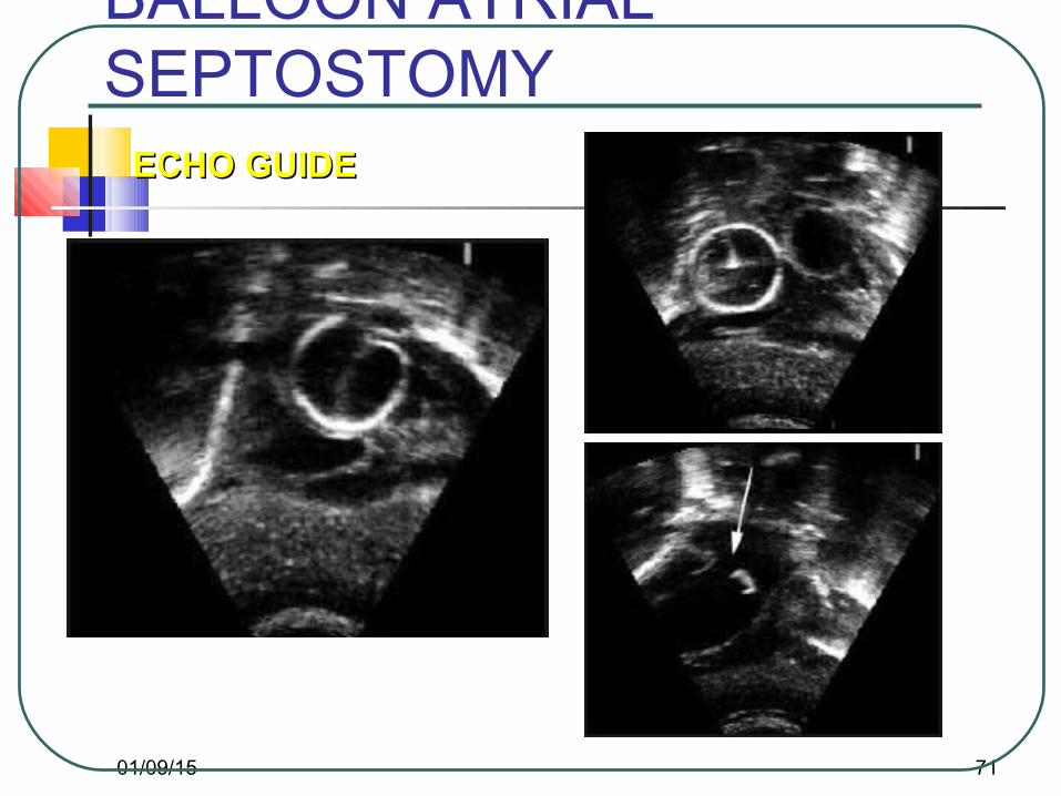
BALLOON ATRIAL SEPTOSTOMY
ECHO GUIDEECHO GUIDE

01/09/15 72
SUCCESSFUL SEPTOSTOMY Cinical improvement Equalization of atrial pressures Change in arterial O2 saturation Angiogram before and after septostomy Increase in arterial pressure Decrease in pulmonary arterial pressure Balloon calibration of defect Echo visualization of defect

01/09/15 73
BALLOON DILATATION OF STENOTIC VALVES
Balloon dialtation of
Aortic valve
Pulmonary valve
Coarctation of aorta

01/09/15 74
BALLOON DILATATION OF THE AORTIC VALVE Palliative procedure
Procedural success is almost 100%
Mortality may be high if associated conditions
Endocardial fibroelastosis LV hypoplasia Others
Risks- aortic regurgitation

01/09/15 75
BALLOON DILATATION OF AORTIC VALVE
Indications
Procedure

01/09/15 76
AORTIC VALVULOPLASTY
PRE DILATATIONPRE DILATATION BALLOON ACROSS BALLOON ACROSS THE AORTIC VALVETHE AORTIC VALVE
POST DILATATIONPOST DILATATIONNO ARNO AR

01/09/15 77
BALLOON DILATATION OF PULMONARY VALVENeonatal critical Pulmonary stenosis Majority present during first week Duct dependant (require PGE1) RV morphology- well developed or
poorly developed Echo assessment Immediate success rate > 80%

01/09/15 78
Tx PULMONARY VALVULOPLASTY

01/09/15 79
Pulmonary valvotomy and VSD closurePulmonary valvotomy and VSD closure
Effective balloon diameter should be 1.2 to 1.4 times Effective balloon diameter should be 1.2 to 1.4 times the measured Pulmonary Valve Annulusthe measured Pulmonary Valve Annulus

01/09/15 80
Pulmonary valvotomy and VSD closurePulmonary valvotomy and VSD closure
Effective balloon diameter should be 1.2 to 1.4 times Effective balloon diameter should be 1.2 to 1.4 times the measured Pulmonary Valve Annulusthe measured Pulmonary Valve Annulus

01/09/15 81
Pulmonary valvotomy and VSD closurePulmonary valvotomy and VSD closure
Effective balloon diameter should be 1.2 to 1.4 times Effective balloon diameter should be 1.2 to 1.4 times the measured Pulmonary Valve Annulusthe measured Pulmonary Valve Annulus
01/09/15 82
BALLOON DILATATION OF COARCTATION Controversial role in neonates Indications
Symptomatic newborn CCF Failure to thrive Upper extremity hypertension Severe LV dysplasia Severe PAH

01/09/15 83

01/09/15 84

01/09/15 85

01/09/15 86

01/09/15 87

01/09/15 88
COIL EMBOLIZATION
Indications Large AVM Aortopulmonary collaterals Systemic venous anomalies PDA (if large, unrestrictive- surgery ideal)

01/09/15 89
STENTING OF PDA
Ductal dependent pulmonary blood flow Difficult procedure Limited experience Intimal proliferation is universal thus
need for reintervention

01/09/15 90
Device Closures
PDA ASD MUSCULAR VSD PERI MEMBRANOUS VSD IN OTHER PLACES e.g
Coronary AV fistulaPeri valvular leaks

01/09/15 91
Closure of PDA
Device closure – for almost any duct Contra indications
Large PDA in a neonate
Associated reversal of shuntAssociated heart disease requiring
surgery PROCEDURE

01/09/15 92

01/09/15 93

01/09/15 94
Coil closure of PDA
Coil closure for small ducts Coil made up of stainless steel wire with
Dacron strands – promote thrombosis Cheap
PROCEDURE

01/09/15 95
DETACHABLE COIL

01/09/15 96
Device closure of ASD
Amplatzer septal occluder device is most widely used
Only used for Secundum ASD Continuous TEE monitoring Adequate rim is required otherwise any size
of ASD can be closed
PROCEDURE

01/09/15 97
(ASD)(ASD)

01/09/15 98
Approach

01/09/15 99
The Device
The device is made up of a NITINOL (Nickel-Titanium Naval Ordanance Laboratory).Dacron fibre - within the device

01/09/15 100

01/09/15 101

01/09/15 102
Device closure of VSD
Amplatzer device for Muscular VSD Amplatzer device for Peri membranous
VSD – defect >5 mm away from aortic valve
PROCEDURE

01/09/15 103

01/09/15 104
Pediatric Cardiology: Summary•1
•11938-Essentially no Rx for CHD–PPioneers-pathologists, Pediatricians, Cardiologists, Surgeons, Imaging experts, Intensivists, Interventionalists•22008-Rx for virtually all CHD, BUT mort./morb.:–vVentricular function–aArrhythmia– Valves/conduits–pPulm hypertension•N

01/09/15 105
FUTURE OF INTERVENTIONAL CARDIOLOGY
1. Percutaneous pulmonary valve replacement2. Percutaneous aortic valve replacement3. VSD closure devices4. Percutaneous banding devices5. RF perforation6. Drug eluting stents7. Fetal intervention- therapy for aortic
stenosis


![[Codientu.org] Cong Nghe Sua Chua May Cong Cu](https://img.pdfslide.net/doc/110x75/55cf941e550346f57b9fc133/codientuorg-cong-nghe-sua-chua-may-cong-cu.jpg)