Embed Size (px)
Citation preview
Activation of Rat Alveolar Macrophage-DerivedLatent Transforming Growth Factor b-1 by PlasminRequires Interaction with Thrombospondin-1 and itsCell Surface Receptor, CD36
Teshome Yehualaeshet,* Robert O’Connor,†
Julia Green-Johnson,‡ Sabine Mai,‡
Roy Silverstein,¶ Joanne E. Murphy-Ullrich,§ andNasreen Khalil*‡
From the Departments of Medicine* and Pathology† and the
Manitoba Institute of Cell Biology,‡ University of Manitoba,
Winnipeg, Manitoba, Canada; the Department of Pathology,§
University of Alabama at Birmingham, Birmingham, Alabama;
and the Division of Hematology/Oncology,¶ Cornell Medical
Center, New York, New York
Transforming growth factor-b-1 (TGF-b1) is secretedby cells in a latent form (L-TGF-b1) noncovalentlybound to a latency-associated peptide. Activated alve-olar macrophages obtained from rat lungs after bleo-mycin-induced pulmonary injury released increasedamounts of active TGF-b1 as well as plasmin, a pro-tease, and thrombospondin-1 (TSP-1), a trimeric gly-coprotein. Previously we had demonstrated that plas-min was critical to the activation of L-TGF- b1. In thepresent study we demonstrated that TSP-1 is also im-portant for the activation of L-TGF- b1 because theactivation can be inhibited by anti-TSP-1 monoclonalantibody. Proteins obtained from alveolar macro-phage cell lysates immunoprecipitated with antibod-ies specific for TSP-1 were identified on immunoblotsas LAP and TGF-b1, indicating that TSP-1/L-TGF-b1complexes are present on alveolar macrophages.However, in the presence of plasmin both latency-associated peptide and TGF-b1 were decreased in thesame cell lysates, indicating that L-TGF-b1 associatedwith TSP-1 is released by plasmin. Using immunoflu-orescence and antibodies to TGF-b1 and CD36, a re-ceptor for TSP-1, there was colocalization of TGF-b1with CD36. Because TSP-1 but not TGF-b1 is a naturalligand for CD36, these findings suggest that the L-TGF-b1 in a complex with TSP-1 localizes to the mac-rophage cell surface when TSP-1 interacts with itsreceptor, CD36. Furthermore, the association of TSP-1/L-TGF-b1 complex with CD36 is necessary to theactivation of L-TGF-b1 because antibodies to CD36prevent the colocalization of TGF-b1 with CD36 asobserved by immunofluorescence and inhibit activa-tion of the L-TGF-b1 by explanted alveolar macro-
phages. These findings suggest that activation of L-TGF-b1 by plasmin occurs at the cell surface ofactivated alveolar macrophages and requires a TSP-1/CD36 interaction. (Am J Pathol 1999, 155:841–851)
At sites of lung injury before connective tissue synthesisthere is an influx of activated macrophages.1,2 Whenactivated, macrophages secrete a number of pro-inflam-matory and fibrogenic cytokines.1,2 Of these cytokines,transforming growth factor-b-1 (TGF-b1), a multifunc-tional peptide, is one of the most potent regulators ofinflammation and connective tissue synthesis.3 TGF-b1 issynthesized as a large 390-amino acid precursor thatundergoes a number of intracellular processing steps,including cleavage of the latent-associated peptide(LAP), to produce the mature 25-kd TGF-b1 protein.4
However, with rare exceptions, when TGF-b1 is secretedby cells it remains noncovalently associated in a 1:1 ratiowith its LAP.4 The noncovalent association of TGF-b1 withits LAP renders the TGF-b1 unable to interact with itsreceptor and, therefore, biologically inactive.4 TGF- b1and its receptors are ubiquitously expressed, and, be-cause TGF-b1 has numerous biological effects, the abilityof a cell to activate L-TGF-b1 on secretion may be animportant regulatory mechanism of TGF- b1 action in vivo.
We had previously demonstrated that after pulmonaryinjury induced by the antineoplastic antibiotic bleomycin,explanted alveolar macrophages generated maximalquantities of biologically active TGF-b1 and plasmin 7days after bleomycin induced pulmonary injury.5 Further-more, the secretion of active TGF-b1 was totally inhibitedby the presence of a2-antiplasmin, a naturally occurringinhibitor of plasmin.5 When large quantities of plasminwere added to activated alveolar macrophages, therewas further activation of the L-TGF-b1.5 However, whenplasmin was added to the L-TGF-b1 in cell-free CM, no
Supported by the Medical Research Council of Canada and the Respi-ratory Health Network of Centers of Excellence (to N. K.) and NationalInstitutes of Health HL50061 (to J. E. M.-U.).
Accepted for publication May 25, 1999.
Address reprint requests to Dr. Nasreen Khalil, B.C. Centre for DiseaseControl, University of British Columbia, 655 West 12th Avenue, Vancou-ver, British Columbia, V5Z 4R4 Canada.
American Journal of Pathology, Vol. 155, No. 3, September 1999
Copyright © American Society for Investigative Pathology
841
9further activation of L-TGF-b1 occurred.5 Our findingssuggested that the generation of plasmin is important inthe posttranslational activation of alveolar macrophage-derived L-TGF- b1 during an inflammatory pulmonaryinjury response and that the activation requires the pres-ence of intact macrophages.5
In this paper, we demonstrate that alveolar macro-phages also secrete increased quantities of TSP-1, aglycoprotein previously reported to activate L-TGF-b1both in the presence of cells and in cell-free solution.6
When alveolar macrophages were cultured in the pres-ence of neutralizing antibodies to TSP-1, the posttransla-tional activation of L-TGF-b1 was abrogated. Further-more, antibodies to the TSP-1 receptor, CD36, alsoabrogated activation of alveolar macrophage-derived L-TGF-b1. Our findings support a model in which L-TGF-b1is held at the cell surface by a TSP-1/CD36 interactionand is processed by plasmin generated by activatedalveolar macrophages.
Materials and Methods
Animals
Female Sprague-Dawley rats, which were free of respira-tory disease and weighed between 250 and 300 g, wereobtained from the University of Manitoba vivarium. Ineach experiment, all rats were matched for age andweight.
Reagents
Bleomycin (Blenoxane) was a gift from Bristol-MyersSquibb (Evansville, IN). Neutralizing antibody to TGF-b1–3 was obtained from Genzyme (Cambridge, MA).Antibody to TGF-b1 used for Western blot analysis wasobtained from Santa Cruz Biotechnology (Santa Cruz,CA). The recombinant anti-human LAP antibody was ob-tained from R&D Systems (Minneapolis, MN). TSP-1 de-pleted of TGF-b activity (sTSP-1) and monoclonal anti-bodies to human platelet TSP-1 depleted of associatedTGF- b1 (mAb 133) used in the enzyme-linked immu-nosorbent assay (ELISA) and TSP-1 immunoprecipitateswere either used by or obtained from Dr. Murphy-Ull-rich.6,7 Anti-TSP-1 antibody used in experiments to neu-tralize TSP-1 from activated alveolar macrophages wasobtained from Sigma (St. Louis, MO). The CD36 antibody,5F1, was provided by the Fifth International Workshop onLeukocyte Differentiation Antigens.8
Bleomycin Administration
This procedure is described in detail elsewhere.5,9,10 Forsome experiments rats were sacrificed at several timeintervals after bleomycin or normal saline treatment,5,9,10
whereas for other experiments alveolar macrophageswere harvested 7 days after bleomycin administration.The latter time point was used based on our findings thatalveolar macrophages are maximally stimulated at thistime to secrete active TGF-b1.5
Macrophage Cultures
This procedure is described in detail elsewhere.5,10 Thelungs were lavaged to obtain cells for culture of alveolarmacrophages. Alveolar macrophages were maintained inserum-free media containing Gentamicin (4 mg/100 ml;Roussel, Montreal, PQ), Fungizone (100 ml/100 ml; GibcoBRL, Grand Island, NY) and 0.2% clotted bovine calfplasma (BCP; National Biological Laboratory Limited, Du-gald, MB). The macrophages were cultured in the ab-sence or presence of a number of reagents consisting ofanti-TSP-1 antibody, 5F1 (anti-CD36 antibody), CD36synthetic peptide (aa 93–110), or sTSP-1. In experimentsto determine the effects of exogenous sTSP-1, alveolarmacrophages were cultured with varying quantities ofsTSP-1 for 2 hours before the collection of conditionedmedia (CM). In addition, CM from the same alveolarmacrophages cultured in parallel was incubated in acell-free solution with sTSP-1 for 2 hours. Incubation ofCM with sTSP-1 for 2 hours was chosen based on ourprevious findings, that sTSP-1 can activate L-TGF-b1within 2 hours.11 In some experiments the cells werecultured in the absence or presence of 5F1, the CD36antibody, before the addition of sTSP-1. In experiments todetermine whether or not both plasmin and TSP-1 arerequired to activate alveolar macrophage-derived L-TGF-b1, the alveolar macrophages were cultured with sTSP-1in the absence or presence of aprotinin, an inhibitor ofplasmin activity.12 After 20 hours of incubation in 5% CO2
at 37°C, the media was collected in the presence ofprotease inhibitors (leupeptin 0.5 mg/ml, Amersham,Poole, UK; aprotinin 1 mg/ml, and pepstatin 1 mg/ml, bothfrom Sigma, Oakville, ON), and frozen at 280°C untilready for TGF-b quantitation.5,10
CCL-64 Mink Lung Epithelial Growth InhibitionAssay for TGF-b
The CCL-64 growth inhibition assay to identity and quan-titate TGF-b has been described.5,9,10,13. Briefly, to sub-confluent cells in 0.2% BCP and resuspended in a-MEM,0.2% BCP, 10 mmol/L Hepes at pH 7.4, penicillin (25mg/ml) and streptomycin (25 mg/ml), and cultured as 5 3104 cells per 0.5 ml in 24-well costar dishes (Flow Labo-ratories, Inc., Mississauga, ON) was added neutral CM orCM that was acidified and subsequently neutralized. Af-ter 22 hours the cells were pulsed with 0.25 Ci of 5-[125I]iodo 29-deoxyuridine (ICN Pharmaceutical, Costa Mesa,CA) for 2 to 3 hours at 37°C and eventually lysed with 1ml of 1 N NaOH for 30 minutes at room temperature andthe 125I-UdR was counted in a g counter (LKB Instru-ments, Gaithersburg, MD). A standard curve using por-cine TGF-b1 was included in each assay. For confirma-tion of TGF-b activity, neutralizing monoclonal antibody toTGF-b1–3 (Genzyme) was added before the addition ofthe CM5,9,10,13 and resulted in abrogation of all TGF-bactivity.
842 Yehualaeshet et alAJP September 1999, Vol. 155, No. 3
Detection and Quantitation of TSP-1 by DirectELISA
The wells of 96-well plates (Falcon tissue culture plates)were coated with 200 ng/well of the mAb 133 (anti-sTSP-1). For each experiment, a standard curve containingwells with 200 ml of CM and several concentrations ofsTSP-1, ranging from 15–150 ng/well, was included. Toquantitate TSP-1 in CM, 200 ml of alveolar macrophagederived CM in carbonate buffer, pH 9.6, was incubatedovernight at 4°C. The next day, the wells were washedthree times with PBS, 0.05%, Tween 220. Nonspecificbinding sites were blocked by incubating with 250 ml/wellof 1% BSA for 1 hour at 37°C. The wells were thenwashed with PBS and incubated with 200 ml/well of mAb133 (7.5 mg/ml) in PBS-Tween for 90 minutes at 37°C. Thewells were then washed and incubated with 30 ng/ml ofalkaline phosphatase-labeled goat anti-mouse lgG for 90minutes at 37°C, and then assayed for color developmentusing the Sigma 104 AP substrate. Color developmentwas stopped by adding 50 ml of 2N NaOH, and absor-bency at 405 nm was read using a Bio-Tek ELISA reader.
Preparation of Synthetic CD36 Peptides
The CD36 peptide, YRVRFLAKENVTQDAEDNC(93–110)was synthesized, based on the work of Leung et al.14 Thepeptide was synthesized with an Applied Biosystemsmodel 431A peptide synthesizer using Fmoc (N-(9-Flu-oreny D-methoxycarbonyl) chemistry and purified by re-verse high pressure liquid chromatography, using a C18column.
Localization of CD36 and TGF-b1 byImmunofluorescence
Alveolar macrophages were obtained by bronchoalveo-lar lavage 7 days after intratracheal normal saline orbleomycin administration, and were adjusted to 1 3 106
cells/ml. For some experiments, alveolar macrophagesobtained after bleomycin administration were cultured inthe presence of anti-CD36 antibody (20 mg per 106 mac-rophages) for 30 minutes before the immunofluoresenceprocedure. The immunofluorescence analysis was per-formed as previously described.15 Briefly, cytospinsmears of 1 3 105 cells suspension were fixed with 3.7%formaldehyde for 10 minutes and washed with PBS. Non-specific binding was blocked by 100% lamb serum(Gibco BRL) for 5 minutes. The cells were then incubatedwith anti-TGF-b1 (Santa Cruz Biotechnology) and anti-CD36 antibody, both at a concentration of 1 mg/ml, for 45minutes. After washing with PBS the cells were incubatedfor 30 minutes with polyclonal anti-rabbit antibody conju-gated to tetramethylrhodamine-isothiocyanate (TRITC) orconjugated with fluorescein isothiocyanate (FITC) mono-clonal anti-mouse IgM as secondary antibodies for thedetection of TGF-b1 and CD36 antibodies, respectively.Both secondary antibodies were used as 1 mg/ml. Theslides were washed three times with PBS. Nuclear stain-ing was done using 49, 69 Diamidino-2-phenylindole
(DAPI) at a concentration of 1 mg/ml for 5 minutes. Theslides were mounted in anti-bleach (12% glycerol, 4.8%Mowiol 4–88 (Hoechst), 2.4% DABCO (1, 4 Diazabicylo[2.2.2]-octane (Fluka) in 0.2 Mol/L Tris/HCl, pH 5 8.5).Image analysis was performed using a Zeiss Axiophotmicroscope, equipped with a cooled CCD camera CH250/a (Optikon/Photometrics), driven by IPLabs Spec-trum software version 3.1, Signal Analytics). For TRITC-conjugated antibodies, the filter combination was BP540/FT580/LP590, resulting in a red emission identifying thelocation of TGF-b1on the macrophage surface. For theFITC-conjugated antibody, the filter combination was450–490/FT510/515–565, resulting in a green emissionidentifying the location of CD36 on the macrophage sur-face. For DAPI, the filter combination was G365/FT395/LP420, resulting in a blue emission. To determinewhether TGF-b1 and CD36 were colocalized, the imageswere obtained by a pixel overlap, which was achievedwith IPLab Spectrum/Multiprobe (V.3.1, Signal Analytics,Fairfax, VA). In areas where the TGF-b1 was localized tothe same region as CD36, the emission was yellow.
Protein Extraction and Immunoprecipitation
Alveolar macrophages obtained from rats that had beentreated with normal saline or bleomycin 7 days earlierwere lysed by incubating the cells for 20 minutes on icein a RIPA lysis buffer (50 mmol/L Tris-Cl, pH 7.5; 150mmol/L NaCl, 1% Nonidet P-40, 0.1% sodium deoxy-cholate and a cocktail of protease inhibitors; phenylmeth-ylsulfonyl fluoride 1 mmol/L, leupeptin 1 mg/ml, and apro-tinin 0.1 mg/ml, all from Sigma). In some experiments thealveolar macrophages were cultured overnight in the ab-sence or presence of a2-antiplasmin or aprotinin (Sigma),both inhibitors of plasmin activity.12 The lysate was cen-trifuged for 20 minutes at 12,000 3 g at 4°C, the super-natant collected, and the total protein content determinedby the Bradford dye-binding assay (Bio-Rad, Missis-sauga, ON). The total protein extract (300 mg) and 10 mgof anti-sTSP-1 antibody (mAb 133) or IgG as an isotypecontrol for the anti-sTSP-1 antibody was incubated over-night at 4°C. After a further incubation of 2 hours with 30ml of Protein G Plus/Protein A-Agarose (Calbiochem, SanDiego, CA) the immune complexed beads were col-lected. The beads were washed four times with RIPAbuffer and placed in a final suspension with 25 ml ofLaemmli buffer and boiled for 10 minutes. The superna-tant containing the precipitated proteins was then usedfor Western blot analysis.
Western Blot Analysis
The protein samples (25 ml) were electrophoresed on10% sodium dodecyl sulfate-polyacrylamide gel electro-phoresis (SDS-PAGE) in a MiniPROTEAN II Electrophore-sis Cell (Bio-Rad, Hercules, CA). Rainbow colored pro-tein molecular weight markers (Amersham) were runparallel to each blot as an indicator of the molecularweight. Equality in loading of protein was evaluated usingsilver staining (not shown). The separated proteins were
Activation of Macrophage-Derived L-TGF-b1 by TSP-1 843AJP September 1999, Vol. 155, No. 3
transferred at 50 V overnight onto nitrocellulose mem-brane (Gibco BRL) in a Mini trans-Blot chamber withtransfer buffer (25 mmol/L Tris Cl, 192 mmol/L glycine,and 20% methanol). The nitrocellulose membrane wasblocked for 1 hour by using 5% instant skim milk powderin TBS. For detection of LAP a 1:500 dilution of anti-recombinant human LAP antibody (anti-rh LAP; R&D Sys-tems) in 1% instant skim milk powder was used. Afterwashing, the nitrocellulose membrane was incubatedwith horseradish peroxidase linked with the secondaryantibody (Goat anti-mouse IgG; Bio-Rad) as recom-mended by the manufacturer. Finally the washed blotswere exposed to enhanced chemiluminescence detec-tion system (Amersham) and recorded on an autoradio-graph (Kodak X-Omat film). Before reprobing, the nitro-cellulose membrane was incubated at 50°C for 30minutes with a stripping buffer (100 mmol/L 2-mercapto-ethanol, 2% sodium dodecyl sulfate, and 62.5 mmol/LTris-HCl, pH 6.7). The blots were rinsed twice with TBS.To ensure the removal of antibodies, membranes wereincubated with the enhanced chemiluminescence detec-tion reagents and exposed to film (Kodak). No band wasdetected, confirming that all antibodies were stripped offthe membrane. The same nitrocellulose membrane wasblocked using 5% instant skim milk powder in TBS. Fordetection of TGF-b1, 0.5 mg/ml of rabbit polyclonal IgGTGF-b1 antibody (Santa Cruz Biotechnology) was usedas above.
Statistical Analysis
Statistical analysis using analysis of variance was done byDr. Bob Tate, Biostatistical Unit, University of Manitoba.
Results
Activation of Alveolar Macrophage-Derived L-TGF-b1 by Thrombospondin (TSP)-1
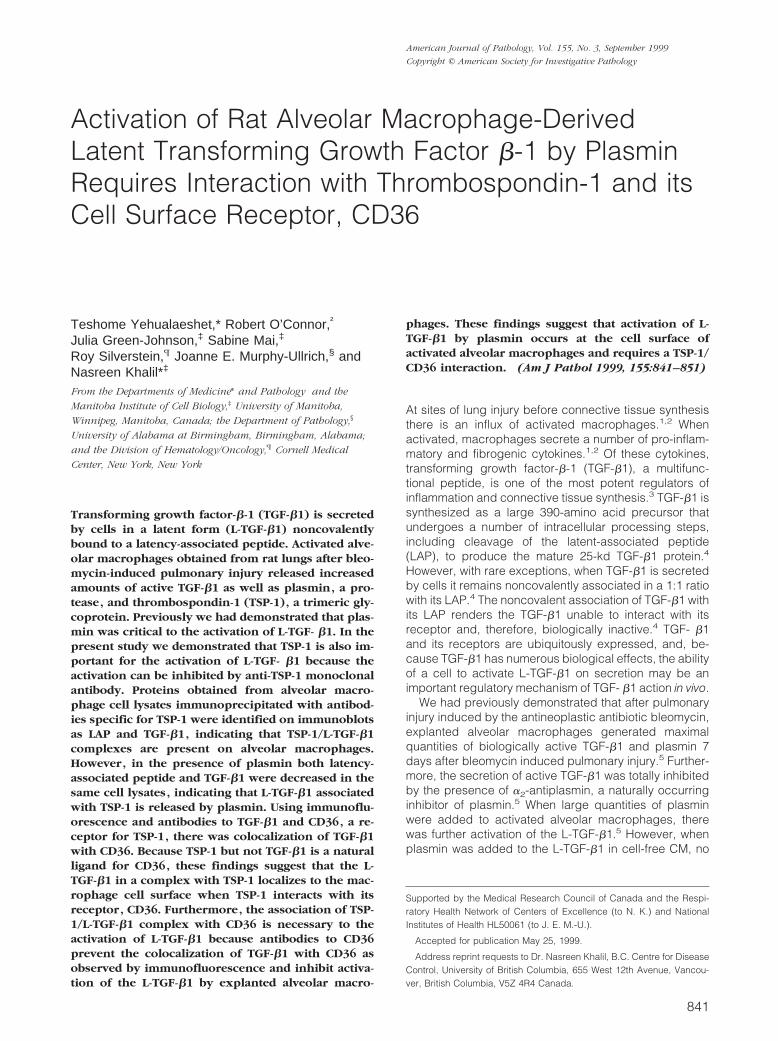
In addition to plasmin, TSP-1, a large trimeric glycopro-tein,16 had also been demonstrated to be a physiologicalsubstance that can activate L-TGF-b1.6,17 TSP-1 wasconstitutively secreted by alveolar macrophages, but af-ter bleomycin injury, TSP-1 secretion was further in-creased and reached maximal levels 7 days after bleo-mycin administration (Figure 1A). The secretion of TSP-1declined rapidly thereafter, and by 28 days after bleomy-cin administration, the secretion returned to that of controllevels of alveolar macrophages from normal saline-treated rats. To determine whether the presence of TSP-1in the CM was necessary for the activation of L-TGF-b1,alveolar macrophages were cultured in the absence orpresence of anti-TSP-1 monoclonal antibody. Anti-TSP-1antibody inhibited the activation of L-TGF- b1 but had noeffect on the secretion of the latent form of TGF-b1 (Fig-ure 1B) by the alveolar macrophages. Neutral CM ofalveolar macrophages activated by in vivo bleomycin in-jury contained 67.6 6 7.9 pg of TGF-b1 per 106 cells.After the addition of 50 mg/ml of the isotype control foranti-TSP-1, 50.13 6 13.4 pg of TGF-b1 per 106 waspresent in neutral CM (P , 0.934). Because TSP-1 hasbeen reported to activate L-TGF-b1 in solution,6 we nextdetermined if alveolar macrophage-derived L-TGF-b1could be directly activated in CM by sTSP-1. After 20hours in culture, activated alveolar macrophages se-creted large quantities of L-TGF-b1.5 The addition ofsTSP-1 to this cell-free L-TGF-b1 unexpectedly dimin-
Figure 1. Activation of alveolar macrophage derived L-TGF-b1 by sTSP-1. A: Quantity of TSP-1 secreted by explanted alveolar macrophages obtained from ratsat varying lengths of time after bleomycin administration. Each point is the mean of samples from 3 to 6 rats. The P value is ,0.0003 when the quantity of TSP-1produced by alveolar macrophages from normal saline-treated rats is compared to TSP-1 produced by alveolar macrophages from rats treated with bleomycin 7days earlier. B: The effects of anti-TSP-1 antibody on the activation of alveolar macrophage-derived L-TGF-b1. Alveolar macrophages obtained 7 days afterbleomycin administration were cultured in the absence of anti-TSP-1 antibody or in the presence of varying concentrations. The TGF-b1 present in neutral CM(M) represents bioactive TGF-b1; that present in acidified then neutralized CM (f) represents the total TGF-b1 in the same sample. Each point is the mean ofsamples from 6 rats. The quantity of TGF-b1 in neutral CM compared to the quantity of TGF-b1 in acidified CM has a P # 0.001 when the anti-TSP-1 antibodyused was 50 mg/ml/106 macrophages, and P # 0.003 when the anti-TSP-1 antibody used was 5.0 or 0.5 mg/ml/106 macrophages. There was no statistical differencein the quantity of TGF-b1 in neutral and acidified CM when no anti-TSP-1 antibody was present. C: Induction of activation of L-TGF-b1 by addition of sTSP-1.Alveolar macrophages were obtained 7 days after bleomycin administration and cultured for 20 hours before the addition of sTSP-1 (0–4 mg/ml) to alveolarmacrophages (f) or the CM (M) from parallel cultures of alveolar macrophages. After a 2-hour incubation with sTSP-1, the TGF- b1 in neutral and acidified CM(not shown) of the same sample was determined. The -fold increase in the quantity of TGF-b1 present was calculated by using the quantity of active TGF-b1present in the absence of sTSP-1 as the denominator and the quantity of TGF-b1 present after the addition of sTSP-1 as the numerator. Each point is the meanof samples from 3 rats. The -fold increase in the quantity of TGF-b1 in neutral CM when alveolar macrophages were treated with 0.4 or 4.0 mg/ml of sTSP-1,compared to TGF-b1 in CM when no sTSP-1 was present, has P value # 0.05.
844 Yehualaeshet et alAJP September 1999, Vol. 155, No. 3
ished the quantity of active TGF-b1 that could be de-tected by our bioassay (Figure 1C). In contrast, sTSP-1added directly to the same CM but, in the presence ofalveolar macrophages, further activated L-TGF-b1 (Fig-ure 1C). These findings demonstrate that TSP-1 is effec-tive in promoting the activation of alveolar macrophage-derived L-TGF-b1 in the presence of intact macrophages.
Cell Surface Association of L-TGF- b1 to CD36Is Required for Activation of L-TGF-b1
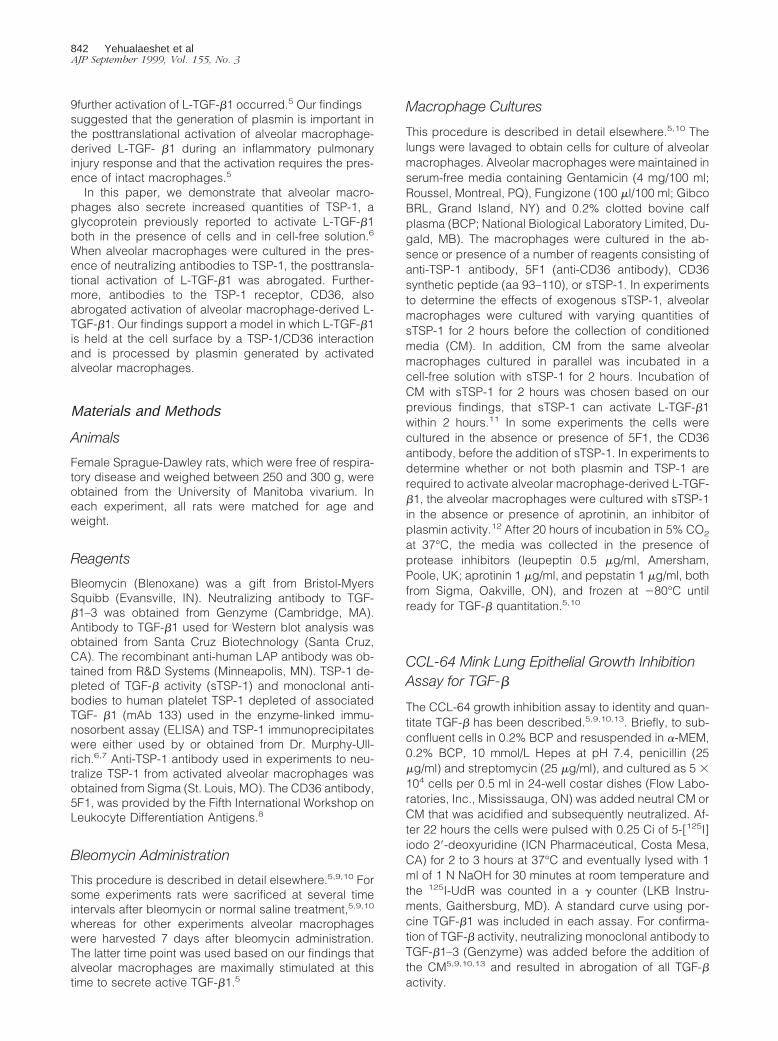
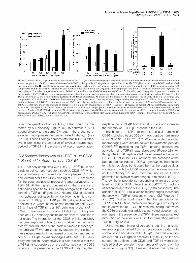
TSP-1 not only complexes with L-TGF-b1,6,7,11 but it alsobinds to cell surface receptors such as CD3618,19 whichare prominently expressed on macrophages.18,19 Wenext determined if the CD36 binding of TSP-1 is requiredfor the posttranslational processing and activation of L-TGF- b1. At the highest concentration, the presence ofantibodies specific to CD36 totally abrogated the activa-tion of L-TGF-b1 (Figure 2A). Neutral CM of alveolarmacrophages activated by in vivo bleomycin injury con-tained 70 6 10 pg of TGF-b1 per 106 cells, while after theaddition of 20 mg/ml of the isotype control for anti-CD36,55 6 1 pg of TGF-b1 per 106 cells was present (P ,0.949). There was an induction of L-TGF- b1 in the pres-ence of CD36 antibody but the mechanism of induction isnot clear. The interaction of the CD36 with its antibodyhas been reported to lead to an oxidative burst in mono-cytes20 and induction of the protein tyrosine kinases fyn,lyn, and yes.21 We are presently determining if either ofthese events results in increased production and secre-tion of L-TGF-b1 by macrophages after anti-CD36 anti-body interaction. Alternatively it is also possible that theL-TGF-b1 is sequestered on the cell surface via the CD36receptor. The presence of the CD36 antibody may then
displace the L-TGF-b1 from the cell surface and increasethe quantity of L-TGF-b1 present in the CM.
The binding of TSP-1 to the extracellular domain ofCD36 is blocked by a CD36 synthetic peptide from aminoacids 93–110 (CD3693–110).14 When activated alveolarmacrophages were incubated with the synthetic peptideCD3693–110 mimicking the TSP-1 binding domain, theactivation of L-TGF-b1 was abrogated (Figure 2B). Al-though the CD36 peptide 93–110 decreased activation ofL-TGF-b1, unlike the CD36 antibody, the presence of thepeptide did not induce L-TGF-b1 generation. The reasonfor this is not clear, but it could be that the peptide doesnot interact with the CD36 receptor in the same manneras the antibody20,21 and, therefore, not cause furtheractivation of alveolar macrophages to release L-TGF-b1.The synthetic peptide corresponding to an area unre-lated to CD36-TSP-1 interaction, CD36204–288, had noeffect on the activation of L-TGF- b1(data not shown). Theaddition of sTSP-1 to alveolar macrophages increasedthe activity of TGF-b1 present in neutral CM (Figures 1Cand 2C). Further confirmation that the association ofTSP-1 with CD36 on alveolar macrophages was impor-tant in activation of L-TGF- b1 was the finding that, whenthe CD36 antibody was added to activated alveolar mac-rophages in the presence of sTSP-1, there was a markeddiminution of the effects of sTSP-1 in generating matureTGF-b1 (Figure 2C).
Using indirect immunofluorescent staining, alveolarmacrophages obtained from rats previously treated withnormal saline had detectable TGF-b1 (red emission; Fig-ure 3A) and CD36 (green emission; Figure 3B) on the cellsurface. In addition, both CD36 and TGF-b1 were colo-calized (yellow emission) to a number of regions on thesame cells (Figure 3C). However, alveolar macrophages
Figure 2. Effects of anti-CD36 antibody on the activation of L-TGF-b1. Alveolar macrophages obtained 7 days after bleomycin administration were cultured in theabsence or presence of different concentrations of anti-CD36 antibody or the CD36 synthetic peptide 93–110. A: The TGF-b1 present in neutral CM M and acidifiedthen neutralized CM f from the same sample was quantitated. Each point is the mean of samples from 4 rats. The quantity of TGF-b1 present in neutral CMcompared to TGF-b1 in acidified CM has a P value #0.0001 when the antibody was 20 mg/ml/106 macrophages, and P # 0.01 when the antibody was 10 mg/ml/106
macrophages. The other comparisons between TGF-b1 in neutral and acidified CM were not significant. B: The effects of CD36 synthetic peptide aa 93–110 onthe activation of L-TGF-b1. Alveolar macrophages were cultured in the absence or presence of several concentrations of the synthetic peptide of CD36 aa 93–110.TGF-b1 in neutral M and acidified, then neutralized CM f was quantitated. All points are the mean of 4 to 6 animals. The quantity of TGF-b1 in neutral CM,compared to the TGF-b1 in acidified CM of the same sample, had a P value #0.0006 only when CD36 peptide 93–110 was present. C: Effects of anti-CD36 antibodyon the activation of L-TGF-b1 in the presence of sTSP-1. Alveolar macrophages were cultured in the absence or presence of 20 mg/ml/106 macrophages ofanti-CD36 antibody, and in the absence or presence of 0.4 mg/ml/106 macrophages of sTSP-1. The TGF- b1 present in neutral CM was quantitated. Each pointis the mean of samples from 4–6 rats. TGF-b1 in neutral CM of alveolar macrophages from bleomycin (BLM)-treated rats compared to normal saline (N/S)-treatedrats has a P value #0.07. TGF-b1 in neutral CM of alveolar macrophages from BLM-treated rats cultured in the absence of sTSP-1, compared to when sTSP-1 waspresent, has a P value #0.02. TGF-b1 in neutral CM of alveolar macrophages from BLM treated rats cultured with sTSP-1, compared to when sTSP-1 and anti-CD36antibody was also present, has a P value #0.006.
Activation of Macrophage-Derived L-TGF-b1 by TSP-1 845AJP September 1999, Vol. 155, No. 3
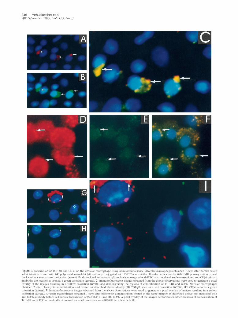
Figure 3. Localization of TGF-b1 and CD36 on the alveolar macrophage using immunofluoresence. Alveolar macrophages obtained 7 days after normal salineadministration treated with (A) polyclonal anti-rabbit lgG antibody conjugated with TRITC reacts with cell surface-associated anti-TGF-b1 primary antibody, andthe location is seen as a red coloration (arrow). B: Monoclonal anti-mouse lgM antibody conjugated with FITC reacts with cell surface-associated anti-CD36 primaryantibody; the location is seen as a green coloration (arrow). C: Immunofluorescent images obtained from the above observations were used to generate a pixeloverlay of the images resulting in a yellow coloration (arrow) and demonstrating the regions of colocalization of TGF-b1 and CD36. Alveolar macrophagesobtained 7 after bleomycin administration and treated as described above identify (D) TGF-b1 seen as a red coloration (arrow); (E) CD36 seen as a greencoloration (arrow). F: Immunofluorescent images obtained from the above observations were used to generate a pixel overlay of images resulting in a yellowcoloration (arrow). Alveolar macrophages obtained 7 days after bleomycin administration treated in the same manner as described above but incubated withanti-CD36 antibody before cell surface localization of (G) TGF-b1 and (H) CD36. A pixel overlay of the images demonstrates either no areas of colocalization ofTGF-b1 and CD36 or markedly decreased areas of colocalization (arrows) on a few cells (I).
846 Yehualaeshet et alAJP September 1999, Vol. 155, No. 3
obtained 7 days after in vivo bleomycin injury had amarked increase in the number of cells with cell surfacelocalization of TGF-b1 (Figure 3D) and CD36 (Figure 3E),and the colocalization of TGF-b1 and CD36 was alsoincreased (Figure 3F). When alveolar macrophages ob-tained 7 days after bleomycin injury were cultured in thepresence of anti-CD36 antibody before the immunofluo-rescent staining the presence of TGF-b1 (Figure 3G) andCD36 (Figure 3H) was markedly reduced and was notdetected consistently on the same cells (Figure 3, G andH) as observed in Figure 3, A-F. Furthermore, the colo-calization of TGF-b1 and CD36 was either not present ormarkedly decreased (Figure 3I). Although alveolar mac-rophages from normal saline-treated rats expressed littleTGF-b1 and CD36 there was almost complete colocal-ization with CD36, whereas after anti-CD36 pretreatmentof alveolar macrophages there was markedly less colo-calization of CD36 and TGF-b1. These findings suggestthat the association of TGF-b1 with CD36 occurs in aspecific manner rather than randomly.
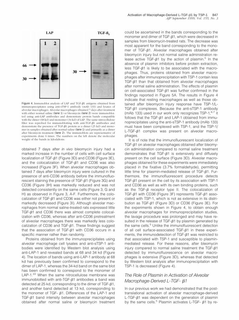
Proteins obtained from the immunoprecipitates usingalveolar macrophage cell lysates and anti-sTSP-1 anti-bodies were identified by Western blot analysis usinganti-LAP-1 and revealed bands at 68 and 34 kd (Figure4). The location of bands using anti-LAP-1 antibody at 68kd has previously been confirmed to correspond to thedimer of LAP-1, whereas the 34-kd band on the same blothas been confirmed to correspond to the monomer ofLAP-1.22 When the same nitrocellulose membrane wasimmunoblotted with anti-TGF-b1 antibodies a band wasdetected at 25 kd, corresponding to the dimer of TGF-b1,and another band detected at 13 kd, corresponding tothe monomer of TGF- b1. Differences of the LAP-1 andTGF-b1 band intensity between alveolar macrophagesobtained after normal saline or bleomycin treatment
could be ascertained in the bands corresponding to themonomer and dimer of TGF-b1, which were decreased insamples from bleomycin-treated rats. The decrease wasmost apparent for the band corresponding to the mono-mer of TGF-b1. Alveolar macrophages obtained afterbleomycin injury but not normal saline administration re-lease active TGF-b1 by the action of plasmin.5 In theabsence of plasmin inhibitors before protein extraction,less TGF-b1 is likely to be associated with the macro-phages. Thus, proteins obtained from alveolar macro-phages after immunoprecipitation with TSP-1 contain lessTGF-b1 than that obtained from alveolar macrophagesafter normal saline administration. The effects of plasminon cell-associated TGF-b1 was further confirmed in thefindings reported in Figure 5A. The results in Figure 4indicate that resting macrophages as well as those ob-tained after bleomycin injury response have TSP-1/L-TGF-b1 complexes. Because the anti-sTSP-1 antibody(mAb 133) used in our work only recognizes TSP-1,6,7 itfollows that the TGF-b1 and LAP-1 obtained from immu-noprecipitates using the anti-sTSP-1 antibody (mAb 133)must have been complexed with TSP-1, and the TSP-1/L-TGF-b1 complex was present on alveolar macro-phages.
It is of note that the immunofluorescent localization ofTGF-b1 on alveolar macrophages obtained after bleomy-cin administration compared to normal saline treatmentdemonstrates that TGF-b1 is extensively and diffuselypresent on the cell surface (Figure 3D). Alveolar macro-phages obtained for these experiments were immediatelyplaced in the fixative (3.7% formaldehyde), permittinglittle time for plasmin-mediated release of TGF-b1. Fur-thermore, the immunofluorescent procedure detectsTGF-b1 present on the cell by its association with TSP-1and CD36 as well as with its own binding proteins, suchas the TGF-b receptor type II. The colocalization ofTGF-b1 with CD36 (Figure 3F) represents TGF-b1 asso-ciated with TSP-1, which is not as extensive in its distri-bution as TGF-b1 (Figure 3D) or CD36 (Figure 3E). Forthe experiments shown in Figure 4, to obtain enoughalveolar macrophages for immunoprecipitation studies,the lavage procedure was prolonged and may have re-sulted in the release of TGF-b1 by plasmin generated bythe same cells.5 Unlike the immunofluorescent detectionof all cell surface-associated TGF-b1 in these experi-ments, the immunodetection of TGF-b1 was restricted tothat associated with TSP-1 and susceptible to plasmin-mediated release. For these reasons, after bleomycininjury compared to normal saline treatment the TGF-b1detected by immunofluorescence on alveolar macro-phages is extensive (Figure 3D), whereas that detectedby Western blot analysis after immunoprecipitation withTSP-1 is decreased (Figure 4).
The Role of Plasmin in Activation of AlveolarMacrophage-Derived L-TGF- b1
In our previous work we had demonstrated that the post-translational activation of alveolar macrophage-derivedL-TGF-b1 was dependent on the generation of plasminby the same cells.5 Plasmin activates L-TGF- b1 by re-
Figure 4. Immunoblot analysis of LAP and TGF-b1 antigens obtained fromimmunoprecipitates using anti-sTSP-1 antibody (mAb 133) and lysates ofalveolar macrophages. Alveolar macrophages obtained 7 days after treatmentwith either normal saline (lane 1) or bleomycin (lane 2) were immunoblot-ted using anti-LAP antibodies and demonstrate protein bands compatiblewith the dimer (68 kd) and monomer (34 kd) of LAP. The same nitrocellulosefilter was reprobed for immunoblotting with anti-TGF-b1 antibodies anddemonstrate the presence of TGF-b1 protein as a dimer (25 kd) and mono-mer in samples obtained after normal saline (lane 1) and primarily as a dimerafter bleomycin treatment (lane 2). The immunoblots are representative ofexperiments done 6 times. The numbers on the left denote the molecularweight of the bands in kilodaltons.
Activation of Macrophage-Derived L-TGF-b1 by TSP-1 847AJP September 1999, Vol. 155, No. 3
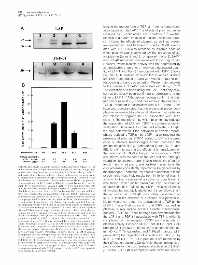
leasing the mature form of TGF- b1 from its noncovalentassociation with its LAP.4 The effects of plasmin can beinhibited by a2-antiplasmin and aprotinin.12,23 a2-Anti-plasmin is a natural inhibitor of plasmin, whereas aproti-nin inhibits the effects of plasmin as well as trypsin,a-chymotrypsin, and kallikrein.23 The L-TGF-b1 associ-ated with TSP-1 is also released by plasmin becausewhen plasmin was inactivated by the presence of a2-antiplasmin (lanes 2 and 3) or aprotinin (lane 3), LAP-1and TGF-b1 remained complexed with TSP-1 (Figure 5A).However, when plasmin activity was not neutralized bya2-antiplasmin or aprotinin, there was a decreased quan-tity of LAP-1 and TGF-b1 associated with TSP-1 (Figure5A, lane 1). In addition we found that in lanes 1–4 usinganti-LAP-1 antibodies a band was visible at 100 kd cor-responding to bands observed on Western blot analysisto the presence of LAP-1 associated with TGF-b1.24–26
The detection of a band using anti-LAP-1 antibody at 68kd has previously been confirmed to correspond to thedimer of LAP-1.22 Although our findings confirm that plas-min can release TGF-b1 and thus diminish the quantity ofTGF-b1 detected in association with TSP-1 (lane 1), wehave also demonstrated that the prolonged presence ofplasmin in overnight cultures of alveolar macrophagescan release or degrade the LAP associated with TSP-1(lane 1). The mechanism by which plasmin may regulatethe association of LAP with TSP-1 is currently under in-vestigation. Because TSP-1 can itself activate L-TGF-b1,we next determined if the activation of alveolar macro-phage derived L-TGF-b1 by sTSP-1 also required thepresence of plasmin. sTSP-1 added to CM in the pres-ence of alveolar macrophages further increased theamount of active TGF-b1 generated (Figures 1C, 2C, and5B). It is of interest that the effects of a2-antiplasmin onthe reduction of TGF-b activity in the presence of TSP-1(not shown) was the same as that of aprotinin. Although,in addition to plasmin, aprotinin also inhibits the effects oftrypsin, a-chymotrypsin, and kallikrein, plasmin is theonly protease consistently reported to be generated bymacrophages. Therefore, the effects of aprotinin in theseexperiments most likely results from inhibition of plasminactivity. In the presence of aprotinin or a2-antiplasmin(not shown), which inhibit plasmin activity, the inductionof activation of L-TGF-b1 by sTSP-1 was significantlydiminished but not totally abolished. It then follows that ifthe activation of L-TGF-b1 was totally dependent onsTSP-1, then the absence or presence of a plasmin in-hibitor would not affect the activation of L-TGF-b1 bysTSP-1. These findings confirm that TSP-1, as well asplasmin, is required to activate alveolar macrophage-derived L-TGF- b1. These findings also demonstrate thatthe LAP-1 and TGF-b1 associated with TSP-1, which iscomplexed with its receptor, CD36, are the targets forplasmin activity. Because sTSP-1, anti-TSP-1, and CD36peptide 93–110 have no effect on the generation of plas-min (D Xu, T Yehualaeshet, and N Khalil, manuscript inpreparation) the regulation of activation of L-TGF-b1 bysTSP-1, anti-TSP-1, or CD36 peptide is not mediated bytheir effects on plasmin. Collectively, these findings sup-port a model for the posttranslational activation of L-TGF-b1 where L-TGF- b1 is complexed with TSP-1 and held at
Figure 5. The effects of plasmin inhibitors on the expression of LAP, TGF-b1and activation of L-TGF-b1. Immunoblot analysis of LAP and TGF-b1 anti-gens obtained from immunoprecipitates using anti-TSP-1 antibody (mAb133)and lysates of alveolar macrophages cultured in the absence or presence ofa2-antiplasmin or aprotinin. A, top: Alveolar macrophages obtained 7 daysafter bleomycin administration cultured in the absence (lane 1) or presenceof a2-antiplasmin at 1024 units/ml (lane 2), a2-antiplasmin 1021 unit/ml(lane 3), or aprotinin 0.01 mg/mm l (lane 4) were immunoblotted withanti-LAP antibodies and demonstrate protein bands compatible with L-TGF-b(100 kd) and the dimer (68 kd) of LAP. A, bottom: The same nitrocellulosefilter was reprobed for immunoblotting with anti-TGF-b1 antibodies anddemonstrates the dimer (25 kd) of TGF-b1. The culture conditions of alveolarmacrophages used for lanes 1–4 are described above. The immunoblots arerepresentative of experiments done 3 times. The numbers on the left denotethe molecular weight of the bands in kilodaltons. B: Effects of aprotinin onthe activation of L-TGF-b1 in the presence of sTSP-1. Alveolar macrophagesobtained 7 days after bleomycin administration were cultured in the absenceor presence of 0.01 mg/mm l/106 macrophages of aprotinin and/or in theabsence or presence of 0.4 mg/ml/106 macrophages of sTSP-1. The TGF-b1present in neutral and acidified then neutralized CM was quantitated and thepercentage of active TGF-b1 was calculated. Each point is the mean of 3 to7 rats. Percentage of active TGF-b1 in CM of alveolar macrophages fromBLM-treated rats compared to normal saline N/S-treated rats or the CM ofalveolar macrophages obtained after BLM treatment cultured with aprotininhave a P value #0.0001. Percentage of active TGF-b1 in CM of alveolarmacrophages cultured in the presence of sTSP-1 compared to when no TSP-1or aprotinin were present has a P value #0.0004. Percentage of activeTGF-b1 in CM of alveolar macrophages obtained after BLM treatment and noin vitro treatment, compared to when sTSP-1 and aprotinin was also present,has a P value #0.0976. Percentage of active TGF-b1 in CM of alveolarmacrophages cultured with sTSP-1 compared to when sTSP-1 and aprotininwere present has a P value ,0.0001.
848 Yehualaeshet et alAJP September 1999, Vol. 155, No. 3
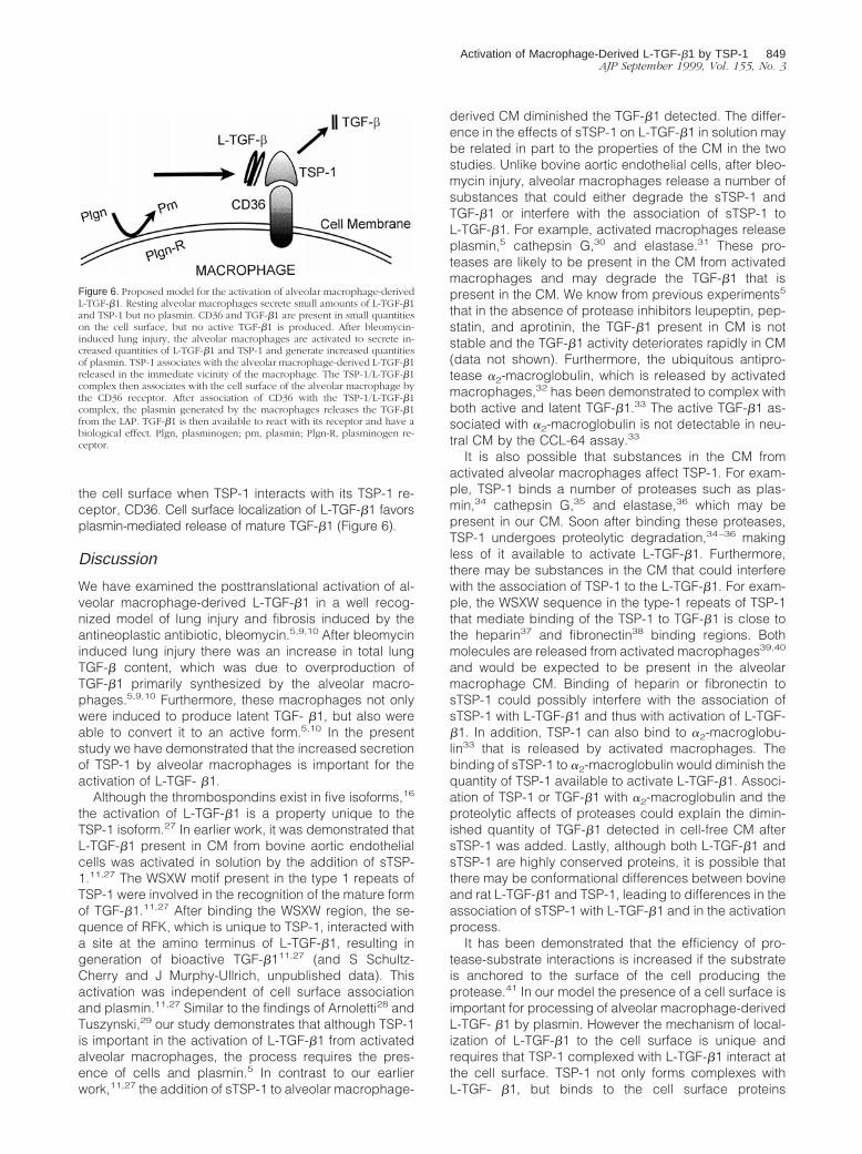
the cell surface when TSP-1 interacts with its TSP-1 re-ceptor, CD36. Cell surface localization of L-TGF-b1 favorsplasmin-mediated release of mature TGF-b1 (Figure 6).
Discussion
We have examined the posttranslational activation of al-veolar macrophage-derived L-TGF-b1 in a well recog-nized model of lung injury and fibrosis induced by theantineoplastic antibiotic, bleomycin.5,9,10 After bleomycininduced lung injury there was an increase in total lungTGF-b content, which was due to overproduction ofTGF-b1 primarily synthesized by the alveolar macro-phages.5,9,10 Furthermore, these macrophages not onlywere induced to produce latent TGF- b1, but also wereable to convert it to an active form.5,10 In the presentstudy we have demonstrated that the increased secretionof TSP-1 by alveolar macrophages is important for theactivation of L-TGF- b1.
Although the thrombospondins exist in five isoforms,16
the activation of L-TGF-b1 is a property unique to theTSP-1 isoform.27 In earlier work, it was demonstrated thatL-TGF-b1 present in CM from bovine aortic endothelialcells was activated in solution by the addition of sTSP-1.11,27 The WSXW motif present in the type 1 repeats ofTSP-1 were involved in the recognition of the mature formof TGF-b1.11,27 After binding the WSXW region, the se-quence of RFK, which is unique to TSP-1, interacted witha site at the amino terminus of L-TGF-b1, resulting ingeneration of bioactive TGF-b111,27 (and S Schultz-Cherry and J Murphy-Ullrich, unpublished data). Thisactivation was independent of cell surface associationand plasmin.11,27 Similar to the findings of Arnoletti28 andTuszynski,29 our study demonstrates that although TSP-1is important in the activation of L-TGF-b1 from activatedalveolar macrophages, the process requires the pres-ence of cells and plasmin.5 In contrast to our earlierwork,11,27 the addition of sTSP-1 to alveolar macrophage-
derived CM diminished the TGF-b1 detected. The differ-ence in the effects of sTSP-1 on L-TGF-b1 in solution maybe related in part to the properties of the CM in the twostudies. Unlike bovine aortic endothelial cells, after bleo-mycin injury, alveolar macrophages release a number ofsubstances that could either degrade the sTSP-1 andTGF-b1 or interfere with the association of sTSP-1 toL-TGF-b1. For example, activated macrophages releaseplasmin,5 cathepsin G,30 and elastase.31 These pro-teases are likely to be present in the CM from activatedmacrophages and may degrade the TGF-b1 that ispresent in the CM. We know from previous experiments5
that in the absence of protease inhibitors leupeptin, pep-statin, and aprotinin, the TGF-b1 present in CM is notstable and the TGF-b1 activity deteriorates rapidly in CM(data not shown). Furthermore, the ubiquitous antipro-tease a2-macroglobulin, which is released by activatedmacrophages,32 has been demonstrated to complex withboth active and latent TGF-b1.33 The active TGF-b1 as-sociated with a2-macroglobulin is not detectable in neu-tral CM by the CCL-64 assay.33
It is also possible that substances in the CM fromactivated alveolar macrophages affect TSP-1. For exam-ple, TSP-1 binds a number of proteases such as plas-min,34 cathepsin G,35 and elastase,36 which may bepresent in our CM. Soon after binding these proteases,TSP-1 undergoes proteolytic degradation,34–36 makingless of it available to activate L-TGF-b1. Furthermore,there may be substances in the CM that could interferewith the association of TSP-1 to the L-TGF-b1. For exam-ple, the WSXW sequence in the type-1 repeats of TSP-1that mediate binding of the TSP-1 to TGF-b1 is close tothe heparin37 and fibronectin38 binding regions. Bothmolecules are released from activated macrophages39,40
and would be expected to be present in the alveolarmacrophage CM. Binding of heparin or fibronectin tosTSP-1 could possibly interfere with the association ofsTSP-1 with L-TGF-b1 and thus with activation of L-TGF-b1. In addition, TSP-1 can also bind to a2-macroglobu-lin33 that is released by activated macrophages. Thebinding of sTSP-1 to a2-macroglobulin would diminish thequantity of TSP-1 available to activate L-TGF-b1. Associ-ation of TSP-1 or TGF-b1 with a2-macroglobulin and theproteolytic affects of proteases could explain the dimin-ished quantity of TGF-b1 detected in cell-free CM aftersTSP-1 was added. Lastly, although both L-TGF-b1 andsTSP-1 are highly conserved proteins, it is possible thatthere may be conformational differences between bovineand rat L-TGF-b1 and TSP-1, leading to differences in theassociation of sTSP-1 with L-TGF-b1 and in the activationprocess.
It has been demonstrated that the efficiency of pro-tease-substrate interactions is increased if the substrateis anchored to the surface of the cell producing theprotease.41 In our model the presence of a cell surface isimportant for processing of alveolar macrophage-derivedL-TGF- b1 by plasmin. However the mechanism of local-ization of L-TGF-b1 to the cell surface is unique andrequires that TSP-1 complexed with L-TGF-b1 interact atthe cell surface. TSP-1 not only forms complexes withL-TGF- b1, but binds to the cell surface proteins
Figure 6. Proposed model for the activation of alveolar macrophage-derivedL-TGF-b1. Resting alveolar macrophages secrete small amounts of L-TGF-b1and TSP-1 but no plasmin. CD36 and TGF-b1 are present in small quantitieson the cell surface, but no active TGF-b1 is produced. After bleomycin-induced lung injury, the alveolar macrophages are activated to secrete in-creased quantities of L-TGF-b1 and TSP-1 and generate increased quantitiesof plasmin. TSP-1 associates with the alveolar macrophage-derived L-TGF-b1released in the immediate vicinity of the macrophage. The TSP-1/L-TGF-b1complex then associates with the cell surface of the alveolar macrophage bythe CD36 receptor. After association of CD36 with the TSP-1/L-TGF-b1complex, the plasmin generated by the macrophages releases the TGF-b1from the LAP. TGF-b1 is then available to react with its receptor and have abiological effect. Plgn, plasminogen; pm, plasmin; Plgn-R, plasminogen re-ceptor.
Activation of Macrophage-Derived L-TGF-b1 by TSP-1 849AJP September 1999, Vol. 155, No. 3
CD3618,19 and CD51.42 Unlike CD51, CD36, which is an88-kd membrane glycoprotein, is consistently found onthe surfaces of monocytes and macrophages.18,19 Noprevious report has demonstrated that CD36 binds TGF-b1, but CD36 expression functions as a receptor forTSP-1,43 collagen,43 and a ligand exposed on the surfaceof erythrocytes infected with the parasite Plasmodiumfalciparum.43 The CD36-TSP-1 interaction has been re-ported to be involved in platelet-monocyte adhesion,44
platelet-tumor-cell adhesion,45 platelet aggregation,46
and macrophage uptake of apoptotic cells.47 The find-ings in this study demonstrate for the first time that theCD36-TSP-1 interaction is important in the activation ofalveolar macrophage-derived L-TGF- b1. This is becausethe presence of an antibody to CD36 in cultures of acti-vated alveolar macrophages diminishes TGF-b1 localiza-tion to the cell surface and totally abrogates the activationof L-TGF-b1. Because CD36 has not been reported tobind L-TGF- b1, the colocalization of TGF-b1 with CD36must occur when TSP-1 that is complexed with L-TGF- b1interacts with its receptor, CD36. There was further con-firmation that the interaction of TSP-1 to CD36 is impor-tant for activation of L-TGF-b1, in that the presence of theCD36 antibody in cultures of alveolar macrophages in-cubated with sTSP-1 resulted in inhibition of the en-hanced activation of L-TGF- b1 observed when sTSP-1alone was added to alveolar macrophages.
It is of interest that a peptide mimicking the region ofCD36 between amino acids 93 and 110, previously de-scribed as critical for the interaction of TSP-1 withCD36,14 also diminished the generation of active TGF-b1in vitro. In addition, when the CD36 peptide 93–110 isadministered to rats concomitantly with bleomycin, alve-olar macrophages do not generate active TGF-b1,48 andthere is less inflammation and fibrosis in the lungs48
(T Yehualaeshet, R O’Connor, A Begleiter, J Murphy-Ullrich, R Silverstein, N Khalil, manuscript in preparation).These observations suggest that the interaction of TSP-1with L-TGF-b1 is important for activation of alveolar mac-rophage-derived L-TGF- b1 in vivo and may subsequentlyregulate pulmonary inflammation and fibrosis. Eventhough the interaction of TSP-1 and CD36 is necessaryfor the activation of L-TGF-b1, the plasmin generated bythe same macrophages is also important for activation ofL-TGF-b1, because plasmin releases LAP-1 and TGF-b1complexed with TSP-1. Although TSP-1 can itself acti-vate L-TGF-b1, our findings suggest that after a pulmo-nary injury, activated alveolar macrophages generateTSP-1 that functions to localize L-TGF- b1 to their cellsurfaces. Localization of L-TGF-b1 to the alveolar macro-phages’ cell surface must lead to optimization of condi-tions that favor activation of L-TGF-b1 by plasmin that isreleased into the vicinity of the alveolar macrophages.5
Furthermore, because plasmin can also be bound to thecell surface,41 the association of L-TGF-b1 to the cellsurface would lead to localizing L-TGF-b1 in close prox-imity to the cell surface-bound plasmin and thus facilitatethe release of L-TGF-b1 by the actions of plasmin.
Several types of cells in the lung, such as endothelialcells,49 epithelial cells,50 and fibroblasts,51 can besources of TGF-b after bleomycin injury. It is of interest
that recently Munger et al52 described a protease- andTSP-1-independent activation of L-TGF-b1 in bleomycin-induced lung injury in mice.52 Their findings suggest thatthe arginine-glycine-aspartic acid sequences in the LAP-1of TGF-b1 associates with the integrin avb6 on alveolarepithelial cells. Munger et al have proposed a model ofactivation of L-TGF-b1 wherein the interaction ofL-TGF-b1 with avb6 is followed by the association ofavb6 with the actin cytoskeleton, leading to a conforma-tional change in L-TGF-b1 attached to the cell surface.The conformational change in L-TGF-b1 leads to themature TGF-b1 interacting with its receptor, TGF-b re-ceptor type II.52 It is not known if bleomycin administra-tion to rats, a different species of rodent, regulates avb6expression on alveolar epithelial cells, nor whether avb6is important in the activation of L-TGF-b1 in our model.Nonetheless, because the presence of active TGF-b1parallels the inflammatory changes seen in this mod-el,5,9,10 our findings suggest that alveolar macrophagesmay be an additional source of active TGF-b1. In conclu-sion, our findings demonstrate that plasmin5 and TSP-1,which are diminished early in the bleomycin injury re-sponse, may result in terminating the activation of alveo-lar macrophage-derived L-TGF- b1, as well as its inflam-matory and fibrotic effects. This then suggests that theregulation of inflammation that is mediated by TGF-b1 isdependent on the posttranslational activation of alveolarmacrophage-derived L-TGF-b1 by both plasmin andTSP-1.
Acknowledgments
We thank Arnold H. Greenberg for many helpful discus-sions and review of the manuscript; Carol Whitman, An-gela Kemp, and Antonio Pallero for technical assistance;Dr. Bob Tate for the statistical analysis of the data; andArlene Klassen and Heather Buhler for typing the manu-script.
References
1. Rappolee DA, Werb Z: Macrophage derived growth factors. CurrTopics Microbiol Immumol 1992, 181:87–140
2. Stein M, Keshav A: The versatility of macrophages. Clin Exp Allergy1992, 22:19–27
3. Wahl SM: Transforming growth factor b: the good, the bad and theugly. J Exp Med 1994, 108:1587–1590
4. Gleizes PE, Munger JS, Nunes I, Harpel JG, Mazzieri R, Noguera S,Rifkin DB: TGF-b latency: biological significance and mechanism ofactivation. Stem Cells 1997, 15:190–197
5. Khalil N, Corne S, Whitman C, Yacyshyn H: Plasmin regulates theactivation of cell associated latent TGF-b1 secreted by rat alveolarmacrophages after in vivo bleomycin injury. Am J Resp Mol Cell Biol1996, 15:252–259
6. Schultz-Cherry S, Murphy-Ullrich JE: Thrombospondin causes acti-vation of latent transforming growth factor-b secreted by endothelialcells by a novel mechanism. J Cell Biol 1993, 122:923–932
7. Murphy-Ullrich JC, Schultz-Cherry S, Hook M: Transforming growth fac-tor-b complexes with thrombospondin. Mol Biol Cell 1992, 3:181–188
8. Silverstein RL, La Salla J, Pearce SE: CD36 cluster report. LeucocyteTyping V: White Cell Differentiation Antigens. Edited by SchlossmanSF, Boumsell L, Gilks W, Harlan JM, Kishimoto T, Morimoto C, Ritz O,Silverstein RL, Springer TA, Tedder TF, and Todd RF. Oxford, OxfordUniversity Press, 1995, 1271:1274–1275
850 Yehualaeshet et alAJP September 1999, Vol. 155, No. 3

9. Khalil N, Bereznay O, Sporn MB, Greenberg AH: Macrophage pro-duction of transforming growth factor-b and fibroblast collagen synthesisin chronic pulmonary inflammation. J Exp Med 1989, 170:727–737
10. Khalil N, Whitman C, Zuo L, Danielpour D, Greenberg AH: Regulationof alveolar macrophage transforming growth factor-b secretion bycorticosteroids in bleomycin-induced pulmonary inflammation in therat. J Clin Invest 1993, 92:1812–1818
11. Schultz-Cherry S, Lawler J, Murphy-Ullrich JE: The type-1 repeats ofthrombospondin-1 activates latent transforming growth factor-b.J Biol Chem 1994, 269:26783–26788
12. Witman B: On the reaction of plasmin or plasmin-streptokinase com-plex with aprotinin or a2-antiplasmin. Thromb Res 1980, 17:143–152
13. Danielpour D, Hart LL, Flanders KC, Roberts AB, Sporn MB: Immu-nodetection and quantitation of the two forms of transforming growthfactor-b (TGF-b1 and TGF-b2) secreted by cells in culture. J CellPhysiol 1989, 138:78–86
14. Leung LL, Wei-Xing L, McGregor JL, Albrecht G, Howard RJ: CD36peptides enhance or inhibit CD36 thrombospondin binding: a two-step process of ligand receptor interaction. J Biol Chem 1992, 267:18244–18250
15. Mai S: Overexpression of C-myc precedes amplification of geneencoding dihydrofolate reductase. Gene 1994, 148:253–260
16. Bornstein P: Thrombospondins: structure and regulation of expres-sion. FASEB J 1992, 6:3290–3299
17. Yee JA, Yan L, Dominguez JC, Allan EH, Martin TJ: Plasminogen-dependent activation of latent transforming growth factor b (TGF-b)by growing cultures of osteoblast-like cells. J Cell Physiol 1993,157:528–534
18. Huh YH, Pearch SF, Yesner LM, Schindler JL, Silverstein RL: Regu-lating expression of CD36 during monocyte-to-macrophagedifferentiation: potential role of CD36 in foam cell formation. Blood1996, 87:2020–2028
19. Yesner LM, Huh HY, Pearch SF, Silverstein RL: Regulation of mono-cyte CD36 and thrombospondin-1 expression by soluble mediators.Arterioscler Thromb Vasc Biol 1996, 16:1019–1025
20. Trezzini C, Jungi TW, Spycher MD, Maly FE, Rao P: Human mono-cytes CD36 and CD16 are signalling molecules: evidence from stud-ies using antibody-induced chemiluminescence as a tool to probesignal transduction. Immunology 1990, 71:29–37
21. Huang MM, Bolen JB, Barnewell JW, Shattil SJ, Brugge JS: Mem-brance glycoprotein IV (CD36) is physically associated with Fyn, Lynand Yes protein-tyrosine kinases in human platelets. Proc Natl AcadSci USA 1991, 88:7844–7848
22. Yang Y, Dignam JD, Gentry LE: Role of carbohydrate structures in thebinding of b1-latency-associated peptide to ligands. Biochem 1997,36:11923–11932
23. Huber D, Philipp J, Fontana A: Protease Inhibitors interfere with thetransforming growth factor-b-dependent but not the transforminggrowth factor-b-independent pathway of tumor cell-mediated immu-nosuppression. J Immunol 1992, 148:277–284
24. Olofsson A, Miyazono K, Kanzaki T, Coloselti P, Engstrom U, HeldinC-H: Transforming growth factor-b1, -b2, and -b3 secreted by ahuman glioblastoma cell line: identification of small and differentforms of large latent complexes. J Biol Chem 1992, 267:1948–1958
25. Mcmahon GA, Dignam JD, Gentry LE: Structural characterization ofthe latent complex between transforming growth factor b1 and b1-latency-associated peptide. Biochem J 1996, 313:343–351
26. Okada F, Yamaguchi K, Ichihara A, Nakamura T: Purification andstructural analysis of the latent form of transforming growth factor-bfrom rat platelets. J Biochem 1989, 106:304–310
27. Schultz-Cherry S, Chen H, Mosher DF, Misenheimer, TM, KrutzschHC, Roberts DD, Murphy-Ullrich JE: Regulation of transforminggrowth factor-b activation by discrete sequences of throm-bospondin-1. J Biol Chem 1995, 270:7304–7310
28. Arnoletti JP, Albo D, Granick MS, Solomon MP, Rothman NL, Tuszyn-ski AB: Thrombospondin and transforming growth factor b-1 increaseexpression of urokinase-type plasminogen activator and plasmino-gen activator inhibitor 1 in human MDA-MB-231 breast cancer cells.Cancer 1995, 76:998–1005
29. Tuszynski GP, Nicosia RF: The role of thrombospondin-1 in tumorprogression and angiogenesis. BioEssays 1996, 18:71–75
30. Welgus, HG, Connolly NL, Senior RM: 12-0-Tetradecanoyl-phorbol-13-acetate-differentiated U937 cells express a macrophage-like pro-file of neutral proteinases: high levels of secreted collagenase and
collagenase inhibitors accompany low levels of intracellular elastaseand cathepsin-G. J Clin Invest 1986, 77:1675–1681
31. Valentine R, Fisher GL: Characteristics of bovine alveolar macro-phage elastase. J Leukocyte Biol 1984, 35:449–457
32. Bonner JC, Hoffman M, Brody AR: Alpha2-macroglobulin secreted byalveolar macrophages serves as a binding protein for a macrophage-derived homologue of platelet-derived growth factor. Am J RespirCell Mol Biol 1989, 1:171–179
33. Souchelnitskiy S, Chambaz EM, Feige JJ: Thrombospondins selec-tively active one of the two latent forms of transforming growth fac-tor-b present in adrenocortical cell-conditioned medium. Endocrine1995, 136:5118–5126
34. Anonick PK, Yoo JK, Webb, DJ, Gonias SL: Characterization of theantiplasmin activity of human thrombospondin-1 in solution. BiochemJ 1993, 289:903–909
35. Rabhi-Sabile S, Pidard D, Lawler J, Renesto P, Chignard M, LegrandC: Proeolysis of thrombospondin during cathepsin-G induced plateletaggregation: functional role of the 165-kd carboxy-terminal fragment.FEBS Lett 1996, 386:82–88
36. Hogg PJ, Owensby DA, Mosher DF, Misenheimer TM, ChestermanCN: Thrombospondin is a tight-binding, competitive inhibitor of neu-trophil elastase. J Biol Chem 1993, 268:7139–7146
37. Murphy-Ullrich JE, Giurusiddappa S, Franzier WA, Hook M: Heparin-binding peptides from thrombospondins 1 and 2 contain focal adhe-sion-labilizing activity. J Biol Chem 1993, 268:26784–26789
38. Sipes JM, Giuo N, Negre E, Yogel T, Krutzsch HC, Roberts DD:Inhibition of fibronectin binding and fibronectin-mediated cell adhe-sion to collagen by a peptide from the second type I repeats ofthrombospondin. J Cell Biol 1993, 121:469–477
39. Kolset SO, Larsen T: Sulphur-containing macromolecules in culturedmonocyte-like cells. Acta Histochem 1988, 84:67–75
40. Driscoll KE, Maurer JK, Higgins J, Poynter J: Alveolar macrophagecytokine and growth factor production in a rat model of crocidolite-induced pulmonary inflammation and fibrosis. J Toxicol EnvironHealth 1995, 46:155–169
41. Vassalli, JD, Wohlwend A, Belin D: Urokinase-catalyzed plasminogen acti-vation at the monocyte/macrophage cell surface: a localized and regulatedproteolylic system. Curr Topics Microbiol Immunol 1992, 181:65–86
42. Clezardin P, Frappart L, Clerget M, Pechoun C, Delmas PD: Expres-sion of thrombospondin (TSP-1) and its receptors (CD36 and 51) innormal, hyperplastic and neoplastic human breast. Cancer Res 1993,53:1421–1430
43. Daviet L, Mcgregor JT: Vascular biology of CD36: role of this newadhesion molecule family in different disease states. Thromb Hae-most 1997, 78:65–69
44. Silverstein RL, Asch AS, Nachman RL: GPIV mediates throm-bospondin-dependent platelet-monocyte adhesion. J Clin Invest1989, 84:546–552
45. Silverstein RL, Baird M, Lo SK, Yesner LM: Sense and antisensecDNA transfection of CD36 (glycoprotein IV) in melanoma cells. J BiolChem 1992, 267:16607–16612
46. Leung, LL: Role of thrombospondin in platelet aggregation. J ClinInvest 1984, 74:1764–1772
47. Ren Y, Silverstein RL, Allen J, Savill J: CD36 gene transfer conferscapacity for phagocytosis of cells undergoing apoptosis: J Exp Med1995, 181:1857–1862
48. Yehaulaeshet T, Douglas D, O’Connor R, Khalil N: In vitro and in vivoinhibition of activation of alveolar macrophage-derived latent trans-forming growth factor-b 1 (TGF-b1) after bleomycin lung injury. Am JRespir Crit Care Med 1998, 157:A265 (abstr.)
49. Phan SH, Gharaee-Kermani M, Wolber F, Ryan US: Stimulation of ratendothelial cell transforming growth factor-b production by bleomy-cin. J Clin Invest 1991, 87:148–154
50. Khalil N, O’Connor R, Flanders K, Shing W, Whitman C: Autocrineregulation of type-2 epithelial cell proliferation by transforming growthfactor-b during bleomycin induced lung injury in the rat. Am J Physiol1994, 77:L498–507
51. Cutroneo K, Breen E, Absher M, Phan S: Bleomycin regulation oftransforming growth factor-b mRNA in rat lung fibroblasts and sub-populations. Chest 1991, 99:65 (suppl)
52. Munger J, Huang X, Kawakatsu H, Griffiths MJD, Datton SL, Wu J,Pittet J-F, Kaminski N, Garat C, Mathay MA, Rifkin DB, Sheppard D:The integrin avb6 binds and activates TGF-b1: a mechanism forregulating pulmonary inflammation and fibrosis. Cell 1999, 96:319–328
Activation of Macrophage-Derived L-TGF-b1 by TSP-1 851AJP September 1999, Vol. 155, No. 3