Embed Size (px)
DESCRIPTION
細胞接着の概要
Citation preview

Crafoord days
The Crafoord Prize
in Polyarthrit is 2004
20-22 Sept 2004
Œ
P r o g r a m m e – A b s t r a c t s – T h e C r a f o o r d l e c t u r e s

Crafoord days20-22 Sept 2004
P r o g r a m m e – A b s t r a c t s – T h e C r a f o o r d l e c t u r e s
Cell Migration in Health and Disease

C R A F O O R D S Y M P O S I U M
Cell Migration in Health and Disease
Monday 20 September in Lund, Tuesday 21 September in Stockholm
09.00 Opening of the Symposium
Gunnar Öquist, Secretary General, Royal Swedish Academy of Sciences
09.10 Integrin Cell Adhesion Molecules in Health and Disease page 12
Timothy A. Springer, Crafoord Laureate 2004 10.00 Coffee break
10.30 Control of Leukocyte Traffic page 20
Sirpa Jalkanen, University of Turku, Finland
11.15 Control of Interstitial Fluid Pressure and Acute Edema page 21 – Role of Integrins
Kristofer Rubin, University of Uppsala, Uppsala
12.00-13.00 Lunch
13.00-14.00 Poster session
14.00 Regulation of Human Neutrophil Apoptosis page 22
Tommy Andersson, Malmö University Hospital, Malmö
14.45 Coffee break
15.15 Cell Adhesion and Migration in Tumor Progression page 23
Richard O. Hynes, MIT, Cambridge, USA
16.00-16.45 Towards an Understanding of Leukocyte page 6 Trafficking in Physiology and Disease
Eugene C. Butcher, Crafoord Laureate 2004
3
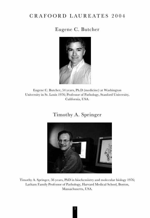
C R A F O O R D L A U R E A T E S 2 0 0 4
Eugene C. Butcher
Eugene C. Butcher, 54 years, Ph.D (medicine) at Washington University in St. Louis 1976; Professor of Pathology, Stanford University,
California, USA.
Timothy A. Springer
Timothy A. Springer, 56 years, PhD in biochemistry and molecular biology 1976; Latham Family Professor of Pathology, Harvard Medical School, Boston,
Massachusetts, USA.

4
Inflammation and Immunity
A short introduction to the 2004 Crafoord Prize
Eugene Butcher and Timothy Springer have elucidated the function of cell adhesion molecules that are expressed in white blood cells and direct their exodus across the blood vessel wall into tissue where they are crucial for the defence against disease.
Cell adhesion molecules (CAMs) are cell surface proteins expressed by many cells including white blood cells. The CAMs are activated by specific signals from peripheral tissues in response to injury or infection and their effector functions are important for processes of inflammation and immunity.
Examples of CAMs are selectins and integrins. The selectins are glycoproteins, consisting of a single chain. They enable the blood cells to bind to activated en-dothelium in the blood vessel wall. As a consequence, the blood cell can ”roll” along the surface of the vessel and eventually adhere more firmly. The integrins consist of two sub-units – an alpha and a beta chain – and function as receptors on the white blood cells. The activity of the various molecules eventually allow the now flattened blood cell to migrate between the cells in the vessel wall to the site of the disease process (Figure 1)
Selectins
Chemoattractants
Diseased tissue
Blood vessel
SelectinIntegrin
Integrins
Figure 1. Selectins in the cellmembranes of white blood cells allow them to adhere to the blood vessel wall in the area around an injurious process. Aided by the integrins they leave the blood vessel and migrate to the damaged area where they act to eliminate the cause of injury.

5
Eugene Butcher has identified the selectins and their interaction with integrins that make them come to a halt, from high velocities along a stretch of some mil-limetres. Butcher has characterised the components of the process, including the ligands and receptors forming the bonds, and has formulated the now classical mul-ti-step model to describe it.
Timothy Springer demonstrated the crucial role of the CAMs in cellular immu-nity. The integrins can rapidly increase in number when a blood cell has identified an antigen, and improve the defence. Springer showed that the integrins constitute a molecular family all consisting of an alpha chain and a beta chain. These exist in various configurations that can combine into a large number of defined variants with different specificities against highly variable targets. In recent years Spring-er has worked to determine the structures at high resolution and to define the functional domains. Like Butcher, he has produced multi-step models to reconcile the structure-function relationships. In recent years he has studied the molecular mechanisms that open and lock integrin in various conformations. If a disulphide binding (sulphur-based) is introduced, an integrin can be locked in an open posi-tion and its function is lost. The flow of white blood cells in inflammation can then be controlled.
New treatment strategies for inflammatory diseaseBoth Butcher and Springer have made successful attempts to apply their findings to the treatment of medical illness. In some cases advanced trials are in progress.
Butcher has shown that treatment with antibodies to the protein LFA-1 pre-vented or cured cerebral malaria in mice. The VCAM-1 protein plays an impor-tant part in the slow but progressive disease multiple sclerosis (MS), and Butcher’s group have showed how antibodies to this protein has stopped the development of disease in some trials.
Springer’s findings have inspired numerous companies working on new cures against rheumatism, psoriasis, asthma, intestinal disorders, haematological disor-ders and AIDS.

6
T H E C R A F O O R D L E C T U R E S 2 0 0 4
Towards an Understanding of Leukocyte Trafficking in Physiology and Disease
Eugene C. Butcher, Stanford University, Stanford, California, USA
Your Majesties, President of the Academy, members of the Crafoord family, mem-bers of the Academy, ladies and gentlemen, it is a great honor to be awarded the Crafoord Prize by the Royal Swedish Academy of Sciences, and I am pleased to be sharing this prize today with my co-laureate, Timothy Springer.
In accepting this honor, I thought I would share with you a personal perspective on the path that led to our studies of leukocyte trafficking and its role in the im-mune system. I wanted to be a scientist from an early age, and I was (and am) intrigued by everything from fundamental physics to the fundamental questions in biology. My brother, who is here to share this day with me, knew he was to be an astronomer since the age of 9. I on the other hand was much less focused, and found I couldn’t chose among the many fascinating mysteries of Nature until much later. By college, though, I had decided that biology was for me. I was attracted I think by the inherent wonder of life, it was and is still truly amazing to me that such a complex and counterintuitive entity as a human can exist, with eyes, ears and brain having all evolved and developed in parallel, and all actually working, at least most of the time. I went on to Medical School, largely to delay deciding what specific question to work on (or, putting it in a more positive light, to give me a better per-spective from which to chose an “Important Question”).
The question I became fascinated with was how do cells recognize each other? And how do they know where to go? During processes such as embryogenesis, neu-rogenesis, and wound healing cells migrate and interact in highly specific and or-chestrated fashion. How do they do this?
When I finished Medical School, the only tools available for defining molecules involved in such events were immunologic, antisera were being used to inhibit cell functions, and to identify functional proteins so I decided I had to join an immuno-logy laboratory. When I went to train in pathology at Stanford University, however, the well-known immunology groups were all full. This turned out to be a stroke of luck, however, because I ended up with the recently tenured Irv Weissman. Irv’s lab was an extraordinary environment for me, providing the combination of freedom and intellectual enthusiasm I needed. I was also very fortunate because, soon after I arrived at Stanford, Judy Woodruff published a seminal paper in JEM, describing

7
(from my perspective) the most thrilling and accessible system ever devised for studying cell-cell recognition. They showed that lymphocytes, rocked in the cold for a mere 15 minutes or so on frozen sections of rat lymph nodes, would recognize and bind with remarkable selectivity to specialized high endothelial venules (HEV), the same vessels that Gowans and Marchesi had shown in the 1960’s recruit lymphocy-tes from the blood. A fantastically simple yet, as it turned out, powerful technique for studying the cellular and molecular mechanisms of leukocyte trafficking.
One of the most exciting discoveries from our early studies was that lymphocytes can tell the difference between blood vessels in different organs in the body, sugges-ting a way that immune cells can be targeted to specific tissues (Figure 1a, b). It ap-peared that differences in blood vessels themselves could explain lymphocyte traf-ficking behaviours that had been described by pioneers in field, such as the ability of some lymphocytes to selectively recirculate through the intestines or through the skin, or to home preferentially to the gut wall. Later studies revealed recognition systems for joint, BALT and brain and skin endothelium. Using Occam’s razor, we
Lymphocytes homing in a high endothelial venule (HEV) in vivo
Tissue selective lymphocyte-endothelial recognition:Lymphocytes can distinguish between HEV in different sites. Here a clonal lymphoma cell line adheres to HEV in sections of lymph nodes but not Peyer’s patches
Peripheral lymph nodes
Endothelialcells
Lymphocytes
Intestinal Peyer’s patch
Lymphocytes
HEV
Endothelial cells
Lymphocytes
Homing receptors
Tissue selective vascular ligands (addressins)
MAdCAM-1 PNAd (CHO) E-selectin
α4β7 L-selectin CLA
intestinal lymph node skin
Figure 1A
Lymphocytes homing in a high endothelial venule (HEV) in vivo
Tissue selective lymphocyte-endothelial recognition:Lymphocytes can distinguish between HEV in different sites. Here a clonal lymphoma cell line adheres to HEV in sections of lymph nodes but not Peyer’s patches
Peripheral lymph nodes
Endothelialcells
Lymphocytes
Intestinal Peyer’s patch
Lymphocytes
HEV
Figure 1B
Figure 2. The ”lock-and-key” model

8
hypothesized a simple lock-and-key model of lymphocyte-endothelial recognition, in which lymphocytes express homing receptors that can target their migration from the blood by interaction with tissue–selective endothelial “address signals” or vascular addressins (Figure 2).
This model, although wrong, drove a search for the postulated homing receptors. We used the new monoclonal antibody technology to make an antibody to the pu-tative lymph node homing receptor: this molecule, the first identified lymphocyte-endothelial cell adhesion molecule, is now called the L-selectin, or CD62L. In sub-sequent studies, we identified tissue-selective HEV antigens, including a sulfated carbohydrate epitope defining the L-selectin ligands (“sulfoadhesins”) of the Perip-heral Node HEV Addressin, PNAd (which is also expressed by chronically inflamed venules in many sites); and the mucosal addressin cell adhesion molecule MAd-CAM-1, which binds the lymphocyte intestinal homing receptor, an integrin called α47. One antibody we made to human HEV, HECA452, was serendipitously found (by a particularly observant fellow) to cross react with a cutaneous T cell antigen (CLA) expressed on skin homing lymphocytes: this turned out to be an epitope as-sociated with E-selectin-binding carbohydrates. This E-selectin-CLA interaction is a hallmark of T cell recruitment in skin inflammation, for example in psoriasis.
These tissue-selective lymphocyte and vascular adhesion molecules offer a way to control inflammation locally or regionally. Immune responses require the re-cruitment of lymphocytes from the blood, and in the case of pathological immune activity, for example in arthritis, blocking lymphocyte recruitment can suppress the unwanted inflammation. Indeed, antibodies or small molecule inhibitors of several of these lymphocyte adhesion pathways have made their way into animal models, and in some instances clinical trials, for autoimmune diseases including arthritis, diabetes, inflammatory bowel diseases, psoriasis, and asthma. As specific examples, antibodies to α47 are in clinical trials for colitis; and antibodies to sulfoadhesin are in development for several diseases of chronic inflammation.
In spite of its successes, it soon became clear that this early lock-and-key hypo-thesis was simply wrong. The problem was that several lymphocyte homing recep-tors were also expressed by other white cells with very different homing properties, such as monocytes and neutrophils. Antibodies to L-selectin even inhibited neu-trophil recruitment into inflamed skin. Obviously, the real mechanism was more complex.
By this time it was well established by Tim Springer and others that the integrins he had identified, in particular the beta 2 integrin LFA-1, could participate in leu-kocyte binding to activated endothelium; and Alf Hamann had shown that LFA-1 had an accessory role in lymphocyte homing as well. We found that activated neu-trophils rapidly shed L-selectin while upregulating beta 2 integrins, suggesting that

9
the molecules might act sequentially. Perhaps most telling were studies by Karl Arfors, a pioneer in the field of microcirculation, who showed with John Harlan that antibodies to beta 2 integrins prevented neutrophil sticking (stopping on the wall of inflamed vessels), but that the neutrophils still rolled along the endothelium. We naturally hypothesized that L-selectin might be responsible for this rolling and, in a collaboration with Ulrich von Andrian, then in Karls’ lab, we showed that rolling (and subsequent activation-dependent arrest) in a rabbit model were indeed inhi-bited by anti-L-selectin antibodies. This established sequential roles for L-selectin and integrins in a multi-step molecular process of neutrophil adhesion to vessels at sites of inflammation.
At the same time (and unbeknownst to us), Mike Lawrence in Tim’s lab was pursuing elegant in vitro models that demonstrated that the vascular selectins could mediate tethering and rolling of neutrophils on slides, and that this rolling, as in vivo, was essential for activation-dependent integrin-mediated arrest of cells under flow. In fact, Uli von Andrian first presented our results, and Tim theirs, at the same meeting in Cold Spring Harbor in 1991. I stayed home from the meeting to walk the hills and write, because I had become incredibly excited by the idea that this might be a general model for leukocyte-endothelial cell recognition and homing---a model that could explain everything we’d learned about lymphocyte homing and its regulation in physiology and disease.
One problem with generalizing the paradigm was that we had no idea what ac-tivating signals on endothelium could trigger lymphocyte integrins. We knew that chemoattractant receptors, specialized receptors similar to those that we use for taste, sight and smell, were responsible for activating neutrophil integrins for stick-ing and arrest, and we had evidence from our own studies with pertussis toxin that similar receptors might be important for lymphocyte arrest on HEV as well. But no potent chemoattractants for circulating lymphocytes were known. In another stroke of luck, I complained about this to a friend who just happened to know that Genen-tech (Tom Schall, building on his earlier work with Alan Krensky at Stanford) had just identified a novel lymphocyte chemoattractant related to the neutrophil activa-tor IL8, defining as it turns out what is now called the “chemokine family”. This was enough for me to stick my neck out and propose that these might be the missing lymphocyte adhesion-triggers required for a general model. (Although it actually took years before we and others identified chemokines that trigger lymphocyte ar-rest on HEV, ultimately this large chemoattractant family has proven important to the homing of every lymphocyte subset studied, and of monocytes, eosinophils, and indeed all white blood cell types).
The hypothesis I put forth was that, for lymphocytes and other white blood cells as well as neutrophils, endothelial interaction might be an active process involving several sequential but equally important events, rolling, chemokine or chemoatt-

10
ractant-induced activation, rapid activation-dependent integrin-mediated arrest, and potentially a fourth step in which chemoattractants could also regulate migration across the endothelial barrier into the surrounding tissues, completing the process. The idea was that, if several diffe-rent receptors could be used interchangeably at each of these steps (which they can, see Figure 3 and Table 1), it would provide an efficient, combinatorial mechanism for generating diversity and specificity in homing.
This model explains how several different homing
Others?
Others?Chemerin?Cathelicidin?
Specificity and diversity from combinatorial association of adhesion, activating and chemoattractant
In
terc
hang
eabl
e re
cept
or-li
gand
pai
rs
1-tethering and rolling 2-activation 3-arrest
P-selectin-CHO
LFA-1--ICAM-1Mac-1--?ICAMs
E-selectin-CHO *PSGL 1
P-selectin-Sulfoadhesinα4β7---MAdCAM-1
α4β7---MAdCAM-1
α4β1---VCAM-1
α4β1---VCAM-1Lipid mediatorsFormyl peptidesC5a
?----VAP-1 CD44--HA
Chemokines
Chemerin?Cathelicidin?
Lipid mediatorsFormyl peptides
Chemokines
4-diapedesis
(N~30?) (N~30?)
cell target site tethering slow rolling
activation (chemokine or attractant)
arrest (activated integrins)
naïve CD4 Tcells
lymph node L-selectin-PNAd L-selectin CCR7-CCL21 LFA-1-ICAM
CD8 T cells lymph nodeHEV
? - VAP1 ± L-selectin-PNAd ?CCL21 ?LFA-1
naïve CD4 T cells
Peyer'spatch
L-selectin-MAdCAM-1 CHO
a4b7-MAdCAM CCR7-CCL21 a4b7/LFA-1
naïve B cells Peyer'spatch
L-selectin-MAdCAM-1 CHO
a4b7-MAdCAM CXCL13,CXClL12,CCL21
a4b7/LFA-1
skin homingmemory CD4cells
skin CLA-E-selectin a4b1-VCAM-1 CCR4-CCL17CCR10-CCL27
a4b1-VCAM1LFA-1-ICAM1
small intestinalmemory T cells
smallintestines
a4b7-MAdCAM-1 CCR9-CCL25 ?LFA-1
Memory CD4 Tcells
joints(arthritis)_
L-selectin-PNAd?P-selectin
a4b1-VCAM1 ?-VAP1
?CCR7, ?CCR2,?others
ICAM-1?a4b1-VCAM1
IgAplasmablasts
small intestines
a4b7-MAdCAM-1 CCR9-CCL25CCR10-CCL28
?LFA-1
IgAplasmablasts
lung a4b1-VCAM-1 CCR10-CCL28 LFA-1
IgGplasmablasts
systemic (non mucosal)
selectins?, a4b1-VCAM-1 LFA-1?
Eosinophils various E-, P- or L-selectin a4b1/a4b7 eotaxins a4? And beta2integrins
Monocytes various E-, P- or L-selectin a4b1 MCP chemokines,C5a, formylpeptides
a4? And beta2integrins
Neutrophils various E-, P- or L-selectin IL8, formylpeptides, C5a,leukotrienes
beta 2integrins
Figure 3. A general model of leukocyte recruitment as a multistep process
Table 1. Some examples to illustrate combinations of adhesion and activating factors in selective cell trafficking programs

11
events could use a common receptor (e.g. L- selectin is used in lymphocyte, neu-trophil, eosinophil and monocyte recruitment: but specificity can be determined by the use of different chemoattractants to trigger arrest). It also makes it easy to see how new homing specificities can evolve simply by combining pre-existing receptors in new ways. And it implies that leukocyte recruitment can be blocked for thera-peutic ends by inhibiting any of the 3 or 4 steps. This general multistep paradigm, with numerous variations and refinements, has been confirmed in many different models of cell trafficking, by our lab and many others.
From a desire to study a simple cell-cell recognition event, we had been led to the discovery of a truly beautiful, combinatorial mechanism for targeting many and perhaps all cells of the immune system in their migration from the blood. There may be hundreds of different trafficking programs, allowing specific control of im-mune cell localization (and thus of immune responses) as a function of the tissue site, the inflammatory state, the leukocyte type and its differentiation. Certainly, we now know combinations of adhesion and chemoattractant receptors that can ex-plain the localization of naïve lymphocytes and subsets of memory cells and dendri-tic cells to lymph nodes or to Peyer’s patches; of memory T cells for gut antigens to the small intestines, and of cutaneous memory lymphocytes to skin; of IgA and IgG plasmablasts; of monocytes, eosinophils and more. Many of the molecules involved are now being pursued as therapeutic targets to treat inflammatory diseases, inclu-ding multiple sclerosis, colitis, psoriasis, asthma and arthritis.
In addition to therapeutics aimed at blocking homing and chemokine receptors, future efforts may take control of the expression of the receptors, redirecting the immune response by altering cellular positioning and interactions. Redirection of immune cells may allow us to improve the efficiency of immune responses after vaccination; or to shut down autoimmune responses. We may even be able to turn leukocytes into Trojan horses to deliver therapeutics to sites of our choosing in the body.
It has been with great pleasure that I have seen many of our observations serving as the basis, directly or indirectly, for efforts to develop novel therapies for inflam-matory diseases. My grandmother suffered greatly from rheumatoid arthritis, and although my passion will always be for the pure beauty of science, it is also my per-sonal hope to have contributed in some way to reducing the suffering and disability of this terrible disease. I thank the Crafoord family for their generous support of such efforts. And I cannot end without extending my deep appreciation and indeb-tedness to the people who made it possible for me to be here, the many wonderful members of my laboratory who have contributed their ideas and talents over the years, my scientific colleagues and collaborators in academia and industry, and es-pecially my wife and family for supporting me with humor and some bemusement in my obsessive dedication to science.

12
Integrin Cell Adhesion Molecules in Health and Disease
Timothy A. Springer, Dept. of Pathology, Harvard Medical School, Boston, USA
Your Majesties, President of the Academy, members of the Crafoord family, mem-bers of the Academy, ladies and gentlemen, I am humbled to be invited to address you, and express my deepest gratitude to the Royal Swedish Academy of Sciences for the Crafoord Prize.
I am now facing one of the greatest challenges of my life – explaining what I do to a lay audience. I am not too good at doing this in words, so I am going to use movies, and even a modern dance, to bring the cells and molecules I work with to life.
I am trained in biochemistry – the science of the molecules of life. And I also work in immunology, the study of how we develop immunity to bacteria and viruses, and fight infections. I have been lucky to work at Harvard Medical School and CBR Biomedical Research Institute, where opportunities abound to make connections between the molecules of life and the diagnosis and treatment of disease in pa-tients.
I was fortunate to participate in the development of a new research field, cell adhesion molecules of the immune system. The initial stage was discovering or identifying molecules on the surface of white blood cells that allow them to bind, stick, or adhere, to use three different names, to other cells. While most of my col-leagues were looking for molecules that were unique to white cells or lymphocytes, and allowed them to recognize foreignness, I was looking for cell adhesion molecu-les that allowed them to recognize the context of the cell on which the foreignness was displayed.
The cells that compose a multicellular organism must organize themselves into tissues and organs that work together for the good of the whole. This organization and communication requires cell adhesion molecules that sense the context that a cell finds itself in the body, and can control the movement of the cells between diffe-rent niches. While this is now accepted fact, it wasn’t at all accepted when I started working on it, and some people thought that physics and chemistry, without any molecular specificity, might allow cells to come together and sort into organs, much as oil or clay can separate out of water. However, we found that specific proteins were required, and that when these were blocked, killer lymphocytes could not do their jobs in recognizing the foreignness that signifies an infected cell.
Let us look at a movie of a white blood cell killing an infected cell. The white cell moves around, first gingerly touching other cells, and when it recognizes foreign-

13
ness, it grabs hold tightly, and delivers a lethal potion that kills the cell. It can then detach, and go on and kill other cells. With the light microscope, we cannot see any of the molecules on the surface of the white blood cell, but the dynamic movements of its surrounding surface membrane suggests that many protein molecules on the membrane must undergo choreographed movements to enable adhesion to the tar-get cell. There must be dynamic regulation, so that the adhesion molecules can alternately hold onto one cell, then let go, and then hold on to yet another cell.
In the first stage of our work, we identified the molecules that were required for killing, because blocking these molecules with antibody probes prevented the func-tion of killing. We called these lymphocyte function-associated molecules 1, 2, and 3 (LFA-1, LFA-2, and LFA-3). Later we identified intercellular adhesion molecules, or ICAMs. In a later stage of our work, we showed that these molecules really func-tioned as adhesion molecules, and not in some other step such as killing, and were specific receptors and counter-receptors for one another. In other words, LFA-1 on a white cell bound to ICAM on an infected cell. Similarly, LFA-2 or CD2 on a white cell bound to LFA-3 on an infected cell (Figure 1). At the time, it was quite a reve-lation that adhesion molecules, as well as receptors for foreignness, were required for protective immune responses. Remarkably, blocking either of the two adhesion pathways was almost as effective as blocking foreignness receptors in preventing immune responses.
We proposed that these pathways must also be utilized in immune responses that go awry and cause harm, such as in the autoimmune diseases rheumatoid arthritis and psoriasis. We reasoned that blocking the action of these molecules could be an effective treatment for autoimmune diseases. Indeed, this vision has now been rea-lized. In early 2003, the FDA approved an LFA-3 decoy called Amevive (alefacept) developed by Biogen for moderate-to-severe plaque psoriasis. Amevive works by looking like LFA-3, and blocking LFA-2 (Figure 2). The generic name for this drug
LFA-1
White Cell
Killer or HelperWhite CellInteraction
Foreign orAntigen-PresentingCell
LFA-2 LFA-3
AgR/CD3
CD8
HLAClass
ICAM-1
ICAM-2
An ensemble of surface molecules, both antigen receptors and adhesion molecules, are required for efficient white cell recognition of foreignness. MAb to each can individually inhibit responses.
Figure 1. Molecular Basis of White Cell (T Lymphocyte) Interactions (circa 1983)

14
is derived from our name LFA-3, i.e. al-ef-a-cept = LFA + (inter)cept. Further-more, yet another drug developed by Genentech was approved in late 2003, also for moderate-to-severe plaque psoriasis. This drug is an antibody to LFA-1 (Figure 3). These drugs have reversed psoriasis and dramatically improved the quality of life for many who have suffered the debilitating effects of this disease, and with redu-ced side effects compared to previous therapies. Extending treatment to other ty-pes of autoimmune diseases with these drugs is currently being explored. Although I did not participate in drug development, both drugs were directly derived from my basic research discoveries, and therefore 2003 was a very exciting year for me.
There is a conundrum that I glossed over. If white cells recognize contextual adhesion molecules on other cells, as well as foreignness, don’t they waste most of
LFA-1
White Cell
Killer or HelperWhite CellInteraction
Foreign orAntigen-PresentingCell
LFA-2 LFA-3LFA-3 Fc
AgR/CD3
CD8
HLAClass
ICAM-1
ICAM-2
Fusion protein containing LFA-3extracellular domain and Fc portion of IgG1 (Amevive; Alefacept = LFA-cept). Found effective in phase III clinical trials of moderate to
severe plaque psoriasis. FDA approved in Jan2003, currently being sold by Biogen.
LFA-1 Ab
White Cell
Killer or HelperWhite CellInteraction
Foreign orAntigen-PresentingCell
LFA-2 LFA-3Fc
AgR/CD3
CD8
HLAClass
ICAM-1
ICAM-2
Antibody to LFA-1 found effective in phase III clinical trials for psoriasis (Raptiva,
Efalizumab). FDA approved in Oct 2003, currently being sold by Genentech.
Figure 2. Molecular Basis of White Cell (T Lymphocyte) Anti-adhesive Therapeutics (2003)
Figure 3. Molecular Basis of White Cell (T Lymphocyte) Anti-adhesive Therapeutics (2003)

15
their time on the wrong cells? At a cocktail party, wouldn’t they spend most of their time with uninteresting cells with no foreignness, and never meet the foreigners they are supposed to recognize and kill? The answer to this problem, we found, is that the adhesion molecule LFA-1 binds very weakly or not at all in its resting state, but if a foreignness receptor gets activated, this activates signals inside the cell. These signals tickle the feet of the integrin inside the cell, and this gets com-municated to the legs of the integrin, which extend and cause the binding site in its head to change shape and bind ICAM tightly (Figure 4). In turn, when LFA-1 binds ICAM, this sends further signals into the cell that tell it is in the right context for killing, and essentially say “full speed ahead.”
In my lab and others, further molecules were discovered that bore a family re-semblance to LFA-1, and these proteins are now called the integrin family. They are present on cells in all tissues in all multicellular animals, ranging from sponges that arose over a billion years ago, to us. Integrins are essential for development of an egg cell into a multicellular organism, and a wide range of processes throughout life including cell migration and wound healing.
ICAM
Interacting cell
Foreignnessrecognition
Activationsignal
recognition
Integrinshape change
Binding to ligand (ICAM)
Intracellularsignals
Integrin activation, inside-out
Signaling byintegrins,outside-in
White cell
Figure 4. Activation of integrins

16
The importance of integrins on white blood cells was underscored when we found patients with an inherited deficiency of integrins. These patients have recurring bacterial infections, and often die in infancy. Their white cells cannot leave the bloodstream to fight infections. Along with Eugene Butcher, we found that a multi-step process is required for white cells to leave the circulation and emigrate into sites of infection. In the first step, cells roll along the vessel wall. Then activating molecules bind to the white cells. Finally, integrins get activated and mediate firm adhesion to the vessel wall, and provide traction for emigration out of the vessel into tissue. Remarkably, we were able to reproduce these steps with purified adhe-sion molecules in artificial flow vessels in the lab. Initially, the lymphocytes roll on a special adhesion molecule called a selectin. When activation signals are added, they signal the lymphocytes to make their integrins sticky. When this happens, the integrins rapidly bind ligands like ICAM on the vessel wall and cause the lympho-cytes to stick firmly (Figure 5). In the next step, the cells migrate out of the vessel, and use the integrins like feet to walk out of the blood vessel and then around in the tissue until they find the infectious agents. The activity of integrins is coordi-nated to aid the walking process. The integrins become stickiest in the direction that the cell senses activation stimuli. We have probes that show the integrins get selectively turned on in membrane projections that are newly formed in the direc-tion in which the cell is attracted by activating stimuli , so that new adhesions to the surface on which the cell is walking can form in the direction in which the cell is moving.
CarbohydrateLigand
G Protein-CoupledReceptor Inactive Integrin Active Integrin
Step 2. Addition of chemoattractant
activator to perfusate solution
P- Selectin ICAM-1
Step 1. Rolling Step 3. Firm Adhesion
Figure 5. Reconstitution of Rolling, Activation, and Firm Adhesion in Vitro

17
Recently, we and other labs have looked at integrins under the electron micros-cope and by X-ray diffraction in crystals. We have learned that integrins must un-dergo unusually large changes in structure in order to pass signals back and forth across the cell membrane. There are two kinds of integrins, one of which contains an extra “I domain,” but overall they undergo very similar shape changes (Figure 6). We have determined the structure of the sticky part of LFA-1, its I domain, and shown how it changes shape when it becomes sticky for ICAM-1.
In even more recent work, we have studied an integrin on platelets. This integrin becomes activated during bleeding so that it binds fibrin, and allows the platelets to aggregate and plug up holes in blood vessels. However, improper activation of this integrin can also cause thrombosis in heart attacks and stroke. Therefore, drugs have been developed that bind to this integrin for prevention and treatment of co-ronary artery thrombosis. We have determined structures that show exactly how these drugs bind, and also reveal the highly active conformation of integrin. With other work, this reveals how integrins work as protein machines in transmitting information across membranes. They have a head that binds ligands and two long legs that cross the membrane. In the resting shape, the legs are highly bent. When the integrin becomes active, a signal is transmitted between the ligand binding site and one of the upper legs. When the binding site changes shape to bind tightly to ligand, this transmits a signal that causes one of the upper legs to separate mar-kedly from the other at the knees. This causes the integrin to extend and stand up
Bent, non-stickyintegrin
Extended, medium-sticky integrin
Extended,highly-stickyintegrin
Membrane
Outside
Inside cell
Head
Upper leg
Lower leg
Figure 6. Integrin Shape Change In Signaling across the Cell Membrane

18
on the cell surface. This positions the active head far above the surface in a good position to bind ligand (Figure 6). Separation of the legs is transmitted into the cell to signal further activation. Integrins are actually not fixed in one of these shapes, but can dynamically equilibrate between them, as will be better brought out next. In summary, we understand much about how signals are transmitted between the integrin head and legs outside the cell, and also through the membrane, since the integrin feet inside the cell separate when the legs do.
To bring integrins to life, I have collaborated on a modern dance in which two integrins are the stars. We also see two activation signals, and a ligand, to which the integrins will stick when they get activated. Imagine that these integrins are on the membrane of two different cells in your bloodstream. They are continually on patrol as they circulate in your blood. Integrins change shape, up and down, even when they are at rest and are not yet active. Ligands are always eager to bind, but not integrins. Integrins require activation before they want to stick. The ligand wants to adhere, but the integrins are not sufficiently aroused.
Suddenly activation signals arrive on the scene, meaning something bad is hap-pening offstage, like a blood vessel rupture or an infection. These signals travel through the blood and alert the cells.
Now, the activation signals enter the cells, and excite the integrins to become interested in binding ligand. The integrins change into a different, highly active shape that is specialized for ligand binding.
The integrins now want to, and do bind ligand, but binding is reversible. When only one integrin binds a ligand at a time, it is easy for the ligand to get away.
Now two integrins bind the same ligand, and stick to it more avidly. Now it is very hard for the ligand to get away, and the integrins can cause the blood to clot, or leukocytes to fight infection.
Binding of multiple integrins causes further activation of the cell. Because the integrins can bind ligand, they tell the cell it is doing something important, and urge it on to do even more, including to divide and make more cells to finish the job.
Thus integrins enable cells to integrate events outside the cell with events inside the cell, and for cells to communicate with their nearest neighbors. Basic research on adhesion molecules has resulted in the development of new drugs to treat au-toimmune disease and heart attacks, and many more drugs directed to integrins are under development. The research I have described was a team effort involving many PhD students, postdoctoral fellows, and collaborators. I am sorry that I do not have time to name them all now, but I am most pleased that some of them are here today to share in this honor.

19
A B S T R A C T S

20
Control of Leukocyte Traffic
Sirpa Jalkanen, MediCity, University of Turku, Turku, Finland
Lymphocytes continuously recirculate between the blood and tissues in search of their cognate antigens. These cells, together with polymorphonuclear granulocytes, also rapidly accumulate at sites of inflammation. The physiological and inflamma-tion-induced leukocyte trafficking processes have several features in common. In both cases the blood-borne leukocyte initially makes transient tethering contacts with the endothelial lining of blood vessels. These interactions result in reduction of leukocyte velocity and in characteristic rolling behavior of the cell. The rolling leukocyte can then become activated. Only the activated leukocyte has the ability to stably adhere to the endothelial cell and seek for the interendothelial junctions through which it can penetrate the vessel wall and thus enter the tissue. There-after, the cell percolates through the tissue stroma, and in case of lymphocytes, it finally leaves the tissue via efferent lymphatics and is carried back to systemic circulation.
Leukocyte emigration into tissue is the hallmark of inflammation. It is the pro-totype response to a wide variety of noxious stimuli as diverse as acute and chronic infection, autoimmune disorders, ischemia-reperfusion injury, graft rejection and defence reactions against malignant cells. These conditions include a wide range of clinically important disorders as exemplified by bowel infections, arthritis, dia-betes, heart infarction, kidney rejection and cancer development just to mention a few. Analogously, malignant cells need to migrate to distant sites of the body to form metastases. They seem to use similar or comparable mechanisms as leukocy-tes do to travel into different tissues.
Within these disease entities, adhesion molecules can be utilized in developing diagnostic aids and therapeutical agents for many purposes.The concept of anti-adhesive therapies has proven to be valid in few efforts performed to date. Howe-ver, finding an optimal target for drug development is a huge challenge. We have identified and characterised novel molecules responsible for leukocyte migration into sites of inflammation. Moreover, we have elucidated mechanisms that normal lymphocytes use when exiting the tissues and cancer cells use for metastasis forma-tion. These results can be benefited in the development of new types of drugs for the treatment of harmful inflammations and cancer.

21
Control of Interstitial Fluid Pressure and Acute Edema – Role of Integrins
Kristofer Rubin, Dept. of Medical Biochemistry and Microbiology, Uppsala University, Uppsala, Sweden
The current concept in interstitial physiology holds that the interstitium acts as a passive fluid reservoir. During the last decade data have been presented suggesting that this model should be revised. It has become evident that connective tissue cells actively control the interstitial fluid pressure (IFP) and thereby fluid flux over the blood vessel wall. A rapid lowering of IFP plays a fundamental role in the develop-ment of edema in burns and in the initial swelling during inflammatory reactions. Integrin-mediated contacts provide a common pathway by which cells can control IFP. Present data suggest that connective tissue cells exert this control by a process related to fibroblast-mediated compaction of three-dimensional collagen lattices in vitro. Inflammatory swelling can be modulated both by exogenous and endogenous substances, thereby suggesting that connective tissue cells can serve as targets for pharmacological intervention aimed to control edema. Furthermore, these new concepts in interstitial physiology and means to regulate IFP may be of importance for drug delivery into carcinoma, where a pathologically elevated IFP seems to limit the uptake of therapeutic drugs.

22
Regulation of Human Neutrophil Apoptosis
Tommy Andersson, Malmö University Hospital, Malmö, Sweden
The human neutrophil is the most abundant granulocyte and the major type of cell involved in an acute inflammatory response. Neutrophils are armed with vari-ous systems of enzymes, that can find and kill pathogens, but unfortunately, these ”weapons” cannot distinguish between the host tissues and the ”invaders.” The-refore, an extensive neutrophil reaction leads to continuous release of toxic me-tabolites, which causes successive self-destruction of host tissues and possibly also organ failure. Such a series of destructive events has been implicated in diseases such as rheumatoid arthritis, myocardial infarction/reperfusion injury, atheroge-nesis, asthma, cystic fibrosis, emphysema, and vasculitis. Resolution of an acute inflammatory process depends on termination of neutrophil emigration from blood vessels and clearance of extravasated neutrophils and their metabolic products. Outside the blood vessels, neutrophils spontaneously undergo apoptosis, and are therefore removed by phagocytic cells at the site of inflammation. Neutrophil apo-ptosis can be modulated by several factors in the local environment, such as the Fas ligand (FasL), but the mechanisms involved are poorly understood.
In this presentation, I describe and elucidate intracellular signalling mechanisms that are involved in regulation of spontaneous and Fas-induced apoptosis in human neutrophils. Using two different methods it was possible to detect constitutive ac-tivity of p38 mitogen-activated protein kinase (p38) in newly isolated neutrophils. The p38 survival signal was transiently lost during both spontaneous and Fas-in-duced apoptosis, an event that favoured induction of the apoptotic process. During the transient loss of p38 activity there was a temporary Fas-induced increase in phosphatidylinositol 3-kinase (PI3K) activity, which also had a pro-apoptotic im-pact on the neutrophils. In addition, my experiments showed that the active form of p38 associates with caspase 8 and caspase 3, an interaction that led to p38-indu-ced phosphorylation of serine-362 and serine-150 on these caspases. These bioche-mical modifications impair the activities, and possibly also the stability, of caspase 8 and 3 and thereby weaken the capacity of these enzymes to induce apoptosis. Finally, I will demonstrate that the short-lived decrease in the phosphorylation levels of p38 and caspase 3 is regulated by protein phosphatase type 2A (PP2A). By exerting that effect, PP2A increases the activity of caspase 3 and thereby enables the Fas-induced apoptotic response in human neutrophils.

23
Cell Adhesion and Migration in Tumor Progression
Richard Hynes, Howard Hughes Medical Institute, Massachusetts Institute of Technology, Cambridge. MA USA
Invasion and metastasis (collectively malignancy) are the processes that make can-cer a dangerous disease. We understand much less about them than we do about the initiation and development of primary tumors, yet malignancy is what kills. So, there is a pressing need to understand the molecular and cellular changes that contribute to invasion and metastasis. These processes involve loss of positional controls, which are intrinsically more complex than the loss of growth controls in-volved in primary tumor growth. Invasion requires both loss of adhesion for “home base” and acquisition of invasive and migratory properties, which themselves re-quire acquisition of novel adhesive interactions. Good examples exist in human tu-mors both of loss of adhesion (e.g., cadherins in colon and stomach carcinomas) and gain of adhesion (e.g., integrins in malignant melanomas and many carcinomas). Changes in cell adhesion probably also contribute to the arrest and extravasation of tumor cells from the vasculature. Despite these anecdotal cases, we do not have a good picture of the changes in cell adhesion that contribute to the many steps required for a successful metastasis. In part this is because the processes are com-plex; in part it is because we have until very recently lacked a sufficiently complete picture of the molecules involved in cell adhesion. The situation has changed radi-cally in recent years and we now have the possibility to attempt a detailed inventory of the changes in cell adhesion associated with the multiple steps of invasion and metastasis.
The coordinated action of adhesion molecules and cytokines during leukocyte traffic is one of the best understood cellular adhesion processes and offers a model for mechanisms used by tumor cells during their metastatic spread. It turns out that some tumor cells do indeed exploit molecular players familiar from the leu-kocyte adhesion cascade in metastatic spread and that host adhesion molecules contribute as well, both to metastatic spread and in the response of the innate immune system to tumor growth. This can be demonstrated using mice lacking various cell adhesion receptors. Screens of metastatic cells for alterations in gene expression also reveal important alterations in adhesion and chemokine receptors and also many alterations in molecules controlling cell migration. Analyses of this sort offer promising opportunities both for understanding tumor progression and malignancy and for developing therapeutic approaches.

24

T H E R O Y A L S W E D I S H A C A D E M Y O F S C I E N C E S is an independent, non-governmental organisation founded in 1739. The major aims are to promote research in mathematics and the natural sciences.
The Academy participates in and promotes international scientific cooperation through its seven scientific institutes by publishing scientific journals, by distributing scientific infor-mation and by promoting contacts between scientists and society. Prizes and grants are awarded annually from funds held in trust by the Academy. The Nobel Prizes in Physics and Chemistry have been awarded by the Academy since 1901 and the Prize in Economic Sciences in memory of Alfred Nobel since 1968.
The Academy has about 350 Swedish members of whom 167 must be under 65. There are also 167 foreign members. Members belong to one of the Academy’s ten classes.
The head of the Academy is the President, assisted by three vice-presidents – all elected to these honorary positions for a certain period. Together with the Secretary General, who is a fulltime employee, they form a Presidium.
Academy work is carried on in the classes and in perma-nent or ad hoc committees. The Academy has a secretariat of about 30 employees, headed by the Secretary General.
The Academy has bilateral agreements on exchange of scientists with academies in other countries, and represents Sweden in the International Council for Science (ICSU). The Academy also administers the Swedish national committees, which handle contacts with ISCU’s international scientific unions.
P.O. Box 50005, SE-104 05 Stockholm, SwedenPhone: +46 8 673 95 00, Fax: +46 8 15 56 70E-mail: [email protected], Web site: www.kva.se ISSN 0283-2747