Embed Size (px)
Citation preview
Cell, Vol. 87, 1059–1068, December 13, 1996, Copyright 1996 by Cell Press
A Drosophila Neurexin Is Required for SeptateJunction and Blood-Nerve BarrierFormation and Function
Stefan Baumgartner,*‖ J. Troy Littleton,†‖ chor cells to the extracellular matrix or adjacent cells(adherens junctions, focal contacts, and desmosomes);Kendal Broadie,‡# Manzoor A. Bhat†, Ruth Harbecke,§and those that function as selective-permeability barri-Judith A. Lengyel,§ Ruth Chiquet-Ehrismann,*ers, separating apical and basal boundaries (tight junc-Andreas Prokop,‡ and Hugo J. Bellen†
tions and septate junctions [SJs]) (Bock and Clark,*Friedrich Miescher-Institute1987). To understand the function of these junctionsPostfach 2543further, a definition of the molecular components andCH-4002 Baseltheir putative roles is imperative. The distribution andSwitzerlandtype of cell junctions in Drosophila comprise cell–cell,†Howard Hughes Medical Institutecell–substrate, adherens, gap, and SJs (Lane and Skaer,Department of Molecular and Human Genetics1980; Tepass and Hartenstein, 1994). SJs have beenBaylor College of Medicinedescribed throughout invertebrate phyla and show aHouston, Texas 77030characteristic intercellular ladder-like structure in elec-‡Department of Zoologytron micrographs (Lane and Skaer, 1980). As in otherUniversity of Cambridgeinsects, SJs in Drosophila are common to all epitheliaDowning Streetand can be subdivided into twotypes: smooth SJs (sSJs;Cambridge, CB2 3EJin gut endoderm and Malpighian tubules) and pleatedUnited KingdomSJs (pSJs; in ectodermally derived epithelia, i.e., glial§Department of Molecular, Cell,sheaths, epidermis, trachea, salivary glands, ectoder-and Developmental Biologymal parts of the alimentary canal, and imaginal discs)University of California(Lane and Skaer, 1980; Fristrom, 1982; Tepass andLos Angeles, California 90095Hartenstein, 1994).
Two proteins have been shown to be associated withSJs in Drosophila: Band 4.1-Coracle (Fehon et al., 1994)
Summary and Discs-large (DLG; Woods and Bryant, 1991). TheDrosophila 4.1-Coracle protein, a homolog of vertebrate
Septate and tight junctions are thought to seal neigh- protein 4.1, is specifically localized to a subpopulationboring cells together and to function as barriers be- of pSJs (Fehon et al., 1994). Protein 4.1 in vertebratestween epithelial cells. We have characterized a novel is thought to attach the cell cytoskeleton to transmem-member of the neurexin family, Neurexin IV (NRX), brane proteins, such as glycophorin C, and to localizewhich is localized to septate junctions (SJs) of epithe- signaling proteins (e.g., p55) to specific subcellular con-lial and glial cells. NRX is a transmembrane protein tact sites (review Lux and Palek, 1995). The product ofwith a cytoplasmic domain homologous to glycopho- the discs-large (dlg) gene contains PDZ-domains, an
SH3 domain, and a domain with homology to guanylaterin C, a protein required for anchoring protein 4.1 inkinases, suggesting a role in cell signaling (Woods andthe red blood cell. Absence of NRX results in mislocal-Bryant, 1991). Homologous proteins in vertebrates in-ization of Coracle, a Drosophila protein 4.1 homolog,clude the tight junction–associated proteins ZO1 andat SJs and causes dorsal closure defects similar toZO2 (Itoh et al., 1993; Willott et al., 1993; Jesaitis andthose observed in coracle mutants. nrx mutant em-Goodenough, 1994). Mutations in Band 4.1-Coracle arebryos are paralyzed, and electrophysiological studiesembryonic lethal and exhibit cell migration defects dur-indicate that the lack of NRX in glial–glial SJs causesing dorsal closure (Fehon et al., 1994), whereas muta-a breakdown of the blood-brain barrier. Electron mi-tions in dlg cause a breakdown of SJs in imaginal cells,croscopy demonstrates that nrx mutants lack the lad-with a loss of apicobasal cell-polarity and overgrowthder-like intercellular septa characteristic of pleated(Woods and Bryant, 1991) and defects in synapse forma-SJs (pSJs). These studies identify NRX as the firsttion (Lahey et al., 1994). Hence, based on genetic andtransmembrane protein of SJ and demonstrate a re-morphological studies (Lane and Skaer, 1980; Lane,quirement for NRX in the formation of septate-junction1991), SJs have been proposed to play a role in thesepta and intercellular barriers.formation of barriers to prevent paracellular flow be-tween epithelial cells, in cell adhesion, and in inter- and
Introduction intracellular communication. It has also been suggestedthat SJs may be the insect equivalent of tight junctions
Cell–cell adhesion and communication are essential for in vertebrates (Willott et al., 1993; Kirkpatrick and Peifer,multicellular development and function. Specialized 1995).junctions have been visualized and classified at the ul- We have identified and characterized a new integraltrastructural level in most cell types. These junctions protein of SJs, Neurexin IV (NRX), the first member ofare divided into those that mediate cell communication the neurexin gene family isolated in a nonmammalian(gap junctions and chemical synapses); those that an- species. Neurexins were first identified as synapse-spe-
cific extracellular receptors for latrotoxin, a componentof black widow spider venom that triggers massive exo-‖ These authors contributed equally to this work.cytosis (Petrenko et al., 1991). Three neurexin genes,# Present address: Department of Biology, University of Utah, Salt
Lake City, Utah 84112. described in vertebrates, have large extracellular do-
Cell1060
Figure 1. Cloning and Structure of nrx
(A) Approximately 50 kb of cloned DNA fromthe nrx chromosomal region defined by fourgenomic phages are presented with theEcoRI restriction sites. The EcoRI site com-mon to cDNA and genomic DNA is chosenas 0. The proximal molecular break point ofDf(3L)vin4 is indicated by an arrow. Southernanalysis and genomic sequencing data indi-cate that nrx harbors at least four exons.(B) Schematic drawing of the domain struc-tures and comparison of identities betweenDrosophila NRX, human Neurexin IV, and ratNeurexin Ia and IIIa. Only partial sequencesfrom human Neurexin IV were available andwere drawn where they align to NRX. Thetop row of numbers indicates percentages ofidentity, and the bottom row indicates per-centages of similarity. Note that in NRX andhuman Neurexin IV, one laminin G domain(LG) and one EGF-like motif (E) are replacedby a DS domain.
mains with laminin G motifs and epidermal growth factor of Drosophila cDNA libraries and allowed isolation of(EGF) repeats and are expressed exclusively in the brain cDNAs that showed homology to vertebrate neurexins(Ushkaryov et al., 1992; Ushkaryov and Sudhof, 1993). (Brown and Kafatos, 1988). The longest clone, a 5 kbBased on their structure and the finding that rat brain cDNA (cnrx 9), was mapped to 68F. In addition, genomiccontains more than 1000 neurexin isoforms generated phage clones were isolated, permitting cloning and re-by alternative splicing (Ullrich et al., 1995), it has been striction mapping of more than 50 kb of genomic DNAproposed that neurexins could serve as cell surface (Figure 1A). Structural analyses revealed that the tran-receptors for synaptic targeting in specific subsets of scription unit spans 12 kb of genomic DNA and containsneurons. at least 4 introns (Figure 1A).
Interestingly, the role of neurexins may not be con- The predicted translation product is a protein of 1283fined to the nervous system. Recently, an intracellular amino acids (145 kDa) (Figure 1B). The initiation methio-ligand of neurexins, the CASK protein, was identified. nine is followed by a secretory signal sequence. TheCASK is expressed in neuronal and nonneuronal tissues primary structure of the protein also contains a domain(Hata et al., 1996). This protein shares extensive homol- characteristic of a transmembrane-spanning segmentogy with the SJ-associated protein DLG, the tight junc- close to the C-terminus, suggesting that the protein istion–associated proteins ZO1 and ZO2, and the postsyn- a transmembrane protein with a large extracellular do-aptic density protein PSD95/SAP90 (Woods and Bryant, main. As shown in Figure 1B, the protein shows a domain1991; Cho et al., 1992; Itoh et al., 1993; Tsukita et al., structure like that of the vertebrate neurexins. The only1993; Willott et al., 1993; Woods and Bryant, 1993a; major difference is in the amino-terminal domain, whereJesaitis and Goodenough, 1994). Taken together, these a laminin domain and an EGF-like repeat are replaceddata suggest that the vertebrate neurexin family likely by a recently identified discoidin (DS) domain, thoughtincludes additional members that may participate in a to represent a lectin-binding domain. The similarity tovariety of cell-junctional complexes, including tight junc- neurexins is highest in the EGF modules and lower intions and synapses. the laminin G domains. In addition, the intracellular do-
To dissect the role of neurexins in vivo, we have initi-main of NRX displays 68% similarity with the intracellular
ated a mutational analysis of NRX in Drosophila. NRXdomain of glycophorin C.
is localized to pSJs of ectodermally derived epithelialA database search using the Drosophila nrx cDNA
and glial cells and is a novel specific marker for thesesequence showed a significant homology to humanjunctions. Through mutational analysis, electrophysiol-cDNA sequences at 17q21 (Brody et al., 1995; Friedmanogy, and morphological studies, we demonstrate thatet al., 1995). The domain structure of the human geneNRX plays an essential role in SJ formation and functionfrom 17q21 appears more similar than rat neurexins toin glia and epithelial cells. This study defines SJs asDrosophila nrx and contains an N-terminal DS domainessential cellular components required for embryonic(Figure 1B). The human neurexin is expressed in brain,development and blood-brain barrier formation and pro-kidney, and lung (Figure 2C). These data suggest thatvides novel in vivo insights into the broad role that thethis gene is a human homolog of the fly nrx and indicateneurexin family may play in the function of cellular junc-the existence of a fourth neurexin in vertebrates whosetions.expression is not limited to the nervous system. There-fore, we have named the human homolog neurexin IVResultsand the fly homolog nrx IV, here abbreviated nrx.
Since reports have demonstrated extensive alterna-Cloning and Sequence of a Noveltive splicing of neurexins in vertebrates, we testedDrosophila neurexinwhether alternative splicing occurs within the nrx tran-Degenerate primers complementary to sequences of an
EGF domain were used in polymerase chain reactions scription unit. Developmental Northern blot analysis
NRX and Septate Junctions1061
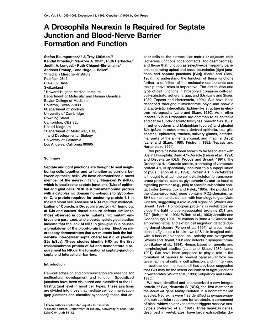
Figure 2. Northern and Western Analyses
(A) Northern-blot analysis of different stages of the Drosophila life cycle with an nrx cDNA. Each lane contains 5 mg of poly A1 RNA (hoursafter egg lay, L1 and L3 are first- and third-instar larva, Pup 5 pupal stages, A 5 adults). A 5 kb transcript was identified. The Dm ras 64probe was used as a loading control. Transcript lengths were determined with RNA standards.(B) The nrx gene does not undergo extensive alternative splicing. RT-polymerase chain reactions using different primer combinations coveringalmost the whole transcript. In each case, a single band was observed.(C) Human Northern blot of various embryonic tissues using human neurexin IV cDNAs. A single 6 kb transcript is detected in brain, kidney,and lung.(D) Western analysis of NRX protein of 7–22 hr old embryos. One prominent band of 150–155 kDa was observed, consistent with the predictedmolecular weight of the protein.
with nrx cDNA probes indicates that the gene is tran- expression first detectable approximately 1 hr prior totheir formation (Tepass and Hartenstein, 1994).scribed only zygotically, with its peak of expression be-
tween 6 and 18 hr after egg lay (Figure 2A). Therefore, To determine in which compartment NRX is localized,we carried out double-labeling experiments. In epider-we reverse transcribed mRNA by polymerase chain re-
actions, applying a comprehensive set of primers. In all mis and hindgut, NRX is localized apicolaterally, adja-cent to Crumbs, which delimits the zonula adherenscases, only a single band of the expected length was
amplified (Figure 2B), suggesting that extensive alterna- (Grawe et al., 1996). These two proteins are not coex-pressed (Figure 3E), placing NRX apicolaterally. Doubletive splicing does not occur in Drosophila nrx.
A polyclonal antiserum was raised against a C-termi- labeling of salivary glands with anti-FAS (Woods andBryant, 1993b) and anti-NRX shows that both proteinsnal peptide. Western analyses show a single band of
150–155 kDa, in close agreement with the predicted colocalize at salivary SJs (data not shown). Double label-ing with anti-NRX and anti-D4.1/Coracle antisera dem-size of 145 kDa (Figure 2D). This serum is specific to
NRX, as no protein can be detected in Western blots of onstrates that NRX precisely colocalizes with this SJ-restricted protein in all tissues tested except PNS andembryos that carry a deficiency removing the nrx
gene (Df(3L)BK9/Df(3L)vin8) or other nrx mutations (see CNS, where D4.1-Coracle is only expressed in a fewcells. This colocalization was confirmed in a series ofbelow).optical sections throughout the embryo using confocalmicroscopy. In the chordotonal organs, NRX is ex-NRX is Localized to pSJs
We analyzed the expression pattern of the nrx gene pressed in the cells surrounding the scolopales whereSJs have been localized (S. D. Carlson et al., submitted).using whole-mount in situ hybridization and antibody
stainings. With both methods, the nrx gene product can In addition, NRX is distributed in a ring at the apical endof many cell types, as shown for SJs (Lane and Skaer,first be detected at late stage 11/early stage 12. NRX is
localized to epithelial cells of ectodermal origin such as 1980). Postembryonically, in third-instar larvae, stainingcan also be detected in areas where SJs have beenepidermis, the tracheal system, pharynx, esophagus,
proventriculus, hindgut, salivary glands, and cells of the reported: the subperineural sheath of the larval CNS(Lane et al., 1977), imaginal disc cells (Locke, 1965), andperipheral and central nervous system (P-CNS) (Figure
3). Staining in the PNS is localized to the scolopales of salivary glands (data not shown). Thus, NRX is localizedto all pSJs documented.the lateral chordotonal organs (Figures 3D and 5B) and
is restricted to glial cells. Double-labeling experiments As SJs represent sites of intercellular contact, NRXmight function as a cell adhesion molecule. Adhesionshow that nrx-expressing cells in the CNS and along
the peripheral nerves express glial-specific cell markers properties of proteins can be tested in cell culture usingS2 cell adhesion assays (Bieber, 1994). Western blotbut not neuronal markers. Two types of epithelia do
not express NRX: amnioserosa and Malpighian tubules. analysis of uninduced Drosophila S2 cell lines that donot show cell aggregation demonstrated that NRX isInterestingly, all tissues that express NRX in the embryo
are characterized by the presence of pSJs, with NRX normally expressed in these cells. Therefore, NRX is not
Cell1062
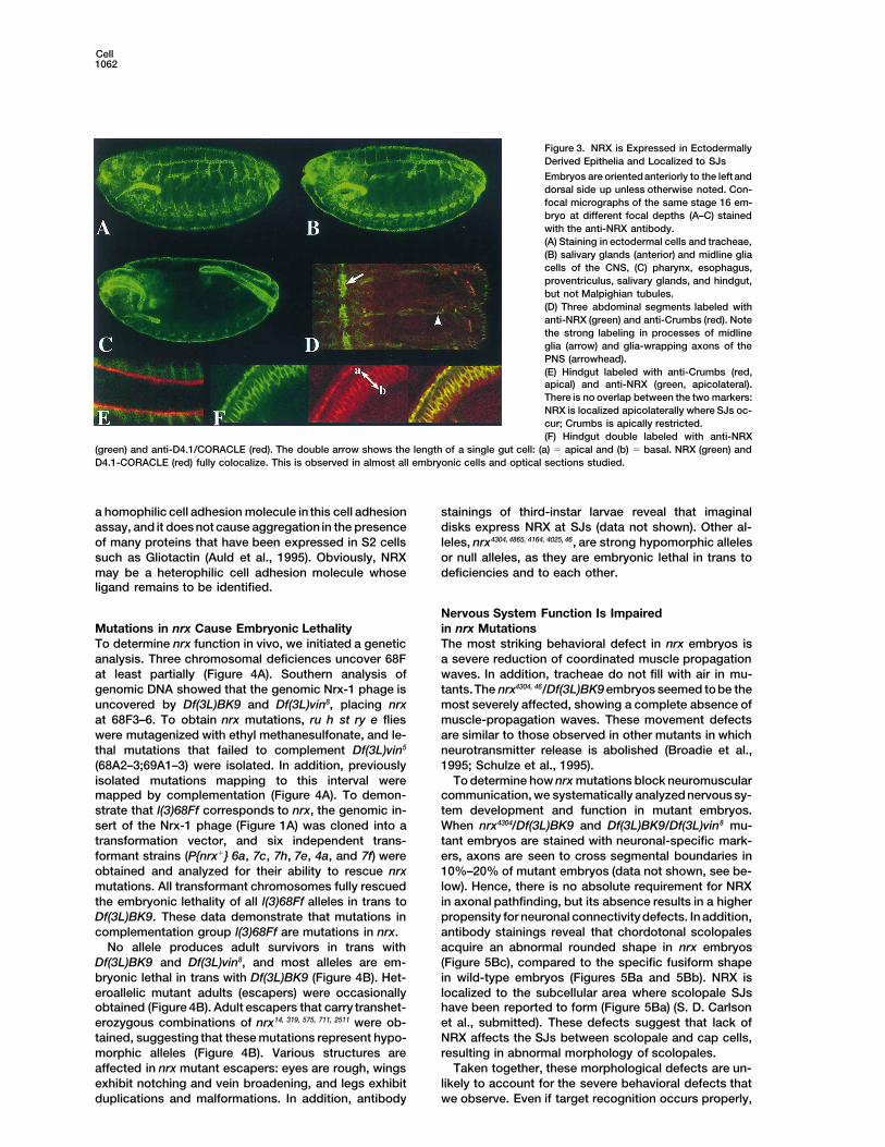
Figure 3. NRX is Expressed in EctodermallyDerived Epithelia and Localized to SJs
Embryos are orientedanteriorly to the leftanddorsal side up unless otherwise noted. Con-focal micrographs of the same stage 16 em-bryo at different focal depths (A–C) stainedwith the anti-NRX antibody.(A) Staining in ectodermal cells and tracheae,(B) salivary glands (anterior) and midline gliacells of the CNS, (C) pharynx, esophagus,proventriculus, salivary glands, and hindgut,but not Malpighian tubules.(D) Three abdominal segments labeled withanti-NRX (green) and anti-Crumbs (red). Notethe strong labeling in processes of midlineglia (arrow) and glia-wrapping axons of thePNS (arrowhead).(E) Hindgut labeled with anti-Crumbs (red,apical) and anti-NRX (green, apicolateral).There is no overlap between the two markers:NRX is localized apicolaterally where SJs oc-cur; Crumbs is apically restricted.(F) Hindgut double labeled with anti-NRX
(green) and anti-D4.1/CORACLE (red). The double arrow shows the length of a single gut cell: (a) 5 apical and (b) 5 basal. NRX (green) andD4.1-CORACLE (red) fully colocalize. This is observed in almost all embryonic cells and optical sections studied.
a homophilic cell adhesion molecule in this cell adhesion stainings of third-instar larvae reveal that imaginaldisks express NRX at SJs (data not shown). Other al-assay, and it does not cause aggregation in the presence
of many proteins that have been expressed in S2 cells leles, nrx4304, 4865, 4164, 4025, 46, are strong hypomorphic allelesor null alleles, as they are embryonic lethal in trans tosuch as Gliotactin (Auld et al., 1995). Obviously, NRX
may be a heterophilic cell adhesion molecule whose deficiencies and to each other.ligand remains to be identified.
Nervous System Function Is Impairedin nrx MutationsMutations in nrx Cause Embryonic Lethality
To determine nrx function in vivo, we initiated a genetic The most striking behavioral defect in nrx embryos isa severe reduction of coordinated muscle propagationanalysis. Three chromosomal deficiences uncover 68F
at least partially (Figure 4A). Southern analysis of waves. In addition, tracheae do not fill with air in mu-tants.The nrx4304, 46/Df(3L)BK9 embryos seemed tobe thegenomic DNA showed that the genomic Nrx-1 phage is
uncovered by Df(3L)BK9 and Df(3L)vin8, placing nrx most severely affected, showing a complete absence ofmuscle-propagation waves. These movement defectsat 68F3–6. To obtain nrx mutations, ru h st ry e flies
were mutagenized with ethyl methanesulfonate, and le- are similar to those observed in other mutants in whichneurotransmitter release is abolished (Broadie et al.,thal mutations that failed to complement Df(3L)vin5
(68A2–3;69A1–3) were isolated. In addition, previously 1995; Schulze et al., 1995).To determine how nrx mutations block neuromuscularisolated mutations mapping to this interval were
mapped by complementation (Figure 4A). To demon- communication, we systematically analyzed nervous sy-tem development and function in mutant embryos.strate that l(3)68Ff corresponds to nrx, the genomic in-
sert of the Nrx-1 phage (Figure 1A) was cloned into a When nrx4304/Df(3L)BK9 and Df(3L)BK9/Df(3L)vin8 mu-tant embryos are stained with neuronal-specific mark-transformation vector, and six independent trans-
formant strains (P{nrx1} 6a, 7c, 7h, 7e, 4a, and 7f) were ers, axons are seen to cross segmental boundaries in10%–20% of mutant embryos (data not shown, see be-obtained and analyzed for their ability to rescue nrx
mutations. All transformant chromosomes fully rescued low). Hence, there is no absolute requirement for NRXin axonal pathfinding, but its absence results in a higherthe embryonic lethality of all l(3)68Ff alleles in trans to
Df(3L)BK9. These data demonstrate that mutations in propensity for neuronal connectivity defects. In addition,antibody stainings reveal that chordotonal scolopalescomplementation group l(3)68Ff are mutations in nrx.
No allele produces adult survivors in trans with acquire an abnormal rounded shape in nrx embryos(Figure 5Bc), compared to the specific fusiform shapeDf(3L)BK9 and Df(3L)vin8, and most alleles are em-
bryonic lethal in trans with Df(3L)BK9 (Figure 4B). Het- in wild-type embryos (Figures 5Ba and 5Bb). NRX islocalized to the subcellular area where scolopale SJseroallelic mutant adults (escapers) were occasionally
obtained (Figure 4B). Adult escapers that carry transhet- have been reported to form (Figure 5Ba) (S. D. Carlsonet al., submitted). These defects suggest that lack oferozygous combinations of nrx14, 319, 575, 711, 2511 were ob-
tained, suggesting that these mutations represent hypo- NRX affects the SJs between scolopale and cap cells,resulting in abnormal morphology of scolopales.morphic alleles (Figure 4B). Various structures are
affected in nrx mutant escapers: eyes are rough, wings Taken together, these morphological defects are un-likely to account for the severe behavioral defects thatexhibit notching and vein broadening, and legs exhibit
duplications and malformations. In addition, antibody we observe. Even if target recognition occurs properly,
NRX and Septate Junctions1063
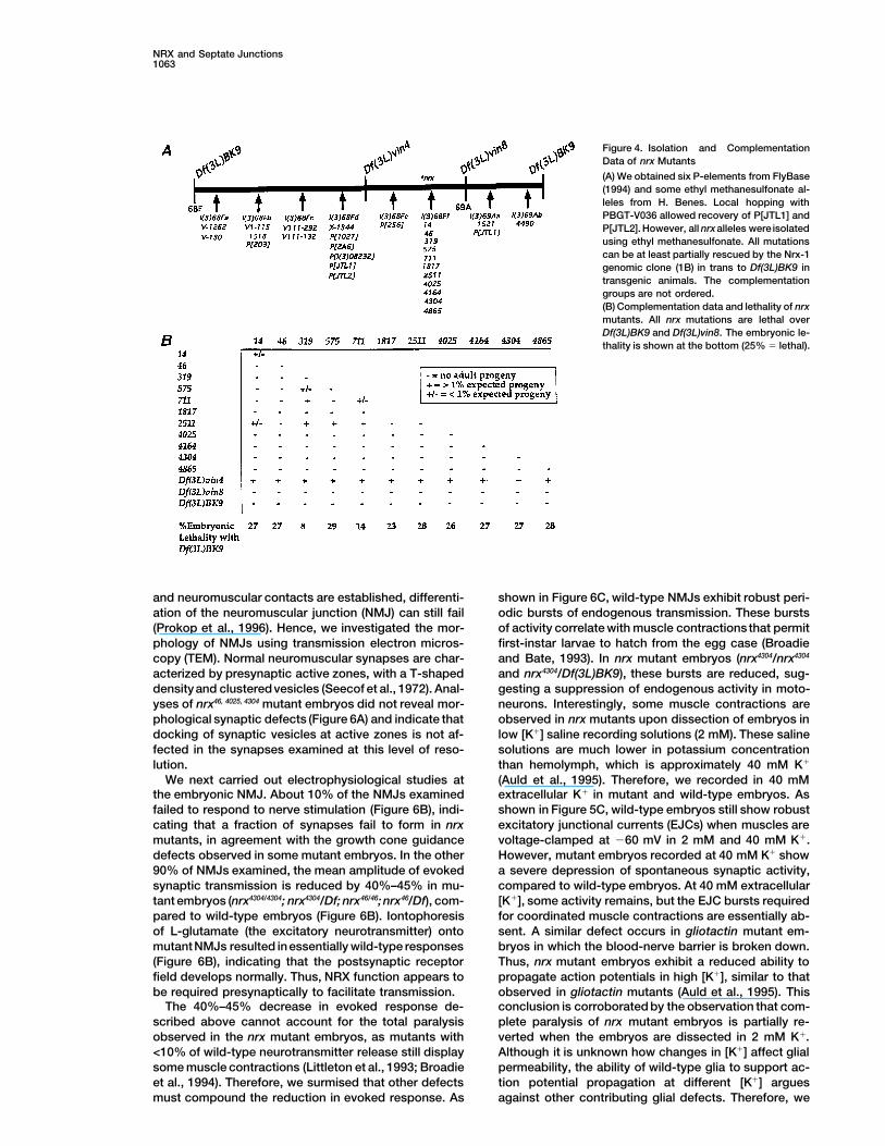
Figure 4. Isolation and ComplementationData of nrx Mutants
(A) We obtained six P-elements from FlyBase(1994) and some ethyl methanesulfonate al-leles from H. Benes. Local hopping withPBGT-V036 allowed recovery of P[JTL1] andP[JTL2]. However, all nrxalleles were isolatedusing ethyl methanesulfonate. All mutationscan be at least partially rescued by the Nrx-1genomic clone (1B) in trans to Df(3L)BK9 intransgenic animals. The complementationgroups are not ordered.(B) Complementation data and lethality of nrxmutants. All nrx mutations are lethal overDf(3L)BK9 and Df(3L)vin8. The embryonic le-thality is shown at the bottom (25% 5 lethal).
and neuromuscular contacts are established, differenti- shown in Figure 6C, wild-type NMJs exhibit robust peri-odic bursts of endogenous transmission. These burstsation of the neuromuscular junction (NMJ) can still fail
(Prokop et al., 1996). Hence, we investigated the mor- of activity correlate with muscle contractions that permitfirst-instar larvae to hatch from the egg case (Broadiephology of NMJs using transmission electron micros-
copy (TEM). Normal neuromuscular synapses are char- and Bate, 1993). In nrx mutant embryos (nrx4304/nrx4304
and nrx4304/Df(3L)BK9), these bursts are reduced, sug-acterized by presynaptic active zones, with a T-shapeddensityand clusteredvesicles (Seecof et al., 1972). Anal- gesting a suppression of endogenous activity in moto-
neurons. Interestingly, some muscle contractions areyses of nrx46, 4025, 4304 mutant embryos did not reveal mor-phological synaptic defects (Figure 6A) and indicate that observed in nrx mutants upon dissection of embryos in
low [K1] saline recording solutions (2 mM). These salinedocking of synaptic vesicles at active zones is not af-fected in the synapses examined at this level of reso- solutions are much lower in potassium concentration
than hemolymph, which is approximately 40 mM K1lution.We next carried out electrophysiological studies at (Auld et al., 1995). Therefore, we recorded in 40 mM
extracellular K1 in mutant and wild-type embryos. Asthe embryonic NMJ. About 10% of the NMJs examinedfailed to respond to nerve stimulation (Figure 6B), indi- shown in Figure 5C, wild-type embryos still show robust
excitatory junctional currents (EJCs) when muscles arecating that a fraction of synapses fail to form in nrxmutants, in agreement with the growth cone guidance voltage-clamped at 260 mV in 2 mM and 40 mM K1.
However, mutant embryos recorded at 40 mM K1 showdefects observed in some mutant embryos. In the other90% of NMJs examined, the mean amplitude of evoked a severe depression of spontaneous synaptic activity,
compared to wild-type embryos. At 40 mM extracellularsynaptic transmission is reduced by 40%–45% in mu-tant embryos (nrx4304/4304; nrx4304/Df; nrx46/46; nrx46/Df), com- [K1], some activity remains, but the EJC bursts required
for coordinated muscle contractions are essentially ab-pared to wild-type embryos (Figure 6B). Iontophoresisof L-glutamate (the excitatory neurotransmitter) onto sent. A similar defect occurs in gliotactin mutant em-
bryos in which the blood-nerve barrier is broken down.mutantNMJs resulted inessentially wild-type responses(Figure 6B), indicating that the postsynaptic receptor Thus, nrx mutant embryos exhibit a reduced ability to
propagate action potentials in high [K1], similar to thatfield develops normally. Thus, NRX function appears tobe required presynaptically to facilitate transmission. observed in gliotactin mutants (Auld et al., 1995). This
conclusion is corroborated by the observation that com-The 40%–45% decrease in evoked response de-scribed above cannot account for the total paralysis plete paralysis of nrx mutant embryos is partially re-
verted when the embryos are dissected in 2 mM K1.observed in the nrx mutant embryos, as mutants with<10% of wild-type neurotransmitter release still display Although it is unknown how changes in [K1] affect glial
permeability, the ability of wild-type glia to support ac-some muscle contractions (Littleton et al., 1993; Broadieet al., 1994). Therefore, we surmised that other defects tion potential propagation at different [K1] argues
against other contributing glial defects. Therefore, wemust compound the reduction in evoked response. As
Cell1064
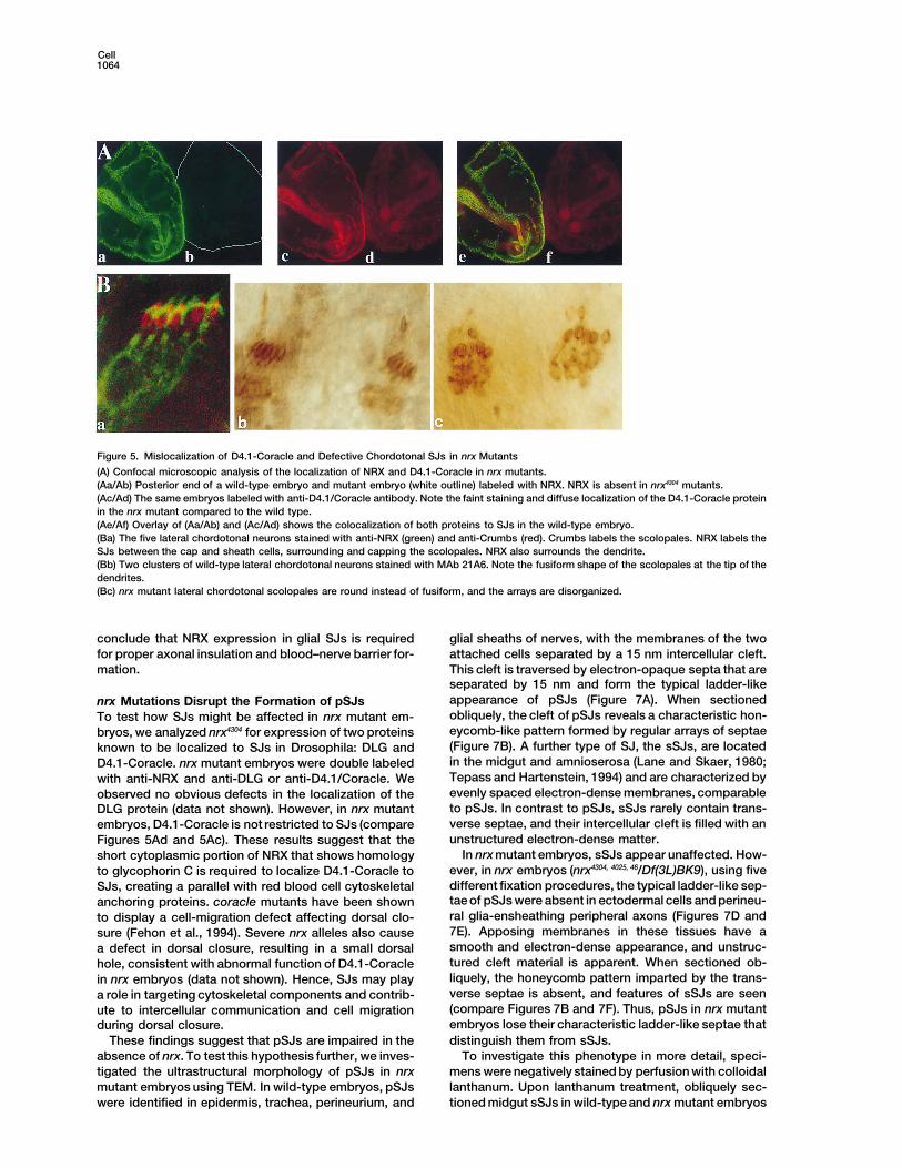
Figure 5. Mislocalization of D4.1-Coracle and Defective Chordotonal SJs in nrx Mutants
(A) Confocal microscopic analysis of the localization of NRX and D4.1-Coracle in nrx mutants.(Aa/Ab) Posterior end of a wild-type embryo and mutant embryo (white outline) labeled with NRX. NRX is absent in nrx4304 mutants.(Ac/Ad) The same embryos labeled with anti-D4.1/Coracle antibody. Note the faint staining and diffuse localization of the D4.1-Coracle proteinin the nrx mutant compared to the wild type.(Ae/Af) Overlay of (Aa/Ab) and (Ac/Ad) shows the colocalization of both proteins to SJs in the wild-type embryo.(Ba) The five lateral chordotonal neurons stained with anti-NRX (green) and anti-Crumbs (red). Crumbs labels the scolopales. NRX labels theSJs between the cap and sheath cells, surrounding and capping the scolopales. NRX also surrounds the dendrite.(Bb) Two clusters of wild-type lateral chordotonal neurons stained with MAb 21A6. Note the fusiform shape of the scolopales at the tip of thedendrites.(Bc) nrx mutant lateral chordotonal scolopales are round instead of fusiform, and the arrays are disorganized.
conclude that NRX expression in glial SJs is required glial sheaths of nerves, with the membranes of the twoattached cells separated by a 15 nm intercellular cleft.for proper axonal insulation and blood–nerve barrier for-
mation. This cleft is traversed by electron-opaque septa that areseparated by 15 nm and form the typical ladder-likeappearance of pSJs (Figure 7A). When sectionednrx Mutations Disrupt the Formation of pSJsobliquely, the cleft of pSJs reveals a characteristic hon-To test how SJs might be affected in nrx mutant em-eycomb-like pattern formed by regular arrays of septaebryos, we analyzed nrx4304 for expression of two proteins(Figure 7B). A further type of SJ, the sSJs, are locatedknown to be localized to SJs in Drosophila: DLG andin the midgut and amnioserosa (Lane and Skaer, 1980;D4.1-Coracle. nrx mutant embryos were double labeledTepass and Hartenstein, 1994) and are characterized bywith anti-NRX and anti-DLG or anti-D4.1/Coracle. Weevenly spaced electron-dense membranes, comparableobserved no obvious defects in the localization of theto pSJs. In contrast to pSJs, sSJs rarely contain trans-DLG protein (data not shown). However, in nrx mutantverse septae, and their intercellular cleft is filled with anembryos, D4.1-Coracle is not restricted to SJs (compareunstructured electron-dense matter.Figures 5Ad and 5Ac). These results suggest that the
In nrx mutant embryos, sSJs appear unaffected. How-short cytoplasmic portion of NRX that shows homologyever, in nrx embryos (nrx4304, 4025, 46/Df(3L)BK9), using fiveto glycophorin C is required to localize D4.1-Coracle todifferent fixation procedures, the typical ladder-like sep-SJs, creating a parallel with red blood cell cytoskeletaltae of pSJs were absent in ectodermal cells and perineu-anchoring proteins. coracle mutants have been shownral glia-ensheathing peripheral axons (Figures 7D andto display a cell-migration defect affecting dorsal clo-7E). Apposing membranes in these tissues have asure (Fehon et al., 1994). Severe nrx alleles also causesmooth and electron-dense appearance, and unstruc-a defect in dorsal closure, resulting in a small dorsaltured cleft material is apparent. When sectioned ob-hole, consistent with abnormal function of D4.1-Coracleliquely, the honeycomb pattern imparted by the trans-in nrx embryos (data not shown). Hence, SJs may playverse septae is absent, and features of sSJs are seena role in targeting cytoskeletal components and contrib-(compare Figures 7B and 7F). Thus, pSJs in nrx mutantute to intercellular communication and cell migrationembryos lose their characteristic ladder-like septae thatduring dorsal closure.distinguish them from sSJs.These findings suggest that pSJs are impaired in the
To investigate this phenotype in more detail, speci-absence of nrx. To test this hypothesis further, we inves-mens were negatively stained by perfusion with colloidaltigated the ultrastructural morphology of pSJs in nrxlanthanum. Upon lanthanum treatment, obliquely sec-mutant embryos using TEM. In wild-type embryos, pSJs
were identified in epidermis, trachea, perineurium, and tioned midgut sSJs in wild-type and nrx mutant embryos
NRX and Septate Junctions1065
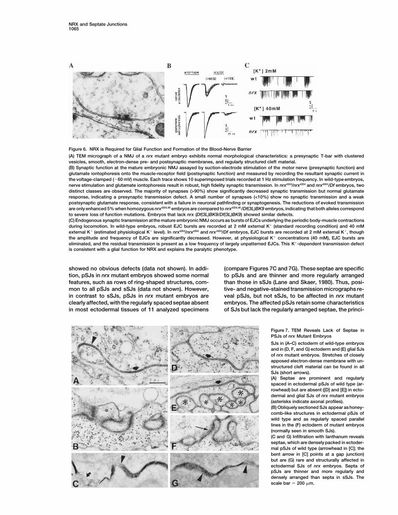
Figure 6. NRX is Required for Glial Function and Formation of the Blood-Nerve Barrier
(A) TEM micrograph of a NMJ of a nrx mutant embryo exhibits normal morphological characteristics: a presynaptic T-bar with clusteredvesicles, smooth, electron-dense pre- and postsynaptic membranes, and regularly structured cleft material.(B) Synaptic function at the mature embryonic NMJ assayed by suction-electrode stimulation of the motor nerve (presynaptic function) andglutamate iontophoresis onto the muscle-receptor field (postsynaptic function) and measured by recording the resultant synaptic current inthe voltage-clamped (260 mV) muscle. Each trace shows 10 superimposed trials recorded at 1 Hz stimulation frequency. In wild-type embryos,nerve stimulation and glutamate iontophoresis result in robust, high fidelity synaptic transmission. In nrx4304/nrx4304 and nrx4304/Df embryos, twodistinct classes are observed. The majority of synapses (>90%) show significantly decreased synaptic transmission but normal glutamateresponse, indicating a presynaptic transmission defect. A small number of synapses (<10%) show no synaptic transmission and a weakpostsynaptic glutamate response, consistent with a failure in neuronal pathfinding or synaptogenesis. The reductions of evoked transmissionare only enhanced 5% when homozygous nrx4304,46 embryos are compared to nrx4304,46 /Df(3L)BK9 embryos, indicating that both alleles correspondto severe loss of function mutations. Embryos that lack nrx (Df(3L)BK9/Df(3L)BK9) showed similar defects.(C) Endogenous synaptic transmission at the mature embryonic NMJ occurs as bursts of EJCs underlying the periodic body-muscle contractionsduring locomotion. In wild-type embryos, robust EJC bursts are recorded at 2 mM external K1 (standard recording condition) and 40 mMexternal K1 (estimated physiological K1 level). In nrx4304/nrx4304 and nrx4304/Df embryos, EJC bursts are recorded at 2 mM external K1, thoughthe amplitude and frequency of EJCs are significantly decreased. However, at physiological K1 concentrations (40 mM), EJC bursts areeliminated, and the residual transmission is present as a low frequency of largely unpatterned EJCs. This K1-dependent transmission defectis consistent with a glial function for NRX and explains the paralytic phenotype.
showed no obvious defects (data not shown). In addi- (compare Figures 7C and 7G). These septae are specificto pSJs and are thinner and more regularly arrangedtion, pSJs in nrx mutant embryos showed some normal
features, such as rows of ring-shaped structures, com- than those in sSJs (Lane and Skaer, 1980). Thus, posi-tive- and negative-stained transmission micrographs re-mon to all pSJs and sSJs (data not shown). However,
in contrast to sSJs, pSJs in nrx mutant embryos are veal pSJs, but not sSJs, to be affected in nrx mutantembryos. The affected pSJs retain some characteristicsclearly affected, with the regularly spacedseptae absent
in most ectodermal tissues of 11 analyzed specimens of SJs but lack the regularly arranged septae, the princi-
Figure 7. TEM Reveals Lack of Septae inPSJs of nrx Mutant Embryos
SJs in (A–C) ectoderm of wild-type embryosand in (D, F, and G) ectoderm and (E) glial SJsof nrx mutant embryos. Stretches of closelyapposed electron-dense membrane with un-structured cleft material can be found in allSJs (short arrows).(A) Septae are prominent and regularlyspaced in ectodermal pSJs of wild type (ar-rowhead) but are absent ([D] and [E]) in ecto-dermal and glial SJs of nrx mutant embryos(asterisks indicate axonal profiles).(B) Obliquely sectioned SJs appear as honey-comb-like structures in ectodermal pSJs ofwild type and as regularly spaced parallellines in the (F) ectoderm of mutant embryos(normally seen in smooth SJs).(C and G) Infiltration with lanthanum revealsseptae, which are densely packed in ectoder-mal pSJs of wild type (arrowhead in [C]; thebent arrow in [C] points at a gap junction)but are (G) rare and structurally affected inectodermal SJs of nrx embryos. Septa ofpSJs are thinner and more regularly anddensely arranged than septa in sSJs. Thescale bar 5 200 mm.
Cell1066
ple feature distinguishing pSJs from sSJs. These results Carlson, 1992). The phenotype of nrx mutants indicatesthat the septae of pSJs are required to form the blood–show that NRX is an integral component of pSJs and is
either part of the transverse septae or required for their brain–nerve barrier in vivo, providing further evidencethat tight junctions in vertebrates and SJs in insects areformation.functionally similar (Willott et al., 1993). At the electro-physiological level, the blood–nerve barrier defect isDiscussionsimilar to that observed in embryos mutant for gliotactin,a gene expressed in glial cells and required to insulateNRX Is a Novel Marker for pSJsmotoneurons (Auld et al., 1995). Gliotactin shows 29%We have isolated a new member of the neurexin family,identity and 50% similarity to vertebrate neuroligin-I,neurexin IV. NRX has the conserved modular arrange-which has been shown to be the ligand of certain verte-ment of the domains of neurexins I–III (Ushkaryov etbrate neurexins (Ichtchenko et al., 1995). However, givenal., 1992; Ushkaryov and Sudhof, 1993) but exhibits athe failure of S2 cells, which express Gliotactin and NRXdifferent amino-terminal domain, in which a DS domainto aggregate, it remains to be determined if these tworeplaces a laminin G domain and an EGF-like repeat.proteins interact directly in Drosophila.The DS domain was originally found in the discoidin
Two other defects were also revealed in the electro-proteins of Dictyostelium and appears to bind carbohy-physiological studies: a decrease in evoked neurotrans-drates (Ito et al., 1994). The overall domain structure ofmitter release, compared to wild-type embryos, and oc-Drosophila NRX appears to be conserved in vertebrates,casional failure to form synapses. The first defect couldand a homologous human gene is expressed in neuronalbe due to a docking defect of synaptic vesicles or to aand nonneuronal embryonic tissues, unlike other verte-reduced activation of vesicle fusion upon Ca21 influx.brate neurexins.However, Drosophila NRX does not localize to syn-NRX is localized to pSJs and is the first marker toapses, and we therefore surmise that this defect is likelyidentify these junctions specifically at all stages and insecondary to glial dysfunction. Second, <10% of theall cells documented to contain pSJs. Multiple lines ofsynapses fail to form, suggesting defects in neuronalevidence suggest that NRX is essential for pSJ formationpathfinding or synapse formation. Glial cells have beenand function. First, NRXis expressed inall tissues knownimplicated in growth cone guidance, and the misroutingto have pSJs and appears about 1 hr prior to their ap-defects observed in nrx mutant embryos strengthen thispearance. NRX is not expressed in tissues devoid ofhypothesis. Finally, the defects observed in the scolopa-pSJs, where zonula adherens or sSJs have been docu-les of chordotonal neurons and localization of NRX tomented. Second, the protein is localized to peripheralSJs of cap and sheath cells surrounding the scolopalesand subperineural glial junctions, which are known tosuggest that this recently described peripheral blood–contain pSJs and have been proposed to form the Dro-nerve barrier (S. D. Carlson et al., submitted) is alsosophila blood–nerve–brain barrier. Third, the proteindisrupted in nrx mutants.does not colocalize with Crumbs, an apical marker that
A second major function of SJs is to provide a me-delimits the zonula adherens (Wodarz et al., 1995).chanical link between cells, being in part responsibleFourth, NRX colocalizes with Fasciclin III, an SJ markerfor the densely packed arrangement of epithelialin salivary glands, and D4.1-Coracle, which labels allsheaths. This function is obviously affected in dlg mu-known pSJs except those of the nervous system. Fifth,tants, causing a breakdown of epithelial sheaths andmutants that lack NRX are paralyzeddue toa breakdownsubsequent overgrowth (Woods and Bryant, 1991). Inof the blood–nerve barrier, indicating a functional defectcontrast, tissues remain intact, and DLG still localizesin SJs. Sixth, loss-of-function nrx alleles cause a mislo-to sites of membrane contact in nrx mutant embryos.calization of D4.1-Coracle in tissues where they areOur TEM analysis demonstrates that nrx mutations docoexpressed, and nrx mutants show a similar pheno-not affect characteristics of SJs that are shared by pSJstype to loss of function coracle alleles. Finally, TEMand sSJs, such as the evenly spaced extracellular cleftanalysis demonstrates that the typical ladder-like septaand the electron-dense appearance of the membranes.that define pSJs are absent in nrx mutants.Therefore, it is unlikely that NRX functions in the adhe-sive properties required to connect the two apposing
nrx Mutant Embryos Reveal Novel Functions for SJs membranes, consistent with the lack of aggregation inBased on our observations, we can begin to describe S2 cells expressing NRX. Instead, NRX is likely to formspecific functions that pSJs are likely to play during or anchor the ladder-like septae characteristic of pSJs.development. First, pSJs are found in ectodermal epi- Finally, pSJs have been suggested to be involved inthelia that are in contact with the environment and in cell signaling. The data suggest a role for neurexins inglia that ensheath the nervous system. It is likely that addition to barrier formation. The requirement of NRXthese cells protect the embryo and the nervous system for proper localization of the Drosophila homolog ofby serving as a selective-diffusion barrier. In vertebrates, protein 4.1, Coracle, an SJ-associated protein, showstight junctions have been proposed to serve this role. that NRX forms a link between the extracellular environ-However, such junctions are virtually absent in insects, ment and intracellular components of SJs. This link hasand it has been proposed that pSJs might serve this important functional implications, as it affects dorsalbarrier function instead. Each SJ may confer partial im- closure and cell migration late in embryonic develop-permeability, so that together they form a barrier that ment. It is interesting to note that human dlg proteinprotects neurons from high potassium concentration binds to protein 4.1 (Lue et al., 1994), and that glycopho-
rin C, protein 4.1, and p55, a guanylate kinase domainin the hemolymph of insects (Hoyle, 1952; Juang and
NRX and Septate Junctions1067
Atkinson, Becky Scott, and Michael Macini for help with confocalcontaining protein, have been shown to form a complexmicroscopy. We thank Richard Atkinson, Kwang Choi, Kyung Cho,in vertebrates (Marfatia et al., 1994). The function ofKaren Schulze, Mark Wu, and Artur Kania for suggestions. We thankthis complex has not been precisely defined. The CASKRick Fehon, Helen Benes, Todd Laverty, Kathy Matthews, and the
protein, another guanylate kinase domain containing Indiana Stock Center for fly strains. A. P. and K. B. thank Michaelprotein, has been shown to bind previously identified Bate, in whose laboratory their work was carried out, and A. P.
thanks H. Skaer and N. J. Lane for discussions. This work wasvertebrate neurexins and to be expressed in nonneu-supported by an NIH training grant to J. T. L., an NIH grant (HD09948)ronal tissues (Hata et al., 1996). Hence, three proteinsto J. A. L, and an NIH grant to H. J. B. A. P. was funded by awith PDZ, guanylate kinase, and SH3 domains (p55,Human Capital and Mobility Fellowship (European Union) and by aDLG, and CASK) are associated with SJs or interact withFellowship from the Lloyd’s of London Tercentenery Foundation.
neurexins and have been proposed to be involved in K. B. was a Research Fellow of Girton College, Cambridge, whoseinter- and intracellular signaling. Defects in these signal- work was supported by the Wellcome Trust and an Alfred P. Sloan
Fellowship. H. J. B. is an Associate Investigator of the Howarding pathways may underlie the dorsal closure defectsHughes Medical Institute. Reprint requests should be sent to H. J. B.in nrx mutants. We are presently focusing on character-
izing the severe defects in adult structures observed inReceived April 21, 1996; Revised October 10, 1996.partial loss of function nrx alleles that further implicate
NRX and SJs in imaginal development and cellular com- Referencesmunication.
Auld, V.J., Fetter, R.D., Broadie, K., and Goodman, C.S. (1995). Glio-tactin, a novel transmembrane protein on peripheral glia, is requiredExperimental Proceduresto form the blood-nerve barrier in Drosophila. Cell 81, 757–767.
Fly Stocks and Mutagenesis Bieber, A.J. (1994). Analysis of cellular adhesion in cultured cells.The vin deficiencies are described by Hoogwerf et al. (1988). To In Drosophila melanogaster: Practical Uses in Cell and Molecularobtain mutations in nrx, ru h st ry e males were treated with ethyl Biology, L.S.B. Goldstein and E.A. Fyrberg, eds. (San Diego, Califor-methanesulfonate (Lewis and Bacher, 1968). Approximately 10,000 nia: Academic Press), pp. 683–713.mutagenized chromosomes were tested for lethality over Df(3L)vin5 Bock, G., and Clark, S. (1987). Junctional Complexes of Epithelialand Df(3L)vin7. Mutations that failed to complement Df(3L)vin5 and Cells (New York: John Wiley & Sons).Df(3L)vin7 were tested over Df(3L)BK9, Df(3L)vin4, and Df(3L)vin8.
Broadie, K.S., and Bate, M. (1993). Development of the embryonicAs shown in Figure 4A, this allowed us to identify and map mutationneuromuscular synapse of Drosophila melanogaster. J. Neurosci.1518 to l(3)68Fb, as well as 11 mutations between the proximal13, 144–166.break points of Df(3L)vin4 and Df(3L)vin8. Mutations 1521 and 4490Broadie, K., Bellen, H.J., DiAntonio, A., Littleton, J.T., and Schwarz,identify two complementation groups to the right of nrx. Other muta-T.L. (1994). The absence of synaptotagmin disrupts excitation-tions shown in Figure 4A are described in the legend.secretion coupling during synaptic transmission. Proc. Natl. Acad.Sci. USA 91, 10727–10731.Isolation of Genomic and cDNA Clones
Degenerate polymerase chain reaction primers derived from the Broadie, K., Prokop, A., Bellen, H.J., O’Kane, C.J., Schulze, K.L.,and Sweeney, S.T. (1995). Syntaxin and synaptobrevin functionEGF-like repeats (forward primer coding for amino acids S, [G, A,
S, R], [H, K], G, [L, N], C; reverse primer coding for amino acids C, downstream of vesicle docking in Drosophila. Neuron 15, 663–673.[N, D], C, [R, E, H], [V, W], S) allowed amplification of a 1320 bp Brody, L.C., Abel, K.J., Castilla, L.H., Couch, F.J., McKinley, D.R.,fragment harboring two laminin G domains flanked by two EGF- Yin G., Ho, P., Merajver, S., Chandrasekharappa, S.C., Xu, J., et al.like repeats that showed homology to rat neurexins. This permitted (1995). Construction of a transcription map surrounding the BRCA1isolation of cDNAs and genomic fragments. locus of human chromosome 17. Genomics 25, 238–247.
Brown, N.H., and Kafatos, F.C. (1988). Functional cDNA librariesGeneration of Antibodies and Immunohistochemical Staining from Drosophila embryos. J. Mol. Biol. 203, 425–437.A peptide corresponding to the 15 aa from the C-terminal (1269–
Cho, K.-O., Hunt, C.A., and Kennedy, M.B. (1992). The rat brain1283) was used to generate a polyclonal antiserum in rabbits. Thispostsynaptic density fraction contains a homolog of the Drosophilaserum was affinity purified and used at 1:500 dilution.discs-large tumor suppressor protein. Neuron 9, 929–942.
Fehon, R.G., Dawson, I.A., and Artravanis-Tsakonas, S. (1994). AElectrophysiologyDrosophila homologue of membrane-skeleton protein 4.1 is associ-The electrophysiological recordings were carried out as describedated with septate junctions and is encoded by the coracle gene.in Broadie and Bate, 1993, Broadie et al., 1995, and Auld et al.,Development 120, 545–557.1995. All records were made from muscle 6 in abdominal segment
A2 in mature embryos (22–24 hr AEL). FlyBase (1994). The Drosophila genetic database. Nucleic AcidsRes., 22, 3456–3458.
Electron Microscopy Friedman, L.S., Ostermeyer, E.A., Lynch, E.D., Welsch, P., Szabo,Late stage 17 embryos were injected with 5% glutaraldehyde, fol- C. I., Meza, J.E., Anderson, L.A., Dowd, P., Lee, M.K., Rowell, S.E.,lowed by 1 hr postfixation in 2.5% glutaraldehyde (Prokop and Tech- et al. (1995). 22 genes from chromosome 17q21: cloning, sequencing,nau, 1993). Preparations were washed, fixed for 1 hr in 1% osmium and characterization of mutations in breast cancer families and tu-in dH2O, washed, and treated en bloc with an aqueous 2% solution mors. Genomics 25, 256–263.of uranyl acetate for 30 min. In lanthanum infiltration experiments, Fristrom, D.K. (1982). Septate junctions in imaginal disks of Dro-mature embryos were injected with 5% glutaraldehyde. For subse- sophila: a model for the redistribution of septa during cell re-quent steps, solutions contained 2% lanthanum nitrate. The uranyl- arrangement. J. Cell Biol. 94, 77–87.acetate step was omitted. Specimens were dehydrated and trans-
Grawe, F., Wodarz, A., Lee, B., Knust, E., and Skaer, H. (1996). Theferred to Araldite. Serial sections of 30–50 nm thickness wereDrosophila genes crumbs and stardust are involved in the biogene-obtained, postcontrasted with lead citrate for 5–10 min, and ana-sis of adherens junctions. Development 122, 951–959.lyzed on a Jeol 200CX.Hata, Y., Butz, S., and Sudhof, T.C. (1996). Cask: a novel dlg/PSD95homologue with an n-terminal calmodulin-dependent protein kinaseAcknowledgmentsdomain identified by interaction with Neurexins. J. Neurosci. 16,2488–2494.We thank Doris Martin for technical assistance, Markus Noll for
libraries, and Beth Ostermeyer for human nrx IV. We thank Richard Hoogwerf, A.M.,Akam, M., and Roberts, D. (1988). Agenetic analysis
Cell1068
of the rose-gespleten region (68C8–69B5) of Drosophila melanogas- and electrophysiological studies of Drosophila syntaxin-1A demon-strate its role in nonneuronal secretion and neurotransmission. Cellter. Genetics 118, 665–670.80, 311–320.Hoyle, G. (1952). High blood potassium in insects in relation to nerveSeecof, R.L., Teplitz, R.L., Gerson, I., Ikeda, K., and Donady, J.J.conduction. Nature 169, 281–282.(1972). Differentiation of neuromuscular junctions in cultures of em-Ichtchenko, K., Hata, Y., Nguyen, T., Ullrich, B., Missler, M., Moo-bryonic Drosophila cells. Proc. Natl. Acad. Sci. USA 69, 566–570.maw, C., and Sudhof, T.C. (1995). Neuroligin 1: a splice site-specificTepass, U., and Hartenstein, V. (1994). The development of cellularligand for B-Neurexins. Cell 81, 435–443.junctions in the Drosophila embryo. Dev. Biol. 161, 563–596.Ito, N., Phillips, S.E.V., Yadav, K.D.S., and Knowles, P.F. (1994).Tsukita, S., Itoh, M., Nagafuchi, A., Yonemura, S., and Tsukita, S.Crystal structure of a free radical enzyme, galactose oxidase. J.(1993). Submembranous junctional plaques proteins include poten-Mol. Biol. 238, 794–814.tial tumor suppressor molecules. J. Cell Biol. 123, 1049–1053.
Itoh, M.S., Nagafuchi, S., Yonemura, R., Kitani-Yasuda, T., and Tsuk-Ullrich, B., Ushkaryov, Y.A., and Sudhof, T.C. (1995). Cartographyita, S. (1993). The 220 kDa protein colocalizing with cadherins inof neurexins: more than 1000 isoforms generated by alternativenon-epithelial cells is identical to Z0–1, a tight junction-associatedsplicing and expressed in distinct subsets of neurons. Neuron 14,protein in epithelial cells: cDNA cloning and immunoelectron micros-497–507.copy. J. Cell Biol. 121, 491–502.Ushkaryov, Y.A., and Sudhof, T.C. (1993). Neurexin IIIa: extensiveJesaitis, L.A., and Goodenough, D.A. (1994). Molecular characteriza-alternative splicing generates membrane-bound and soluble forms.tion and tissue distribution of Z0–2, a tight junction protein homolo-Proc. Natl. Acad. Sci. USA 90, 6410–6414.gous to Z0–1 and the Drosophila discs-large tumor supressor pro-Ushkaryov, Y.A., Petrenko, A.G., Geppert, M., and Sudhof, T.C.tein. J. Cell Biol. 124, 949–961.(1992). Neurexins: synaptic cell surface proteins related to the
Juang, J.-L., and Carlson, S.D. (1992). A blood-brain barrier withouta-latrotoxin receptor and laminin. Science 257, 50–56.
tight junctions in the fly central nervous system in the early postem-Willott, E., Balda, M.S., Fanning, A.S., Jameson, B., VanItallie, C.,bryonic stage. Cell Tissue Res. 270, 95–103.andAnderson, J.M. (1993). The tight junction protein Z0–1 is homolo-
Kirkpatrick, C., and Peifer, M. (1995). Not just glue: cell-cell junctions gous to the Drosophila discs large tumor suppressor protein ofas cellular signalling centers. Curr. Opin. Genet. Dev. 5, 56–65. septate junctions. Proc. Natl. Acad. Sci. USA 90, 7834–7838.Lahey, T., Gorczyca, M., Jia, X.-X., and Budnik, V. (1994). The Dro- Wodarz, A., Hinz, U., Engelbert, M., and Knust, E. (1995). Expressionsophila tumor suppressor gene dlg is required for normal synaptic of Crumbs confers apical character on plasma membrane domainsbouton structure. Neuron 13, 823–835. of ectodermal epithelia of Drosophila. Cell 82, 67–76.Lane, N.J. (1991). Cytoskeletal associations with intercellular junc- Woods, D.F., and Bryant,P.J. (1991). The discs-large tumor suppres-tions in arthropods. In Form and Function inZoology, G. Lanzavecchi sor gene of Drosophila encodes a guanylate kinase homolog local-and R. Valvassori, eds. (Munich: Modena), pp. 87–102. ized at septate junctions. Cell 66, 451–464.Lane, N.J., and Skaer, H.B. (1980). Intercellular junctions in insect Woods, D.F., and Bryant, P.J. (1993a). Apical junctions and celltissues. In Advances in Insect Physiology, M.J. Berridge, J.E. Tre- signalling in epithelia. J. Cell Sci. Suppl. 17, 171–181.herne, and V.B. Wigglesworth, eds. (London: Academic Press), pp. Woods, D.F., and Bryant, P.J. (1993b). ZO-1, DlgA and PSD-35–213. 95SAP90: homologous proteins in tight, septate and synaptic cellLane, N.J., Skaer, H.I., andSwales, L.S. (1977). Intercellular junctions junctions. Mech. Dev. 44, 85–89.in the central nervous system of insects. J. Cell Sci. 26, 175–199.
Lewis, E.B., and Bacher, F. (1968). Mutagenesis with ethyl methane- EMBL Accession Numbersulfonate. Drosoph. Inf. Serv. 43, 193.
The accession number for the nrx sequence is X86685. The acces-Littleton, J.T., Stern, M., Schulze, K., Perin, M., and Bellen, H.J.sion numbers for the human expressed sequence tags in Figure 1B(1993). Mutational analysis of Drosophila synaptotagmin demon-are T27170, U18000, U17905, Z42448, and U18017.strates its essential role in Ca21-activated neurotransmitter release.
Cell 74, 1125–1134.
Locke, M. (1965). The structure of septate desmosomes. J. Cell Biol.25, 166–168.
Lue, R.A., Marfatia, S.M., Branton, D., and Chishti, A.H. (1994). Clon-ing and characterization of hdlg: the human homologue of the Dro-sophila disc large tumor suppressor binds to protein 4.1. Proc. Natl.Acad. USA 91, 9818–9822.
Lux, S.E., and Palek, J. (1995). Disorders of the red cell membrane.In Blood Principles and Practice of Hematology, R.I. Handin, S.E.Lux, and T.P. Stossel, eds. (Philadelphia: J.B. Lippincott Company).
Marfatia, S.M., Lue, R.A., Branton, D., and Chishti, A.H. (1994). Invitro binding studies suggest a membrane-associated complex be-tween erythroid p55, protein 4.1, and glycophorin C. J. Biol. Chem.269, 8631–8634.
Petrenko, A.G., Perin, M.S., Bazbek, A., Davletov, B.A., Ushkaryov,Y.A., Geppert, M.,and Sudhof, T.C. (1991).Binding of synaptotagminto the a-latrotoxin receptor implicates both in synaptic vesicle exo-cytosis. Nature 353, 65–68.
Prokop, A., and Technau, G.M. (1993). Cell transplantation. In Cellu-lar Interaction in Development: A Practical Approach, D. Hartley,ed. (London: Oxford University Press), pp. 33–57.
Prokop, A., Landgraf, M., Rushton, E., Broadie, K., and Bate, M.(1996). Presynaptic development at the Drosophila neuromuscularjunction: assembly and localization of presynaptic active zones.Neuron, 17 617-626.
Schulze, K., Broadie, K., Perin, M., and Bellen, H.J. (1995). Genetic