Embed Size (px)
Citation preview
A Homozygous CARD9 Mutation in a Family with Susceptibility toFungal Infections
Erik-Oliver Glocker, M.D., Andre Hennigs, Mohammad Nabavi, M.D., Alejandro A. Schäffer,Ph.D., Cristina Woellner, M.Sc., Ulrich Salzer, M.D., Dietmar Pfeifer, Ph.D., Hendrik Veelken,M.D., Klaus Warnatz, M.D., Fariba Tahami, M.Sc., Sarah Jamal, M.Sc., Annabelle Manguiat,M.Sc., Nima Rezaei, M.D., Ali Akbar Amirzargar, M.D., Alessandro Plebani, M.D., NicoleHannesschläger, B.Sc., Olaf Gross, Ph.D., Jürgen Ruland, M.D., and Bodo Grimbacher, M.D.
AbstractBACKGROUND—Chronic mucocutaneous candidiasis may be manifested as a primaryimmunodeficiency characterized by persistent or recurrent infections of the mucosa or the skin withcandida species. Most cases are sporadic, but both autosomal dominant inheritance and autosomalrecessive inheritance have been described.
METHODS—We performed genetic studies in 36 members of a large, consanguineous five-generation family, in which 4 members had recurrent fungal infections and an additional 3 membersdied during adolescence, 2 after invasive infection of the brain with candida species. All 36 familymembers were enrolled in the study, and 22 had blood samples taken for DNA analysis.Homozygosity mapping was used to locate the mutated gene. In the 4 affected family members(patients) and the 18 unaffected members we sequenced CARD9, the gene encoding the caspaserecruitment domain-containing protein 9, carried out T-cell phenotyping, and performed functionalstudies, with the use of either leukocytes from the patients or a reconstituted murine model of thegenetic defect.
RESULTS—We found linkage (lod score, 3.6) to a genomic interval on chromosome 9q, includingCARD9. All four patients had a homozygous point mutation in CARD9, resulting in a prematuretermination codon (Q295X). Healthy family members had wild-type expression of the CARD9protein; the four patients lacked wild-type expression, which was associated with low numbers ofTh17 cells (helper T cells producing interleukin-17). Functional studies based on geneticreconstitution of myeloid cells from Card9−/− mice showed that the Q295X mutation impairs innatesignaling from the antifungal pattern-recognition receptor dectin-1.
CONCLUSIONS—An autosomal recessive form of susceptibility to chronic mucocutaneouscandidiasis is associated with homozygous mutations in CARD9.
Chronic mucocutaneous candidiasis is characterized by impaired clearance of fungal infectionsand results in colonization and infections of the mucosa or skin, predominantly with Candidaalbicans.1,2 A variety of clinical conditions, such as infection with the humanimmunodeficiency virus or the use of corticosteroids, favor the development of chronicmucocutaneous candidiasis, but the disease may also be a primary immunodeficiency arisingfrom unknown genetic defects.1,3 In chronic mucocutaneous candidiasis, the most commoninfections are due to C. albicans; however, patients may also have an increased susceptibility
Address reprint requests to Dr. Grimbacher at the Department of Immunology and Molecular Pathology, Royal Free Hospital andUniversity College London, Pond St., London NW3 2QG, United Kingdom, or at [email protected]. Glocker and Hennigs contributed equally to this article.No potential conflict of interest relevant to this article was reported.
NIH Public AccessAuthor ManuscriptN Engl J Med. Author manuscript; available in PMC 2010 April 29.
Published in final edited form as:N Engl J Med. 2009 October 29; 361(18): 1727–1735. doi:10.1056/NEJMoa0810719.
NIH
-PA Author Manuscript
NIH
-PA Author Manuscript
NIH
-PA Author Manuscript
to dermatophytes.1,3 Severe complications rarely develop in patients with chronicmucocutaneous candidiasis, although reports on invasive infections with candida species —Cryptococcus neoformans or Histoplasma capsulatum — have been published.4–6
Research conducted in the eight decades since the first report on primary chronicmucocutaneous candidiasis appeared7 has shown that it is a heterogeneous syndrome that maybe accompanied by endocrine and inflammatory disorders, including hypothyroidism andadrenocortical failure. 2 Most cases of chronic mucocutaneous candidiasis are sporadic, butmultiplex families with dominant8–12 and recessive13,14 inheritance have been described.
Recurrent and severe candidiasis can have a defining role in primary immunodeficiencies. Theautoimmune polyendocrinopathy–candidiasis–ectodermal dystrophy (APECED) syndrome iscaused by biallelic mutations in AIRE, the autoimmune regulator gene. Heterozygous mutationsin the signal transducer and activator of transcription 3 gene (STAT3) cause the hyper-IgEsyndrome, another multisystem disorder in which candidiasis is a common clinical feature.17–20 Genetic linkage of an autosomal dominant candidiasis–thyroiditis syndrome tochromosome 2 has been reported,12 and candidiasis associated with a low expression ofintercellular adhesion molecule 1 (ICAM-1) has been traced to chromosome 11,21 but in bothcases the causative genes remain unknown.
We undertook genetic studies of a large, consanguineous Iranian family with multiple casesof chronic mucocutaneous candidiasis to determine whether a mutated gene was associatedwith this form of nonsyndromic candidiasis. Recent work has shown that innate antifungalimmunity in mice is controlled by a signaling pathway that does not involve toll-like receptors.Mice lacking either the C-type lectin receptor dectin-1 (encoded by the Clec7a gene) or theintracellular adapter molecule Card9, which is essential for dectin-1 signaling, have impairedantifungal immunity.22,23
We identified a homozygous mutation in CARD9 that results in a loss-of-function mutationdue to a premature stop codon in the coding sequence. Experiments in the murine Card9−/−
model showed that only wild-type CARD9 — not the mutated human CARD9 gene found inpatients — could restore cytokine production in response to the triggering of dectin-1, a pattern-recognition receptor for fungal cell-wall antigens.
METHODSSTUDY PARTICIPANTS
We enrolled 36 members of a large, consanguineous Iranian family in the study. Blood sampleswere obtained for DNA analysis, and the participants were classified as likely to be affectedor likely to be unaffected according to the results of physical examination and microbiologicculture. Laboratory personnel were unaware of the classification of the samples. Theparticipants provided written informed consent on forms approved by local ethics committees.DNA samples from 50 healthy Iranian donors and 180 healthy white donors of othernationalities were used as controls.
GENOTYPING AND ANALYSISDNA samples from five family members deemed likely to be affected and eight deemed likelyto be unaffected were genotyped with use of the Affymetrix 250k NspI single-nucleotide-polymorphism (SNP) mapping array, as described previously.24 Genotypes of the SNP arrayswere assigned with the use of the Bayesian Robust Linear Modeling and Mahalanobis(BRLMM) distance method, which was implemented as described in the Genotyping Consolesoftware and introduced as an improvement over the RLMM method.25
Glocker et al. Page 2
N Engl J Med. Author manuscript; available in PMC 2010 April 29.
NIH
-PA Author Manuscript
NIH
-PA Author Manuscript
NIH
-PA Author Manuscript
To further evaluate one region on chromosome 9 that was suggestive of linkage, fourmicrosatellite markers were genotyped on 13 available samples, as described previously.26
Single-marker lod scores were computed with Fastlink27–29; multipoint lod scores werecomputed with Superlink.30,31 (For details on the use of the BRLMM method, Fastlink, andSuperlink, see the Supplementary Appendix, available with the full text of this article atNEJM.org.)
MOLECULAR GENETICS AND T-CELL PHENOTYPINGDNA from the study participants was isolated, and coding regions of CARD9 (Ensemblnumber, ENSG00000187796) were amplified and sequenced. Total RNA was isolated andtranscribed into complementary DNA (cDNA). SYBR green-based real-time polymerase-chain-reaction quantification of CARD9 was performed with the use of standard curves.Screening for the Q295X mutation with the use of restriction-enzyme digestion was carriedout in heterozygous and homozygous healthy family members as well as in healthy controls.
T-cell phenotyping, regulatory T-cell staining, and detection of Th17 cells (helper T cellsproducing interleukin-17) were performed in accordance with protocols published previously.32–36 Extracts of peripheral-blood mononuclear cells from patients with a deficiency of theCARD9 protein were stained with a polyclonal goat anti-CARD9 antibody. (Details of theseprocedures are available in the Supplementary Appendix.)
RETROVIRAL TRANSDUCTION OF HUMAN CARD9 VARIANTSCARD9 cDNA was generated from human peripheral-blood mononuclear cells and cloned intoa retroviral expression vector (based on murine stem-cell virus) expressing green fluorescentprotein.37 The Q295X mutation was introduced with the use of site-directed mutagenesis.Retroviruses were generated by transfecting the Phoenix ecotropic-packaging cell line andwere used to infect bone marrow cells as described previously.37 Bone marrow cells weredifferentiated into macrophages and stimulated with either curdlan, a selective dectin-1 agonist,or ultrapure lipopolysaccharide. Concentrations of tumor necrosis factor α (TNF-α) in thesupernatants were analyzed with the use of an enzyme-linked immunosorbent assay.22,23,38
RESULTSPATIENTS’ MEDICAL HISTORY
The pedigree (Fig. 1A) for the consanguineous Iranian family shows that multiple memberswere affected by chronic mucocutaneous candidiasis through a presumably autosomalrecessive mode of inheritance. Recurrent fungal infections were diagnosed clinically in eightfamily members, three of whom died in early adolescence — two with proven and one withpresumed invasive candida infection of the brain. None of the eight infected patients hadunusual bacterial or viral infections, suggesting that the host defense against these pathogenswas normal.
The index patient (Patient 2B2) was a 19-year-old man who had had oral candidiasis (thrush)since the age of 3 years. Candida infection was confirmed with the use of microbiologic testing,and prophylaxis with ketoconazole was ongoing. He was otherwise healthy.
Patient 2B1, a sibling of Patient 2B2, had had intermittent thrush since early childhood.Seizures developed suddenly, with loss of consciousness, when he was 18 years old.Hydrocephalus developed, and he died of candida meningitis at the age of 19 years.
Patient 2M, the mother of Patients 2B1 and 2B2, was 50 years old at the time of our study andhad had vaginal candidiasis since the age of 42 years. In addition to being infected with C.
Glocker et al. Page 3
N Engl J Med. Author manuscript; available in PMC 2010 April 29.
NIH
-PA Author Manuscript
NIH
-PA Author Manuscript
NIH
-PA Author Manuscript
albicans, she had a 5-year history of dermatophytosis of her hands and neck. She also hadintermittent aphthous lesions, type 2 diabetes mellitus, and nephrolithiasis.
Patient 1M, the sister of Patient 2M, had had oral and vaginal candidiasis since early childhood.She also had tinea corporis on her chest and neck. She was otherwise healthy.
Patient 5F, the affected brother of Patients 1M and 2M, had had dermatophytosis sincechildhood, with little improvement in response to local treatment. Both of his daughters weregiven a postmortem diagnosis of invasive candida infection.
The older daughter (Patient 5G1) had a ventricular septal defect during infancy and had ageographic tongue, which is suggestive of chronic candidiasis. A unilateral paresthesiadeveloped when she was 13 years old, and she died from what was vaguely defined as a braintumor, with severe skull destruction, at the age of 15 years.
Her sister (Patient 5G2) had recurrent, severe, refractory thrush starting in early childhood. At15 years of age, a severe headache and fevers developed, followed by diplopia. A brain tumorwas suspected, but candida meningoencephalitis was identified during surgery. She died 6months after the onset of symptoms.
None of the patients with invasive fungal infections had a condition or took any medicationthat predisposed them to infection. The three deceased patients could not be enrolled in thestudy because of the lack of available tissue samples.
Patient 1B1, a child of Patient 1M and a cousin of the index patient (Patient 2B2), had had onemild episode of candida infection in adulthood. In our genetic analysis, we considered Patient1B1 to be affected. We found that the episode of infection in Patient 1B1 was the result of aphenocopy, which is consistent with the fact that it was clinically much milder than theinfections in all the other affected family members (see the Supplementary Appendix for thedefinition of phenocopy).
CELL COUNTS AND T-CELL PHENOTYPINGIn Patients 1M, 2M, and 5F, complete blood counts were within the normal range, as were totalcounts of CD3+ T cells, CD4+ T cells, CD8+ T cells, memory T cells, follicular helper T cells,effector memory T cells, regulatory T cells, B cells, and natural killer cells (for results seeTable S1 in the Supplementary Appendix). Basal levels of serum immunoglobulin were alsonormal. In Patient 2B2, the results of a delayed-type hypersensitivity skin test were negativefor tuberculin but positive for candida.
AIRE was shown to be of wild-type sequence in Patient 2B2. The absence of a specificimmunologic disorder led us to use a positional cloning approach to identify the patients’underlying genetic defect.
GENETIC LINKAGE ANALYSISAnalysis of the SNP genotypes showed a region of perfect segregation on chromosome 9 (137.5to 138.8 Mbp in human genome build 36), provided that Patient 1B1’s episode of candidainfection was the result of a phenocopy. This finding was confirmed by genotyping fourmicrosatellite markers, yielding a peak multipoint lod score of 3.6 (see the SupplementaryAppendix).
There were 121 genes in the maximal linkage interval defined by the microsatellite markersD9S2157 (135.0 Mbp) and D9S1838 (139.8 Mbp). Among these 121 genes, 41 are located inthe perfectly segregating 1.3-Mbp subinterval suggested by the SNP data. After exploring the
Glocker et al. Page 4
N Engl J Med. Author manuscript; available in PMC 2010 April 29.
NIH
-PA Author Manuscript
NIH
-PA Author Manuscript
NIH
-PA Author Manuscript
literature, we identified CARD9 — which is among the 41 ideally located genes (see Table S2in the Supplementary Appendix) — as a functional candidate because Card9−/− mice aresusceptible to fungal infections (for details, see the Supplementary Appendix). 22,39
HOMOZYGOUS MUTATIONS IN CARD9We sequenced CARD9 in the four affected patients and 18 other relatives and identified in allaffected persons a single homozygous point mutation from C to T in exon 6 at codon 295,resulting in a premature termination codon (Q295X). Patient 1B1 and his healthy relatives hadeither a heterozygous Q295X mutation or wild-type alleles only (Fig. 1B).
To assess the frequency of this previously unknown genetic abnormality in CARD9 and toexclude the possibility of a genetic variation, we examined the affected site in 50 unrelatedhealthy Iranians and 180 unrelated healthy white subjects by means of a sequencing assay ora restriction-enzyme assay. None of the 230 controls had the Q295X mutation in CARD9.
CARD9 mRNA LEVELS AND PROTEIN EXPRESSIONAmong CARD9 wild-type cells, average levels of CARD9 mRNA were highest in monocytes,followed by granulocytes, B cells and T cells, and the colon-cell line HT-29. The peripheral-blood mononuclear cells from our patients still had substantial levels of mutated CARD9mRNA, thereby escaping nonsense-mediated RNA decay (see the Supplementary Appendix).
To examine the effect of the CARD9 Q295X mutation at the protein level, we assessed theexpression of CARD9 in peripheral-blood mononuclear cells from the patients, using Westernblotting. As compared with unrelated healthy controls and homozygous or heterozygoushealthy family members, patients with the homozygous Q295X mutation completely lackedexpression of the wild-type CARD9 protein (Fig. 2), indicating the detrimental consequencesof the mutation. However, expression of the truncated CARD9 (amino acid positions 1 through294) protein cannot be ruled out, since the polyclonal antibody used is directed against the C-terminal part of CARD9.
EFFECT OF CARD9 Q295X ON SIGNAL TRANSDUCTIONPrimary bone marrow cells from Card9-deficient mice were retrovirally transduced with humanwild-type and mutated (Q295X) CARD9, differentiated into macrophages in vitro, andanalyzed with the use of flow cytometry (see the Supplementary Appendix). We thenstimulated the transduced or nontransduced cells with the β-glucan preparation curdlan as aspecific and selective agonist for dectin-1 or with the TLR4 ligand lipopolysaccharide andmeasured TNF-α production to test innate immune cell activation.38 As is consistent withprevious data, Card9−/− cells showed severe defects in dectin-1–triggered TNF-α production,although they responded normally to stimulation with lipopolysaccharide (Fig. 3A).22
Expression of human full-length CARD9 corrected the dectin-1 signaling defect in Card9−/−
cells, indicating that the human protein can complement the murine mutation. Expression ofthe human mutant CARD9 Q295X did not increase TNF-α production, on stimulation withdectin-1, above the level in uninfected cells or those transduced with green fluorescent proteinonly, showing that CARD9 Q295X is a loss-of-function mutation (Fig. 3B).
HOMOZYGOUS Q295X MUTATIONS AND TH17 CELLSSince Th17 cells40 are important for antifungal immunity and Card9−/− mice have animpairment in Th17 polarization,22,38 we compared Th17 cells in our four patients with thosein family members with wild-type CARD9 and nine healthy controls. The mean proportion ofTh17 cells in the four affected patients was significantly lower than that in healthy controls(mean, 0.2% of CD4+CD45RO+ interleukin-17+interferon-γ− cells; P = 0.004) (Fig. 4). Healthy
Glocker et al. Page 5
N Engl J Med. Author manuscript; available in PMC 2010 April 29.
NIH
-PA Author Manuscript
NIH
-PA Author Manuscript
NIH
-PA Author Manuscript
controls and family members with wild-type CARD9 had an average of 1.2% Th17 cells exvivo.
DISCUSSIONPattern-recognition receptors of the innate immune system bind components of microbes andinitiate intracellular signal cascades that result in the activation of transcription factors, up-regulation of defense-associated target genes, and release of cytokines. Dectin-1 is atransmembrane pattern-recognition receptor that senses the β-glucan component of fungal cellwalls.23,41–43 On ligand binding, dectin-1 sends signals through an immunoreceptor tyrosine-based activation motif (ITAM), which becomes phosphorylated by Src family kinases (proto-oncogenic tyrosine kinases), leading to the recruitment and activation of the spleen tyrosinekinase (Syk).44,45
Dectin-1–Syk engages CARD9, which together with B-cell leukemia–lymphoma 10 (BCL10)and mucosa-associated lymphoid tissue 1 (MALT1) forms an intracellular signaling complexthat in cells recognizing fungi leads to the activation of the transcription factor nuclear factorκB and mitogen-activated protein kinases.22,46–48 This signaling pathway is operative inmyeloid cells and promotes the production of key cytokines, including interleukin-1β,interleukin-6, and interleukin-23, which are required to control antifungal immune responses.24,38,49–52 Apart from dectin-1, the C-type lectins dectin-2 and macrophage-inducible C-typelectin (MINCLE) may also recognize fungi, engage the ITAM adapter FcRγ for Syk activation,and transmit signals through the CARD9 pathway. 23,53–55 Thus, CARD9 plays a central rolein antifungal defense by receiving signals from several antifungal pattern-recognition receptorsand stimulating proinflammatory responses. Since murine Card9 deficiency results insusceptibility to fungal infections,22,39 this signaling pathway seems to be conserved betweenmice and humans.
Our study shows that a homozygous point mutation in CARD9, resulting in a prematuretermination codon and a loss of function in the adapter protein CARD9, is associated with asusceptibility to fungal infections, as evidenced by a chronic mucocutaneous candidiasisphenotype. In the family in our study, two members died from a fungal infection and a thirdpresumably died from a similar cause. Further studies may clarify whether human CARD9deficiency accounts only for recurrent mucosal infections or also accounts for an increasedsusceptibility to severe invasive fungal infections. In this consanguineous family, we cannotexclude the possibility that a second genetic defect may have contributed to a more severephenotype in the deceased family members.
Unfortunately, we were unable to study viable cells from the family members in vitro becauseof logistical constraints. However, to understand the function of the human mutated CARD9gene, we used an in vivo model with cells from Card9−/− mice and showed that the truncatedhuman CARD9 protein fails to correct the dectin-1 signaling defect. In contrast, the humanwild-type CARD9 protein restores the dectin-1–Card9 pathway in murine Card9−/−
macrophages.
In Card9−/− mice, stimulation of dendritic cells with the cell-wall component zymosan or wholeC. albicans cells results in a considerable reduction in the release of cytokines, includinginterleukin-2, interleukin-6, interleukin-10, and TNF-α, and decreased numbers of Th17 cells,which are implicated in adaptive antifungal immunity.22,38 All CARD9−/− patients hadsignificantly reduced numbers of Th17 cells, further supporting the notion that CARD9-mediated signaling contributes to Th17-cell differentiation. Th17 cells and their production ofinterleukin-17 have been shown to play a pivotal role in mucosal host defense againstcandidiasis in mice.56,57 Moreover, Eyerich et al. reported decreased numbers of Th17 cells
Glocker et al. Page 6
N Engl J Med. Author manuscript; available in PMC 2010 April 29.
NIH
-PA Author Manuscript
NIH
-PA Author Manuscript
NIH
-PA Author Manuscript
in two sporadic cases of chronic mucocutaneous candidiasis, 58 but the role of these cells inhuman anti-fungal immunity remains elusive. If the lack of Th17 cells and their cytokines werecritical for the pathogenesis of mucosal candidiasis, one could speculate that in patients witha low total CD4 count, such as in the low-CD4 syndrome, another rare primaryimmunodeficiency, or in patients with the acquired immunodeficiency syndrome, the lack ofCD4 differentiation into Th17 cells is critical for maintaining the mucosal host defense againstcandida. Patients with the hyper-IgE syndrome, who lack Th17 cells because of heterozygousmutations in STAT3, also have recurrent candidiasis.18,20 Whether Th17 cells are alsoimplicated in the pathogenesis of candidiasis in APECED is currently being studied. Thephenotype of susceptibility to fungal infections in human CARD9 deficiency serves as anotherexample of a rare primary immunodeficiency that gives insight into the signaling pathwaysinvolved in immune regulation.
Supplementary MaterialRefer to Web version on PubMed Central for supplementary material.
AcknowledgmentsSupported in part by a European Commission Marie Curie Excellence Grant (MEXT-CT-2006-042316); the younginvestigator award of 2008 from the European Society for Immunodeficiencies; the Intramural Research Program ofthe National Institutes of Health (NIH), National Library of Medicine; a Max Eder Program Grant from DeutscheKrebshilfe (to Dr. Ruland); and Sonderforschungsbereich grants from the Deutsche Forschungs-gemeinschaft (to Dr.Ruland).
We thank Judy Levin and Charlotte Holden of the NIH for administrative support and encouragement and Dr. HansStauss for his critical reading of an earlier version of this article.
References1. Kirkpatrick CH. Chronic mucocutaneous candidiasis. Pediatr Infect Dis J 2001;20:197–206. [PubMed:
11224843]2. Lilic D. New perspectives on the immunology of chronic mucocutaneous candidiasis. Curr Opin Infect
Dis 2002;15:143–7. [PubMed: 11964914]3. Eggimann P, Garbino J, Pittet D. Epidemiology of Candida species infections in critically ill non-
immunosuppressed patients. Lancet Infect Dis 2003;3:685–702. [PubMed: 14592598]4. Drouhet E, Dupont B. Chronic mucocutaneous candidosis and other superficial and systemic mycoses
successfully treated with ketoconazole. Rev Infect Dis 1980;2:606–19. [PubMed: 6255540]5. Kauffman CA, Shea MJ, Frame PT. Invasive fungal infections in patients with chronic mucocutaneous
candidiasis. Arch Intern Med 1981;141:1076–9. [PubMed: 7247593]6. van ’t Wout JW, de Graeff-Meeder ER, Paul LC, Kuis W, van Furth R. Treatment of two cases of
cryptococcal meningitis with fluconazole. Scand J Infect Dis 1988;20:193–8. [PubMed: 2840732]7. Thorpe ES, Handley HE. Chronic tetany and chronic mycelial stomatitis in a child aged four and one-
half years. Am J Dis Child 1929;38:328–38.8. Canales L, Middlemas RO III, Louro JM, South MA. Immunological observations in chronic
mucocutaneous candidiasis. Lancet 1969;2:567–71. [PubMed: 4185535]9. Kroll JJ, Einbinder JM, Merz WG. Mucocutaneous candidiasis in a mother and son. Arch Dermatol
1973;108:259–62. [PubMed: 4579501]10. Sams WM Jr, Jorizzo JL, Snyderman R, et al. Chronic mucocutaneous candidiasis: immunologic
studies of three generations of a single family. Am J Med 1979;67:948–59. [PubMed: 316285]11. Loeys BL, Van Coster RN, Defreyne LR, Leroy JG. Fungal intracranial aneurysm in a child with
familial chronic mucocutaneous candidiasis. Eur J Pediatr 1999;158:650–2. [PubMed: 10445344]12. Atkinson TP, Schäffer AA, Grimbacher B, et al. An immune defect causing dominant chronic
mucocutaneous candidiasis and thyroid disease maps to chromosome 2p in a single family. Am JHum Genet 2001;69:791–803. [PubMed: 11517424]
Glocker et al. Page 7
N Engl J Med. Author manuscript; available in PMC 2010 April 29.
NIH
-PA Author Manuscript
NIH
-PA Author Manuscript
NIH
-PA Author Manuscript
13. Wells RS, Higgs JM, Macdonald A, Valdimarsson H, Holt PJ. Familial chronic muco-cutaneouscandidiasis. J Med Genet 1972;9:302–10. [PubMed: 4562433]
14. Ahonen P, Myllarniemi S, Sipila I, et al. Clinical variation of autoimmune polyendocrinopathy–candidiasis–ectodermal dystrophy (APECED) in a series of 68 patients. N Engl J Med1990;322:1829–36. [PubMed: 2348835]
15. The Finnish-German APECED Consortium. An autoimmune disease, APECED, caused by mutationsin a novel gene featuring two PHD-type zinc-finger domains. Nat Genet 1997;17:399–403. [PubMed:9398840]
16. Nagamine K, Peterson P, Scott HS, et al. Positional cloning of the APECED gene. Nat Genet1997;17:393–8. [PubMed: 9398839]
17. Minegishi Y, Saito M, Tsuchiya S, et al. Dominant-negative mutations in the DNA-binding domainof STAT3 cause hyper-IgE syndrome. Nature 2007;448:1058–62. [PubMed: 17676033]
18. Holland SM, DeLeo FR, Elloumi HZ, et al. STAT3 mutations in the hyper-IgE syndrome. N Engl JMed 2007;357:1608–19. [PubMed: 17881745]
19. Van Scoy RE, Hill HR, Ritts RE, Quie PG. Familial neutrophil chemotaxis defect, recurrent bacterialinfections, mucocutaneous candidiasis, and hyperimmunoglobulinemia E. Ann Intern Med1975;82:766–71. [PubMed: 1138587]
20. Grimbacher B, Holland SM, Gallin JI, et al. Hyper-IgE syndrome with recurrent infections — anautosomal dominant multisystem disorder. N Engl J Med 1999;340:692–702. [PubMed: 10053178]
21. Mangino M, Salpietro DC, Zuccarello D, et al. A gene for familial isolated chronic nail candidiasismaps to chromosome 11p12–q12.1. Eur J Hum Genet 2003;11:433–6. [PubMed: 12774035]
22. Gross O, Gewies A, Finger K, et al. Card9 controls a non-TLR signalling pathway for innate anti-fungal immunity. Nature 2006;442:651–6. [PubMed: 16862125]
23. Taylor PR, Tsoni SV, Willment JA, et al. Dectin-1 is required for β-glucan recognition and controlof fungal infection. Nat Immunol 2007;8:31–8. [PubMed: 17159984]
24. Pfeifer D, Woellner C, Petersen A, et al. The hyper-IgE syndrome is not caused by a microdeletionsyndrome. Immunogenetics 2007;59:913–26. [PubMed: 18000661]
25. Rabbee N, Speed TP. A genotype calling algorithm for affymetrix SNP arrays. Bioinformatics2006;22:7–12. [PubMed: 16267090]
26. Finck A, Van der Meer JWM, Schäffer AA, et al. Linkage of autosomal-dominant common variableimmunodeficiency to chromosome 4q. Eur J Hum Genet 2006;14:867–75. [PubMed: 16639407]
27. Lathrop GM, Lalouel J-M, Julier C, Ott J. Strategies for multilocus linkage analysis in humans. ProcNatl Acad Sci U S A 1984;81:3443–6. [PubMed: 6587361]
28. Cottingham RW Jr, Idury RM, Schäffer AA. Faster sequential genetic linkage computations. Am JHum Genet 1993;53:252–63. [PubMed: 8317490]
29. Schäffer AA, Gupta SK, Shriram K, Cottingham RW Jr. Avoiding recomputation in linkage analysis.Hum Hered 1994;44:225–37. [PubMed: 8056435]
30. Fishelson M, Geiger D. Exact genetic linkage computations for general pedigrees. Bioinformatics2002;18(Suppl 1):S189–S198. [PubMed: 12169547]
31. Silberstein M, Tzemach A, Dovgolevsky N, Fishelson M, Schuster A, Geiger D. Online system forfaster multipoint linkage analysis via parallel execution on thousands of personal computers. Am JHum Genet 2006;78:922–35. [PubMed: 16685644]
32. Giovannetti A, Mazzetta F, Caprini E, et al. Skewed T-cell receptor repertoire, decreased thymicoutput, and predominance of terminally differentiated T cells in ataxia telangiectasia. Blood2002;100:4082–9. [PubMed: 12393664]
33. Giovannetti A, Pierdominici M, Mazzetta F, et al. Unravelling the complexity of T cell abnormalitiesin common variable immunodeficiency. J Immunol 2007;178:3932–43. [PubMed: 17339494]
34. Liu W, Putnam AL, Xu-Yu Z, et al. CD127 expression inversely correlates with FoxP3 andsuppressive function of human CD4+ T reg cells. J Exp Med 2006;203:1701–11. [PubMed:16818678]
35. Seddiki N, Santner-Nanan B, Martinson J, et al. Expression of interleukin (IL)-2 and IL-7 receptorsdiscriminates between human regulatory and activated T cells. J Exp Med 2006;203:1693–700.[PubMed: 16818676]
Glocker et al. Page 8
N Engl J Med. Author manuscript; available in PMC 2010 April 29.
NIH
-PA Author Manuscript
NIH
-PA Author Manuscript
NIH
-PA Author Manuscript
36. Milner JD, Brenchley JM, Laurence A, et al. Impaired T(H)17 cell differentiation in subjects withautosomal dominant hyper-IgE syndrome. Nature 2008;452:773–6. [PubMed: 18337720]
37. Miething C, Grundler R, Fend F, et al. The oncogenic fusion protein nucleophosmin- anaplasticlymphoma kinase (NPM-ALK) induces two distinct malignant phenotypes in a murine retroviraltransplantation model. Oncogene 2003;22:4642–7. [PubMed: 12879008]
38. LeibundGut-Landmann S, Gross O, Robinson MJ, et al. Syk- and CARD9-dependent coupling ofinnate immunity to the induction of T helper cells that produce interleukin 17. Nat Immunol2007;8:630–8. [PubMed: 17450144]
39. Hsu Y-MS, Zhang Y, You Y, et al. The adaptor protein CARD9 is required for innate immune responseto intracellular pathogens. Nat Immunol 2007;8:198–205. [PubMed: 17187069]
40. Romagnani S. Human Th17 cells. Arthritis Res Ther 2008;10:206. [PubMed: 18466633]41. Herre J, Marshall ASJ, Caron E, et al. Dectin-1 uses novel mechanisms for yeast phagocytosis in
macrophages. Blood 2004;104:4038–45. [PubMed: 15304394]42. Rogers NC, Slack EC, Edwards AD, et al. Syk-dependent cytokine induction by Dectin-1 reveals a
novel pattern recognition pathway for C type lectins. Immunity 2005;22:507–17. [PubMed:15845454][Erratum, Immunity 2005;22:773–4.]
43. Willment JA, Brown GD. C-type lectin receptors in antifungal immunity. Trends Microbiol2008;16:27–32. [PubMed: 18160296]
44. Underhill DM, Goodridge HS. The many faces of ITAMs. Trends Immunol 2007;28:66–73. [PubMed:17197236]
45. Brown GD. Dectin-1: a signalling non-TLR pattern-recognition receptor. Nat Rev Immunol2006;6:33–43. [PubMed: 16341139]
46. Colonna M. All roads lead to CARD9. Nat Immunol 2007;8:554–5. [PubMed: 17514206]47. Hruz P, Eckmann L. Caspase recruitment domain-containing sensors and adaptors in intestinal innate
immunity. Curr Opin Gastroenterol 2008;24:108–14. [PubMed: 18301258]48. Bertin J, Guo Y, Wang L, et al. CARD9 is a novel caspase recruitment domain-containing protein
that interacts with BCL10/CLAP and activates NF-κB. J Biol Chem 2000;275:41082–6. [PubMed:11053425]
49. Gantner BN, Simmons RM, Canavera SJ, Akira S, Underhill DM. Collaborative induction ofinflammatory responses by dectin-1 and Toll-like receptor 2. J Exp Med 2003;197:1107–17.[PubMed: 12719479]
50. Goodridge HS, Simmons RM, Underhill DM. Dectin-1 stimulation by Candida albicans yeast orzymosan triggers NFAT activation in macrophages and dendritic cells. J Immunol 2007;178:3107–15. [PubMed: 17312158]
51. Dillon S, Agrawal S, Banerjee K, et al. Yeast zymosan, a stimulus for TLR2 and dectin-1, inducesregulatory antigen-presenting cells and immunological tolerance. J Clin Invest 2006;116:916–28.[PubMed: 16543948]
52. Ma CS, Chew GYJ, Simpson N, et al. Deficiency of Th17 cells in hyper IgE syndrome due to mutationsin STAT3. J Exp Med 2008;205:1551–7. [PubMed: 18591410]
53. Sato K, Yang X-L, Yudate T, et al. Dectin- 2 is a pattern recognition receptor for fungi that coupleswith the Fc receptor γ chain to induce innate immune responses. J Biol Chem 2006;281:38854–66.[PubMed: 17050534]
54. Bugarcic A, Hitchens K, Beckhouse AG, Wells CA, Ashman RB, Blanchard H. Human and mousemacrophage-inducible C-type lectin (Mincle) bind Candida albicans. Glycobiology 2008;18:679–85. [PubMed: 18509109]
55. Yamasaki S, Ishikawa E, Sakuma M, Hara H, Ogata K, Saito T. Mincle is an ITAM-coupled activatingreceptor that senses damaged cells. Nat Immunol 2008;9:1179–88. [PubMed: 18776906]
56. Huang W, Na L, Fidel PL, Schwarzenberger P. Requirement of interleukin-17A for systemic anti-Candida albicans host defense in mice. J Infect Dis 2004;190:624–31. [PubMed: 15243941]
57. Conti HR, Shen F, Nayyar N, et al. Th17 cells and IL-17 receptor signaling are essential for mucosalhost defense against oral candidiasis. J Exp Med 2009;206:299–311. [PubMed: 19204111]
Glocker et al. Page 9
N Engl J Med. Author manuscript; available in PMC 2010 April 29.
NIH
-PA Author Manuscript
NIH
-PA Author Manuscript
NIH
-PA Author Manuscript
58. Eyerich K, Foerster S, Rombold S, et al. Patients with chronic mucocutaneous candidiasis exhibitreduced production of Th17-associated cytokines IL-17 and IL-22. J Invest Dermatol2008;128:2640–5. [PubMed: 18615114]
APPENDIXThe authors’ affiliations are as follows: the Department of Immunology and MolecularPathology, Royal Free Hospital and University College London, London (E.-O.G., C.W., F.T.,S.J., A.M., B.G.); the Departments of Rheumatology and Clinical Immunology (A.H., U.S.,K.W.) and Hematology and Oncology (D.P., H.V.), University Hospital Freiburg, Freiburg,Germany; Semnan University of Medical Science, Semnan, Iran (M.N.); the National Centerfor Biotechnology Information, National Institutes of Health, Department of Health and HumanServices, Bethesda, MD (A.A.S.); the Growth and Development Research Center, Center ofExcellence for Pediatrics, Children’s Medical Center (N.R.), and the ImmunogeneticLaboratory, Department of Immunology (A.A.A.), School of Medicine, Tehran University ofMedical Sciences, Tehran, Iran; Clinica Pediatrica, Università di Brescia and Istituto MedicinaMolecolare Angelo Nocivelli, Spedali Civili, Brescia, Italy (A.P.); Medizinische Klinik 3,Klinikum rechts der Isar, Technische Universität München, Munich, Germany (N.H., O.G.,J.R.); and the Laboratory of Signaling in the Immune System, Helmholtz Zentrum München— German Research Center for Environmental Health, Neuherberg, Germany (J.R.)
Glocker et al. Page 10
N Engl J Med. Author manuscript; available in PMC 2010 April 29.
NIH
-PA Author Manuscript
NIH
-PA Author Manuscript
NIH
-PA Author Manuscript
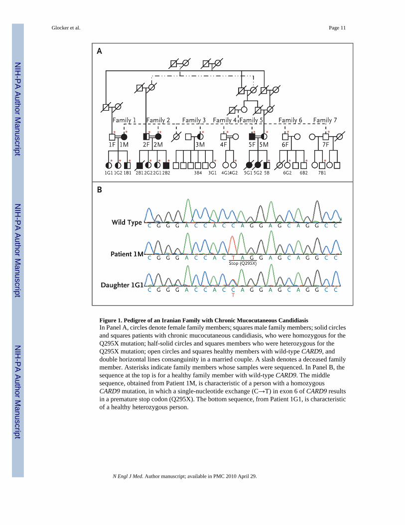
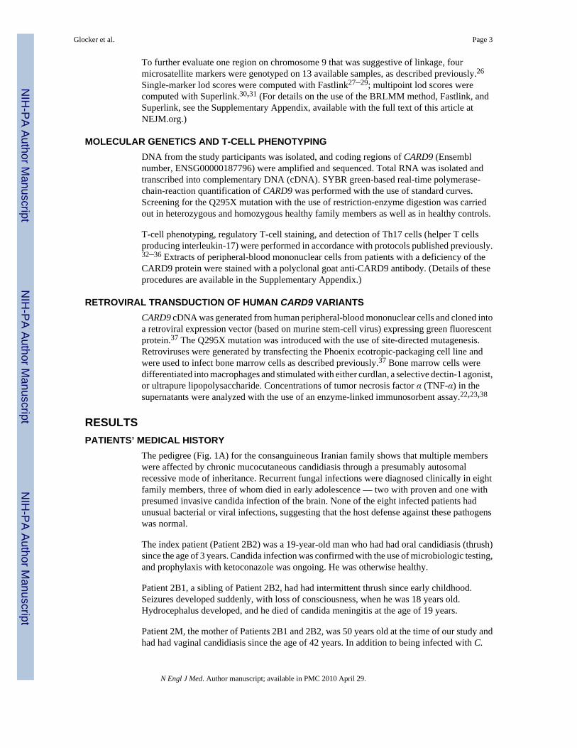
Figure 1. Pedigree of an Iranian Family with Chronic Mucocutaneous CandidiasisIn Panel A, circles denote female family members; squares male family members; solid circlesand squares patients with chronic mucocutaneous candidiasis, who were homozygous for theQ295X mutation; half-solid circles and squares members who were heterozygous for theQ295X mutation; open circles and squares healthy members with wild-type CARD9, anddouble horizontal lines consanguinity in a married couple. A slash denotes a deceased familymember. Asterisks indicate family members whose samples were sequenced. In Panel B, thesequence at the top is for a healthy family member with wild-type CARD9. The middlesequence, obtained from Patient 1M, is characteristic of a person with a homozygousCARD9 mutation, in which a single-nucleotide exchange (C→T) in exon 6 of CARD9 resultsin a premature stop codon (Q295X). The bottom sequence, from Patient 1G1, is characteristicof a healthy heterozygous person.
Glocker et al. Page 11
N Engl J Med. Author manuscript; available in PMC 2010 April 29.
NIH
-PA Author Manuscript
NIH
-PA Author Manuscript
NIH
-PA Author Manuscript
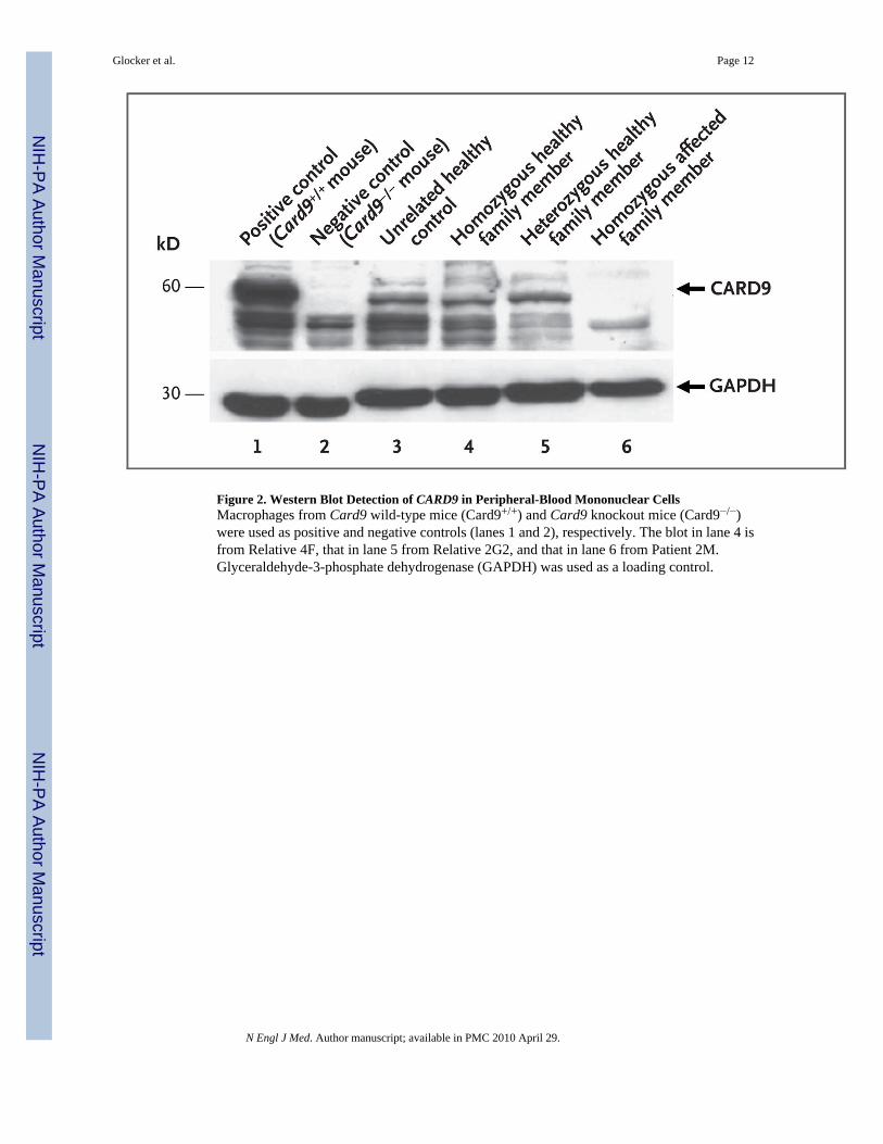
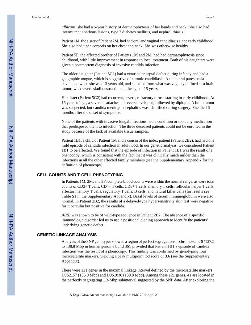
Figure 2. Western Blot Detection of CARD9 in Peripheral-Blood Mononuclear CellsMacrophages from Card9 wild-type mice (Card9+/+) and Card9 knockout mice (Card9−/−)were used as positive and negative controls (lanes 1 and 2), respectively. The blot in lane 4 isfrom Relative 4F, that in lane 5 from Relative 2G2, and that in lane 6 from Patient 2M.Glyceraldehyde-3-phosphate dehydrogenase (GAPDH) was used as a loading control.
Glocker et al. Page 12
N Engl J Med. Author manuscript; available in PMC 2010 April 29.
NIH
-PA Author Manuscript
NIH
-PA Author Manuscript
NIH
-PA Author Manuscript
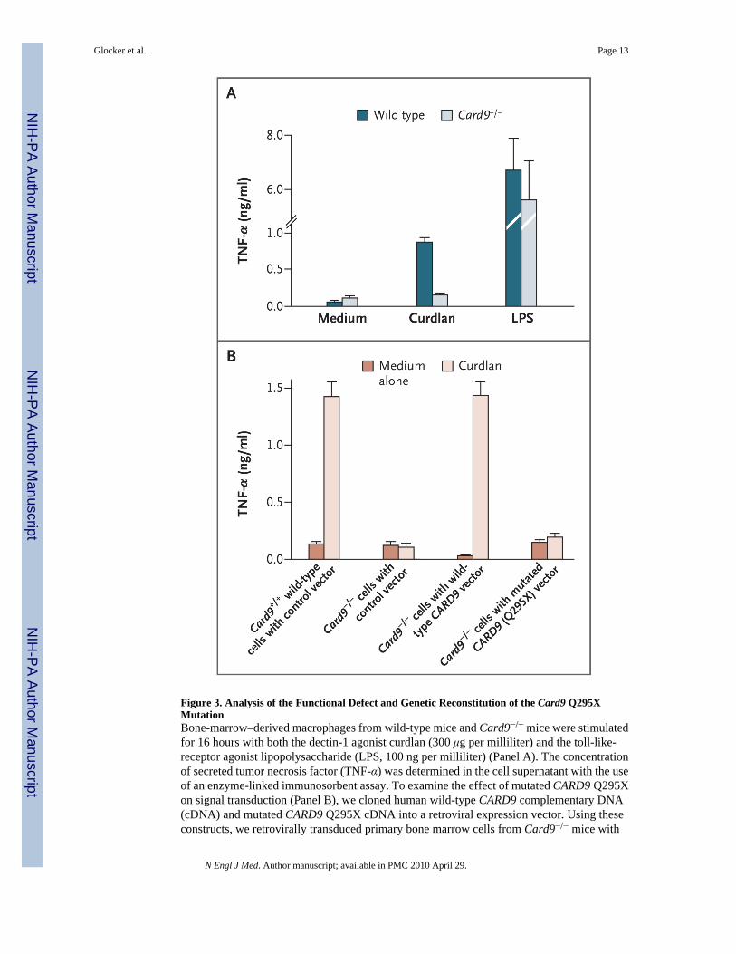
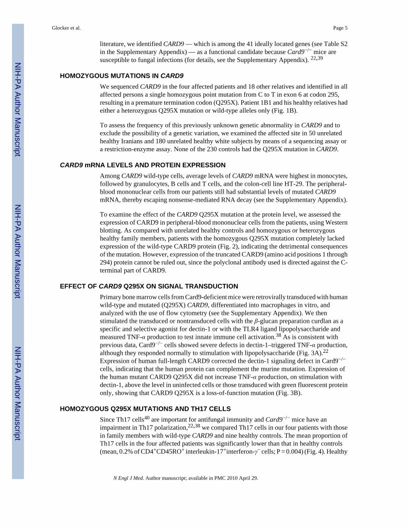
Figure 3. Analysis of the Functional Defect and Genetic Reconstitution of the Card9 Q295XMutationBone-marrow–derived macrophages from wild-type mice and Card9−/− mice were stimulatedfor 16 hours with both the dectin-1 agonist curdlan (300 μg per milliliter) and the toll-like-receptor agonist lipopolysaccharide (LPS, 100 ng per milliliter) (Panel A). The concentrationof secreted tumor necrosis factor (TNF-α) was determined in the cell supernatant with the useof an enzyme-linked immunosorbent assay. To examine the effect of mutated CARD9 Q295Xon signal transduction (Panel B), we cloned human wild-type CARD9 complementary DNA(cDNA) and mutated CARD9 Q295X cDNA into a retroviral expression vector. Using theseconstructs, we retrovirally transduced primary bone marrow cells from Card9−/− mice with
Glocker et al. Page 13
N Engl J Med. Author manuscript; available in PMC 2010 April 29.
NIH
-PA Author Manuscript
NIH
-PA Author Manuscript
NIH
-PA Author Manuscript
either wild-type CARD9 or the mutated CARD9 Q295X. To establish a control, we transducedbone marrow cells from Card9+/+ wild-type mice and Card9−/− mice with a control vectoronly. The transduced bone marrow cells were then differentiated into macrophages in vitro.After stimulation of the macrophages with curdlan for 16 hours, the concentration of secretedTNF-α was determined. The macrophages of Card9−/− mice transduced with human wild-typeCARD9 secreted as much TNF-α upon stimulation with curdlan as did the control macrophagesof wild-type Card9+/+ mice with the control vector. In contrast with these cells, themacrophages of Card9−/− mice transduced with mutated human CARD9 Q295X or with acontrol vector only did not respond with increased secretion of TNF-α upon stimulation withcurdlan, a finding showing that CARD9 Q295X is a loss-of-function mutation that cannotcorrect the dectin-1/Card9 signaling pathway in the cells of Card9−/− mice. T bars indicatestandard deviations in three independent experiments.
Glocker et al. Page 14
N Engl J Med. Author manuscript; available in PMC 2010 April 29.
NIH
-PA Author Manuscript
NIH
-PA Author Manuscript
NIH
-PA Author Manuscript
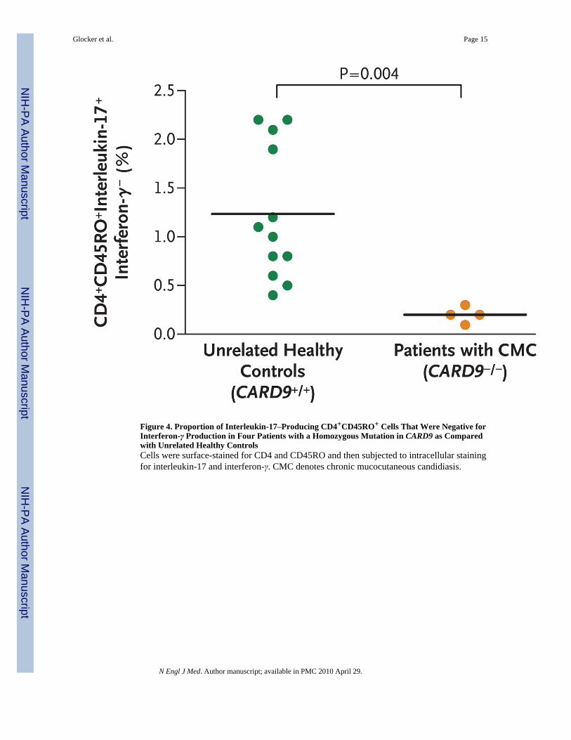
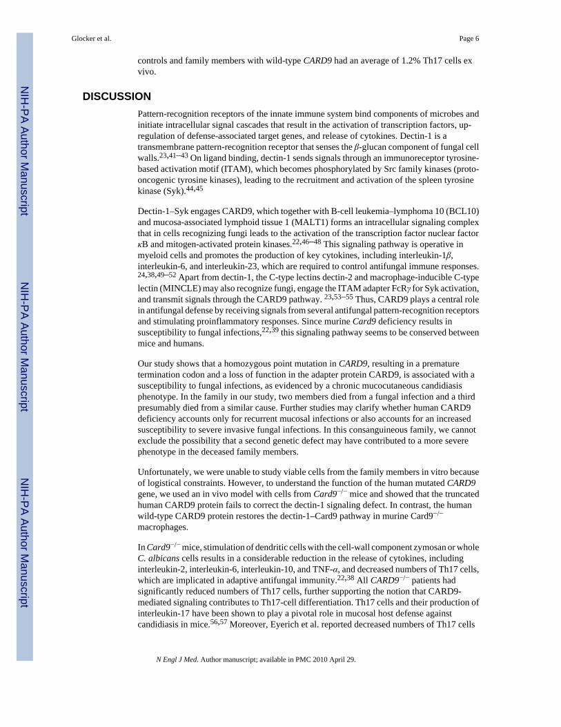
Figure 4. Proportion of Interleukin-17–Producing CD4+CD45RO+ Cells That Were Negative forInterferon-γ Production in Four Patients with a Homozygous Mutation in CARD9 as Comparedwith Unrelated Healthy ControlsCells were surface-stained for CD4 and CD45RO and then subjected to intracellular stainingfor interleukin-17 and interferon-γ. CMC denotes chronic mucocutaneous candidiasis.
Glocker et al. Page 15
N Engl J Med. Author manuscript; available in PMC 2010 April 29.
NIH
-PA Author Manuscript
NIH
-PA Author Manuscript
NIH
-PA Author Manuscript