Embed Size (px)
Citation preview

A Mouse Model of Generalized non-Herlitz JunctionalEpidermolysis Bullosa
Jason A. Bubier, Thomas J. Sproule, Lydia Petell, Cameron Webb, Jo-David Fine, Derry C.Roopenian#,‡, and John P. Sundberg‡,X,**The Jackson Laboratory, 600 Main St, Bar Harbor, ME 04609#The National Epidermoloysis Bullosa Registry, Vanderbilt University School of Medicine,Nashville, TNXSkin Disease Research Center, Vanderbilt University School of Medicine, Nashville, TN
AbstractEpidermolysis bullosa (EB) is a class of intractable, rare, genetic disorders characterized by fragileskin and blister formation as a result of dermal-epidermal mechanical instability. EB presents withconsiderable clinical and molecular heterogeneity. Viable animal models of junctionalepidermolysis bullosa (JEB), that both mimic the human disease and survive beyond the neonatalperiod, are needed. We identified a spontaneous, autosomal recessive mutation (Lamc2 jeb) due toa Murine Leukemia Virus long terminal repeat insertion in Lamc2 that results in a hypomorphicallele with reduced levels of LAMC2 protein. These mutant mice develop a progressive blisteringdisease validated at the gross and microscopic levels to closely resemble generalized non-HerlitzJEB. The Lamc2 jeb mice display additional extracutaneous features such as loss of bonemineralization and abnormal teeth, as well as a respiratory phenotype that is recognized but not aswell characterized in humans. This model faithfully recapitulates human JEB and provides animportant preclinical tool to test novel therapeutic approaches.
INTRODUCTIONThe skin and mucus membranes represent the front-line physical barrier and immunedefense for the protection of visceral tissues and organs. Skin is composed of an epidermisand dermis which are held together at the basement membrane by numerous adhesionmolecules. Epidermolysis bullosa (EB) is a name applied to a heterogeneous group ofinherited skin disorders in which minor trauma leads to blistering of the skin and mucousmembranes, due to defects in the structure and expression any one of these basementmembrane proteins as well as intra-epidermal proteins, desmoplakin and plakophilin 1(PKP1) (Fine and Hintner, 2009; Fine et al., 2008a; Uitto et al., 1997).
Depending upon where the tissue separation occurs, EB can be subdivided into four maingroups: EB simplex, in which blister formation is caused by keratin 5 or 14 mutations,leading to the disruption of the basal keratinocytes; junctional EB (JEB) in which tissuecleavage arises in the lamina lucida; and dystrophic EB, in which cleavage occurs beneath
**Corresponding author: John P. Sundberg, D.V.M., Ph.D., The Jackson Laboratory, 600 Main Street, Bar Harbor, ME 04609-1500,Phone: 207-288-6410, FAX: 207-288-6073/6078, [email protected].‡Authors contributed equally to this work.CONFLICT OF INTERESTThe authors declare no conflict of interest.
NIH Public AccessAuthor ManuscriptJ Invest Dermatol. Author manuscript; available in PMC 2010 December 27.
Published in final edited form as:J Invest Dermatol. 2010 July ; 130(7): 1819–1828. doi:10.1038/jid.2010.46.
NIH
-PA Author Manuscript
NIH
-PA Author Manuscript
NIH
-PA Author Manuscript

the lamina densa of the dermo-epidermal basement membrane zone (Fine et al., 2008a;Petronius et al., 2003). A fourth type, Kindler syndrome, is characterized by cleavage withinand beneath the basement membrane zone, and results from mutations in the Kindlin-1 gene(possibly mouse Fermt1) (Fine et al., 2008b; Ussar et al., 2006).
JEB results from mutations within collagen 17a1 (COL17A1) or any of the genes encodingfor the three subunits of laminin-332 (Aumailley et al., 2005), a heterotrimericmacromolecule composed of laminin α3 (LAMA3), laminin β3 (LAMB3), and laminin γ2(LAMC2). Generalized JEB is subclassified into two major subtypes, based on clinical andmolecular criteria. The more severe Herlitz subtype, associated commonly with death duringinfancy or early childhood, is usually characterized by the presence of compoundheterozygote mutations leading to premature termination codons within any of the threelaminin-332 subunit genes. Non-Herlitz JEB is generally less severe and is associated withbetter prognosis. The majority of cases of non-Herlitz JEB result from less severe mutations(missense; in-frame splicing) within the laminin-332 genes (Fine et al., 2008a; Uitto andRichard, 2004).
Because EB is a heritable disease with potentially deadly sequelae, gene therapy may offer adesirable alternative for its treatment (Ferrari et al., 2005). Such therapy, however, is in itsinfancy and must be developed and validated in animal models before it would be justifiableto apply to humans.
Animal models of skin disease are proving invaluable in the study of humangenodermatoses. JEB-like phenotypes have been previously described in dogs (Capt et al.,2005; Dunstan et al., 1988; Nagata et al., 1997; Nagata et al., 1995) and horses (Frame etal., 1988; Spirito et al., 2002). A viable mouse model of JEB would be invaluable, since itcan manipulated experimentally and genetically. Targeted disruption of laminin alpha 3,Lama3 (Lama3tm1Crt) (Ryan et al., 1999), or laminin gamma 2, Lamc2 (Lamc2tm1Uit) (Menget al., 2003), as well as the spontaneous intragenic insertion of an intracisternal A particle(IAP) element into laminin beta 3, Lamb3 (Lamb3IAP) (Kuster et al., 1997), results in a JEB-like phenotype in mice, but all lead to perinatal lethality, thus reducing their experimentalutility. A recently produced targeted mouse mutant in collagen XVII (Col17a1tm1Shzu)develops a JEB phenotype. However, most of these mice die within 2 weeks of birth (Nishieet al., 2007). A hypomorphic allele of collagen VII (Col7A1tm1Lbt) has been shown to resultin a dystrophic EB phenotype; most of these mice die before 28 days (Fritsch et al., 2008).The only long-term viable EB mouse model currently available is an inducible model for theDowling-Meara variant of generalized EB simplex (Cao et al., 2001). Here we describe apreviously unreported model for JEB in which a hypomorphic allele of the gene Lamc2results in viable mice that progressively develop a syndrome remarkably similar to thatobserved in humans.
RESULTSHistopathology of Lamc2jeb mutant mice
Abnormal mice were first noted in an inbred stock of 129X1/SvJ (129) mice. Theydeveloped conspicuously hyperemic, ulcerated pinnae (ears) with similar changes on theirtails. These traits were apparent with 100% penetrance in this inbred stock with a meanonset of 154 +/− 4.3 days (n=23) but were never detected in other 129 stocks. Completenecropsies (Relyea et al., 2000; Seymour et al., 2004), were initially performed on twoaffected and two clinically normal, six month old male and female littermates. Lesions werelimited to the skin on several parts of the body (Fig. 1A-C). Histologically, the ears wereseverely ulcerated with granulation tissue forming at the base of the ulcers (Fig. 1E).Dermal-epidermal separation at the level of the basement membrane was present (Fig. 1E).
Bubier et al. Page 2
J Invest Dermatol. Author manuscript; available in PMC 2010 December 27.
NIH
-PA Author Manuscript
NIH
-PA Author Manuscript
NIH
-PA Author Manuscript

The most diagnostic areas were the tail (Fig. 1D) and footpads (Fig. 1F), where the skin wasnot ulcerated. Subepidermal separation, with little to no dermal inflammation, was present.Large areas of epidermis lifted off of the tail (Fig. 1C), confirming the presence of markedmechanical fragility of the skin. The histological features of the footpads in the same mouseranged from areas of normality, to acute blister formation, or healing by scarring. Dorsalthoracic skin was normal or had areas of ulceration with adjacent dermal-epidermalseparation (not shown). Histological, histochemical, and immunohistochemical (IHC)analyses supported the diagnosis of JEB. There was no evidence of any keratinocytesadhering to the base of the cleft, thereby excluding a variant of EB simplex. The mostabundant follicular and interfollicular epidermal keratins, most notably keratins 5 and 14,were expressed normally, as would be expected in JEB skin (data not shown). This was alsotrue for mouse specific keratin 6, which is normally expressed in the companion layer of thehair follicle (as it was in these mice) and only in hyperplastic epidermis (data not shown). Inparticular, immunohistochemical localization of type IV collagen to the base of the blister(not shown), indicative of intralamina lucida cleavage confirms the diagnosis of JEB(Megahed, 2004). Immunofluorescence blister mapping revealed that_integrin alpha 6(ITGA6) and bullous pemphigoid antigen 180 (COL17A1) expression were located on theroof of the blister while type VII collagen (COL7A1) expression was exclusively present onthe blister floor (Fig. 2), thereby localizing the blister to the region where laminin type 5(laminin-332), which includes laminin gamma 2, is located. These observations closelyparallel those seen in skin lesions from patients with JEB. In addition, the presence ontransmission electron microscopy (TEM) of intralamina lucida cleavage and reducednumbers of rudimentary-appearing hemidesmosomes further support the diagnosis that thisis a model for generalized non-Herlitz type JEB (Fig. 3).
Mapping Lamc2 jeb
The autosomal recessive junctional epidermolysis bullosa mutation (gene symbol Lamc2 jeb)arose spontaneously. To map the mutated gene, we performed an intraspecific cross betweenC57BL/6J (B6) wild type (+/+) (females) mice and 129-Lamc2jeb/Lamc2jeb mutant malemice. All F1 progeny were unaffected. These F1 progeny were then backcrossed (129-Lamc2jeb/Lamc2 jeb X B6 +/+)F1 x 129-Lamc2 jeb /Lamc2 jeb mice, with ~50% of theiroffspring being affected, thus confirming the action of a single autosomal recessiveMendelian trait. To map the mutated gene locus, DNAs from the backcross mice wereanalyzed using a DNA pooling genome scanning method (Taylor et al., 1994) usingallelically discriminant dinucleotide repeat markers mapping to 15-20 cM intervalsthroughout the genome. Seventeen affected, 11 males/6 females, 18 unaffected littermates,11 males/7 females, parental 129, B6, and F1 hybrids were used for initial mapping. Thislow resolution mapping technique revealed that the mutation was linked to a ~16 Mb regionon mouse Chromosome (Chr) 1 between 143.810 and 160.466 Mb. Once the preliminarymap position for the Lamc2 jeb locus was established, we developed a high resolution map ofthe region surrounding this locus with the goal of mapping Lamc2 jeb to an interval in whichcandidate genes could be evaluated for “positional cloning”. DNA from each F2 mouse wasallele typed for the most proximal and distal recombinant markers within the ~16 Mbinterval. As the mutation arose in 129 mice, the most informative mice were Lamc2 jeb
homozygotes with at least one B6 allelic marker in the interval. This mapping narrowed themutation to a ~5 Mb region between 152.110 and 155.702 (75-82 cM) on Chr 1 to a regionincluding Lamc1 and Lamc2 (Fig. 4A). Lamc1 encodes for the laminin gamma 1 subunit, acomponent of 10 of the 11 laminin family members. Absence of Lamc1 results in embryoniclethality due to the failure of endoderm differentiation (Bader et al., 2005; Smyth et al.,1999; Willem et al., 2002). However, as summarized earlier, mutations in Lamc2 areassociated with forms of JEB (Meng et al., 2003).
Bubier et al. Page 3
J Invest Dermatol. Author manuscript; available in PMC 2010 December 27.
NIH
-PA Author Manuscript
NIH
-PA Author Manuscript
NIH
-PA Author Manuscript

The Lamc2jeb mutation is caused by a retroviral insertionBecause both Lamc1 and Lamc2 were considered candidate genes for explaining theLamc2jeb mutation, we first evaluated gene expression differences using quantitative real-time PCR. Five primers sets specific to Lamc1, as well as five specific to Lamc2. NeitherLamc1 nor Lamc2 showed a significant change in expression level between Lamc2jeb/Lamc2jeb and + controls when three mutants and three biological control replicates werecompared (data not shown). Northern blot analysis of skin poly A RNA probed with aLamc1 fragment revealed the presence of only the expected 7.6 kb band (data not shown).Lamc2 is reported to encode for two transcripts (5.1 and 2.4 kb), the smaller of which is notexpected in adult skin because it exhibits very specific tissue and developmental expression(Airenne et al., 1996; Airenne et al., 2000; Lee et al., 2001). Skin of wild type 129 miceyielded the expected dominant 5.1 kb transcript when probed with a DNA fragment thatspanned exons 1-17. By contrast, 129-Lamc2 jeb homozygous mice yielded a majortranscript of 6.5 kb and potentially a weak band consistent with the 5.1 kb wild-typetranscript (Fig. 4C, left panel). When probed with a more 3′ fragment spanning exons 11-21,two major mutant bands were observed, one again consistent with the 6.5 kb band and asecond band of approximately 4.5 kb (Fig. 4C, right panel). This lower size band, detectedwith the exon 11-21 probe but not with the exon 1-17 probe, suggests that the mutation canalso result in a second alternative transcript that initiates 3′ of exon 17.
Sequencing of all exons failed to reveal a change between the jeb and 129 wild type Lamc2;however, sequencing of all introns revealed the presence of a single Murine Leukemia VirusLong Terminal Repeat (MLVLTR) insertion of 560 bp within the 18th intron (between exons18 and 19, 900 bp from the end of 18 and 130 bp before the beginning of exon 19) of Lamc2(Fig. 4B). Restriction fragment length polymorphism (RFLP) analysis of genomic DNA bySouthern blotting was consistent with this single LTR insertion being the only detectedgenetic change in the mutant Lamc2 locus (Fig. 4B). Given the increased size of the majorLamc2 jeb transcript observed by Northern blotting, we hypothesized that this increase isbecause of the incorporation of the LTR as_part of the transcript. Indeed, by use ofoligonucleotide primer pairs nested in the LTR and either 3′ or 5′ exonic sequence for RT-PCR, we detected and sequence verified cDNA products containing the intron and LTR(Supp. Figs. 1 and 2). This transcript includes 5′ exons, does not splice out intron 18 and theLTR, and introduces a TAG translational stop codon in intron 18 (Supp. Figs. 1 and 2).However, a correctly spliced WT transcript is also produced at low abundance as it has beendetected repeatedly by RT-PCR and sequencing (Supp. Figs. 1 and 3). Consistent with thislow abundance transcript, a low abundance LAMC2 protein of the correct WT size is foundin skin extract of Lamc2 jeb mice by Western blot but negligibly in skin of mice carrying thenull Lamc2tm1Utt allele (Meng et al., 2003) (Fig.4D). Immunoreactive LAMC2 protein isalso detected in situ_with antibodies reactive to two different regions of the protein (Fig. 5).
Finally, to confirm that the mutation was within Lamc2, a genetic complementation test wasperformed by crossing Lamc2 jeb /Lamc2 jeb mice to mice heterozygous for the Lamc2tm1Ui
gene inactivation allele of Lamc2 (Meng et al., 2003). All resulting Lamc2 jeb /Lamc2_tm1Utt
heterozygotes but not Lamc2jeb /+ or Lamc2tm1Utt/+ littermates developed the characteristicblisters by two months of age confirming that the mutations were allelic (Fig. 4E). Thisexperiment provided formal genetic proof that Lamc2jeb is the mutant allele. The combinedresults support the model in which the Lamc2jeb mutation is caused by the insertion of aretroviral LTR. This insertion results in abnormal transcripts, but also permits an apparentlyintact but low abundance Lamc2 WT transcript that results in a greatly reduced level ofLAMC2 protein as detected by Western blot (Fig. 4D). Lamc2jeb is therefore a hypomorphicLamc2 allele with the reduced amount of LAMC2 protein being sufficient to permit longterm viability.
Bubier et al. Page 4
J Invest Dermatol. Author manuscript; available in PMC 2010 December 27.
NIH
-PA Author Manuscript
NIH
-PA Author Manuscript
NIH
-PA Author Manuscript

Effect on Bone MineralizationIn order to evaluate if the Lamc2 hypomorphic mouse displayed any of the bonemineralization features observed in humans (Fewtrell et al., 2006), mice were analyzed bydual energy x-ray absorptiometry. As summarized in Table 1, when mice are asymptomaticearly on, as measured by gross ear blistering, there were no statistically significantdifferences in bone mineral composition (BMC) and bone mass density (BMD). However,as mice age and develop blisters, Lamc2jeb/Lamc2 jeb mice display significant decreases inboth of these parameters.
Tooth InvolvementGeneralized enamel hypoplasia is a principal feature of both Herlitz and non-Herlitz JEB(Wright et al., 1993). As seen in Fig. 6A, the incisors of Lamc2jeb/Lamc2 jeb mice hadsubstantial pitting, as detected by scanning electron microscopy.
Respiratory InvolvementLamc2 is highly expressed in lung ((http://symatlas.gnf.org/SymAtlas/). In addition to theskin manifestations, Lamc2jeb homozygous mutant mice were dyspneic, suggestingpulmonary function abnormalities. No tracheal abnormalities were observed by H&Estaining or TEM (data not shown), although abnormal tracheal hemidesmosomes werereported in the neonatal homozygous Lamc2 tm1Utt mice (Nguyen et al., 2006). In order toassess pulmonary mechanics, Lamc2jeb/Lamc2 jeb mice were analyzed by the forcedoscillation technique. This process allows for the evaluation of lung function by evaluationof the inflation and deflation phases of each breath and the construction of a classicpressure-volume (PV) curve (Salazar and Knowles, 1964). As shown in Fig. 6B, for apositive end-expiratory pressure (PEEP) of 3 the Lamc2jeb/Lamc2 jeb mice have functionallung defects, resulting in decreased resistance and/or elastance and overall_lung restrictionthat are similar to those reported in the cystic fibrosis transmembrane conductance regulator(Cftr) deficient mouse (B6.129P2-Cftrtm1Unc/J) (Cohen et al., 2004).
DISCUSSIONWe describe a previously unreported, spontaneous mutation in Lamc2 that results in aphenotype of cutaneous blister formation with subsequent ulceration that is remarkable tothe extent to which it mimics and is instructive toward understanding human JEB. Affectedmice develop blisters as they age but many heal by forming scars under the re-epithelializedsurface such that they do not succumb to fluid loss and electrolyte imbalance that wouldoccur with extensive loss of an intact skin barrier. Blister formation above the plane ofCOL4A1 and COL7A1 and below the plane of ITGA6 and COL17A1 expression supportthe diagnosis of JEB.
Retroviral insertion within introns is a common mutagenic mechanism in mice (Helms et al.,2005; Seperack et al., 1995; Stoye et al., 1988). Cloning and sequence analysis of Lamc2revealed that our mouse with the generalized non-Herlitz JEB phenotype had a single MLVLTR insertion within intron 18 of the Lamc2 gene. This insertion gives rise to an abnormallylarge (6.5 kb) major transcript abrupts the normal excision of intron 18 and introduces aTAG translational stop codon (Supp. Fig. 1). While transcripts with a translational stopcodon introduced distantly (>55 bases) from the terminal intron/exon junctions oftenexperience nonsense-mediated decay, there are exceptions (Frankel et al., 2009; Zhao et al.,2005). The apparent stability of the transcript caused by the MLV LTR insertion may haveoverridden this survey mechanism.
Bubier et al. Page 5
J Invest Dermatol. Author manuscript; available in PMC 2010 December 27.
NIH
-PA Author Manuscript
NIH
-PA Author Manuscript
NIH
-PA Author Manuscript

The MLV insertion also leads to the initiation of a smaller (4.5 kb) alternative transcriptinitiating between exons 18 and 21, perhaps using the known alternate promoter locatedwithin exon 19 (Lee et al., 2001). Any protein produced from this truncated transcript isunlikely to have a biological effect as it would lack amino acids including the necessary N-terminal signal sequence. However, a correctly spliced but weakly expressed Lamc2transcript is detected by Northern and Western blots, and is confirmed by less quantitative insitu immunofluorescence analyses of the dermal-epidermal junction. The fact that the skinlesions are observed only in Lamc2jeb/Lamc2jeb mice is consistent with a recessive mutation,and argues against dominant effects that could arise by the above-described aberrantLamc2jeb transcripts. This is further supported by the complementation test showing thatLamc2tm1Uit/Lamc2jeb compound heterozygotes develop blisters similar to the Lamc2jeb
homozygote. The low abundance, apparently intact transcript is the most parsimoniousexplanation for this hypomorphic allele._Hypomorphic alleles arising from alternate splicingor exon skipping of LAMC2 and LAMB3 resulting in a reduced WT transcript have also beenreported for JEB in humans (Castiglia et al., 2001; Nakano et al., 2002; Pulkkinen et al.,1994).
Classical histological studies evaluated the teeth of JEB patients and concluded that the teethdefects were due to extensive disruption of the ameloblast function and life cycle (Arwill etal., 1965). Unlike humans, rodent incisors are continuously growing. Mice with a targetedknock-out allele of Lamc3 resulted in disrupted ameloblast differentiation and decreasedenamel deposition (Ryan et al., 1999). Recent work on the collagen XVII-mutant mouse,Col17a1tm1Shzu, concluded that collagen XVII is also required for tooth enamel formation(Asaka et al., 2008). Laminin-332 is known to have a critical function in the adhesion of thedental epithelial cells with the enamel (Yoshiba et al., 1998). Lamc2jeb mice developeddental abnormalities consistent with that observed in humans (Arwill et al., 1965; Kirkhamet al., 2000; Wright et al., 1996; Yoshiba et al., 1998). Our results are consistent with a rolefor LAMC2 as part of Laminin-332 for proper enamel formation.
They also show the loss of bone mineralization which is also a known characteristic of moreseverely affected humans with JEB (Fewtrell et al., 2006). Finally, there was a conspicuousrespiratory dysfunction in Lamc2jeb mice. Forty percent of JEB patients, regardless ofsubtype, have moderate to severe tracheolaryngeal involvement (Fine et al., 2007). Of these,a substantial minority, primarily those with Herlitz JEB, die as a direct result of the severityof the upper airway disease while others die of “pneumonia” and other undefined respiratoryailments (Fine et al., 2007). Importantly, respiratory disease in human JEB patients has yetto be described at the level of a quantitative functional assay and this mouse thereforeprovides a unique opportunity whereby pulmonary function and JEB can be meticulouslystudied. In addition, since this previously unreported spontaneous mouse model for JEBsurvives well into adulthood, it provides a useful rodent model for many other levels ofinvestigation, including a mechanistic description of JEB.
Finally, the previously unreported Lamc2 JEB model described here holds promise forevaluating the efficacy and safety of gene therapy. Recent success with ex vivo gene therapyfor the treatment of EB appear to be promising (Mavilio et al., 2006) as well as bonemarrow transplantation approaches to correct skin defects (Tolar et al., 2009). The Lamc2jeb
mice described here provide the biological tool needed to validate these approaches beforetesting in human patients.
Bubier et al. Page 6
J Invest Dermatol. Author manuscript; available in PMC 2010 December 27.
NIH
-PA Author Manuscript
NIH
-PA Author Manuscript
NIH
-PA Author Manuscript

MATERIALS AND METHODSMice
Mice were maintained in conventional specific-pathogen free barrier facilities. Routine andskin specific microbiological work done on 129.X1-Lamc2jeb/Lamc2jeb mice with cutaneousulcers revealed that no known mouse infectious agent was involved in the pathogenesis ofJEB. The Jackson Laboratory follows husbandry practices in accordance with the AmericanAssociation for the Accreditation of Laboratory Animal Care and all work was done withthe approval of our Institutional Animal Care and Use Committee. Mice were sex and agematched for each experiment. The mutation arose during the production of 129X1-Fcgrttm1Dcr/Dcr JR#3980 mice and was isolated as 129X1-Lamc2jeb JR#6859 and B6-Lamc2jeb JR#7061 mice. Lamc2tm1Uit (JR#7058) mice were a kind gift from J. Uitto(Thomas Jefferson University).
Gene MappingTail tips were amputated, DNA extracted using a modified hot NaOH protocol (Truett et al.,2000), and DNA pooled from 12 mutant mice. DNA was also collected from 10 clinicallynormal F2 progeny and 2 parental mice (129-Lamc2jeb/Lamc2jeb) females and B6 +/+ males.Equal amounts of DNA from the 12 mutant mice were mixed to make a pooled sample. Asimilar pooled sample was assembled from the 12 normal siblings. These two pools weretested for simple sequence length polymorphisms (SSLPs) allele ratios as compared toparental and F1 controls using 87 markers spaced at 7-35cM (mean 14.4 cM spacing)throughout the genome (23). Samples were amplified on a MJ research PTC-1000 ThermalCycler with initial incubation at 95°C for 5 min, followed by 40 cycles of melting at 95°Cfor 15s, annealing at 60°C for 20s at 70°C for 30s; followed by holding the samples at 4 °C.Each reaction had a volume of 15 μl containing 3 ng DNA template and 0.025 units/μl TaqDNA Polymerase (Eppendorf, NY). The final concentration of the other reagents were asfollows: each primer at 0.4 μM (MWG, High Point NC), 1.5 mM MgCl, 50 mM KCl, 1 mMß-mercaptoethanol, 25 mM TAPS (N-tris-(hydroxy-methyl)-methyl-3-amino-propanesulfonic acid, sodium salt), 200 μM each dNTP (Promega, Madison WI). All noveldinucleotide repeat based mapping markers are available at MGI(http://www.informatics.jax.org). Markers were electrophoresed in the presence of ethidiumbromide and run on a 0.7% agarose with 1.5% Synergel (Diversified BioTech, Boston MA)in Tris-acetate-EDTA. Gels were run at 160 V for 1.5-2 hr and photographed using mediumwave UV Syngene Ingenius UV (Frederick, MD) digital camera system.
HistopathologyOrgans were fixed overnight in Fekete’s acid-alcohol-formalin fixative, rinsed extensively,held overnight in distilled water, and stored in 70% ethanol until processed. Organs weretrimmed, embedded in paraffin, serially sectioned at 6 μm, and then stained withhemotoxylin and eosin (H&E) or periodic acid Schiff.
Immunofluorescence ImagingWhole ears were cut longitudinally and each half and prepared was rinsed in OCT (Tissue-Tek) until saturated (about 30 seconds to 1 minute). One half of the ear tissue was embeddedcut side down into the partially frozen OCT in the base mold. The section was allowed tofreeze after which more OCT was added and stored at −80°C. Samples were cut between 10and 12 μm thick onto super frost plus slides (Fisher). Slides were stored at −20°C until used.For antibody staining the sectioned tissues were fixed in ice cold acetone for 10 minutes,washed three times for 10 minutes in phosphate buffered saline (PBS), and then allowed todry. Primary antibodies were added with 3% fetal bovine serum for 2 hours at room
Bubier et al. Page 7
J Invest Dermatol. Author manuscript; available in PMC 2010 December 27.
NIH
-PA Author Manuscript
NIH
-PA Author Manuscript
NIH
-PA Author Manuscript

temperature in a humid chamber. The slides were washed in PBS, incubated in the dark withfluorescence-labeled secondary antibodies for 1 hour, washed and cover slipped using anti-fade gel mounting media (Sigma-Aldrich, St. Louis, MO). Imaging was performed using aSP5 Leica confocal microscope at 63x. The following primary antibodies were used: rabbitanti mouse BP180 and COL7A1 (gifts of Z. Liu), rabbit anti mouse LAMA3, LAMB3,LAMC2 LE1-3 (AA 22-186, exons 2-6), LAMC2 LE4-6 (AA 460-606, exons 11-13, a giftof T. Sasaki) and rat anti-mouse ITGA6 (GeneTex, San Antonio, TX). All secondaryantibodies were anti-rabbit or anti-rat Alexa Fluor 546 (Invitrogen/Molecular Probes,Carlsbad, CA).
Electron MicroscopySamples were prepared according to routine methods previously described (Bechtold, 2000).Skin samples were fixed in 2.5% glutaraldehyde in 0.1M phosphate buffer (PB) pH 7.2overnight at 4°C. Samples were then washed 2 × 15 minutes in PB and soaked in 2%osmium tetroxide overnight at 4°C followed again by 2 × 15 minute washes in PBS.Samples for TEM were serially dehydrated in ethanol followed by propylene oxideincubation twice for 30 minutes. The samples were then treated with propylene oxide:epon-araldite (1:1) for 24 hrs with rotation followed by 24 hrs of rotation in pure resin. Sampleswere then cured for 1 day at 70°C, sectioned, and examined by on a JEM-1230 TEM (JEOL,Tokyo, Japan). For scanning electron microscopy, teeth samples were prepared inKarnovsky’s fixative and mounted with double-stick tape on aluminum stubs, sputter-coatedwith a 4 nm layer of gold, and examined at 20kV at a working distance of approximately 15mm on a Hitachi S3000N VP Scanning Electron Microscope (Hitachi Science Systems,Japan).
Transcript Analysis and SequencingFor Northern blot, total RNA was isolated using the standard Trizol reagent method(Invitrogen). Poly A+ enrichment was performed by passing over oligo(dT) cellulosecolumn (Applied Biosystems/Ambion, Foster City, CA). Two micrograms of RNA per lanewas analyzed by Northern blot. 1 ug of total RNA was used to make cDNA using messagesSensor RT (Applied Biosystems/Ambion). The cDNA was used to produce probes forLamc1 and Lamc2 using primers for Lamc1F, Lamc1R, Lamc2.2F, Lamc2.18R, Lamc2.12F,Lamc2.22R is described in Supplemental Table 1. Additional primers used for quantitativeReal Time PCR on cDNA produced from both WT and 129X1/SvJ Lamc2jebmice are listedin Supplemental Table 1. Gene expression differences were determined using the GlobalPattern Recognition (GPR) software (Akilesh et al., 2003). For sequencing, total RNA wasisolated from spleen using RNAqueous 4-PCR (Ambion). Oligo(dT)18 and random decamerprimed cDNA was synthesized using ThermoScript RT (Invitrogen). Sequencing of Lamc2was done using gene specific primers on an Applied Biosystems 3700 and analyzed usingSequencher 4.8 (Gene Codes Corporation, Ann Arbor, MI).
FlexiVent MeasurementsMice are anesthetized with ketamine/dormitor at doses based on body weight. To preventthe mice from breathing against the respirator, the mice were treated with pancuroniumbromide in NaCl as a muscle relaxant at 0.2mg/kg. Once the mouse was properlyanesthetized, a small incision was made between the 3rd and 5th tracheal rings whereupon a1.27 cm long tracheal cannula was inserted. The cannula was secured in place with suturematerial. The mouse was then fixed to the flexiVent ventilator (SCIREQ, Montreal, CA) andventilated at 200 breaths/min with a tidal volume of 10 ml/kg body weight. Once the mousewas breathing passively, 2 consecutive sigh breaths were performed to open the airways andlungs. Baseline R, C, and E measures were recorded. Sigh breaths were performedthroughout the experiment to keep airways and lungs open.
Bubier et al. Page 8
J Invest Dermatol. Author manuscript; available in PMC 2010 December 27.
NIH
-PA Author Manuscript
NIH
-PA Author Manuscript
NIH
-PA Author Manuscript
Bone Density MeasurementsPeripheral dual-energy X-ray absorptiometry (pDXA; PIXI-mus, GE-Lunar, Madison WI)was used to assess bone mineral density and bone mineral composition, with head exclusion.This methodology is routinely used in and validated in small animals (Bouxsein et al.,2002). Mice were anesthetized with tribromoethanol at 180 mg/kg intraperitoneal, allowing5 minutes for sedation before scans.
Western Blot AnalysisSkin lysates were prepared in 20 mM Tris-HCl (pH 7.5), 135 mM NaCl, 1.5 mM MgCl2, 1mM EGTA, 1% Triton X-100, and 10% glycerol supplemented with a complete proteaseinhibitor cocktail (Gene Technology, Somerville, MA). Total protein was quantified usingBCM protein analysis kit (Thermo Scientific/Pierce, Rockford, IL). 10-100 ug was separatedon a BioRad Criterion XT 4-12% Bis-Tris gel, electroblotted onto a PVDF membrane(BioRad, Hercules, CA), and probed with a 1:500 dilution of LAMC2 sc-28330 anti-humanLAMC2 antibody (Santa Cruz, Santa Cruz, CA). The secondary antibody used (1:1000) wasgoat anti mouse IgG conjugated to HRP (Southern Biotech, Birmingham, AL) withdetection using Pierce ECL western blot substrate. Images were collected on a FujiLAS1000 CCD luminescent imaging system, with band intensities quanified using ImageJ(NIH). As a loading control the blot was stripped and re-probed with rabbit anti-mouse actin(Sigma) and goat anti-rabbit conjugated HRP.
Supplementary MaterialRefer to Web version on PubMed Central for supplementary material.
AcknowledgmentsThis work was supported by Dystrophic Epidermolysis Research Association (DebRA) - International and TheNational Institutes of Health (DK56597, DCR; AR49288, JPS). The authors thank L. E. King, Jr. for discussions onthe comparative aspects of this mouse model with the human disease, and A. Brown and especially W. Frankel foradvice regarding MLVs and genomic analyses. We appreciate Drs. J. Uitto and J. Klement for making theLamc2tm1Uit mouse available, T. Sasaki for anti- LAMA3, LAMB3, and LAMC2 antibodies, and Z. Liu for theanti-BP180 and anti COL7A1 antibodies. We also thank K.A. Silva and J. Miller for initial sample preparation andimmunohistochemical analysis as well as L. Rowe and M. Barter for SSLP mapping services. We thank A.Nicholson and J. Ryan for pulmonary physiology analyses, B. King and T. Sterns for statistical analysis, and L.Bechtold and P. Finger for EM sample preparation.
Abbreviations
EB epidermolysis bullosa
jeb junctional epidermolysis bullosa
Lamc2 laminin gamma 2 gene
Lamc2jeb mouse laminin gamma 2 allelic mutation
MLV Murine Leukemia Virus
ReferencesAirenne T, Haakana H, Sainio K, Kallunki T, Kallunki P, Sariola H, et al. Structure of the human
laminin gamma 2 chain gene (LAMC2): alternative splicing with different tissue distribution of twotranscripts. Genomics 1996;32:54–64. [PubMed: 8786121]
Bubier et al. Page 9
J Invest Dermatol. Author manuscript; available in PMC 2010 December 27.
NIH
-PA Author Manuscript
NIH
-PA Author Manuscript
NIH
-PA Author Manuscript

Airenne T, Lin Y, Olsson M, Ekblom P, Vainio S, Tryggvason K. Differential expression of mouselaminin gamma2 and gamma2* chain transcripts. Cell Tissue Res 2000;300:129–37. [PubMed:10805082]
Akilesh S, Shaffer DJ, Roopenian D. Customized molecular phenotyping by quantitative geneexpression and pattern recognition analysis. Genome Res 2003;13:1719–27. [PubMed: 12840047]
Arwill T, Olsson O, Bergenholtz A. Epidermolysis Bullosa Hereditaria. 3. A Histologic Study ofChanges in Teeth in the Polydysplastic Dystrophic and Lethal Forms. Oral Surg Oral Med OralPathol 1965;19:723–44. [PubMed: 14298560]
Asaka T, Akiyama M, Domon T, Nishie W, Natsuga K, Fujita Y, et al. Type XVII Collagen is a KeyPlayer in Tooth Enamel Formation. Am J Pathol. 2008
Aumailley M, Bruckner-Tuderman L, Carter WG, Deutzmann R, Edgar D, Ekblom P, et al. Asimplified laminin nomenclature. Matrix Biol 2005;24:326–32. [PubMed: 15979864]
Bader BL, Smyth N, Nedbal S, Miosge N, Baranowsky A, Mokkapati S, et al. Compound geneticablation of nidogen 1 and 2 causes basement membrane defects and perinatal lethality in mice. MolCell Biol 2005;25:6846–56. [PubMed: 16024816]
Bechtold, LS. Ultrastructural evaluation of mouse mutations. In: Sundberg, JP.; Boggess, D., editors.Systematic characterization of mouse mutations. CRC Press; Boca Raton: 2000. p. 121-9.
Bouxsein ML, Rosen CJ, Turner CH, Ackert CL, Shultz KL, Donahue LR, et al. Generation of a newcongenic mouse strain to test the relationships among serum insulin-like growth factor I, bonemineral density, and skeletal morphology in vivo. J Bone Miner Res 2002;17:570–9. [PubMed:11918215]
Cao T, Longley MA, Wang XJ, Roop DR. An inducible mouse model for epidermolysis bullosasimplex: implications for gene therapy. J Cell Biol 2001;152:651–6. [PubMed: 11157990]
Capt A, Spirito F, Guaguere E, Spadafora A, Ortonne JP, Meneguzzi G. Inherited junctionalepidermolysis bullosa in the German Pointer: establishment of a large animal model. J InvestDermatol 2005;124:530–5. [PubMed: 15737193]
Castiglia D, Posteraro P, Spirito F, Pinola M, Angelo C, Puddu P, et al. Novel mutations in theLAMC2 gene in non-Herlitz junctional epidermolysis bullosa: effects of laminin-5 assembly,secretion, and deposition. J Invest Dermatol 2001;117:731–9. [PubMed: 11564184]
Cohen JC, Lundblad LK, Bates JH, Levitzky M, Larson JE. The “Goldilocks effect” in cystic fibrosis:identification of a lung phenotype in the cftr knockout and heterozygous mouse. BMC genetics2004;5:21. [PubMed: 15279681]
Dunstan RW, Sills RC, Wilkinson JE, Paller AS, Hashimoto KH. A disease resembling junctionalepidermolysis bullosa in a toy poodle. Am J Dermatopath 1988;10:442–7. Dunstan, R. W., R. C.Sills, J. E. Wilkinson, A. S. Paller, and K. H. Hashimoto. 1988. [PubMed: 3228192]
Ferrari S, Pellegrini G, Mavilio F, De Luca M. Gene therapy approaches for epidermolysis bullosa.Clin Dermatol 2005;23:430–6. [PubMed: 16023940]
Fewtrell MS, Allgrove J, Gordon I, Brain C, Atherton D, Harper J, et al. Bone mineralization inchildren with epidermolysis bullosa. The British journal of dermatology 2006;154:959–62.[PubMed: 16634901]
Fine, J-D.; Hintner, H. Life with epidermolysis bullosa (EB): etiology, diagnosis, multidisciplinarycare, and therapy. Vol. xix. Springer: Wien; New York: 2009. p. 338
Fine JD, Eady RA, Bauer EA, Bauer JW, Bruckner-Tuderman L, Heagerty A, et al. The classificationof inherited epidermolysis bullosa (EB): Report of the Third International Consensus Meeting onDiagnosis and Classification of EB. Journal of the American Academy of Dermatology 2008a;58:931–50. [PubMed: 18374450]
Fine JD, Johnson LB, Weiner M, Suchindran C. Tracheolaryngeal Complications of InheritedEpidermolysis Bullosa: Cumulative Experience of the National Epidermolysis Bullosa Registry.Laryngoscope 2007;117:1652–60. [PubMed: 17762793]
Fine JD, Johnson LB, Weiner M, Suchindran C. Cause-specific risks of childhood death in inheritedepidermolysis bullosa. The Journal of pediatrics 2008b;152:276–80. [PubMed: 18206702]
Frame SR, Harrington DD, Fessler J, Frame PF. Hereditary junctional mechanobullous disease in afoal. J Am Vet Med Assoc 1988;193:1420–4. [PubMed: 3209456]
Bubier et al. Page 10
J Invest Dermatol. Author manuscript; available in PMC 2010 December 27.
NIH
-PA Author Manuscript
NIH
-PA Author Manuscript
NIH
-PA Author Manuscript

Frankel WN, Yang Y, Mahaffey CL, Beyer BJ, O’Brien TP. Szt2, a novel gene for seizure threshold inmice. Genes Brain Behav 2009;8:568–76. [PubMed: 19624305]
Fritsch A, Loeckermann S, Kern JS, Braun A, Bosl MR, Bley TA, et al. A hypomorphic mouse modelof dystrophic epidermolysis bullosa reveals mechanisms of disease and response to fibroblasttherapy. The Journal of clinical investigation 2008;118:1669–79. [PubMed: 18382769]
Helms C, Pelsue S, Cao L, Lamb E, Loffredo B, Taillon-Miller P, et al. The Tetratricopeptide repeatdomain 7 gene is mutated in flaky skin mice: a model for psoriasis, autoimmunity, and anemia.Experimental biology and medicine (Maywood, NJ 2005;230:659–67.
Kirkham J, Robinson C, Strafford SM, Shore RC, Bonass WA, Brookes SJ, et al. The chemicalcomposition of tooth enamel in junctional epidermolysis bullosa. Arch Oral Biol 2000;45:377–86.[PubMed: 10739859]
Kuster JE, Guarnieri MH, Ault JG, Flaherty L, Swiatek PJ. IAP insertion in the murine LamB3 generesults in junctional epidermolysis bullosa. Mamm Genome 1997;8:673–81. [PubMed: 9271670]
Lee G, Kim MG, Yim JB, Hong SH. Alternative transcriptional initiation and splicing of mouseLamc2 message. Mol Cells 2001;12:380–90. [PubMed: 11804339]
Lee JW, Beebe K, Nangle LA, Jang J, Longo-Guess CM, Cook SA, et al. Editing-defective tRNAsynthetase causes protein misfolding and neurodegeneration. Nature 2006;443:50–5. [PubMed:16906134]
Mavilio F, Pellegrini G, Ferrari S, Di Nunzio F, Di Iorio E, Recchia A, et al. Correction of junctionalepidermolysis bullosa by transplantation of genetically modified epidermal stem cells. Nat Med2006;12:1397–402. [PubMed: 17115047]
Megahed, M. Histopathology of Blistering Diseases. Vol. 405. Springer-Verlag; Beline HeidelbergNew York: 2004.
Meng X, Klement JF, Leperi DA, Birk DE, Sasaki T, Timpl R, et al. Targeted inactivation of murinelaminin gamma2-chain gene recapitulates human junctional epidermolysis bullosa. J InvestDermatol 2003;121:720–31. [PubMed: 14632187]
Nagata M, Iwasaki T, Masuda H, Shimizu H. Non-lethal junctional epidermolysis bullosa in a dog.The British journal of dermatology 1997;137:445–9. [PubMed: 9349347]
Nagata M, Shimizu H, Masunaga T, Nishikawa T, Nanko H, Kariya K, et al. Dystrophic form ofinherited epidermolysis bullosa in a dog (Akita Inu). The British journal of dermatology1995;133:1000–3. [PubMed: 8547021]
Nakano A, Lestringant GG, Paperna T, Bergman R, Gershoni R, Frossard P, et al. Junctionalepidermolysis bullosa in the Middle East: clinical and genetic studies in a series of consanguineousfamilies. Journal of the American Academy of Dermatology 2002;46:510–6. [PubMed: 11907499]
Nguyen NM, Pulkkinen L, Schlueter JA, Meneguzzi G, Uitto J, Senior RM. Lung development inlaminin gamma2 deficiency: abnormal tracheal hemidesmosomes with normal branchingmorphogenesis and epithelial differentiation. Respir Res 2006;7:28. [PubMed: 16483354]
Nishie W, Sawamura D, Goto M, Ito K, Shibaki A, McMillan JR, et al. Humanization of autoantigen.Nature Medicine 2007;13:378–83.
Petronius D, Bergman R, Ben Izhak O, Leiba R, Sprecher E. A comparative study ofimmunohistochemistry and electron microscopy used in the diagnosis of epidermolysis bullosa.Am J Dermatopathol 2003;25:198–203. [PubMed: 12775981]
Pulkkinen L, Christiano AM, Airenne T, Haakana H, Tryggvason K, Uitto J. Mutations in the gamma2 chain gene (LAMC2) of kalinin/laminin 5 in the junctional forms of epidermolysis bullosa.Nature genetics 1994;6:293–7. [PubMed: 8012393]
Relyea, MJ.; Sundberg, JP.; Ward, JM. Immunohistochemical and immunofluorescence methods. In:Sundberg, JP.; Boggess, D., editors. Systematic approach to evaluation of mouse mutations. CRCPress; Boca Raton, FL: 2000. p. 131-44.
Ryan MC, Lee K, Miyashita Y, Carter WG. Targeted disruption of the LAMA3 gene in mice revealsabnormalities in survival and late stage differentiation of epithelial cells. J Cell Biol1999;145:1309–23. [PubMed: 10366601]
Salazar E, Knowles JH. An Analysis of Pressure-Volume Characteristics of the Lungs. Journal ofapplied physiology 1964;19:97–104. [PubMed: 14104296]
Bubier et al. Page 11
J Invest Dermatol. Author manuscript; available in PMC 2010 December 27.
NIH
-PA Author Manuscript
NIH
-PA Author Manuscript
NIH
-PA Author Manuscript

Seperack PK, Mercer JA, Strobel MC, Copeland NG, Jenkins NA. Retroviral sequences located withinan intron of the dilute gene alter dilute expression in a tissue-specific manner. The EMBO journal1995;14:2326–32. [PubMed: 7774591]
Seymour, R.; Ichiki, T.; Mikaelian, I.; Boggess, D.; Silva, KA.; Sundberg, JP. Necropsy methods. In:Hedrich, HJ., editor. Laboratory mouse. Academic Press; London: 2004. p. 495-516.
Smyth N, Vatansever HS, Murray P, Meyer M, Frie C, Paulsson M, et al. Absence of basementmembranes after targeting the LAMC1 gene results in embryonic lethality due to failure ofendoderm differentiation. J Cell Biol 1999;144:151–60. [PubMed: 9885251]
Spirito F, Charlesworth A, Linder K, Ortonne JP, Baird J, Meneguzzi G. Animal models for skinblistering conditions: absence of laminin 5 causes hereditary junctional mechanobullous disease inthe Belgian horse. J Invest Dermatol 2002;119:684–91. [PubMed: 12230513]
Stoye JP, Fenner S, Greenoak GE, Moran C, Coffin JM. Role of endogenous retroviruses as mutagens:the hairless mutation of mice. Cell 1988;54:383–91. [PubMed: 2840205]
Taylor BA, Navin A, Phillips SJ. PCR-amplification of simple sequence repeat variants from pooledDNA samples for rapidly mapping new mutations of the mouse. Genomics 1994;21:626–32.[PubMed: 7959741]
Tolar J, Ishida-Yamamoto A, Riddle M, McElmurry RT, Osborn M, Xia L, et al. Amelioration ofepidermolysis bullosa by transfer of wild-type bone marrow cells. Blood 2009;113:1167–74.[PubMed: 18955559]
Truett GE, Heeger P, Mynatt RL, Truett AA, Walker JA, Warman ML. Preparation of PCR-qualitymouse genomic DNA with hot sodium hydroxide and tris (HotSHOT). BioTechniques 2000;29:52,4. [PubMed: 10907076]
Uitto J, Pulkkinen L, McLean WH. Epidermolysis bullosa: a spectrum of clinical phenotypesexplained by molecular heterogeneity. Mol Med Today 1997;3:457–65. [PubMed: 9358473]
Uitto J, Richard G. Progress in epidermolysis bullosa: genetic classification and clinical implications.Am J Med Genet C Semin Med Genet 2004;131C:61–74. [PubMed: 15468152]
Ussar S, Wang HV, Linder S, Fassler R, Moser M. The Kindlins: subcellular localization andexpression during murine development. Experimental cell research 2006;312:3142–51. [PubMed:16876785]
Willem M, Miosge N, Halfter W, Smyth N, Jannetti I, Burghart E, et al. Specific ablation of thenidogen-binding site in the laminin gamma1 chain interferes with kidney and lung development.Development 2002;129:2711–22. [PubMed: 12015298]
Wright JT, Hall KI, Deaton TG, Fine JD. Structural and compositional alteration of tooth enamel inhereditary epidermolysis bullosa. Connect Tissue Res 1996;34:271–9. [PubMed: 9084636]
Wright JT, Johnson LB, Fine JD. Development defects of enamel in humans with hereditaryepidermolysis bullosa. Arch Oral Biol 1993;38:945–55. [PubMed: 8297258]
Yoshiba N, Yoshiba K, Aberdam D, Meneguzzi G, Perrin-Schmitt F, Stoetzel C, et al. Expression andlocalization of laminin-5 subunits in the mouse incisor. Cell Tissue Res 1998;292:143–9.[PubMed: 9506922]
Zhao L, Longo-Guess C, Harris BS, Lee JW, Ackerman SL. Protein accumulation andneurodegeneration in the woozy mutant mouse is caused by disruption of SIL1, a cochaperone ofBiP. Nature genetics 2005;37:974–9. [PubMed: 16116427]
Bubier et al. Page 12
J Invest Dermatol. Author manuscript; available in PMC 2010 December 27.
NIH
-PA Author Manuscript
NIH
-PA Author Manuscript
NIH
-PA Author Manuscript

Figure 1. Gross and histological lesions seen in 6 month old 129-Lamc2jeb/Lamc2jeb mutant miceA. Ulcers are evident on the tips of the pinnae resulting in distortion. B. Foot pads arehyperemic and ulcerated. C. The tail has multiple ulcers of various size. D. Ear tips innormal +/+ mice. The long, straight auricular cartilage and relatively straight pinna ofuniform thickness (A, bar = 500 μm). Higher magnification of the distal end (bar = 100 μm).Deformed ear from a Lamc2jeb/Lamc2jeb mutant mouse. The central figure (bar = 500 μm) isa low magnification of the deformed distal pinna. Note the marked soft tissue thickening dueto ulceration, inflammation, and exudation. The auricular cartilage is severely distorted dueto contracture from scarring. Boxed areas on either side (bar = 100 μm) are magnified toshow marked epidermal hyperplasia (marked acanthosis and mild orthokeratotichyperkeratosis) with dermal-epidermal separation. The same is seen with separation aroundthe telogen hair follicle. E. Tail skin illustrated the most dramatic lesions. While WT tailskin had a normal moderately thickened epidermis (compared to normal truncal skin) withtight dermal-epidermal adhesion (bar = 200 μm, bar = 100 μm), Lamc2jeb/Lamc2jeb mice at6 months of age had complete separation of the dermis from the epidermis (bar = 1 mm,bar=100 μm). F. Normal footpads in 6 month old +/+ mice have a thick orthokeratotichyperkeratotic stratum corneum overlying a thick Malphigian layer that is firmly attached tothe underlying dermis. By contrast, Lamc2jeb/Lamc2jeb mutant mice have dermal-epidermalseparation in footpads (bar = 200 μm)
Bubier et al. Page 13
J Invest Dermatol. Author manuscript; available in PMC 2010 December 27.
NIH
-PA Author Manuscript
NIH
-PA Author Manuscript
NIH
-PA Author Manuscript

Figure 2. Blister mapping confirms separation at level of lamina lucidaExpression of basement membrane zone proteins integrin alpha 6, collagen type XVII, andcollagen type VII. Immunofluorescence of 129X1/SvJ +/+ and Lamc2jeb/Lamc2jeb tail skinusing 63x lens on a Leica Confocal microscope at 546nm (red) adjusting threshold and gainto maximize dynamic range. Images on left are at 546nm on right are DAPI and bright fieldoverlay. The blister cavity is indicated by a star. (bar = 50 μm) Dotted line represents dermalepidermal boundary.
Bubier et al. Page 14
J Invest Dermatol. Author manuscript; available in PMC 2010 December 27.
NIH
-PA Author Manuscript
NIH
-PA Author Manuscript
NIH
-PA Author Manuscript
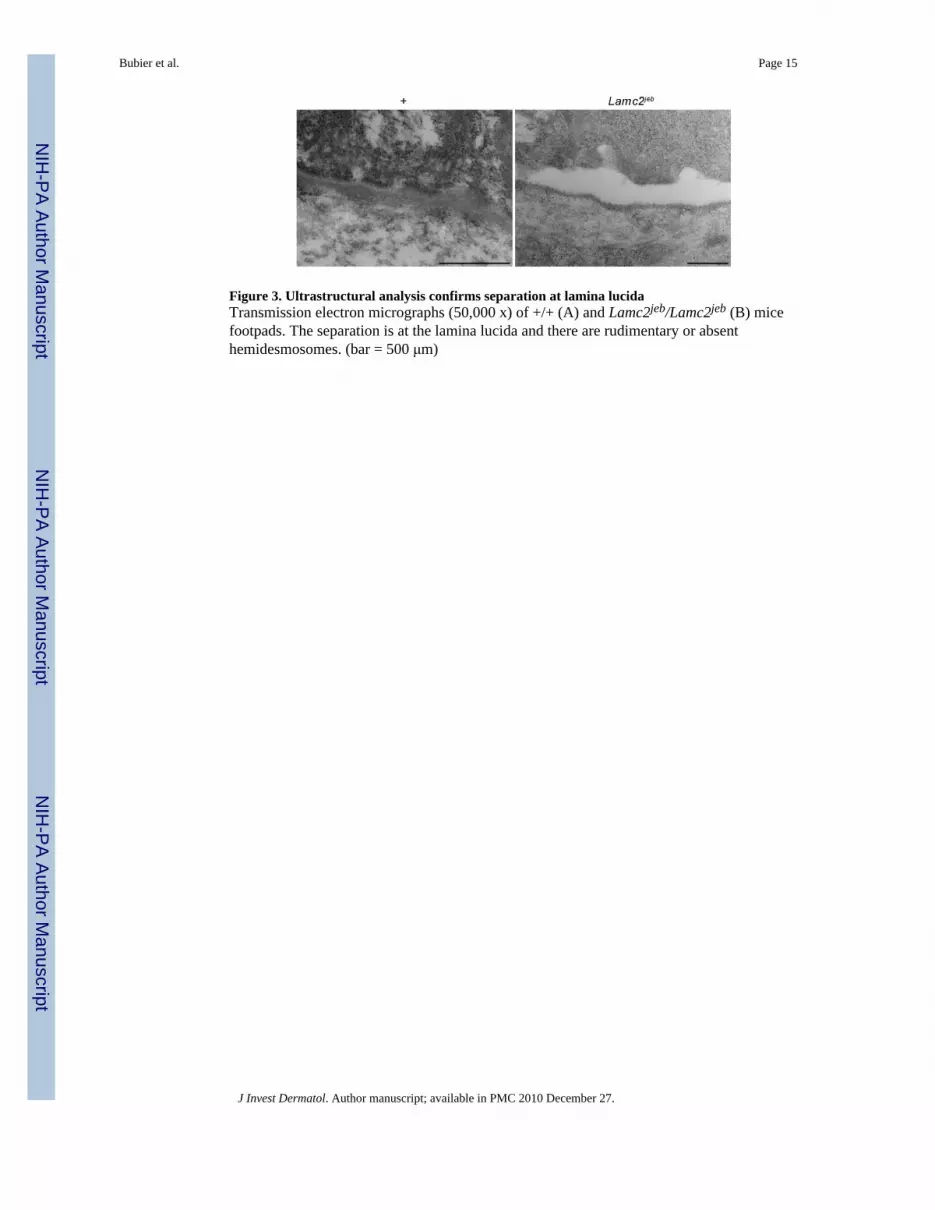
Figure 3. Ultrastructural analysis confirms separation at lamina lucidaTransmission electron micrographs (50,000 x) of +/+ (A) and Lamc2jeb/Lamc2jeb (B) micefootpads. The separation is at the lamina lucida and there are rudimentary or absenthemidesmosomes. (bar = 500 μm)
Bubier et al. Page 15
J Invest Dermatol. Author manuscript; available in PMC 2010 December 27.
NIH
-PA Author Manuscript
NIH
-PA Author Manuscript
NIH
-PA Author Manuscript
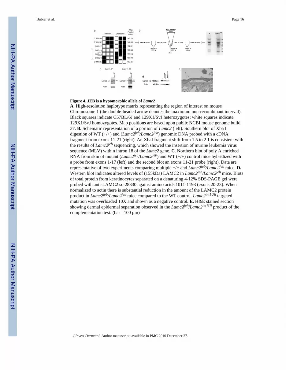
Figure 4. JEB is a hypomorphic allele of Lamc2A. High-resolution haplotype matrix representing the region of interest on mouseChromosome 1 (the double-headed arrow denotes the maximum non-recombinant interval).Black squares indicate C57BL/6J and 129X1/SvJ heterozygotes; white squares indicate129X1/SvJ homozygotes. Map positions are based upon public NCBI mouse genome build37. B. Schematic representation of a portion of Lamc2 (left). Southern blot of Xba Idigestion of WT (+/+) and (Lamc2jeb/Lamc2jeb) genomic DNA probed with a cDNAfragment from exons 11-21 (right). An XbaI fragment shift from 1.5 to 2.1 is consistent withthe results of Lamc2jeb sequencing, which showed the insertion of murine leukemia virussequence (MLV) within intron 18 of the Lamc2 gene. C. Northern blot of poly A enrichedRNA from skin of mutant (Lamc2jeb/Lamc2jeb) and WT (+/+) control mice hybridized witha probe from exons 1-17 (left) and the second blot an exons 11-21 probe (right). Data arerepresentative of two experiments comparing multiple +/+ and Lamc2jeb/Lamc2jeb mice. D.Western blot indicates altered levels of (155kDa) LAMC2 in Lamc2jeb/Lamc2jeb mice. Blotsof total protein from keratinocytes separated on a denaturing 4-12% SDS-PAGE gel wereprobed with anti-LAMC2 sc-28330 against amino acids 1011-1193 (exons 20-23). Whennormalized to actin there is substantial reduction in the amount of the LAMC2 proteinproduct in Lamc2jeb/Lamc2jeb mice compared to the WT control. Lamc2tm1Uit targetedmutation was overloaded 10X and shown as a negative control. E. H&E stained sectionshowing dermal epidermal separation observed in the Lamc2jeb/Lamc2tm1Ui product of thecomplementation test. (bar= 100 μm)
Bubier et al. Page 16
J Invest Dermatol. Author manuscript; available in PMC 2010 December 27.
NIH
-PA Author Manuscript
NIH
-PA Author Manuscript
NIH
-PA Author Manuscript
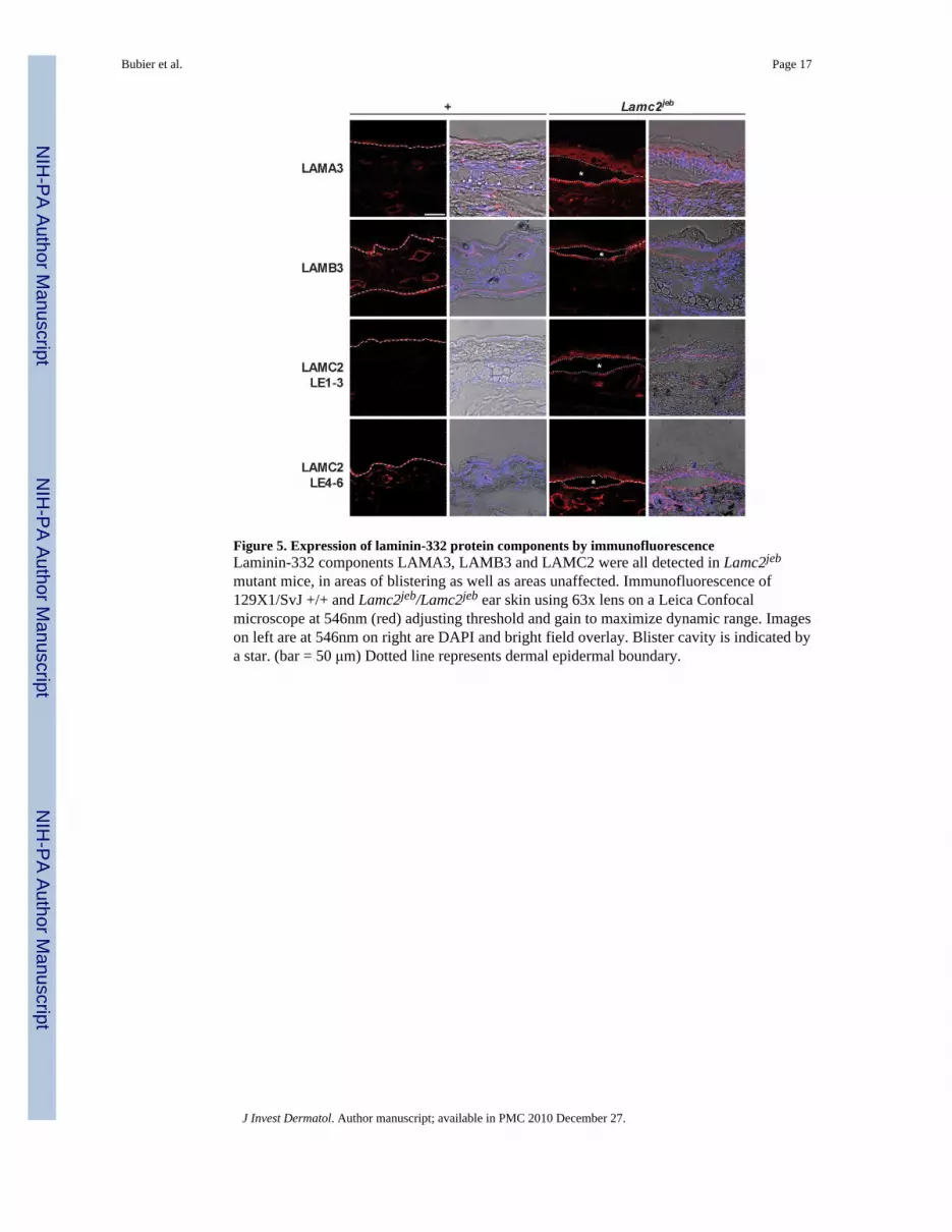
Figure 5. Expression of laminin-332 protein components by immunofluorescenceLaminin-332 components LAMA3, LAMB3 and LAMC2 were all detected in Lamc2jeb
mutant mice, in areas of blistering as well as areas unaffected. Immunofluorescence of129X1/SvJ +/+ and Lamc2jeb/Lamc2jeb ear skin using 63x lens on a Leica Confocalmicroscope at 546nm (red) adjusting threshold and gain to maximize dynamic range. Imageson left are at 546nm on right are DAPI and bright field overlay. Blister cavity is indicated bya star. (bar = 50 μm) Dotted line represents dermal epidermal boundary.
Bubier et al. Page 17
J Invest Dermatol. Author manuscript; available in PMC 2010 December 27.
NIH
-PA Author Manuscript
NIH
-PA Author Manuscript
NIH
-PA Author Manuscript

Figure 6. Non-cutaneous skin phenotypes of Lamc2 jebmiceA. SEM of WT and Lamc2jeb teeth at 35x (bar = 1 mm) and 500x (bar = 100 μm) reveals thesurface pitting of the enamel of the teeth in the mutant mice. B. Average lung pressure-volume loops post challenge. The average (±SE) of 8 129X1/SvJ and 9 Lamc2jeb/Lamc2jeb
analyzed by forced oscillation technique (at PEEP 3) show clear differences in resistanceand/or elastance between WT and Lamc2 jeb/Lamc2 jeb mice. X-axis represents pressure incm H2O and y-axis represents volume in mL. p < 0.001 by ANOVA.
Bubier et al. Page 18
J Invest Dermatol. Author manuscript; available in PMC 2010 December 27.
NIH
-PA Author Manuscript
NIH
-PA Author Manuscript
NIH
-PA Author Manuscript

NIH
-PA Author Manuscript
NIH
-PA Author Manuscript
NIH
-PA Author Manuscript
Bubier et al. Page 19
Table 1
Lamc2jeb mutation results in late onset reduction of bone mineral content
BMD (g/cm2) ± SEM BMC (g) ± SEM
9 Month 129X1 (n=10) 0.0575± 0.0036 0.6162±0.0465
9 Month 129X1 Lamc2jeb/Lamc2jeb (n=5) 0.0580±0.0024 0.6160±0.0742
p-value 0.669 0.987
12 Month 129X1 (n=5) 0.0591± 0.0021 0.6472±0.0488
12 Month 129X1 Lamc2jeb/Lamc2jeb (n=5) 0.0510±0.0021 0.532±0.0197
p-value* 0.0003 0.0039
*Two-tailed T-test
J Invest Dermatol. Author manuscript; available in PMC 2010 December 27.


