Embed Size (px)
Citation preview

From the Lübeck Institute of
Experimental Dermatology
Director: Prof. Dr. S. Ibrahim
Diagnosis and models of epidermolysis bullosa acquisita
Cumulative Dissertation
for Fulfillment of
Requirements
for the Doctoral Degree
of the University of Lübeck
From the Department of Medicine
submitted by
Artem Vorobyev
from Rybinsk, Russian Federation
Lübeck, 2016

First referee: Prof. Dr. med. Ralf Ludwig
Second referee: Prof. Dr. med. Peter König
Third referee: Prof. Dr. med. G. Hofbauer
Date of oral examination: 09.11.2016
Approved for printing: 09.11.2016

Diagnosis and models of epidermolysis bullosa aquisita
Introduction
Autoimmune blistering diseases (AIBD) are characterized by the presence of
circulating and tissue-bound antibodies to structural components of the skin and
mucous membranes. Clinical symptoms of these diseases are dependent on the
targeted antigen. All AIBD according to the affected antigen can be divided into
2 major groups: pemphigus and pemphigoid diseases (Table 1).
Disease group Disease Antigen
Pemphigus diseases Pemphigus vulgaris desmoglein 1
desmoglein 3
Pemphigus foliaceus desmoglein 1
Paraneoplastic
pemphigus
envoplakin, periplakin,
desmoglein 1, desmoglein 3,
desmoplakin I/II 2
makroglobulin-like 1,
desmocollin
IgA-pemphigus dsg1, dsg3, desmocollin 1
Pemphigoid
diseases
Bullous pemphigoid BP180, BP230
Pemphigoid gestationis BP180
Mucous membrane
pemphigoid
BP180, BP230, laminin 332,
-Integrin, collagen type
VII
Linear IgA-dermatosis LAD-1, BP180 NC16A,
collagen type VII
Anti-p200 pemphigoid laminin 1, p200-antigen
Epidermolysis bullosa
acquisita
collagen type VII
Dermatitis herpetiformis
Duhring
epidermal transglutaminase
(TG3)
tissue transglutaminase
(TG2)
Table 1. Classification of autoimmune bullous diseases
Pemphigus diseases are characterized by the presence of antibodies against
desmosomal proteins and intraepidermal blisters in histological examination
(Figure 1 A, B, C). Pemphigoid diseases are caused by the antibodies,
recognizing components of hemidesmosomes. Histologically subepidermal
blisters are observed (Figure 1 D, E, F).
Figure 1. Typical clinical picture of pemphigus vulgaris shows multiple erosions (A). Histologically
intraepidermal blisters are observed (B), in DIF typical intercellular depositions of IgG and
complement (C). Clinical picture of bullous pemphigoid wih blisters, erosions, urticarial plaques
(D). Microscopically subepidermal blisters (E) and linear deposions of IgG and complement C3
(F) are typical signs of pemphigoid diseases.
A B C
F E D

The "gold standard" of the diagnostic of autoimmune bullous diseases is
detection of autoantibody-depositions in the epidermis (pemphigus diseases) or
at the dermal-epidermal junction zone (pemphigoid diseases) in direct
immunofluorescence. In pemphigoid diseases for the further differentiation of
autoantibody specificity, indirect immunofluorescence on 1M NaCl-split skin can
be used. After incubation of normal skin in 1M NaCl-solution, the split in the
lamina lucida occurs, allowing detection of distinct antigens. At the roof of artificial
blister BP180 and BP230 are detected, at the floor - col7, p200 and laminin 332
(Figure 2).
Figure 2. Indirect immunofluorescence on 1M salt-split skin. On the left - binding of autoantibodies
to the floor of the artificial blister (col7, p200, laminin 332), on the right - to the roof of the blister
(BP180, BP230)
Further specification of antigens is possible using serological assays such as
enzyme-linked immunosorbent assay (ELISA) or western blot. For many
frequently detected antigens, such as BP180 and BP230 ELISA kits are
commercially available and assays can be performed in most of the laboratories
in several hours. For rare diseases, such as EBA, anti-p200 pemphigoid or anti-
laminin 332 pemphigoid no commercial kits have been available. Serologic
diagnosis of epidermolysis bullosa acquisita is made by western blot with extract
of human dermis, where 290 kDa (whole molecule of collagen type VII) and 145
kDa (NC1-domain) proteins are detected. Alternatively, ELISA with

immunodominant NC1 region of type VII collagen was previously tested, but is
not widely available. Because western blot analysis is technically demanding and
time consuming, it is available only in few diagnostic laboratories in the world,
complicating diagnosis of epidermolysis bullosa acquisita.
During my work in the autoimmune diagnostics laboratory at the department of
dermatology, University clinic Schleswig-Holstein I was involved in the analysis
of the samples of a patient with clinical symptoms of epidermolysis bulosa
dystrophica, such as nail loss and acral contractures (Figure 3).
Figure 3. Nail loss and acral contractures in a patient with EBA
Histopathologically, subepidermal blisters were detected, in the direct
immunoflurescence linear depositions of IgG and complement C3 along the
dermal-epidermal junction zone, suggesting the diagnosis of autoimmune
subepidermal blistering disease. In further analysis of the autoantibody-specificity
the targeted protein was recognized as type VII collagen (col7), antigen in
epidermolysis bullosa acquisita (EBA). EBA is a rare chronic autoimmune
subepidermal blistering disease of skin and mucous membranes. Its' incidence is
estimated as 0,25-0,5 per 1 million/year. EBA is characterized by the presence
of antibodies against col7. Col7 is a major component of anchoring fibrils,

connecting epidermis to dermis. Different clinical phenotypes of EBA are
described, such as classic mechano-bullous and inflammatory variants.
However, no pathogenic differences determining different clinical phenotypes
have been described, most likely due to the low incidence of disease.
After that work I got interested in the pathogenesis of epidermolysis bullosa
acquisita and its' different clinical phenotypes. For the further investigations, we
collected to the moment biggest international cohort of epidermolysis bullosa
acquisita patients (73 patients from Germany, Netherlands, Japan, South Korea,
Great Britain).
This collection made the development of diagnostic tools possible (see 1 and 2).
In addition, this cohort was the basis for fine epitope-mapping study (see 3).
Study 1.
Publication:
Komorowski L, Müller R, Vorobyev A, Probst C, Recke A, Jonkman MF,
Hashimoto T, Kim SC, Groves R, Ludwig RJ, Zillikens D, Stöcker W, Schmidt E.
Sensitive and specific assays for routine serological diagnosis of epidermolysis
bullosa acquisita. J Am Acad Dermatol. 2013 Mar;68(3):e89-95 (IF: 5,004)
In collaboration with our partners from Euroimmun, Lübeck, we used our cohort
of previously collected 73 EBA patients' sera to establish two sensitive and
specific assays for detection of circulating anti-col7 antibodies. The diagnosis of
epidermolysis bullosa acquisita was confirmed using indirect
immunofluorescence of 1m salt-split skin and western blot with human dermal
extract.
In detail, NC1 domain of type VII collagen was previously described as major
antigenic epitope in epidermolysis bullosa acquisita. However, several cases of
autoantibody reactivity to non-collagenous NC2 as well as to collagenous domain
were reported. Because of low number of autoantibody reactivity outside NC1,
we decided to use only NC1 domain in this assay.
Full-lengh non-collagenous NC1 domain, major antigen site of type VII collagen
was cloned and expressed in HEK293 cells. The protein was coated on ELISA
plates and tested with the sera of 73 EBA patients, to the moment largest reported
EBA cohort. 395 control sera, including BP, anti-p200 pemphigoid, anti-laminin
332 pemphigoid and pemphigus vulgaris sera, were used.
Alternatively to ELISA, NC1-transfected HEK293 cells were coated on glass and
used for indirect immunofluorescence microscopy. Treatment with secondary
antibody allows the visualization of anti-collagen VII antibody bound to NC1-
expressing HEK293 cells.
After optimisation of the ELISA cut-off, sensitivity and specificity of 98,7% were
reached. In the indirect immunofluorescence sensitivity of 91,8% and specificity
of 99,8% were observed.
Conclusion:
In this study we could develop two novel serological assays from diagnosis of
epidermolysis bullosa acquisita, both demonstrating high sensitivity and
specificity.

Study 2.
Publication:
Kim JH, Kim YH, Kim S, Noh EB, Kim SE, Vorobyev A, Schmidt E, Zillikens D,
Kim SC. Serum levels of anti-type VII collagen antibodies detected by enzyme-
linked immunosorbent assay in patients with epidermolysis bullosa acquisita are
correlated with the severity of skin lesions. J Eur Acad Dermatol Venereol. 2013
Feb;27(2):e224-30 (IF: 3,105)
Serological assays are indispensable not only for the diagnostics of autoimmune
bullous skin diseases, but also for the follow-up and treatment control. Previously,
it has been reported that ELISA values are correlating with disease severity in
bullous pemphigoid and pemphigus vulgaris, however, no studies on this matter
in EBA have been performed. To address this question, we tested sera of 30 EBA
patients in an ELISA with NC1 and NC2 domains (MBL, Nagoya, Japan) as well
as in indirect immunofluorescence microscopy assay using NC1-domain
expressing HEK293 cells (Euroimmmun, Lübeck, Germany). In this study we
could confirm the previously reported sensitivity of 91,8 % and specificity of 98,1
% of the ELISA system. Indirect immunofluorescence on HEK293 cells showed
comparable results. In our cohort of 30 EBA patients, ELISA values as well as IIF
titres positively correlated to EBA severity. However, comparing ELISA and IIF
titres before treatment and after remission, ELISA demonstrated better
correlation with disease severity, while in IIF in one patient autoantibody titre
increased after remission. In time course experiment, using the samples from one
EBA patient collected at 7 different time points, ELISA values better represented
disease severity that IIF titre.
Conclusion:

In this study we could demonstrate that serological assays, such as ELISA or IIF
on HEK 293 cells are useful for the evaluation of EBA severity and follow up
controls, as both assays positively correlated with disease severity score.

Study 3.
Publication:
Vorobyev A, Ujiie H, Recke A, Buijsrogge JJ, Jonkman MF, Pas HH, Iwata H,
Hashimoto T, Kim SC, Hoon Kim J, Groves R, Samavedam U, Gupta Y, Schmidt
E, Zillikens D, Shimizu H, Ludwig RJ. Autoantibodies to multiple epitopes on the
Non-Collagenous-1 domain of type VII collagen induce blisters. J Invest
Dermatol. 2015 Jun;135(6):1565-73 (IF: 7,216)
Clinically, different EBA phenotypes are described: (i) classical mechanobullous,
and (ii) inflammatory type, including bullous pemphigoid-, Brunsting-Perry
pemphigoid-, linear IgA dermatosis-, and mucous membrane pemphigoid-like
variants. Mechanobullous variant of EBA resembles clinical picture of hereditary
epidermolysis bullosa and is characterized by blisters at the trauma-prone sites,
scarring, and milia formation. Inflammatory type of epidermolysis bullosa
acquisita demonstrates erythematous plaques and tense blisters, mimicking
bullous pemphigoid.
The pathogenesis of epidermolysis bullosa is relatively well characterised. First,
antibodies bind to col7 at the dermal-epidermal junction, forming immune
complexes. These immune complexes trigger complement activation cascade,
which, in turn, recruits leukocytes to dermal-epidermal junction. Activated
neutrophils secrete proteases, produce reactive oxygen species (ROS) and
cause damage to dermal-epidermal junction, leading to blister formation.
However, although these events are relatively well characterized, pathogenic
mechanisms causing different clinical phenotypes are still not understood. It has
been previously reported that NC1-domain of col7 interacts with collagen I,

collagen IV and laminin 332, so the perturbations of these interactions by
autoantibodies could be one of the possible explanations.
To address this question, we created overlapping recombinant proteins, covering
whole NC1 domain.
These recombinant proteins were tested in immunoblot assay with 69 clinically
characterized EBA patients' sera. Autoantibodies of EBA patients recognized
clusters of epitopes throughout the NC1 domain. To test if targeted epitopes
correlate with clinical phenotype, homogeneity distribution analysis and
Pearson's correlation analysis were used. No difference in epitope-recognition
pattern and clinical phenotype of the patients could be detected. However,
interestingly, epitope-recognition was strongly dependent on gender and age of
the patients. In our opinion, antibody reactivity to NC2- and collagenous domains
of collagen VII can not explain these differences, as only few patients have been
described with autoantibody reactivity outside NC1 domain. We think another
factors, such as neutrophil ability to mount an inflammatory response, may play
a major role.
To test the pathogenic relevance of the epitope-specific antibodies, polyclonal
rabbit anti-human col7 antibodies were produced. For the analysis of specificity
of raised anti-collagen VII antibodies, these antibodies were tested in western
blot with the proteins used for immunization. Interestingly, for several epitopes,
binding of antibodies outside of the relevant epitope has been observed (Figure
7). This phenomenon of intramolecular cross-reactivity could be explained by
high homology of FNIII-like domains and could contribute to pathogenicity of
specific antibodies.

To test pathogenic relevance of single antibodies, ex vivo dermal-epidermal
separation assay was used. Cryosections of normal human skin were treated with
rabbit anti-human anti-col7 antibodies, followed by incubation with normal human
PMNs. In this assay all of the used antibodies caused dermal-epidermal
separation, which was dependent on PMNs, as incubation of skin sections with
antibody alone did not cause split formation. For in vivo validation, antibodies
against two subdomains of NC1 domain of type VII collagen were affinity-purified
and injected into mice carrying null mutations of mouse COL7 and the human
COL7 transgene. Both of these antibodies bound to the dermal-epidermal
junction and induced typical histological changes and clinical phenotype in mice,
for the first time demonstrating pathogenicity of anti-human collagen VII
antibodies in vivo.
Conclusion:
This study provides novel insights into pathogenesis and diagnostics of
epidermolysis bullosa acquisita, which will improve our understanding, diagnosis
and treatment of this rare autoimmune disease.

Summary
Autoimmune blistering diseases (AIBD) are characterized by the presence of
tissue-bound and circulating antibodies directed to the structural components of
skin and mucous membranes. AIBD can be divided into two groups. Pemphigus
diseases are characterized by the presence of autoantibodies to the desmosomal
proteins; in the pemphigoid diseases autoantibodies target the components of
dermal-epidermal junction. One of the typical pemphigoid diseases is
epidermolysis bullosa acquisita.
Epidermolysis bullosa acquisita is a rare chronic autoimmune blistering disease
of skin and mucous membranes, characterized by the presence of antibodies to
collagen VII (col7), major component of anchoring fibrils. Col7 is a heterotrimer,
consisting of three identical alpha-chains, each consisting of N-terminal non-
collagenous NC1 domain, collagenous domain and C-terminal non-collagenous
NC2 domain. In the previous studies, NC1 domain was described as major
antigenic region of col7. Binding of autoantibodies to col7 causes formation of
immune complexes and activation of complement cascade, which, in turn,
recruits neutrophils to dermal-epidermal junction. Neutrophils secrete proteases
and release ROS, causing tissue damage and blister formation. Clinically,
mechanobullous and inflammatory EBA phenotypes are described.
Even though the pathogenesis of epidermolysis bullosa is relatively well
characterised, pathogenic mechanisms causing different clinical phenotypes are
still not understood. One of the possible explanations could be the perturbations
of interaction of col7 NC1 with collagen I, collagen IV and laminin 332 by binding
of autoantibodies to distinct subdomains of NC1. To address this question we
collected to this moment largest reported cohort of 73 EBA patients. We cloned

and expressed recombinant overlapping proteins, covering whole NC1 domain of
col7. The proteins were used for western blot assays, where no correlation of
clinical phenotype with epitope-recognition pattern was detected. Interestingly,
strong correlation of epitope-recognition pattern with gender and age of the
patients was observed.
For the further validation of pathogenicity of antibodies against distinct NC1
subdomains, polyclonal rabbit anti-human col7 antibodies were raised. To test
the crossreactivity of these antibodies, they were investigated in western blot
assay with all of the recombinant proteins. Interestingly, intramolecular cross-
reactivity was detected, what could be explained by high homology of FNIII-like
subdomains of col7. All of the raised rabbit anti-human col7 antibodies
demonstrated pathogenicity ex vivo in dermal-epidermal separation cryosection
assay. For the in vivo validation, two antibodies were affinity-purified and injected
into col7-humanized mice, causing clinical and histological phenotypes, similar to
human EBA. Thus, in vivo pathogenicity of antibodies to human col7 was reported
for the first time.
Diagnostic of EBA is based on the detection of tissue-bound and/or circulating
autoantibodies to col7. Due to low incidence of EBA, diagnosis of this disease
remains an issue, as no commercial kits for its serological diagnostic were
available. Detection of circulating anti-col7 autoantibodies was possible only with
technically demanding western blot analyses of human dermal extract or
recombinant NC1 domain of col7, available only in several diagnostic laboratories
in the world. Using our cohort of EBA patients, we were able to develop two novel
serological diagnostic tools, namely ELISA with recombinant NC1 domain and
Biochip with NC1-transfected HEK 293 cells. In our further study we could

demonstrate, that these tools can also be used for the evaluation of disease
severity and treatment control.
In our studies using to the moment largest cohort of EBA patients, we could
develop two novel diagnostic tools and imrove our understanding of pathogenesis
of epidermolysis bullosa acquisita.

Deutsche Zusammenfassung
Blasenbildende Autoimmundermatosen (AIBD) sind durch die Präsenz der
gewebsgebundenen und zirkulierenden Antikörper gegen Strukturproteine der
Haut und Schleimhäuten gekennzeichnet. AIBD stellen eine heterogene Gruppe
von Erkrankungen dar, die in zwei Gruppen aufgeteilt werden können.
Pemphiguserkrankungen werden durch Antikörper gegen desmosomale
Proteine verursacht, was sich histopathologisch als intraepidermale Spaltbildung
darstellt. Bei den Pemphigoiderkrankungen zeigt sich eine subepidermale
Spaltbildung, und die Autoantikörper sind gegen Strukturproteine der dermo-
epidermalen Junktionszone gerichtet.
Eine repräsentative Erkrankung aus der Gruppe der Pemphigoid-Erkrankungen
stellt die Epidermolysis bullosa acquisita dar.
Epidermolysis bullosa acquisita is eine chronische blasenbildende
Autoimmunerkrankung der Haut und den Schleimhäuten, die durch
Autoantikörper gegen Kollagen VII verursacht ist. Kollagen VII ist ein
Heterotrimer, der aus drei identischen Alpha-Ketten besteht. Jede Alpha-Kette ist
aus einer N-terminalen nicht kollagenosen NC1 Domäne, einer kollagenosen
Domäne und einer C-terminalen nicht kollagenosen NC2 Domäne
zusammengesetzt. In den vorherigen Studien wurde die NC1-Domäne bereits als
der wichtigste antigene Bereich des Kollagen VII Molekül beschrieben. Die
Bindung der Autoantikörper an Kollagen VII verursacht die Bildung der
Immunkomplexen und Aktivierung der Komplementkaskade. Danach werden die
Neutrophile zur dermal-epidermalen Junktionszone rekrutiert. Anschließend
findet die Produktion der Proteasen und die Freisetzung der reaktiven
Sauerstoffspezies durch die Neutrophile statt, was zur Gewebeschädigung und
Blasenbildung führt.

Klinisch wurde der klassische mechanobullöse Phänotyp und der entzündliche
Phänotyp der Epidermolysis bullosa acquisita beschrieben. Obwohl die
Pathogenese der Epidermolysis bullosa acquisita relativ gut charakterisiert ist,
die pathogenetische Mechanismen, die zu verschiedenen klinischen Phänotypen
der Erkrankung führen sind noch nicht bekannt. Eine der möglichen Erklärungen
könnte die Störung der Interaktion von Kollagen VII mit Kollagen I, Kollagen IV
und Laminin 332 durch Bindung der Autoantikörper zu spezifischen Subdomänen
der NC1-Domäne sein. Um diese Frage zu beantworten wurde die bisher größte
Kohorte aus 73 Epidermolysis bullosa acquisita Patienten gesammelt.
Die rekombinanten überlappenden Proteine, die die gesamte NC1-Domäne
abdecken, wurden von uns kloniert und exprimiert. Die Proteine wurden in
Western Blot Untersuchungen mit Patientenseren getestet, wo keine Korrelation
zwischen dem klinischen Phänotyp und dem Muster der durch die Antikörper
gebundenen Epitopen festgestellt werden konnte. Interessanterweise,
signifikante Korrelation des Musters der Epitopen mit Alter und Geschlecht der
Patienten wurde bemerkt.
Um die Pathogenität der Antikörper gegen die spezifischen Fragmente der NC1-
Domäne zu validieren, wurden in Kaninchen die poliklonale anti-humane
Kollagen VII-Antikörper erzeugt. Um die Kreuzreaktivität dieser Antikörper zu
untersuchen, wurde ein Western Blot mit allen rekombinanten Proteinen
durchgeführt. Interessanterweise, wurde eine intramolekulare Kreuzreaktivität
festgestellt, die durch eine hohe Homologie zwischen FNIII-Subdomäne erklärt
werden könnte. Jeder dieser Kaninchen anti-human Kollagen VII Antikörper
zeigte eine dermal-epidermale Trennung ex vivo bei der Untersuchung mit den
Kryoschnitten normaler humaner Haut. Für die in vivo Validierung, zwei dieser
Antikörper wurden affinitätsaufgereinigt und in die Kollagen VII-humanisierte

Mäuse injiziert. Klinisch und histologisch war der Phänotyp der humanen
Epidemolysis bullosa acquisita ähnlich. Dadurch wurde die Pathogenität der
gegen humanen Kollagen VII gerichteten Antikörper zum ersten Mal
beschrieben.
Diagnosis der Epidermolysis bullosa acquisita wird durch Nachweis
gewebsgebundener und/oder zirkulierender Antikörper gegen Kollagen VII
gestellt. Wegen der geringen Inzidenz der Epidermolysis bullosa acquisita ist die
Diagnostik dieser Erkrankung immer noch eine Herausforderung. Zu der Zeit der
Untersuchung standen noch keine kommerzielle Diagnostikmethoden zur
Vefügung. Nachweis von zirkulierenden anti-Kollagen VII Antikörper war nur mit
der technisch sehr anspruchsvollen Western Blot Untersuchung des Extraktes
humaner Dermis oder rekombinantem Kollagen VII möglich. Diese sind nur in
einzelnen Diagnostiklaboren weltweit möglich. Mit unserer Kohorte der
Epidermolysis bullosa acquisita Patienten wurden zwei neuen serologischen
Diagnostikverfahren (ELISA mit rekombinantem NC1 und Biochip mit NC1-
transfizierten HEK 293 Zellen) etabliert. In unserer nächsten Studie konnten wir
zeigen, dass diese Verfahren auch für die Einschätzung des Schwierigkeitsgrads
der Erkrankung sowie Behandlungskontrolle geeignet sind.
In unseren Studien mit der größten Kohorte der Epidermolysis bullosa acquisita
Patienten konnten wir zwei neuen Diagnostikmethoden entwickeln und unsere
Kenntnisse der Pathogenese der Erkrankung verbessern.

Publications:
1. Vorobyev A, Ujiie H, Recke A, Buijsrogge JJ, Jonkman MF, Pas HH, Iwata
H, Hashimoto T, Kim SC, Hoon Kim J, Groves R, Samavedam U, Gupta Y,
Schmidt E, Zillikens D, Shimizu H, Ludwig RJ. Autoantibodies to Multiple
Epitopes on the Non-Collagenous-1 Domain of Type VII Collagen Induce Blisters.
J Invest Dermatol. 2015 Jun;135(6):1565-73
2. Iwata H, Pipi E, Möckel N, Sondermann P, Vorobyev A, van Beek N, Zillikens
D, Ludwig RJ. Recombinant soluble CD32 supresses disease progression in
experimental Epidermolysis bullosa aquisista. J Invest Dermatol. 2015
Mar;135(3):916-9.
3. Recke A, Trog LM, Pas HH, Vorobyev A, Abadpour A, Jonkman MF, van
Zandbergen G, Kauderer C, Zillikens D, Vidarsson G, Ludwig RJ. Recombinant
Human IgA1 and IgA2 Autoantibodies to Type VII Collagen Induce Subepidermal
Blistering Ex Vivo. J Immunol. 2014 Aug 15;193(4):1600-8.
4. Iwata H, Bieber K, Tiburzy B, Chrobok N, Kalies K, Shimizu A, Leineweber S,
Ishiko A, Vorobyev A, Zillikens D, Köhl J, Westermann J, Seeger K, Manz R,
Ludwig RJ. B cells, dendritic cells, and macrophages are required to induce an
autoreactive CD4 helper T cell response in experimental epidermolysis bullosa
acquisita. J Immunol. 2013 Sep 15;191(6):2978-88.
5. Kim JH, Kim YH, Kim S, Noh EB, Kim SE, Vorobyev A, Schmidt E, Zillikens
D, Kim SC. Serum levels of anti-type VII collagen antibodies detected by enzyme-
linked immunosorbent assay in patients with epidermolysis bullosa acquisita are
correlated with the severity of skin lesions. J Eur Acad Dermatol Venereol. 2013
Feb;27(2):e224-30.
6. Komorowski L, Müller R, Vorobyev A, Probst C, Recke A, Jonkman MF,
Hashimoto T, Kim SC, Groves R, Ludwig RJ, Zillikens D, Stöcker W, Schmidt E.
Sensitive and specific assays for routine serological diagnosis of epidermolysis
bullosa acquisita.J Am Acad Dermatol. 2013 Mar;68(3):e89-95.
7. Meissner C, Hoefeld-Fegeler M, Vetter R, Bellutti M, Vorobyev A, Gollnick H,
Leverkus M. Severe acral contractures and nail loss in a patient with mechano-
bullous Epidermolysis bullosa acquisita. Eur J Dermatol. 2010 Jul-Aug;20(4):543-
4.
Acknowledgment:
I would like to acknowledge all people supporting me during my PhD work.
This work would not have been possible without support of my mentor Prof. Dr.
Ralf Ludwig. I appreciate his guidance and valuable comments throughout my
research work.
I would like to express my acknowledgment to Prof. Dr. Detlef Zillikens, director
of the Department of Dermatology, for hosting this thesis.
I must express my gratitude to Prof. Dr. Dr. Enno Schmidt for his valuable advice,
suggestions and help in my PhD work.
I would like to thank Dr. Andreas Recke for introducing me into molecular biology
methods and his contribution to the design of the epitope mapping project.
Furthermore I would like to acknowledge Yask Gupta for his valuable help in
statistical analysis and introducing me into R software.
Moreover, I would like to thank all my colleagues and friends for their unbelievable
support during my work.
Finally, I would like to thank my family for their patience and providing me with
their moral support all the time.

Curriculum vitae
Personal details
Name Artem Vorobyev
Date of birth 14.04.1982
e-mail [email protected]
Nationality Russian
Education and qualifications
12/2012-present Physician, Department of Dermatology, University of
Lübeck
04/2009-12/2012 PhD student, Department of Dermatology, University of
Lübeck
10/2007-03/2008 Internship, Department of Dermatology,
Hospital Saint-Louis, Paris, France
09/2005-08/2007 Specialisation in dermatology, Medical University of
Yaroslavl, Russia
09/1999-08/2005 Studies of human medicine, Medical University of
Yaroslavl, Russia

Severe acral contractures and nail loss
in a patient with mechano-bullous
Epidermolysis bullosa acquisita
Epidermolysis bullosa acquisita (EBA) is a chronicacquired autoimmune blistering disease with highly vari-able clinical manifestations [1]. The classic mechano-bullous non-inflammatory type presents with acral blistersand erosions. Inflammatory variants of EBA can resemblebullous or cicatricial pemphigoid, and severe mucosalinvolvement may be present, leading to severe complica-tions, such as oesophageal stenosis [2]. Therapeutically,EBA represents a major challenge, requiring high doselong term immunosuppressive therapy, with modest longterm success [3].We describe a 50-year-old patient in whom a mechano-bullous form of EBA was diagnosed 12 years ago [4].First symptoms were blister formation on mechanicallystressed areas (figure 1A). Furthermore, aggravated blisterformation of the hands was evident within hours after sunexposure. Histopathology of biopsies revealed subepidermalbullae and a dense inflammatory infiltrate in the upper der-mis. Direct immunofluorescence (DIF) microscopy showedlinear depositions of IgG and C3 at the basement membranezone, and indirect IF microscopy showed binding of circu-lating IgG (figure 1F), but not IgA antibodies to the dermalside of the split (figures 1G, H). Immunoblot analysis withIgG4 isotype-specific secondary antibodies showed that thepatient’s serum specifically reacted with the recombinantNC-1 domain of type VII collagen. ELISA using recombi-nant BP230, BP180 NC16A domains, as well as ANA,ENA, or Abs against single or double stranded DNA,were negative. We diagnosed a predominantly mechano-bullous form of EBA. The patient refused consent for anoral corticosteroid therapy and did not present again forseveral years. He noted an aggravation of the symptomsupon intensive manual work. 9 years after diagnosis henoted progressive atrophic scarring of his hands (figure 1B).After the initial diagnosis 12 years before, therapy withintravenous immunoglobulins (1.2 g kg-1 in monthly inter-vals over a total of 18 months) was initiated. Subsequently,he was treated with mycophenolate mofetil (2 g daily p.o.)and colchicine (1 mg daily) [5] for several years. Later,recurrent oesophageal strictures required balloon dilation.12 years after the initial diagnosis, the patient presentedwith severe contractures of the hands and feet (figures 1C-E). The palmar side of his hands demonstrated severe der-matogenic contractures and nail loss (figure 1E). For this westarted systemic corticosteroid treatment in combinationwith azathioprine (1.5 mg/kg body weight). Based uponprevious successful reports [6, 7], we initiated 4 cycles of aB-cell depleting therapy with Rituximab (375 mg/m2 infu-sion once a week) and achieved remission of the inflamma-tory clinical symptoms over 4 months. Circulating autoanti-body titers against the immuno-dominant (NC-1) domainsof type VII collagen (titer of 1:1600 at presentation) wereundetectable within 4-6 months, and the skin vulnerabilitywas substantially improved.A loss of function of type VII collagen due to mutationswithin the COL7A1 gene leads to different forms of epi-
A B
D
C
E
F
G
H
Figure 1. Mechano-bullous form of Epidermolysis bullosaacquisita leads to severe acral contractures. A) Erythematouspapules and plaques with crusting of the right hand at the timeof diagnosis. B) Right hand with initial contractures ofthe hand, severe atrophy and scars of the dorsal sideof the hands; 9 years after diagnosis. The contracture of the PIPjoint was 30° on both sides and the range of motionin the MCP joint 20°-70° on both sides. C) Developmentof severe acral contractures 12 years after diagnosis. Laterallook at the wrist with confluent erythematosus plaques.D) Palmar aspect of both hands with severe contracturesof the distal and interphalangeal joints. E) Total destructionof the nail organs at digit 2-5 of the right hand. F-H) IndirectIF microscopy on 1 M NaCl split human skin (original mag-nification × 200 for F-H) demonstrated binding of circulatingIgG antibodies (F) to the dermal side of the split, whereas IgAreactivity (H) was absent in our patient. H) A positive controlpatient with known IgA EBA demonstrates binding of IgAautoantibodies to the dermal side of the split.
EJD, vol. 20, n° 4, July-August 2010 543
dermolysis bullosa [8]. We demonstrate that over the pro-longed course presented here a predominantly mechano-bullous EBA can lead to severe contractures and mutila-tions that may resemble a dystrophic form of Epidermo-lysis bullosa. To the best of our knowledge, this is the firstdocumented case of such major skin complications ofEBA in the literature, arguing for an early aggressiveimmunosuppression for such forms of EBA. Althoughthe patient was not continuously affected in his daily lifeby the extent of active disease, the mechano-bullous formof EBA can lead to serious acral complications. We willnow aim to mobilize the patient once clinical activity ofthe disease is controlled by immunosuppressive and B-cell depleting therapy. Future studies will evaluate theeffectiveness of immunosuppression, not only for acuteinflammation [9], but also for prevention of mutilations.We speculate that immunosuppression and thus reductionof circulating and tissue-bound autoantibodies to collagentype VII may delay the scarring and contractions that wedocument in this report. ■
Acknowledgement. We thank Dr. Enno Schmidt (Univer-sity of Lübeck, Department of Dermatology) for helpfulsuggestions and critical reading of the manuscript.Financial support: none. Conflict of interest: none.
1Department of Dermatology
and Venereology, Otto-von-
Guericke-University Magdeburg,
Germany2Department of Dermatology,
Venereology, and Allergology,
Medical Faculty Mannheim,
University of Heidelberg, Theodor-
Kutzer-Ufer 1-3, 68167 Mannheim,
Germany3Department of Dermatology,
University of Lübeck
heidelberg.de>
Carl MEISSNER1
Marc
HOEFELD-FEGELER1
Robert VETTER1
Michael BELLUTTI1
Artem VOROBYEV3
Harald GOLLNICK1
Martin LEVERKUS1,2
1. Sitaru C. Experimental models of epidermolysis bullosa acquisita.ExpDermatol 2007; 16: 520-31.2. Shipman AR, Agero AL, Cook I, et al. Epidermolysis bullosa acqui-sita requiring multiple oesophageal dilatations. ClinExpDermatol2008; 33: 787-9.3. Schmidt E, Brocker EB, Goebeler M. Rituximab in treatment-resistant autoimmune blistering skin disorders. ClinRevAllergy Immu-nol 2008; 34: 56-64.4. Jappe U, Zillikens D, Bonnekoh B, Gollnick H. Epidermolysis bul-losa acquisita with ultraviolet radiationsensitivity. BrJ Dermatol 2000;142: 517-20.5. Bibas R, Gaspar NK. Ramos-e-Silva. Colchicine for dermatologicdiseases. JDrugs Dermatol 2005; 4: 196-204.6. Niedermeier A, Eming R, Pfutze M, et al. Clinical response ofsevere mechanobullous epidermolysis bullosa acquisita to combinedtreatment with immunoadsorption and rituximab (anti-CD20 monoclo-nal antibodies). Arch Dermatol 2007; 143: 192-8.7. Schmidt E, Benoit S, Brocker EB, Zillikens D, Goebeler M. Success-ful adjuvant treatment of recalcitrant epidermolysis bullosa acquisitawith anti-CD20 antibody rituximab. Arch Dermatol 2006; 142:147-50.8. Dang N, Murrell DF. Mutation analysis and characterization ofCOL7A1 mutations in dystrophic epidermolysis bullosa. Exp Dermatol2008; 17: 553-68.9. Takae Y, Nishikawa T, Amagai M. Pemphigus mouse model as atool to evaluate various immunosuppressive therapies. Exp Dermatol2009; 18: 252-60.
doi:10.1684/ejd.2010.1002
Secondary syphilis presenting in a red
tattoo
A 26-year-old Chinese male presented with a reddishbrown scaly maculopapular eruption on a red tattoo onhis trunk, and with slight pruritus for about 6 months.The initial lesions occurred on his palms. The red areaof the tattoo was involved although the dark blue area ofthe tattoo was free of lesions. One year before, the patienthad a dragon tattooed on his trunk with bulk dark ink anda few cinnabars, by a professional tattoo artist. He had ahistory of unprotected sexual activities during theprevious 12 months. No history of genital ulcer or othercutaneous or systemic disease could be elicited from thepatient. There was no history of hair loss.Physical examination revealed reddish brown macules andpapules with slight scales, which covered most of the redarea of the tattoo. The dark blue area was not involved(figure 1A). A scaly circular rash was present on hispalms. There was no palpable lymphadenopathy. Mucousmembranes of the mouth and pharynx were unremarkable.A biopsy from the lesion on the red tattoo revealed para-keratosis, acanthosis and numerous neutrophils presentwithin the epidermis; a diffused infiltrate of plasma cellsassociated with red and black pigment-containing macro-phages was present in the upper dermis (figure 1B).Warthin-Starry staining was performed and was negativefor spirocheta. Laboratory tests including blood, urine,stools, hepatic and renal functions were all within normallimits. A patch test for cinnabar was negative. Repeatedfungal tests under microscopy and cultures were negative.The result of rapid plasma reagin (RPR) was positive withtitre 1:32. Treponema pallidum hemagglutination assay(TPHA) exhibited a positive result with titre 1:2560.HIV antibody was negative. A diagnosis of secondarysyphilis was made. Procaine benzylpenicillin was pre-scribed, 800,000 units per day for 2 weeks. Lesions ofthe trunk and palms cleared completely about 4 weekslater. The RPR titre decreased to 1:2.Syphilis is a chronic sexually transmitted disease causedby Treponema pallidum. Its incidence has risen in the lastfew decades [1]. Up to now, the relationship betweentattoos and syphilis is uncertain. It was believed thatthe needles used during tattooing and the saliva used
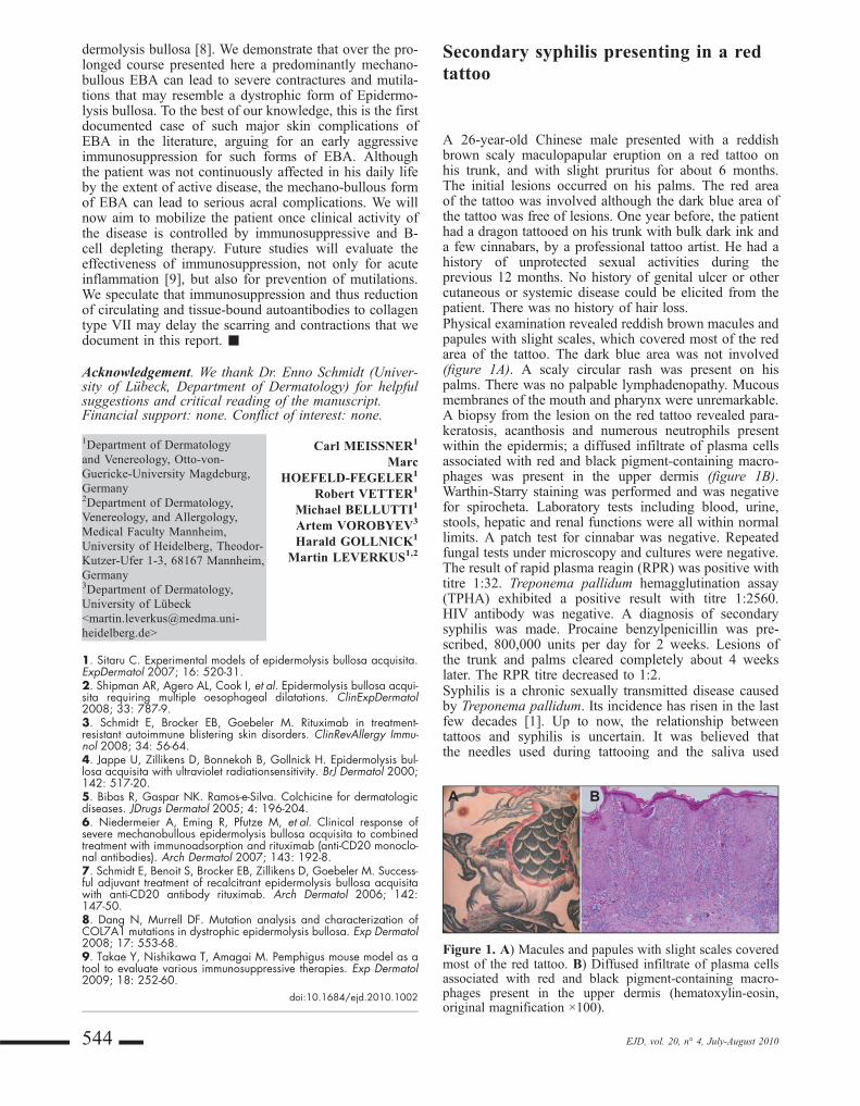
A B
Figure 1. A) Macules and papules with slight scales coveredmost of the red tattoo. B) Diffused infiltrate of plasma cellsassociated with red and black pigment-containing macro-phages present in the upper dermis (hematoxylin-eosin,original magnification ×100).
544 EJD, vol. 20, n° 4, July-August 2010
Sensitive and specific assays for routine serological
diagnosis of epidermolysis bullosa acquisita
Lars Komorowski, PhD,aRalf M€uller, PhD,
bArtem Vorobyev, MD,
bChristian Probst, PhD,
a
Andreas Recke, MD,b Marcel F. Jonkman, MD, PhD,c Takashi Hashimoto, MD,d Soo-Chan Kim, MD,e
Richard Groves, MD, FRCP,f Ralf J. Ludwig, MD,b Detlef Zillikens, MD,b Winfried St€ocker, MD,a
and Enno Schmidt, MD, PhDb,g
Luebeck, Germany; Groningen, The Netherlands; Kurume, Japan; Seoul, Korea;
and London, United Kingdom
Background: Epidermolysis bullosa acquisita (EBA) is a severe autoimmune subepidermal blisteringdisease characterized by autoantibodies against the N-terminal collagenous domain (NC1) of type VIIcollagen (Col VII).
Objective: Development of reliable assays for the detection of anti-Col VII-NC1 antibodies.
Methods: NC1 was expressed in human HEK293 cells and used as target antigen in an enzyme-linkedimmunosorbent assay (ELISA) and in an immunofluorescence assay (IFA). These two assays were probedin a large cohort of patients with EBA (n = 73), bullous pemphigoid (BP, n = 72), anti-p200 pemphigoid (n =24), anti-laminin 332 mucous membrane pemphigoid (MMP, n = 15), pemphigus vulgaris (PV, n = 24), andhealthy control subjects (n = 254).
Results: The cut-off for the ELISA was optimized for accuracy by receiver-operating characteristics (areaunder the curve [AUC] = 0.9952). IgG reactivity against NC1 was detected in 69 of 73 EBA (94.5%) and 5control sera (2 healthy controls and 3 BP patients), resulting in a specificity of 98.7%. The IFA showed asensitivity of 91.8% and specificity of 99.8%. Reproducibility of the ELISA was demonstrated by an intra-class correlation coefficient of 0.97. IgG subclass analyses by ELISA revealed IgG1, IgG2, IgG3, and IgG4anti-NC1 reactivity in 83.6%, 85.3%, 37.7%, and 83.6% of EBA sera, respectively.
Limitations: The novel assays were not evaluated prospectively and their use in monitoring serum levelsduring the disease course was not tested.
Conclusion: The two assays are highly specific and sensitive to diagnose EBA. Their diagnosticcompetence was demonstrated in a large cohort of well-characterized EBA sera. ( J Am Acad Dermatol2013;68:e89-95.)
Key words: autoantibody; ELISA; immunofluorescence; type VII collagen.
From the Institute of Experimental Immunology, EUROIMMUN
AG,a Department of Dermatology,b and Comprehensive Center
for Inflammation Medicine,g University of Luebeck; Department
of Dermatology, University Medical Center Groningen, Univer-
sity of Groningenc; Department of Dermatology, Kurume Uni-
versity School of Medicine and Kurume University Institute of
Cutaneous Cell Biologyd; Department of Dermatology and
Cutaneous Biology Research Institute, Yonsei University College
of Medicine, Gangnam Severance Hospital,e Seoul; and the
Department of Immunodermatology, St John’s Institute of
Dermatology, St Thomas’ Hospital,f London.
Drs Komorowski and M€uller contributed equally and are listed in
alphabetical order.
Funding sources: This work was supported by the Schleswig-Hol-
stein Cluster of Excellence in Inflammation Research (DFG EXC
306/1) to E.S., D.Z., and R.L and the Graduiertenkolleg ‘‘Mod-
ulation of Autoimmunity’’ (GRK 1727/1) to A.V.
Disclosure: Drs Probst and St~ocker are employees and share-
holders of EUROIMMUN AG. Dr Komorowski is an employee of
EUROIMMUN AG.
Accepted for publication December 24, 2011.
Correspondence to: Enno Schmidt, MD, PhD, Department of
Dermatology, University of L€ubeck, Ratzeburger Allee 160,
23538 L€ubeck, Germany. E-mail: [email protected].
Published online February 20, 2012.
0190-9622/$36.00
� 2012 by the American Academy of Dermatology, Inc.
doi:10.1016/j.jaad.2011.12.032
e89
INTRODUCTIONEpidermolysis bullosa acquisita (EBA) is a rare
chronic subepidermal bullous autoimmune diseasecharacterized by autoantibodies against type VIIcollagen (Col VII).1,2 Clinically, mechanobullous(trauma-induced) and inflammatory variants (resem-bling bullous pemphigoid or mucous membranepemphigoid) are differenti-ated.3-5 Diagnosis is madeby the presence of IgG and/or IgA antibodies at the der-moepidermal junction by di-rect immunofluorescence(IF) microscopy.6-8 Recently,a diagnostic ‘‘u-serrated’’binding pattern at the der-moepidermal junction hasbeen described that may dif-ferentiate EBA from othersubepidermal blistering au-toimmune diseases by directIF microscopy.9,10 Circulating autoantibodies in EBApatients bind to the floor of 1mol/L NaCl-split normalhuman skin by indirect IF microscopy and recognizethe 290-kd type VII collagen byWestern blottingwithhuman dermis.1,2,11
Col VII, the main constituent of anchoring fibrils,is a homotrimer of 3 identical a-chains. Each 290-kdchain is composed of a central collagenous triplehelical rod flanked by an N-terminal 145-kd non-collagenous domain (NC1) and a 34-kd NC2 domain(Fig 1). A portion of the NC2 domain is removedwhen Col VII molecules form tail-to-tail dimers thatare stabilized by disulfide bonding between theremaining NC2 domains.12,13 The NC1 domain haspreviously been identified as the immunodominantregion of Col VII.14-18
The clinical picture together with positiveu-serrated binding pattern by direct IF microscopyare sufficient for the diagnosis.9,10 However, serol-ogy is supportive when positive, and mandatory ifthe serration pattern is not recognized. At present,serological diagnosis is made by the detection ofserum autoantibodies against Col VII by Westernblotting with extract of human dermis, conditionedmedium of human WISH cells, and the pepsinizedhuman protein.2,5,19 Alternatively, the immunodo-minant NC1 domain has previously been employedby ELISA to specifically detect circulating anti-Col VIIantibodies in 24, 15, and 49 EBA patients, respec-tively.18,20,21 To date, none of these test systems iswidely available.
In our study, applying a large cohort of well-characterized EBA sera, we developed two highly
specific and sensitive assays for the detection ofserum anti-Col VII autoantibodies. of which the IFmicroscopy test will be widely available.
METHODSHuman sera
Sera from patients with EBA (n = 73) werecollected at the dermatologydepartments in Luebeck(Germany), Groningen (TheNetherlands), Kurume(Japan), Seoul (SouthKorea), and London (UK).All EBA sera (1) were takenfrom patients with a compat-ible clinical picture in theactive stage of the disease,(2) were labeled the dermalside of human salt-split skinby indirect IF microscopy,and (3) reacted either with a
290-kd protein by Western blotting with extract ofhuman dermis22 or failed to react against the p200protein, laminin g1, and laminin 332 by Westernblotting with dermal extract, recombinant C-termi-nus of laminin g1, or extracellular matrix of culturedhuman keratinocytes, respectively.23-25
As control sera from patients with bullouspemphigoid (BP, n = 72), anti-laminin 332 mucousmembrane pemphigoid (MMP, n = 15), anti-p200/laminin g1 pemphigoid (n = 24), and pemphigusvulgaris (PV, n = 24) as well as from healthy blooddonors (HBD, n = 254) were used. All sera werestored at �208C until assayed. Patients and controlsubjects gave written consent to participate in thisstudy, which was adherent to the Declaration ofHelsinki Guidelines and which was approved by thelocal Ethics Committee (10-017).
Cloning and expression of the NC1 domain ofhuman type VII collagen
Full-length cDNA of the NC1 domain of humanCol VII alpha 1 (accession number NM_000094)was amplified using appropriate primers (MWGBiotech, Ebersberg, Germany). The cDNA wasligated with vector pTriEx-1 (Novagen, Darmstadt,Germany) to give the construct pTriEx-1-pre-Col7A1NC1-His. Alternatively, a linker DNA codingfor the transmembrane domain of desmoglein1 was introduced into pTriEx-1-NcoI/XhoI, result-ing in the construct pTriEx-1-pre-Col7A1NC1-TM.Both constructs were verified by DNA sequencing(MWG Biotech). Expression and purificationof the soluble human NC1 domain of Col VII
CAPSULE SUMMARY
d Patients with epidermolysis bullosa
acquisita have autoantibodies against
collagen VII.
d We have developed 2 novel assays to
determine them.
d The assays can help in the clinical
practice to establish the correct
diagnosis and monitor therapy.
J AM ACAD DERMATOL
MARCH 2013e90 Komorowski et al

(ColVII-NC1) from culture supernatant was con-ducted as described for the production of recom-binant desmoglein ectodomains.26
ELISA using the recombinant NC1 domain ofhuman type VII collagen
Microtiter plates (Greiner, Frickenhausen,Germany) were coated with recombinant ColVII-NC1; serum samples were diluted 1:101.Subsequently, horse radish peroxidase-conjugateddetection antibodies (anti pan-IgG, anti-IgG1, anti-IgG2, anti-IgG3, anti-IgG4; The Binding Site,Schwetzingen, Germany) were applied and IgGbinding was visualized with TMB substrate(EUROIMMUN). Optical density (OD) was measuredat 450 nm (reference, 620 nm). Samples were run induplicate and results were expressed as meanvalues. The cut-off for positivity was validated andoptimized by receiver-operating characteristics(ROC). A highly positive index patient serum wasused to generate a standard curve consisting of 3calibrators (1:50, 1:100, and 1:200 dilution) coveringthe linear range of the assay (0-4 RU/mL). Relativeunits (RU) were calculated from the OD values bythis standard curve.
Immunofluorescence microscopy usingmembrane-bound recombinant NC1 domain ofhuman type VII collagen
Alternatively to the production of soluble NC1,HEK293 were transfected with pTriEx-1-pre-Col7A1NC1-TM while growing on cover glasses.After 48 hours, cells were fixed with 1% (wt/vol)formaldehyde, acetone, ethanol, or mixtures thereof.Coated cover glasses were cut into millimeter-sizedfragments (biochips) and used side by side withuntransfected cells as substrates by indirect IF mi-croscopy (Fig 2, A). Slides were incubated withhuman sera diluted 1:10 according to the standardprotocol for indirect IF microscopy (Euroimmun).
Statistical analysisFor statistical analyses Gnu R open access soft-
ware was used (R Development Core Team 2009;R Foundation for Statistical Computing, Vienna,
Austria). The cutoff which maximized test accuracywas calculated by ROC analysis with package‘‘DiagnosisMed’’ and inter-test reliability with pack-age ‘‘psych’’.
RESULTSThe analysis of the 73 EBA and 395 control sera by
ELISA revealed a high overall diagnostic perfor-mance, which is detailed in Table I. IgG reactivityto ColVII-NC1 was found in 69 of 73 (94.5%) EBAsera, 3 of the 72 BP sera (4.2%), 2 of 254 HBD sera(0.5%), and none of the PV, MMP, and anti-p200/laminin g1 pemphigoid sera (Fig 3). To evaluate thereproducibility of the novel ELISA, the intraclasscorrelation coefficient (ICC) was calculated for intra-assay and interassay variation. The intra-assay vari-ation determined by quadruplicate measurementson the same plate with sera (n = 55) covering a widerange of ELISA reactivities resulted in an ICC2 forrandomly selected measurements of 0.975 (95%confidence interval: 0.961-0.984) (Fig 4, upper pan-el ). The interassay variation, based on 8 differentsera assayed in 6 separate experiments on differentdays, revealed an ICC3 for separate experiments of0.973 (95% confidence interval 0.932-0.994), dem-onstrating a very good reproducibility for the novelassay (Fig 4, lower panel ). IgG subclass reactivitieswere optimized using EBA sera with the highestremaining volume (n = 61) and randomly selectedHBD sera (n = 151). IgG1, IgG2, IgG3, and IgG4subclass autoantibodies against ColVII-NC1 weredetected in 83.6%, 85.3%, 37.7%, and 83.6% of EBAsera, respectively.
As an alternative to ELISA, indirect IF microscopyusing ColVII-NC1 expressed on the surface ofHEK293 cells was used to analyze EBA and controlsera (Fig 2, B). Fixation experiments revealed thebest performance for formalin-only fixed cells due tothe eradication of reactivities against intracellular
Fig 1. Schematic diagram of type VII collagen (Col VII,top) and the HEK293 cell-expressed recombinant noncollagenous domain-1 (NC1) of Col VII used for ELISA(middle) and immunofluorescence microscopy (bottom).Col VII is a homotrimer consisting of an amino-terminalsignal sequence (black box), the NC1 domain, a collage-nous helical domain, and the NC2 domain. Amino acidnumbers are indicated above. Gray box, 63His tag;diamond, transmembrane domain of desmoglein 1.
Abbreviations used:
BP: bullous pemphigoidCol VII: type VII collagenDEJ: dermoepidermal junctionEBA: epidermolysis bullosa acquisitaELISA: enzyme-linked immunosorbent assayIF: immunofluorescenceNC1: noncollagenous domain 1PBS: phosphate-buffered saline
J AM ACAD DERMATOL
VOLUME 68, NUMBER 3Komorowski et al e91

components (that is, nuclear constituents targeted bycommon antinuclear antibodies), thereby easingevaluation of low-titer sera (data not shown).Storing experiments revealed that dust-freestoring of desiccated slides containing ColVII-NC1eexpressing and control cells at �208C for upto 12 months did not affect the outcome of individualtests (data not shown).
Applying this novel IF microscopy test, 67 of 73(91.8%) EBA sera, one of 72 (1.4%) BP sera, and noneof the 154 HBD, 24 PV, 15 MMP, and 24 anti-p200/laminin g1 pemphigoid sera were positive, resultingin a sensitivity of 91.8% (CI = 83.2%-96.2%) and aspecificity of 99.8% (CI = 97.6%-100%). Full repro-ducibility was demonstrated on the basis of 3 subse-quent slide lots and all criteria for a CE labeling couldbe fulfilled.
DISCUSSIONPatients with EBA need to be differentiated from
patients with other subepidermal blistering disorders
since EBA is usually more difficult to treat comparedwith, for example, BP and anti-p200/lamining1 pemphigoid19 and may be associated with in-flammatory bowel disease (reviewed in Hundorfeanet al27). The commonly employed indirect IF micros-copy using tissue substrates is not sufficient for thedifferentiation of EBA from pemphigoid diseases,but combining it with serration pattern analysis bydirect IF microscopy, it might be conclusive.10,28
Nevertheless, demonstration of binding of autoanti-bodies to ColVII is definite for the diagnosis.Therefore, a number of ColVII-specific assays,mostly immunoblot and ELISA protocols, havebeen developed.2,5,14-18,20,21 In particular, Chenet al18 have previously developed a highly sensitiveand specific ELISA based on the NC1 domain ofColVII expressed in human HEK293 cells. Theysubsequently demonstrated a higher diagnostic sen-sitivity of this assay compared with indirect IFmicroscopy on salt-split skin and Western blottingwith recombinant and cell-derived ColVII.18
Fig 2. Slide with 10 reaction fields containing HEK293 cells expressing non-collagenousdomain-1 of type VII collagen on cell surface via the transmembranous domain of desmoglein1 (HEK293-ColVII-NC1) and untransfected control cells (HEK293). In the reaction field,additional substrates may be included containing other relevant target antigens or tissues (eg,primate esophagus [esoph.] and human salt-split skin. Autoantibodies in the serum of a patientwith EBA labeled ColVII-NC1-expressing cells but not non-ColVII-NC1-expressing cells used asinternal control (lower left panel ). No reactivity of both ColVII-NC1-expressing and non-ColVII-NC1-expressing cells is seen with serum from a healthy blood donor (lower right panel ).
J AM ACAD DERMATOL
MARCH 2013e92 Komorowski et al

Available diagnostic studies, however, includedonly a limited number of EBA and control sera andreported test systems remained in the realm ofspecialized laboratories. The aim of the presentstudy was to develop a simple, sensitive, specific,and widely available assay for the serological diag-nosis of EBA. In a first step, we established the ELISAby Chen et al18 using the HEK293 celleexpressedNC1 domain of ColVII as the diagnostic ‘‘gold stan-dard’’. After optimization of the ELISA cut-off formaximum test accuracy by ROC, the largest cohort ofEBA sera to date (n = 73), complemented by asubstantial control group of relevant diseases (n =135) and a large number of sera from healthyindividuals (n = 254), were probed. Our ELISAshowed a high sensitivity of 98.7% and specificity(98.7%) comparable with the ELISA by Chen et al18 aswell as others 20,29 When the same sera were appliedin the novel biochip IF test, sensitivity and specificityof 91.8% and 99.8%, respectively, were observed.Importantly, none of the sera from patients with anti-laminin 332 MMP, anti-p200/laminin g1 pemphigoid,and PV and only 2 of 72 BP sera contained anti-ColVII-NC1 reactivity. One of these 2 BP sera alsorecognized the 290-kd band by immunoblotting withdermal extract and showed both dermal and epider-mal binding by indirect IF microscopy on humansalt-split skin, suggesting a possible overlap of BPand EBA. This serumwas excluded from analyses forspecificity in both the ELISA and the IF microscopytest.
In order to employ the novel IF microscopy test inthe routine diagnosis of EBA, it was also optimizedwith regard to cell fixation, reproducibility, andstorability. It has recently been CE-labeled, withFood and Drug Administration approval in progress.Moreover, within the same reaction field, the mini-ature biochips containing ColVII-NC1eexpressingand control HEK293 cells can be placed next to
biochips containing other relevant target antigens ortissues, for example, primate esophagus and salt-split human skin, as shown in Fig 2, A. The biochiptechnique is currently developed to allow the simul-taneous testing of up to 16 different substrates with atest volume of only 25 �l of 1:10 diluted serum.
In the present study, we focused on autoantibodyreactivity against the NC1 domain of ColVII. In fact,only a few EBA patients have been described withautoantibody reactivity against the NC2 domain orthe central rod.22,30-33 Tanaka et al30 and Ishii et al33
reported on 1 and 5 patients, respectively, withexclusive reactivity against the central rod of ColVII. Furthermore, Tanaka et al30 described 2 patientswith NC2-specific autoantibodies, but no NC1 reac-tivity. These observations led to the combined use ofthe NC1 and NC2 domains in a recently developedELISA. In the relatively large group of 49 EBA sera,only one serum exclusively reacted with the NC2domain.21 In the present study (data not shown) andearlier reports, all EBA sera recognized the NC1domain.14,15,18,20,29 We conclude that including theNC2 domain in the diagnostic assessment of EBApatients, particularly produced with a bacterial ex-pression system, may only lead to a minimal increasein sensitivity while the specificity may be lower (eg,
Table I. characteristics of the novel ColVII-NC1
ELISA
Parameters ColVII-NC1 95% CI
AUC 0.9905 0.9905-0.9999
Sensitivity 0.9452 0.8674-0.9785
Specificity 0.9897 0.9738-0.9960
Cut off*,y 0.68
Accuracy 0.981 0.963-0.99
Maximum sum of
sensitivity and sensitivity
1.931 1.863-1.971
AUC, Area under the curve; CI, confidence interval.
*Cut-off values are presented in relative units per milliliter.yThe optimal cut-off was determined by optimization of
maximum accuracy.
Fig 3. ELISA reactivities of sera from patients with EBAand controls. IgG reactivity to the noncollagenousdomain-1 of type VII collagen was found in sera from 69of 73 (94.5%) EBA patients, 2 of 254 (0.8%) healthy blooddonors (HBD), 3 of 72 (4.1%) bullous pemphigoid (BP)patients and none of 24, 15, and 24 patients with pemphi-gus vulgaris (PV), anti-laminin 332 mucous membranepemphigoid (MMP), and anti-p200/laminin g1 pemphi-goid, respectively. Dashed line indicates the cut-off value.A typical standard curve revealed a linear range of theELISA (0-4 RU/mL; calibrators diluted 1:50, 1:100 and1:200) and is shown as insert. RU was calculated from theOD values by this standard curve.
J AM ACAD DERMATOL
VOLUME 68, NUMBER 3Komorowski et al e93

98.1% in the ELISA reported by Saleh et al21 com-pared with 98.7% and 99.8% in the present study. Toevaluate the potential of the novel assays to providehelp for treatment decisions during the course of thedisease, future studies are required that correlateserum anti-ColVII antibody levels with the diseaseactivity during the follow-up of patients.
In summary, the two new assays are highlyvaluable for the serological diagnosis of EBA.
We thank Vanessa Krull, L€ubeck, for her excellenttechnical assistance.
REFERENCES
1. Woodley DT, Briggaman RA, O’Keefe EJ, Inman AO, Queen LL,
Gammon WR. Identification of the skin basement-membrane
autoantigen in epidermolysis bullosa acquisita. N Engl J Med
1984;310:1007-13.
2. Woodley DT, Burgeson RE, Lunstrum G, Bruckner-Tuderman L,
Reese MJ, Briggaman RA. Epidermolysis bullosa acquisita
antigen is the globular carboxyl terminus of type VII procol-
lagen. J Clin Invest 1988;81:683-7.
3. Roenigk HH Jr, Ryan JG, Bergfeld WF. Epidermolysis bullosa
acquisita. Report of three cases and review of all published
cases. Arch Dermatol 1971;103:1-10.
4. Caux F. Diagnosis and clinical features of epidermolysis
bullosa acquisita. Dermatol Clin 2011;29:485-91.
5. Woodley DT, Remington J, Chen M. Autoimmunity to type VII
collagen: epidermolysis bullosa acquisita. Clin Rev Allergy
Immunol 2007;33:78-84.
6. Nieboer C, Boorsma DM, Woerdeman MJ, Kalsbeek GL.
Epidermolysis bullosa acquisita. Immunofluorescence, elec-
tron microscopic and immunoelectron microscopic studies in
four patients. Br J Dermatol 1980;102:383-92.
7. Yaoita H, Briggaman RA, Lawley TJ, Provost TT, Katz SI.
Epidermolysis bullosa acquisita: ultrastructural and immuno-
logical studies. J Invest Dermatol 1981;76:288-92.
8. Zambruno G, Manca V, Kanitakis J, Cozzani E, Nicolas JF,
Giannetti A. Linear IgA bullous dermatosis with autoanti-
bodies to a 290 kd antigen of anchoring fibrils. J Am Acad
Dermatol 1994;31:884-8.
9. Vodegel RM, Jonkman MF, Pas HH, de Jong MC. U-ser-
rated immunodeposition pattern differentiates type VII
collagen targeting bullous diseases from other subepider-
mal bullous autoimmune diseases. Br J Dermatol 2004;151:
112-8.
10. Buijsrogge JJA, Diercks GFH, Pas HH, Jonkman MF. The many
faces of epidermolysis bullosa acquisita after serration pattern
analysis by direct immunofluorescence microscopy. Br J
Dermatol 2011;165:92-8.
11. Gammon WR, Briggaman RA, Woodley DT, Heald PW, Wheeler
CE Jr. Epidermolysis bullosa acquisitaea pemphigoid-like
disease. J Am Acad Dermatol 1984;11:820-32.
12. Burgeson RE. Type VII collagen, anchoring fibrils, and epider-
molysis bullosa. J Invest Dermatol 1993;101:252-5.
13. Keene DR, Sakai LY, Bachinger HP, Burgeson RE. Type III
collagen can be present on banded collagen fibrils regardless
of fibril diameter. J Cell Biol 1987;105:2393-402.
14. Lapiere JC, Woodley DT, Parente MG, Iwasaki T, Wynn KC,
Christiano AM, et al. Epitope mapping of type VII collagen.
Identification of discrete peptide sequences recognized by
sera from patients with acquired epidermolysis bullosa. J Clin
Invest 1993;92:1831-9.
15. Gammon WR, Murrell DF, Jenison MW, Padilla KM, Prisayanh
PS, Jones DA, et al. Autoantibodies to type VII collagen
recognize epitopes in a fibronectin-like region of the
noncollagenous (NC1) domain. J Invest Dermatol 1993;100:
618-22.
16. Tanaka T, Furukawa F, Imamura S. Epitope mapping for
epidermolysis bullosa acquisita autoantibody by molecularly
cloned cDNA for type VII collagen. J Invest Dermatol 1994;102:
706-9.
17. Jones DA, Hunt SW III, Prisayanh PS, Briggaman RA, Gammon
WR. Immunodominant autoepitopes of type VII collagen are
short, paired peptide sequences within the fibronectin type III
homology region of the noncollagenous (NC1) domain.
J Invest Dermatol 1995;104:231-5.
18. Chen M, Chan LS, Cai X, O’Toole EA, Sample JC, Woodley DT.
Development of an ELISA for rapid detection of anti-type VII
collagen autoantibodies in epidermolysis bullosa acquisita.
J Invest Dermatol 1997;108:68-72.
19. Schmidt E, Zillikens D. Modern diagnosis of autoimmune
blistering skin diseases. Autoimmun Rev 2010;10:84-9.
Fig 4. Reproducibility of ELISA measurements. The intra-assay variation was determined by quadruplicate mea-surements on the same plate with sera (n = 55) covering abroad range of ELISA reactivities. The intraclass coefficientfor randomly selected measurements (ICC2) was 0.975(95% confidence interval [CI]: 0.961-0.984). Each dotrepresents a single measurement. Results were sortedfrom left to right according to the median of quadruplicatemeasurements (upper panel ). For determination of inter-assay variation, 8 different sera were measured in 6different experiments on different days. The intraclasscoefficient for jointly measured values (ICC3) was 0.973(95% I: 0.932-0.994).
J AM ACAD DERMATOL
MARCH 2013e94 Komorowski et al

20. Muller R, Dahler C, Mobs C, Wenzel E, Eming R, Messer G, et al.
T and B cells target identical regions of the non-collagenous
domain 1 of type VII collagen in epidermolysis bullosa
acquisita. Clin Immunol 2010;135:99-107.
21. Saleh MA, Ishii K, Kim YJ, Murakami A, Ishii N, Hashimoto T,
et al. Development of NC1 and NC2 domains of Type VII
collagen ELISA for the diagnosis and analysis of the time
course of epidermolysis bullosa acquisita patients. J Dermatol
Sci 2011;62:169-75.
22. Schmidt E, Hopfner B, Chen M, Kuhn C, Weber L, Brocker EB,
et al. Childhood epidermolysis bullosa acquisita: a novel
variant with reactivity to all three structural domains of type
VII collagen. Br J Dermatol 2002;147:592-7.
23. Zillikens D, Kawahara Y, Ishiko A, Shimizu H, Mayer J, Rank CV,
et al. A novel subepidermal blistering disease with autoanti-
bodies to a 200-kDa antigen of the basement membrane
zone. J Invest Dermatol 1996;106:465-70.
24. Lazarova Z, Sitaru C, Zillikens D, Yancey KB. Comparative
analysis of methods for detection of anti-laminin 5 autoanti-
bodies in patients with anti-epiligrin cicatricial pemphigoid.
J Am Acad Dermatol 2004;51:886-92.
25. Groth S, Recke A, Vafia K, Ludwig RJ, Hashimoto T, Zillikens D,
et al. Development of a simple enzyme-linked immunosorbent
assay for the detection of autoantibodies in anti-p200 pem-
phigoid. Br J Dermatol 2011;164:76-82.
26. Schmidt E, D€ahnrich C, Rosemann A, Probst C, Komorowski L,
Saschenbrecker S, et al. Novel ELISA systems for antibodies to
desmoglein 1 and 3: correlation of disease activity with serum
autoantibody levels in individual pemphigus patients. Exp
Dermatol 2010;19:458-63.
27. Hundorfean G, Neurath MF, Sitaru C. Autoimmunity against
type VII collagen in inflammatory bowel disease. J Cell Mol
Med 2010;14:2393-403.
28. Terra JB, Pas HH, Hertl M, Dikkers FG, Kamminga N, Jonkman
MF. IF serration pattern analysis as a diagnostic criterion in
anti-laminin-332 mucous membrane pemphigoid---immuno-
pathological findings and clinical experience in 10 Dutch
patients. Br J Dermatol 2011;165:815-22.
29. Chen M, Doostan A, Bandyopadhyay P, Remington J, Wang X,
Hou Y, et al. The cartilage matrix protein subdomain of type
VII collagen is pathogenic for epidermolysis bullosa acquisita.
Am J Pathol 2007;170:2009-18.
30. Tanaka H, Ishida-Yamamoto A, Hashimoto T, Hiramoto K, Harada
T, Kawachi Y, et al. A novel variant of acquired epidermolysis
bullosa with autoantibodies against the central triple-helical
domain of type VII collagen. Lab Invest 1997;77:623-32.
31. Chen M, Keene DR, Costa FK, Tahk SH, Woodley DT. The
carboxyl terminus of type VII collagen mediates antiparallel
dimer formation and constitutes a new antigenic epitope for
epidermolysis Bullosa acquisita autoantibodies. J Biol Chem
2001;276:21649-55.
32. Fukumoto T, Umekawa T, Higuchi M, Hashimoto T, Shumann H,
Bruckner-Tuderman L, et al. Childhood epidermolysis bullosa
acquisita with autoantibodies against all 3 structural domains
of type VII collagen. J Am Acad Dermatol 2004;50:480-2.
33. Ishii N, Yoshida M, Ishida-Yamamoto A, Fritsch A, Elfert S,
Bruckner-Tuderman L, et al. Some epidermolysis bullosa
acquisita sera react with epitopes within the triple-helical
collagenous domain as indicated by immunoelectron micros-
copy. Br J Dermatol 2009;160:1090-3.
J AM ACAD DERMATOL
VOLUME 68, NUMBER 3Komorowski et al e95

ORIGINAL ARTICLE
Serum levels of anti-type VII collagen antibodies detected
by enzyme-linked immunosorbent assay in patients with
epidermolysis bullosa acquisita are correlated with the
severity of skin lesions
J.H. Kim,† Y.H. Kim,‡ S. Kim,§ E.B. Noh,† S-E. Kim,† A. Vorobyev,– E. Schmidt,– D. Zillikens,–
S-C. Kim†,*
†Department of Dermatology and Cutaneous Biology Research Institute, Yonsei University College of Medicine, Seoul, Korea‡INC Research, Wilmington, NC, USA§Department of Laboratory Medicine, Yonsei University College of Medicine, Seoul, Korea–Department of Dermatology, University of Lubeck, Lubeck, Germany
*Correspondence: S.-C. Kim. E-mail: [email protected]
Abstract
Background Epidermolysis bullosa acquisita (EBA) is a chronic autoimmune subepidermal bullous disease
characterized by circulating autoantibodies against type VII collagen. Detecting these autoantibodies is crucial for
the diagnosis of this disease, and is also useful for measuring disease activity. Enzyme-linked immunosorbent assay
(ELISA), a quantitative method to measure anti-type VII collagen antibody levels, is currently available to diagnose
EBA.
Objective The aim of this study was to investigate the relationship of ELISA with overall clinical severity.
Methods Sera from patients with EBA (n = 30), bullous pemphigoid (n = 20), anti-laminin c1 pemphigoid (n = 9)
and healthy donors (n = 24) were tested using ELISA, using the recombinant non-collagenous 1 (NC1) and 2 (NC2)
domains of type VII collagen. Relationships between clinical characteristics, indirect immunofluoroscence (IIF) titres
and ELISA values were investigated.
Results The sensitivity and specificity of the EBA ELISA were 96.7% and 98.1%, respectively. There was no
significant difference between ELISA results for classic and inflammatory types. The severity of skin involvement was
positively correlated with both ELISA value (r = 0.87, P < 0.01) and IIF titre (r = 0.59, P < 0.01). Time sequence
analysis in four patients with EBA showed that ELISA values reflect disease activity better than IIF titres.
Conclusions Type VII collagen ELISA using the NC1 and NC2 domains is useful for diagnosing EBA and
monitoring disease severity.
Received: 28 March 2012; Accepted: 23 May 2012
Conflict of Interest
None.
Funding Sources
None.
Introduction
Epidermolysis bullosa acquisita (EBA) is a chronic subepidermal
bullous disease characterized by autoantibodies against type VII
collagen, which is the major component of the anchoring fibrils
that connect the basement membrane zone to the papillary
dermis.1,2 Clinically, the two main clinical types of EBA are
characterized by classic mechanobullous and inflammatory vesicu-
lobullous features. Mechanobullous EBA manifests as trauma-
induced blistering, skin fragility, scarring and milia, whereas
inflammatory EBA manifests as non-traumatic blisters that mimic
other bullous diseases, including bullous pemphigoid (BP),
mucous membrane pemphigoid (MMP) and linear IgA dermato-
sis (LAD).3
Type VII collagen consists of a triple-helical domain flanked by a
large 145 kDa non-collagenous amino-terminal (NC1) domain
and a small 34 kDa non-collagenous carboxyl-terminal (NC2)
ª 2012 The Authors
JEADV 2013, 27, e224–e230 Journal of the European Academy of Dermatology and Venereology ª 2012 European Academy of Dermatology and Venereology
DOI: 10.1111/j.1468-3083.2012.04617.x JEADV

domain.4–6 Most EBA autoantibodies recognize epitopes within the
NC1 domain of type VII collagen,7 whereas a small number of EBA
autoantibodies recognize the NC2 and collagenous domains.8–11
The routine screening test for diagnosing EBA is direct and
indirect immunofluorescence (DIF and IIF) microscopy. Partic-
ularly, salt-split IIF in immunofluorescence can distinguish
EBA from BP. Patients with EBA have immune deposits on
the dermal side of salt-split skin, whereas in BP, deposits are
on the epidermal side. This technique, however, does not dis-
tinguish EBA from anti-laminin 332 MMP or anti-laminin c1
pemphigoid, as these diseases also show immune deposition
on the dermal side of salt-split skin. Serration pattern analysis
in DIF helps to differentiate EBA from other subepidermal
bullous disease.12 EBA sera recognize the 290 kDa type VII
collagen protein in immunoblotting studies. This reactivity, in
fact, confirms the diagnosis of EBA; however, immunoblotting
studies are practically difficult because they are time consum-
ing and technically demanding. To overcome these problems,
anti-type VII collagen enzyme-linked immunosorbent assays
(ELISA) systems were developed.
As ELISA was first introduced as a diagnostic tool of pem-
phigus,13,14 many ELISA studies have been conducted to diag-
nose various autoimmune bullous diseases, including BP (BP
180 and 230),15,16 paraneoplastic pemphigus (envoplakin and
periplakin),17 dermatitis herpetiformis (epidermal and tissue
transglutaminase) 18,19 and anti-laminin c1 pemphigoid (lami-
nin c1).20 Previous studies demonstrated that ELISAs for detect-
ing autoantibodies in pemphigus and BP are more sensitive
than immunoblotting and more related to disease activity than
IIF.21–25 ELISAs were also developed to detect autoantibodies in
EBA using different recombinant proteins of the NC1 domain
of type VII collagen.26–29 Recently, recombinant NC1 and NC2
domains of type VII collagen were also used in a commercial
ELISA for EBA.30 Previous reports demonstrated that EBA ELI-
SAs showed high sensitivity and specificity; however, only a
limited number of cases have so far been reported regarding
the relationship between ELISA values and disease activity.
Moreover, the relationship between titres of EBA ELISA and
overall clinical scores has not been characterized. In this study,
we investigated the usefulness of EBA ELISA using the recombi-
nant NC1 and NC2 domains of type VII collagen to diagnose
EBA and measure disease activity.
Materials and methods
Patients and controls
Before beginning treatment, we obtained serum samples from 30
EBA patients showing (1) blisters and erosions on the skin and ⁄or
mucosa; (2) subepidermal blister formation by histopathology; (3)
linear deposits of IgG autoantibodies along the dermal-epidermal
junction using DIF; (4) circulating IgG autoantibodies on the der-
mal side of 1 mol ⁄L salt-split skin using IIF and (5) reactivity
against the 290 kDa protein by immunoblotting using dermal
extract or recombinant NC1 protein. The sera were also tested for
laminin 332 and laminin c1 immunoreactivity using immunoblot-
ting to rule out other subepidermal bullous diseases that react to
the dermal side of salt-split skin using IIF.20,31 Furthermore, we
performed IIF using HEK 293 cells transfected with the NC1
recombinant domain (Euroimmun, Lubeck, Germany). Sera from
healthy blood donors were used as controls (n = 24). Additional
controls included patients with BP (n = 20) and anti-laminin c1
pemphigoid (n = 9).
A retrospective medical record review was performed on 24
EBA patients. We evaluated gender, age of onset, clinical type, oral
mucosal involvement, IIF titre, methylprednisolone (MPD) dose
and disease severity score (Table 1). We further assessed ELISA
values and IIF titres over a time course from four patients to
investigate their sequential correlation with disease severity.
Clinical severity assessment and remission
Disease severity was evaluated based on retrospective chart reviews
and photographs. We modified the pemphigus disease area index
(PDAI) by assessing the cutaneous and mucosal disease extent
before beginning treatment. The modified PDAI score is a contin-
uous scale ranging from 0 to 40 (Table 2). Changes in skin
involvement of one patient who had seven serial sera were mea-
sured using a four-point scoring system: grade 0, quiescent (no
lesion) status; grade 1, 0–10% skin involvement; grade 2, 10–30%
skin involvement; grade 3, 30–60% skin involvement and grade 4,
over 60% skin involvement. We defined remission as the presence
of transient new lesions that heal within 1 week with minimal
therapy for at least 2 months. Minimal therapy was defined as less
than or equal to 8 mg ⁄day of MPD with or without adjuvant ther-
apy, including dapsone and colchicine.32
ELISA for detecting antibodies against type VII collagen
To measure the levels of antibodies against type VII collagen,
we used the EBA ELISA kit (MBL, Nagoya, Japan). Two puri-
fied recombinant antigens (NC1 and NC2) were combined and
coated in the same well of an ELISA microplate. The antibody
titre was measured according to the manufacturer’s instructions.
The following formula was used to compare the index values:
Index (units per millilitre of serum) = (OD of tested serum –
OD of negative control) ⁄ (OD of positive control – OD of nega-
tive control) · 100. The pre-determined cut-off value by manu-
facturer for anti-type VII collagen antibodies was 6.14 U ⁄mL.
Sera were tested in duplicate and all steps were carried out at
room temperature.
Statistical analysis
Continuous variables were summarized with median (range)
or mean (standard deviation) based on the distribution. The
Wilcoxon rank-sum test was performed for two group compari-
sons and Spearman correlation (r) was used to investigate asso-
ª 2012 The Authors
JEADV 2013, 27, e224–e230 Journal of the European Academy of Dermatology and Venereology ª 2012 European Academy of Dermatology and Venereology
A correlation of ELISA with disease severity in EBA e225

Table 1 Patient characteristics
No. Gender ⁄
Age (y)
Clinical type Oral mucosal
involvement
Initial MPD
dose (mg ⁄ d)
Other IS drugs
(mg ⁄ d)
Severity
score
ELISA
(U ⁄mL)
NC1-transfected
HEK cell
1 F ⁄ 73 Inflammatory (BP-like) Positive 8 None 31 70.2 Positive
2 F ⁄ 67 Inflammatory (MMP-like) Positive 8 None 10 25.2 Positive
3 F ⁄ 38 Inflammatory (BP-like) Positive 8 None 17 22.7 Positive
4 M ⁄ 49 Mechanobullous Positive 16 None 30 65.5 Positive
5 F ⁄ 46 Inflammatory (BP-like) Positive 72 MMF 150 mg 28 41 Positive
6 M ⁄ 40 Mechanobullous Positive 20 CsA 100 mg 26 99 Positive
7 F ⁄ 79 Mechanobullous Positive 0 None 13 20 Positive
8 F ⁄ 42 Mechanobullous Positive 2 None 17 32.8 Positive
9 F ⁄ 67 Mechanobullous Positive 8 None 16 32 Positive
10 F ⁄ 69 Mechanobullous Positive 24 None 22 126.8 Positive
11 M ⁄ 61 Mechanobullous Positive 32 None 18 95.8 Positive
12 F ⁄ 68 Inflammatory (BP-like) Positive 8 None 30 133.5 Positive
13 M ⁄ 74 Mechanobullous Negative 8 None 12 3 Negative
14 M ⁄ 56 Inflammatory (BP-like) Positive 8 None 22 37.5 Positive
15 M ⁄ 41 Mechanobullous Positive 8 None 38 152.9 Positive
16 F ⁄ 68 Mechanobullous Positive 16 None 38 78.3 Positive
17 F ⁄ 59 Inflammatory (BP-like) Positive 16 CsA 100 mg 30 55 Positive
18 M ⁄ 44 Inflammatory (BP-like) Positive 16 None 11 14.4 Positive
19 F ⁄ 71 Inflammatory (BP-like) Positive 16 None 14 63.8 Positive
20 F ⁄ 68 Mechanobullous Positive 8 None 26 105.9 Positive
21 F ⁄ 76 Mechanobullous Positive 10 Rituximab 1 cycle 27 140.8 Positive
22 F ⁄ 54 Mechanobullous Negative 8 Rituximab 1 cycle 21 45.2 Positive
23 F ⁄ 74 Mechanobullous Positive 8 None 17 25.3 Positive
24 F ⁄ 83 Mechanobullous Positive 8 None 9 24.6 Positive
BP, bullous pemphigoid; MMP, mucous membrane pemphigoid; MPD, methylprednisolone; IS, immunosuppressive; MMF, mycophenolate mofetil;
CsA, cyclosporine A
Table 2 Modified Pemphigus Disease Area Index (PDAI) for patients with epidermolysis bullosa acquisita
Anatomic location Erosion ⁄ blisters or new erythema
<Skin>
0 Absent
1 1–3 lesions, none >5 cm diameter
2 >3 lesions, none >5 cm diameter
3 >3 lesions, and ⁄ or at least one >5 cm diameter
<Mucosa>
0 Absent
5 1–3 lesions
10 >3 lesions
Scalp ⁄ 3
Face ⁄ 3
Neck ⁄ 3
Chest ⁄ 3
Abdomen ⁄ 3
Back, buttocks ⁄ 3
Arms ⁄ 3
Hands ⁄ 3
Legs ⁄ 3
Feet ⁄ 3
Mucosa ⁄ 10
Total points ⁄ 40
ª 2012 The Authors
JEADV 2013, 27, e224–e230 Journal of the European Academy of Dermatology and Venereology ª 2012 European Academy of Dermatology and Venereology
e226 Kim et al.

ciations. Statistical analyses were performed using SAS 9.2 and R
2.13.0 under the alpha level of 0.05.
Results
ELISA using recombinant NC1 and NC2 proteins of type
VII collagen
A total of 30 EBA patients who presented at the Department of
Dermatology in Gangnam Severance Hospital between April 1993
and June 2011 were included in this study. The mean (SD) age of
EBA patients was 57.9 (15.4) years. Twenty nine of 30 sera had
positive ELISA results. One of the sera from the control group
showed ELISA values beyond the cut-off value (6.14 U ⁄mL). The
sensitivity and specificity of the ELISA were 96.7% and 98.1%,
respectively (Fig. 1). IIF using HEK 293 cells transfected with NC1
were also 96.7% positive.
To assess the relationship between clinical features and ELISA
values, we divided EBA patients into mechanobullous and inflam-
matory type (Table 1). The median ELISA values of the fifteen
mechanobullous type and nine inflammatory type EBA patients
were 65.5 (3.0, 152.9) and 31.4 (0.9, 149.3), respectively. The
ELISA values for mechanobullous and inflammatory type EBA
were not significantly different (P = 0.15).
Correlation between index values and disease severity
Disease severity was positively correlated with both ELISA values
and IIF titres (r = 0.87, P < 0.01 and r = 0.59, P < 0.01). Only
ELISA values were correlated with the initial corticosteroid dose
(r = 0.53, P < 0.01). There was a correlation between ELISA val-
ues and IIF titres (r = 0.82, P < 0.01) (Fig. 2).
Disease progression was compared with the IIF titres and
ELISA values to investigate, which is more closely related to the
disease course. First, ELISA values and IIF titres from three
patients were measured before treatment and after remission
(Fig. 3a,b, respectively). ELISA values declined in all patients
after remission, whereas IIF titre increased in one patient after
remission. Second, we performed a retrospective chart review of
another patient who had sera collected serially. A 40-year-old
man, who had blisters and erosions on the whole body
including oral mucosa, has been treated with prednisolone
(10–25 mg ⁄d), colchicine (1.2 mg ⁄d) and dapsone (100–150
mg ⁄d) based on his disease severity. When the prednisolone
was tapered to 10 mg, he experienced an active flare. ELISA
and IIF tests were performed seven times during 10 years of
follow-up. We found that ELISA values are better correlated
with disease activity than IIF titres (Fig. 4).
Discussion
Although the pathomechanism of blister formation in EBA
remains unclear, several studies have provided evidence that the
autoantibodies against type VII collagen are pathogenic. When
rabbit and human antibodies specific to the NC1 domain of type
VII collagen were passively transferred to mice, they induced sub-
epidermal blister formation and nail dystrophy mimicking
EBA.33,34 The active mouse model of EBA was established by
immunizing mice with the recombinant NC1 domain of murine
type VII collagen.35,36In addition, a transient neonate EBA resulted
from a vertical transfer of maternal IgG autoantibodies against
type VII collagen from a mother with EBA, suggesting that these
transmitted autoantibodies induced blister formation in the neo-
nate.37 In addition, serial titres of anti-type VII collagen IgG from
each patient with EBA reflect disease activity.26,30,38 Although pre-
viously, levels of autoantibodies to type VII collagen, detected
using ELISA, have been shown to correlate with disease activity at
different time points in individual patients, we herein demonstrate
a correlation of autoantibody serum levels with overall disease
activity in a large cohort of EBA patients.
Different ELISAs for the detection of autoantibodies in EBA have
been developed using recombinant type VII collagen. Chen et al.28
reported 100% reactivity by an ELISA using eukaryote-derived
recombinant NC1 domain of type VII collagen. In another study of
EBA, ELISA using baculovirus-derived recombinant NC1 had 67%
sensitivity and 100% specificity.26 Recently, Komorowski et al.29
yielded 94.5% sensitivity and 98.7% specificity by an ELISA using
recombinant NC1 expressed in human HEK 293 cells. In this study,
an ELISA against recombinant NC1 and NC2 domains showed
Figure 1 ELISA values in serum samples from patients with epi-
dermolysis bullosa acquisita (EBA), bullous pemphigoid (BP),
anti-laminin c1 pemphigoid (laminin c1) and healthy blood
donors (NHS). The sensitivity and specificity of the EBA ELISA
system were 96.7% and 98.1%, respectively. The dashed line
indicates the cut-off value (6.14 U ⁄mL) and the solid line indi-
cates the median value (41.3 U ⁄mL).
ª 2012 The Authors
JEADV 2013, 27, e224–e230 Journal of the European Academy of Dermatology and Venereology ª 2012 European Academy of Dermatology and Venereology
A correlation of ELISA with disease severity in EBA e227

high sensitivity (96.7%) and specificity (98.1%). Our findings are
comparable with those of a previous study reporting 91.8%
sensitivity and 98.1% specificity using the same ELISA system.30 IIF
using HEK 293 cells transfected with NC1 showed similar sensi-
tivity, which means that almost all sera in this study have auto-
antibodies against NC1 domain of type VII collagen. Nevertheless,
it would be advisable to use EBA ELISA containing recombi-
nant NC1 and NC2 domains rather than using recombinant NC1
domain only, as some EBA patients have autoantibodies
against NC2 domain only.8,9 IIF titre, ELISA value, initial MPD
dose and disease severity score were not significantly different
between mechanobullous and inflammatory type EBA (data not
shown).
Both EBA ELISA and IIF values are positively correlated with
disease severity scores. In our study, three aspects suggest that
ELISA results better correlate with disease activity than IIF. First,
(a) (b)
(c) (d)
Figure 2 The relationships between (a) ELISA value or (b) indirect immunofluorescence (IIF) titre and disease severity score. ELISA
values (r = 0.87, P < 0.01) and IIF titres (r = 0.59, P < 0.01) correlated with disease severity scores. (c) ELISA values was also corre-
lated with IIF titres (r = 0.82, P < 0.01). (d) ELISA values correlated with initial methylprednisolone (MPD) dose (r = 0.53, P < 0.01),
but IIF titres did not.
ª 2012 The Authors
JEADV 2013, 27, e224–e230 Journal of the European Academy of Dermatology and Venereology ª 2012 European Academy of Dermatology and Venereology
e228 Kim et al.

we observed that only ELISA, not IIF, correlated with the initial
steroid dose, which usually reflects EBA severity.39 Second, we
compared ELISA values and IIF titres from three patients
with EBA before treatment and after remission. The ELISA values
for all three patients declined after remission, whereas one
patient’s IIF titre increased after remission (Fig. 3). Finally, sera
collected from one patient at seven different times also showed
that ELISA values better represent disease severity than IIF titres
(Fig. 4).
In conclusion, the ELISA using recombinant NC1 and NC2
proteins is useful for the diagnosis of EBA and evaluation of dis-
ease severity.
Figure 4 Clinical course, methylprednisolone (MPD) dose, indirect immunofluorescence (IIF) titres and ELISA values from January
2001 to January 2011 of one patient with epidermolysis bullosa acquisita.
Figure 3 Indirect immunofluorescence (IIF) titres (a) and ELISA values (b) of three patients before treatment and after remission.
ª 2012 The Authors
JEADV 2013, 27, e224–e230 Journal of the European Academy of Dermatology and Venereology ª 2012 European Academy of Dermatology and Venereology
A correlation of ELISA with disease severity in EBA e229

References1 Roenigk HH Jr, Ryan JG, Bergfeld WF. Epidermolysis bullosa acquisita.
Report of three cases and review of all published cases. Arch Dermatol
1971; 103: 1–10.
2 Woodley DT, Briggaman RA, O’Keefe EJ et al. Identification of the skin
basement-membrane autoantigen in epidermolysis bullosa acquisita.
N Engl J Med 1984; 310: 1007–1013.
3 Kim JH, Kim YH, Kim SC. Epidermolysis bullosa acquisita: a retrospec-
tive clinical analysis of 30 cases. Acta Derm Venereol 2011; 91: 307–312.
4 Chung HJ, Uitto J. Type VII collagen: the anchoring fibril protein at fault
in dystrophic epidermolysis bullosa. Dermatol Clin 2010; 28: 93–105.
5 Christiano AM, Rosenbaum LM, Chung-Honet LC et al. The large
non-collagenous domain (NC-1) of type VII collagen is amino-terminal
and chimeric. Homology to cartilage matrix protein, the type III
domains of fibronectin and the A domains of von Willebrand factor.
Hum Mol Genet 1992; 1: 475–481.
6 Greenspan DS. The carboxyl-terminal half of type VII collagen, includ-
ing the non-collagenous NC-2 domain and intron ⁄ exon organization of
the corresponding region of the COL7A1 gene. Hum Mol Genet 1993;
2: 273–278.
7 Lapiere JC, Woodley DT, Parente MG et al. Epitope mapping of type
VII collagen. Identification of discrete peptide sequences recognized by
sera from patients with acquired epidermolysis bullosa. J Clin Invest
1993; 92: 1831–1839.
8 Chen M, Keene DR, Costa FK et al. The carboxyl terminus of type VII
collagen mediates antiparallel dimer formation and constitutes a new
antigenic epitope for epidermolysis Bullosa acquisita autoantibodies.
J Biol Chem 2001; 276: 21649–21655.
9 Ishii N, Yoshida M, Hisamatsu Y et al. Epidermolysis bullosa acquisita
sera react with distinct epitopes on the NC1 and NC2 domains of type
VII collagen: study using immunoblotting of domain-specific recombi-
nant proteins and postembedding immunoelectron microscopy. Br J
Dermatol 2004; 150: 843–851.
10 Tanaka H, Ishida Yamamoto A, Hashimoto T et al. A novel variant of
acquired epidermolysis bullosa with autoantibodies against the central
triple-helical domain of type VII collagen. Lab Invest 1997; 77: 623–632.
11 Ishii N, Yoshida M, Ishida-Yamamoto A et al. Some epidermolysis bull-
osa acquisita sera react with epitopes within the triple-helical collage-
nous domain as indicated by immunoelectron microscopy. Br J
Dermatol 2009; 160: 1090–1093.
12 Buijsrogge JJ, Diercks GF, Pas HH et al. The many faces of epidermoly-
sis bullosa acquisita after serration pattern analysis by direct immuno-
fluorescence microscopy. Br J Dermatol 2011; 165: 92–98.
13 Amagai M, Hashimoto T, Shimizu N et al. Absorption of pathogenic
autoantibodies by the extracellular domain of pemphigus vulgaris anti-
gen (Dsg3) produced by baculovirus. J Clin Invest 1994; 94: 59–67.
14 Ishii K, Amagai M, Hall RP et al. Characterization of autoantibodies in
pemphigus using antigen-specific enzyme-linked immunosorbent assays
with baculovirus-expressed recombinant desmogleins. J Immunol 1997;
159: 2010–2017.
15 Kromminga A, Sitaru C, Hagel C et al. Development of an ELISA for
the detection of autoantibodies to BP230. Clin Immunol 2004; 111:
146–152.
16 Zillikens D, Mascaro JM, Rose PA et al. A highly sensitive enzyme-
linked immunosorbent assay for the detection of circulating anti-BP180
autoantibodies in patients with bullous pemphigoid. J Invest Dermatol
1997; 109: 679–683.
17 Probst C, Schlumberger W, Stocker W et al. Development of ELISA for
the specific determination of autoantibodies against envoplakin and
periplakin in paraneoplastic pemphigus. Clin Chim Acta 2009; 410:
13–18.
18 Rose C, Armbruster FP, Ruppert J et al. Autoantibodies against epider-
mal transglutaminase are a sensitive diagnostic marker in patients with
dermatitis herpetiformis on a normal or gluten-free diet. J Am Acad
Dermatol 2009; 61: 39–43.
19 Dieterich W, Laag E, Bruckner Tuderman L et al. Antibodies to tissue
transglutaminase as serologic markers in patients with dermatitis her-
petiformis. J Invest Dermatol 1999; 113: 133–136.
20 Groth S, Recke A, Vafia K et al. Development of a simple enzyme-
linked immunosorbent assay for the detection of autoantibodies in
anti-p200 pemphigoid. Br J Dermatol 2011; 164: 76–82.
21 Lehman JS, Camilleri MJ, Gibson LE. Epidermolysis bullosa acquisita:
concise review and practical considerations. Int J Dermatol 2009; 48:
227–235. quiz 35–36.
22 Leuci S, Gurcan HM, Ahmed AR. Serological studies in bullous pem-
phigoid: a literature review of antibody titers at presentation and in
clinical remission. Acta Derm Venereol 2010; 90: 115–121.
23 Lee EH, Kim YH, Kim S. Usefulness of enzyme-linked immunosor-
bent assay using recombinant BP180 and BP230 for serodiagnosis
and monitoring disease activity of bullous pemphigoid. Ann Dermatol
2012; 24: 45–55.
24 Abasq C, Mouquet H, Gilbert D et al. ELISA testing of anti-desmoglein
1 and 3 antibodies in the management of pemphigus. Arch Dermatol
2009; 145: 529–535.
25 Schmidt E, Obe K, Brocker EB et al. Serum levels of autoantibodies to
BP180 correlate with disease activity in patients with bullous pemphig-
oid. Arch Dermatol 2000; 136: 174–178.
26 Muller R, Dahler C, Mobs C et al. T and B cells target identical regions
of the non-collagenous domain 1 of type VII collagen in epidermolysis
bullosa acquisita. Clin Immunol 2010; 135: 99–107.
27 Chen M, Doostan A, Bandyopadhyay P et al. The cartilage matrix pro-
tein subdomain of type VII collagen is pathogenic for epidermolysis
bullosa acquisita. Am J Pathol 2007; 170: 2009–2018.
28 Chen M, Chan LS, Cai X et al. Development of an ELISA for rapid
detection of anti-type VII collagen autoantibodies in epidermolysis bull-
osa acquisita. J Invest Dermatol 1997; 108: 68–72.
29 Komorowski L, Muller R, Vorobyev A et al. Sensitive and specific assays
for routine serological diagnosis of epidermolysis bullosa acquisita.
J Am Acad Dermatol 2012; doi: 10.1016/j.jaad.2011.12.032. [Epub ahead
of print].
30 Saleh MA, Ishii K, Kim YJ et al. Development of NC1 and NC2
domains of Type VII collagen ELISA for the diagnosis and analysis of
the time course of epidermolysis bullosa acquisita patients. J Dermatol
Sci 2011; 62: 169–175.
31 Rose C, Schmidt E, Kerstan A et al. Histopathology of anti-laminin 5
mucous membrane pemphigoid. J Am Acad Dermatol 2009; 61: 433–440.
32 Kim J.H., Kim YH, Kim SC. Epidermolysis bullosa acquisita: a retrospec-
tive clinical analysis of 30 cases. Acta Derm Venereol 2011; 91: 307–312.
33 Sitaru C, Mihai S, Otto C et al. Induction of dermal-epidermal separa-
tion in mice by passive transfer of antibodies specific to type VII colla-
gen. J Clin Invest 2005; 115: 870–878.
34 Woodley DT, Ram R, Doostan A et al. Induction of epidermolysis bull-
osa acquisita in mice by passive transfer of autoantibodies from
patients. J Invest Dermatol 2006; 126: 1323–1330.
35 Ludwig RJ, Recke A, Bieber K et al. Generation of antibodies of distinct
subclasses and specificity is linked to H2s in an active mouse model of
epidermolysis bullosa acquisita. J Invest Dermatol 2011; 131: 167–176.
36 Sitaru C, Chiriac MT, Mihai S et al. Induction of complement-fixing
autoantibodies against type VII collagen results in subepidermal blister-
ing in mice. J Immunol 2006; 177: 3461–3468.
37 Abrams ML, Smidt A, Benjamin L et al. Congenital epidermolysis bull-
osa acquisita: vertical transfer of maternal autoantibody from mother to
infant. Arch Dermatol 2011; 147: 337–341.
38 Sato M, Ishitsuka A, Shibuya Y et al. Time-course of the change in titre
of antibodies against type VII collagen in a patient with epidermolysis
bullosa acquisita. Acta Derm Venereol 2012; doi: 10.2340/00015555-
1339. [Epub ahead of print].
39 Lyakhovitsky A, Baum S, Scope A et al. The impact of stratifying initial
dose of corticosteroids by severity of pemphigus vulgaris on long-term
disease severity. Int J Dermatol 2011; 50: 1014–1019.
ª 2012 The Authors
JEADV 2013, 27, e224–e230 Journal of the European Academy of Dermatology and Venereology ª 2012 European Academy of Dermatology and Venereology
e230 Kim et al.
Autoantibodies to Multiple Epitopes on theNon-Collagenous-1 Domain of Type VIICollagen Induce BlistersArtem Vorobyev1, Hideyuki Ujiie2, Andreas Recke1, Jacqueline J.A. Buijsrogge3, Marcel F. Jonkman3,Hendri H. Pas3, Hiroaki Iwata1, Takashi Hashimoto4, Soo-Chan Kim5, Jong Hoon Kim5, Richard Groves6,Unni Samavedam1, Yask Gupta1, Enno Schmidt1, Detlef Zillikens1, Hiroshi Shimizu2 and Ralf J. Ludwig1
Epidermolysis bullosa acquisita (EBA) is an autoimmune blistering disease of the skin and mucous membranes,characterized by autoantibodies against type VII collagen (COL7), a major component of anchoring fibrils.Different clinical EBA phenotypes are described, including mechanobullous and inflammatory variants. Most EBApatients’ sera react with epitopes located within the non-collagenous 1 (NC1) domain of human COL7. However,it has remained unclear whether antibody binding to these different epitopes is pathogenically relevant. Toaddress this issue, we generated recombinant proteins covering the entire NC1 domain. IgG reactivity with theseproteins was analyzed in sera of 69 EBA patients. Most recognized clusters of epitopes throughout the NC1domain. No correlation was detected between antibody specificity and clinical phenotype. To study thepathogenicity of antibodies specific to different NC1 subdomains, rabbit antibodies were generated. All theseantibodies caused dermal–epidermal separation ex vivo. Antibodies against two of these subdomains wereinjected into mice carrying null mutations of mouse COL7 and the human COL7 transgene and inducedsubepidermal blisters. We here document that autoantibodies to COL7, independent of the targeted epitopes,induce blisters both ex vivo and in vivo. In addition, using COL7-humanized mice, we provide in vivo evidence ofpathogenicity of autoantibodies binding to human COL7.
Journal of Investigative Dermatology (2015) 135, 1565–1573; doi:10.1038/jid.2015.51; published online 12 March 2015
INTRODUCTIONAutoimmune diseases are characterized by the presence ofautoreactive T or B cells that mediate an uncontrolledinflammatory response and are responsible for clinical diseasemanifestation (Rose and Bona, 1993). Over the past decades,incidence of autoimmune diseases has constantly increased indeveloped countries (Bach, 2002). Epidermolysis bullosaacquisita (EBA) is a prototypical, organ-specific autoimmunedisease. EBA is characterized by the presence of circulating andtissue-bound antibodies to type VII collagen (COL7), a majorcomponent of anchoring fibrils (Ludwig, 2013; Sakai et al.,
1986; Schmidt and Zillikens, 2013; Woodley et al., 1984;Woodley et al., 1986). Pathogenicity of anti-COL7 autoanti-bodies has been demonstrated in several experimental models(Ludwig et al., 2013). Clinically, different EBA phenotypes aredescribed: (i) classical mechanobullous (Roenigk et al., 1971),and (ii) inflammatory type, including bullous pemphigoid-(Gammon et al., 1984), Brunsting-Perry pemphigoid- (Kurzhalset al., 1991), linear IgA dermatosis- (Zambruno et al., 1994),and mucous membrane pemphigoid-like variants (Dahl, 1979).Whereas events leading to blister formation in the inflammatoryEBA variant are relatively well characterized, it remains unclearwhich processes cause blistering in mechanobullous EBA––e.g.,perturbation of interactions of COL7 and other components ofthe dermal–epidermal junction (Gupta et al., 2012; Ishii et al.,2010; Ludwig, 2012).Detailed analysis of the autoantibody response in EBA
patients showed that, in the majority of cases, IgG anti-COL7autoantibodies are detected. Characterization of IgG isotypesdemonstrated the prevalence of IgG1 and IgG4 subclasses(Bernard et al., 1991; Oostingh et al., 2005). In addition, IgAanti-COL7 reactivity, either exclusively or in combinationwith IgG autoantibodies, is observed in 50–60% of patients(Buijsrogge et al., 2011; Gandhi et al., 2000; Kim et al., 2011).Yet, this understanding of the autoimmune response has notbeen useful to differentiate among the mechanobullous and
ORIGINAL ARTICLE
1Department of Dermatology, University of Lübeck, Lübeck, Germany;2Department of Dermatology, Hokkaido University Graduate School ofMedicine, Sapporo, Japan; 3Department of Dermatology, University MedicalCenter Groningen, University of Groningen, Groningen, The Netherlands;4Department of Dermatology, Kurume University School of Medicine andKurume University Institute of Cutaneous Cell Biology, Fukuoka, Japan;5Department of Dermatology and Cutaneous Biology Research Institute,Gangnam Severance Hospital, Yonsei University College of Medicine, Seoul,Korea and 6Department of Immunodermatology, St John’s Institute ofDermatology, St Thomas’ Hospital, London, UK
Correspondence: Artem Vorobyev, Lübeck Institute of ExperimentalDermatology (LIED), Ratzeburger Allee 160, Lübeck 23538, Germany.E-mail: [email protected]
Received 27 August 2013; revised 1 January 2015; accepted 5 January 2015;accepted article preview online 17 February 2015; published online 12 March2015
© 2015 The Society for Investigative Dermatology www.jidonline.org 1565
the inflammatory EBA variants (Gandhi et al., 2000). Epitopemapping of the COL7-specific B-cell immune response in EBApatients identified the NC1 domain of COL7 as the majorantigenic site (Gammon et al., 1993; Jones et al., 1995;Tanaka et al., 1994). Binding of patient autoantibodies to thecollagenous or the NC2 domain is rarely observed (Ishii et al.,2004; Saleh et al., 2011). Further epitope mapping of EBApatients’ sera identified 4 major antigenic epitopes within theNC1 domain (Chen et al., 2007; Gammon et al., 1993;Gandhi et al., 2000; Ishii et al., 2004; Lapiere et al., 1993).However, it remained unclear whether the specificities of theanti-COL7 antibodies are associated with certain clinical EBAphenotypes.The pathogenic relevance of patient’s anti-COL7 IgG has
been demonstrated by its ability to induce dermal–epidermalseparation when incubated with cryosections of human skin inthe presence of neutrophils (Sitaru et al., 2002a) and byinduction of subepidermal blisters in mice, which wereinjected with patient antibodies, affinity-purified against theNC1 domain (Woodley et al., 2006). Experimental EBA canalso be induced in mice by transfer of antibodies to epitopeslocated within the murine sixth–ninth fibronectin-3 (FNIII)-likerepeat or by immunization with the same antigen (Sitaru et al.,2006; Sitaru et al., 2005). Furthermore, transfer of patientautoantibodies, affinity-purified against the cartilage matrixprotein (CMP) domain, also induced EBA after transfer intomice (Chen et al., 2007). Recently, pathogenicity of anti-vonWillebrand factor 2 (anti-vWFA2) antibodies has been demon-strated, both by transfer of autoantibodies and by immunizationof mice with vWFA2 (Iwata et al., 2013). Collectively, serafrom EBA patients bind to many epitopes located within theNC1 domain, but pathogenicity has only been demonstrated forfew of these. It remains unclear whether the broad epitoperecognition observed in EBA patients is an epiphenomenonresulting from intramolecular epitope spreading or whether thisbroad recognition pattern is required for blister induction.In a cohort of 69 EBA patients as well as in ex vivo and
in vivo models of EBA, we therefore here addressed whether(i) the different clinical EBA phenotypes are associated withcertain autoantibody specificities, and (ii) whether autoanti-bodies to certain domains of NC1 are capable to inducesubepidermal blisters.
RESULTSExpression of recombinant subdomains of COL7 NC1Recombinant overlapping proteins for NC1 subdomains andNC2 domain were successfully produced in E. coli RosettaDE3 (Supplementary Figure S1). CMP-FNIII1 and FNIII9-vWFA2 fragments were insoluble. After refolding, CMP-FNIII1protein could be obtained in sufficient amounts and purity forrabbit immunization. FNIII9-vWFA2 protein purity did notallow using this protein for raising rabbit antibodies. Correctsize of all proteins was confirmed by SDS-PAGE gel analysis(Supplementary Figure S1).
EBA patients’ sera recognize multiple epitopes within NC1For determination of epitope recognition patterns of patientautoantibodies, we analyzed IgG reactivity with recombinant
proteins of NC1 domain. In preliminary studies, IgG1, IgG2,IgG3, and IgG4 secondary antibodies for detection ofreactivity to the recombinant fragments were evaluated forspecificity. Although the use of secondary antibodies specificfor IgG1, IgG2, and IgG3 showed high reactivity with normalhuman sera, the use of IgG4 secondary antibodies did not oronly rarely show this unspecific binding (not shown). This is inline with the routine diagnosis of EBA in our laboratory, wherean anti-IgG4 antibody is used for the same reason (Schmidtand Zillikens, 2013). Furthermore, here (Supplementary TableS1 online) and elsewhere (Komorowski et al., 2013) IgG4reactivity correlated well with the COL7-specific binding ofother IgG to this protein. Therefore, subsequent epitopemapping studies were carried out using anti-human IgG4 as asecondary antibody. In most cases, the IgG4 reactivity patternin sera from EBA patients was not restricted to single epitopes,but rather showed clusters of reactivity, recognizing multiplesubdomains of NC1 domain (Figure 1). Detailed analysis ofthe number of epitopes recognized by EBA sera showed thatmost sera (58%) had autoantibody reactivity to two stretches(defined as a continuous recognition of subsequent recombi-nant proteins). The mean length of such stretches consisted onan average of 3.5±2.7 recombinant proteins. Sera of 16patients recognized a single stretch with a length of 7.2± 2.7recombinant proteins; autoantibodies from 13 patients werespecific to three different stretches, mean length 2.0± 1.5. Wealso generated recombinant NC2. In 41 tested sera from ourcohort of EBA patients, 9 reacted to this epitope.
Gender and age, but not the clinical EBA variant, correlate withautoantibody specificityTo test whether certain epitope recognition patterns arespecific for the different clinical phenotypes of EBA, weperformed homogeneity distribution analysis and Pearson'scorrelation coefficient analysis. Interestingly, no statisticallysignificant correlation was found between the recognizedepitopes within the NC1 domain and the clinical phenotype.However, IgG4 binding pattern was dependent on age andgender of the patients (Figure 2). Younger age was associatedwith recognition of C-terminally located recombinantproteins, as well as NC2. Male gender was associated withrecognition of FNIII2-FNIII3 and FNIII3-FNIII4 fragments.Antibodies from female EBA patients bound more to FNIII8-FNIII9 (Figure 2).
Autoantibodies against specific human NC1 subdomains showintramolecular cross-reactivity and varying cross-reactivitywith murine skinFor functional studies, polyclonal rabbit anti-COL7 antibodieswere generated by immunizing rabbits with all recombinantproteins spanning the human NC1 domain with the exceptionof FNIII9-vWFA2. Subdomains of NC1 domain of COL7consist of the flanking CMP and vWFA2 domains and9 FNIII-like repeats; these subdomains share some homology(Parente et al., 1991). We first evaluated the possibility ofintramolecular cross-reactivity of autoantibodies directed tospecific domains within NC1. For this, we performed westernblotting analysis using affinity-purified anti-NC1 antibodies
A Vorobyev et al.The Non-Collagenous-1 Domain of Type VII Collagen Harbors Multiple Pathogenic Epitopes
1566 Journal of Investigative Dermatology (2015), Volume 135

from rabbit sera and recombinant proteins. All antibodiesshowed reactivity to the respective antigen. Interestingly, forseveral antibodies, we observed binding to NC1 subdomainsoutside of the used immunogen. For example, FNIII1-FNIII2was recognized not only by the corresponding anti-CMP-FNIII1, anti-FNIII1-FNIII2, and anti-FNIII2-FNIII3 antibodiesbut also by anti-FNIII4-FNIII5, anti-FNIII5-FNIII6, and anti-FNIII7-FNIII8 antibodies. (Figure 3a and b). Antibodies to NC2were also generated. However, these did not bind to humansalt-split skin (not shown). The presence of intermolecularcross-reactivity was also observed in the immunapheresismaterial of an EBA patient (Supplementary Figure S2A and Bonline).
Next, as NC1 portions of murine and human share somehomology, we evaluated whether antibodies to human COL7can also recognize murine COL7. All our polyclonal anti-COL7 antibodies bound to the dermal side of human salt-splitskin. Endpoint titers ranged from 1:1,600 (anti-FNIII2-FNIII3)to 1:25,600 (anti-FNIII4-FNIII5 and anti-FNIII7-FNIII8) (Figure 3c).Using murine skin as a substrate for indirect immunofluo-rescence microscopy, we observed binding of most anti-bodies (Figure 3c). However, on murine skin, a similar endpointtiter was only observed for anti-CMP-FNIII1, whereas nobinding to murine skin was observed for anti-FNIII3-FNIII4and anti-FNIII7-FNIII8. All other antibodies showed a lowerendpoint titer on murine, compared with human, skin.
Antibodies to different epitopes of COL7 induce dermal–epidermal separation ex vivoWe next determined the ability of anti-COL7 autoantibodiesto induce dermal–epidermal separation ex vivo by incubatingour anti-COL7 autoantibodies with cryosections of humanskin in the presence of leukocytes. Interestingly, all anti-COL7autoantibodies recruited leukocytes to the dermal–epidermaljunction and caused dermal–epidermal separation, located inlamina lucida (Figure 4,Supplementary Figure S3 online). Thisis in line with previous observations of intra-lamina lucidablister formation in EBA patients (Fine et al., 1989). For mostimmune sera tested, the extent of dermal–epidermal separa-tion correlated with antibody concentration (Figure 4). Preim-mune sera neither recruited leukocytes to the dermal–epidermaljunction, nor induced dermal–epidermal separation.
60
50
40
30
Patients
(n
)
20
10
0
CMP FNIII2 FNIII4 FNIII6 FNIII8 vWFA2
FNIII9FNIII7FNIII5
Epitopes
CMP-FNIII1
CMP-FNIII1
FNIII3-FNIII4
FNIII3-FNIII4
FNIII4-FNIII5
FNIII2-FNIII3
FNIII1-FNIII2
FNIII6-FNIII7
FNIII5-FNIII6
FNIII6-FNIII7
FNIII7-FNIII8
FNIII8-FNIII9
FNIII9-vWFA2
NC2 30 KDa
Normal
human
sera
EBA patients
30 KDa
30 KDa
20 KDa
20 KDa
20 KDa
20 KDa
20 KDa
20 KDa
20 KDa
20 KDa
FNIII7-FNIII8
FNIII8-FNIII9FNIII5-FNIII6FNIII2-FNIII3
FNIII1-FNIII2 FNIII4-FNIII5
FNIII9-vWFA2
FNIII3FNIII1
Figure 1. Epidermolysis bullosa acquisita (EBA) patients’ sera recognize
multiple epitopes within the non-collagenous 1 (NC1) domain. (a) Schematic
representation of the epitope-specific reactivity of 69 EBA patients’ sera tested
by western blotting. The y axis corresponds to the number of EBA patient sera
reacting with the epitopes specified on the x axis. (b) Representative images of
western blotting analysis of selected EBA patients’ sera. Bands show reactivity
of 18 EBA sera with different recombinant proteins of human type VII collagen
NC1. As negative controls, randomly selected normal human sera were used.
CMP-FNIII1
0.5
(0.000078)
0.28
(0.04)
–0.45
(0.00074)
–0.46
(0.00059)
–0.36
(0.0074)
–0.29
(0.033)
–0.56
(0.00025)
–0.5
–1
0
0.5
1
Male
Female Young Mechanobullous
phenotype
Old Inflammatory
phenotype
FNIII3-FNIII4
FNIII4-FNIII5
FNIII2-FNIII3
FNIII1-FNIII2
FNIII5-FNIII6
FNIII6-FNIII7
FNIII7-FNIII8
FNIII8-FNIII9
FNIII9-vWFA2
NC2
Gender Age Phenotype
Figure 2. Correlation of gender, age, and clinical phenotype with
autoantibody reactivity. Heatmap table showing Pearson’s correlation
coefficient of epitope-specific autoantibody reactivity with gender, age, and
clinical phenotype. Nonsignificant values are shown as blank. Top line of the
numbers in each box corresponds to Pearson’s correlation coefficient and
bottom line (in brackets) to P-value. In the left column, red and green colors
correspond to male and female gender, in the middle column to older and
younger age, and in the right column to inflammatory and mechanobullous
phenotypes, respectively.
A Vorobyev et al.The Non-Collagenous-1 Domain of Type VII Collagen Harbors Multiple Pathogenic Epitopes
www.jidonline.org 1567

To validate these findings using human sera, we affinity-purified IgG antibodies direct against the different epitopeswithin the NC1 domain from immunoapheresis material fromone EBA patient. Here, we could isolate IgG antibodies
against all NC1 fragments used within the study. The obtainedmaterial was not sufficient to be tested in the above ex vivodermal–epidermal separation assay. To test for a possibleneutrophil activating capability of these autoantibodies, we
CMP-FNIII1
Pro
tein
s
Pro
tein
s
CMP-
FNIII1
CMP-
FNIII1
CMP-FNIII1 h
CMP-FNIII1 m
FNIII3-FNIII4
FNIII4-FNIII5
FNIII2-FNIII3
FNIII1-FNIII2
Antibodies
a
c
b
FNIII5-FNIII6
FNIII6-FNIII7
FNIII7-FNIII8
++
+ ++
+++
+++
+++
++
+
+ + +
+
+
+
+
+
+
++
++
+ +
FNIII8-FNIII9
FNIII3-
FNIII4
FNIII3-
FNIII4
FNIII4-
FNIII5
FNIII4-
FNIII5
FNIII2-
FNIII3
FNIII2-
FNIII3
FNIII1-
FNIII2
FNIII1-
FNIII2
FNIII5-
FNIII6
FNIII5-
FNIII6
FNIII6-
FNIII7
FNIII6-
FNIII7
FNIII7-
FNIII8
FNIII7-
FNIII8
FNIII8-
FNIII9
FNIII8-
FNIII9
FNIII3-FNIII4 m
FNIII3-FNIII4 h
FNIII4-FNIII5 h
FNIII2-FNIII3 m
FNIII2-FNIII3 h
FNIII1-FNIII2 m
FNIII1-FNIII2 h
FNIII5-FNIII6 h FNIII6-FNIII7 h FNIII7-FNIII8 h FNIII8-FNIII9 h
FNIII4-FNIII5 m FNIII5-FNIII6 m FNIII6-FNIII7 m FNIII7-FNIII8 m FNIII8-FNIII9 m
CM
P-F
NIII1
FN
III3
-FN
III4
FN
III4
-FN
III5
FN
III2
-FN
III3
FN
III1
-FN
III2
FN
III5
-FN
III6
FN
III6
-FN
III7
FN
III7
-FN
III8
FN
III8
-FN
III9
Immune rabbit
anti-col7 sera
Normal
rabbit
sera
Human Murine
C-1 3,2003,200
3,200
3,200
25,600
25,600
12,800
6,400
6,400 1,600
6,400
160
640
400
–
–
1,600
1,6001-2
2-3
3-4
4-5
5-6
6-7
7-8
8-9
Figure 3. The presence of intramolecular cross-reactivity of antibodies to type VII collagen (COL7) non-collagenous 1 (NC1) and cross-reactivity of antibodies
between human and murine skin. (a) Summary and (b) corresponding images of western blotting analyses, showing cross-reactivity of immune sera with
recombinant fragments of human COL7. Marked in red is reactivity outside the antigen used for immunization. (c) Summary and corresponding images of end
point titers of the generated rabbit immune sera, specific to different subdomains of COL7 NC1, on human 1 M salt-split skin (marked with h) and murine skin
(marked with m) by indirect immunofluorescence microscopy. Bar= 100 μm. Serum dilutions used for the shown images were adjusted to a 100-fold lower
dilution compared with the end point titer of each epitope-specific serum determined on human skin as substrate. For immunofluorescence images on human and
murine skin, the same serum dilutions were used. The summary table on the left side of top panel shows end point titers of the subdomain-specific sera on human
salt-split skin and murine skin. Abbreviations: C-1, CMP-FNIII1, 1-2, FNIII1-FNIII2, all other abbreviations correspond to this format.
A Vorobyev et al.The Non-Collagenous-1 Domain of Type VII Collagen Harbors Multiple Pathogenic Epitopes
1568 Journal of Investigative Dermatology (2015), Volume 135
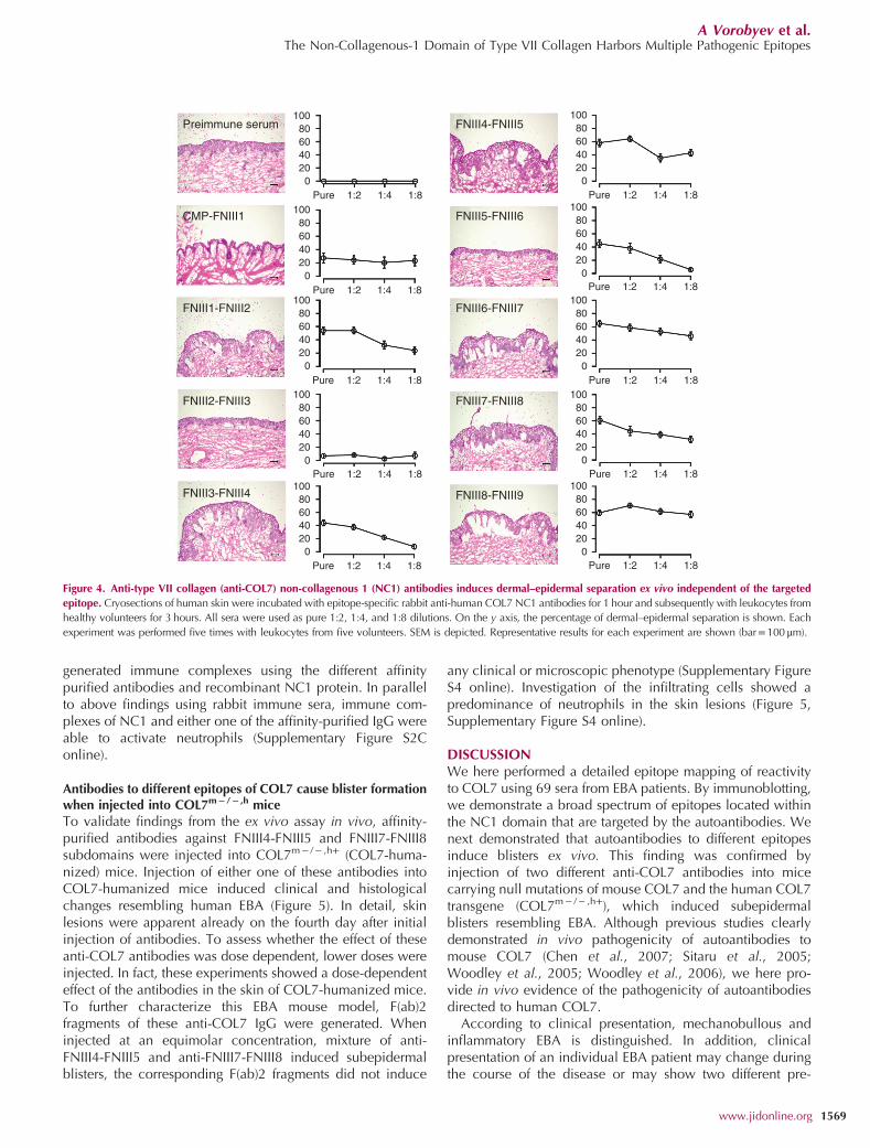
generated immune complexes using the different affinitypurified antibodies and recombinant NC1 protein. In parallelto above findings using rabbit immune sera, immune com-plexes of NC1 and either one of the affinity-purified IgG wereable to activate neutrophils (Supplementary Figure S2Conline).
Antibodies to different epitopes of COL7 cause blister formationwhen injected into COL7m− /− ,h miceTo validate findings from the ex vivo assay in vivo, affinity-purified antibodies against FNIII4-FNIII5 and FNIII7-FNIII8subdomains were injected into COL7m− /− ,h+ (COL7-huma-nized) mice. Injection of either one of these antibodies intoCOL7-humanized mice induced clinical and histologicalchanges resembling human EBA (Figure 5). In detail, skinlesions were apparent already on the fourth day after initialinjection of antibodies. To assess whether the effect of theseanti-COL7 antibodies was dose dependent, lower doses wereinjected. In fact, these experiments showed a dose-dependenteffect of the antibodies in the skin of COL7-humanized mice.To further characterize this EBA mouse model, F(ab)2fragments of these anti-COL7 IgG were generated. Wheninjected at an equimolar concentration, mixture of anti-FNIII4-FNIII5 and anti-FNIII7-FNIII8 induced subepidermalblisters, the corresponding F(ab)2 fragments did not induce
any clinical or microscopic phenotype (Supplementary FigureS4 online). Investigation of the infiltrating cells showed apredominance of neutrophils in the skin lesions (Figure 5,Supplementary Figure S4 online).
DISCUSSIONWe here performed a detailed epitope mapping of reactivityto COL7 using 69 sera from EBA patients. By immunoblotting,we demonstrate a broad spectrum of epitopes located withinthe NC1 domain that are targeted by the autoantibodies. Wenext demonstrated that autoantibodies to different epitopesinduce blisters ex vivo. This finding was confirmed byinjection of two different anti-COL7 antibodies into micecarrying null mutations of mouse COL7 and the human COL7transgene (COL7m− /− ,h+), which induced subepidermalblisters resembling EBA. Although previous studies clearlydemonstrated in vivo pathogenicity of autoantibodies tomouse COL7 (Chen et al., 2007; Sitaru et al., 2005;Woodley et al., 2005; Woodley et al., 2006), we here pro-vide in vivo evidence of the pathogenicity of autoantibodiesdirected to human COL7.According to clinical presentation, mechanobullous and
inflammatory EBA is distinguished. In addition, clinicalpresentation of an individual EBA patient may change duringthe course of the disease or may show two different pre-
CMP-FNIII1
FNIII3-FNIII4
FNIII4-FNIII5Preimmune serum100
80
60
40
20
0
Pure 1:2 1:4 1:8
100
80
60
40
20
0
Pure 1:2 1:4 1:8100
80
60
40
20
0
Pure 1:2 1:4 1:8
100
80
60
40
20
0
Pure 1:2 1:4 1:8100
80
60
40
20
0
Pure 1:2 1:4 1:8
100
80
60
40
20
0
Pure 1:2 1:4 1:8
100
80
60
40
20
0
Pure 1:2 1:4 1:8
100
80
60
40
20
0
Pure 1:2 1:4 1:8
100
80
60
40
20
0
Pure 1:2 1:4 1:8
100
80
60
40
20
0
Pure 1:2 1:4 1:8
FNIII2-FNIII3
FNIII1-FNIII2
FNIII5-FNIII6
FNIII6-FNIII7
FNIII7-FNIII8
FNIII8-FNIII9
Figure 4. Anti-type VII collagen (anti-COL7) non-collagenous 1 (NC1) antibodies induces dermal–epidermal separation ex vivo independent of the targeted
epitope. Cryosections of human skin were incubated with epitope-specific rabbit anti-human COL7 NC1 antibodies for 1 hour and subsequently with leukocytes from
healthy volunteers for 3 hours. All sera were used as pure 1:2, 1:4, and 1:8 dilutions. On the y axis, the percentage of dermal–epidermal separation is shown. Each
experiment was performed five times with leukocytes from five volunteers. SEM is depicted. Representative results for each experiment are shown (bar=100 μm).
A Vorobyev et al.The Non-Collagenous-1 Domain of Type VII Collagen Harbors Multiple Pathogenic Epitopes
www.jidonline.org 1569

sentations simultaneously. The clinical distinction betweenmechanobullous and inflammatory EBA has so far, however,not been reflected by laboratory findings––e.g., autoantibodysubclasses (Gupta et al., 2012). As binding of autoantibodiesto the vWFA2-domain of NC1 leads to disruption of COL7/COL1 interactions, as shown by nuclear magnetic resonance(Leineweber et al., 2011), and as interactions of COL7with both COL4 and laminin-332 have been described(Brittingham et al., 2006; Chen et al., 1997; Rousselle et al.,1997), we hypothesized that autoantibody binding to certainepitopes within the NC1 domain of COL7 is associated with adistinct EBA phenotype. However, in our cohort of 69 EBApatients, we found no correlation of clinical phenotype with
the fine specificity of anti-COL7 IgG. We do not think thatreactivity to the collagenous domain of COL7 can explainthese findings, as only very few patients had been describedto have reactivity to epitopes located within the collagenousdomain and/or the hinge region of COL7 (Ishii et al., 2009).Hence, other factors are likely to cause the different clinicalphenotypes. In an antibody-transfer model of the disease,inbred mouse lines showed different susceptibilities to blisterinduction; e.g., in C57BL6/J, the extent of clinical disease washigher compared with BALB/C mice (Kasperkiewicz et al.,2012). As the immune system differs among inbred mouselines, mechanobullous EBA may develop in patients who donot mount a strong inflammatory reaction in response to
Anti-FNIII4-FNIII5
Ab 3X500 µg
Clin
ical pic
ture
His
tolo
gy
lgG
C3
Anti-FNIII7-FNIII8
Ab 3X500 µg Control
FNIII7-FNIII8FNIII4-FNIII5
3x500 µg
3x250 µg
3x100 µg
3x500 µg
3x250 µg
3x100 µg
20
15
10
Dis
ease
score
0
5
20
15
10
Dis
ease
score
0
5
Day 8Day 4 Day 12 Day 8Day 4 Day 12
Figure 5. Transfer of anti-FNIII4-FNIII5 or anti-FNIII7-FNIII8 antibodies induces blisters in type VII collagen (COL7)-humanized mice. COL7-humanized mice,
injected with 3×500 μg of (a, d) FNIII4-FNIII5- or (b, e) FNIII7-FNIII8-specific antibodies, developed blisters and erosions clinically and subepidermal splits
histologically, whereas (c, f) control mice remained healthy (bar=100 μm). By direct immunofluorescence microscopy, COL7-humanized mice injected with FNIII4-
FNIII5- or FNIII7-FNIII8-specific antibodies showed (g, h) strong IgG and (j, k) weak C3 deposits, respectively, at the dermal–epidermal junction, whereas no
immunoreactants were found in (i, l) control mice (bar=25 μm). Graphs show disease scores corresponding to body surface area affected by skin lesions after injecting
COL7-humanized mice with 3×500 μg, 3×250 μg, or 3×100 μg of (m) anti-FNIII4-FNIII5, or (n) anti-FNIII7-FNIII8 affinity-purified antibodies. SDs are indicated.
A Vorobyev et al.The Non-Collagenous-1 Domain of Type VII Collagen Harbors Multiple Pathogenic Epitopes
1570 Journal of Investigative Dermatology (2015), Volume 135

binding of autoantibodies to COL7, if the autoantibody targetsa structurally relevant epitope. Careful clinical observationstogether with stringent immunophenotyping––e.g., analyzingthe patients’ neutrophils for the ability to mount an oxidativeburst upon stimulation with autoimmune complexes––willallow testing this assumption. In line with this assumption, amajor interindividual variation of immune complex-inducedneutrophil activation has been described (Recke et al., 2010).After demonstrating a broad epitope recognition of EBA
patient IgG, we next evaluated the potential of antibodiestargeting different epitopes of NC1 to induce subepidermalsplits. When these antibodies were tested for reactivity with theproteins used for immunization, binding to epitopes outsidethese proteins was observed for anti-FNIII3-FNIII4, anti-FNIII5-FNIII6, anti-FNIII6-FNIII7, anti-FNIII7-FNIII8, and anti-FNIII8-FNIII9 antibodies. This phenomenon may be explained by thehigh homology among the FNIII-like repeat domains. Hence, inEBA patients, an autoantibody may cross-react with differentepitopes on COL7. In addition to inter- and intramolecularepitope spreading (Di Zenzo et al., 2011), this binding of anindividual antibody to different epitopes underscores thecomplexity of the autoimmune response in acquired bullousdermatoses. Although the epitope mapping study used IgG4 asa surrogate marker for the total IgG reactivity to COL7 forsensitivity and specificity purposes, we here either used wholepatient IgG or used whole immune IgG from rabbits.We further showed that all antibodies generated here,
which were directed to different epitopes of human COL7NC1, led to dermal–epidermal separation ex vivo, as reportedpreviously using other fragments of COL7 (Csorba et al.,2010). Split formation was dependent on the presence ofneutrophils, whereas incubation of human skin with anti-bodies alone did not cause pathology. This is in line withprevious observations demonstrating that patients’ antibodies,purified against the entire human NC1 domain, depend onthe presence of neutrophils (Sitaru et al., 2002a), morespecifically, on generation of reactive oxygen species (Chiriacet al., 2007) and proteases (Shimanovich et al., 2004). Tovalidate the blister-inducing potential of the generatedantibodies in vivo, rabbit anti-FNIII4-FNIII5 or anti-FNIII7-FNIII8 was injected into COL7-humanized mice. Both theseantibody preparations bound to the dermal–epidermal junc-tion, led to complement deposition, and induced subepider-mal blisters in a dose-dependent manner, resembling theinflammatory variant of human EBA. This blistering pheno-type underscores the pathogenic relevance of the usedantibodies, as mere binding of autoantibodies to the dermal–epidermal junction, i.e. formation of immune complexes,does not inevitably lead to blister formation. For example, thisis reflected by the lack of blister formation, despite of IgGdeposition at the dermal–epidermal junction in mouse modelsof EBA (Ludwig et al., 2012; Sitaru et al., 2006) and bullouspemphigoid (Liu et al., 1995). This EBA animal model alsoprovides direct evidence that binding of antibodies to humanCOL7 induces blistering in vivo. Previous mouse models ofEBA demonstrated pathogenicity of antibodies, directed tomouse or human COL7, that bound murine COL7 in vivo(Chen et al., 2007; Sitaru et al., 2006; Sitaru et al., 2005;
Woodley et al., 2005). The pathogenicity of different IgGsubclasses, as well as IgA directed against COL7, still remainsa controversial issue: although some data suggest a potentialof IgG4 to induce subepidermal splits ex vivo (Mihai et al.,2007), others have demonstrated opposite findings (Reckeet al., 2010). Furthermore, IgA has recently been demon-strated to lead to subepidermal splits ex vivo (Recke et al.,2014; van der Steen et al., 2012). On the basis of thedeveloped animal model here, these findings can now beaddressed in vivo to investigate the potential pathogeniccontribution of IgA, as well as IgG subclasses in EBA.In summary, our mapping studies with NC1 and NC2
domains of COL7 show that autoantibodies in EBA patientsrecognize a broad spectrum of epitopes within COL7 NC1.No correlation was detected between antibody specificity andclinical phenotype. Both ex vivo and using COL7 humanizedmouse in vivo, these autoantibodies induce subepidermalblisters, independent of their specificity. Furthermore, wedemonstrate intramolecular cross-reactivity of autoantibodiesdirected to a specific epitope within the NC1 domain.
MATERIALS AND METHODSA more detailed description of the materials and methods used is
provided in the Supplementary Materials and Methods online.
Patient sera and human skinSerum samples from 69 EBA patients for detection of circulating anti-
COL7 antibodies were used in this study. As the presence of
circulating anti-COL7 antibodies was required for epitope mapping
studies, EBA patients diagnosed by detection of an u-serrated pattern
in the absence of circulating autoantibodies (Buijsrogge et al., 2011)
were not included. Sera from healthy volunteers served as controls.
Neonatal human foreskin, obtained from routine circumcision, was
snap, cut in 6 μm sections, and mounted on SuperFrost slides
(Thermo Scientific, Braunschweig, Germany). Prior to all procedures,
written informed consent was obtained from all patients. The study
was approved by the ethics committee of the University of Lübeck
and was performed according to the Declaration of Helsinki.
MiceCOL7m− /− ,h+ (COL7-humanized) mice that carry the homozygous
null mutations of mouse Col7a1 genes and the transgene of human
COL7A1 were generated as described previously (Ito et al., 2009).
COL7-humanized mice aged 6–8 weeks were intraperitoneally
injected with 100–500 μg in a total of rabbit antibody specific to
NC1 domain of human COL7 or phosphate-buffered saline (PBS) at
days 0, 2, and 4. The extent of blistering was evaluated at days 4, 8,
and 12 after the initial IgG injection. Skin samples for hematoxylin
and eosin staining and direct immunofluorescence microscopy and
serum samples were obtained at day 12. All animal procedures were
conducted according to guidelines of the Hokkaido University
Institutional Animal Care and Use Committee under an approved
protocol.
Cloning of cDNA clones for human NC1 and NC2 domains ofCOL7 recombinant proteinsDNA sequence of the NC1 and NC2 domains was obtained from
Uniprot database (www.uniprot.org).
A Vorobyev et al.The Non-Collagenous-1 Domain of Type VII Collagen Harbors Multiple Pathogenic Epitopes
www.jidonline.org 1571

Expression and purification of recombinant proteinsE. coli Rosetta DE3 cells were transfected with plasmids. Recombi-
nant proteins were purified by affinity chromatography using chitin
beads (New England Biolabs, Ipswich, MA). As proteins CMP-FNIII1
and FNIII9-vWFA2 were purified from inclusion bodies as described
(Hirose et al., 2011), they were refolded using oxidized-reduced
glutathione buffer, dialyzed against protein elution buffer, and
purified by affinity chromatography over chitin beads.
Western blotting with patient seraPlease see Supplementary Materials and Methods online.
Rabbit polyclonal antibody productionRabbit polyclonal antibodies against different fragments of COL7
were produced as previously described (Sitaru et al., 2005).
IgG was purified from preimmune and postimmune serum using
protein G-agarose as described (Sitaru et al., 2005). Corresponding F
(ab)2 fragments were generated using the F(ab)2 preparation kit
(Thermo Scientific, Rockford, IL) according to the manufacturer’s
protocol.
Indirect immunofluorescence microscopyIndirect immunofluorescence studies were performed on murine and
1 M NaCl-split human skin. Cryosections of mouse skin and human
salt-split skin of 6-μm thickness were incubated with serial dilutions
of immune rabbit serum for 1 hour at room temperature, washed with
PBS, and then treated with 1:100 diluted FITC-conjugated polyclonal
swine-anti rabbit Ig (Dako, Hamburg, Germany).
Affinity purification of antibodies from rabbit serumRecombinant proteins were dialyzed in 0.1 M MOPS, pH 7.5, and
coupled to Affi-Gel 10 or 15 (according to isoelectric point of
proteins) following the manufacturer’s protocol (Bio-Rad, Munich,
Germany). IgG previously purified from postimmune serum was
affinity-purified using corresponding Affi-Gel coupled recombinant
proteins, then dialyzed against PBS, and concentrated by Amicon
Ultra (30 KDa, Millipore, Billerica, MA).
Affinity purification of antibodies from EBA patient serumPlasmapheresis material from EBA patient was affinity-purified using
corresponding Affi-Gel coupled recombinant proteins, then dialyzed
against PBS, and concentrated by Amicon Ultra (30 KDa, Millipore,
Billerica, MA).
Purification of normal human leukocytesLeukocytes were isolated from heparin-anticoagulated blood of five
different healthy volunteers following a published protocol (Sitaru
et al., 2002b). Leukocytes used in this study (determined by trypan
blue) had a viability of ⩾ 90%.
Immune-complex activation of human leukocytes in vitroPlease see Supplementary Materials and Methods online.
Determination of dermal–epidermal separation ex vivoCryosections of 6-μm thickness human foreskin were incubated with
different dilutions of rabbit serum (pure, 1:2, 1:4, 1:8, 1:16) for 1 hour
at 37 °C, washed with PBS, and subsequently incubated with normal
human leukocytes from five different healthy donors at 37 °C for
3 hours. As a negative control, preimmune rabbit serum was used.
Subsequently, sections were washed and stained with hematoxylin
and eosin. Percentage of dermal–epidermal separation was evaluated
by two investigators unaware of the section treatment.
Statistical analysisR was used for statistical analyses (R Foundation for Statistical
Computing, Vienna, Austria). A P-valueo0.05 was considered significant.
Correlation of epitopes with gender, age, and clinical phenotype was
assessed by homogeneity distribution analysis and Pearson’s corre-
lation. For Pearson’s correlation, the coefficient analysis WGCNA
package was used. Homogeneity distribution analysis was performed
using homals package.
CONFLICT OF INTERESTThe authors state no conflict of interest.
ACKNOWLEDGMENTSThis work was supported by the Research Training Groups “Modulation ofAutoimmunity” (DFG GRK1727/1) and “Genes, Environment and Inflamma-tion” (DFG GRK1743/1).
SUPPLEMENTARY MATERIAL
Supplementary Material is linked to the online version of the paper at http://www.nature.com/jid
REFERENCES
Bach JF (2002) The effect of infections on susceptibility to autoimmune andallergic diseases. N Engl J Med 347:911–20
Bernard P, Prost C, Aucouturier P et al. (1991) The subclass distribution of IgGautoantibodies in cicatricial pemphigoid and epidermolysis bullosaacquisita. J Invest Dermatol 97:259–63
Brittingham R, Uitto J, Fertala A (2006) High-affinity binding of the NC1 domainof collagen VII to laminin 5 and collagen IV. Biochem Biophys ResCommun 343:692–9
Buijsrogge JJ, Diercks GF, Pas HH et al. (2011) The many faces of epidermolysisbullosa acquisita after serration pattern analysis by direct immunofluores-cence microscopy. Br J Dermatol 165:92–8
Chen M, Doostan A, Bandyopadhyay P et al. (2007) The cartilage matrix proteinsubdomain of type VII collagen is pathogenic for epidermolysis bullosaacquisita. Am J Pathol 170:2009–18
Chen M, Marinkovich MP, Veis A et al. (1997) Interactions of the amino-terminal noncollagenous (NC1) domain of type VII collagen withextracellular matrix components. A potential role in epidermal-dermaladherence in human skin. J Biol Chem 272:14516–22
Chiriac MT, Roesler J, Sindrilaru A et al. (2007) NADPH oxidase is required forneutrophil-dependent autoantibody-induced tissue damage. J Pathol 212:56–65
Csorba K, Sesarman A, Oswald E et al. (2010) Cross-reactivity of autoantibodiesfrom patients with epidermolysis bullosa acquisita with murinecollagen VII. Cell Mol Life Sci 67:1343–51
Dahl MG (1979) Epidermolysis bullosa acquisita–a sign of cicatricialpemphigoid? Br J Dermatol 101:475–84
Di Zenzo G, Thoma-Uszynski S, Calabresi V et al. (2011) Demonstration ofepitope-spreading phenomena in bullous pemphigoid: results of aprospective multicenter study. J Invest Dermatol 131:2271–80
Fine JD, Tyring S, Gammon WR (1989) The presence of intra-lamina lucidablister formation in epidermolysis bullosa acquisita: possible role ofleukocytes. J Invest Dermatol 92:27–32
Gammon WR, Briggaman RA, Woodley DT et al. (1984) Epidermolysisbullosa acquisita–a pemphigoid-like disease. J Am Acad Dermatol 11:820–32
A Vorobyev et al.The Non-Collagenous-1 Domain of Type VII Collagen Harbors Multiple Pathogenic Epitopes
1572 Journal of Investigative Dermatology (2015), Volume 135
Gammon WR, Murrell DF, Jenison MW et al. (1993) Autoantibodies to type VIIcollagen recognize epitopes in a fibronectin-like region of the noncolla-genous (NC1) domain. J Invest Dermatol 100:618–22
Gandhi K, Chen M, Aasi S et al. (2000) Autoantibodies to type VII collagen haveheterogeneous subclass and light chain compositions and theircomplement-activating capacities do not correlate with the inflammatoryclinical phenotype. J Clin Immunol 20:416–23
Gupta R, Woodley DT, Chen M (2012) Epidermolysis bullosa acquisita. ClinDermatol 30:60–9
Hirose M, Recke A, Beckmann T et al. (2011) Repetitive immunization breakstolerance to type XVII collagen and leads to bullous pemphigoid in mice.J Immunol 187:1176–83
Ishii N, Hamada T, Dainichi T et al. (2010) Epidermolysis bullosa acquisita:what's new? J Dermatol 37:220–30
Ishii N, Yoshida M, Hisamatsu Y et al. (2004) Epidermolysis bullosa acquisitasera react with distinct epitopes on the NC1 and NC2 domains of type VIIcollagen: study using immunoblotting of domain-specific recombinantproteins and postembedding immunoelectron microscopy. Br J Dermatol150:843–51
Ishii N, Yoshida M, Ishida-Yamamoto A et al. (2009) Some epidermolysisbullosa acquisita sera react with epitopes within the triple-helicalcollagenous domain as indicated by immunoelectron microscopy. Br JDermatol 160:1090–3
Ito K, Sawamura D, Goto M et al. (2009) Keratinocyte-/fibroblast-targeted rescueof Col7a1-disrupted mice and generation of an exact dystrophicepidermolysis bullosa model using a human COL7A1 mutation. Am JPathol 175:2508–17
Iwata H, Bieber K, Tiburzy B et al. (2013) B cells, dendritic cells, and macrophagesare required to induce an autoreactive CD4 helper T cell response inexperimental epidermolysis bullosa acquisita. J Immunol 191:2978–88
Jones DA, Hunt SW 3rd, Prisayanh PS et al. (1995) Immunodominantautoepitopes of type VII collagen are short, paired peptide sequenceswithin the fibronectin type III homology region of the noncollagenous(NC1) domain. J Invest Dermatol 104:231–5
Kasperkiewicz M, Nimmerjahn F, Wende S et al. (2012) Genetic identificationand functional validation of FcgammaRIV as key molecule inautoantibody-induced tissue injury. J Pathol 228:8–19
Kim JH, Kim YH, Kim SC (2011) Epidermolysis bullosa acquisita: a retrospectiveclinical analysis of 30 cases. Acta Derm Venereol 91:307–12
Komorowski L, Muller R, Vorobyev A et al. (2013) Sensitive and specific assaysfor routine serological diagnosis of epidermolysis bullosa acquisita. J AmAcad Dermatol 68:e89–95
Kurzhals G, Stolz W, Meurer M et al. (1991) Acquired epidermolysis bullosawith the clinical feature of Brunsting-Perry cicatricial bullous pemphigoid.Arch Dermatol 127:391–5
Lapiere JC, Woodley DT, Parente MG et al. (1993) Epitope mapping of type VIIcollagen. Identification of discrete peptide sequences recognized by serafrom patients with acquired epidermolysis bullosa. J Clin Invest 92:1831–9
Leineweber S, Schonig S, Seeger K (2011) Insight into interactions of the von-Willebrand-factor-A-like domain 2 with the FNIII-like domain 9 of collagenVII by NMR and SPR. FEBS Lett 585:1748–52
Liu Z, Diaz LA, Swartz SJ et al. (1995) Molecular mapping of a pathogenicallyrelevant BP180 epitope associated with experimentally induced murinebullous pemphigoid. J Immunol 155:5449–54
Ludwig RJ (2012) Model systems duplicating epidermolysis bullosa acquisita: amethodological review. Autoimmunity 45:102–10
Ludwig RJ (2013) Clinical presentation, pathogenesis, diagnosis, and treatmentof epidermolysis bullosa acquisita. ISRN Dermatology 2013:25
Ludwig RJ, Kalies K, Kohl J et al. (2013) Emerging treatments for pemphigoiddiseases. Trends Mol Med 19:501–12
Ludwig RJ, Muller S, Marques A et al. (2012) Identification of quantitative traitloci in experimental epidermolysis bullosa acquisita. J Invest Dermatol132:1409–15
Mihai S, Chiriac MT, Herrero-Gonzalez JE et al. (2007) IgG4 autoantibodiesinduce dermal-epidermal separation. J Cell Mol Med 11:1117–28
Oostingh GJ, Sitaru C, Zillikens D et al. (2005) Subclass distribution of type VIIcollagen-specific autoantibodies in patients with inflammatory boweldisease. J Dermatol Sci 37:182–4
Parente MG, Chung LC, Ryynanen J et al. (1991) Human type VII collagen:cDNA cloning and chromosomal mapping of the gene. Proc Natl Acad SciU S A 88:6931–5
Recke A, Sitaru C, Vidarsson G et al. (2010) Pathogenicity of IgG subclassautoantibodies to type VII collagen: induction of dermal-epidermalseparation. J Autoimmun 34:435–44
Recke A, Trog LM, Pas HH et al. (2014) Recombinant human IgA1 and IgA2autoantibodies to type VII collagen induce subepidermal blistering ex vivo.J Immunol 193:1600–8
Roenigk HH Jr., Ryan JG, Bergfeld WF (1971) Epidermolysis bullosa acquisita.Report of three cases and review of all published cases. Arch Dermatol103:1–10
Rose NR, Bona C (1993) Defining criteria for autoimmune diseases (Witebsky'spostulates revisited). Immunol Today 14:426–30
Rousselle P, Keene DR, Ruggiero F et al. (1997) Laminin 5 binds the NC-1domain of type VII collagen. J Cell Biol 138:719–28
Sakai LY, Keene DR, Morris NP et al. (1986) Type VII collagen is a majorstructural component of anchoring fibrils. J Cell Biol 103:1577–86
Saleh MA, Ishii K, Kim YJ et al. (2011) Development of NC1 and NC2domains of type VII collagen ELISA for the diagnosis and analysis of thetime course of epidermolysis bullosa acquisita patients. J Dermatol Sci 62:169–75
Schmidt E, Zillikens D (2013) Pemphigoid diseases. Lancet 381:320–32
Shimanovich I, Mihai S, Oostingh GJ et al. (2004) Granulocyte-derived elastaseand gelatinase B are required for dermal-epidermal separation induced byautoantibodies from patients with epidermolysis bullosa acquisita andbullous pemphigoid. J Pathol 204:519–27
Sitaru C, Chiriac MT, Mihai S et al. (2006) Induction of complement-fixingautoantibodies against type VII collagen results in subepidermal blisteringin mice. J Immunol 177:3461–8
Sitaru C, Kromminga A, Hashimoto T et al. (2002a) Autoantibodies to type VIIcollagen mediate Fcgamma-dependent neutrophil activation and inducedermal-epidermal separation in cryosections of human skin. Am J Pathol161:301–11
Sitaru C, Mihai S, Otto C et al. (2005) Induction of dermal-epidermal separationin mice by passive transfer of antibodies specific to type VII collagen. J ClinInvest 115:870–8
Sitaru C, Schmidt E, Petermann S et al. (2002b) Autoantibodies to bullouspemphigoid antigen 180 induce dermal-epidermal separation in cryosec-tions of human skin. J Invest Dermatol 118:664–71
Tanaka T, Furukawa F, Imamura S (1994) Epitope mapping for epidermolysisbullosa acquisita autoantibody by molecularly cloned cDNA for type VIIcollagen. J Invest Dermatol 102:706–9
van der Steen LP, Bakema JE, Sesarman A et al. (2012) Blocking Fcalphareceptor I on granulocytes prevents tissue damage induced by IgAautoantibodies. J Immunol 189:1594–601
Woodley DT, Briggaman RA, O'Keefe EJ et al. (1984) Identification of the skinbasement-membrane autoantigen in epidermolysis bullosa acquisita.N Engl J Med 310:1007–13
Woodley DT, Chang C, Saadat P et al. (2005) Evidence that anti-type VIIcollagen antibodies are pathogenic and responsible for the clinical,histological, and immunological features of epidermolysis bullosa acqui-sita. J Invest Dermatol 124:958–64
Woodley DT, O'Keefe EJ, Reese MJ et al. (1986) Epidermolysis bullosa acquisitaantigen, a new major component of cutaneous basement membrane, is aglycoprotein with collagenous domains. J Invest Dermatol 86:668–72
Woodley DT, Ram R, Doostan A et al. (2006) Induction of epidermolysis bullosaacquisita in mice by passive transfer of autoantibodies from patients.J Invest Dermatol 126:1323–30
Zambruno G, Manca V, Kanitakis J et al. (1994) Linear IgA bullous dermatosiswith autoantibodies to a 290 kd antigen of anchoring fibrils. J Am AcadDermatol 31:884–8
A Vorobyev et al.The Non-Collagenous-1 Domain of Type VII Collagen Harbors Multiple Pathogenic Epitopes
www.jidonline.org 1573