Embed Size (px)
Citation preview

Cell, Vol. 60, 921-926, March 23, 1990, Copyright 0 1990 by Cell Press
A Novel Pathway of DNA End-to-End Joining
Silke Thode, Antje Schafer, Petra Pfeiffer, and Walter Vielmetter Institute of Genetics University of Cologne 5000 Cologne 41 Federal Republic of Germany
Summary
Repair mechanisms related to Illegitimate recombi- nation can join nonhomologous DNA ends that ter- minate as protruding single strands (PSS). Here we analyze in Xenopus egg extracts joining reactions be- tween 3’ PSS termini and various partner termlni. In junctions, 3’ PSS termlni are preserved by fill-in DNA synthesis, although their 5’ recessed ends cannot serve as a primer. Alternative priming from a partner terminus ligeted to the 3’ PSS end appears unlikely, because no single strand-specific DNA ligases are de- tectable. We show that fill-in of 3’ PSS termini precedes ligation and can even be primed in the absence of any ligation. Therefore, priming requites precise alignment of terminus pairs by a novel mechanism. We postulate that this is achieved by unique DNA binding proteins that align ends in various types of joining reactions.
Introduction
illegitimate recombination covers a diversity of reactions promoting rearrangements of DNA sequences within chromosomal DNA. A basic feature of the pertinent path- ways is physical breakage of DNA molecules and rejoin- ing of DNA ends (for review, see Roth et al., 1985). Apart from damage-induced breakage, DNA ends may be generated by copy and repair processes of DNA mole- cules (for review, see Roth and Wilson, 1988), and they may be eliminated by processes of random end-to-end joining that link nonhomologous DNA termini (Wilson et al., 1982; Wake et al., 1984). Thus, nonhomologous join- ing mechanisms are necessary tools in the illegitimate transfer of chromosomal DNA sequences. Illegitimate break-join mechanisms have particular importance in the recombination machinery of higher eukaryotes. In con- trast to the situation in prokaryotes and certain yeasts, DNA integration into vertebrate chromosomes proceeds orders of magnitude more frequently by illegitimate than by homologous recombination (Folger et al., 1984; Roth and Wilson, 1985; Thomas et al., 1986).
Mechanisms for end-to-end joining manage to generate seamless junctions from nonhomologous DNA ends that terminate in either protruding single strands (PSS) or blunt ends. DNA sequence analyses of naturally occur- ring junctions (for review, see Roth and Wilson, 1986) show that joining tends to preserve the original se- quences. Changes, mainly restricted to the immediate vi-
cinity of breakpoints, comprise small numbers of base ex- changes, nucleotide losses, and occasional nucleotide additions (Roth et al., 1985; Landau et al., 1987). Small ter- minal sequence homologies of 1-5 nucleotides have been considered to be involved in joining processes (Stringer, 1982; Marvo et al., 1983; Ruley and Fried, 1983; Roth et al., 1985; Roth and Wilson, 1986).
Complete information on the original sequences pro- cessed into naturally occurring junctions is difficult to ob- tain and was rarely verified (Gahlmann and Doerfler, 1983; Wiikie and Palmiter, 1987). A different approach starts from DNA substrates with defined nonhomologous termi- nus configurations that are joined either in vivo in mam- malian cells (Roth and Wilson, 1988) or in vitro in Xenopus egg extracts (Pfeiffer and Vielmetter, 1968). Analysis of joined products in these systems reveals strikingly similar pathways of junction formation, explaining several of the structural properties found in natural junctions. The main features of junction formation are the unexpected joining modes by which terminal PSS interact. Thus far, two oper- ationally different traits for the joining of nonhomologous termini have been observed (Pfeiffer and Vielmetter, 1988).
One trait concerns the joining of DNA termini with PSS ends of equal polarity (3’ PSS-3’ PSS, 5’ PSS-5’ PSS). These form overlap structures that may even invade termi- nal duplex regions of joining partners (Roth and Wilson, 1986). The degree of overlap is set by fortuitously match- ing base pairs, and overlap structures undergo final mis- match and fill-in corrections. To explain the peculiar over- lap mode and its register setting by even single base matches, the existence of as yet unknown terminal align- ment proteins was postulated (Pfeiffer and Vielmetter, 1988).
A second trait concerns the joining of DNA termini in which PSS ends are paired with blunt ends or PSS ends of opposite polarity (blunt-8PSS, blunt-B PSS, 5’PSS-3’ PSS). In junctions of these configurations PSS sequences are preserved by fill-in DNA synthesis (Roth and Wilson, 1986; Pfeiffer and Vielmetter, 1988). At the5’recessed end of a 3’ PSS terminus, priming of fill-in DNA synthesis by normal, 5’-toa’-directed DNA synthesis is not feasible. However, a fill-in primer could in principle be provided by a partner terminus ligated to the 3’ PSS end by a single strand-specific DNA ligase. Known DNA ligases (Korn- berg, 1980) can at best occasionally attach single nucleo- tide protrusions to blunt ends (Wiaderkiewicz and Ruiz- Carillo, 1987). Therefore, joining systems contain either a yet to be discovered single strand-specific DNA ligase or a special priming mechanism.
In this paper we analyze the problem of fill-in priming during the joining of 3’ PSS ends to partner termini in ex- tracts from Xenopus laevis eggs. This system is devoid of a single strand-specific DNA ligase activity and can prime fill-in of 3’ PSS ends in the absence of any terminal liga- tion. We discuss the presence of (the above mentioned) alignment proteins that may provide priming conditions by prealigning the involved termini.

Cell 922
io -I In+ Li%f ~@,o t: 0000 3 0000 Figure 1. Examples of Gel Assays for Joined or Ligated Reaction Products from Homolo- gous and Nonhomologous Substrates
Joining reactions of substrates were performed
- Id under standard conditions as described in Ex-
- Id perimental Procedures. Gel bands are visual-
-0c - Id
-0c ized by in situ hybridization of gels probed with
--Im -0c 32P-pSP65 DNA.
-lm (A) and (B): Reactions of substrates in PPL or
-Im T4 DNA ligase (control) show that (A) the ho- mologous substrate BamHl (= 5’ PSS) is li-
- ccc -ccc gated to oc and ccc monomers and (B) the non- -ccc homologous substrate Smal-Pstl (= blunt-3
A 6 C D E PSS) is not joined to oc or ccc monomers ex- cept by the extract (control). (C), (D), and (E): Extract-promoted joining reac-
tions with or without the addition of ddNTPs (as indicated) show (C) for the homologous substrate Sal1 (= 5’ PSSTCGA) that ligation is unaffected by ddNTP addition. Nonhomologous substrates (D) BamHI-Pstl and (E) Aval-Pstl (both = 5’ PSS-3’ PSS) are key configurations in the analysis of the joining process, Except in ddNTP-free extract only in (E), ligation to oc monomers occurs in the presence of certain ddNTPs by a pattern outlined in Figure 2C’. Lane specifications: (M) pSP65 DNA marker, Substrates are incubated in: (PPL) extract-derived fractions of partially purified DNA ligase activity, (T4) T4 DNA ligase, (EX) complete Xenopus egg extract, and@,@,@, and@complete extract plus single ddNTP types added as indicated. Band specifications: (Id) linear dimer, (oc) open circular monomer, (Im) linear monomer, (ccc) covalently closed circular monomer. Multimers above Id are not considered in these assays.
Results
Linear DNA Substrates Are Joined to Form Distinguishable Circular Products The joining of DNA substrates in Xenopus egg extracts was investigated by the use of established assay proce- dures (Pfeiffer and Vieimetter, 1988). They detect ligation and joining by circularization of linear substrate DNA moi- ecules with defined terminus structures. The linear DNA substrates we have used are the nonhomologous types blunt-g’ PSS, blunt-3 PSS, and 5’ PSS-3’ PSS with four nucleotide long PSS ends. These substrates are prepared by two successive restriction cuts within the polylinker of plasmid pSP85 (Melton et al., 1984) while homologous control substrates are correspondingly generated by sin- gle restriction cuts. In some substrates 3’ PSS or blunt ends are capped with a dideoxynucieoside triphosphate (ddNTP) by a terminal deoxynucieotidyl transferase re- action.
Extract-promoted joining converts substrates into various mono- and multimeric reaction products distinguished from one another and from linear input DNA by gel elec- trophoretic separation (Figure 1). Multimeric forms may be catenated simply by ligation of equal DNA ends and thus are useful to diagnose ligation. Only circular monomers are considered to be true joined reaction products. They arise in complete reactions as ccc (covalentiy closed cir- cular) monomers, which are regularly accompanied by the nicked or gapped intermediates, the oc (open circular) monomers. In certain incomplete (fill-in inhibited) reac- tions, exclusively oc monomers are generated by ligation.
Fill-In DNA Synthesis Can Se Differentially Inhibited by ddNTPs The problem of ligation of PSS termini may be directly analyzed in complete extracts by inhibiting fill-in reactions
so that PSS ends are retained in a (partially) single- stranded state during joining. Fill-in DNA synthesis during joining reactions can be specifically interrupted by sup- plementing extracts with ddNTPs to block DNA synthesis promoted by DNA polymerase(s) j3 (y) at sites of ddNTP incorporation into DNA (van der Viiet and Kwant, 1978; Kornberg, 1980; Miller and Chinault, 1982). We showed previously that ddNTPs efficiently inhibit the extract- promoted formation of ccc monomers from nonhomoio- gous substrates without affecting the ligation of homoio- gous substrates (Figure 1C). Thus, ddNTP inhibition can be used as a reliable tool to study extract-promoted joining of PSS termini, with the advantage that fill-in lengths can be set differentially by incorporation of selected ddNTPs at suitable positions of PSS sequences during extract- promoted joining reactions. This leads in different sub- strates to characteristic patterns of inhibition for the forma- tion of ccc and oc monomers (Figure 2).
Joining Reactions Depend upon Fill-In DNA Synthesis Inhibition patterns for joining reactions in ddNTP-supple- mented extracts were studied for three substrate types (Figure 2). As an example of adjoined gel assays, the par- ticularly interesting substrates of the 5’ PSS-3’ PSS type are documented in Figures 1D and 1E. All three substrate types undergo complete joining in ddNTP-free extracts to form oc and ccc monomers. This corresponds to previous results, which have shown by cionai sequence analysis that in these substrates, PSS sequences are preserved in the junctions of reaction products (Pfeiffer and Vielmetter, 1988).
Incomplete fill-in of PSS termini upon ddNTP incorpora- tion prevents the formation of ccc monomers. Thus, ccc suppression indicates successful ddNTP incorporation. However, ddNTP inhibition does not completely suppress

DNA End-to-End Joining 923
I I c;.Anl Pst I ~bbbb~..di I I
I---- I- i2)CCQG) tgc@I+ I-
Figure 2. Joining Patterns for Substrates A to C’ in Complete Extract under Conditions of ddNTP Inhibition
Substrates A to C’are depicted by their terminal configurations. Below each substrate scheme the expected positions of ddNTP incorporation and their adjoined fill-in patterns are shown for the ddNTPs indicated by an encircled letter. Formation or absence of oc and ccc monomers in the single reactions is indicated by + or - Note that both oc and ccc monomers are exclusively generated in untreated extracts (lines 1). ddNTP-treated extracts (lines 2-5) only produce oc monomers at certain settings of fill-in inhibition.
the formation of oc monomers. In some cases, depending upon ddNTP positions in PSS sequences of particular substrates, oc monomers are readily formed. This indi- cates that single-strand ligation has taken place. By these diagnostic criteria, the joining of substrates A to C’ was analyzed in complete extracts in separate samples to which either no ddNTP or inhibitory amounts of one selected ddNTP type were added. Subslrate 7Lpe A Blunt-B PSS substrates can directly extend their 3’ recessed ends by fill-in DNA synthesis to create ligatable blunt ends. Incorporation of a ddNTP at the utmost termi- nal 5’ PSS nucleotide (Figure 2A) leads to a completely filled-in 3’ dideoxy-terminated “pseudoblunt” end. Forma- tion of oc monomers indicates that this structure can be readily ligated to a blunt end by its dideoxy-free strand. Upon ddNTP incorporation at any other site at the 5’ PSS terminus, 5’ PSS residues are retained due to incomplete fill-in (Figure 2A). Under these conditions no oc mono- mers are formed, which indicates that the extract contains no DNA ligase activities that could ligate 5’ PSS ends to a blunt-end partner.
SUbStr8te Typ8S B 8nd B Blunt-3’ PSS substrates lack a fill-in primer for the direct fill-in of 3’ PSS ends. Upon ddNTP incorporation at any site of the 3’ PSS end oc monomers are formed (Figures 28 and 28’). This observation raises the possibility that li- gation does not occur unless there has been the filling-in of at least a single nucleotide. SubStf8te Types C and C 5’ PSS-3’ PSS substrates carry oppositely directing termi- nal single strands, of which the 5’ PSS but not the 3’ PSS end is accessible to direct fill-in DNA synthesis. No oc monomers arise by ddNTP inhibition at any site in the 5’ PSS end, not even if complete fill-in generates a pseudo- blunt end (Figure 1D; Figure 2C). Evidently, this structure cannot be ligated to a 3’ PSS partner, although it is ligated to a blunt end (Figure 2A). This may be taken as decisive evidence that complete extracts have no capacity to ligate blunt ends directly to 3’ single-strand ends.
The same unexpected situation as observed for blunt-3 PSS substrates (Figures 28 and 283 arises if in 5’ PSS-3’ PSS substrates the 3’ PSS end is submitted to ddNTP in- hibition to fill in 5’ PSS ends completely and 3’ PSS ends partially. This situation fully restores oc monomer forma- tion (Figure 1E; Figure 2C’). Therefore, partial fill-in of the 3’ PSS end, even by insertion of a single ddNTP at the ut- most terminal position, suffices to convert it into a ligata- ble structure. Thus, the ability for ligation apparently de- pends upon 3’ PSS fill-in. Consequently, fill-in must precede ligation. We are therefore driven to the conclu- sion that fill-in DNA synthesis is primed by some unortho- dox mechanism in the absence of covalent DNA linkage between partner termini.
Blunt-3’ PSS SUb8tr8tSS Are Joined under Conditions of Blocked 3’ PSS Ligation The concept of unorthodox priming of fill-in was more rigorously analyzed for the blunt-3’ PSS substrate in the complete absence of primary ligation by simply capping it at its 3’PSS end with a dideoxy-G (see scheme in Figure 3A). Surprisingly, this terminus configuration is readily joined by complete extracts to form oc monomers. Apart from the trivial explanation of nucleolytic degradation of the capped 3’ PSS end, the only apparent way to achieve molecular circularization would be to fill in the capped 3 PSS end. This then would lead to a continuous strand con- nection via the “fill-in” strand (see scheme in Figure 38).
Extract-joined reaction products of the dideoxy-Gcap- ped substrate were cloned, and 17 clones were submitted to DNA sequence analysis. Junctions in 85% of the clones retain the 3’ PSS sequence, including the dideoxy-G posi- tion. This is lacking in 17% of the clones, because during substrate preparation tailing reactions can be driven to only 80%-90% completeness. The remaining 17% repre- sent cloned junctions whose 3’ PSS ends were degraded to variable degrees (up to 18 nucleotides) during the join- ing reaction.
The sequence integrity of the junctional region was veri- fied by Southern blot hybridization of clonal DNA by using the l&mer oligonucleotide (oligo) 2 (see scheme in Figure 3A) as a probe. Oligo 2 is complementary to the intact

Cdl 924
A [QAQCQQQQACQTCQQ)UWO~
SfMl
~~&cjyxxmx;“~~
xi7
(~ZTCQCCC~TQCAQC~)OIICPZ
Figure 3. Terminal Configuration of a Blunt-3’ PSSddG Substrate
(A) In the Smal-Pstl (= blunt-8 PSS) substrate configuration the 3’ PSS end within the “dideoxy” strand is capped by a dideoxy-G (framed G). This configuration is depicted in alignment to the sequences of oligos 1 and 2, which are used as probes IO analyze the junctional in- tegrity of joined products. (B) Proposed mechanism for the joining of the dideoxy-G-capped sub- strate: (1) Fill-in (Mack arrowheads) is induced within the “fill-in” strand by “unorthodox priming” in the absence of blunt-3 PSS ligation. (2) Subsequent ligation (open diamond) at the 5’ recessed end provides continuity of the fill-in strand, while within the dideoxy strand a nick per- sists next to the dideoxy-G position. This retains the substrate as an oc monomer.
clonal copy of the “dideoxy” strand, which extends right and left from the junctional break point. Under stringent hybridization conditions, only this sequence hybridizes. Since 24 of the 36 tested clones (66%) respond with a positive hybridization signal, they must possess an intact dideoxy strand. That in fact only clones wlth intact junc- tional DNA sequences yield positive hybridization signals is exemplified in Figure 4.
To ensure that these results are not simply due to clon- ing artifacts induced by the bacterial host, we analyzed extract-joined products directly without cloning. For this, gel-separated single strands of the linearized products Figure 5. Oligonucleotide Hybridization Analysis of Direct (Not Cloned)
Joined Products Derived from Blunt-3’ PSS-ddG Substrate
Reaction products of the Smal-Pstl-ddG substrate linearized with Pvul and separated on denaturing agarose gels were analyzed by oligonu- cleotide hybridization (see Figure 3) to verify: first, the fill-in strand con- tinuity and sequence integrity by a positive signal if probed with oligo 1, and second, the dideoxy strand disruption at the dide0xy-G discon- tinuity by a negative response if probed by oligo 2. Sizes of single- stranded fragments are: 3 kb for the continuous fill-in strand, and 1.4 and 1.6 kb for the two dideoxy strand fragments. After blotting on Hy bond N, separated single-stranded products were stringently hybrid- ized in successive steps with: (A) oligo 1, (8) oligo 2, and (C) linearized =PvpSP65 DNA. Lane specifications: Size markers (hybridization controls) derived from junctionally intact clone 1 (see Figure 4) were restricted with (1) Pvul (3 kb).(2) Pvul and EcoRl(l.6 kb), and (3) Pvul and Hindlll(1.4 kb). The essential (Pvul-restricted) reaction products are (4) Smal-Pstl and (5) Smal-Pstl-ddG. Negative hybridization controls were derived from (6) junctional-defective clone 6 (see Figure 4) restricted with Pvul, (7) Pvul and EcoRI, and (6) Pvul and Hindlll.
Figure 4. Southern Blot Analyses of Cloned Joined Products Derived from a Blunt-3’ PSS-ddG Substrate
Cloned reaction products of the substrate Smal-Pstl-ddG are strin- gently hybridized with oligo 2 to distinguish intact from defective junc- tions The bottom line refers to results for adjoined sequence analyses available for clones 1, 2, and 6-9: +, complete junctional sequence including dideoxy-G position; -, the junctional sequence misses at least the dide0xy-G position. Band assigments: (oc) open circular monomer, (ccc) covalently closed circular monomers, which are the di- agnostic derivatives, and (ss) single-stranded monomer.
were assayed by Southern blot hybridization (Figure 5). The following 15mer oligonucleotides served as probes: oligo 1, complementary to the fill-in strand, and oligo 2, complementary to the dideoxy strand (see scheme in Fig- ure 3A). The result in Figure 5A shows that oligo 1 effi- ciently hybridizes to the fill-in strand, which therefore must contain the whole complement of the dideoxy strand (in-

DNA End-to-End Joining 925
eluding the dideoxy-G position). In contrast, oligo 2 (Fig- ure 58) as expected, fails to hybridize to the dideoxy strand, because genuine strands retain a nick next to the dide0xy-G cap (see scheme in Figure 3A) so that two dis- connected single-stranded fragments are generated. This confirms that the dideoxy-G cap is still present in the reac- tion products.
Taken together, these experiments demonstrate that the joining system can prime fill-in DNA synthesis at the 3 PSS terminus of a blunt-3 PSS configuration in the ab- sence of any primary ligation. Thus, an unorthodox prim- ing mechanism for fill-in DNA synthesis must be seriously considered.
Extract-Derived DNA Ligases Pail to Ligate PSS to Blunt Ends or Ends Overlapping by a Single Nucleotide Dissection of complete extracts into ligation-active compo- nents may provide direct information on inherent special properties of DNA ligases required for the joining of non- homologous substrates. Therefore, extract fractions ob- tained by fractionation over DEAE cellulose were assayed for their ability to ligate homologous and to join non- homologous substrates and to perform DNA synthesis (see Experimental Procedures). The partially purified DNA ligase (PPL), derived from a subset of fractions, con- tains a 20-fold enriched collection of virtually the total liga- tion activities across fractionation profiles. The absence of any DNA synthesis capacity in the PPL subset allows the direct detection of single strand-specific DNA ligases with substrates carrying PSS ends that remain single stranded during ligation assays.
PPL efficiently ligates homologous substrates to form ccc and oc monomers similarly, as does T4 DNA ligase (Figure 1A). However, PPL fails to ligate blunt-8 PSS sub- strates (Figure 18) as well as blunt-5 PSS and 5’ PSS-3 PSS substrates (data not shown). Therefore, PPL lacks measurable amounts of single strand-specific ligation ac- tivities that could ligate PSS ends of either polarity.
rerminal configuration generates generates ---Jy-y
Figure 6. Pattern of Joined Products for Blunt-3’ PSS and Blunt- ddT-3’ PSS Substrates Reacted in Complete Extract or PPL
Terminal configurations of substrates are depicted on the left. Right columns list formation (yes) or absence (no) of oc and ccc monomers as reaction products in extract or PPL. Joining (ligation) assays were performed under standard conditions as described in Experimental Procedures.
The result that PPL lacks single strand-specific DNA li- gases fully complements experiments in complete ex- tracts in which DNA synthesis is inhibited by ddNTPs. These experiments imply that PSS ends of blunt-3 PSS substrates can be ligated only if they overlap by at least one fill-in-generated, matching nucleotide (Figures 28, 2B’, and 2C’). A substrate that generates this situation ar- tificially was constructed: the blunt-8 PSS substrate was capped at its blunt end with a dideoxy-T complementing the terminal A of the 3’ PSS end (Figure 6). Alone by its structure, this substrate is inert toward any fill-in reaction. This modified substrate turned out to be readily ligated to form oc monomers in complete extracts, but not in PPL or any other fraction over the entire DEAE column profile. Thus, a terminal matching base pair alone is insufficient to promote the ligation to blunt ends. Apparently, the liga- tion of the modified substrate requires at least one addi- tional supporting component that apparently has become separated from DNA ligases during fractionation.
Discussion
Extracts from X. laevis eggs have the capacity to join non- homologous DNA termini efficiently (Pfeiffer and Vielmet- ter, 1966) by pathways strikingly similar to those found in vivo in mammalian cells (Roth and Wilson, 1966). Thus, this system appears to be part of a general route for illegiti- mate recombination common to vertebrate cells. The spectrum of possible terminus interactions of this system has been described (Pfeiffer and Vielmetter, 1966). The pertinent mechanisms are still poorly understood.
Single-Strand Ends Cannot Directly Be Ligated to Partner Termini In joining reactions of blunt-3’ PSS and related substrate types junctional PSS sequences are preserved by fill-in DNA synthesis. The crucial point is how to fill in 3’ PSS termini, whose 5’ recessed ends cannot provide a primer, if 5’-to-3’-directed DNA synthesis is considered to be the only acceptable synthesis mode (Kornberg, 1960). As an alternative it was suggested that fill-in might be primed from a partner blunt end attached to a 3’ PSS end by single-strand ligation (Roth and Wilson, 1966; Pfeiffer and Vielmetter, 1966). We show here that this simple scheme does not hold.
PPL fractions, containing the entire ligation activity of the extract, are unable to ligate PSS to blunt ends (Figure 1B). PPL apparently lacks ligation-activating components, which are unlikely to be just selectively lost single strand- specific DNA ligases. This view is strongly supported by the fact that ddNTP-induced partial fill-in inhibition, which retains PSS ends during joining in complete extracts in a (partially) single-stranded state, prevents ligation between blunt and PSS ends (Figures 2A and 2C). Taken together, these facts are certainly not in favor of the concept of di- rect single-strand ligation.
Fill-In DNA Synthesis Pn?cedes 3’ PSS Ligation During the joining of 5’ PSS-3’ PSS substrates, the appro- priate ddNTP setting converts 5’ PSS ends into 3’dideoxy-
terminated pseudoblunt ends (Figure 2C). These may be considered as key configurations that block any terminal priming of DNA synthesis. In contrast to a regular blunt end (Figure 2B), a pseudoblunt end cannot be joined to a 3’ PSS end, although it may well be ligated to a partner blunt end (Figure 2A). Its primer-blocking capacity is over- come and ligation is fully restored by ddNTP settings that allow fill-in to proceed into the 3’ PSS terminus side by even a single nucleotide (Figure 2C’; see also blunt-3 PSS substrates, Figure 28). This conclusively shows that the partner terminus contributes to the priming event. Fill- in is therefore a prerequisite for the ligation of partner ter- mini. The essential condition is apparently a matching base-paired duplex structure at the 3’ PSS terminus for which, remarkably, a single base match suffices. Fill-in it- self is a ligation-independent process that must precede ligation.
Strong support for the concept of ligation-independent fill-in came from the use of a blunt-3 PSS substrate, whose dideoxy-G-capped 3’ PSS end prohibits primary li- gation (Figure 3A). Extracts readily join this substrate via direct fill-in of the 3’ PSS end (Figure 38) as was fully con- firmed by detailed junctional analyses (Figures 4 and 5). We therefore conclude that in a blunt-3’ PSS configura- tion, fill-in of the 3’ PSS terminus can be primed from the blunt end in the complete absence of primary ligation.
Fill-In Preceding Blunt-3’ PSS Ligation Requires Support of Alignment Proteins How can fill-in DNA synthesis of the 3’ PSS end be primed from a blunt-end partner prior to ligation? On first sight, DNA strand priming and extension at the blunt end could be due to nucleotide addition by terminal deoxynucleo- tidy1 transferase (Baltimore, 1974; Kunkel et al., 1966) or “nontemplated” DNA polymerase reactions (Clark et al., 1967; Clark, 1966). However, these reactions cannot be the answer, because they fail to produce faithfully the copied complements of 3’ PSS partner termini. Even if they could copy faithfully, this would in any case require physical contact with the partner terminus carrying the complementary sequence information. Therefore, it ap- pears conclusive that in the absence of primary ligation, two termini must be juxtaposed long enough to prime fill- in DNA synthesis successfully. This contact time must be orders of magnitude larger than that required for diffu- sion-controlled collision of free DNA ends. Therefore, an efficient priming process necessarily requires the exis- tence of a helper. We postulate that this helper is a DNA binding protein that can align terminus pairs precisely and hold them in contact to make priming feasible. We pro- pose “alignment protein” as a provisional term.
Alignment Proteins May Also Support the Ligation Process Attachment of single complementary dideoxynucleotides to the blunt end of the blunt-3’ PSS substrate (Figure 6) simulates a joining configuration in which the first nucleo- tide at the tip of the 3’ PSS end has been filled in (see Fig- ure 20. Failure of this configuration to be ligated by PPL implies that a single base match does not provide suffi-
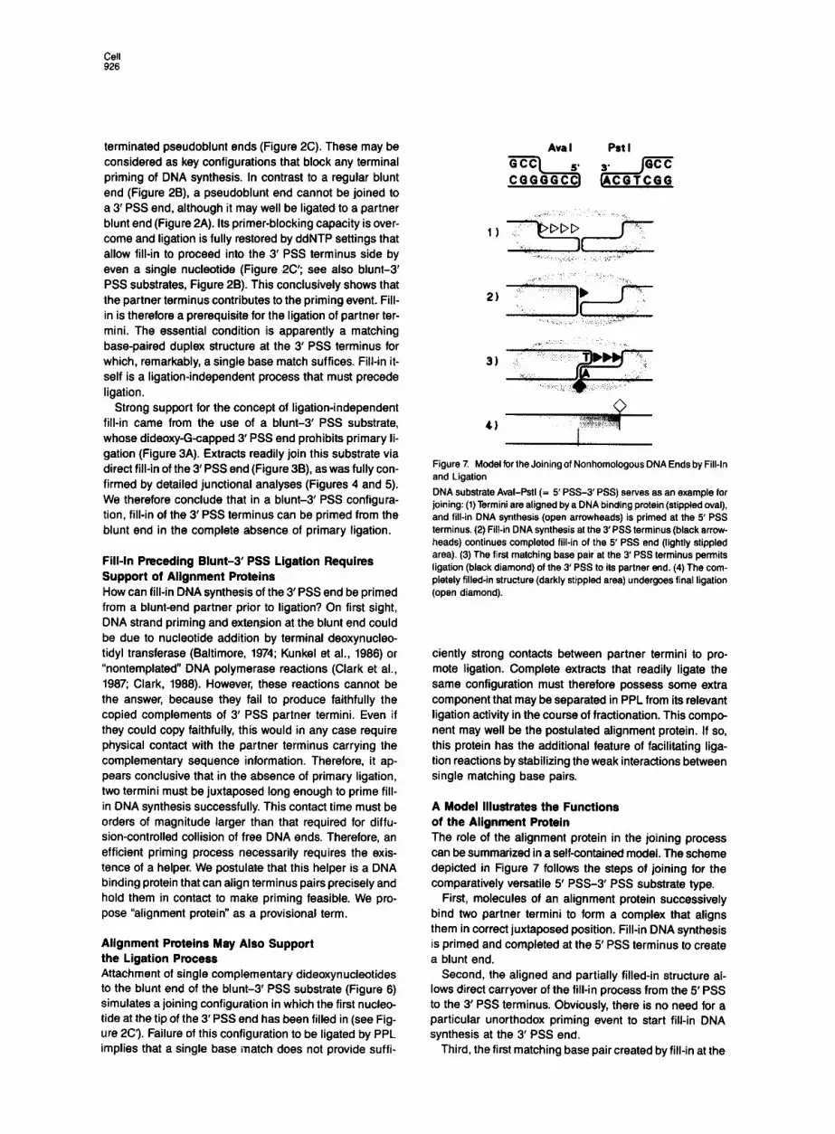
Ava I Pst I
4)
Figure 7. Model for the Joining of Nonhomologous DNA Ends by Fill-In and Ligation
DNA substrate Aval-Pstl (= 5’ PSS-3’ PSS) serves as an example for joining: (1) Termini are aligned by a DNA binding protein (stippled oval), and fill-in DNA synthesis (open arrowheads) is primed at the 5’ PSS terminus. (2) Fill-in DNA synthesis at the 3’PSS terminus (black arrow- heads) continues completed fill-in of the 5’ PSS end (lightly stippled area). (3) The first matching base pair at the 3’ PSS terminus permits ligation (black diamond) of the 3’ PSS to its partner end. (4) The com- pletely filled-in structure (darkly stippled area) undergoes final ligation (open diamond).
ciently strong contacts between partner termini to pro- mote ligation. Complete extracts that readily ligate the same configuration must therefore possess some extra component that may be separated in PPL from its relevant ligation activity in the course of fractionation. This compo- nent may well be the postulated alignment protein. If so, this protein has the additional feature of facilitating liga- tion reactions by stabilizing the weak interactions between single matching base pairs.
A Model Illustrates the Functions of the Alignment Protein The role of the alignment protein in the joining process can be summarized in a self-contained model. The scheme depicted in Figure 7 follows the steps of joining for the comparatively versatile 5’ PSS-3’ PSS substrate type.
First, molecules of an alignment protein successively bind two partner termini to form a complex that aligns them in correct juxtaposed position. Fill-in DNA synthesis is primed and completed at the 5’ PSS terminus to create a blunt end.
Second, the aligned and partially filled-in structure al- lows direct carryover of the fill-in process from the 5’ PSS to the 3’ PSS terminus. Obviously, there is no need for a particular unorthodox priming event to start fill-in DNA synthesis at the 3’ PSS end.
Third, the first matching base pair created by fill-in at the

DNA End-lo-End Joining 927
3’ PSS end is a necessary and sufficient condition to in- duce primary ligation.
And fourth, the intact duplex junction is finally gener- ated by completion of 3’ PSS fill-in and ligation of the re- maining nick. The order of the third and fourth steps might be reversed.
The Alignment Protein Is Still a Hypothetical Entity Detailed specifications of the postulated alignment protein are not clear at present. It may belong to a known class of proteins, i.e., DNA polymerases. Similarly, the align- ment protein may be associated with DNA polymerase or DNA ligase functions or both. We previously postulated the existence of a protein recognizing terminal DNA struc- tures (Pfeiffer and Vielmetter, 1999). This was required to explain why egg extracts are able to align PSS ends of equal polarity by an overlap structure, which can be set by even single matching base pairs. Unusual structural properties are required for a protein that is capable of achieving not only this but also precise terminal align- ment. A search for a protein with these unusual properties is in progress. The availability of a comparatively simple assay using specific substrates makes us hopeful that this protein can be isolated.
Experimental Procedures
Preqaration of Egg Extracts and DNA Substrates Extracts from artificially fertilized X. laevis eggs and DNA substrates derived from plasmid pSP65 were prepared as described by Pfeiffer and Vielmetter (1968).
Joinlng Asaays with and without Inhibition of Fill-In DNA Syntheels by ddNTPs Joining assays and T4 DNA ligase controls were performed in a total reaction volume of 10 PI containing 1 ng of DNA substrate per ul (Pfeiffer and Vielmetter, 1986) by incubation at 13OC for 90 min. They were stopped by addition of 30 ul of 1% (w/v) SDS in TE-Stop (20 mM Tris-HCI [pH 7.81, 10 mM EDTA) and heated at 85OC for 10 min. Fill-in DNA synthesis in joining assays was blocked by the addition of 1 ul of 2 mM ddNTP (Pharmacia) in H&l to joining samples. Final ddNTP concentrations (200 uM) exceeded dNTP concentrations in the extract precursor pool by a factor of about 10, estimated from values published for intact eggs (Woodland and Pestell, 1972).
Visualization of Jolned Products in Agankse Gel Assays For gel assays 4 ul aliquots of the supernatant of centrifuged SDS- stopped samples (corresponding to 1 ng of DNA substrate input) were incubated together with 2 pl of TE (10 mM Tris-HCI [pH 7.51, 1 mM EDTA) and 4 ul of prostop (2 ng/ul proteinase K, 1.6 mM Tris-HCI [pH 8],46% [v/v] glycerol, 16 mM EDTA, 0.01% [w/v] BPB, 0.01% [w/v] XC) at 3PC for 30 min. After heating to 65OC for 10 min and subsequent chilling on ice, these samples were electrophoresed on 1% (w/v) agarose minigels in TA (40 mM Tris-acetate [pH 81, 2 mM EDTA) con- taining 1 W/ml ethidium bromide at 10 V/cm. Gels were soaked for 5 min in GSO (0.25 N HCI, 1.5 M NaCI), for 30 min in GSl (0.5 N NaOH, 1.5 M NaCI), and for IO min in GS2 (1 M Tris-HCI [pH 7.51, 1.5 M NaCI) prior to vacuum heat drying at 8oOC for 30 min. Dried denatured gels were hybridized in situ using a modification of the protocol of Shinnick et al. (1975). The hybridization mix (6x SSC, 1% [whr] SDS, 50 mM Tris-HCI [pH 7.51, 4 mM EDTA, 2 ug/ml carrier DNA) contained linear pSP65 DNA as probe that had been radioactively labeled with [@PI dCTP (3000 CilmMol; Amersham) by the random primer extension method (Amersham, Multiprime DNA labeling kit). After hybridization at 65OC, overnight gels were washed in WS (8x SSC. 0.5% [w&l SDS) twice at room temperature for 20 min and once at 65OC for 10 min and subsequently exposed with duplicate screens to Kodak X-OMAT AR films at -70°C for at least 6 hr.
PPL frum Complete Egg Extracts Complete undiluted egg extract was centrifuged at 15,096 x g at 4OC for 30 min. Supernatants free of cell debris and yolk were adjusted to a final concentration of 0.5 mM PMSF and 0.2 uM pepstatin (Serva). Dialysis of the centrifuged extract was carried out on microdialysis filters (0.025 pm, Millipore) against buffer B (10 mM Tris-HCI [pH 81, 10% (v/v) glycerol, 10 mM NaCI, 10 mM 8-mercaptoethanol, 1 mM EDTA). The dialyzed extract was loaded onto a DEAE cellulose column equilibrated with the same buffer. Protein fractions were eluted with a 10-500 mM NaCl gradient and dialyzed against buffer D (70 mM Tris-HCI [pH 81, 5 mM j3-mercaptoethanol, 10 mM MgCIr). Ligation- active fractions were pooled and are referred to as PPL.
Assays for Ligation Activity In PPL Fractions DEAE cellulose column fractions (8 pl) were added to 10 ng of DNA substrate in 1 pl of TE and 1 d of 10x BA (0.5 mg/ml BSA, 10 mM ATP (pH 7.61). Incubation and termination of reactions and gel assays were performed as described above.
Assays for DNA Polymeraee Activities in PPL Fractions Assays measuring activities of unspecified DNA polymerases were carried out in a total volume of 106 ul containing the to be determined extract fractions in 50 mM Tris-HCI (pH 8.0). 5 mM 8-mercaptoethanol, 8 mM Mg acetate, 0.2 mglml BSA, 80 fug/ml “activated” calf thymus DNA (Sigma), 25 uM each of dATP, dTTP dGTP, dCTP (Pharmacia), and 50 &i/ml 13H]dTTP (48 Cilmmol, Amersham). The reaction mix- ture was incubated for 30 min at 3pC and stopped by heat inactivation at 95’C for 5 min. Acid-precipitable material was collected on What- man GWA filters, and the 3H incorporation was determined in a scintil- lation counter.
The activity of 8(u) DNA polymerases was determined by specific in- activation of a(6) DNA polymerases by the addition of 5 pg/ml aphidico- lin (Sigma), which causes more than 90% of inactivation (Kornberg, 1982).
Preparation of Dideoxy-Capped Nonpolar DNA Substrates The usual procedure (Pfeiffer and Vielmetter, 1966) using two succes- sive restriction cuts to obtain nonhomologous substrates from plasmid pSP85 was modified to generate dideoxycappd substrates. Dideoxy capping after the first and before the second cut results in cap attach- ment exclusively at the 3’OH side of the first cut. Two dideoxy-capped blunt-3’ PSS substrates, Smal-ddT-Pstl and Smal-Pstl-ddG, were constructed.
Capping reactions were performed by the incubation of pSP85 plas- mid DNA linearized by Smal (or Pstl) and the desired ddNTP at a molar ratio of lo3 ddNTP/substrate 3’ OH (Olson and Harvey, 1975) in the presence of terminal deoxynucleotidyl transferase (TdT; BRL) at 3 Ulpg DNA in TdT reaction buffer (BRL) at 3pC for at least 2 hr. Under these conditions usually 80%-90% of TdT-treated molecules were modified. The reaction was stopped by the addition of EDTA to a final concentra- tion of 40 mM. After extraction with phenol and chloroform and precipi- tation with ethanol, samples were treated with T4 DNA ligase to ligate DNA molecules that had escaped the capping reaction. Capped linear DNA monomers were purified from agarose gels and subsequently cut with the second restriction enzyme (Pstl or Smal, respectively), thus generating the final capped DNA substrate.
Southern Blot Analysts and Ollgonucleotlde Hybridization of Jolned Products Preparative joining assays were performed with 120 pl of complete ex- tract in a total reaction volume of 150 pl at a DNA substrate (Smal-Pstl- ddG) input concentration of 1 ng/ul. Reactions were stopped by the ad- dition of 250 pl of 05 ug/pl proteinase K (Sigma) in TNE (20 mM This-HCI [pH 7.51, 0.3 M NaCI, 10 mM EDTA) containing 1% (w/v) SDS immediately followed by incubation at 85°C for 30 min. After extraction with phenol and chloroform total nucleic acids were precipitated with ethanol. For denaturing agarose gels the samples were digested with Pvul (Boehringer Mannheim), which linearizes plasmid pSP86 approx- imately opposite the polylinker. After another extraction with phenol and precipitation with ethanol, samples were adjusted to 0.5 M NaOH, heated to 65OC for 5 min. and electrophoresed on 1% (w/v) agarose gels in NE (30 mM NaOH, 2 mM EDTA) at 10 V/cm. Southern transfer of the DNA to Hybond N filters (Amersham) was performed with TS (0.4

M NaOH, 0.6 M NaCI) overnight. Filters were subsequently neutralized in NS (0.5 M lbs-HCI [pH 7.51, 1 M NaCI), dried at 55OC for 1 hr, and irradiated with UV light (302 nm) for IO min. Prehybridization of filters was performed in H-mix (6x SSC. 0.2% [w/v] BSA, 0.2% [w/v] PVR 0.2% [w/v] Ficoll) containing 0.1 mglml carrier DNA at 51% for 2 hr. Fif- teen picamoles of IBmer oligonucleotides (oligo 1 or 2. see Figure 4A; T, = 54%) 5’ end-labeled with 100 uCi of [Y-~PJATP (5000 Ci/mmol; Amersham) by T4 polynucleotide kinase (Boehringer Mannheim), served as a probe for hybridization of filters, which was performed in 30 ml of H-mix containing 0.1 mg/ml carrier DNA at 51% overnight. Filters were then washed in WS2 (6x SSC, 1% [w/v] SDS) at 51°C for 15 min, at 52°C for 5 min, and at 54% for 3 min. Autoradiography was performed as described above. Filters were dehybridized by incuba- tion in H-mix at 65% for 2 hr. Complete removal of the probe was con- trolled by autoradiography of filters overnight.
Cloning of Joined Pmdwts and Analysis of Clones Cloning procedures, miniscale preparation of clonal piasmid DNA, and sequencing protocols were the same as described by Pfeiffer and Viel- metter (1986). For hybridization of cloned joined products with a 5’- %P-end-labeled 15mer oligonucleotide. undigested clonal plasmid DNA was electrophoresed on 1% (w/v) agarose gels in TA at 10 V/cm. After electrophoresis gels were soaked in GSO, GSl, and GS2 as de- scribed above, and DNA was blotted onto Hybond N filters with 20x SSC as the transfer solution. After washing in 2x SSC, filters were dried at 5fYC for 1 hr and irradiated with UV light (302 nm) for 10 min. Prehybridization, hybridization, and rehybridization were performed as above.
Acknowledgments
We thank Dr. Dagmar Barthels and Dr. Wolfgang Wille for helpful sug- gestions and instructions on oligonucleotide hybridization, Dr. Boer- ries Kemper for advice and practical support concerning protein purifi- cation, Dr. Robert Jack for critically reading our manuscript, and last, but not least, Ute Zylajew for her excellent technical assistance. This work was supported by grant Vi 23/t-l from the Deutsche Forschungs- gemeinschaft to W. V
The costs of publication of this article were defrayed in part by the payment of page charges. This article must therefore be hereby marked “advertisement” in accordance with 16 U.S.C. Section 1734 solely to indicate this fact.
Received November 15, 1969; revised January 16, 1990.
References
Baltimore, D. (1974). Is terminal deoxynucleotidyl transferase a somatic mutagen in lymphocytes? Nature 248, 409-411. Clark, J. M. (1966). Novel non-temple&l nucleotide addition reactions catalyzed by prokaryotic and eukaryotic DNA polymerases. Nucl. Acids Res. 16, 9677-9666. Clark, J. M., Joyce, C. M., and Beardsley, G. P (1987). Novel blunt-end addition reactions catalyzed by DNA polymerase I of Escherichis co/i. J. Mol. Biol. f98, 123-127. Folger, K. R., Thomas, K. R., and Capecchi, M. R. (1964). Analysis of homologous recombination in cultured mammalian cells. Cold Spring Harbor Symp. Quant. Biol. 49. 123-136. Gahlmann, A., and Doerfler, W. (1983). Integration of viral DNA into the genome of the adenovirus type P-transformed hamster cell line HE5 without loss or alteration of cellular nucleotides. Nucl. Acids Res. II, 7347-7361. Kornberg, A. (1980). DNA Replication. Second edition (San Francisco: Freeman). Kornberg, A. (1962). Supplement to DNA Replication. Second edition (San Francisco: Freeman). Kunkel, T A., Gopinathan, K. P, Dube, D. K., Snow, E. T, and Loeb, L. A. (1966). Rearrangements of DNA mediated by terminal transfer- ase. Proc. Nat). Acad. Sci. USA 83, 1667-1871. Landau, N. FL, Schatz, 0. G., Rosa, M., and Baltimore, D. (1967). In- creased frequency of N-region insertion in a murine pre-B-cell line in-
fected with a terminal deoxynucleotidyl transferase retroviral expres- sion vector. Mol. Cell. Biol. 7, 3237-3243. Marvo, S. L., King, S. R., and Jaskunas, S. Ft. (1983). Role of short regions of homology in intermolecular illegitimate recombination events. Proc. Natl. Acad. Sci. USA 80, 1391-1395. Melton, D. A., Krieg, l? A., Rebagliati, M. A.. Maniatis, T, Zinn, K., and Greene, M. R. (1964). Efficient in vitro synthesis of biologically active RNA and RNA hybridization probes from plasmids containing a bacte- riophage SP6 promotor. Nucl. Acids Res. 12, 7035-7056. Miller, M. R., and Chinault, D. N. (1962). The rolesof DNApolymerases a, 6 and y in DNA repair synthesis induced in hamster and human cells by different DNA damaging agents. J. Biol. Chem. 257,10204-10209. Olson, K., and Harvey, C. (1975). Determination of the 3’ terminal nucleotide of DNA fragments. Nucl. Acids Res. 2, 319-325. Pfeiffer, P, and Vielmetter, W. (1986). Joining of nonhomologous DNA double strand breaks in vitro. Nucl. Acids Res. 76, 907-924. Roth, D. B., and Wilson, J. H. (1985). Relative rates of homologous and nonhomologous recombination in transfected DNA. Proc. Natl. Acad. Sci. USA 82,3355-3359. Roth, D. 8.. and Wilson, J. H. (1966). Nonhomologous recombination in mammalian cells: role for short sequence homologies in the joining reaction. Mol. Cell. Biol. 6, 4295-4304. Roth, D. B., and Wilson, J. H. (1968). Illegitimate ecombination in mam- malian cells. In Genetic Recombination, R. Kucherlapati and G. R. Smith, eds. (Washington, DC.: American Society for Microbiology), pp. 621-653. Roth, D. B., Porter, T M., and Wilson, J. H. (1965). Mechanisms of non- homologous recombination in mammalian cells. Mol. Cell. Biol. 5, 2599-2607. Ruley, H. E., and Fried, M. (1983). Clustered illegitimate recombination events in mammalian cells involving very short sequence homologies. Nature 304, 161-184. Shinnick, T. M., Lund, E., Smithies, O., and Blattner, F. R. (1975). Hy- bridization of labeled RNA to DNA in agarose gels. Nucl. Acids Res. 2, 1911-1929. Stringer, J. R. (1962). DNA sequence homology and chromosomal de- letion at a site of SV40 DNA integration. Nature 296, 363-366. Thomas, K. R., Folger, K. R., and Capecchi, M. A. (1966). High fre- quency targeting of genes to specific sites in the mammalian genome. Cell 44, 419-426. van der Vliet. P C., and Kwant. M. M. (1978). Role of DNA polymerase y in adenovirus DNA replication. Nature 276, 532-534. Wake, C. T, Gudewicz, T, Porter, T, White, A., and Wilson, J. H. (1984). How damaged is the biologically active subpopulation of transfected DNA? Mol. Cell. Biol. 4, 387-396. Wiaderkiewicz, R., and Rub-Carrillo, A. (1987). Mismatch and blunt to protruding-end joining by DNA ligases. Nucl. Acids Res. 75, 7631- 7848. Wilkie, T M., and Palmiter, R. D. (1987). Analysis of the integrant in MyK-103 transgenic mice in which males fail to transmit the integrant. Mol. Cell. Biol. 7; 1646-1655. Wilson, J. H., Berget, F!, and Pipas, J. M. (1962). Somatic cells effi- ciently join unrelated DNA segments end-to-end. Mol. Cell. Biol. 2, 1258-1269. Woodland, H. R., and Pestell, R. 0. W. (1972). Determination of the nucleoside triphosphate contents of eggs and oocytes of Xenopus lsevis. Biochem. J. 727, 597-605.


