Embed Size (px)
Citation preview

A Portrait of Tissue Phosphoprotein Stability inthe Clinical Tissue Procurement Process*Virginia Espina‡§, Kirsten H. Edmiston¶, Michael Heiby‡, Mariaelena Pierobon‡�,Manuela Sciro‡**, Barbara Merritt¶, Stacey Banks¶, Jianghong Deng‡,Amy J. VanMeter‡, David H. Geho‡ ‡‡, Lucia Pastore§§, Joel Sennesh§§,Emanuel F. Petricoin III‡, and Lance A. Liotta‡
Little is known about the preanalytical fluctuations ofphosphoproteins during tissue procurement for molecularprofiling. This information is crucial to establish guide-lines for the reliable measurement of these analytes. Todevelop phosphoprotein profiles of tissue subjected tothe trauma of excision, we measured the fidelity of 53signal pathway phosphoproteins over time in tissue spec-imens procured in a community clinical practice. Thisinformation provides strategies for potential surrogatemarkers of stability and the design of phosphoproteinpreservative/fixation solutions. Eleven different specimencollection time course experiments revealed augmenta-tion (�20% from the time 0 sample) of signal pathwayphosphoprotein levels as well as decreases over timeindependent of tissue type, post-translational modifica-tion, and protein subcellular location (tissues includedbreast, colon, lung, ovary, and uterus (endometrium/myo-metrium) and metastatic melanoma). Comparison acrosstissue specimens showed an >20% decrease of proteinkinase B (AKT) Ser-473 (p < 0.002) and myristoylatedalanine-rich C-kinase substrate protein Ser-152/156 (p <0.0001) within the first 90-min postexcision. Proteins inapoptotic (cleaved caspase-3 Asp-175 (p < 0.001)), prolif-eration/survival/hypoxia (IRS-1 Ser-612 (p < 0.0003),AMP-activated protein kinase � Ser-108 (p < 0.005), ERKThr-202/Tyr-204 (p < 0.003), and GSK3�� Ser-21/9 (p <0.01)), and transcription factor pathways (STAT1 Tyr-701(p < 0.005) and cAMP response element-binding proteinSer-133 (p < 0.01)) showed >20% increases within 90-minpostprocurement. Endothelial nitric-oxide synthase Ser-1177 did not change over the time period evaluated withbreast or leiomyoma tissue. Treatment with phosphataseor kinase inhibitors alone revealed that tissue kinasepathways are active ex vivo. Combinations of kinase andphosphatase inhibitors appeared to stabilize proteins that
exhibited increases in the presence of phosphatase inhib-itors alone (ATF-2 Thr-71, SAPK/JNK Thr-183/Tyr-185,STAT1 Tyr-701, JAK1 Tyr-1022/1023, and PAK1/PAK2 Ser-199/204/192/197). This time course study 1) establishesthe dynamic nature of specific phosphoproteins in ex-cised tissue, 2) demonstrates augmented phosphoryla-tion in the presence of phosphatase inhibitors, 3) showsthat kinase inhibitors block the upsurge in phosphoryla-tion of phosphoproteins, 4) provides a rational strategy forroom temperature preservation of proteins, and 5) consti-tutes a foundation for developing evidence-based tissueprocurement guidelines. Molecular & Cellular Proteom-ics 7:1998–2018, 2008.
Elucidation of pathogenic protein signaling networks withintumors offers tremendous promise as a means to individualizemolecular targeted cancer therapy. Despite this promise, thedata obtained from a diagnostic assay applied to humantissue must be stable, reproducible, monitored, and validated;otherwise a clinical decision may be based on incorrect orbiased information.
Modulation of ongoing cellular kinase activity representsone of the most rapidly growing arenas in new drug discovery.Identification of specific phosphoprotein signaling aberrationscan be used for the development of targeted therapies forpatients with lung, breast, colon, or other cancer (1–6). Pro-filing the tumor phosphoproteome using human tumor biopsyspecimens is an important component of the strategy forindividualized cancer therapy (1–6). Nevertheless unless weunderstand preanalytical variables affecting the fidelity of thephosphoproteome we will fail to utilize this rich repertoire ofbiological information.
The perishability of tissue molecules may be profoundlyinfluenced by the timing and treatment of the tissue after it isharvested. Tissue is generally snap frozen to perform pro-teomics research studies. In a busy clinical setting, it may beimpossible to immediately preserve procured tissue in liquidnitrogen. Moreover the time delay from patient excision topathologic examination and molecular analysis is often notrecorded and may vary from 30-min to many hours dependingon the time of day, length of the procedure, time for patho-logic examination, and number of concurrent cases.
From the ‡Center for Applied Proteomics and Molecular Medicine,George Mason University, Manassas, Virginia 20110, �Clinica Chiru-rcica II, Universita degli Studi di Padova, 35128 Padova (Padova),Italy, **Centro di Referimento Oncologico, National Cancer Institute,Aviano Hospital, 33081 Aviano (Pordenone), Italy, and ¶Inova CancerCenter and §§Department of Pathology, Inova Fairfax Hospital, FallsChurch, Virginia 22042
Received, December 31, 2007, and in revised form, May 21, 2008Published, MCP Papers in Press, July 30, 2008, DOI 10.1074/
mcp.M700596-MCP200
Research
© 2008 by The American Society for Biochemistry and Molecular Biology, Inc.1998 Molecular & Cellular Proteomics 7.10This paper is available on line at http://www.mcponline.org
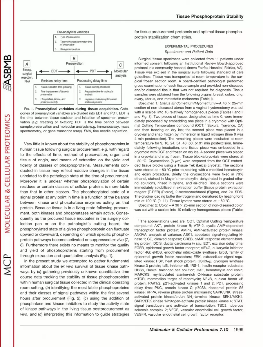
Very little is known about the stability of phosphoproteins inhuman tissue following surgical procurement, e.g. with regardto the effects of time, method of preservation, organ andtissue of origin, and means of extraction on the yield andfidelity of classes of phosphoproteins. Measurements con-ducted in tissue may reflect reactive changes in the tissueunrelated to the pathologic state at the time of procurement.Moreover it is unclear whether phosphorylation of certainresidues or certain classes of cellular proteins is more labilethan that in other classes. The phosphorylated state of asignal protein at any point in time is a function of the balancebetween kinase and phosphatase enzymes acting on thatprotein. Because tissue is in a living state following procure-ment, both kinases and phosphatases remain active. Conse-quently as the procured tissue incubates in the surgery col-lection pan or on the pathologist’s cutting board, thephosphorylated state of a given phosphoprotein can fluctuateupward or downward, depending on which specific phospho-protein pathways become activated or suppressed ex vivo (7,8). Furthermore there exists no means to monitor the qualityand yield of phosphoproteins starting from procurementthrough extraction and quantitative analysis (Fig. 1).
In the present study we attempted to gather fundamentalinformation about the ex vivo survival of tissue kinase path-ways by (a) gathering previously unknown quantitative timecourse data tracking the stability of tissue phosphoproteinswithin human surgical tissue collected in the clinical operatingroom setting, (b) identifying the most labile phosphoproteinsand their classes of signal pathways within the first severalhours after procurement (Fig. 2), (c) using the addition ofphosphatase and kinase inhibitors to study the activity stateof kinase pathways in the living tissue postprocurement exvivo, and (d) interpreting this information to guide strategies
for tissue procurement protocols and optimal tissue phospho-protein stabilization chemistries.
EXPERIMENTAL PROCEDURES
Specimens and Patient Data
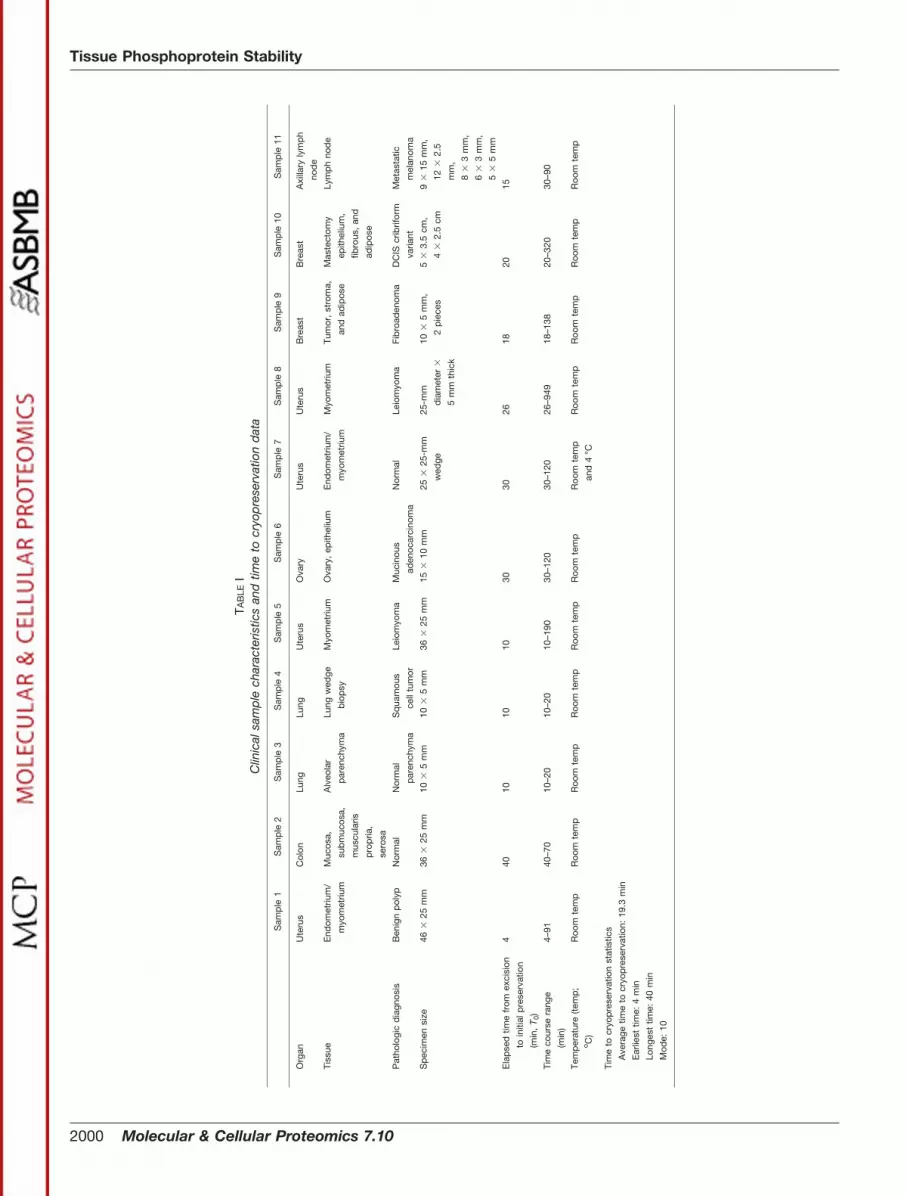
Surgical tissue specimens were collected from 11 patients underinformed consent following an Institutional Review Board-approvedprotocol in a community hospital (Inova Fairfax Hospital, Fairfax, VA).Tissue was excised in the surgical suite following standard of careguidelines. Tissue was transported at room temperature to the sur-gical frozen section room. A board-certified pathologist performedgross examination of each tissue sample and provided non-diseasedand/or diseased tissue that was not required for diagnosis. Tissuesamples were obtained from the following organs: breast, colon, lung,ovary, uterus, and metastatic melanoma (Table I).
Specimen 1: Uterus (Endometrium/Myometrium)—A 46 � 25-mmsection of non-diseased uterus from a vaginal hysterectomy was cutwith a scalpel into 16 relatively homogeneous pieces (Tables I and IIand Fig. 3). Two pieces of tissue, designated as time 0, were imme-diately processed by embedding one piece in a cryomold with Opti-mal Cutting Temperature compound (OCT;1 Sakura, Torrence, CA)and then freezing on dry ice; the second piece was placed in acryovial and snap frozen by immersion in liquid nitrogen (time 0 was4 min postexcision). The remaining pieces were incubated at roomtemperature for 9, 16, 24, 34, 48, 60, or 91 min postexcision. Imme-diately following incubation, one tissue piece was embedded in acryomold with OCT and frozen on dry ice. A second piece was placedin a cryovial and snap frozen. Tissue blocks/cryovials were stored at�80 °C. Cryosections (8 �m) were prepared from the OCT-embed-ded tissue blocks using a Tissue Tek (Leica) cryostat. The sectionswere stored at �80 °C prior to staining with a modified hematoxylinand eosin procedure. Briefly the cryosections were fixed in 70%ethanol, stained in Mayer’s hematoxylin, dehydrated in graded etha-nol solutions, rinsed in xylene, and air-dried. Tissue sections wereimmediately solubilized in extraction buffer (tissue protein extractionreagent (T-PER) (Pierce), 2-mercaptoehtanol (Sigma), and 2� SDS-Tris-glycine loading buffer (Invitrogen)) and denatured by heating for 8min at 100 °C (9–11). Tissue lysates were stored at �80 °C.
Specimen 2: Colon—A 36 � 25-mm section of non-diseased colonwas cut with a scalpel into 10 relatively homogeneous pieces (Tables
1 The abbreviations used are: OCT, Optimal Cutting Temperaturecompound; AKT, protein kinase B; ATF-2, cyclic AMP-dependenttranscription factor protein; AMPK, AMP-activated protein kinase;ANOVA, analysis of variance; ASK1, apoptosis signal-regulating ki-nase 1; CC, cleaved caspase; CREB, cAMP response element-bind-ing protein; DCIS, ductal carcinoma in situ; EDT, excision delay time;EGFR, epidermal growth factor receptor; eIF4G, eukaryotic initiationfactor 4G; eNOS, endothelial nitric-oxide synthase; ErbB, family ofepidermal growth factor receptors; ERK, extracellular signal-regu-lated kinase; HSP, heat shock protein; GSK3��, glycogen synthasekinase 3 protein; I�B, inhibitor �B, IRS-1, insulin receptor substrate;HBSS, Hanks’ balanced salt solution; H&E, hematoxylin and eosin;MARCKS, myristoylated alanine-rich C-kinase substrate protein;mTOR, mammalian target of rapamycin; NF�B, nuclear factor �Bprotein; PAK1/2, p21-activated kinases 1 and 2; PDT, processingdelay time; PKC, protein kinase C; p70S6, ribosomal protein S6kinase; RPPA, reverse phase protein microarray; SAPK/JNK, stress-activated protein kinase/c-Jun NH2-terminal kinase; SEK1/MKK4,SAPK/ERK kinase 1/mitogen-activate protein kinase kinase 4; STAT,signal transducers and activator of transcription; TSC2, tuberoussclerosis complex 2; VEGF, vascular endothelial cell growth factor;VEGFR, vascular endothelial cell growth factor receptor.
FIG. 1. Preanalytical variables during tissue acquisition. Cate-gories of preanalytical variables are divided into EDT and PDT. EDT isthe time between tissue excision and initiation of specimen preser-vation (e.g. freezing or fixation); PDT is the time period betweensample preservation and molecular analysis (e.g. immunoassay, massspectrometry, or gene transcript array). FNA, fine needle aspiration.
Tissue Phosphoprotein Stability
Molecular & Cellular Proteomics 7.10 1999
TAB
LEI
Clin
ical
sam
ple
char
acte
ristic
san
dtim
eto
cryo
pre
serv
atio
nd
ata
Sam
ple
1S
amp
le2
Sam
ple
3S
amp
le4
Sam
ple
5S
amp
le6
Sam
ple
7S
amp
le8
Sam
ple
9S
amp
le10
Sam
ple
11
Org
anU
teru
sC
olon
Lung
Lung
Ute
rus
Ova
ryU
teru
sU
teru
sB
reas
tB
reas
tA
xilla
ryly
mp
h
nod
e
Tiss
ueE
ndom
etriu
m/
myo
met
rium
Muc
osa,
sub
muc
osa,
mus
cula
ris
pro
pria
,
sero
sa
Alv
eola
r
par
ench
yma
Lung
wed
ge
bio
psy
Myo
met
rium
Ova
ry,
epith
eliu
mE
ndom
etriu
m/
myo
met
rium
Myo
met
rium
Tum
or,
stro
ma,
and
adip
ose
Mas
tect
omy
epith
eliu
m,
fibro
us,
and
adip
ose
Lym
ph
nod
e
Pat
holo
gic
dia
gnos
isB
enig
np
olyp
Nor
mal
Nor
mal
par
ench
yma
Sq
uam
ous
cell
tum
or
Leio
myo
ma
Muc
inou
s
aden
ocar
cino
ma
Nor
mal
Leio
myo
ma
Fib
road
enom
aD
CIS
crib
rifor
m
varia
nt
Met
asta
tic
mel
anom
a
Sp
ecim
ensi
ze46
�25
mm
36�
25m
m10
�5
mm
10�
5m
m36
�25
mm
15�
10m
m25
�25
-mm
wed
ge
25-m
m
dia
met
er�
5m
mth
ick
10�
5m
m,
2p
iece
s
5�
3.5
cm,
4�
2.5
cm
9�
15m
m,
12�
2.5
mm
,
8�
3m
m,
6�
3m
m,
5�
5m
m
Ela
pse
dtim
efr
omex
cisi
on
toin
itial
pre
serv
atio
n
(min
,T 0
)
440
1010
1030
3026
1820
15
Tim
eco
urse
rang
e
(min
)
4–91
40–7
010
–20
10–2
010
–190
30–1
2030
–120
26–9
4918
–138
20–3
2030
–90
Tem
per
atur
e(te
mp
;o C
)
Roo
mte
mp
Roo
mte
mp
Roo
mte
mp
Roo
mte
mp
Roo
mte
mp
Roo
mte
mp
Roo
mte
mp
and
4°C
Roo
mte
mp
Roo
mte
mp
Roo
mte
mp
Roo
mte
mp
Tim
eto
cryo
pre
serv
atio
nst
atis
tics
Ave
rage
time
tocr
yop
rese
rvat
ion:
19.3
min
Ear
liest
time:
4m
in
Long
est
time:
40m
in
Mod
e:10
Tissue Phosphoprotein Stability
2000 Molecular & Cellular Proteomics 7.10

TAB
LEII
Initi
altim
eco
urse
exp
erim
ents
with
hum
antis
sue
colle
cted
ina
com
mun
ityho
spita
l
UE
,�20
%in
crea
sew
ithin
first
60m
inp
oste
xcis
ion;
UM
,�20
%in
crea
seb
etw
een
60an
d12
0m
inp
oste
xcis
ion;
UL,
�20
%in
crea
sem
ore
than
120
min
pos
texc
isio
n;D
E,�
20%
dec
reas
ew
ithin
first
60m
inp
oste
xcis
ion;
DM
,�
20%
dec
reas
eb
etw
een
60an
d12
0m
inp
oste
xcis
ion;
DL,
�20
%d
ecre
ase
mor
eth
an12
0m
inp
oste
xcis
ion;
bla
nk,
not
test
ed.
Pro
tein
Func
tion
Sig
nific
ant
chan
ges
over
time
cour
sera
nge
pos
texc
isio
n
Sam
ple
1:ut
erus
,b
enig
n;4–
91m
inS
amp
le2:
colo
n,no
rmal
;40
–70
min
Sam
ple
3:lu
ngp
aren
chym
a;10
–20
min
Sam
ple
4:lu
ngsq
uam
ous
cell
carc
inom
a;10
–20
min
Sam
ple
5:ut
erin
ele
iom
yom
a;10
–190
min
Sam
ple
7:ut
erus
,b
enig
n;30
–120
min
BA
D(S
er-1
12)
Ap
opto
sis
UE
aa
aa
a
Cas
pas
e-3,
clea
ved
(Asp
-175
)A
pop
tosi
sU
EU
E/D
MD
EU
EU
EU
EC
asp
ase-
7,cl
eave
d(A
sp-1
98)
Ap
opto
sis
UE
/DL
Cas
pas
e-9,
clea
ved
(Asp
-330
)A
pop
tosi
sD
LU
EC
HK
1(S
er-3
45)
Cel
lcyc
leU
MC
yclin
AC
ellc
ycle
UM
Ace
tylC
oAC
arb
oxyl
ase
(Ser
-79)
Hyp
oxia
/isch
emia
aa
eNO
S(S
er-1
177)
Hyp
oxia
/isch
emia
UE
HIF
-1�
Hyp
oxia
/isch
emia
DE
/UL
p38
MA
PK
(Thr
-180
/Tyr
-182
)H
ypox
ia/is
chem
iaU
ED
EU
EU
Ea
DE
VE
GFR
2(T
yr-9
51)
Hyp
oxia
/isch
emia
DL
AK
T(S
er-4
73)
Pro
lifer
atio
n/su
rviv
alD
ED
ED
ED
EU
ED
EA
KT
(Thr
-308
)P
rolif
erat
ion/
surv
ival
UE
DE
aU
EU
Ea
EG
FR(T
yr-1
148)
Pro
lifer
atio
n/su
rviv
alU
ED
Ea
UE
a
eIF4
G(S
er-1
108)
Pro
lifer
atio
n/su
rviv
alU
EE
RK
1/2
(Thr
-202
/Tyr
-204
)P
rolif
erat
ion/
surv
ival
UE
DE
aU
EU
ED
EG
SK
3�(S
er-2
1/9)
Pro
lifer
atio
n/su
rviv
alU
Ea
UE
UE
a
IRS
-1(S
er-6
12)
Pro
lifer
atio
n/su
rviv
alU
ED
ED
EU
EU
Ea
PD
K1
(Ser
-241
)P
rolif
erat
ion/
surv
ival
UE
PK
C�
/�II
(Thr
-638
/641
)P
rolif
erat
ion/
surv
ival
UE
AS
K1
(Ser
-83)
Str
ess/
infla
mm
atio
nU
EU
E/U
MA
TF-2
(Thr
-71)
Str
ess/
infla
mm
atio
na
I�B
-�(S
er-1
52/1
56)
Str
ess/
infla
mm
atio
na
aD
EU
EU
ED
E/U
LJA
K1
(Tyr
-102
2/10
23)
Str
ess/
infla
mm
atio
nM
AR
CK
S(S
er-1
52/1
56)
Str
ess/
infla
mm
atio
nD
ED
ED
ED
ED
LD
E/D
MS
AP
K/J
NK
(Thr
-183
/Tyr
-185
)S
tres
s/in
flam
mat
ion
UE
Src
fam
ily(T
yr-4
16)
Str
ess/
infla
mm
atio
nD
EC
RE
B(S
er-1
33)
Tran
scrip
tion
fact
orU
Ea
aa
UE
a
STA
T1(T
yr-7
01)
Tran
scrip
tion
fact
orU
ED
EU
EU
EU
Ea
STA
T3(T
yr-7
05)
Tran
scrip
tion
fact
orU
E
aN
och
ange
.
Tissue Phosphoprotein Stability
Molecular & Cellular Proteomics 7.10 2001

I and II and Fig. 4). Tissue was processed as described for specimen1. Time 0 was 40 min postexcision. The remaining pieces wereincubated at room temperature for 45, 50, 60, or 70 min postexcision.
Specimens 3 and 4: Lung (Non-diseased Lung Alveolar Paren-chyma with Patient-matched Squamous Cell Carcinoma)—Twopieces of a lung wedge biopsy, each 10 � 5 mm, were provided fromthe same patient (Tables I and II and Fig. 5). Sample 3 was non-diseased lung parenchyma, and sample 4 was squamous cell carci-noma. Tissue was processed as described for specimen 1. Time 0was 10 min postexcision. The remaining pieces were incubated atroom temperature for 15 and 20 min postexcision.
Specimen 5: Uterus Leiomyoma (Myometrium)—A 36 � 25-mmuterine leiomyoma sample was cut with a scalpel into eight relatively
homogeneous pieces (Tables I and II). Time 0 was 10 min postexci-sion. Tissue pieces were incubated at room temperature for 21, 40,70, 100, 130, 160, and 190 min postexcision prior to embedding inOCT. Cryosectioning, tissue staining, and lysis were performed asdescribed for specimen 1.
Specimen 6: Ovary Tumor—We collected duplicate samples from apatient diagnosed with an ovarian mucinous adenocarcinoma (TablesI and II and Fig. 6). A 20 � 20-mm section of ovary tissue was cut witha scalpel into 10 relatively homogeneous pieces. Time 0 was 30 minpostexcision. Duplicate tissue pieces were incubated at room tem-perature for 50, 60, 90, or 120 min postexcision prior to snap freezingin liquid nitrogen.
Snap frozen tissue samples were weighed on an analytical balance(Mettler-Toledo) and then pulverized using a Bio-Pulverizer (ResearchProducts International) or a mortal and pestle on dry ice. Proteinextraction buffer was added directly to the frozen pulverized tissue.The lysates were denatured by heating at 100 °C for 8 min. Lysateswere stored at �80 °C.
Evaluation of Storage Temperature and Preservative Solutions:Specimen 7: Uterus (Endometrium and Myometrium)
To evaluate the effect of storage temperature conditions on post-translationally modified proteins over time, a 25 � 25-mm cross-section of intact uterine tissue (endometrium/myometrium) was cutinto 24 relatively homogeneous pieces (Tables I and II). Time 0 was 30min postexcision. Four pieces of tissue were incubated at 4 °C for 45,60, 90, or 120 min postexcision (Fig. 7A). An additional four pieces oftissue were incubated at room temperature (Fig. 7B). Tissue wasprocessed as described for specimen 5.
To evaluate the effect of preservative chemistries and phosphataseinhibitors on cellular histomorphology, one of the remaining 15 piecesof tissue was immersed in each of the following solutions 45 minpostexcision (see Fig. 13). Base solutions were Cytolyt� (Cytyc Corp.,Marlborough, MA), ethanol (Sigma), methanol (Sigma), or Hanks’ bal-anced salt solution (HBSS) (ThermoFisher, Pittsburgh, PA) with one ormore of the following additives: polyethylene glycol (ThermoFisher),CompleteTM protease inhibitor tablet (Roche Applied Science), so-dium orthovanadate (Sigma), and �-glycerophosphate (EMD Bio-
FIG. 2. Inter- and intraslide precision of reverse phase proteinmicroarrays. Averages of the replicate spot intensities at each dilu-tion were calculated between arrays (n � 8, mean � S.D.) and withina single array (n � 12, mean � S.D.). Between-run precision (blackcircles) of the RPPA ranged from 4.0% (coefficient of variation) in themiddle of the linear range to 17.8% at the lower limit of detection witha correlation coefficient of R2 � 0.9761. Within-run precision (inset,white squares) ranged from 5.0 to 18.0% (coefficient of variation)within the linear range with a correlation coefficient of R2 � 0.9693. Asa model system, sensitivity of the arrays was 3,660 molecules ofphosphorylated VEGFR Tyr-951 (�2 S.D. above background).
FIG. 3. Uterine tissue room temperature time course. Uterinetissue was incubated at room temperature and analyzed by reversephase protein microarray for fluctuations in post-translationally mod-ified proteins. Increases and decreases in specific protein levels arenoted over time compared with the initial (time 0) tissue aliquot thatwas frozen 4 min after excision (represented as 100%). Fluctuations�20% were considered significant because this coincided with 2 S.D.wherein 1 S.D. is �10% of the mean as shown in precision studies(Fig. 2).
FIG. 4. Colon tissue room temperature time course. Tissuespecimens incubated at room temperature beyond time 0 (40 minpostexcision) exhibited fluctuations in specific phosphoprotein ana-lyte levels over time. The initial time 0 aliquot (100%) was frozenimmediately, whereas the subsequent tissue aliquots were frozen atvarious times after room temperature incubation. White, 40 min pos-texcision; speckled, 45 min; hatched, 50 min; checked, 60 min; black,70 min postexcision. Solid lines represent �20% change from thetime 0 value.
Tissue Phosphoprotein Stability
2002 Molecular & Cellular Proteomics 7.10

sciences/Calbiochem). Tissue was removed from solution after 24 h,embedded in OCT, and frozen on dry ice. Tissue was processed asdescribed for specimen 1.
Precision Studies Using Homogeneous Tissue: Specimen 8:Uterus Leiomyoma (Myometrium)
A 25 � 5-mm cross-section of a uterine leiomyoma was cut into 24uniform, 4 � 4.5-mm cores using a 4-mm Keyes dermal punch(Tables I and III and Fig. 8, A–H). At time 0 (26 min postexcision), threereplicate samples were placed in a cryovial and snap frozen in liquidnitrogen. Replicate samples were incubated at room temperature andsnap frozen at 44, 60, 119, 146, 191, and 949 min postexcision (TableIII). Cryosectioning and tissue lysis was performed as for specimen 6.
Effects of Phosphatase and Kinase Inhibitors
Specimen 9: Breast (Fibroadenoma) with Surrounding Stroma/Ad-ipose Tissue—Two 10 � 5-mm breast tissue samples containingtumor, stroma, and adipose tissue were cut into six pieces with ascalpel (Table I and Fig. 11). Six samples were immersed in a solutionof ethanol, polyethylene glycol, HBSS, sodium orthovanadate,�-glycerophosphate, staurosporine, and genistein 138 min postexci-sion. Duplicate samples were removed from solution after 5 min, 90min, and 24 h; embedded in OCT; and frozen on dry ice. Cryosectionswere prepared as described for specimen 1.
Specimen 10: Breast (Ductal Carcinoma in Situ) with SurroundingStroma/Adipose Tissue—Two breast tissue samples (5 � 3.5 cm and4 � 2.5 cm) were cut into 40 relatively homogenous pieces. At time 0,20 min postexcision, duplicate samples were placed in a cryovial andsnap frozen in liquid nitrogen. Duplicate samples were incubated atroom temperature for 44, 50, 80, 110, 140, 230, 260, and 320 minpostexcision and snap frozen in liquid nitrogen (Tables I and IV). Theremaining 22 samples were immersed in either HBSS (ThermoFisher),HBSS with sodium orthovanadate (Sigma) and �-glycerophosphate(Calbiochem), or HBSS with staurosporine (Sigma) and genistein(Sigma) 42 min postexcision (Fig. 9). Duplicate tissue samples wereremoved from each solution after 10, 20, 30, 60, 90, or 120 min andsnap frozen in liquid nitrogen. Tissue lysis was performed as forspecimen 6.
Specimen 11: Metastatic Melanoma to Axillary Lymph Node—Du-plicate tissue samples were embedded in OCT and frozen at time 0(15 min postexcision). Replicate tissue pieces were immersed insolutions containing either (a) HBSS; (b) HBSS, polyethylene glycol,orthovanadate, and �-glycerophosphate; (c) HBSS, ethanol, polyeth-ylene glycol, genistein, and staurosporine; or (d) HBSS, polyethyleneglycol, orthovanadate, �-glycerophosphate, genistein, and stauros-porine. Tissue samples were removed 15, 30, and 90 min postimmer-sion and processed as for specimen 1 (Figs. 10 and 12).
Reverse Phase Protein Microarrays (RPPAs)
Protein Microarray Construction—Cellular lysates were printed onglass-backed nitrocellulose array slides (FAST Slides, Whatman, Flo-rham Park, NJ) using an Aushon 2470 arrayer (Aushon BioSystems,Burlington, MA) equipped with 350-�m pins as described previously(9). Cellular lysates prepared from A431 � epidermal growth factor,HeLa � pervanadate (BD Biosciences), or Jurkat � calyculin (CellSignaling Technology, Danvers, MA) cell lines were printed on eacharray for quality control assessments. Human endothelial � pervana-date (BD Biosciences) cellular lysates were printed on arrays forsensitivity and precision comparisons (Fig. 2). Immunostaining wasperformed as described previously (9) with antibodies listed in TableV. Polyclonal and monoclonal antibodies were purchased from CellSignaling Technology, BIOSOURCE/Invitrogen, BD Biosciences, andUpstate/Millipore (Billerica, MA). Total protein per microarray spotwas determined with a SYPRO Ruby protein stain (Invitrogen/Molec-ular Probes) according to the manufacturer’s directions and imagedwith a charge-coupled device camera (NovaRay, Alpha Innotech, SanLeandro, CA or Eastman Kodak Co. 4000MM, Carestream Health,New Haven, CT).
Statistics—Analysis of variance (ANOVA) was used to compare themean values between time 0 samples and all other time points (SASversion 9.1.3, SAS Institute Inc., Cary, NC). p values �0.05 wereconsidered significant. S.D. was calculated for replicate samples.
RESULTS
Reverse Phase Protein Microarray Sensitivity and Preci-sion—Sensitivity and precision of the RPPA using recombi-
FIG. 5. Lung tissue room temperature time course. Lung tissueincubated at room temperature shows fluctuations (�20%) during ashort (20-min) time span compared with time 0 (10 min). A, squamouscell lung cancer. B, patient matched normal lung parenchyma. White,10 min postexcision; speckled, 15 min; gray, 20 min.
Tissue Phosphoprotein Stability
Molecular & Cellular Proteomics 7.10 2003

nant prostate-specific antigen molecules, microdissected tis-sue samples, and fine needle aspirates have been reportedpreviously (3, 10, 11). Because of variation in robotic arrayprinting devices we determined inter- and intraslide precisionfor the Aushon 2470 arrayer using human endothelial celllysates treated with pervanadate as a model of phosphoryla-ted VEGF receptor sensitivity and precision. Human endothe-lial cells are known to express �100,000 VEGF receptors/cell.Sensitivity of the arrays using anti-VEGF receptor (VEGFR)Tyr-951 as the primary antibody was found to be 3,660 re-ceptor molecules as indicated by the ability to detect signalgreater than 2 S.D. above background (Fig. 2). To determineinterslide precision, human endothelial cells treated with per-vanadate were printed on RPPAs in 2-fold serial dilutions(undiluted to 1:2,048) in duplicate on eight slides and probedwith anti-VEGFR Tyr-951. The mean spot intensity for eachdilution was calculated and plotted as a dose-response curve
based on the estimated number of VEGF receptors per cell.Excellent dose-response curves were observed between ar-rays (coefficient of variation, 4.0–17.8%, n � 8, R2 � 0.9761).Within-run variation (n � 12) was found to be within 5.0–18.1% with good linearity (R2 � 0.9693) (Fig. 2, inset).
Post-translationally Modified Cell Signaling Proteins—53signal pathway proteins representing phosphorylated,cleaved, or total protein end points from various subcellularcompartments (membrane-bound receptors, cytoplasm, mi-tochondria, and nucleus) were quantitatively measured byRPPA (Table V). Antibodies were extensively validated forspecificity (12). End points were selected based on their ex-pected involvement in cellular signaling pathways: hypoxia/ischemia, proliferation/survival, adhesion/cytoskeleton struc-ture, stress/inflammation, or apoptosis. Post-translationalmodifications, such as caspase cleavage or phosphorylationof specific serine, threonine, or tyrosine residues, were spe-
FIG. 6. Replicate ovarian tissue room temperature time course. A, proteins in stress, adhesion, proliferation/survival, cell cycle, andhypoxia pathways show significant changes over time compared with time 0 (30 min postexcision) n � 2, (mean � 2 S.D.). ANOVA comparisonof means is shown: JAK1 Tyr-1022/1023 (60 min, p � 0.0430; 90 min, p � 0.0156), PAK1 Ser-199/204/PAK2 Ser-192/197 (60 min, p � 0.0418;90 min, p � 0.0222; 120 min, p � 0.0179), TSC2 Tyr-1571 (90 min, p � 0.0129; 120 min, p � 0.0095), and CHK1 Ser-345 (50 min, p � 0.0238;60 min, p � 0.0272; 90 min, p � 0.0285; 120 min, p � 0.0172). B, ATF-2 Thr-71 (50 min, p � 0.0503; 60 min, p � 0.0496; 90 min, p � 0.0187;120 min, p � 0.0075) and eNOS Ser-1177 (50 min, p � 0.0082; 90 min, p � 0.0158; 120 min, p � 0.0149). Statistically significant differencesare denoted by asterisks (*). FAK, focal adhesion kinase.
Tissue Phosphoprotein Stability
2004 Molecular & Cellular Proteomics 7.10

TAB
LEIII
Eva
luat
ion
ofp
ost-
tran
slat
iona
llym
odifi
edp
rote
ins
over
time
for
uter
ine
leio
myo
ma
tissu
e
UE
,�20
%in
crea
sew
ithin
first
60m
inp
oste
xcis
ion;
UM
,�20
%in
crea
seb
etw
een
60an
d12
0m
inp
oste
xcis
ion;
UL,
�20
%in
crea
sem
ore
than
120
min
pos
texc
isio
n;D
E,�
20%
dec
reas
ew
ithin
first
60m
inp
oste
xcis
ion;
DM
,�
20%
dec
reas
eb
etw
een
60an
d12
0m
inp
oste
xcis
ion;
DL,
�20
%d
ecre
ase
mor
eth
an12
0m
inp
oste
xcis
ion.
Pro
tein
Func
tion
Sig
nific
ant
chan
ges
over
time
pva
lues
for
sam
ple
8,ut
erin
ele
iom
yom
a(n
�24
tissu
ep
iece
s),
attim
ep
oste
xcis
ion
(tim
e0
�26
min
)
44m
in(n
�3)
60m
in(n
�3)
119
min
(n�
4)14
6m
in(n
�4)
191
min
(n�
4)94
9m
in(n
�5)
Ad
duc
in(S
er-6
62)
Ad
hesi
on/c
ytos
kele
ton
DE
/DL
0.00
130.
0437
0.01
660.
002
Ann
exin
IIA
dhe
sion
/cyt
oske
leto
nU
L0.
0088
0.04
08C
aten
in( �
)(S
er-3
3/37
/Thr
-41)
Ad
hesi
on/c
ytos
kele
ton
UL
0.00
340.
0278
E-c
adhe
rinA
dhe
sion
/cyt
oske
leto
na
aa
aa
aa
FAK
(Tyr
-576
/577
)A
dhe
sion
/cyt
oske
leto
nU
M/J
L0.
0366
0.23
9P
AK
1(S
er-1
99/2
04)/
PA
K2
(Ser
-192
/197
)A
dhe
sion
/cyt
oske
leto
na
aa
aa
aa
BA
D(S
er-1
12)
Ap
opto
sis
UM
0.00
71B
AX
Ap
opto
sis
UM
/UL
0.00
520.
0009
�0.
0001
0.00
6B
CL-
2(S
er-7
0)A
pop
tosi
sU
L0.
0026
0.03
59C
asp
ase-
3,cl
eave
d(A
sp-1
75)
Ap
opto
sis
aa
aa
aa
a
Cas
pas
e-7,
clea
ved
(Asp
-198
)A
pop
tosi
sU
L0.
0029
Cas
pas
e-9,
clea
ved
(Asp
-330
)A
pop
tosi
sD
E0.
0462
0.00
29C
HK
1(S
er-3
45)
Cel
lcyc
leU
M0.
0141
0.07
25A
cety
l-C
oAca
rbox
ylas
e(S
er-7
9)H
ypox
ia/is
chem
iaU
E/U
M0.
0218
0.00
37A
MP
K�
1(S
er-4
85)
Hyp
oxia
/isch
emia
UM
/UL
0.00
640.
002
AM
PK
�1
(Ser
-108
)H
ypox
ia/is
chem
iaU
M/U
L0.
0212
0.00
610.
0002
0.01
61eN
OS
(Ser
-117
7)H
ypox
ia/is
chem
iaa
aa
aa
aa
HIF
-1�
Hyp
oxia
/isch
emia
DE
/DL
0.10
430.
0029
0.00
1H
SP
90H
ypox
ia/is
chem
iaa
aa
aa
aa
4EB
P1
(Ser
-65)
Pro
lifer
atio
n/su
rviv
alU
L0.
0004
�0.
0001
0.00
01A
KT
(Ser
-473
)P
rolif
erat
ion/
surv
ival
aa
aa
aa
a
AK
T(T
hr-3
08)
Pro
lifer
atio
n/su
rviv
alU
L0.
0245
c-A
BL
(Thr
-735
)P
rolif
erat
ion/
surv
ival
DE
/UM
/UL
0.00
290.
0103
0.01
43E
GFR
(Tyr
-114
8)P
rolif
erat
ion/
surv
ival
aa
aa
aa
a
eIF4
G(S
er-1
108)
Pro
lifer
atio
n/su
rviv
ala
aa
aa
aa
Erb
B2/
HE
R2
(Tyr
-124
8)P
rolif
erat
ion/
surv
ival
UL
0.00
15E
RK
1/2
(Thr
-202
/Tyr
-204
)P
rolif
erat
ion/
surv
ival
UE
/UM
/UL
0.00
010.
0002
�0.
0001
0.00
030.
0035
GS
K3�
(Ser
-21/
9)P
rolif
erat
ion/
surv
ival
UL
0.01
31IR
S-1
(Ser
-612
)P
rolif
erat
ion/
surv
ival
aa
aa
aa
a
PD
K1
(Ser
-241
)P
rolif
erat
ion/
surv
ival
DE
0.03
55P
KC
�/�
II(T
hr-6
38/6
41)
Pro
lifer
atio
n/su
rviv
ala
aa
aa
aa
Tub
erin
/TS
C2
(Tyr
-157
1)P
rolif
erat
ion/
surv
ival
aa
aa
aa
a
AS
K1
(Ser
-83)
Str
ess/
infla
mm
atio
nU
M/U
L0.
0358
0.00
160.
0044
ATF
-2(T
hr-7
1)S
tres
s/in
flam
mat
ion
aa
aa
aa
a
JAK
1(T
yr-1
022/
1023
)S
tres
s/in
flam
mat
ion
aa
aa
aa
a
MA
RC
KS
(Ser
-152
/156
)S
tres
s/in
flam
mat
ion
aa
aa
aa
a
NF�
B(S
er-5
36)
Str
ess/
infla
mm
atio
na
aa
aa
aa
SA
P/J
NK
(Thr
-183
/Tyr
-185
)S
tres
s/in
flam
mat
ion
UE
/UM
/UL
0.04
370.
0025
�0.
0001
�0.
0001
0.00
04S
EK
1/M
KK
4(S
er-8
0)S
tres
s/in
flam
mat
ion
aa
aa
aa
a
STA
T1(T
yr-7
01)
Tran
scrip
tion
fact
orU
M/U
L0.
0371
0.01
06S
TAT3
(Tyr
-705
)Tr
ansc
riptio
nfa
ctor
UM
/UL
�0.
0001
�0.
0001
0.00
02
aN
osi
gnifi
cant
chan
ge.
Tissue Phosphoprotein Stability
Molecular & Cellular Proteomics 7.10 2005

cifically chosen as representative analytes for assessing fluc-tuations in cellular proteins over time.
Procurement and Time to Preservation Vary in a HospitalClinical Setting—The initial specimen collection time courseexperiments were designed to examine, in a community hos-pital clinical setting, current specimen collection practices formolecular studies (Table I). The average time we could pro-cure and commence tissue processing was 19.3 min postex-cision for uterus, colon, lung (normal and squamous cell car-cinoma), ovarian, breast, and metastatic melanoma tissue.The earliest time to process tissue was 4 min (uterus, speci-men 1), whereas the longest time was 40 min (colon, speci-men 2). Tissue sample size varied from 10 � 5 mm (lung,specimens 3 and 4) to 4 � 3.5 cm (breast specimen 10).
Tissue Is Alive and Reacting to Environmental StressesPostprocurement—All tissue specimens evaluated revealed
augmentation (�20% from the time 0 sample) of phospho-protein levels as well as decreases over time independent oftissue type, post-translational modification, and subcellularlocation (Tables II–IV and Figs. 3–8). Uterine tissue (specimen1; Fig. 3) showed �20% decreases for AKT Ser-473 andMARCKS Ser-152/156 within the first 60 min postexcision.Proteins in apoptotic (BAD Ser-112 and cleaved caspase-3Asp-175), hypoxia (p38 Thr-180/Tyr-182), proliferation/sur-vival (AKT Thr-308, EGFR Tyr-1148, ERK Thr-202/Tyr-204,and GSK3�� Ser-21/9), and transcription factor pathways(STAT1 Tyr-701 and CREB Ser-133) showed �20% increaseswithin the initial 30 min postprocurement. These increasesappeared to fluctuate over time, presumably as the tissuereacted to immediate environmental stresses. These findingswere not specific to uterus; colon and lung (histologicallynormal or tumor) tissue samples also showed fluctuations in
FIG. 7. 4 °C and room temperaturetime course. A, 4 °C time course fornormal uterine tissue. B, room tempera-ture time course for normal uterine tis-sue. Each analyte listed above the graphis displayed in a different color and sym-bol as designated. Increases and de-creases in specific protein levels arenoted over time compared with the initial(time 0) tissue aliquot that was frozen 30min after excision (represented as100%). Each time point represents anindividual piece of tissue (mean of repli-cate array spots, n � 2). AcCoA, acetyl-CoA carboxylase.
Tissue Phosphoprotein Stability
2006 Molecular & Cellular Proteomics 7.10

post-translationally modified protein levels over time (Table IIand Figs. 4 and 5). Time 0 for the colon sample (specimen 2)was 40 min; thus early perturbations may have occurred priorto specimen processing. The lung time course (specimens 3and 4) consisted of three time points because of the smallvolume of tissue available for analysis. Patient-matched tumorand normal tissue did not show identical responses over time.
Squamous cell carcinoma tissue (Fig. 5A) showed decreasesin apoptotic proteins (CC3 Asp-175) with concomitant in-creases in proliferation/survival (AKT Thr-308, EGFRTyr-1148, GSK3�� Ser-21/9, ERK Thr-202/Tyr-204, andIRS-1 Ser-612), stress/inflammation (STAT1 Tyr-701 and I�BSer-32), and hypoxia/ischemia (p38 Thr-180/Tyr-182) pro-teins. Normal tissue showed increases in proliferation/survival
FIG. 8. Protein fluctuations over time by cell signaling pathway. Leiomyoma tissue replicates incubated at room temperature andanalyzed by RPPA reveal pathway- and protein-specific increases and decreases over time (n � 3 for 44 and 60 min; n � 4 for 119, 146, and191 min; n � 5 for 949 min; �S.D. as a percentage of time 0 sample). Cell signaling pathways represented are: apoptosis (A), adhesion (B),hypoxia (C), glucose/insulin signaling (D), proliferation/survival (E), cell cycle (F), stress/inflammation (G), and transcription factors (H). FAK,focal adhesion kinase; AcCoA, acetyl-CoA carboxylase.
Tissue Phosphoprotein Stability
Molecular & Cellular Proteomics 7.10 2007

TAB
LEIV
Tim
eco
urse
ofb
reas
ttis
sue
(DC
IS,
stro
ma,
and
adip
ose)
pos
texc
isio
n
UE
,�20
%in
crea
sew
ithin
first
60m
inp
oste
xcis
ion;
UM
,�20
%in
crea
seb
etw
een
60an
d12
0m
inp
oste
xcis
ion;
UL,
�20
%in
crea
sem
ore
than
120
min
pos
texc
isio
n;D
E,�
20%
dec
reas
ew
ithin
first
60m
inp
oste
xcis
ion;
DM
,�
20%
dec
reas
eb
etw
een
60an
d12
0m
inp
oste
xcis
ion;
DL,
�20
%d
ecre
ase
mor
eth
an12
0m
inp
oste
xcis
ion.
Pro
tein
Func
tion
Sig
nific
ant
chan
ges
over
time
pva
lues
for
sam
ple
10,
bre
ast
DC
IS,
epith
eliu
m,
and
adip
ose
tissu
e(n
�18
tissu
ep
iece
s),
attim
ep
oste
xcis
ion
(tim
e0
�20
min
)
44m
in(n
�2)
50m
in(n
�2)
80m
in(n
�2)
110
min
(n�
2)14
0m
in(n
�2)
230
min
(n�
2)26
0m
in(n
�2)
320
min
(n�
2)
Cat
enin
( �)
(Ser
-33/
37/T
hr-4
1)A
dhe
sion
/cyt
oske
leto
nD
E/D
M/D
L0.
0013
0.00
830.
0006
0.00
850.
0063
0.01
66E
-cad
herin
Ad
hesi
on/c
ytos
kele
ton
UE
/UM
/UL
0.00
050.
0001
0.00
050.
0319
0.00
01P
AK
1(S
er-1
99/2
04)/
PA
K2
(Ser
-192
/197
)A
dhe
sion
/cyt
oske
leto
nU
L0.
0304
BA
D(S
er-1
12)
Ap
opto
sis
DL
0.01
320.
0064
Cas
pas
e-3,
clea
ved
(Asp
-175
)A
pop
tosi
sU
E/U
M/U
L�
0.00
010.
004
�0.
0001
0.00
180.
0211
0.00
99C
asp
ase-
7,cl
eave
d(A
sp-1
98)
Ap
opto
sis
UE
/UM
/UL
0.03
0.01
390.
0098
Cas
pas
e-9,
clea
ved
(Asp
-330
)A
pop
tosi
sU
E/U
M0.
0033
30.
0094
CH
K1
(Ser
-345
)C
ellc
ycle
aa
aa
aa
aa
a
Ace
tyl-
CoA
carb
oxyl
ase
(Ser
-79)
Hyp
oxia
/isch
emia
UE
/UL
0.00
490.
0003
0.04
84A
MP
K�
1(S
er-4
85)
Hyp
oxia
/isch
emia
UE
/UL/
DL
0.01
10.
0328
0.01
10.
0493
AM
PK
�1
(Ser
-108
)H
ypox
ia/is
chem
iaU
E/U
L/D
L0.
005
0.00
540.
032
eNO
S(S
er-1
177)
Hyp
oxia
/isch
emia
aa
aa
aa
aa
a
HS
P90
Hyp
oxia
/isch
emia
UL
0.03
41V
EG
Fre
cep
tor-
2(T
yr-1
175)
Hyp
oxia
/isch
emia
UE
/UM
/UL
0.01
880.
004
0.00
24A
KT
(Ser
-473
)P
rolif
erat
ion/
surv
ival
UE
/DL
0.02
880.
0016
0.00
890.
0021
0.03
93A
KT
(Thr
-308
)P
rolif
erat
ion/
surv
ival
DM
/DL
0.00
660.
0076
0.01
66c-
AB
L(T
hr-7
35)
Pro
lifer
atio
n/su
rviv
ala
aa
aa
aa
aa
EG
FRP
rolif
erat
ion/
surv
ival
DM
/DL
0.03
820.
0297
EG
FR(T
yr-1
148)
Pro
lifer
atio
n/su
rviv
alD
E/D
M/D
L0.
0139
0.01
130.
0132
0.00
40.
0072
0.01
260.
0034
0.01
6eI
F4G
(Ser
-110
8)P
rolif
erat
ion/
surv
ival
UE
/UM
/UL
0.02
90.
0111
0.02
5E
rbB
2P
rolif
erat
ion/
surv
ival
DM
/DL
0.02
70.
0471
0.02
710.
0394
ER
K1/
2(T
hr-2
02/T
yr-2
04)
Pro
lifer
atio
n/su
rviv
alD
E/D
M/D
L0.
0197
0.02
70.
0065
0.01
290.
0116
0.02
46G
SK
3�(S
er-2
1/9)
Pro
lifer
atio
n/su
rviv
alD
M/D
L0.
0203
0.00
280.
0091
0.03
09H
ER
3(T
yr-1
289)
Pro
lifer
atio
n/su
rviv
alU
L0.
0241
IRS
-1(S
er-6
12)
Pro
lifer
atio
n/su
rviv
alU
E/U
M/U
L0.
0221
0.00
210.
0092
0.00
03m
TOR
(Ser
-248
1)P
rolif
erat
ion/
surv
ival
aa
aa
aa
aa
a
PK
C�
/�II
(Thr
-638
/641
)P
rolif
erat
ion/
surv
ival
DM
/DL
0.04
020.
0451
0.01
940.
0464
AS
K1
(Ser
-83)
Str
ess/
infla
mm
atio
nD
E/D
M/D
L0.
0055
0.00
850.
003
0.00
210.
0055
I�B
-�(S
er-3
2/36
)S
tres
s/in
flam
mat
ion
DE
/DM
/DL
0.02
660.
0107
0.02
550.
0042
0.00
260.
0137
0.00
190.
0093
MA
RC
KS
(Ser
-152
/156
)S
tres
s/in
flam
mat
ion
UE
/UM
/DM
/DL
0.00
130.
0001
0.00
010.
0087
0.00
230.
0029
SA
PK
/JN
K(T
hr-1
83/T
yr-1
85)
Str
ess/
infla
mm
atio
nD
M/U
L0.
0347
0.00
31C
RE
B(S
er-1
33)
Tran
scrip
tion
fact
orU
M/D
L0.
005
0.01
090.
0023
0.00
120.
011
0.00
39S
TAT1
(Tyr
-701
)Tr
ansc
riptio
nfa
ctor
UE
/UM
/UL
0.00
790.
0057
�0.
0001
0.01
560.
0068
0.00
02S
TAT3
(Ser
-727
)Tr
ansc
riptio
nfa
ctor
aa
aa
aa
aa
a
aN
osi
gnifi
cant
chan
ge.
Tissue Phosphoprotein Stability
2008 Molecular & Cellular Proteomics 7.10

(EGFR Tyr-1148 and GSK3�� Ser-21/9), hypoxia/ischemia(p38 Thr-180/Tyr-182), and stress/inflammation (STAT1 Tyr-701) with concomitant decreases in I�B Ser-32 (Fig. 5B).
Ovarian tissue (specimen 6) procured, incubated, and ana-lyzed in duplicate exhibited fluctuations over time for a varietyof protein end points associated with stress, adhesion, pro-liferation/survival, cell cycle, and hypoxia pathways (Fig. 6).ANOVA comparison of means for the time 0 sample (30 minpostexcision) showed changes over time as compared withtissue incubated at room temperature for the following endpoints: JAK1 Tyr-1022/1023 (60 min, p � 0.0430; 90 min, p �
0.0156), PAK1 Ser-199/204/PAK2 Ser-192/197 (60 min, p �
0.0418; 90 min, p � 0.0222; 120 min, p � 0.0179), TSC2Tyr-1571 (90 min, p � 0.0129; 120 min, p � 0.0095), andCHK1 Ser-345 (50 min, p � 0.0238; 60 min, p � 0.0272; 90min, p � 0.0285; 120 min, p � 0.0172), ATF-2 Thr-71 (50min, p � 0.0503; 60 min, p � 0.0496; 90 min, p � 0.0187;120 min, p � 0.0075), and eNOS Ser-1177 (50 min, p �
0.0082; 90 min, p � 0.0158; 120 min, p � 0.0149).Classes of Phosphoprotein Pathway Fluctuations in Various
Tissues—As summarized in Table I the tissue time coursespecimens analyzed in this study included a wide variety oftissues and pathologies. An important question was: doclasses of phosphoproteins exist that fluctuate in a similarmanner independent of tissue type? For specimens 1–5 andspecimen 7, general classes of proteins exhibited increases
over time compared with the time 0 sample (Table II). In-creases were noted for CC3 Asp-175 (apoptosis) in four of sixspecimens; p38 Thr-180/Tyr-182 (hypoxia/ischemia) in threeof six specimens; AKT Thr-308 (three of five specimens), ERKThr-202/Tyr-204 (three of six specimens), GSK3�� Ser-21/9(three of five specimens), and IRS-1 Ser-612 (three of sixspecimens) (proliferation/survival); and STAT1 Tyr-701 (tran-scription factor) in four of six specimens. Loss of phospho-protein levels was noted in five of six specimens for AKTSer-473 (proliferation/survival) and MARCKS Ser-152/156(stress/inflammation). Uterine tissue (specimen 7) incubatedat either 4 °C (Fig. 7A) or room temperature (Fig. 7B) exhibitedsimilar perturbations, including activation and deactivation, inseveral classes of kinases over time. Although the magnitudeof the changes for CC3 Asp-175, CC9 Asp-330, and ASK1Ser-83 were somewhat reduced over time for the samplesincubated at 4 °C, a marked change from the initial sample(time 0) was observed, similar to the increases noted at roomtemperature.
Leiomyoma tissue (specimen 8; Table III and Fig. 8, A–H)provided homogeneous tissue for precision studies with bio-logical and experimental replicates. ANOVA comparison ofmeans between the initial time 0 sample and every other timepoint revealed significant decreases (p � 0.05) over time forAdducin Ser-662 in four of six time points and HIF-1� in threeof six time points. Significant increases (individual p values are
FIG. 9. Phosphatase inhibitors aug-ment phosphorylation levels in tissue,whereas kinase inhibitors decreasephosphorylation levels. Reverse phaseprotein microarray analysis of breast tis-sue incubated for 10–120 min in Hanks’balanced salt solution only (white), withphosphatase inhibitors (PI) sodium or-thovanadate and �-glycerophosphate(red), or with kinase inhibitors (KI) stau-rosporine and genistein (blue) is shown.A, EGFR Tyr-1148; B, STAT1 Tyr-701; C,SAPK/JNK Thr-183/Tyr-185 ; D, cleavedcaspase-7 Asp-198 (n � 2, mean �S.D.).
Tissue Phosphoprotein Stability
Molecular & Cellular Proteomics 7.10 2009

listed in Table III and range from p �0.04 to �0.001) over timecompared with time 0 were seen with BAX in four of six timepoints, AMPK�1 Ser-108 in four of six time points, 4EBP1Ser-65 in three of six time points, ERK Thr-202/Tyr-204 in fiveof six time points, ASK1 Ser-83 in three of six time points,SAPK/JNK Thr-183/Tyr-185 in five of six time points, andSTAT3 Tyr-705 in three of six time points. No significantdifferences were noted for E-cadherin, PAK1 Ser-199/204/
PAK2 Ser-192/197, CC3 Asp-175, eNOS Ser-1177, HSP 90,AKT Ser-473, EGFR Tyr-1148, eIF4G Ser-1108, IRS-1 Ser-612, PKC�� II Thr-638/641, TSC2 Tyr-1571, ATF-2 Thr-71,JAK1 Tyr-1022/1023, MARCKS Ser-152/156, NF�B Ser-536,and SEK1/MKK4 Ser-80 over the evaluated time period.
Heterogeneous breast tissue (specimen 10; Table IV) exhib-ited a different activation/deactivation protein profile over timecompared with the leiomyoma tissue. Decreases (p � 0.05,
FIG. 10. Tissue treated with phos-phatase inhibitors exhibits increasedphosphorylation of serine, threonine,and tyrosine residues. RPPA analysisof metastatic melanoma tissue incu-bated for 30 min (A) or 90 min (B) pos-texcision in Hanks’ balanced salt solu-tion alone (green circles); HBSS withphosphatase inhibitors polyethylene gly-col, sodium orthovanadate, and �-glyc-erophosphate (PI; red squares); andHBSS with kinase inhibitors polyethyl-ene glycol, staurosporine, and genistein(KI; blue triangles) (n � 2, mean as apercentage of the HBSS mean value) isshown. 85% of the end points were ele-vated with phosphatase inhibitor treat-ment or decreased with kinase inhibitortreatment by 20% compared with un-treated tissue (HBSS) (ANOVA, p �0.05). Six end points did not change witheither inhibitor treatment: CC9 Asp-330,EGFR Tyr-1148, CC7 Asp-198, p70S6Thr-389, PKC�� Thr-638/641, and SHCTyr-317. FAK, focal adhesion kinase;AcCoA, acetyl-CoA carboxylase; PTEN,phosphatase and tensin homologue de-leted on chromosome 10.
Tissue Phosphoprotein Stability
2010 Molecular & Cellular Proteomics 7.10

individual p values are listed in Table IV) were noted in �-cate-nin Ser-33/37/Thr-41 in six of eight time points, EGFR Tyr-1148 in eight of eight time points, ERK Thr-202/Tyr-204 in sixof eight time points, ErbB2 in four of eight time points,GSK3�� Ser-21/9 in four of eight time points, ASK1 Ser-83 infive of eight time points, PKC�� II Thr-638/641 in four of eighttime points, and I�B Ser-32 in eight of eight time points.Increases were seen in E-cadherin in five of eight time points,CC3 Asp-175 in six of eight time points, AMPK�1 Ser-485 infour of eight time points, IRS-1 Ser-612 in four of eight timepoints, and STAT1 Tyr-701 in six of eight time points.
MARCKS Ser-152/156 and CREB Ser-133 exhibited in-creases in the early/middle time points with decreases notedat later time points (more than 230 min postexcision). Nosignificant differences were noted for CHK1 Ser-345, eNOSSer-1177, c-ABL Thr-735, mTOR Ser-2481, or STAT3Ser-727.
Phosphatase and Kinase Inhibitor Treatment of Tissue Re-veals Active and Reactive Kinase Pathways Postprocure-ment—As a model for evaluating the effects of cell-permeablephosphatase inhibitors on cell signaling kinases we treatedA549 wild type EGFR lung cancer cells and H1975 EGFR
FIG. 11. Kinase inhibitors plus phos-phatase inhibitors stabilize phospho-proteins over 90 min. Breast fibroade-noma with surrounding stroma tissuewas incubated in a solution containing aprecipitating fixative, a permeation en-hancer, balanced salt solution, andphosphatase and kinase inhibitors. Pro-teins indicated by an asterisk (*) did notshow changes (�20%) over time in thepreservative solution indicating attenua-tion of phosphatase and kinase activityin the tissue samples. Light blue, 5-minincubation; dark blue, 90-min incuba-tion. (n � 2, mean � S.D.). AcCoA,acetyl-CoA carboxylase.
Tissue Phosphoprotein Stability
Molecular & Cellular Proteomics 7.10 2011

L858R mutant cells with 1 mM pervanadate solution. Sus-tained increases in serine and tyrosine phosphorylation in thecell line models were noted (data not shown).
To assess the effects of phosphatase inhibitors within intacttissue in a liquid-based preservative solution, we immersedfresh tissue in HBSS with or without phosphatase inhibitors(sodium orthovanadate and �-glycerophosphate) or kinaseinhibitors (genistein and staurosporine). Duplicate breast tis-sue (epithelium, tumor, and adipose tissue) (specimen 10)samples incubated in HBSS plus phosphatase inhibitors con-firmed the presence of elevated levels of phosphoproteinsover time compared with untreated tissue (HBSS only) be-cause of the continued activity of kinases (Fig. 9). EGFRTyr-1148, STAT1 Tyr-701, and SAPK/JNK Thr-183/Tyr-185exhibited elevated phosphorylation in the presence of phos-phatase inhibitors over time compared with untreated tissue.The same end points showed a lack of elevation over time inthe presence of kinase inhibitors. Phosphatase and kinaseinhibitors did not show a notable effect on CC7 Asp-198compared with incubation in HBSS only perhaps because ofthe lack of a direct phosphorylation event required forcaspase activation (Fig. 9D).
We repeated this experiment with metastatic melanomatissue (specimen 11). As shown in Fig. 10, for 39 phospho-protein end points measured, 85% were elevated followingphosphatase inhibitor treatment, whereas 85% were sup-pressed following kinase inhibitor treatment (Fig. 10). Thistreatment effect was most prominent at early time points inkeeping with the early elevation of phosphoprotein end pointsshown in Fig. 9. At 90 min, phosphatase inhibitor treatmentcontinued to augment the levels of a subset of proteins com-
pared with untreated tissue. A subset, including JNK Thr-183/Tyr-185, ASK1 Ser-183, SHIP1 Tyr-1020, eIF4G Ser-1108,and S6 ribosomal protein Ser-240/244, remained elevatedafter 90 min compared with the untreated tissue. In contrast,the levels of cleaved caspases were not altered by the kinaseor phosphatase inhibitor treatment of the tissue (Fig. 10).These data strongly support the conclusion that kinase path-ways containing the phosphoproteins being measured areactive and reactive in the tissue for at least 90 min followingtissue procurement.
Phosphatase plus Kinase Inhibitors Stabilize Phosphopro-tein Fluctuations over 90 Min—Because the experimentsabove demonstrated that kinase pathways remained reactiveex vivo we tested the hypothesis that a combination of phos-phatase and kinase inhibitors would suppress phosphopro-tein fluctuations by blocking both kinase and phosphataseactivities on the phosphoprotein substrates. Six breast fibro-adenoma tissue pieces (specimen 9) were immersed in asolution of ethanol, polyethylene glycol, HBSS, sodium or-thovanadate, �-glycerophosphate, staurosporine, and genis-tein 138 min postexcision. Morphological examination of thetissue samples at 5 min, 90 min, and 24 h postincubationin the fixative solution containing phosphatase and kinaseinhibitors revealed the presence of mainly adipose tissue inthe 24-h sample (hematoxylin and eosin (H&E) stain; data notshown). Therefore, data for the 24-h time point was not usedfor comparison because of lack of tissue homogeneity withthe samples fixed for 5 and 90 min. Proteins that we expectedto increase �20% (ATF-2 Thr-71, SAPK/JNK Thr-183/Tyr-185, STAT1 Tyr-701, JAK1 Tyr-1022/1023, and PAK1 Ser-199/204/PAK2 Ser-192/197) based on observations from
FIG. 12. A prototype protein preserv-ative/fixative solution minimizes fluc-tuations of phosphoproteins in tissue.RPPA analysis of metastatic melanomatissue immersed in a solution containinga precipitating fixative, a permeation en-hancer, balanced salt solution, andphosphatase and kinase inhibitors isshown (blue square, 30 min; pink trian-gle, 60 min; red triangle, 90 min; n � 2,mean � range). FAK, focal adhesionkinase.
Tissue Phosphoprotein Stability
2012 Molecular & Cellular Proteomics 7.10

specimens 10 and 11 appeared to be stabilized over the90-min time period in the presence of kinase and phospha-tase inhibitors (Fig. 11). In contrast, these same phosphopro-teins increased in the presence of a phosphatase inhibitoralone (Fig. 9). This stabilization of labile phosphoproteins wasconfirmed in a separate tissue sample examined at 30, 60,and 90 min postprocurement (metastatic melanoma) (Fig. 12).
Histomorphology and Phosphoprotein Preservation in thePresence of Kinase and/or Phosphatase Inhibitors—Histologypreservation and morphology was compared for uterine en-dometrium/myometrium (specimen 7) tissue incubated incandidate preservative solutions containing phosphatase andprotease inhibitors with methanol or ethanol with a standardfrozen section preparation (Fig. 13). Adequate cellular mor-phology was seen with an H&E stain for the frozen section(Fig. 13A). Tissue incubated in either the methanol-based orethanol-based solution containing phosphatase and proteaseinhibitors revealed excellent cellular color and nuclear detailwith minimal cell shrinkage (Fig. 13, B and C).
DISCUSSION
This study provides previously unknown quantitative infor-mation about the stability of 53 tissue phosphoproteins andsignal pathway proteins at room temperature following col-lection of surgical specimens in the setting of a communityhospital. The results support the conclusion that kinase path-ways remain active and reactive during the immediate ex vivoperiod. These data have profound implications for tissue col-lection protocols, tissue banking, and development of pre-servative chemistries for molecular profiling of proteins.
Tissue Acquisition and Accessibility—Tissue is generallyprocured for pathologic examination in three main settings: (a)surgery in a hospital-based operating room, (b) biopsy con-ducted in an outpatient clinic, and (c) image-directed needlebiopsies or needle aspirates conducted in a radiologic suite.The first questions we needed to address in this study were:1) what is the accessibility of tissue for research in a commu-nity hospital setting? and 2) how long are tissue proteins,particularly phosphoproteins, stable?
Fig. 1 depicts the two categories of variable time periodsthat define the stability intervals for tissue procurement. Timepoint A is defined as the moment tissue is excised from the
TABLE VProteins evaluated by reverse phase protein microarray in the time
course studies
CST, Cell Signaling Technology; BD, BD Biosciences; Upstate,Millipore/Upstate; Biosource, Invitrogen/BIOSOURCE; A, apopto-sis; B, transcription factors; C, cell cycle; D, hypoxia/ischemia; E,adhesion/cytoskeletal; F, stress/inflammation; G, proliferation/sur-vival; FAK, focal adhesion kinase; MAPK, mitogen-activated proteinkinase.
Protein/subcellular locationAntibody
vendor/speciesFunction
Cytoplasm4EBP1 (Ser-65) CST/rabbit GAcetyl-CoA carboxylase
(Ser-79)CST/rabbit D
AKT (Ser-473) CST/rabbit GAKT (Thr-308) CST/rabbit GAMPK�1 (Ser-485) CST/rabbit DAMPK� (Ser-108) CST/rabbit DASK1 (Ser-83) CST/rabbit Fc-ABL (Thr-735) CST/rabbit GBAD (Ser-112) CST/rabbit ACaspase-3, cleaved (Asp-175) CST/rabbit ACaspase-7, cleaved (Asp-198) CST/rabbit ACaspase-9, cleaved (Asp-330) CST/rabbit AE-cadherin CST/rabbit EeIF4G (Ser-1108) CST/rabbit GeNOS (Ser-1177) CST/rabbit DERK 1/2 (Thr-202/Tyr-204) CST/rabbit GFAK (Tyr-576/577) CST/rabbit EGSK3�/� (Ser-21/9) CST/rabbit GHIF-1� BD/mouse DHSP 90 CST/rabbit DI� B-� (Ser-32/36) CST/mouse FIRS-1 (Ser-612) CST/rabbit GJAK1 (Tyr-1022/1023) CST/rabbit FMARCKS (Ser-152/156) CST/rabbit FmTOR (Ser-2481) CST/rabbit GNF�B p65 (Ser-536) CST/rabbit Fp38 MAPK (Thr-180/Tyr-182) CST/rabbit DPAK1 (Ser-199/204)/PAK2
(Ser-192/197)CST/rabbit E
PDK1 (Ser-241) CST/rabbit GPKC�/� II (Thr-638/641) CST/rabbit GSAPK/JNK (Thr-183/Tyr-183) CST/rabbit FSEK1/MKK4 (Ser-80) CST/rabbit FSrc family (Tyr-146) CST/rabbit FSTAT1 (Tyr-701) Upstate/rabbit BSTAT3 (Ser-727) CST/rabbit BSTAT3 (Tyr-705) CST/rabbit BTuberin/TSC2 (Tyr-1571) CST/rabbit G
MembraneAdducin (Ser-662) CST/rabbit EAnnexin II BD/mouse EEGFR CST/rabbit GEGFR (Tyr-1148) Biosource/rabbit GErbB2 CST/rabbit GErbB2/HER2 (Tyr-1248) CST/rabbit GHER3 (Tyr-1289) CST/rabbit GVEGF receptor-2 (Tyr-1175) CST/rabbit DVEGFR 2 (Tyr-951) CST/rabbit D
TABLE V—continued
Protein/subcellular locationAntibody
vendor/speciesFunction
NucleusATF-2 (Thr-71) CST/rabbit F�-Catenin (Ser-33/37/Thr-41) CST/rabbit ECHK1 (Ser-345) CST/rabbit CCREB (Ser-133) CST/rabbit BCyclin A BD/mouse C
MitochondriaBCL-2 (Ser-70) CST/rabbit ABAX CST/rabbit A
Tissue Phosphoprotein Stability
Molecular & Cellular Proteomics 7.10 2013

patient and becomes available ex vivo for analysis and proc-essing. The postexcision delay time (EDT) is the time fromtime point A to the time that the specimen is placed in astabilized state, e.g. immersed in fixative or snap frozen inliquid nitrogen, time point B. Given the complexity of patientcare settings, during the EDT the tissue may reside at roomtemperature in the operating room or on the pathologist’scutting board, or it may be refrigerated in a specimen con-tainer. The second variable time period is the processingdelay time (PDT). At the beginning of this interval the tissue isimmersed in a preservative solution or stored in a freezer. Atthe end of this interval, time point C, the tissue is subjected toprocessing for molecular analysis. In addition to the uncer-tainty about the length of these two time intervals, a host ofknown and unknown variables can influence the stability oftissue molecules during these time periods. These include 1)temperature fluctuations prior to fixation or freezing; 2) pre-servative chemistry and rate of tissue penetration; 3) size ofthe tissue specimen; 4) extent of handling, cutting, and crush-ing of the tissue; 5) fixation and staining prior to microdissec-tion or histologic processing; and 6) tissue hydration anddehydration.
Tissue was selected for study based on 1) availability (ex-cess tissue not required to render diagnosis), 2) organ/tissuetype (epithelial/fibrous), and 3) disease status (normal/benign/diseased). The majority of the diseased tissue sample (squa-mous cell carcinoma, breast fibroadenoma, and metastaticmelanoma) was required for diagnosis. Therefore the samplesize available for research for the diseased tissue (cancer) wasmuch smaller compared with normal/benign tissue samples(uterus/leiomyoma and colon). Laser microdissection was notperformed on these samples in order to analyze the totalrepertoire of all cell signaling classes in a variety of cellularphenotypes. H&E-stained tissue sections were evaluated forconfirmation of tissue type (data not shown), and contiguoustissue cryosections were analyzed as a means of maintainingtissue block homogeneity.
The average time from specimen excision to preservationwas 19.3 min with a range of 4–40 min. Factors affectingthese times were (a) proximity of the surgery suite to thepathology area, (b) number and size of specimens submitted,(c) request for pathology frozen section evaluation, (d) numberof concurrent surgical cases, and (e) efficiency of samplecollection coordination between surgical, pathology, and re-search staff.
Collection of research samples should not interfere withstandard of care in a clinical setting. As such, routine speci-men collection protocols for diagnosis were followed in allcases. This requirement profoundly limited the volume ofclinical tissue that could be released for research purposes.Samples were routinely transported in closed polypropyleneor polystyrene containers without additional fluids. We ob-served that a high degree of coordination was needed be-tween surgical, pathology, and research teams to effectively
FIG. 13. A precipitating fixative with phosphatase and proteaseinhibitors retains full tissue histomorphology. A, frozen section H&Estain of uterine endometrium and myometrium. B, H&E stain of uterineendometrium and myometrium postincubation in a preservative solutioncontaining methanol, polyethylene glycol, protease inhibitor, sodiumorthovanadate, and �-glycerophosphate. C, H&E stain of uterine endo-metrium and myometrium postincubation in a preservative solution con-taining ethanol, polyethylene glycol, protease inhibitor, sodium orthovana-date, and �-glycerophosphate. G, endometrial glands; S, stroma.
Tissue Phosphoprotein Stability
2014 Molecular & Cellular Proteomics 7.10

communicate research project requirements such as speci-men type and handling. Community hospitals and clinics of-ten rotate staff assignments, unlike research entities in whichdedicated staff oversee a project, necessitating timely com-munication between the groups. We noted some commonpathology gross examination practices that should, at thevery least, be noted as part of the research record because oftheir potential impact on molecular analyses. One practice weobserved was the rinsing of tissue in running tap water toremove excess blood prior to the initial dissection of thespecimen (specimen 6, ovarian mucinous adenocarcinoma).This practice may cause tissue osmotic shock (rapid hydra-tion and changes in temperature). Depending on the sensitiv-ity of the research assay, this may confound the molecularanalysis.
Tissue Signal Pathway Phosphoproteins Fluctuate ex Vivoat Room Temperature—Previously quantitative data did notexist for the effects of time, method of preservation, organand tissue of origin, and means of surgical extraction on theyield and fidelity of classes of phosphoproteins in humantissue procured in a typical surgical environment. To date,clinical preservation practices (e.g. to preserve proteins) rou-tinely rely on protocols that are decades old, such as formalinfixation, and are designed to preserve specimens for histo-logic examination (13, 14). Although it is now possible toextract proteins from formalin-fixed tissue, formalin pene-trates tissue at a rate of less than 1 mm/h (13). During this timedelay period the portion of living tissue deeper than severalmillimeters would be expected to undergo significant fluctu-ations (Figs. 3–8) with respect to phosphoprotein analytes(15). Because the dimensions of the tissue and the depth ofthe block for which the samples are obtained are unknownvariables, formalin fixation would be expected to cause pro-nounced variability in phosphoprotein levels for moleculardiagnostics.
For the few studies that have been conducted in the pastconcerning the stability of proteins and phosphoproteins intissue, the main analytical tool has been immunohistochem-istry with incidental use of Western blots (7, 8). Baker et al. (7)reported disparities of AKT Ser-473 staining in human surgicalsections compared with biopsy samples, all of which werefixed in 10% buffered formalin. One of the uncontrolled as-pects of this prior study was the standardization of tissuesample size such that the formalin penetration rate and thusthe rate of protein fixation were not similar in both study sets.Thus conclusions regarding the rate of decline in AKT Ser-473activity should be judged in the context of tissue size, formalinpenetration rate, and the depth of the tissue that was used foranalysis in relation to overall size of the tissue block.
Immunohistochemistry is valuable for cellular localizationinformation but cannot be considered a true quantitative toolat least because immunohistochemistry lacks sensitivity andquantitation for phosphoproteins (16). This reflects the factthat antibody reactivity is a complex product of antigen avail-
ability within the permeabilized cell, the chemistry and timingof fixation, level of cross-linking, and antigen retrieval. Immu-nohistochemistry is thousands of times less sensitive thanalternative molecular procedures, such as direct extractionand solubilization for protein microarray analysis (10, 11).Western blotting is non-quantitative and is 100–1000 timesless sensitive compared with quantitative protein microarrayanalysis.
Measurement of protein signaling molecules may providekey information for individualized therapy because the proteinsignaling molecules constitute the actual drug targets of mo-lecular inhibitors. Despite this promise, molecular profiling cannever be translated to patient benefit unless the fundamentalproblem of how to minimize preanalytical fluctuations of pro-teins, particularly post-translationally modified proteins, in tis-sue samples is solved. The instant a tissue biopsy is removedfrom a patient the cells within the tissue react and adapt to theabsence of vascular perfusion, ischemia, hypoxia, acidosis,accumulation of cellular waste, absence of electrolytes, andtemperature changes. In as little as 30 min postexcision (Figs.3–5), drastic changes can occur in the protein signaling path-ways of the biopsy tissue as the tissue remains in the oper-ating room suite or on the pathologist’s cutting board. Toaddress the uncertainties of molecular preservation, freezingthe tissue has been the major conventional method to pre-serve RNA and protein molecules (17, 18). The tissue isplaced in liquid nitrogen or embedded in cryoprotectant me-dium and transported on dry ice. Although freezing may pre-serve RNA and protein pathway signatures, it is impractical forroutine use in the clinic as many physician offices and oper-ating rooms are not equipped with these reagents. Moreoverfrozen material is inadequate for routine pathological diagno-sis, which requires chemical fixation of the tissue to maintaincellular morphology. In addition, freezing does not solve theproblem of the unknown or unknowable time delay followingremoval of the tissue from the patient during surgery.
We have shown, with biological and experimental repli-cates, the dynamic nature of cell signaling pathways postex-cision (Figs. 6, 8, and 9). Tissue biopsies can no longer beviewed as non-reactive or unchanging after procurement. Theexcised living tissue is likely reacting to environmental stressand the wounding insult. Cascades of molecular events due tocytokine release, hypotension, vascular stasis, decreases inglucose concentration, and temperature and oxygen satura-tion potentially contribute to the previously unrecognized ac-tivation and/or deactivation of stress, hypoxia, survival, andapoptotic pathways within excised tissue (Fig. 14).
Conventional wisdom dictates that tissue should be storedat 4 °C if a processing delay is expected or likely. Based solelyon classical enzyme kinetics one would surmise that thispractice would delay or suppress cellular kinase and phos-phatase activity in tissue samples (19). In the case of replicateuterine tissue samples incubated at 4 °C and room tempera-ture (Fig. 7) fluctuations in kinase substrates were exhibited at
Tissue Phosphoprotein Stability
Molecular & Cellular Proteomics 7.10 2015

both temperatures, although there was apparent reduction ofkinase activity over time for the refrigerated samples. In theabsence of preservative chemistries or immediate processing(freezing), the recommendation is to freeze tissue within 20min of excision to preserve the state of phosphoproteins. Theeffect of refrigeration in comparison with room temperaturestorage is the subject of an on-going, in-depth study.
Each tissue specimen behaved somewhat differently overtime with regard to phosphoprotein fluctuations (Tables II–IV).Presumably this is a reflection of the survival struggle of thetissue overlaid by the pathological state and the cellular com-position of the individual tissue. As expected, this time coursestudy revealed tissue phosphoprotein fluctuations that werepotentially influenced by neoplastic state or tissue of originand other factors that appear independent of tissue histology.Those tissue-specific classes of proteins that changed overtime may reflect the predominant pathways normally in use inthat particular tissue. Interestingly in both leiomyoma andbreast tissue (DCIS with surrounding adipose and stromaltissue) eNOS Ser-1177 was the only protein in common ofthose studied that did not change over time.
These data constitute the first systematic attempt to quan-titatively explore the stability of disease-relevant phosphopro-teins in freshly obtained human tissue specimens. As such avariety of specimen tissue types and pathologic states wereinvestigated to answer basic questions such as (a) are thephosphoproteins changing at all?, (b) what is the direction ofchange?, (c) what is the reason for the phosphoproteinchanges?, and (d) how can this guide the design of improvedroom temperature preservative chemistries? We did obtainanswers to these fundamental questions. Nevertheless thedata generated are limited to the specimen types available.Because we did not evaluate all classes of tissue and organs,
nor a wide variety of pathologies, nor a series of matchednormal and cancer specimens from the same patient, theseconclusions cannot be generalized with regard to organ- orcancer-specific phosphoprotein differences. These findingssupport the important need for future in-depth investigation ofthe fluctuations of protein signaling and post-translationalmodifications ex vivo following tissue procurement. Gatheringthese data are a necessary step before a phosphoprotein endpoint is selected for use as a clinical predictor (see guidelinesfor tissue procurement in Table VI). Moreover the ex vivofluctuations in the reactive and reacting signal pathways ofcancer cells may be different compared with their normalcounterpart. This knowledge could provide insights into theadaptation of cancer cells in the native tissue to hypoxic andmetabolic stress.
Phosphoprotein Stability: the Balance between Kinases andPhosphatases: Implications for Preservation Chemistry—Phosphorylation and dephosphorylation of structural and reg-ulatory proteins are major intracellular control mechanisms(20). Protein-tyrosine phosphatases remove phosphategroups from phosphorylated tyrosine residues of proteins.Protein-tyrosine phosphatases display diverse structural fea-tures and play important roles in the regulation of cell prolif-eration, differentiation, cell adhesion and motility, and cy-toskeletal function (21). At any point in time within the cellularmicroenvironment, the phosphorylated state of a protein is afunction of the local stoichiometry of associated kinases andphosphatases specific for the phosphorylated residue. Duringthe ex vivo time period, if the cell remains alive, phosphoryl-ation of certain kinase substrates may transiently increase
FIG. 14. Molecular stages and time line of tissue cell death.Post-tissue excision, cascades of cellular kinases are activated anddeactivated as tissue reacts to wounding, ischemia, inflammation,environmental stresses, hypoxia, and nutrient depletion. temp,temperature.
TABLE VIConcepts in tissue procurement and preservation
Tissue protein biomarkers: preanalytical procurementvalidation and guidelines for minimizing preanalytical
variables during clinical tissue procurement
1. Tissue procurement protocols must be based on therecognition that the excised tissue is alive and reactive toex vivo stress. Kinase pathways are active and reactiveuntil the tissue cells are stabilized.
2. Based on time course analysis of phosphoproteins, tissueshould be stabilized as soon as possible after excision.Taking into consideration the average time forprocurement in a community hospital, the recommendedmaximum elapsed time is 20 min from excision tostabilization (e.g. flash freezing, thermal denaturation, orchemical stabilization).
3. For preservation of kinase pathway proteins, stabilizationmethods should be designed to block both sides of thekinase/phosphatase kinetics. Blocking the phosphatasesonly can cause false elevation of the phosphorylation levelof the analyte.
4. Reactive changes occurring in tissue postexcision cangenerate false elevation as well as false decline in protein(or molecular) analytes. This is a significant source of biasfor clinical biomarker qualification and must be knownbefore a tissue biomarker is selected for clinical use.
Tissue Phosphoprotein Stability
2016 Molecular & Cellular Proteomics 7.10
because of the persistence of functional signaling, activationby hypoxia, or some other stress response signal. On theother hand, the availability of ubiquitous cellular phospha-tases would be expected to ultimately destroy phosphoryla-tion sites given enough time. This conclusion is in keepingwith the time course data that generally revealed a surge ofphosphorylation in early times, consistent with reactive kinasepathways, followed by a general long term loss of all phos-phoprotein analytes as the tissue died and the kinase path-ways ceased to function while the phosphatases remainedactive. Cultured cell lines and human tissue exhibited en-hanced phosphorylation in the presence of phosphatase in-hibitors compared with untreated tissue, whereas the sameproteins incubated with kinase inhibitors did not increase overtime, indicating the explicit need for both kinase and phos-phatase inhibitors in a tissue preservative (Figs. 9 and 10).
These data do not support the use of phosphatase inhibi-tors alone for the stabilization of tissue phosphoproteins.Several studies have been conducted concerning the stabilityof RNA in tissue ex vivo (22–26). These studies indicate thatrefrigeration is superior to room temperature storage and thatthe addition of RNase inhibitors may be useful as RNA pre-servatives. Although this information is applicable to genearray profiling, it has little if any bearing on protein stability ingeneral or phosphoprotein stability specifically at least be-cause gene transcript array data cannot reflect the post-translational state of a protein. For the case of phosphopro-tein preservation, because the tissue kinase pathways remainactive, as shown in Figs. 9 and 10, phosphatase inhibitorsalone will cause a non-physiologic false elevation of phospho-rylation that is not representative of the in vivo state prior toprocurement. In contrast, simultaneous inhibition of kinasesand phosphatases (Figs. 11 and 12) stabilizes phosphopro-teins to a greater degree compared with phosphatases alone.
An important long term goal of clinical proteomics for can-cer diagnostics is to use the chemistry of kinases and phos-phatases to design rational stabilizers for the preservation ofphosphoprotein stability without freezing. There is a need formethods to preserve post-translationally modified proteins,such as phosphoproteins (e.g. within about 20 min of corporalextraction), and for methods to monitor the status (e.g. phos-phorylation state) of proteins during the EDT and PDT periods(Fig. 1). A variety of chemical- and protein-based inhibitors ofphosphatases exist (27, 28). Our data support a room tem-perature preservative chemistry that incorporates both phos-phatase and kinase inhibitors to stabilize phosphoproteins atroom temperature. Initial evaluation of this solution shows thatit is compatible with histological examination of tissue (Fig. 13)and fully stabilizes end points that we expected to increasedramatically over the immediate postprocurement 90-min pe-riod (Figs. 11 and 12). Additionally this prototype stabilizationchemistry did not appreciably alter the total yield of candidatephosphoprotein identifications by mass spectrometry appliedto peripheral blood mononuclear cells compared with un-
treated peripheral blood mononuclear cells (data not shown).If we are to develop a set of surrogate markers for tissuequality, it would be interesting to know what role other mark-ers, or “alarmins,” play in signal transduction of excised tissue(29). From a cellular viewpoint, excision is traumatic injury. Weare currently restricted to evaluating known cell signalingmarkers, although a host of as yet unknown or unrecognizedmolecules may be contributing to cellular response post-trauma. Alarmins, as a class, will undoubtedly provide asource of molecules to study in the future for developing adefinitive portrait of the forensic time course of tissue stability.
A desired standard operating procedure would be to placethe tissue directly in the preservative chemistry solution at thetime of excision. The ideal chemistry would (a) fully preservehistology for pathologic diagnosis, (b) permit cryosectionpreparation, and (c) remain compatible with subsequent for-malin fixation and paraffin embedding if desired. Thus nofurther manipulation and no subdivision of tissue for molecu-lar analysis would be necessary compared with the currentstandard of practice. Continued studies of tissue phospho-protein preservative chemistries to achieve this standard op-erating procedure are ongoing in our laboratory.
* This work was supported, in whole or in part, by a NationalInstitutes of Health grant from the National Cancer Institute program“Innovations in Cancer Sample Preparation” (to L. A. L.). This workwas also supported by George Mason University, Inova Fairfax Hos-pital, and the Istituto Superiore di Sanita, Italy. The costs of publica-tion of this article were defrayed in part by the payment of pagecharges. This article must therefore be hereby marked “advertise-ment” in accordance with 18 U.S.C. Section 1734 solely to indicatethis fact.
§ The data herein was incorporated in the submission of GrantIR21CA125698–01A1 to the National Institutes of Health/NationalCancer Institute Program “Innovations in Cancer Sample Prepara-tion.” To whom correspondence should be addressed: Center forApplied Proteomics and Molecular Medicine, George Mason Univer-sity, 10900 University Blvd., MS 1A9, Manassas, VA 20110. Tel.:703-993-8062; Fax: 703-993-8606; E-mail: [email protected].
‡‡ Present address: Merck & Co., Inc., P. O. Box 4, Imaging Re-search, West Point, PA 19486.
REFERENCES
1. Araujo, R. P., Liotta, L. A., and Petricoin, E. F. (2007) Proteins, drug targetsand the mechanisms they control: the simple truth about complex net-works. Nat. Rev. Drug Discov. 6, 871–880
2. Collins, M. O., Yu, L., and Choudhary, J. S. (2007) Analysis of proteinphosphorylation on a proteome-scale. Proteomics 7, 2751–2768
3. Gulmann, C., Espina, V., Petricoin, E., III, Longo, D. L., Santi, M., Knutsen,T., Raffeld, M., Jaffe, E. S., Liotta, L. A., and Feldman, A. L. (2005)Proteomic analysis of apoptotic pathways reveals prognostic factors infollicular lymphoma. Clin. Cancer Res. 11, 5847–5855
4. Haab, B. B. (2005) Antibody arrays in cancer research. Mol. Cell. Proteom-ics 4, 377–383
5. Petricoin, E. F., III, Bichsel, V. E., Calvert, V. S., Espina, V., Winters, M.,Young, L., Belluco, C., Trock, B. J., Lippman, M., Fishman, D. A., Sgroi,D. C., Munson, P. J., Esserman, L. J., and Liotta, L. A. (2005) Mappingmolecular networks using proteomics: a vision for patient-tailored com-bination therapy. J. Clin. Oncol. 23, 3614–3621
6. Schmelzle, K., and White, F. M. (2006) Phosphoproteomic approaches toelucidate cellular signaling networks. Curr. Opin. Biotechnol. 17,406–414
Tissue Phosphoprotein Stability
Molecular & Cellular Proteomics 7.10 2017

7. Baker, A. F., Dragovich, T., Ihle, N. T., Williams, R., Fenoglio-Preiser, C.,and Powis, G. (2005) Stability of phosphoprotein as a biological markerof tumor signaling. Clin. Cancer Res. 11, 4338–4340
8. De Marzo, A. M., Fedor, H. H., Gage, W. R., and Rubin, M. A. (2002)Inadequate formalin fixation decreases reliability of p27 immunohisto-chemical staining: probing optimal fixation time using high-density tissuemicroarrays. Hum. Pathol. 33, 756–760
9. Petricoin, E. F., III, Espina, V., Araujo, R. P., Midura, B., Yeung, C., Wan, X.,Eichler, G. S., Johann, D. J., Jr., Qualman, S., Tsokos, M., Krishnan, K.,Helman, L. J., and Liotta, L. A. (2007) Phosphoprotein pathway mapping:Akt/mammalian target of rapamycin activation is negatively associatedwith childhood rhabdomyosarcoma survival. Cancer Res. 67, 3431–3440
10. Rapkiewicz, A., Espina, V., Zujewski, J. A., Lebowitz, P. F., Filie, A., Wulfku-hle, J., Camphausen, K., Petricoin, E. F., III, Liotta, L. A., and Abati, A.(2007) The needle in the haystack: application of breast fine-needleaspirate samples to quantitative protein microarray technology. Cancer111, 173–184
11. Paweletz, C. P., Charboneau, L., Bichsel, V. E., Simone, N. L., Chen, T.,Gillespie, J. W., Emmert-Buck, M. R., Roth, M. J., Petricoin, I. E., andLiotta, L. A. (2001) Reverse phase protein microarrays which capturedisease progression show activation of pro-survival pathways at thecancer invasion front. Oncogene 20, 1981–1989
12. Major, S. M., Nishizuka, S., Morita, D., Rowland, R., Sunshine, M., Shanka-varam, U., Washburn, F., Asin, D., Kouros-Mehr, H., Kane, D., andWeinstein, J. N. (2006) AbMiner: a bioinformatic resource on availablemonoclonal antibodies and corresponding gene identifiers for genomic,proteomic, and immunologic studies. BMC Bioinformatics 7, 192
13. Kiernan, J. A. (2000) Formaldehyde, formalin, paraformaldehyde and glut-araldehyde: what they are and what they do. Microsc. Today 00, 8–12
14. Titford, M. E., and Horenstein, M. G. (2005) Histomorphologic assessmentof formalin substitute fixatives for diagnostic surgical pathology. Arch.Pathol. Lab. Med. 129, 502–506
15. Becker, K. F., Schott, C., Hipp, S., Metzger, V., Porschewski, P., Beck, R.,Nahrig, J., Becker, I., and Hofler, H. (2007) Quantitative protein analysisfrom formalin-fixed tissues: implications for translational clinical researchand nanoscale molecular diagnosis. J. Pathol. 211, 370–378
16. Wong, S. C., Chan, J. K., Lo, E. S., Chan, A. K., Wong, M. C., Chan, C. M.,Lam, M. Y., and Chan, A. T. (2007) The contribution of bifunctionalSkipDewax pretreatment solution, rabbit monoclonal antibodies, andpolymer detection systems in immunohistochemistry. Arch. Pathol. Lab.Med. 131, 1047–1055
17. Gillespie, J. W., Best, C. J., Bichsel, V. E., Cole, K. A., Greenhut, S. F.,Hewitt, S. M., Ahram, M., Gathright, Y. B., Merino, M. J., Strausberg,
R. L., Epstein, J. I., Hamilton, S. R., Gannot, G., Baibakova, G. V., Calvert,V. S., Flaig, M. J., Chuaqui, R. F., Herring, J. C., Pfeifer, J., Petricoin,E. F., Linehan, W. M., Duray, P. H., Bova, G. S., and Emmert-Buck, M. R.(2002) Evaluation of non-formalin tissue fixation for molecular profilingstudies. Am. J. Pathol. 160, 449–457
18. Mutter, G. L., Zahrieh, D., Liu, C., Neuberg, D., Finkelstein, D., Baker, H. E.,and Warrington, J. A. (2004) Comparison of frozen and RNALater solidtissue storage methods for use in RNA expression microarrays. BMCGenomics 5, 88
19. Voet, D., and Voet, J. G. (1995) Biochemistry, 2nd Ed., pp. 331–410, JohnWiley & Sons, New York
20. Khan, I. H., Mendoza, S., Rhyne, P., Ziman, M., Tuscano, J., Eisinger, D.,Kung, H. J., and Luciw, P. A. (2006) Multiplex analysis of intracellularsignaling pathways in lymphoid cells by microbead suspension arrays.Mol. Cell. Proteomics 5, 758–768
21. Stone, R. L., and Dixon, J. E. (1994) Protein-tyrosine phosphatases. J. Biol.Chem. 269, 31323–31326
22. Ohashi, Y., Creek, K. E., Pirisi, L., Kalus, R., and Young, S. R. (2004) RNAdegradation in human breast tissue after surgical removal: a time-coursestudy. Exp. Mol. Pathol. 77, 98–103
23. Paska, C., Bogi, K., Szilak, L., Tokes, A., Szabo, E., Sziller, I., Rigo, J., Jr.,Sobel, G., Szabo, I., Kaposi-Novak, P., Kiss, A., and Schaff, Z. (2004)Effect of formalin, acetone, and RNAlater fixatives on tissue preservationand different size amplicons by real-time PCR from paraffin-embeddedtissue. Diagn. Mol. Pathol. 13, 234–240
24. Imbeaud, S., Graudens, E., Boulanger, V., Barlet, X., Zaborski, P., Eveno,E., Mueller, O., Schroeder, A., and Auffray, C. (2005) Towards standard-ization of RNA quality assessment using user-independent classifiers ofmicrocapillary electrophoresis traces. Nucleic Acids Res. 33, e56
25. Srinivasan, M., Sedmak, D., and Jewell, S. (2002) Effect of fixatives andtissue processing on the content and integrity of nucleic acids. Am. J.Pathol. 161, 1961–1971
26. Chu, W. S., Furusato, B., Wong, K., Sesterhenn, I. A., Mostofi, F. K., Wei,M. Q., Zhu, Z., Abbondanzo, S. L., and Liang, Q. (2005) Ultrasound-accelerated formalin fixation of tissue improves morphology, antigen andmRNA preservation. Mod. Pathol. 18, 850–863
27. Neel, B. G., and Tonks, N. K. (1997) Protein tyrosine phosphatases in signaltransduction. Curr. Opin. Cell Biol. 9, 193–204
28. Theodosiou, A., and Ashworth, A. (2002) MAP kinase phosphatases. Ge-nome Biol. 3, REVIEWS3009
29. Bianchi, M. (2007) DAMPs, PAMPs and alarmins: all we need to know aboutdanger. J. Leukoc. Biol. 81, 1–5
Tissue Phosphoprotein Stability
2018 Molecular & Cellular Proteomics 7.10