Embed Size (px)
Citation preview
A selective small molecule inhibitor of c-Met, PHA-665752,reverses lung premalignancy induced by mutant K-ras
Yanan Yang1, Marie Wislez1,7, Nobukazu Fujimoto1, Ludmila Prudkin1, Julie G. Izzo2,Futoshi Uno3, Lin Ji3, Amy E. Hanna1, Robert R. Langley4, Diane Liu5, Faye M. Johnson1,Ignacio Wistuba1,6, and Jonathan M. Kurie1
1Department of Thoracic/Head and Neck Medical Oncology, The University of Texas M. D.Anderson Cancer Center, Houston, Texas2Department of Experimental Therapeutics, The University of Texas M. D. Anderson CancerCenter, Houston, Texas3Department of Thoracic and Cardiovascular Surgery, The University of Texas M. D. AndersonCancer Center, Houston, Texas4Department of Cancer Biology, The University of Texas M. D. Anderson Cancer Center,Houston, Texas5Department of Biostatistics and Applied Mathematics, The University of Texas M. D. AndersonCancer Center, Houston, Texas6Department of Pathology, The University of Texas M. D. Anderson Cancer Center, Houston,Texas7UPRESEA3493, CHU Saint-Antoine, Université Paris-VI, Paris, France
AbstractThe c-Met receptor tyrosine kinase has been implicated in cellular transformation induced bymutant Ras, a commonly activated proto-oncogene in non-small cell lung cancer (NSCLC).However, the role of c-Met has not been defined in K-ras-mutant NSCLC, a disease for which noeffective targeted therapeutic options currently exist. To acquire a greater understanding of itsrole, we used genetic and pharmacologic approaches to inhibit c-Met in mice and cultured cells. InKrasLA1 mice, which develop premalignant lung lesions that progress to multifocal lungadenocarcinomas owing to somatic mutations in K-ras, c-Met was expressed in multiple cell typeswithin premalignant lung lesions, and high concentrations of HGF were detected inbronchoalveolar lavage samples. Short-term treatment with PHA-665752, a c-Met inhibitor,decreased the numbers of premalignant lung lesions and induced apoptosis in tumor cells andvascular endothelial cells within lesions. In cell culture, PHA-665752 induced apoptosis of a lungadenocarcinoma cell line derived from KrasLA1 mice (LKR-13) and a murine lung endothelial cellline (MEC). c-Met depletion by siRNA transfection induced apoptosis of MECs but not LKR-13cells. Collectively, these findings suggest that apoptosis was an on-target effect of PHA-665752 inMECs but not in LKR-13 cells. We conclude that PHA-665752 inhibited lung tumorigenesis inKrasLA1 mice and may provide a novel therapeutic approach to the prevention of K-ras-mutantNSCLC. [Mol Cancer Ther 2008;7(4):952–60]
Copyright © 2008 American Association for Cancer Research.
Requests for reprints: Jonathan M. Kurie, Departments of Thoracic/Head and Neck Medical Oncology, The University of Texas M.D. Anderson Cancer Center, Houston, Texas, 77030. Phone: 713-792-6383; Fax: 713-796-8665. [email protected].
NIH Public AccessAuthor ManuscriptMol Cancer Ther. Author manuscript; available in PMC 2012 June 19.
Published in final edited form as:Mol Cancer Ther. 2008 April ; 7(4): 952–960. doi:10.1158/1535-7163.MCT-07-2045.
NIH
-PA Author Manuscript
NIH
-PA Author Manuscript
NIH
-PA Author Manuscript

IntroductionNon-small cell lung cancer (NSCLC) is the most frequent cause of cancer-related death inthe United States, a result, in part, of a dearth of effective treatments for patients withadvanced NSCLC and for individuals at high risk for the development of this disease (1).Recent advancements in this area include the identification of activating somatic mutationsin the epidermal growth factor receptor (EGFR) and evidence that patients with EGFR-mutant NSCLC frequently experience rapid tumor shrinkage in response to treatment withEGFR TKIs (2). However, EGFR mutations occur in only 10% of NSCLC patients in thiscountry, and effective therapeutics are needed for patients with NSCLC harboring othersomatic mutations, such as mutant K-ras, which accounts for 20–30% of NSCLC (3). Noeffective targeted therapeutic approaches are currently available for patients with K-ras-mutant NSCLC.
Addressing this deficiency for patients with K-ras-mutant NSCLC will requireimprovements in our understanding of the fundamental mechanisms by which K-rasmutations promote lung tumorigenesis. As one possibility, cells transformed by mutant Rashave been shown to be dependent upon the c-Met receptor tyrosine kinase for invasion andmetastasis (4). c-Met contributes to a variety of cellular responses, including survival,proliferation, migration, invasion, and branching morphogenesis (5). Mechanistically, thebinding of c-Met to its ligand hepatocyte growth factor (HGF) induces autophosphorylationof the cytoplasmic domain of c-met at residues that lead to kinase activation (Tyr-1234 and-1235) and the recruitment of adaptor proteins (Tyr-1349 and -1356) that activatedownstream signaling molecules, such as Ras/Raf/mitogen-activated protein kinase/extracellular signal-regulated kinase, phospholipase C-γ, the Shp2 protein tyrosinephosphatase, Crk/CrkL, and phosphoinositol 3-kinase, which is the primary mediator of cellsurvival induced by c-Met (5).
There is a growing body of evidence that c-Met is activated in NSCLC. c-Met is somaticallymutated in intronic regions leading to a loss of Cbl E3-ligase binding, which enhances c-metprotein stability in NSCLC cells (6). Other somatic mutations have been identified withinthe semaphorin and juxtamembrane domains of c-Met that constitutively activate c-metreceptor tyrosine kinase activity and enhance the motility of NSCLC cells (7). In addition, c-Met is activated in NSCLC cells through cell-autonomous mechanisms by tumor-infiltratingneutrophils and lung fibroblasts that express HGF and activate c-Met in a paracrine fashion(8–11). The net effect of the activation of c-Met through these mechanisms is that NSCLCcell proliferation and survival are maintained. Not surprisingly, therefore, the intratumoralHGF expression is a predictor of poor outcome in NSCLC patients (8).
In this study, we investigated the role of c-Met in lung neoplasia induced by mutant K-ras,an important question given the known association of Ras mutations with high c-Metexpression, the frequency of K-ras mutations in NSCLC, and the current lack of effectivetherapeutic and preventive strategies for this disease. We tested the activity of PHA-665752,a selective small molecule inhibitor of c-Met (12), in KrasLA1 mice, which develop lungadenocarcinomas as a result of a somatic mutation (G12D) of K-ras (13). Prior to theappearance of adenocarcinomas, the lungs have multiple premalignant lesions that aredesignated histologically as adenomatous alveolar hyperplasia (AAH) and adenomas that areheavily infiltrated with macrophages, neutrophils, and vascular endothelial cells (14, 15).We chose to study these mice at an early stage of tumorigenesis because inflammatory cellsand stromal fibroblasts that infiltrate NSCLC are reported to secrete HGF (8–11). We foundthat PHA-665752 inhibited lung tumorigenesis in this model and concluded from thesefindings that additional preclinical studies are warranted to explore the efficacy of this andother selective c-Met inhibitors against K-ras-mutant NSCLC.
Yang et al. Page 2
Mol Cancer Ther. Author manuscript; available in PMC 2012 June 19.
NIH
-PA Author Manuscript
NIH
-PA Author Manuscript
NIH
-PA Author Manuscript

Materials and MethodsReagents
LKR-13 cells and MECs were derived and maintained as previously described (15). Wepurchased rabbit polyclonal antibodies against human Factor VIII (DakoCytomation,Carpinteria, CA); SPC (Research Diagnostic, Inc, Concord MA); phosphorylated AKT (Ser473), phosphorylated p42/44 MAPK (Thr 202/Tyr 204), phosphorylated c-met(Tyr1234/1235), total p42/44 MAPK, AKT, and cleaved caspase-3 (Cell SignalingTechnology, Beverly, MA); cleaved PARP and total c-met (Santa Cruz Biotechnology,Santa Cruz, CA); rat monoclonal antibodies against F4/80 and p40 (Serotec, Oxford, UK);and goat polyclonal antibodies against HGF and c-met (R&D Systems). For fluorescencestaining, we used rabbit antibodies against cleaved caspase-3 (FITC; Cell SignalingTechnology) and CD31 (PE; Research Diagnostic, Inc, Concord, MA) and secondary anti-rabbit and anti-rat antibodies (TRITC; Immunology Consultants Laboratory, Inc., Newburg,OR). Nuclei were counter-stained with 4’,6-diamidino-2-phenylindole II (Vysis, Inc.,Downers Grove, IL).
Animal ExperimentsAnimal experiments were done in compliance with the guidelines of The University ofTexas M. D. Anderson Cancer Center. The KrasLA1 mice were provided by Dr. Tyler Jacks(Massachusetts Institute of Technology, Cambridge, MA). Sixteen-week-old K-rasLA1 micewere given intravenous tail vein injections of the c-met inhibitor PHA-665752 (Pfizer) at adosage of either 25 mg/kg/day (high-dose group, n = 5) or 12.5 mg/kg/day (low-dose group,n = 5) for 6 days in a volume of 150 µl; another 5 control mice were given vehicle (L-lactate[pH 4.8] and 10% polyethylene glycol). Treatment was limited to 6 days to avoid the venoustoxicity associated with longer administration (12). At autopsy, the animals’ lungs wereperfused with PBS and removed from the body. One lung was kept at −80°C for proteinextraction and the other was fixed in 4% paraformaldehyde for 30 minutes followed by 10%formalin overnight before being embedded in paraffin as previously described (15). Toobtain the bronchoalveolar lavage specimens, a separate group of 16 week-old KrasLA1 mice(n = 9) and wild-type littermates (n = 6) were killed by cervical dislocation, and then three1-ml aliquots of PBS were injected directly into the trachea. The liquid was recovered bygentle aspiration and centrifuged. The supernatants were recovered and frozen at −80°C.
ELISATo measure concentrations of HGF in bronchoalveolar lavage supernatants from KrasLA1
mice (n = 9) and 129/sv wild-type littermates (n = 6), ELISA was performed according tothe supplier’s instructions (Institute of Immunology, Tokyo, Japan). Results were expressedas the mean concentration (pg/ml) ± standard error of the mean (SEM).
Murine Tissue Microarrays and Immunohistochemical AnalysisMicroarrays were constructed of cores from the formalin-fixed, paraffin-embedded blocks ofall lesions identified by histologic analysis from the mice treated with PHA-665752 (high orlow dose) or vehicle. A single core of tissue 1 mm in diameter was obtained from eachmurine lesion. Four-micrometer-thick sections were deparaffinized, rehydrated, washed withPBS, and subjected to antigen retrieval and staining with primary and secondary antibodies,as previously described (14, 15). Negative controls for immunohistochemical stainingconsisted of the removal of primary antibody. Staining was quantified by two independentinvestigators (M. W. and I. I. W.), who were blinded to the treatment groups. Lesions werescored according to the frequencies of positive cells within lesions (cleaved caspase-3,F4/80, p40, Factor vIII) or a combination of staining intensity and extension
Yang et al. Page 3
Mol Cancer Ther. Author manuscript; available in PMC 2012 June 19.
NIH
-PA Author Manuscript
NIH
-PA Author Manuscript
NIH
-PA Author Manuscript

(phosphorylated Ser473-AKT), as previously described (14, 15). Tissues were visualized atX20 magnification for the scoring with all antibodies, with the exception of cleavedcaspase-3, which was visualized at ×40 magnification.
Immunofluorescence StudiesFor the immunofluorescence studies, 4-µm-thick sections were deparaffinized, rehydrated,and washed with TBS-T. Antigens were retrieved by exposure to antigen retrieval buffer(DakoCytomation) for 30 minutes in a steamer. Samples were blocked for endogenousactivity in 3% H2O2/TBS, avidin/biotin solution (Zymed, South San Francisco, CA) andDAKO serum-free protein block (DakoCytomation) before incubation with the primaryantibodies overnight at 4°C. The slides were then washed with TBS-T and incubated withappropriate biotinylated secondary antibodies for 30 minutes. The fluorescence wasdeveloped using TSA kits (Invitrogen, Carlsbad, CA) according to the manufacturer’sinstructions.
Western Blot AnalysisLKR-13 cells and MECs were subjected to 24 hours of treatment with PHA-665752 at theindicated doses. Lysates of cell lines or mouse tissue containing 30 to 40 µg of protein wereseparated by SDS-PAGE, transferred onto nitrocellulose membranes, and subjected towestern blotting as previously described (14, 15).
Proliferation AssaysTo measure sensitivity to PHA-665752 in vitro, LKR-13 cells or MECs were plated (1,000cells/well) in quadruplicate in 96-well tissue culture plates and incubated for 24 hours beforebeing exposed to different concentrations of PHA-665752 or vehicle (DMSO). Proliferationafter 3 days of incubation was quantified by 3-(4,5 dimethylthiazol-2-thiazyl)-2,5-diphenyl-tetrazolium bromide (MTT, Sigma) assays.
Apoptosis AssaysLKR-13 cells or MECs were treated for 24 h with PHA-665752 and then subjected towestern blotting for cleaved caspase-3 or stained with bisbenzimide (Hoechst 33342), aspreviously described (14, 15). Nuclei were analyzed for chromatin condensation using afluorescence microscope with a UV filter at a magnification of ×40.
c-Met siRNA TransfectionLKR and MEC cells were transfected with scrambled RNA or specific siRNA for c-Met(Dharmacon) using pooled siRNA oligomers (4 oligomers per pool at a final concentrationof 80 pM) using the transfection reagents provided by the manufacturer (Invitrogen). After72 h of transfection, the cell densities and the expression of c-Met and cleavage of caspase-3and PARP were examined.
Statistical AnalysisImmunostaining scores, numbers of lung lesions, and results from proliferation andmigration assays were considered continuous variables. The patients’ clinical variables andtumor histologic features were summarized using standard descriptive statistics andfrequency tabulation. Associations between categorical variables were assessed using chi-squared test and Fisher’s exact test. The Kruskal-Wallis test was used to assess groupdifferences for three-group comparisons and the Mann Whitney nonparametric test for two-group comparisons. P values of ≤0.05 were considered significant. Data were processed withStatView and Survival Tools 5.0 (Abacus Concepts, Berkeley, CA).
Yang et al. Page 4
Mol Cancer Ther. Author manuscript; available in PMC 2012 June 19.
NIH
-PA Author Manuscript
NIH
-PA Author Manuscript
NIH
-PA Author Manuscript

Resultsc-Met Activation in the Lungs of KrasLA1 Mice
We hypothesized that c-Met is required for the malignant progression of tumors in KrasLA1
mice. Prior to testing this, we first examined evidence of c-Met activation in lung tissues byperforming western blot analysis of c-Met phosphorylation on Tyr-1234/1235, anautophosphorylation site (5), in whole-lung lysates from KrasLA1 mice (n = 3) and wild-typelittermates (n = 3; Fig. 1A). Although total c-Met levels were not different, densitometricscanning revealed, on average, 3-fold higher c-met phosphorylation in KrasLA1 mice than incontrols. To investigate whether the higher phosphorylation might be a consequence ofincreased concentrations of c-Met ligand, we quantified HGF levels in bronchoalveolarlavage samples, which contain secretions from tumor cells as well as resident neutrophilsand macrophages. Indeed, HGF levels were higher in samples from KrasLA1 mice (n = 9)than those of wild-type littermates (n = 6; Fig. 1B).
In light of the reported role of c-Met in maintaining the survival of tumor cells as well asother cell types in the microenvironment such as endothelial cells (16), we next determinedwhich cell types within AAH and adenomas expressed c-Met by performingimmunohistochemical and immunofluorescence studies. Immunohistochemical analysis oftotal c-met revealed diffuse expression within the lesions (Fig. 1C, panels a–d), suggestingthat c-Met was expressed by multiple cell types. Consistent with this conclusion, dualimmunofluorescence studies using antibodies against total c-met and markers for specificcell types, including surfactant protein-C for type II alveolar cells, F4/80 for macrophages,and CD31 for vascular endothelial cells, demonstrated staining in all three cell types (Fig.1C panels i, n, and s).
Anti-tumor Effects of PHA-665752 in KrasLA1 MiceKrasLA1 mice were treated for 6 d with PHA-665752 at high-dose (25 mg/kg/day, n = 5) orlow-dose (12.5 mg/kg/day, n = 5) or vehicle (n = 5). The doses of PHA-665752 were chosenbased on a previous study that showed dose-dependent growth inhibition of xenografttumors in nude mice (12). Following the completion of therapy, mice were sacrificed andsubjected to autopsy to quantify the numbers of lung lesions on the pleural surfaces. Onelung was frozen and the other was formalin-fixed for later use in western blot andimmunohistochemical analyses, respectively. For immunohistochemical studies, a tissuemicroarray was constructed with punch-biopsy samples of all identifiable AAH andadenoma lesions of each KrasLA1 mouse treated with PHA-66572 or vehicle.
We found a reduction in the numbers of lesions in mice treated with high-dose but not low-dose PHA-665752 (Fig. 2A), demonstrating dose-dependent anti-tumor effects. We nextinvestigated the lungs for biochemical markers that correlated with the differential effects ofPHA-665752 on the numbers of lung lesions at the two doses. We performedimmunohistochemical analysis of lung tissues to quantify Ser473-phosphorylated AKT,which activates survival signals in KrasLA1 mice (17), and western blotting of whole lungextracts to quantify Tyr1234-phosphorylated c-Met and cleavage of the caspase-3 substratepoly(ADP-ribose)polymerase (PARP), a marker of apoptotic cells. Although the reductionin Tyr1234-phosphorylated c-Met was roughly equivalent in the low- and high-dose groups(Fig. 2B), the reduction in Ser473-phosphorylated AKT (Fig. 2C) and cleavage of PARP(Fig. 2D) were more prominent in mice treated with high-dose than in those treated withlow-dose PHA-665752. Thus, the dose-dependent effects of PHA-665752 on tumor numberscorrelated with its effects on biochemical markers of cell survival and apoptosis but not c-Met phosphorylation.
Yang et al. Page 5
Mol Cancer Ther. Author manuscript; available in PMC 2012 June 19.
NIH
-PA Author Manuscript
NIH
-PA Author Manuscript
NIH
-PA Author Manuscript

We next examined which cell types within the premalignant lesions underwent apoptosisfollowing high-dose PHA-665752 treatment by performing dual-immunofluorescence assaysusing cleaved caspase-3 as a marker of apoptotic cells, which was merged with surfactantprotein-C, F4/80, and CD31 as markers of tumor cells, macrophages, and vascularendothelial cells, respectively. Cleaved caspase-3 staining overlapped with that of SPC (Fig.3A panel d) and CD31 (Fig. 3A panel h) but not F4/80 (data not shown), indicating that itwas the tumor cells and vascular endothelial cells but not macrophages that had undergoneapoptosis. We quantified the percentages of apoptotic cells that were endothelial cells andtumor cells. In tumor sections stained with CC3 and SPC, 84 CC3-positive cells wereidentified, of which 31 (37%) were SPC-positive. In tumor sections stained with CC3 andCD31, 90 CC3-positive cells were identified, of which 10 (11%) were CD31-positive. Thus,apoptotic tumor cells were more prevalent than apoptotic intra-tumoral endothelial cells. Toexamine whether the pro-apoptotic effect of PHA-665752 on endothelial cells resulted in areduction in the numbers of intra-tumoral endothelial cells, we quantified endothelial cellsby performing immunohistochemical analysis of Factor vIII staining, which revealed a dose-dependent reduction in endothelial cells associated with PHA-665752 treatment (Fig. 3B). Incontrast, the numbers of infiltrating macrophages and neutrophils did not change (data notshown). We therefore conclude that PHA-665752 induced the apoptosis of endothelial cellsand tumor cells.
c-Met-dependent and Independent Effects of PHA-665752Although the mice exhibited equivalent reductions in intra-tumoral c-Met phosphorylationfollowing treatment with low-dose or high-dose PHA-665752, only the high-dose groupexperienced a reduction in tumor numbers, suggesting that PHA-665752 inhibitedtumorigenesis, at least in part, through c-Met-independent mechanisms. To furtherinvestigate this possibility, we compared the pro-apoptotic effect of PHA-665752 to that ofc-Met siRNA in LKR-13, an adenocarcinoma cell line derived from KrasLA1 mice, andMEC, an immortalized murine lung endothelial cell line. These cell lines were chosen asmodels of the two cell types found to undergo apoptosis in KrasLA1 mice treated withPHA-665752.
Treatment of MECs with PHA-665752 caused reductions in c-Met phosphorylation after 4 h(Fig. 4A) and cell densities after 72 h (Fig. 4B), which was due in part to apoptosis as shownby western blot analysis of cleaved caspase-3 (Fig. 4C) and Hoechst staining of cells todetect nuclear condensation and fragmentation (data not shown). The effects ofPHA-665752 on c-met phosphorylation in MECs were dose-dependent and correlatedclosely with changes in cell numbers and caspase-3 cleavage, suggesting that these were“on-target” effects of PHA-665752 in this cell type. Transient transfection of MECs with c-Met siRNA achieved a 50% knock-down in c-Met protein levels after 72 h, which decreasedcell numbers by 40% and induced apoptosis as shown by western blotting to detectcaspase-3 (Fig. 4D). Together, these findings suggest that c-Met inhibition inducedapoptosis of MECs. Treatment of LKR-13 cells with PHA-665752 caused dose-dependentreductions in c-Met phosphorylation after 4 h (Fig. 5A) and cell density after 72 h (Fig. 5B)as well as biochemical (Fig. 5C) and morphologic (data not shown) evidence of apoptosis.However, the IC50 dose in LKR-13 cells based on cell density measurements (2.7 µM) wasconsiderably higher than the IC50 dose based on reductions in phosphorylated c-Met (0.63µM), suggesting that cytotoxocity was mediated in part through “off-target” effects.Consistent with this conclusion, despite having achieved an efficient (80%) reduction in c-Met protein levels, transient transfection of LKR-13 cells with c-Met siRNA caused nochange in cell density or biochemical evidence of apoptosis after 72 h (Fig. 5D), indicatingthat c-Met depletion was not sufficient to induce apoptosis of LKR-13 cells. Collectively,these findings suggest that c-Met inhibition was not sufficient to induce apoptosis of
Yang et al. Page 6
Mol Cancer Ther. Author manuscript; available in PMC 2012 June 19.
NIH
-PA Author Manuscript
NIH
-PA Author Manuscript
NIH
-PA Author Manuscript

LKR-13 cells and that PHA-665752 mediated its effects through c-Met-dependent and -independent mechanisms.
DiscussionEffective therapeutic approaches are needed for the prevention and treatment of K-ras-mutant NSCLC. To address this need, here we investigated the efficacy of the c-Metinhibitor PHA665752 in KrasLA1 mice based on previous reports demonstrating a role for c-Met in malignant progression of Ras-mutant cancer cells (4). We found that theconcentration of HGF was increased in bronchoalveolar lavage fluids, c-Met wasubiquitously expressed and highly phosphorylated in lung lesions, and short-term treatmentwith PHA-665752 reduced the numbers of lung lesions and induced apoptosis of tumor cellsand intra-tumoral endothelial cells. We conclude from these findings that c-Met wasactivated during lung tumorigenesis, and PHA-665752 reversed lung premalignant lesions inthis model. Although these findings suggest that PHA-665752-induced apoptosis had asubstantial impact on tumor growth, we have not excluded the possibility that other biologicprocesses, such as proliferative arrest or necrosis of tumor cells, also contributed to thereduction in tumor numbers in these mice.
Several of the findings presented here argue in favor of c-Met as a therapeutic target inKrasLA1 mice, whereas others suggest that c-Met inhibition was not sufficient to inhibittumor growth in this model. First, low-dose PHA-665752 treatment achieved an inhibitionof c-Met phosphorylation that was equivalent to that of high-dose therapy, but low-dosetreatment did not cause tumor regression. Second, the PHA-665752 IC50 value in LKR-13cells based on cell density exceeded the IC50 value based on c-Met phosphorylation. Third,transfection of LKR-13 cells with c-Met siRNA achieved an 80% reduction in c-Met proteinbut did not decrease cell numbers. Together, these findings suggest that the cytotoxic effectsof PHA-665752 against tumor cells in this model were mediated in part through c-Met-independent mechanisms. “Off-target” effects of PHA-665752 reported previously includeinhibition of the Ron (IC50 value 0.9 µM) and Flk-1 (IC50 value 2.5 µm) receptor tyrosinekinases (12). Cell lines that are highly dependent upon c-Met for survival due to over-expression of c-Met typically have PHA-665752 IC50 values less than 1.0 µM (12, 18),suggesting that tumor cell cytotoxic effects observed at doses higher than 1.0 AM are due inpart to c-Met-independent mechanisms.
Our data show that PHA-665752 induced apoptosis of tumor cells and intra-tumoralendothelial cells in mice. Direct effects of PHA-665752 on tumor cells were not likely tohave been the primary contributor to its anti-tumor effect in this study because peak serumPHA-665752 levels in mice administered the same dose range of PHA-665752 areapproximately 1–3 µM.8 This concentration was not sufficient to induce apoptosis ofLKR-13 cells in vitro. Therefore, we favor the conclusion that tumor cell apoptosis wascaused in part by PHA-665752-induced endothelial cell death. Although our findings showthat endothelial cells were not the predominant intra-tumoral cell type undergoing apoptosisin mice, endothelial cells constitute a minority of the total intra-tumoral cell population, andremoval of a small number of endothelial cells could affect a larger number of tumor cellsthat depend upon a single blood vessel for survival.
The findings presented here corroborate a previous report demonstrating anti-angiogeniceffects of PHA-665752 as well as that of another c-Met inhibitor, PF-2341066, and aresupported by a large body of evidence demonstrating a fundamental role for c-Met in tumorangiogenesis (16, 19, 20). c-Met promotes angiogenesis by increasing the transcription of
8J. Christensen, personal communication.
Yang et al. Page 7
Mol Cancer Ther. Author manuscript; available in PMC 2012 June 19.
NIH
-PA Author Manuscript
NIH
-PA Author Manuscript
NIH
-PA Author Manuscript

angiogenic mediators, including, among others, interleukin-8 (IL-8) and vascular endothelialgrowth factor (21–24). The murine functional homologues of IL-8 (KC and MIP-2a) arehighly expressed in the lungs of KrasLA1 mice, and the passive immunization of KrasLA1
mice using neutralizing antibodies against CXCR2, a receptor for these chemokines, inducesapoptosis in vascular endothelial cells within premalignant lung lesions and inhibits lungtumorigenesis (15). IL-8 is also a direct transcriptional target of mutant Ras through itsactivation of NFnB and mitogen-activated protein kinases (25). Additional studies will berequired to identify the role of c-Met as an intermediate in the activation of IL-8 expressionby mutant K-ras.
Additional research is needed to identify those NSCLC patients most likely to benefit fromtreatment with c-Met inhibitors. Some recent reports have shed light on this question.Cancer cell lines with c-Met genomic amplification are highly sensitive to the cytotoxiceffects of PHA-665752 in vitro (18). EGFR-mutant NSCLC cells that initially respond totreatment with EGFR TKIs subsequently develop resistance to treatment in part through c-Met amplification, and combined treatment of these cells with PHA-665752 plus EGFR TKIovercomes this resistance (26). Tumor cell sensitivity to c-Met inhibitors might be enhancedin vivo by anti-angiogenic effects of these agents or by cooperative effects with other anti-cancer agents, such as rapamycin (27). In addition to TKIs, other strategies to inhibit c-Methave been developed that might prove equally or more beneficial, including agents thatinhibit c-Met at the levels of protein expression (ribozymes), ligand binding (decoyreceptors, HGF kringle variant antagonists, or neutralizing antibodies against c-Met orHGF), or kinase activation (small molecule ATP-binding antagonists) have also shown anti-tumor activities in preclinical models (28). These findings constitute ample proof that c-Metshould be considered for clinical trials in the treatment of patients with established cancer.Lastly, the response observed here in the KrasLA1 early neoplastic disease model indicatesthat c-Met inhibition may also have a place in the treatment of early lung cancers in humans.
AcknowledgmentsGrant support: P50 CA70907 (Lung Cancer SPORE) and R01 CA117965.
References1. Jemal A, Siegel R, Ward E, et al. Cancer statistics. CA Cancer J Clint. 2006; 56:106–130.
2. Riely GJ, Politi KA, Miller V, et al. Update on epidermal growth factor receptor mutations in non-small cell lung cancer. Clin Cancer Res. 2006; 12:7232–7241. [PubMed: 17189394]
3. Ronen SA, Blackhall FH, Shepherd FA, et al. K-ras Mutations in non-small-cell lung carcinoma.Clin Lung Cancer. 2006; 8:11–12. [PubMed: 16870039]
4. Webb CP, Taylor GA, Jeffers M, et al. Evidence for a role of Met-HGF/SF during Ras-mediatedtumorigenesis/metastasis. Oncogene. 1998; 17:2019–2025. [PubMed: 9798673]
5. Ma P, Maulik G, Christensen J, et al. c-Met: Structure, function, and potential for therapeuticinhibition. Cancer Metastasis. 2003; 22:309–325.
6. Kong-Beltran M, Seshagiri S, Zha J, et al. Somatic mutations lead to an oncogenic deletion of metin lung cancer. Cancer Res. 2006; 66:283–299. [PubMed: 16397241]
7. Ma P, Jagadeeswaran R, Jagadeesh S, et al. Functional expression and mutations of c-Met and itstherapeutic inhibition with SU11274 and small interfering RNA in non-small cell lung cancer.Cancer Res. 2005; 65:1479–1488. [PubMed: 15735036]
8. Wislez M, Rabbe N, Marchal J, et al. Hepatocyte growth factor production by neutrophilsinfiltrating bronchioloalveolar subtype pulmonary adenocarcinoma: role in tumor progression anddeath. Cancer Res. 2003; 63:1405–1412. [PubMed: 12649206]
Yang et al. Page 8
Mol Cancer Ther. Author manuscript; available in PMC 2012 June 19.
NIH
-PA Author Manuscript
NIH
-PA Author Manuscript
NIH
-PA Author Manuscript

9. Tokunou M, Niki T, Eguchi K, et al. c-Met expression in myofibroblasts: role in autocrineactivation and prognostic significance in lung adenocarcinoma. Am J Pathol. 2001; 158:1451–1463.[PubMed: 11290563]
10. Singh-Kaw P, Zarnegar R, Siegfried JM. Stimulatory effects of hepatocyte growth on normal andneoplastic human bronchial epithelial cells. Am J Pathol. 1995; 158:451–463.
11. Masuya D, Huang C, Liu D, et al. The tumor-stromal interaction between intratumoral c-Met andstromal hepatocyte growth factor associated with tumor growth and prognosis in non-small -celllung cancer patients. Br J Cancer. 2004; 90:1555–1562. [PubMed: 15083185]
12. Christensen JG, Schreck R, Burrows J, et al. A selective small molecule inhibitor of c-Met kinaseinhibits c-Met-dependent phenotypes in vitro and exhibits cytoreductive antitumor activity in vivo.Cancer Res. 2003; 63:7345–7355. [PubMed: 14612533]
13. Johnson L, Mercer K, Greenbaum D, et al. Somatic activation of the K-ras oncogene causes earlyonset lung cancer in mice. Nature. 2001; 410:1111–1116. [PubMed: 11323676]
14. Wislez M, Spencer ML, Izzo JG, et al. Inhibition of mammalian target of rapamycin reversesalveolar epithelial neoplasia induced by oncogenic K-ras. Cancer Res. 2005; 65:3226–3235.[PubMed: 15833854]
15. Wislez M, Fujimoto N, Izzo JG, et al. High expression of ligands for chemokine receptor CXCR2in alveolar epithelial neoplasia induced by oncogenic K-ras. Cancer Res. 2006; 66:4198–4207.[PubMed: 16618742]
16. Desiderio M. Hepatocyte growth factor in invasive growth of carcinomas. Cell Mol Life Sci. 2007;64:1341–1354. [PubMed: 17415522]
17. Fujimoto N, Wislez M, Zhang J, et al. High expression of ErbB family members and their ligandsin lung adenocarcinomas that are sensitive to inhibition of epidermal factor receptor. Cancer Res.2005; 65:11478–11485. [PubMed: 16357156]
18. Smolen GA, Sordella R, Muir B, et al. Amplification of MET may identify a subset of cancers withextreme sensitivity to the selective tyrosine kinase inhibitor PHA- 665752. Proc Natl Acad Sci U SA. 2006; 103:2316–2321. [PubMed: 16461907]
19. Puri N, Khramtsov A, Ahmed S, et al. A selective small molecule inhibitor of c-Met, PHA665752,inhibits tumorigenicity and angiogenesis in mouse lung cancer xenografts. Cancer Res. 2007;67:3529–3534. [PubMed: 17440059]
20. Zou H, Li Q, Lee JH, et al. An Orally Available small-molecule inhibitor of c-Met, PF- 2341066,exhibits cytoreductive antitumor efficacy through antiproliferative and antiangiogenicmechanisms. Cancer Res. 2007; 67:4408–4417. [PubMed: 17483355]
21. Saucier C, Khoury H, Lai KM, et al. The Shc adaptor protein is critical for VEGF induction byMet/HGF and ErbB2 receptors and for early onset of tumor angiogenesis. Proc Natl Acad Sci U SA. 2004; 101:2345–2350. [PubMed: 14983012]
22. Dong G, Chen B, Li ZY, et al. Hepatocyte growth factor/scatter factor-induced activation of MEKand PI3K signal pathways contributes to expression of proangiogenic cytokines interleukin-8 andvascular endothelial growth factor in head and neck squamous cell carcinoma. Cancer Res. 2001;61:5911–5918. [PubMed: 11479233]
23. Ren Y, Cao B, Law S, et al. Hepatocyte growth factor promotes cancer cell migration andangiogenic factors expression: a prognostic marker of human esophageal squamous cellcarcinomas. Clin Cancer Res. 2005; 11:6190–6197. [PubMed: 16144920]
24. Kaposi-Novak P, Lee JS, Gomez-Quiroz L, et al. Met-regulated expression signature defines asubset of human hepatocellular carcinomas with poor prognosis and aggressive phenotype. J ClinInvest. 2006; 116:1582–1595. [PubMed: 16710476]
25. Sparmann A, Bar-Sagi D. Ras-induced interleukin-8 expression plays a critical role in tumorgrowth and angiogenesis. Cancer Cell. 2004; 6:447–458. [PubMed: 15542429]
26. Engelman J, Zejnullahu K, Mitsudomi T, et al. MET amplification leads to gefitinib resistance inlung cancer by activating ERBB3 signaling. Science. 2007; 316:1039–1043. [PubMed: 17463250]
27. Ma PC, Schaefer E, Christensen JG, et al. A selective small molecule inhibitor of c-Met,PHA665752, cooperates with rapamycin. Clin Cancer Res. 2005; 11:2312–2319. [PubMed:15788682]
Yang et al. Page 9
Mol Cancer Ther. Author manuscript; available in PMC 2012 June 19.
NIH
-PA Author Manuscript
NIH
-PA Author Manuscript
NIH
-PA Author Manuscript

28. Christensen JG, Burrows J, Salgia R. c–Met as a target for human cancer and characterization ofinhibitors for therapeutic intervention. Cancer Lett. 2005; 225:1–26. [PubMed: 15922853]
Yang et al. Page 10
Mol Cancer Ther. Author manuscript; available in PMC 2012 June 19.
NIH
-PA Author Manuscript
NIH
-PA Author Manuscript
NIH
-PA Author Manuscript

Figure 1.Analysis of c-Met phosphorylation and HGF concentration in lungs of KrasLA1 mice. A,Tyr-1234/1235-phosphorylation of c-Met in whole-lung lysates was higher in KrasLA1 micethan in wild-type (WT) littermates. Western blot analysis of Tyr1234-phosphorylated c-met(P-met) and total c-met performed on whole-lung lysates from KrasLA1 (n = 3) and wild-type (n = 3) littermates. B, comparison of HGF concentrations in bronchoalveolar lavagesamples reveals higher levels in KrasLA1 mice (n =9) than in wild-type (WT) littermates (n=6). HGF concentrations were measured by ELISA (mean value ± S.E.M). C, c-Met wasexpressed in multiple cell types within premalignant lung lesions of KrasLA1 mice.
Yang et al. Page 11
Mol Cancer Ther. Author manuscript; available in PMC 2012 June 19.
NIH
-PA Author Manuscript
NIH
-PA Author Manuscript
NIH
-PA Author Manuscript

Representative immunohistochemical stains for c-met in two lung adenomas (panels a-d).One adenoma illustrated at 4× (panel a) and 40× (panel b) magnifications to show thepredominantly cytoplasmic staining, and the other adenoma stained in the presence (panel c)or absence (panel d) of primary antibody to demonstrate the specificity of staining (bothillustrated at ×4 magnification). Dual-immunofluoresence staining was performed usingantibodies against total c-met (panels g, l, and q) and markers for specific cell types,including surfactant protein-C (SPC) for tumor cells (panel h), F4/80 for macrophages(panel m), and CD31 for vascular endothelial cells (panel r). c-Met stains were merged withcorresponding stains for SPC (panel i), F4/80 (panel n), and CD31 (panel s). Lightmicroscopic fields (panels e, j, and o) and DAPI stains (panels f, k, and p) are illustrated.Panels e-s are ×10 magnification.
Yang et al. Page 12
Mol Cancer Ther. Author manuscript; available in PMC 2012 June 19.
NIH
-PA Author Manuscript
NIH
-PA Author Manuscript
NIH
-PA Author Manuscript

Figure 2.PHA-665752 inhibited c-met signaling and induced apoptosis in the lungs of KrasLA1 mice.A, PHA-665752 decreased the numbers of premalignant lung lesions. Numbers of lesions onpleural surfaces counted at the time of autopsies of mice in the three treatment groups (mean± S.D.). B, PHA-665752 inhibited c-met phosphorylation. Mice were treated with vehicle,low-dose (low) PHA-665752, or high-dose (high) PHA-665752 (n = 4 for each). Westernblot analysis of whole-lung lysates. Mean densitometric values (-fold) of Tyr-1234-phosphorylated c-met (P-met) for each treatment group were determined relative to that ofvehicle, which was set at 1.0, after correcting for differences in total c-met. C, PHA-665752decreased AKT phosphorylation. Left, representative images (×10 magnification) ofpremalignant lung lesions stained for Ser473-phosphorylated AKT byimmunohistochemistry. Right, scoring of AKT phosphorylation (mean ± S.D.) for the threetreatment groups.
Yang et al. Page 13
Mol Cancer Ther. Author manuscript; available in PMC 2012 June 19.
NIH
-PA Author Manuscript
NIH
-PA Author Manuscript
NIH
-PA Author Manuscript

Figure 3.PHA-665752 induced apoptosis of specific cell types within premalignant lung lesions. A,PHA-665752 induced apoptosis of tumor cells and vascular endothelial cells. Dual-immunofluoresence staining was performed on lung tissues from mice that received high-dose PHA-665752 using antibodies against cleaved caspase-3 (CC3) (panels c and g) andmarkers for specific cell types, including SPC for tumor cells (panel b) and CD31 forvascular endothelial cells (panel f). CC3 stains were merged with corresponding stains forSPC (panel d) and CD31 (panel h). DAPI stains (panels a, e) are illustrated. All images are×40 magnification. B, PHA-665752 decreased the numbers of endothelial cells. Left,representative images of premalignant lesions stained immunohistochemically for FactorvIII (×10 magnification). Right, scoring of Factor vIII-positive cells (means ± S.D.) in lungsections from mice in the three treatment groups.
Yang et al. Page 14
Mol Cancer Ther. Author manuscript; available in PMC 2012 June 19.
NIH
-PA Author Manuscript
NIH
-PA Author Manuscript
NIH
-PA Author Manuscript
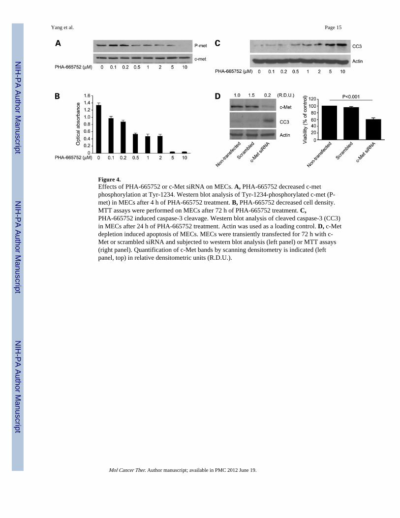
Figure 4.Effects of PHA-665752 or c-Met siRNA on MECs. A, PHA-665752 decreased c-metphosphorylation at Tyr-1234. Western blot analysis of Tyr-1234-phosphorylated c-met (P-met) in MECs after 4 h of PHA-665752 treatment. B, PHA-665752 decreased cell density.MTT assays were performed on MECs after 72 h of PHA-665752 treatment. C,PHA-665752 induced caspase-3 cleavage. Western blot analysis of cleaved caspase-3 (CC3)in MECs after 24 h of PHA-665752 treatment. Actin was used as a loading control. D, c-Metdepletion induced apoptosis of MECs. MECs were transiently transfected for 72 h with c-Met or scrambled siRNA and subjected to western blot analysis (left panel) or MTT assays(right panel). Quantification of c-Met bands by scanning densitometry is indicated (leftpanel, top) in relative densitometric units (R.D.U.).
Yang et al. Page 15
Mol Cancer Ther. Author manuscript; available in PMC 2012 June 19.
NIH
-PA Author Manuscript
NIH
-PA Author Manuscript
NIH
-PA Author Manuscript

Figure 5.Effects of PHA-665752 or c-Met siRNA on LKR-13 cells. A, PHA-665752 decreased c-metphosphorylation at Tyr-1234. Western blot analysis of Tyr-1234-phosphorylated c-met (P-met) in LKR-13 cells after 4 h of PHA-665752 treatment. B, PHA-665752 decreased celldensity. MTT assays were performed on LKR-13 cells after 72 h of PHA-665752 treatment.C, PHA-665752 induced caspase-3 cleavage. Western blot analysis of cleaved caspase-3(CC3) in LKR-13 cells after 24 h of PHA-665752 treatment. Actin was used as a loadingcontrol. D, c-Met depletion induced no evidence of apoptosis. LKR-13 cells were transientlytransfected for 72 h with c-Met or scrambled siRNA and subjected to western blot analysis(left panel) or MTT assays (right panel). Quantification of c-Met bands by scanningdensitometry is indicated (left panel, top) in relative densitometric units (R.D.U.).
Yang et al. Page 16
Mol Cancer Ther. Author manuscript; available in PMC 2012 June 19.
NIH
-PA Author Manuscript
NIH
-PA Author Manuscript
NIH
-PA Author Manuscript


