Embed Size (px)
Citation preview

Santus et al. BMC Pulmonary Medicine 2014, 14:201http://www.biomedcentral.com/1471-2466/14/201
RESEARCH ARTICLE Open Access
A systematic review on tracheostomydecannulation: a proposal of a quantitativesemiquantitative clinical scorePierachille Santus1,7*, Andrea Gramegna2, Dejan Radovanovic1, Rita Raccanelli1, Vincenzo Valenti3, Dimitri Rabbiosi4,Michele Vitacca5 and Stefano Nava6
Abstract
Background: Tracheostomy is one of the most common surgical procedures performed in critical care patientmanagement; more specifically, ventilation through tracheal cannula allows removal of the endotracheal tube (ETT).Available literature about tracheostomy care and decannulation is mainly represented by expert opinions and nocertain knowledge arises from it.
Methods: In lack of statistical requirements, a systematic and critical review of literature regarding tracheostomytube removal was performed in order to assess predictor factors of successful decannulation and to propose apredictive score. We combined 3 terms and a literature search has been performed using the Cochrane CentralRegister of Controlled Trials (CENTRAL); MEDLINE via Ovid SP; EMBASE via Ovid SP; EBSCO. Abstracts were independentlyreviewed: for those studies fitting the inclusion criteria on the basis of the title and abstract, full-text was achieved.We included studies published from January 1, 1995 until March 31, 2014; any sort of review and expert opinionhas been excluded by our survey. English language restriction was applied. Ten studies have been consideredeligible for inclusion in the review and were analysed further.
Results: Cough effectiveness and ability to tolerate tracheostomy tube capping are the most considered parametersin clinical practice; other parameters are taken into different consideration by many authors in order to proceed todecannulation. Among them, we distinguished between objective quantitative parameters and semi-quantitativeparameters more dependent from clinician’s opinion. We then built a score (the Quantitative semi Quantitativescore: QsQ score) based on selected parameters coming from literature.
Conclusions: On our knowledge, this review provides the first proposal of decannulation score system based oncurrent literature that is hypothetical and requires to be validated in daily practice. The key point of our proposalis to give a higher value to the objective parameters coming from literature compared to less quantifiableclinical ones.
Keywords: Tracheostomy, Decannulation, Predictive score, Clinical score, Removal tube
* Correspondence: [email protected] of Life Science, Università degli Studi di Milano. PulmonaryRehabilitation Unit, Fondazione Salvatore Maugeri, Istituto Scientifico diMilano-IRCCS, Via Camaldoli, 64-20138 Milan, Italy7Dipartimento di Scienze della Salute, Università degli Studi di Milano.Pulmonary Rehabilitation Unit, Fondazione Salvatore Maugeri, ScientificInstitute of Milan-IRCCS, Via Camaldoli, 64-20138 Milan, ItalyFull list of author information is available at the end of the article
© 2014 Santus et al.; licensee BioMed Central. This is an Open Access article distributed under the terms of the CreativeCommons Attribution License (http://creativecommons.org/licenses/by/4.0), which permits unrestricted use, distribution, andreproduction in any medium, provided the original work is properly credited. The Creative Commons Public DomainDedication waiver (http://creativecommons.org/publicdomain/zero/1.0/) applies to the data made available in this article,unless otherwise stated.

Santus et al. BMC Pulmonary Medicine 2014, 14:201 Page 2 of 8http://www.biomedcentral.com/1471-2466/14/201
BackgroundTracheostomy is one of the most frequent proceduresapplicated in intensive care unit (ICU) patients: about10% of patients requiring more than 3 days of mechanicalventilation are expected to undergo tracheostomy [1]. Fur-thermore, in the last years the development of less invasivesurgical techniques allowed tracheostomy to be safelyperformed at patient’s bedside and at present thistechniques are growing more and more because of theincreased number of patients requiring difficult orprolonged weaning from endotracheal tube (ETT),due to aging and severe comorbidities [2]. Moreover,possibility of tracheotomy for prevention and betterclinical management of ventilator-associated pneumo-nia was recently evaluated [3].As a result, the correct management of tracheosto-
mized patients and defined procedures for decannulationgrew into a more important clinical issue.
Physiological modifications after tracheostomyIn the following chapter we will discuss the main physio-logical implications for breathing after tracheostomy. Inscientific literature, authors usually compare tracheos-tomy to endotracheal intubation. In our purpose, weintended to compare changes during tracheostomy tospontaneous breathing and the most relevant topics are:
Airflow resistance - Resistance of anatomically normalairways is firstly conditioned by upper airways (up to80% during nose breathing and 50% during mouthbreathing). Therefore, tracheostomy should virtuallyreduce flow resistance bypassing the respiratory upperairways. Actually, data from scientific literature statethe contrary, as the presence of tracheotomy tube,during spontaneous breathing, reduces airway radiusleading to increased flow resistance and, of course,increased work of breathing (WOB) [4,5].This is according to Poiseuille equation, as the resistanceto gas flow through a tube varies inversely with theinternal diameter of the tube (in particular, to the 4thpower of the radius of the tube when flow is laminar).Secondly, secretions (stimulated even more by thepresence of the tube) still play an important role:adhering to the inner lumen of the tube, they can leadto significant luminal narrowing, according to Wilsonet al. [6], 15% of tubes are shrunk three sizes; otherexperiences from literature ultimately come to thesame conclusions [7,8].It has been supposed that the inner diameter of asmaller tracheostomy tube may induce increased flowresistance and WOB, if compared to spontaneousbreathing. The first data from in vivo studies appearedrecently by Valentini et al. trying to define the effect ofsmaller tracheostomy tube sizes on diaphragm effort.
They recorded diaphragm pressure- time product permin (PTPdi/min), tension-time index of the diaphragm(TTdi), and the ratio of respiratory rate to tidal volume(f/VT) for 2 different tracheotomy tube, with an innerdiameters 8 mm and 6.5 mm. The use of a smallerdiameter resulted in an increase of diaphragmatic effort,decrease of VT and an increase of intrinsic PEEP. Inconclusion, the authors assessed that, especially inhard to wean tracheostomized patients, the use of atracheostomy tube of small size can lead to alterationsof some weanibility parameters, otherwise normal withgreater size tubes [9].In addition, a single physiologic study by Criner et al.reported that airways resistance and work of breathingresulted higher with the tube in place, duringspontaneous breathing, when compared to breathingafter decannulation [10].Humidification and heating - Normally upper airwaysplay a fundamental function of heating and humidificationof inspired air. In tracheostomized patients, the airbypasses the nasopharyngeal cavity and enters directlythe tracheobronchial tree; a system of artificialhumidification is needed. In the absence of adequatehumidification, the epithelium of the trachea is involvedin a gradual inflammatory process resulting in squamousmetaplasia and, consequentially , impairment of ciliaryfunction and increased risk of respiratory infection [11].
Physiological modifications after decannulationWhile scientific literature has better investigated trache-otomy effects on respiratory physiology, data aboutrespiratory mechanisms after decannulation are almosttotally lacking. Chadda et al. studied respiratory parame-ters in nine neuromuscular patients after that theupper airways were confirmed to be free of obstruc-tions by fiberoptic bronchoscopy. In this experience,decannulation resulted in an increase of the tidal vol-ume and carbon dioxide partial pressure due to an in-crease of the dead space and work of breathing [12].However, the argument is still controversial. In a study
of few years ago, Dellweg et al. did not find a significantdifference in WOB in favour of tracheostomy whencompared to mouth breathing. Decannulation increasesor decreases airways resistance depending from case tocase and, in particular, from the morphology of theupper airways [13].To our knowledge, there are only few papers on physio-
logical modifications after decannulation; experts havediffering opinion on this topic and so further researchand trials are desirable.
Weaning from tracheostomy – problem generationPlace a tracheostomy enables discharging the patientfrom ICU to a rehabilitation unit [14]. However, despite

Santus et al. BMC Pulmonary Medicine 2014, 14:201 Page 3 of 8http://www.biomedcentral.com/1471-2466/14/201
decannulation is not risk-free, there is evidence of benefitsfor tracheostomy tube removal. The tracheostomy tubemay cause inflammation and stenosis or excessive coughand may impair swallowing by preventing the physio-logical trachea’s elevation against the epiglottis in order toprevent aspiration of food or secretions [15].Furthermore, upper airways are excluded from breath-
ing. At last, in most cases tracheostomized patients areunable to speak; aphonia worsens patient’s quality of lifeand slows down the recovery process, often leading toanxiety and depression.A number of clinically important early and late compli-
cations have been evaluated, including granulation tissue,tracheal stenosis, tracheomalacia, tracheo-esophagealfistula, ventilator-associated pneumonia and aspiration.According to literature, chronic tracheostomy in severeChronic Obstructive Pulmonary Disease (COPD) patientsis associated with a higher frequency of exacerbationsrequiring antibiotic treatment [16]. The clinical relevanceof these complications may lead to death and most ofpatients weaned from mechanical ventilation throughtracheostomy should undergo early decannulation.As it is hard to schematize the approach for decannu-
lation, we decided to perform the following revision ofliterature, with the aim of supporting or retracting currentstatements. Which are then current issues in managingtracheostomy? How does evidence-based medicine handlethe process of weaning from tracheostomy tube? Now-adays clinical habit derives from physiopathological know-ledge, from personal experience and from professionalpractice: there is limited evidence in literature regardingspecifically decannulation processes. Little is known abouthow clinicians decide to decannulate patients. Which arethe criteria for choice?
MethodsCriteria for considering studies for this systematic reviewA literature search has been performed using theCochrane Central Register of Controlled Trials (CENTRAL);MEDLINE via Ovid SP; EMBASE via Ovid SP; EBSCO.Research included, but was not limited to, 3 keywords(tracheostomy, decannulation, weaning). Abstracts wereindependently reviewed: for those studies fitting theinclusion criteria on the basis of the title and abstract,full-text was achieved. Reference lists were also exam-ined for any additional relevant studies not identifiedthrough the former search. We included studies pub-lished from January 1, 1995 until March 31, 2014. Suchstudies examined a population more than 18 years old,who received tracheostomy for any clinical reason,excluded neuromuscular diseases, and hospitalized inany ward (ICU, rehabilitation wards, etc.); any sort ofreview and expert opinion has been excluded by oursurvey. Including English language restriction was
applied. A flow chart diagram of the search strategy andstudy selection is provided in Figure 1.
ResultsThe database search yielded 248 citations publishedbetween January 1995 and September 2012 (with dupli-cates removed). 226 articles were excluded on the titleand abstract. The full-text of potentially relevant arti-cles was achieved for further evaluation. The final num-ber of papers taken into account is 10. Figure 1 showsthe flow-chart explaining how many citations wereexcluded from the analysis and for what reasons. Afteranalyzing the selected literature, we decided for asystematic and critical revision of literature on decan-nulation, due to lack of statistical requirements for ameta-analysis. Specifically, in Table 1, primary and –when present – secondary outcomes proposed by eachauthor have been evaluated for each study.Studies targeting different outcomes from the ones
predictors in decannulation were excluded, althoughthey considered tracheostomy tube removal of enrolledpatients as a step. Surveys were also included. Context-ually, papers exploring new and/or experimental tech-niques for the assessment of risk in trachestomy tuberemoval were excluded [17,18]. Such exclusion wasoperated because the proposed approaches - such asoscillometry impedance measurement or upper airwayresistance measurement – are difficult to apply inevery setting and are not easily reproducible in differ-ent contexts. Nevertheless in this context innovativetechniques, such those before reported, could probably beuseful to improve decannulation procedure in future.First of all, it must be noted that literature is mainly
made of expert opinions and international surveys; thefew reported clinical trials are mainly descriptive (retro-spective and prospective); because of technical and ethicalproblems, Randomized Clinical Trials (RCTs) are totallylacking.It can be noticed from the data that the population
taken into consideration is heterogeneous for age,comorbidities and causes of tracheostomy; it must alsobe emphasized that studies take in consideration bothacute/reversible pathologies and chronic conditions atdifferent levels of severity (for instance, Budweiser et al.[2] considers prolonged weaning patients with persistentrespiratory failure). Secondly, recommendations differbetween clinicians who work in acute facilities andthose who work at chronic wards and between respira-tory therapists and physicians.In our analysis cough effectiveness and ability to toler-
ate tracheostomy tube capping are the most frequentcriteria used by clinicians in order to predict successfuldecannulation. In others studies a different importanceis also given to parameters such as oxygenation and

Figure 1 Flow diagram of the search process. The number of references initially identified through each database was 248. References wereusually excluded for more than one reason by a two consecutive steps.
Santus et al. BMC Pulmonary Medicine 2014, 14:201 Page 4 of 8http://www.biomedcentral.com/1471-2466/14/201
capnia, level of consciousness and neurological state,age, swallowing, quantity and quality of secretions, dur-ation of mechanical ventilation, stability of haematicgases (PaO2 and PaCO2), aetiology of respiratory failureand comorbidities.Bach et al. experienced in a population of patients
with respiratory failure due to different aetiologies someparameters that can predict the success of the tracheos-tomy decannulation. The most important predictive fac-tor is PCF (Peak Cough Flow), in particular at least 160L/min; other important factors are also VC (Vital Cap-acity) and age [19]. Ceriana e al. proposed a prospectivestudy to decide whether to remove tracheotomy in long-term mechanically ventilated patients with respiratoryfailure from different causes. By the use of a decisionalflowchart based on clinical and physiological parameters,
they were able to remove tracheotomy cannula in almost80% of patients with spontaneous breathing without majorclinical complications. Principal parameters consideredwere patient's ability to remove secretions, swallowingfunction, absence of psychiatric diseases, possibility ofreaching spontaneous breathing, and amount of respira-tory space [20].Choate K and Barbetti J conducted a prospective
descriptive study of consecutive patients who receiveda tracheostomy in ICU. Of the 823 decisions for decan-nulation, there were 40 episodes of failed decannula-tion, representing a failure rate of 4.8%. The mainreason for decannulation failure was sputum retentionand ineffective cough [21].Lastly, in a retrospective study on patients requiring
tracheostomy in ICU, Leung and his team identified risk
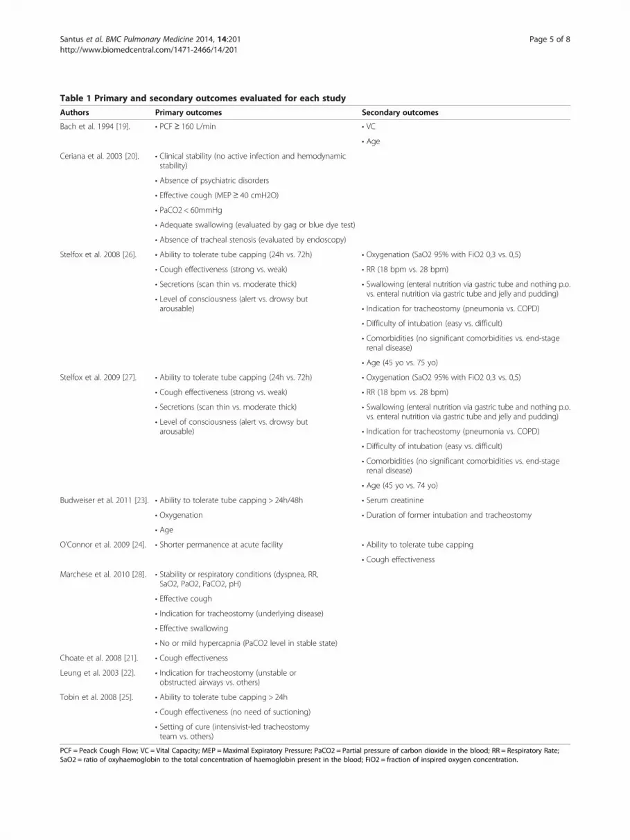
Table 1 Primary and secondary outcomes evaluated for each study
Authors Primary outcomes Secondary outcomes
Bach et al. 1994 [19]. • PCF≥ 160 L/min • VC
• Age
Ceriana et al. 2003 [20]. • Clinical stability (no active infection and hemodynamicstability)
• Absence of psychiatric disorders
• Effective cough (MEP≥ 40 cmH2O)
• PaCO2 < 60mmHg
• Adequate swallowing (evaluated by gag or blue dye test)
• Absence of tracheal stenosis (evaluated by endoscopy)
Stelfox et al. 2008 [26]. • Ability to tolerate tube capping (24h vs. 72h) • Oxygenation (SaO2 95% with FiO2 0,3 vs. 0,5)
• Cough effectiveness (strong vs. weak) • RR (18 bpm vs. 28 bpm)
• Secretions (scan thin vs. moderate thick) • Swallowing (enteral nutrition via gastric tube and nothing p.o.vs. enteral nutrition via gastric tube and jelly and pudding)
• Level of consciousness (alert vs. drowsy butarousable) • Indication for tracheostomy (pneumonia vs. COPD)
• Difficulty of intubation (easy vs. difficult)
• Comorbidities (no significant comorbidities vs. end-stagerenal disease)
• Age (45 yo vs. 75 yo)
Stelfox et al. 2009 [27]. • Ability to tolerate tube capping (24h vs. 72h) • Oxygenation (SaO2 95% with FiO2 0,3 vs. 0,5)
• Cough effectiveness (strong vs. weak) • RR (18 bpm vs. 28 bpm)
• Secretions (scan thin vs. moderate thick) • Swallowing (enteral nutrition via gastric tube and nothing p.o.vs. enteral nutrition via gastric tube and jelly and pudding)
• Level of consciousness (alert vs. drowsy butarousable) • Indication for tracheostomy (pneumonia vs. COPD)
• Difficulty of intubation (easy vs. difficult)
• Comorbidities (no significant comorbidities vs. end-stagerenal disease)
• Age (45 yo vs. 74 yo)
Budweiser et al. 2011 [23]. • Ability to tolerate tube capping > 24h/48h • Serum creatinine
• Duration of former intubation and tracheostomy• Oxygenation
• Age
O’Connor et al. 2009 [24]. • Shorter permanence at acute facility • Ability to tolerate tube capping
• Cough effectiveness
Marchese et al. 2010 [28]. • Stability or respiratory conditions (dyspnea, RR,SaO2, PaO2, PaCO2, pH)
• Effective cough
• Indication for tracheostomy (underlying disease)
• Effective swallowing
• No or mild hypercapnia (PaCO2 level in stable state)
Choate et al. 2008 [21]. • Cough effectiveness
Leung et al. 2003 [22]. • Indication for tracheostomy (unstable orobstructed airways vs. others)
Tobin et al. 2008 [25]. • Ability to tolerate tube capping > 24h
• Cough effectiveness (no need of suctioning)
• Setting of cure (intensivist-led tracheostomyteam vs. others)
PCF = Peack Cough Flow; VC = Vital Capacity; MEP =Maximal Expiratory Pressure; PaCO2 = Partial pressure of carbon dioxide in the blood; RR = Respiratory Rate;SaO2 = ratio of oxyhaemoglobin to the total concentration of haemoglobin present in the blood; FiO2 = fraction of inspired oxygen concentration.
Santus et al. BMC Pulmonary Medicine 2014, 14:201 Page 5 of 8http://www.biomedcentral.com/1471-2466/14/201
Santus et al. BMC Pulmonary Medicine 2014, 14:201 Page 6 of 8http://www.biomedcentral.com/1471-2466/14/201
factors that would indicate a low likelihood of earlydecannulation. The indications for tracheostomy wereprolonged mechanical ventilation, tracheobronchialtoilet or risk of aspiration and unstable or obstructedairways. They concluded that the only indicator forearly decannulation is tracheostomy insertion andother patient related variables are not significant. [22].The usefulness of a tracheostomy retainer (TR) and
the predictors of successful decannulation were alsoevaluated by Budweiser and his team. In percutaneouslytracheostomized patients with prolonged weaning, theuse of a TR seems to facilitate the weaning process. Fur-thermore, also the duration of spontaneous breathingprior to decannulation, age and oxygenation predict therisk of recannulation [23].O’Connor et al. retrospectively examined the process
of decannulation following tracheostomy in patientstransferred to a long-term care hospital for weaningfrom prolonged mechanical ventilation. Decannulationwas successful in 35% of patients and main factors takenin account were ability to tolerate tube capping andcough effectiveness. Patients who failed decannulationhad an earlier placed tracheostomy tube and had also ashorter permanence at the acute facility compared withpatients who were decannulated [24].Similar results were described by Tobin and his team;
the authors also demonstrated that an intensivist-ledtracheostomy team is associated with quicker decannula-tion time and a shorter hospitalization [25].Stelfox et al. performed a cross-sectional survey on
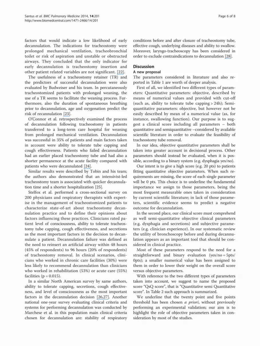
200 physicians and respiratory therapists with expert-ise in the management of tracheostomized patients tocharacterize state-of-art about tracheostomy decan-nulation practice and to define their opinions aboutfactors influencing these practices. Clinicians rated pa-tient level of consciousness, ability to tolerate tracheos-tomy tube capping, cough effectiveness, and secretionsas the most important factors in the decision to decan-nulate a patient. Decannulation failure was defined asthe need to reinsert an artificial airway within 48 hours(45% of respondents) to 96 hours (20% of respondents)of tracheostomy removal. In clinical scenarios, clini-cians who worked in chronic care facilities (30%) wereless likely to recommend decannulation than clinicianswho worked in rehabilitation (53%) or acute care (55%)facilities (p = 0.015).In a similar North American survey by same authors,
ability to tolerate capping, secretions, cough effective-ness, and level of consciousness as the most importantfactors in the decannulation decision [26,27]. Anothernational one-year survey evaluating clinical criteria andsystems for performing decannulation was conducted byMarchese et al. in this population main clinical criteriachosen for decannulation are: stability of respiratory
conditions before and after closure of tracheostomy tube,effective cough, underlying diseases and ability to swallow.Moreover, laryngo-tracheoscopy has been considered inorder to exclude contraindications to decannulation [28].
DiscussionA new proposalThe parameters considered in literature and also re-ported in Table 1 are worth of deeper analysis.First of all, we identified two different types of param-
eters: Quantitative parameters: objective, described bymeans of numerical values and provided with cut-off(such as, ability to tolerate tube capping > 24h); Semi-quantitative parameters: objective, but however not beeasily described by mean of a numerical value (as, forinstance, swallowing function). Our purpose is to sug-gest a clinical score including all parameters – bothquantitative and semiquantitative –considered by availablescientific literature in order to evaluate the feasibility oftracheostomy tube removal.In our idea, objective quantitative parameters shall be
taken into greater account in decisional process. Otherparameters should instead be evaluated, when it is pos-sible, according to a binary system (e.g. dysphagia yes/no).Our intent is to give a high score (e.g. 20 pts) to patients
fitting quantitative objective parameters. When such re-quirements are missing, the score of each single parameterwill be 0 pts. This choice is to underline the fundamentalimportance we assign to those parameters, being themost frequent measurable ones taken in considerationby current scientific literature; in lack of those parame-ters, scientific evidence seems to predict a negativeoutcome for decannulation.In the second place, our clinical score must comprehend
as well semi-quantitative objective clinical parameters(e.g. dysphagia and secretions) and subjective parame-ters (e.g. clinician experience). In our systematic reviewthe utility of bronchoscopy before and during decannu-lation appears as an important tool that should be con-sidered in clinical practice.Most of these parameters respond to the need for a
straightforward and binary evaluation (yes/no = 5pts/0pts); a smaller numerical value has been assigned tothem in order to lower their weight on the overall scoreversus objective parameters.With reference to the two different types of parameters
taken into account, we suggest to name the proposedscore “QsQ score”, that is “Quantitative semi Quantitativescore”. In Table 2 such approach is summarized.We underline that the twenty point and five points
threshold has been chosen a priori, without previouslyperforming an experimental validation; our aim is tohighlight the role of objective parameters taken in con-sideration by most of the studies.

Table 2 QsQ score: Quantitative and semiquantitative parameters
Parameter Cut-off Missing Fitting
Objective quantitative parameters – Main criteria
Cough MEP ≥ 40 cmH2O 0 20
PCF > 160 L/min
Tube capping ≥24 h 0 20
Semi-quantitative parameters – Minor criteria
Level of counsciousness Drowsy/Alert 0 5
Secretion (thick vs. thin) 0 5
Swallowing Impaired/Normal 0 5
Capnia paCO2 < 60 mmHg 0 5
Patent airway Tracheal stenosis < 50% seen by bronchoscopy 0 5
Age <70 0 5
Indication for tracheostomy Others/Pneumonia or airway obstruction 0 5
Comorbidities Present (≥1) or None 0 5
This hypothetical score have the objective quantitative parameters, named ‘major criteria’ , and semi-quantitative or subjective parameters, named ‘minor criteria’.For the proposed interpretation and clinical application see the text in Discussion section.MEP =Maximal Expiratory Pressure; PaCO2 = partial pressure of carbon dioxide in the blood; RR = Respiratory Rate; SaO2 = ratio of oxyhemoglobin to the totalconcentration of hemoglobin present in the blood; FiO2 = fraction of inspired oxygen concentration.
Santus et al. BMC Pulmonary Medicine 2014, 14:201 Page 7 of 8http://www.biomedcentral.com/1471-2466/14/201
Score: hypotheses and interpretationsWe suggest an hypothetical score, that requires discus-sion and a prospective validation study. For a practicaluse we will name objective quantitative parameters‘major criteria’, and semi-quantitative or subjectiveparameters ‘minor criteria’. If all main criteria are sat-isfied, regardless of minor criteria, decannulation withhigh probability of positive outcome can be assumed.If only one of the two major criteria is satisfied, a
careful evaluation of minor criteria should be required,assuming a good probability of positive outcome whenthe majority of minor criteria is satisfied. The sameprobability category reported above could be applied if,in lack of major criteria, all of minor criteria are satis-fied. Finally, if none of the major criteria and less thanthree minor criteria are satisfied, a low probability ofpositive outcome can be assumed.
ConclusionsTracheostomy decannulation represents one of themost important problems in the clinical and home caremanagement of patients which undergo tracheostomy.No validated and specific pathway is followed when per-forming a decannulation and this process is left to theclinical expertise. Considering this, we hypothesized aclinical score, named QsQ, to help clinicians in choos-ing decannulation timing. We underline that this scorehas never been validated in clinical real life and there-fore we suggest evaluating QsQ in further clinical trialsto validate it.
AbbreviationsPCF: Peak Cough Flow; VC: Vital Capacity; MEP: Maximal Expiratory Pressure;PaCO2: partial pressure of carbon dioxide in the blood; RR: Respiratory Rate;SaO2: Ratio of oxyhemoglobin to the total concentration of hemoglobin inblood; FiO2: Fraction of inspired oxygen concentration; QsQ: Quantitativeand semiquantitative score.
Competing interestsThe authors declare that they have no competing interests.
Authors’ contributionsPS conceived the study, was responsible for data collection, performed statisticalanalysis and drafted the manuscript. AG and DR performed statistical analysis anddrafted the manuscript. All authors participated in the study design, datacollection, read, supervised and approved the final manuscript for publication.
AcknowledgementsThe authors thank the Respiratory Physiotherapist Team of RehabilitationUnit – Fondazione Salvatore Maugeri, IRCCS, Milano (Giuseppe Gaudiello,Rossana Ciraudo, Goffredo Alfieri and Grazia Lacala) for their support onconceiving the study and giving suggestions about tracheostomy clinicalmanagement.
Author details1Department of Life Science, Università degli Studi di Milano. PulmonaryRehabilitation Unit, Fondazione Salvatore Maugeri, Istituto Scientifico diMilano-IRCCS, Via Camaldoli, 64-20138 Milan, Italy. 2Department ofPathophysiology and Transplantation, Università degli Studi di Milano, IRCCSFondazione Cà Granda Ospedale Maggiore Policlinico, Via F. Sforza, 33-20122Milan, Italy. 3Department of Biomedical Sciences for Health, Università degliStudi di Milano. Respiratory Unit, Policlinico San Donato IRCCS, Piazza E.Malan, 1-20097 San Donato Milanese, Italy. 4Unit of Maxillo-Facial Surgery.Ospedale San Paolo, Milano, Università degli Studi di Milano, Milano, Via A. diRudinì, 8-20141 Milan, Italy. 5Pulmonary Rehabilitation Unit, FondazioneSalvatore Maugeri, Via Mazzini, 129- 26088 Lumezzane, Brescia, Italy. 6AlmaMater University Department of Clinical, Integrated and ExperimentalMedicine (DIMAS), Respiratory and Critical Care Unit, S. Orsola-MalpighiHospital, Via Albertoni, 10-40138 Bologna, Italy. 7Dipartimento di Scienzedella Salute, Università degli Studi di Milano. Pulmonary Rehabilitation Unit,Fondazione Salvatore Maugeri, Scientific Institute of Milan-IRCCS, Via Camal-doli, 64-20138 Milan, Italy.

Santus et al. BMC Pulmonary Medicine 2014, 14:201 Page 8 of 8http://www.biomedcentral.com/1471-2466/14/201
Received: 2 July 2014 Accepted: 11 December 2014Published: 15 December 2014
References1. Durbin CG Jr: Tracheostomy: why, when, and how? Respir Care 2010,
55:1056–1068.2. MacIntyre NR, Epstein SK, Carson S, Scheinhorn D, Christopher K, Muldoon S:
Management of patients requiring prolonged mechanical ventilation:report of a NAMDRC consensus conference. Chest 2005, 128:3937-3954.
3. Terragni PP, Antonelli M, Fumagalli R: Early vs late tracheotomy forprevention of pneumonia in mechanically ventilated adult ICU patients:a randomized controlled trial. JAMA 2010, 303:1483–1489.
4. Cavo J, Ogura JH, Sessions DG, Nelson JR: Flow resistance in tracheotomytubes. Ann Otol Rhinol Laryngol 1973, 82:827–830.
5. Heffner JE: Tracheotomy application and timing. Clin Chest Med 2003,24:389–398.
6. Wilson AM, Gray DM, Thomas JG: Increases in endotracheal tube resistanceare unpredictable relative to duration of intubation. Chest 2009,136:1006–1013.
7. Epstein SK, Ciubotaru RL: Influence of gender and endotracheal tube sizeon preextubation breathing pattern. Am J Respir Crit Care Med 1996,154:1647–1652.
8. Mehta S, Heffer MJ, Maham N, Nelson DL, Klinger JR, Levy MM: Impact ofendotracheal tube size on preextubation respiratory variables. J Crit Care2010, 25:483–488.
9. Valentini I, Tonveronachi E, Gregoretti C, Mega C, Fasano L, Pisani L, Nava S:Different tracheotomy tube diameters influence diaphragmatic effortand indices of weanability in difficult to wean patients. Respir Care 2012,57:2012–2018.
10. Criner G, Make B, Celli B: Respiratory muscle dysfunction secondary tochronic tracheostomy tube placement. Chest 1987, 91:139–141.
11. Epstein SK: Anatomy and physiology of tracheostomy. Respir Care 2005,50:476–482.
12. Chadda K, Louis B, Benaissa L: Physiological effects of decannulation intracheostomized patients. Intensive Care Med 2002, 28:1761–1767.
13. Dellweg D, Barchfeld T, Haidl P, Appelhans P, Kohler D: Tracheostomydecannulation: implication on respiratory mechanics. Head Neck 2007,29:1121–1127.
14. Scheinhorn DJ, Chao DC, Hassenpflug MS, Gracey DR: Post-ICU weaningfrom mechanical ventilation: the role of long-term facilities. Chest 2001,120:482S–484S.
15. Christopher KL: Tracheostomy decannulation. Respir Care 2005, 50:538–541.16. Clini E, Vitacca M, Bianchi L, Porta R, Ambrosino N: Long term
tracheostomy in severe COPD patients weaned from mechanicalventilation. Respir Care 1999, 44:415–420.
17. Franke KJ, Nilius G, Morgenstern S: Removal of the tracheal tube afterprolonged mechanical ventilation: assessment of risk by oscillatoryimpedance. Respiration 2011, 81:118–123.
18. Gao C, Zhou L, Wei C, Hoffman MR, Li C, Jiang JJ: The evaluation ofphysiologic decannulation readiness according to upper airwayresistance measurement. Otolaryngol Head Neck Surg 2008, 139:535–540.
19. Bach JR, Saporito LR: Indications and criteria for decannulation andtransition from invasive to noninvasive long-term ventilatory support.Respir Care 1994, 39:515–528.
20. Ceriana P, Carlucci A, Navalesi P, Rampulla C, Delmastro M, Piaggi G, DeMattia E, Nava S: Weaning from tracheotomy in long-term mechanicallyventilated patients: feasibility of a decisional flowchart and clinicaloutcome. Intensive Care Med 2003, 29:845–848.
21. Choate K, Barbetti J, Currey J: Tracheostomy decannulation failure ratefollowing critical illness: a prospective descriptive study. Aust Crit Care2009, 22:8–15.
22. Leung R, MacGregor L, Campbell D, Berkowitz RG: Decannulation andsurvival following tracheostomy in an intensive care unit. Ann Otol RhinolLaryngol 2003, 112:853–858.
23. Budweiser S, Baur T, Jorres RA, Kollert F, Pfeifer M, Heinemann F: Predictorsof successful decannulation using tracheostomy retainer in patients withprolonged weaning and persisting respiratory failure. Respiration 2012,84:469–476.
24. O'Connor HH, Kirby KJ, Terrin N, Hill NS, White AC: Decannulation followingtracheostomy for prolonged mechanical ventilation. J Intensive Care Med2009, 24:187–194.
25. Tobin AE, Santamaria JD: An intensivist-led tracheostomy review teamis associated with shorter decannulation time and lenght of stay:a prospective cohort study. Crit Care 2008, 12:R48.
26. Stelfox HT, Crimi C, Berra L, Noto A, Schmidt U, Bigatello LM, Hess D:Determinants of tracheostomy decannulation: an international survey.Crit Care 2008, 12:R26.
27. Stelfox HT, Hess DR, Schmidt UH: A North American survey of respiratorytherapist and physician tracheostomy decannulation practices. Respir Care2009, 54:1658–1664.
28. Marchese S, Corrado A, Scala R, Corrao S, Ambrosino N: Tracheostomy inpatients with long-term mechanical ventilation: a survey. Respir Med2010, 104:749–753.
doi:10.1186/1471-2466-14-201Cite this article as: Santus et al.: A systematic review on tracheostomydecannulation: a proposal of a quantitative semiquantitative clinicalscore. BMC Pulmonary Medicine 2014 14:201.
Submit your next manuscript to BioMed Centraland take full advantage of:
• Convenient online submission
• Thorough peer review
• No space constraints or color figure charges
• Immediate publication on acceptance
• Inclusion in PubMed, CAS, Scopus and Google Scholar
• Research which is freely available for redistribution
Submit your manuscript at www.biomedcentral.com/submit